Note: Descriptions are shown in the official language in which they were submitted.
CA 02514576 2005-07-27
WO 2004/069184
PCT/US2004/002867
1
METHODS FOR TREATING, PREVENTING AND DIAGNOSING
HELICOBACTER INFECTION
TECHNICAL FIELD
The present invention relates generally to bacterial immunogens. In
particular, the invention pertains to Helicobacter cerdo, a new pathogen
isolated
from swine, and methods of treating, preventing and diagnosing Helicobacter
infection using immunogenic proteins and nucleic acids derived from H. cerdo.
BACKGROUND
Gastric disease is an important cause of morbidity and economic loss in swine-
rearing operations (O'Brien, J. (1992) "Gastric ulcers" p.680. In A. D. Leman,
B. E.
Straw, W. L. Mengeling, and S. D. D'Allaire (ed), Diseases of swine. Wolfe,
London,
United Kingdom). Although the cause of porcine gastric disease has not been
previously established, it is most often attributed to diet and/or stress
(O'Brien, J.
(1992) "Gastric ulcers" p.680. In A. D. Leman, B. E. Straw, W. L. Mengeling,
and S.
D. D'Allaire (ed), Diseases of swine. Wolfe, London, United Kingdom).
In 1984, Helicobacter pylori (Hp) emerged as an etiologic agent in human
gastritis/ulcer disease following the documentation of this agent in patients
with
gastritis (Marshall and Warren (1984) Lancet 1:1311-1314). Hp is now
universally
recognized as one of the primary gastric pathogens and the study of this
bacterial
species and the spectrum of diseases associated with it has become a major
focus in
human gastroenterology (Suerbaum and Michetti (2002) N. Eng. J. Med. 347:1175-
1186). Hp is causally associated with chronic superficial (active) type B
gastritis
(Buck (1990) Clin. Micro. Rev. 3:1-12; Blaser (1992) Gasteroenterol. 102:720-
727;
Consensus Statement, 1994, NSAID), independent gastric ulceration (Peterson
(1991)
N Eng. J. Med. 324:1043-1047; Moss and Calam (1992) Gut 33:289- 292; Leung et
al. (1992) Am. J. Clin. Pathol. 98:569 574; Forbes et al. (1994) Lancet
343:258-260),
atrophic gastritis (Nomura et al. (1991) N Engl. J. Med. 325:1132-1136;
Parsonnet et
al. (1991) JNCI 83:640-643; Sipponen (1992) Drugs 52:799-804, 1996), and
gastric
CA 02514576 2005-07-27
WO 2004/069184
PCT/US2004/002867
2
MALT lymphoma (Rodriguez et al. (1993) Acta Gastro-Enterol. Belg. 56
(suppl):47;
Eidt et al. (1994) J. Clin. Pathol. 47:436-439).
Multiple agent antimicrobial therapies have been available for human Hp for
more than a decade. These therapies can be expensive, cumbersome to
administer,
and often do not completely cure the disease. Such therapies would be
impractical in
domestic livestock. Moreover, injudicious use of antimicrobials promotes
emergence
of antibiotic-resistant strains of Hp and Hp resistance to metronidazole and
clarirythromycin has increased (Michetti, (1997) Gut 41:728-730).
Additionally, the
use of antibiotics in food animals is undesirable.
Attempts to treat Hp infection in humans using immunotherapy rather than
chemotherapy has been largely unsuccessful. In particular, induction of
immunity
which mimics the "natural" immune response of convalescent infected humans has
not been successful since human Hp infection can persist indefinitely in spite
of a
strong immune response to Hp (Lee (1996) Gastroenterol. 110:2003-2006). In
mice,
protection has been achieved with sonicates or recombinant proteins such as
ureA and
ureB, vacA and GroEL, given orally with cholera toxin (CT) and heat labile
toxin
(LT) as adjuvants. The focus has been primarily upon the use of purified
and/or
recombinant bacterial proteins as target immunogens in vaccine development
programs. In general, inconsistent and only partial protection has been
achieved. In
rodent systems, mucosal vaccination assisted by CT or LT has emerged as the
favored
route, notwithstanding the fact that these species are highly resistant to
toxic effects of
CT/LT and the resultant rodent data does not directly translate into the human
or
swine experience.
In particular, in piglets immunized and then challenged with Hp, the strongest
pre-challenge indicator of efficacy is the level and presence of Hp-specific
serum/salivary IgG, not IgA (Eaton and Krakowka (1992) Gastroenterol. 103:1580-
1586). Parenteral vaccination stimulates a strong IgG response; oral
vaccination does
not. Parenteral immunization was completely protective in 50% of the piglets
immunized subcutaneously and in 60% of piglets immunized intraperitoneally
(Eaton
et al. (1998) J. Infect. Dis. 178:1399-1405). In contrast, oral vaccination
with: 1) live
bacteria (cleared with antimicrobials prior to challenge), 2) whole intact
killed
CA 02514576 2005-07-27
WO 2004/069184
PCT/US2004/002867
3
bacteria, 3) whole bacterial sonicates and 4) whole bacterial sonicates with
mucosal
LT adjuvant failed to provide a single instance (0 of 27 piglets or 0%) of
protection.
Bacterial cfu were reduced compared to controls but the levels of reduction
did not
reach statistical significance. Thus, in the porcine model of Hp colonization
and acute
gastritis, the parenteral route of vaccination appears to be superior to the
oral route in
both absolute (infected versus uninfected after challenge) and relative
(bacterial cfu in
vaccinates versus nonvaccinated controls) measures of antimicrobial efficacy.
SUMMARY OF THE INVENTION
The present invention is based on the discovery of a novel Helicobacter
pathogen isolated from swine exhibiting gastritis/ulcer disease. This organism
has
been named Helicobacter cerdo (Hc) by the inventors herein. This organism has
been
shown by the inventors to cause gastric disease in young piglets that is
similar to Hp-
associated active gastritis in humans.
Subunit vaccines, including antigens and mixtures of antigens derived from H.
cerdo, provide protection against subsequent infection with Helicobacter
species,
such as H. pylori and H. cerdo. The present invention provides a safe,
efficacious and
economical method of treating and/or preventing Hc infection in swine.
Accordingly, in one embodiment, the subject invention is directed to a
composition comprising a pharmaceutically acceptable vehicle and at least one
Helicobacter cerdo immunogen. In certain embodiments, the at least one H.
cerdo
immunogen is provided in an H. cerdo lysate, such as a lysate produced by
proteolytic
digestion of H. cerdo bacteria. In additional embodiments, the composition
further
comprises an adjuvant.
In another embodiment, the invention is directed to methods of treating or
preventing a Helicobacter infection in a vertebrate subject comprising
administering
to the subject a therapeutically effective amount of a composition as
described above.
In certain embodiments, the vertebrate subject is a porcine subject. In
additional
embodiments, the Helicobacter infection is a Helicobacter cerdo infection. In
yet
further embodiments, the composition is administered parenterally.
CA 02514576 2014-11-20
51440-23
3a
According to one aspect of the present invention, there is provided a
composition comprising a pharmaceutically acceptable vehicle and a
Helicobacter cerdo
lysate comprising at least one Helicobacter cerdo immunogen; wherein said
Helicobacter
cerdo is a porcine Helicobacter gastric isolate which has the following
characteristics: gram
negative, short curved rods, microaerophilic growth pattern, urease enzyme
activity, catalase
enzyme activity, possession of one or more immunogenic polypeptides selected
from the
group consisting of HpaA, FlaA, FlaB, urease, UreA, UreB, hsp60, cytotoxin,
cagA, and
VacA, and a distinct SDS-PAGE profile from Helicobacter pylori under reducing
conditions;
and wherein the H cerdo lysate is produced by pepsin proteolytic digestion of
H cerdo
bacteria.
According to another aspect of the present invention, there is provided a
method of producing a composition comprising: (a) providing a Helicobacter
cerdo lysate
comprising at least one Helicobacter cerdo immunogen; and (b) combining said H
cerdo
lysate with a pharmaceutically acceptable vehicle; wherein said Helicobacter
cerdo is a
porcine Helicobacter gastric isolate which has the following characteristics:
gram negative,
short curved rods, microaerophilic growth pattern, urease enzyme activity,
catalase enzyme
activity, possession of one or more immunogenic polypeptides selected from the
group
consisting of HpaA, FlaA, FlaB, urease, UreA, UreB, hsp60, cytotoxin, cagA,
and VacA, and
a distinct SDS-PAGE profile from Helicobacter pylori under reducing condition;
and wherein
the II cerdo lysate is produced by pepsin proteolytic digestion of II cerdo
bacteria.
According to still another aspect of the present invention, there is provided
a
method of detecting Helicolmicter cerdo infection in a vertebrate subject
comprising: (a)
providing a biological sample from the subject; and (b) reacting said
biological sample with a
Helicobacter cerdo lysate comprising at least one H cerdo immunogen, under
conditions
which allow Helicobacter antibodies, when present in the biological sample, to
bind with said
immunogen(s), thereby detecting the presence or absence of Helicobacter cerdo
infection in
the subject; wherein said Helicobacter cerdo is a porcine Helicobacter gastric
isolate which
has the following characteristics: gram negative, short curved rods,
microaerophilic growth
pattern, urease enzyme activity, catalase enzyme activity, possession of one
or more
immunogenic polypeptides selected from the group consisting of HpaA, FlaA,
FlaB, urease,
CA 02514576 2014-11-20
51440-23
3c
UreA, UreB, hsp60, cytotoxin, cagA, and VacA, and a distinct SDS-PAGE profile
from
Helicobacter pylori under reducing condition.
According to still a further aspect of the present invention, there is
provided
use of the H. cerdo lysate as described herein in an ex vivo method of
detecting Helicobacter
cerdo infection in a vertebrate subject; wherein said Helicobacter cerdo is a
porcine
Helicobacter gastric isolate which has the following characteristics: gram
negative, short
curved rods, microaerophilic growth pattern, urease enzyme activity, catalase
enzyme activity,
possession of one or more immunogenic polypeptides selected from the group
consisting of
HpaA, FlaA, FlaB, urease, UreA, UreB, hsp60, cytotoxin, cagA, and VacA, and a
distinct
SDS-PAGE profile from Helicobacter pylori under reducing condition.
According to yet another aspect of the present invention, there is provided
use
of a composition comprising the Helicobacter cerdo lysate as described herein
for producing
an immunological response against Helicobacter cerdo in a porcine.
According to yet another aspect of the present invention, there is provided a
composition comprising the Helicobacter cerdo lysate as described herein for
use in the
production of an immunological response against Helicobacter cerdo in a
porcine.
CA 02514576 2005-07-27
WO 2004/069184
PCT/US2004/002867
4
In yet another embodiment, the invention is directed to methods of treating or
preventing a Helicobacter cerdo infection in a porcine subject comprising
parenterally
administering to the subject a therapeutically effective amount of a
composition as
described above.
In another embodiment, the invention is directed to a method of producing a
composition comprising:
(a) providing at least one Helicobacter cerdo immunogen; and
(b) combining the H. cerdo immunogen with a pharmaceutically acceptable
vehicle.
In certain embodiments, the at least one H. cerdo immunogen is provided in
an H. cerdo lysate, such as an H. cerdo lysate produced by proteolytic
digestion of H.
cerdo bacteria. In additional embodiments, an adjuvant is also provided.
In yet another embodiment, the invention is directed to a method of detecting
Helicobacter infection in a subject comprising:
(a) providing a biological sample from the subject; and
(b) reacting the biological sample with at least one H. cerdo immunogen,
under conditions which allow Helicobacter antibodies, when present in the
biological
sample, to bind with the immunogen(s),
thereby detecting the presence or absence of Helicobacter infection in the
subject.
In certain embodiments, the method further comprises:
(c) removing unbound antibodies;
(d) providing one or more moieties capable of associating with the bound
antibodies; and
(e) detecting the presence or absence of the one or more moieties,
thereby detecting the presence or absence of H. cerdo infection.
In certain embodiments, the detectable label is a fluorescer or an enzyme. In
additional embodiments, the at least one immunogen is provided in an H. cerdo
lysate. In still further embodiments, the biological sample is a porcine serum
sample.
In additional embodiments, the invention is directed to a method of detecting
Helicobacter cerdo infection in a porcine subject comprising:
CA 02514576 2005-07-27
WO 2004/069184
PCT/US2004/002867
(a) providing a biological sample from the subject; and
(b) reacting the biological sample with at least one H. cerdo immunogen,
under conditions which allow H. cerdo antibodies, when present in the
biological
sample, to bind with the immunogen(s),
5 (c) removing unbound antibodies;
(d) providing one or more moieties capable of associating with the bound
antibodies; and
(e) detecting the presence or absence of the one or more moieties, thereby
detecting the presence or absence of H. cerdo infection.
In still further embodiments, the invention is directed to an antibody
specific
for a Helicobacter cerdo immunogen. In certain embodiments, the antibody is a
polyclonal antibody. In other embodiments, the antibody is a monoclonal
antibody.
In another embodiment, the invention is directed to a Helicobacter cerdo
lysate comprising at least one H. cerdo immunogen. In certain embodiments, the
H.
cerdo lysate is produced by proteolytic digestion of H. cerdo bacteria.
These and other embodiments of the subject invention will readily occur to
those of skill in the art in view of the disclosure herein.
BRIEF DESCRIPTION OF THE DRAWINGS
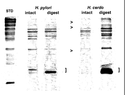
Figure 1 shows SDS-PAGE profiles of intact and digested H. pylon and H.
cerdo preparations. The ">" in the figure illustrates bands present in H.
pylori and
absent from H. cerdo. The "]" indicates low molecular weight protease digest
products.
Figures 2A and 2B show SDS-PAGE separations of intact H. cerdo (2A) and
an H. cerdo digest (2B). An increased amount of low molecular weight material
()
is seen in the digested preparation.
Figures 3A and 3B show a Western blot analysis of intact H. cerdo (3A) and
an H. cerdo digest (3B) separated on a native, non-reducing gel.
Figures 4A and 4B show a Western blot analysis of the antibody reactivity
profile against intact H. cerdo (4A) and an H. cerdo digest (4B). An increased
amount of low molecular weight material is seen in the digest (indicated by
]).
CA 02514576 2011-07-13
51440-23
6
Increased staining intensity is also seen (.9, as well as additional
irnmunoreactive
bands (<).
DETAILED DESCRIPTION OF THE INVENTION
The practice of the present invention will employ, unless otherwise indicated,
conventional techniques of molecular biology, microbiology, bacteriology,
recombinant DNA technology, and immunology, which are within the skill of the
art.
Such techniques are explained fully in the literature. See, e.g., Sambrook,
Fritsch &
Maniatis, Molecular Cloning: A Laboratory Manual, Second Edition (1989); DNA
Cloning,Vols. I and II (D.N. Glover ed. 1985); Oligonucleotide Synthesis M.J.
Gait
ed. 1984); Nucleic Acid Hybridization (B.D. Hames & S.J. Higgins eds. 1984);
Animal Cell Culture (R.K. Freshney ed. 1986); Immobilized Cells and Enzymes
(IRL
Press, 1986); Perbal, B., A Practical Guide to Molecular Cloning (1984); the
series,
Methods In Enzymology (S. Colowick and N. Kaplan eds., Academic Press, Inc.);
and
Handbook of Experimental Immunology, Vols. I-IV (D.M. Weir and C.C. Blackwell
eds., 1986, Blackwell Scientific Publications).
1. DEFINITIONS
In describing the present invention, the following terms will be employed, and
are intended to be defined as indicated below.
It must be noted that, as used in this specification and the appended claims,
the
singular forms "a", "an" and "the" include plural referents unless the content
clearly
dictates otherwise. Thus, for example, reference to "an H. cerdo immunogen"
includes a mixture of two or more such immunogens, and the like.
By "Helicobacter infection" is meant any disorder caused by a Helicobacter
bacterium, including without limitation, H. cerdo, H. pylori and H.
heilniannii, such
as, but not limited to, chronic superficial (active) type B gastritis,
independent gastric
ulceration, peptic, gastric and duodenal ulcers, gastroesophageal ulceration
(GEU),
proventricular ulcers, ulcerative gastric hemorrhage, atrophic gastritis, and
carcinoma
CA 02514576 2005-07-27
WO 2004/069184
PCT/US2004/002867
7
including gastric MALT lymphoma. The term also intends subclinical disease,
e.g.,
where Helicobacter infection is present but clinical symptoms of disease have
not yet
manifested themselves. Subjects with subclinical disease can be asymptomatic
but
are nonetheless at a considerable risk of developing peptic ulcers and/or
gastric
adenocarcinomas. For a review of Helicobacter-associated diseases, see,
Telford et
al., Trends in Biotech. (1994) 12:420-426 and Blaser, M.J., Scientific
American
(February 1996):104-107.
By "an H. cerdo lysate" is meant an extract or lysate derived from an H. cerdo
whole bacterium which includes one or more H. cerdo immunogenic polypeptides,
as
defined below. The term therefore is intended to encompass crude extracts that
contain several H. cerdo immunogens as well as relatively purified
compositions
derived from such crude lysates which include only one or few such immunogens.
Such lysates are prepared using techniques well known in the art, described
further
below.
Representative immunogens that may be present in such lysates, either alone
or in combination, include immunogens with one or more epitopes derived from
H.
cerdo adhesins such as, but not limited to, H. cerdo immunogens corresponding
to a
kDa N-acetyl-neuraminillactose-binding fibrillar haemagglutinin (HpaA), a 63
kDa protein that binds phosphatidylethanolamine and gangliotetraosyl ceramide,
and
20 a conserved fimbrial pilus-like structure as found in H. pylori. See,
e.g., Telford et
al., Trends in Biotech. (1994) 12:420-426 for a description of these antigens.
Other immunogens that may be present in the lysate include immunogens with
one or more epitopes derived from any of the various flagellins corresponding
to the
H. pylori flagellins known as the major flagellin, FlaA and the minor
flagellin, FlaB.
The flagella of H. pylori are composed of FlaA and FlaB, each with molecular
weights of approximately 55 kDa. Immunogens from H. cerdo corresponding to
either or both of FlaA and/or FlaB may be used in the lysates of the present
invention.
Another representative H. cerdo immunogen is an immunogen corresponding
to H. pylori urease which is associated with the outer membrane and the
periplasmic
space of the bacterium. The H. pylori holo enzyme is a large complex made up
of two
subunits of 26.5 kDa (UreA) and 61 kDa (UreB), respectively. H. cerdo
immunogens
CA 02514576 2005-07-27
WO 2004/069184
PCT/US2004/002867
8
with epitopes derived from the holoenzyme, either of the subunits, or a
combination
of the three, can be present in the compositions.
Another representative immunogen that may be present in the lysate or used in
further purified form includes the H. cerdo protein corresponding to the H.
pylori heat
shock protein known as "hsp60." See, e.g., International Publication No. WO
93/18150.
Additionally, the H. cerdo cytotoxin corresponding to the H. pylori cytotoxin
may also be present. This cytotoxin is an ion transport ATPase which includes
87
kDa (monomer) and 972 kDa (decamer) forms. One cytotoxin is commonly termed
"CagA." CagA is associated with the immunodominant antigen and is expressed on
the bacterial surface. The DNA and corresponding amino acid sequences for H.
pylori CagA are known. See, e.g., International Publication No. WO 93/18150,
published 16 September 1993. The native protein shows interstrain size
variability
due to the presence of a variable number of repeats of a 102 bp DNA segment
that
encodes repeats of a proline-rich amino acid sequence. See, Covacci et al.,
Proc.
Natl. Acad. Sci. USA (1993) 90:5791-5795. Accordingly, the reported molecular
weight of CagA ranges from about 120-135 kDa. Hence, if CagA is present in the
lysate, it can be present as any of the various CagA variants, fragments
thereof and
muteins thereof, which retain activity.
Yet another immunogen that may be present in the lysate includes the H.
cerdo VacA protein. The DNA and corresponding amino acid sequences for H.
pylori
VacA are known and reported in, e.g., International Publication No. WO
93/18150,
published 16 September 1993. The gene for the VacA polypeptide encodes a
precursor of about 140 kDa that is processed to an active molecule of about 90-
100
kDa. This molecule, in turn, is slowly proteolytically cleaved to generate two
fragments that copurify with the intact 90 kDa molecule. See, Telford et al.,
Trends
in Biotech. (1994) 12:420-426. Thus, the lysate can include the precursor
protein, as
well as the processed active molecule, active proteolytic fragments thereof or
portions
or muteins thereof, which retain biological activity.
It is to be understood that the lysate can also include other immunogens not
specifically described herein.
CA 02514576 2005-07-27
WO 2004/069184
PCT/US2004/002867
9
The term "polypeptide" when used with reference to an H. cerdo immunogen,
such as VacA, CagA or any of the other immunogens described above, refers to a
VacA, CagA etc., whether native, recombinant or synthetic, which is derived
from
any H. cerdo strain. The polypeptide need not include the full-length amino
acid
sequence of the reference molecule but can include only so much of the
molecule as
necessary in order for the polypeptide to retain immunogenicity and/or the
ability to
treat or prevent H. cerdo infection, as described below. Thus, only one or few
epitopes of the reference molecule need be present. Furthermore, the
polypeptide
may comprise a fusion protein between the full-length reference molecule or a
fragment of the reference molecule, and another protein that does not disrupt
the
reactivity of the H. cerdo polypeptide. It is readily apparent that the
polypeptide may
therefore comprise the full-length sequence, fragments, truncated and partial
sequences, as well as analogs and precursor forms of the reference molecule.
The
term also intends deletions, additions and substitutions to the reference
sequence, so
long as the polypeptide retains immunogenicity.
Thus, the full-length proteins and fragments thereof, as well as proteins with
modifications, such as deletions, additions and substitutions (either
conservative or
non-conservative in nature), to the native sequence, are intended for use
herein, so
long as the protein maintains the desired activity. These modifications may be
deliberate, as through site-directed mutagenesis, or may be accidental, such
as through
mutations of hosts which produce the proteins or errors due to PCR
amplification.
Accordingly, active proteins substantially homologous to the parent sequence,
e.g.,
proteins with 70...80...85...90...95...98...99% etc. identity that retain the
biological
activity, are contemplated for use herein.
The term "analog" refers to biologically active derivatives of the reference
molecule, or fragments of such derivatives, that retain activity, as described
above. In
general, the term "analog" refers to compounds having a native polypeptide
sequence
and structure with one or more amino acid additions, substitutions and/or
deletions,
relative to the native molecule. Particularly preferred analogs include
substitutions
that are conservative in nature, i.e., those substitutions that take place
within a family
of amino acids that are related in their side chains. Specifically, amino
acids are
CA 02514576 2005-07-27
WO 2004/069184
PCT/US2004/002867
generally divided into four families: (1) acidic -- aspartate and glutamate;
(2) basic --
lysine, arginine, histidine; (3) non-polar -- alanine, valine, leucine,
isoleucine, proline,
phenylalanine, methionine, tryptophan; and (4) uncharged polar -- glycine,
asparagine, glutamine, cysteine, serine threonine, tyrosine. Phenylalanine,
5 tryptophan, and tyrosine are sometimes classified as aromatic amino
acids. For
example, it is reasonably predictable that an isolated replacement of leucine
with
isoleucine or valine, an aspartate with a glutamate, a threonine with a
serine, or a
similar conservative replacement of an amino acid with a structurally related
amino
acid, will not have a major effect on the biological activity. For example,
the
10 polypeptide of interest may include up to about 5-10 conservative or non-
conservative
amino acid substitutions, or even up to about 15-25 or 50 conservative or
non-conservative amino acid substitutions, or any number between 5-50, so long
as
the desired function of the molecule remains intact.
A "purified" protein or polypeptide is a protein which is recombinantly or
synthetically produced, or isolated from its natural host, such that the
amount of
protein present in a composition is substantially higher than that present in
a crude
preparation. In general, a purified protein will be at least about 50%
homogeneous
and more preferably at least about 80% to 90% homogeneous.
By "biologically active" is meant an H. cerdo protein that elicits an
immunological response, as defined below.
By "epitope" is meant a site on an antigen to which specific B cells and T
cells
respond. The term is also used interchangeably with "antigenic determinant" or
"antigenic determinant site." An epitope can comprise 3 or more amino acids in
a
spatial conformation unique to the epitope. Generally, an epitope consists of
at least 5
such amino acids and, more usually, consists of at least 8-10 such amino
acids.
Methods of determining spatial conformation of amino acids are known in the
art and
include, for example, x-ray crystallography and 2-dimensional nuclear magnetic
resonance. Furthermore, the identification of epitopes in a given protein is
readily
accomplished using techniques well known in the art, such as by the use of
hydrophobicity studies and by site-directed serology. See, also, Geysen et
al., Proc.
Nad. Acad. Sci. USA (1984) 81:3998-4002 (general method of rapidly
synthesizing
CA 02514576 2005-07-27
WO 2004/069184
PCT/US2004/002867
11
peptides to determine the location of immunogenic epitopes in a given
antigen); U.S.
Patent No. 4,708,871 (procedures for identifying and chemically synthesizing
epitopes of antigens); and Geysen et al., Molecular Immunology (1986) 23:709-
715
(technique for identifying peptides with high affinity for a given antibody).
Antibodies that recognize the same epitope can be identified in a simple
immunoassay
showing the ability of one antibody to block the binding of another antibody
to a
target antigen.
An "immunological response" to a composition or vaccine is the development
in the host of a cellular and/ or antibody-mediated immune response to the
composition or vaccine of interest. Usually, an "immunological response"
includes
but is not limited to one or more of the following effects: the production of
antibodies,
B cells, helper T cells, suppressor T cells, and/or cytotoxic T cells and/or
1,6 T cells,
directed specifically to an antigen or antigens included in the composition or
vaccine
of interest. Preferably, the host will display a protective immunological
response to
the H. cerdo immunogen(s) in question, e.g., the host will be protected from
subsequent infection by the pathogen and such protection will be demonstrated
by
either a reduction or lack of symptoms normally displayed by an infected host
or a
quicker recovery time.
The terms "immunogenic" protein or polypeptide refer to an amino acid
sequence which elicits an immunological response as described above. An
"immunogenic" protein or polypeptide, as used herein, includes the full-length
sequence of the particular H. cerdo immunogen in question, including any
precursor
and mature forms, analogs thereof, or immunogenic fragments thereof. By
"immunogenic fragment" is meant a fragment of the H cerdo immunogen in
question
which includes one or more epitopes and thus elicits the immunological
response
described above.
Immunogenic fragments, for purposes of the present invention, will usually be
at least about 2 amino acids in length, more preferably about 5 amino acids in
length,
and most preferably at least about 10 to 15 amino acids in length. There is no
critical
upper limit to the length of the fragment, which could comprise nearly the
full-length
CA 02514576 2005-07-27
WO 2004/069184
PCT/US2004/002867
12
of the protein sequence, or even a fusion protein comprising two or more
epitopes of
the H. cerdo immunogen in question.
"Homology" refers to the percent identity between two polynucleotide or two
polypeptide moieties. Two DNA, or two polypeptide sequences are "substantially
homologous" to each other when the sequences exhibit at least about 50% ,
preferably
at least about 75%, more preferably at least about 80%-85%, preferably at
least about
90%, and most preferably at least about 95%-98% sequence identity over a
defined
length of the molecules. As used herein, substantially homologous also refers
to
sequences showing complete identity to the specified DNA or polypeptide
sequence.
In general, "identity" refers to an exact nucleotide-to-nucleotide or amino
acid-to-amino acid correspondence of two polynucleotides or polypeptide
sequences,
respectively. Percent identity can be determined by a direct comparison of the
sequence information between two molecules by aligning the sequences, counting
the
exact number of matches between the two aligned sequences, dividing by the
length
of the shorter sequence, and multiplying the result by 100. Readily available
computer programs can be used to aid in the analysis, such as ALIGN, Dayhoff,
M.O.
in Atlas of Protein Sequence and Structure M.O. Dayhoff ed., 5 Suppl. 3:353-
358,
National Biomedical Research Foundation, Washington, DC, which adapts the
local
homology algorithm of Smith and Waterman Advances in Appl. Math. 2:482-489,
1981 for peptide analysis. Programs for determining nucleotide sequence
identity are
available in the Wisconsin Sequence Analysis Package, Version 8 (available
from
Genetics Computer Group, Madison, WI) for example, the BESTFIT, FASTA and
GAP programs, which also rely on the Smith and Waterman algorithm. These
programs are readily utilized with the default parameters recommended by the
manufacturer and described in the Wisconsin Sequence Analysis Package referred
to
above. For example, percent identity of a particular nucleotide sequence to a
reference sequence can be determined using the homology algorithm of Smith and
Waterman with a default scoring table and a gap penalty of six nucleotide
positions.
Another method of establishing percent identity in the context of the present
invention is to use the MPSRCH package of programs copyrighted by the
University
of Edinburgh, developed by John F. Collins and Shane S. Sturrok, and
distributed by
CA 02514576 2005-07-27
WO 2004/069184
PCT/US2004/002867
13
IntelliGenetics, Inc. (Mountain View, CA). From this suite of packages the
Smith-Waterman algorithm can be employed where default parameters are used for
the scoring table (for example, gap open penalty of 12, gap extension penalty
of one,
and a gap of six). From the data generated the "Match" value reflects
"sequence
identity." Other suitable programs for calculating the percent identity or
similarity
between sequences are generally known in the art, for example, another
alignment
program is BLAST, used with default parameters. For example, BLASTN and
BLASTP can be used using the following default parameters: genetic code =
standard;
filter = none; strand = both; cutoff = 60; expect = 10; Matrix = BLOSUM62;
Descriptions = 50 sequences; sort by = HIGH SCORE; Databases = non-redundant,
GenBank + EMBL + DDBJ + PDB + GenBank CDS translations + Swiss protein +
Spupdate + PIR. Details of these programs are well known in the art.
Alternatively, homology can be determined by hybridization of
polynucleotides under conditions which form stable duplexes between homologous
regions, followed by digestion with single-stranded-specific nuclease(s), and
size
determination of the digested fragments. DNA sequences that are substantially
homologous can be identified in a Southern hybridization experiment under, for
example, stringent conditions, as defined for that particular system. Defining
appropriate hybridization conditions is within the skill of the art. See,
e.g., Sambrook
et al., supra; DNA Cloning, supra; Nucleic Acid Hybridization, supra.
A "coding sequence" or a sequence which "encodes" a selected polypeptide, is
a nucleic acid molecule which is transcribed (in the case of DNA) and
translated (in
the case of mRNA) into a polypeptide in vitro or in vivo when placed under the
control of appropriate regulatory sequences. The boundaries of the coding
sequence
are determined by a start codon at the 5' (amino) terminus and a translation
stop codon
at the 3' (carboxy) terminus. A transcription termination sequence may be
located 3'
to the coding sequence.
By "vector" is meant any genetic element, such as a plasmid, phage,
transposon, cosmid, chromosome, virus, virion, etc., which is capable of
replication
when associated with the proper control elements and which can transfer gene
CA 02514576 2005-07-27
WO 2004/069184
PCT/US2004/002867
14
sequences to cells. Thus, the term includes cloning and expression vehicles,
as well
as viral vectors.
By "recombinant vector" is meant a vector that includes a heterologous
nucleic acid sequence which is capable of expression in vitro or in vivo.
The term "transfection" is used to refer to the uptake of foreign DNA by a
cell,
and a cell has been "transfected" when exogenous DNA has been introduced
inside
the cell membrane. A number of transfection techniques are generally known in
the
art. See, e.g., Graham et al. (1973) Virology, 52 :456, Sambrook et al. (1989)
Molecular Cloning, a laboratory manual, Cold Spring Harbor Laboratories, New
York, Davis et al. (1986) Basic Methods in Molecular Biology, Elsevier, and
Chu et
al. (1981) Gene 13:197. Such techniques can be used to introduce one or more
exogenous DNA moieties into suitable host cells.
The term "heterologous" as it relates to nucleic acid sequences such as coding
sequences and control sequences, denotes sequences that are not normally
joined
together, and/or are not normally associated with a particular cell. Thus, a
"heterologous" region of a nucleic acid construct or a vector is a segment of
nucleic
acid within or attached to another nucleic acid molecule that is not found in
association with the other molecule in nature. For example, a heterologous
region of
a nucleic acid construct could include a coding sequence flanked by sequences
not
found in association with the coding sequence in nature. Another example of a
heterologous coding sequence is a construct where the coding sequence itself
is not
found in nature (e.g., synthetic sequences having codons different from the
native
gene). Similarly, a cell transformed with a construct which is not normally
present in
the cell would be considered heterologous for purposes of this invention.
Allelic
variation or naturally occurring mutational events do not give rise to
heterologous
DNA, as used herein.
A "nucleic acid" sequence refers to a DNA or RNA sequence. The term
captures sequences that include any of the known base analogues of DNA and RNA
such as, but not limited to 4-acetylcytosine, 8-hydroxy-N6-methyladenosine,
aziridinylcytosine, pseudoisocytosine, 5-(carboxyhydroxyl-methyl) uracil,
5-fluorouracil, 5-bromouracil, 5-carboxymethylaminomethy1-2-thiouracil, 5-
CA 02514576 2005-07-27
WO 2004/069184
PCT/US2004/002867
carboxymethyl-aminomethyluracil, dihydrouracil, inosine, N6-
isopentenyladenine, 1-
methyladenine, 1-methylpseudo-uracil, 1-methylguanine, 1-methylinosine,
2,2-dimethyl-guanine, 2-methyladenine, 2-methylguanine, 3-methyl-cytosine, 5-
methylcytosine, N6-methyladenine, 7-methylguanine, 5-methylaminomethyluracil,
5 5-methoxy-amino-methyl-2-thiouracil, beta-D-mannosylqueosine, 5'-
methoxycarbonylmethyluracil, 5-methoxyuracil, 2-methylthio-N6-
isopentenyladenine, uracil-5-oxyacetic acid methylester, uracil-5-oxyacetic
acid,
oxybutoxosine, pseudouracil, queosine, 2-thiocytosine, 5-methyl-2-thiouracil,
2-thiouracil, 4-thiouracil, 5-methyluracil, ¨uracil-5-oxyacetic acid
methylester, uracil-
10 5-oxyacetic acid, pseudouracil, queosine, 2-thiocytosine, and 2,6-
diaminopurine.
The term DNA "control sequences" refers collectively to promoter sequences,
polyadenylation signals, transcription termination sequences, upstream
regulatory
domains, origins of replication, internal ribosome entry sites ("IRES"),
enhancers, and
the like, which collectively provide for the replication, transcription and
translation of
15 a coding sequence in a recipient cell. Not all of these control
sequences need always
be present so long as the selected coding sequence is capable of being
replicated,
transcribed and translated in an appropriate host cell.
The term "promoter" is used herein in its ordinary sense to refer to a
nucleotide region comprising a DNA regulatory sequence, wherein the regulatory
sequence is derived from a gene which is capable of binding RNA polymerase and
initiating transcription of a downstream (3'-direction) coding sequence.
Transcription
promoters can include "inducible promoters" (where expression of a
polynucleotide
sequence operably linked to the promoter is induced by an analyte, cofactor,
regulatory protein, etc.), "repressible promoters" (where expression of a
polynucleotide sequence operably linked to the promoter is induced by an
analyte,
cofactor, regulatory protein, etc.), and "constitutive promoters".
"Operably linked" refers to an arrangement of elements wherein the
components so described are configured so as to perform their usual function.
Thus,
control sequences operably linked to a coding sequence are capable of
effecting the
expression of the coding sequence. The control sequences need not be
contiguous
with the coding sequence, so long as they function to direct the expression
thereof.
CA 02514576 2005-07-27
WO 2004/069184
PCT/US2004/002867
16
Thus, for example, intervening untranslated yet transcribed sequences can be
present
between a promoter sequence and the coding sequence and the promoter sequence
can
still be considered "operably linked" to the coding sequence.
For the purpose of describing the relative position of nucleotide sequences in
a
particular nucleic acid molecule throughout the instant application, such as
when a
particular nucleotide sequence is described as being situated "upstream,"
"downstream," "3 prime (3')" or "5 prime (5')" relative to another sequence,
it is to
be understood that it is the position of the sequences in the "sense" or
"coding" strand
of a DNA molecule that is being referred to as is conventional in the art.
By "vertebrate subject" is meant any member of the subphylum chordata,
including, without limitation, mammals such as cattle, sheep, pigs, goats,
horses, and
humans; domestic animals such as dogs and cats; and birds, including domestic,
wild
and game birds such as cocks and hens including chickens, turkeys and other
gallinaceous birds; and fish. The term does not denote a particular age. Thus,
both
adult and newborn animals, as well as fetuses, are intended to be covered.
The terms "effective amount" or "therapeutically effective amount" of a
composition or agent, as provided herein, refer to a nontoxic but sufficient
amount of
the composition or agent to provide the desired "therapeutic effect," such as
to elicit
an immune response as described above, preferably preventing, reducing or
reversing
symptoms associated with the Helicobacter infection. This effect can be to
alter a
component of a disease (or disorder) toward a desired outcome or endpoint,
such that
a subject's disease or disorder shows improvement, often reflected by the
amelioration of a sign or symptom relating to the disease or disorder. For
example, a
representative therapeutic effect can render the subject negative for
Helicobacter
infection when gastric mucosa is cultured for the particular Helicobacter
species in
question, such as H. cerdo. Similarly, biopsies indicating lowered IgG, IgM
and IgA
antibody production directed against the Helicobacter species in question,
such as H.
cerdo are an indication of a therapeutic effect. Similarly, decreased serum
antibodies
against the Helicobacter species in question are indicative of a therapeutic
effect.
Reduced gastric inflammation is also indicative of a therapeutic effect. The
exact
amount required will vary from subject to subject, depending on the species,
age, and
CA 02514576 2005-07-27
WO 2004/069184
PCT/US2004/002867
17
general condition of the subject, the severity of the condition being treated,
and the
particular components of the composition administered, mode of administration,
and
the like. An appropriate "effective" amount in any individual case may be
determined
by one of ordinary skill in the art using routine experimentation.
"Treatment" or "treating" Helicobacter infection includes: (1) preventing the
Helicobacter disease, or (2) causing disorders related to Helicobacter
infection to
develop or to occur at lower rates in a subject that may be exposed to
Helicobacter,
such as H. cerdo, (3) reducing the amount of Helicobacter present in a
subject, and/or
reducing the symptoms associated with Helicobacter infection.
As used herein, a "biological sample" refers to a sample of tissue or fluid
isolated from an individual, including but not limited to, for example, blood,
plasma,
serum, fecal matter, urine, bone marrow, bile, spinal fluid, lymph fluid,
samples of the
skin, external secretions of the skin, respiratory, intestinal, and
genitourinary tracts,
samples derived from the gastric epithelium and gastric mucosa, tears, saliva,
milk,
blood cells, organs, biopsies and also samples of in vitro cell culture
constituents
including but not limited to conditioned media resulting from the growth of
cells and
tissues in culture medium, e.g., recombinant cells, and cell components.
As used herein, the terms "label" and "detectable label" refer to a molecule
capable of detection, including, but not limited to, radioactive isotopes,
fluorescers,
chemiluminescers, enzymes, enzyme substrates, enzyme cofactors, enzyme
inhibitors,
chromophores, dyes, metal ions, metal sols, ligands (e.g., biotin or haptens)
and the
like. The term "fluorescer" refers to a substance or a portion thereof which
is capable
of exhibiting fluorescence in the detectable range. Particular examples of
labels
which may be used under the invention include fluorescein, rhodamine, dansyl,
umbelliferone, Texas red, luminol, acradimum esters, NADPH and a-f3-
galactosidase.
2. MODES OF CARRYING OUT THE INVENTION
Before describing the present invention in detail, it is to be understood that
this
invention is not limited to particular formulations or process parameters as
such may,
of course, vary. It is also to be understood that the terminology used herein
is for the
CA 02514576 2005-07-27
WO 2004/069184
PCT/US2004/002867
18
purpose of describing particular embodiments of the invention only, and is not
intended to be limiting.
Although a number of methods and materials similar or equivalent to those
described herein can be used in the practice of the present invention, the
preferred
materials and methods are described herein.
Central to the present invention is the discovery of a new Helicobacter
species
isolated from swine with gastritis/ulcer disease. This organism, named, H.
cerdo (Hc)
by the inventors herein, produces gastric disease in young piglets that is
similar to the
Hp-associated active gastritis in humans. Moreover, immunogens from H. cerdo
provide protection against subsequent challenge with Helicobacter species and
provide diagnostic reagents for detecting Helicobacter infection, such as H.
cerdo
infection, in vertebrate subjects such as swine. H. cerdo vaccines can be used
against
a wide range of Helicobacter isolates. Moreover, the vaccines are safe,
economic,
have an indefinite shelf life and can be efficiently administered
parenterally.
In order to further an understanding of the invention, a more detailed
discussion is provided below regarding H. cerdo immunogens, as well as various
uses
thereof.
H. cerdo immunogens
The H. cerdo immunogens for use in vaccine and diagnostic compositions can
be produced using a variety of techniques. For example, the immunogens can be
obtained directly from H. cerdo bacteria that have been isolated from swine
using
techniques well known in the art and described in the examples herein.
Generally, H.
cerdo bacteria are obtained from young, weanling swine, typically three weeks
to
eight weeks of age, more typically five to six weeks of age, before the onset
of ulcer
disease. The presence of the bacterium can be detected as described in the
examples,
e.g., by microscopic examination, as well as by detecting the activity of the
enzyme
urease and/or catalase. For example, urease catalyzes the conversion of urea
to
ammonium causing an increase in the pH of the culture medium. The pH change
can
be detected by a color change to the medium due to the presence of a pH
sensitive
indicator. See, e.g., U.S. Patent No. 5,498,528.
CA 02514576 2005-07-27
WO 2004/069184
PCT/US2004/002867
19
H. cerdo immunogens from the bacteria can be provided in a lysate that can be
obtained using methods well known in the art. Generally, such methods entail
extracting proteins from H. cerdo bacteria using such techniques as sonication
or
ultrasonication; agitation; liquid or solid extrusion; heat treatment; freeze-
thaw
techniques; explosive decompression; osmotic shock; proteolytic digestion such
as
treatment with lytic enzymes including proteases such as pepsin, trypsin,
neuraminidase and lysozyme; alkali treatment; pressure disintegration; the use
of
detergents and solvents such as bile salts, sodium dodecylsulphate, TRITON,
NP40
and CHAPS; fractionation, and the like. The particular technique used to
disrupt the
cells is largely a matter of choice and will depend on the culture conditions
and any
pre-treatment used. Following disruption of the cells, cellular debris can be
removed,
generally by centrifugation and/or dialysis.
The immunogens present in such lysates can be further purified if desired,
using standard purification techniques such as but not limited to, column
chromatography, ion-exchange chromatography, size-exclusion chromatography,
electrophoresis, HPLC, immunoadsorbent techniques, affinity chromatography,
immunoprecipitation, and the like. See, e.g., International Publication No. WO
96/12965, published 2 May 1996, for a description of the purification of
several
antigens from H. pylon. Such techniques are also useful for purifying antigens
from
H cerdo.
The H cerdo immunogens can also be generated using recombinant methods,
well known in the art. In this regard, oligonucleotide probes can be devised
based on
the sequences of the H cerdo and/or H pylori genome and used to probe genomic
or
cDNA libraries for H cerdo genes encoding for the antigens useful in the
present
invention. The genes can then be further isolated using standard techniques
and, if
desired, restriction enzymes employed to mutate the gene at desired portions
of the
full-length sequence.
Similarly, H cerdo genes can be isolated directly from bacterial cells using
known techniques, such as phenol extraction, and the sequence can be further
manipulated to produce any desired alterations. See, e.g., Sambrook et al.,
supra, for
a description of techniques used to obtain and isolate DNA. Finally, the genes
CA 02514576 2005-07-27
WO 2004/069184
PCT/US2004/002867
encoding the H. cerdo immunogens can be produced synthetically, based on the
known sequences. The nucleotide sequence can be designed with the appropriate
codons for the particular amino acid sequence desired. In general, one will
select
preferred codons for the intended host in which the sequence will be
expressed. The
5 complete sequence is generally assembled from overlapping
oligonucleotides
prepared by standard methods and assembled into a complete coding sequence.
See,
e.g., Edge, Nature (1981) 292:756; Nambair et al., Science (1984) 223:1299;
Jay et
al., J Biol. Chem. (1984) 259:6311.
Once coding sequences for the desired polypeptides have been isolated or
10 synthesized, they can be cloned into any suitable vector or replicon for
expression in a
variety of systems, including insect, mammalian, bacterial, viral and yeast
expression
systems, all well known in the art. In particular, host cells are transformed
with
expression vectors which include control sequences operably linked to the
desired
coding sequence. The control sequences will be compatible with the particular
host
15 cell used. It is often desirable that the polypeptides prepared using
the above systems
be fusion polypeptides. As with nonfu.sion proteins, these proteins may be
expressed
intracellularly or may be secreted from the cell into the growth medium.
Furthermore, plasmids can be constructed which include a chimeric gene
sequence, encoding e.g., multiple H. cerdo antigens. The gene sequences can be
20 present in a dicistronic gene configuration. Additional control elements
can be
situated between the various genes for efficient translation of RNA from the
distal
coding region. Alternatively, a chimeric transcription unit having a single
open
reading frame encoding the multiple antigens can also be constructed. Either a
fusion
can be made to allow for the synthesis of a chimeric protein or alternatively,
protein
processing signals can be engineered to provide cleavage by a protease such as
a
signal peptidase, thus allowing liberation of the two or more proteins derived
from
translation of the template RNA. The processing protease may also be expressed
in
this system either independently or as part of a chimera with the antigen
and/or
cytokine coding region(s). The protease itself can be both a processing enzyme
and a
vaccine antigen.
CA 02514576 2005-07-27
WO 2004/069184
PCT/US2004/002867
21
Depending on the expression system and host selected, the immunogens of the
present invention are produced by growing host cells transformed by an
expression
vector under conditions whereby the immunogen of interest is expressed. The
immunogen is then isolated from the host cells and purified. If the expression
system
provides for secretion of the immunogen, the immunogen can be purified
directly
from the media. If the immunogen is not secreted, it is isolated from cell
lysates. The
selection of the appropriate growth conditions and recovery methods are within
the
skill of the art.
The H. cerdo immunogens may also be produced by chemical synthesis such
as by solid phase or solution peptide synthesis, using methods known to those
skilled
in the art. Chemical synthesis of peptides may be preferable if the antigen in
question
is relatively small. See, e.g., J. M. Stewart and J. D. Young, Solid Phase
Peptide
Synthesis, 2nd Ed., Pierce Chemical Co., Rockford, IL (1984) and G. Barany and
R.
B. Merrifield, The Peptides: Analysis, Synthesis, Biology, editors E. Gross
and J.
Meienhofer, Vol. 2, Academic Press, New York, (1980), pp. 3-254, for solid
phase
peptide synthesis techniques; and M. Bodansky, Principles of Peptide
Synthesis,
Springer-Verlag, Berlin (1984) and E. Gross and J. Meienhofer, Eds., The
Peptides:
Analysis, Synthesis, Biology, supra, Vol. 1, for classical solution synthesis.
The H. cerdo immunogens, including H cerdo lysates, can be used to produce
antibodies, both polyclonal and monoclonal. If polyclonal antibodies are
desired, a
selected mammal, (e.g., mouse, rabbit, goat, horse, etc.) is immunized with an
immunogen of the present invention, or its fragment, or a mutated immunogen.
Serum from the immunized animal is collected and treated according to known
procedures. See, e.g., Jurgens et al. (1985) 1 Chrom. 348:363-370. If serum
containing polyclonal antibodies is used, the polyclonal antibodies can be
purified by
immunoaffinity chromatography, using known procedures.
Monoclonal antibodies to the H cerdo immunogens, can also be readily
produced by one skilled in the art. The general methodology for making
monoclonal
antibodies by using hybridoma technology is well known. Immortal
antibody-producing cell lines can be created by cell fusion, and also by other
techniques such as direct transformation of B lymphocytes with oncogenic DNA,
or
CA 02514576 2005-07-27
WO 2004/069184
PCT/US2004/002867
22
transfection with Epstein-Barr virus. See, e.g., M. Schreier et al., Hybridoma
Techniques (1980); Hammerling et al., Monoclonal Antibodies and T-cell
Hybridomas (1981); Kennett et al., Monoclonal Antibodies (1980); see also U.S.
Patent Nos. 4,341,761; 4,399,121; 4,427,783; 4,444,887; 4,452,570; 4,466,917;
4,472,500, 4,491,632; and 4,493,890. Panels of monoclonal antibodies produced
against the H. cerdo immunogen of interest, or fragment thereof, can be
screened for
various properties; i.e., for isotype, epitope, affinity, etc. Monoclonal
antibodies are
useful in purification, using itnmunoaffinity techniques, of the individual
antigens
which they are directed against. Both polyclonal and monoclonal antibodies can
also
be used for passive immunization or can be combined with subunit vaccine
preparations to enhance the immune response.
H. cerdo Formulations and Administration
The H. cerdo immunogens of the present invention, including the H. cerdo
lysates, can be formulated into compositions, such as vaccine or diagnostic
compositions, either alone or in combination with other antigens, for use in
immunizing subjects as described below. Methods of preparing such formulations
are
described in, e.g., Remington 's Pharmaceutical Sciences, Mack Publishing
Company,
Easton, Pennsylvania, 18 Edition, 1990. Typically, the vaccines of the present
invention are prepared as injectables, either as liquid solutions or
suspensions. Solid
fonn.s suitable for solution in or suspension in liquid vehicles prior to
injection may
also be prepared. The preparation may also be emulsified or the active
ingredient
encapsulated in liposome vehicles. The active immunogenic ingredient is
generally
mixed with a compatible pharmaceutical vehicle, such as, for example, water,
saline,
dextrose, glycerol, ethanol, or the like, and combinations thereof. In
addition, if
desired, the vehicle may contain minor amounts of auxiliary substances such as
wetting or emulsifying agents and pH buffering agents.
Adjuvants which enhance the effectiveness of the vaccine may also be added
to the formulation. Adjuvants may include for example, muramyl dip eptides,
avridine, aluminum hydroxide, alum, Freund's adjuvant, incomplete Freund's
adjuvant (ICFA), dimethyldioctadecyl ammonium bromide (DDA), oils, oil-in-
water
CA 02514576 2005-07-27
WO 2004/069184
PCT/US2004/002867
23
emulsions, saponins, cytokines, and other substances known in the art. Such
adjuvants are well known and commercially available from a number of sources,
e.g.,
Difco, Pfizer Animal Health, Newport Laboratories, etc.
The H. cerdo immunogens may also be linked to a carrier in order to increase
the immunogenicity thereof. Suitable carriers include large, slowly
metabolized
macromolecules such as proteins, including serum albumins, keyhole limpet
hemocyanin, immunoglobulin molecules, thyroglobulin, ovalbumin, and other
proteins well known to those skilled in the art; polysaccharides, such as
sepharose,
agarose, cellulose, cellulose beads and the like; polymeric amino acids such
as
polyglutamic acid, polylysine, and the like; amino acid copolymers; and
inactive virus
particles.
The H. cerdo immunogens may be used in their native form or their functional
group content may be modified by, for example, succinylation of lysine
residues or
reaction with Cys-thiolactone. A sulfhydryl group may also be incorporated
into the
carrier (or antigen) by, for example, reaction of amino functions with 2-
iminothiolane
or the N-hydroxysuccinimide ester of 3-(4-dithiopyridyl propionate. Suitable
carriers
may also be modified to incorporate spacer arms (such as hexamethylene diamine
or
other bifunctional molecules of similar size) for attachment of peptides.
Furthermore, the H. cerdo immunogens may be formulated into vaccine
compositions in either neutral or salt forms. Pharmaceutically acceptable
salts include
the acid addition salts (formed with the free amino groups of the active
polypeptides)
and which are formed with inorganic acids such as, for example, hydrochloric
or
phosphoric acids, or such organic acids as acetic, oxalic, tartaric, mandelic,
and the
like. Salts formed from free carboxyl groups may also be derived from
inorganic
bases such as, for example, sodium, potassium, ammonium, calcium, or ferric
hydroxides, and such organic bases as isopropylamine, trimethylamine, 2-
ethylamino
ethanol, histidine, procaine, and the like.
Vaccine formulations will contain a "therapeutically effective amount" of the
active ingredient, that is, an amount capable of eliciting an immune response
in a
subject to which the composition is administered. In the treatment and
prevention of
Helicobacter infection, a "therapeutically effective amount" is readily
determined by
CA 02514576 2005-07-27
WO 2004/069184
PCT/US2004/002867
24
one skilled in the art using standard tests. The H. cerdo immunogens will
typically
range from about 1% to about 95% (w/w) of the composition, or even higher or
lower
if appropriate. With the present vaccine formulations, .1 to 500 mg of active
ingredi-
ent per ml, preferably 1 to 100 mg/ml, more preferably 10 to 50 mg/ml, such as
20...25...30...35...40, etc., or any number within these stated ranges, of
injected
solution should be adequate to raise an immunological response when a dose of
.25 to
3 ml per animal is administered.
To immunize a subject, the vaccine is generally administered parenterally,
usually by intramuscular injection. Other modes of administration, however,
such as
subcutaneous, intraperitoneal and intravenous injection, are also acceptable.
The
quantity to be administered depends on the animal to be treated, the capacity
of the
animal's immune system to synthesize antibodies, and the degree of protection
desired. Effective dosages can be readily established by one of ordinary skill
in the
art through routine trials establishing dose response curves. The subject is
immunized
by administration of the vaccine in at least one dose, and preferably two or
more
doses. Moreover, the animal may be administered as many doses as is required
to
maintain a state of immunity to infection.
Additional vaccine formulations which are suitable for other modes of
administration include suppositories and, in some cases, aerosol, intranasal,
oral
formulations, and sustained release formulations. For suppositories, the
vehicle
composition will include traditional binders and carriers, such as,
polyalkaline
glycols, or triglycerides. Such suppositories may be formed from mixtures
containing
the active ingredient in the range of about 0.5% to about 10% (w/w),
preferably about
1% to about 2%. Oral vehicles include such normally employed excipients as,
for
example, pharmaceutical grades of mannitol, lactose, starch, magnesium,
stearate,
sodium saccharin cellulose, magnesium carbonate, and the like. These oral
vaccine
compositions may be taken in the form of solutions, suspensions, tablets,
pills,
capsules, sustained release formulations, or powders, and contain from about
10% to
about 95% of the active ingredient, preferably about 25% to about 70%.
Intranasal formulations will usually include vehicles that neither cause
irritation to the nasal mucosa nor significantly disturb ciliary function.
Diluents such
CA 02514576 2005-07-27
WO 2004/069184
PCT/US2004/002867
as water, aqueous saline or other known substances can be employed with the
subject
invention. The nasal formulations may also contain preservatives such as, but
not
limited to, chlorobutanol and benzalkonium chloride. A surfactant may be
present to
enhance absorption of the subject proteins by the nasal mucosa.
5 Controlled or sustained release formulations are made by
incorporating the
protein into carriers or vehicles such as liposomes, nonresorbable impermeable
polymers such as ethylenevinyl acetate copolymers and Hytrel copolymers,
swellable
polymers such as hydrogels, or resorbable polymers such as collagen and
certain
polyacids or polyesters such as those used to make resorbable sutures. The H.
cerdo
10 immunogens can also be delivered using implanted mini-pumps, well known
in the
art.
The H. cerdo immunogens of the instant invention can also be administered
via a carrier virus which expresses the same. Carrier viruses which will find
use with
the instant invention include but are not limited to the vaccinia and other
pox viruses,
15 adenovirus, and herpes virus. By way of example, vaccinia virus
recombinants
expressing the novel proteins can be constructed as follows. The DNA encoding
the
particular protein is first inserted into an appropriate vector so that it is
adjacent to a
vaccinia promoter and flanking vaccinia DNA sequences, such as the sequence
encoding thymidine kinase (TK). This vector is then used to transfect cells
which are
20 simultaneously infected with vaccinia. Homologous recombination serves
to insert
the vaccinia promoter plus the gene encoding the instant protein into the
viral
genome. The resulting TIC-recombinant can be selected by culturing the cells
in the
presence of 5-bromodeoxyuridine and picking viral plaques resistant thereto.
An alternative route of administration involves gene therapy or nucleic acid
25 immunization. Thus, nucleotide sequences (and accompanying regulatory
elements)
encoding the subject H. cerdo immunogens can be administered directly to a
subject
for in vivo translation thereof. Alternatively, gene transfer can be
accomplished by
transfecting the subject's cells or tissues ex vivo and reintroducing the
transformed
material into the host. DNA can be directly introduced into the host organism,
i.e., by
injection (see International Publication No. WO/90/11092; and Wolff et al.
(1990)
Science 247:1465-1468). Liposome-mediated gene transfer can also be
accomplished
CA 02514576 2005-07-27
WO 2004/069184
PCT/US2004/002867
26
using known methods. See, e.g., Hazinski et al. (1991) Am. J. Respir. Cell
Mol. Biol.
4:206-209; Brigham et al. (1989) Am. J. Med. Sci. 298:278-281; Canonico et al.
(1991) Clin. Res. 39:219A; and Nabel et al. (1990) Science 1990) 249:1285-
1288.
Targeting agents, such as antibodies directed against surface antigens
expressed on
specific cell types, can be covalently conjugated to the liposomal surface so
that the
nucleic acid can be delivered to specific tissues and cells susceptible to
infection.
The compositions of the present invention can be administered prior to,
subsequent to or concurrently with traditional antimicrobial agents used to
treat
Helicobacter disease, such as but not limited to bismuth subsalicylate,
metronidazole,
amoxicillin, omeprazole, clarithromycin, ciprofloxacin, erythromycin,
tetracycline,
nitrofurantoin, ranitidine, omeprazole, and the like. One particularly
preferred
method of treatment is to first administer conventional antibiotics as
described above
followed by vaccination with the compositions of the present invention once
the
Helicobacter infection has cleared.
Diagnostics
The H. cerdo immunogens, including H. cerdo lysates, can also be used as
diagnostics to detect the presence of reactive antibodies directed against the
bacterium
in a biological sample. Furthermore, the immunogens can be used to monitor the
course of antibiotic therapy by comparing results obtained at the outset of
therapy to
those obtained during and after a course of treatment. For example, the
presence of
antibodies reactive with the H. cerdo antigens can be detected using standard
electrophoretic and immunodiagnostic techniques, including immunoassays such
as
competition, direct reaction, or sandwich type assays. Such assays include,
but are
not limited to, Western blots; agglutination tests; enzyme-labeled and
mediated
immunoassays, such as ELISAs; biofin/avidin type assays; radioimmunoassays;
immunoelectrophoresis; immunoprecipitation, etc. The reactions generally
include
revealing labels such as fluorescent, chemiluminescent, radioactive, enzymatic
labels
or dye molecules, or other methods for detecting the formation of a complex
between
the antigen and the antibody or antibodies reacted therewith.
CA 02514576 2005-07-27
WO 2004/069184
PCT/US2004/002867
27
The aforementioned assays generally involve separation of unbound
antibody in a liquid phase from a solid phase support to which antigen-
antibody
complexes are bound. Solid supports which can be used in the practice of the
invention include substrates such as nitrocellulose (e.g., in membrane or
microtiter
well form); polyvinylchloride (e.g., sheets or microtiter wells); polystyrene
latex (e.g,
beads or microtiter plates); polyvinylidine fluoride; diazotized paper; nylon
membranes; activated beads, magnetically responsive beads, and the like.
Typically, a solid support is first reacted with a solid phase component
(e.g., one or more H. cerdo antigens, such as an H. cerdo lysate produced by
proteolytic digestion of H. cerdo bacteria) under suitable binding conditions
such that
the component is sufficiently immobilized to the support. Sometimes,
immobilization
of the antigen to the support can be enhanced by first coupling the antigen to
a protein
with better binding properties. Suitable coupling proteins include, but are
not limited
to, macromolecules such as serum albumins including bovine serum albumin
(BSA),
keyhole limpet hemocyanin, immunoglobulin molecules, thyroglobulin, ovalbumin,
and other proteins well known to those skilled in the art. Other molecules
that can be
used to bind the antigens to the support include polysaccharides, polylactic
acids,
polyglycolic acids, polymeric amino acids, amino acid copolymers, and the
like.
Such molecules and methods of coupling these molecules to the antigens, are
well
known to those of ordinary skill in the art. See, e.g., Brinkley, M.A.,
Bioconjugate
Chem. (1992) 3:2-13; Hashida et al., J. Appl. Biochem. (1984) 6:56-63; and
Anjaneyulu and Staros, International J ofPeptide and Protein Res. (1987)
30:117-
124.
After reacting the solid support with the solid phase component, any non-
immobilized solid-phase components are removed from the support by washing,
and
the support-bound component is then contacted with a biological sample
suspected of
containing ligand moieties (e.g., antibodies toward the immobilized antigens)
under
suitable binding conditions. After washing to remove any non-bound ligand, a
secondary binder moiety is added under suitable binding conditions, where the
secondary binder is capable of associating selectively with the bound ligand.
The
CA 02514576 2005-07-27
WO 2004/069184
PCT/US2004/002867
28
presence of the secondary binder can then be detected using techniques well
known in
the art.
More particularly, an ELISA method can be used, where the wells of a
microtiter plate are coated with the H. cerdo antigen(s). A biological sample
containing or suspected of containing anti-H. cerdo immunoglobulin molecules
is
then added to the coated wells. In assays where it is desired to use one
microfiter
plate, a selected number of wells can be coated with, e.g., a first antigen
moiety, a
different set of wells coated with a second antigen moiety, and so on. In the
alternative, a series of ELISAs can be run in tandem. After a period of
incubation
sufficient to allow antibody binding to the immobilized antigens, the plate(s)
can be
washed to remove unbound moieties and a detectably labeled secondary binding
molecule added. The secondary binding molecule is allowed to react with any
captured sample antibodies, the plate washed and the presence of the secondary
binding molecule detected using methods well known in the art.
Thus, in one particular embodiment, the presence of bound anti-H. cerdo
antigen ligands from a biological sample can be readily detected using a
secondary
binder comprising an antibody directed against the antibody ligands. A number
useful immunoglobulin (Ig) molecules are known in the art and commercially
available. Ig molecules for use herein will preferably be of the IgG or IgA
type,
however, IgM may also be appropriate in some instances. The Ig molecules can
be
readily conjugated to a detectable enzyme label, such as horseradish
peroxidase,
glucose oxidase, Beta-galactosidase, alkaline phosphatase and urease, among
others,
using methods known to those of skill in the art. An appropriate enzyme
substrate is
then used to generate a detectable signal. In other related embodiments,
competitive-
type ELISA techniques can be practiced using methods known to those skilled in
the
art.
Assays can also be conducted in solution, such that the bacterial proteins
and antibodies specific for those bacterial proteins form complexes under
precipitating conditions. In one particular embodiment, the H. cerdo
antigen(s) can
be attached to a solid phase particle (e.g., an agarose bead or the like)
using coupling
techniques known in the art, such as by direct chemical or indirect coupling.
The
CA 02514576 2005-07-27
WO 2004/069184
PCT/US2004/002867
29
antigen-coated particle is then contacted under suitable binding conditions
with a
biological sample suspected of containing antibodies for H. cerdo. Cross-
linking
between bound antibodies causes the formation of particle-antigen-antibody
complex
aggregates which can be precipitated and separated from the sample using
washing
and/or centrifugation. The reaction mixture can be analyzed to determine the
presence or absence of antibody-antigen complexes using any of a number of
standard
methods, such as those immunodiagnostic methods described above.
In yet a further embodiment, an immunoaffinity matrix can be provided,
wherein a polyclonal population of antibodies from a biological sample
suspected of
containing anti-H. cerdo antibodies is immobilized to a substrate. In this
regard, an
initial affinity purification of the sample can be carried out using
immobilized
antigens. The resultant sample preparation will thus only contain anti-H.
cerdo
moieties, avoiding potential nonspecific binding properties in the affinity
support. A
number of methods of immobilizing immunoglobulins (either intact or in
specific
fragments) at high yield and having good retention of antigen binding
activity, are
known in the art. Not being limited by any particular method, immobilized
protein A
or protein G can be used to immobilize immunoglobulins.
Accordingly, once the immunoglobulin molecules have been immobilized
to provide an immunoaffinity matrix, the H. cerdo antigens, having separate
and
distinct labels, are contacted with the bound antibodies under suitable
binding
conditions. After any non-specifically bound antigen has been washed from the
immunoaffinity support, the presence of bound antigen can be determined by
assaying
for each specific label using methods known in the art.
The above-described assay reagents, including the H. cerdo immonogens
(such as an H. cerdo lysate), optionally immobilized on a solid support, can
be
provided in kits, with suitable instructions and other necessary reagents, in
order to
conduct immunoassays as described above. The kit can also contain, depending
on
the particular immunoassay used, suitable labels and other packaged reagents
and
materials (i.e. wash buffers and the like). Standard immunoassays, such as
those
described above, can be conducted using these kits.
CA 02514576 2005-07-27
WO 2004/069184
PCT/US2004/002867
3. EXPERIMENTAL
Below are examples of specific embodiments for carrying out the present
invention. The examples are offered for illustrative purposes only, and are
not
intended to limit the scope of the present invention in any way.
5 Efforts have been made to ensure accuracy with respect to numbers
used
(e.g., amounts, temperatures, etc.), but some experimental error and deviation
should,
of course, be allowed for.
Example 1
10 Isolation of H. cerdo from Porcine Gastric Mucosa
Bacteria were recovered from porcine gastric mucosa under microaerophilic
conditions as follows. Stomachs were removed from young swine and opened by
incision along the greater and lesser curvatures. Contents were removed and
the
mucosa was rinsed with sterile saline washes. Mucosal strips from the
glandular
15 cardia of the lesser curvature and mucosal antrum, 5 x 20 mm, (less the
muscularis),
were removed by sterile dissection and suspended in 5 ml of Brucella broth
(Difco)
supplemented with 10% fetal bovine serum (B-FBS) and the strip was placed in
sterile 7.0 ml glass ten Broeck tissue grinders. The tissues were ground 10
times and
10-fold serial dilutions
20 (10- to 104) were made in B-FBS. 1/10 ml of each dilution was plated
onto agar
plates containing either Skirrow's medium or TSAII (trypticase soy agar with
5%
sheep blood). Plates were incubated in a humid microaerobic environment for 3-
4
days. Suspect Helicobacter species colonies (small pinpoint translucent and
non-
hemolytic) were identified and sub-passed onto fresh agar plates as above.
25 Aliquots of each suspect isolate were stained by the Gram's stain
method and
tested for urease activity (placement of a cotton swab containing the
organisms into
B-FBS containing urea and pH indicator) and into a solution of 1% (v/v)
hydrogen
peroxide in sterile distilled water. Microbes which were Gram negative short
curved
rods which were urease- and catalase-positive were considered to be
Helicobacter
30 species.
CA 02514576 2005-07-27
WO 2004/069184
PCT/US2004/002867
31
On the basis of location (stomach), morphology (Gram negative, short, curved
"gull-wing-like" rods), urease activity and cross-reactivity with an anti-Hp
reagent,
the bacterium isolated was assigned to the genus Helicobacter and named H.
cerdo
(Spanish for "pork").
H. cerdo is distinct from, but antigenically related to Hp, and the larger
spiral
organism, Candidatus Helicobacter suis (Degroote et al. (2000) J. Clin.
Microbiol.
38:1131-1135), another Helicobacter species that is found in normal swine and
swine
with gastritits and is therefore thought to be a nonpathogenic commensal
organism.
Example 2
Infection and Recovery of H. cerdo from Experimentally Infected Swine
Three gnotobiotic piglets were orally infected with H. cerdo at three days of
age and terminated at 35 days of age. A procedure similar to that detailed
above was
used to recover gastric bacteria from the experimentally infected swine. For
this, one-
half of the stomach was sterilely removed and placed into sterile pre-weighed
100
mm3 petri dishes. 5 ml of B-FBS was added and the mucosa was separated from
the
gastric muscularis by blunt dissection and scraping with sterile instruments.
The
muscularis was removed and the petri plates containing the recovered mucosa
were
weighed again. The mucosa and B-PBS were removed and placed into sterile 7.0
ml
glass ten Broeck tissue grinders and ground as above. 10-fold serial dilutions
of the
homogenate were made in B-PBS and 1/10 ml of each dilution was plated in
duplicate
onto TSAI' or blood agar plates. Plates were incubated in a humid microaerobic
environment for 3-4 days.
Suspect Helicobacter species colonies (small pinpoint translucent and non-
hemolytic) were identified on each plate dilution. Discrete colonies were
counted on
the plate/dilution containing between 30 and 300 bacterial colonies. To
determine
bacterial colony forming units (cfu) per gram of gastric mucosa, the number of
colonies counted between the two dilutions was averaged (total colonies
counted
divided by 2) and multiplied by the dilution factor (10m to 104), by 5 (for
the initial
dilution in B-FBS), times 10 (for the initial dilution) to arrive at the total
cfu
CA 02514576 2005-07-27
WO 2004/069184
PCT/US2004/002867
32
recovered. The total cfu was divided by the weight of gastric mucosa and the
resultant number was the bacterial cfu/gram of gastric mucosa.
Tables 1-4 summarize the gross observations (Table 1), histopathologic
changes (Table 2), extra-gastric histopathologic findings (Table 3) and
microbiologic
findings (Table 4) in the infected pigs. All of the tested pigs (3/3) were
culture and
W/S positive in the stomach. 3/3 pigs displayed gastroesophageal ulceration
(GEU)
in nonglandular cardia; 2/3 showed healed antral microulcers; and 3/3
displayed
lyphofollicular antral gastritis. Thus, H. cerdo colonized the gastric mucosa
of the
swine. Additionally, H. cerdo infection was strongly associated with gastric
and
duodenal ulcer disease and produced a persistent gastric bacterial infection
of swine
analogous to H. pylori in humans.
CA 02514576 2005-07-27
WO 2004/069184
PCT/US2004/002867
33
Table 1. A summary of gross observations in gnotobiotic piglets infected with
H.
cerdo and terminated at 35 days of age.
Group & Wt. Gender Excess Lymphoid Submucosal Skin Tests' Ulcers
and/or
Piglet No. (Urns) (M/F) Mucus Follicles Edema 24hr 48 hr
Erosions
--
02-2662 2650 M lb 2 2 -C slight GEU,
lesser red
curvature
02-2663 2700 F 2 2 0 - - GEU,
lesser
curvature,
possible
ulcer in fundus
02-2664 2960 F 2 3 3 - - GEU,
lesser
curvature,
possible
ulcer in antrum
'Skin test antigen consisted of Helicobacter pylori preparation, (10.0 ug
26695 clarified sonicate in 0.1
ml PBS).
bVisually scored as 0= no change from normal; 1 = minimal change; 2 = moderate
change; and 3 =
severe change
'Skin test responses scored as negative (-) or positive (+) with further
description.
CA 02514576 2005-07-27
WO 2004/069184
PCT/US2004/002867
34
Table 2. A summary of histooathologic changes in the stomachs of gnotobiotic
piglets infected with H. cerdo and terminated at 35 days of age.
Anatomical Region of the Stomach
Group
And Piglet Cardia Fundus Antrum Pylorus
ID number H/E W/Sa H/E W/S H/E W/S H/E
W/S
02-2662 3b +c
1 Xd 2 X 0
X GEU possible healed
micro-ulcer
02-2663 3 +/- 0
X 1 X 1 X
02-2664 3 0 X 3 X 0
X
possible healed
micro-ulcer
H/E = hematoxylin and eosin stain; W/S = Warthin-Starry stain
b Subjectively scored as 0 = no change from normal (no inflammation); 1 =
minimal change from
normal; 2 = moderate change from normal; and 3 = severe change from normal.
Scored as (+) for small curved extracellular micro-organisms present on the
gastric luminal surface of
the sections or (-): no microbes seen.
d X - The W/S stains are of poor quality and must be repeated before a
determination of the presence of
organisms can be determined.
GEU: gastroesophageal ulceration in the nonglandular cardia and adjacent
glandular mucosa of the
lesser curvature of the stomach.
CA 02514576 2005-07-27
WO 2004/069184
PCT/US2004/002867
Table 3. A summary of extra-gastric histopathologic fmdings in gnotobiotic
piglets
infected with H. cerdo and terminated at 35 days of age.
5 Group Anatomical Region of the Gastrointestinal Tract
and Skin test sites (ear)
Piglet
ID number esophagus duodenum jejunum ileum colon gastric 24 hr 48 hr
lymph nodes
10 ________________________________________________________________
02-2662 Oa 0 0 reactive 0 reactive 01) 1
Peyer's hyperplasia
15 patches
mononuclear
cell infiltrates
02-2663 0 villous 0 reactive 0 ndc 1 0
20 atrophy Peyer's PMNs &
patches mononuclears
02-2664 0 0 0 reactive reactive reactive 0 1
Peyer's follicles lymphoid
25 mononuclear patches
hyperplasia
cell infiltrates
30 'Subjectively scored as 0 = no change from normal (no inflammation); 1 =
minimal change from
normal; 2 = moderate change from normal; and 3 = severe change from normal on
H/E-stained
sections.
bSkin test sites (ear) scored as 0 (no inflammatory cell infiltrate) or 1
(modest inflammatory cell
35 infiltrates
cnd: not done
CA 02514576 2011-07-13
51440-23
36
Table 4. A summary of microbiologic findings in gnotobiotic piglets infected
with H.
cerdo and terminated at 35 days of age.
Group H. cerdo at Termination (PI) 35) Other
Microbial
and
Piglet No. cfu/gm (x106) Urease Cata Oxi Contaminants
02-2662 5.54 x 10⁵ none
02-2663 + (re-streaks) none
02-2664 5.52 x 10⁶ none
Example 3
Prevention of H. cerdo Infection using an H. cerdo Lysate
An H. cerdo vaccine was prepared using proteolytic digestion to produce an
H cerdo lysate, according to a method similar to the digestion protocol
described in
Waters et al. (2000) Vaccine 18:711-719. In particular, suspensions of H.
cerdo
bacteria propagated in liquid cultures of B-FBS under microaerophilic
conditions
were allowed to reach approximately 109 bacteria per ml. The bacteria were
recovered by centrifugation (2000-3000 x g) for 10 minutes. The spent
supernatant
was discarded and the bacterial pellet was resuspended in a minimal amount of
Dulbecco's phosphate-buffered saline, transferred to a plastic cryo vial and
frozen at -
70 degrees C. While frozen, the bacterial pellet was lyophilized in a
centrifugal
evaporator apparatus (speed vac; Lyophilized bacterial pellets were pooled and
weighed. For bacterial digestion, pepsin (Sigma, St. Louis, MO) at a
concentration of
1.0 pg/m1 was prepared by dilution into 10 rriM HC1, pH 1.9-2.2. 1 },tg of
pepsin was
incubated with 1 mg of lyophilized bacteria for 24-25 hours at 37 degrees C on
a
magnetic stirrer. After completion of digestion, the digest was aliquoted,
labeled and
frozen at -70 degrees C until use.
* Trade-mark
CA 02514576 2005-07-27
WO 2004/069184
PCT/US2004/002867
37
The H. cerdo lysate was formulated into a vaccine composition and used to
vaccinate gnotobiotic pigs as follows. The lysate was diluted to 24-25 mg/ml
in
Dulbecco's phosphate-buffered saline and mixed with 1 ml of adjuvant. The
vaccine
was emulsified in adjuvant and .5 ml of the mixture was injected into the
dorsal
axillas and hips of each piglet. Each piglet received 3 injections at 3, 10
and 17 days
of age (see, Table 5).
The results indicated significant reduction in pathogen loads and disease
sparing in vaccinated pigs, demonstrating the efficacy of this
immunoprophylactic
approach. In particular, as seen in the tables herein, H. cerdo infects
piglets and
persistently colonizes gastric mucosa and segments of the proximal small
intestine.
H. cerdo is associated with gastric ulcer disease. Homologous, parenterally
administered vaccine protected against subsequent oral challenge with
infection by H.
cerdo.
CA 02514576 2005-07-27
WO 2004/069184
PCT/US2004/002867
38
Table 5. Experimental Design and Evaluation
Piglet and Vaccinate at 3, 10 and 17 days of age with: Infect with H. cerdo
on day 21:
Group No. H. pylori digest H. cerdo digest 24 days of age
Isolator no 1
A (n=2) yes yes
B (n=2) yes yes
C (n=2) yes
1. Piglets were terminated approximately 2 weeks after challenge with H. cerdo
(35 days of age).
2. One-half of the stomach was removed, weighed, mucosa scraped free of the
muscularis and weighed
again. A 10% (w/v) homogenate was made and quantitative re-isolation of
organisms was determined
by titration onto microtiter plates. Organisms were confirmed to be of
Helicobacter spp by urease,
catalase assays, Gram's stain and colony morphology.
3. The remaining one-half stomach was examined for histologic evidence of
disease by standard
methods.
, 4. For the two piglets of group C, sterile samples of esophagus,
duodenum, jejenum, ileum, spiral
colon, descending colon and terminal colon was also cultured for the presence
of organisms (positive or
negative, nonquantitative), to determine if H. suis is a stomach-specific
pathogen of swine as H. pylori
is in experimentally infected gnotobiotic swine and also in humans.
CA 02514576 2005-07-27
WO 2004/069184 PCT/US2004/002867
39
Table 6. A summary of gross observations in gnotobiotic piglets vaccinated
with
protease digests, infected with H. cerdo and terminated at 35 days of age.
Group & Wt. Gender Excess Lymphoid Submucosal Ulcers and/or
Piglet No. (Gms) (M/F) Mucus Follicles Edema Erosions
Vaccinated with H. pylon digest and infected with H cerdo
02-3481 2750 F Oa +/- 0 none
02-3482 3400 M 1 0 0 none
Vaccinated with H cerdo digest and infected with H. cerdo
02-3484 2840 M 1 1 1 GEU-mild
02-3485 3410 M 1 2 1 possible GEU &
ulcer
Unvaccinated and infected with H cerdo
02-3483 2970 F 1 3 1 massive GEU,
hemorrhage
02-3486 3520 M 1 3 1 small GEU
a Visually scored as 0= no change from normal; +/- = possible change from
normal; 1 = minimal
change; 2 = moderate change; and 3 = severe change
Isotype-specific ELISAs were performed in order to detect serum antibodies
directed against Helicobacter species antigen in sera from H. cerdo- and H.
pylori-
infected pigs as described in Krakowka et al. (1987) Infect. Immun. 55:2789-
2796;
Krakowka et al. (1996) Vet. Immunol. Immunopathol 55:2789-2796; and Eaton et
al.
(1992) Gastroenterol 103:1580-1586. The vaccine in saline alone without
adjuvant
or combined with the adjuvants described further below stimulated IgG isotype-
specific antibodies. Moreover, sera from H. cerdo-infected and H. pylori-
infected
pigs cross-reacted in the ELISA when either bacterial antigen was used. See,
Tables
7-9.
CA 02514576 2005-07-27
WO 2004/069184
PCT/US2004/002867
Table 7. ELISA (IgG) serum antibody responses to lysates of Helicobacter
species in
gnotobiotic piglets vaccinated three times with H. pylori proteolytic digest,
orally
infected with a suboptimal amount of H. pylori and terminated at 24 days of
age.
5 ____________________________________________________________
Group Helicobacter pylon antigen: Helicobacter cerdo antigen:
&
Piglet Pre-vaccination Pre-challenge Terminal Pre-vaccination Pre-challenge
Terminal
10 ___________________________________________________________
Group A: Vaccinated three times with Protease Digest in Squalene and
challenged with H pylori
01-4161 --- 0.86 1.02 --- 0.74 0.81
01-4162 --- 1.07 1.35 --- 1.18 1.45
01-4163 --- 1.02 1.25 --- 0.92 1.05
Group B: Vaccinated three times with saline alone and challenged with H pylori
01-4164 --- --- --- --- --- ---
01-4165 --- --- --- --- --- ---
01-4166 --- --- --- --- --- ---
Group C: Vaccinated three times with Protease Digest in saline and challenged
with H pylori
01-4167 --- 1.10 1.23 --- 1.01 1.21
01-4168 --- 0.41 0.60 --- 0.28 0.42
01-4169 --- 1.19 1.16 --- 1.05 1.12
Interpretation(s)
1. The ELISA OD values were corrected for background of roughly 0.1-0.2 OD
units; there was no
significant difference between Helicobacter sp antigens in ELISA assays.
2. The challenge dose of H. pylori inoculum was below the colonization
threshold for gnotobiotic
piglets, even though all vaccinates (proteolytic digest in squalene or saline,
Groups A and Groups C)
seroconverted after vaccinations (Pre-challenge sera) and ELISA titers had
increased slightly by
termination.
,
3. A "priming" effect of vaccination may be evident if the responses to the
vaccine digests in either the
squalene adjuvant or in saline (Groups A and C) is compared to the lack of
response, even after
subinfectious challenge, in the challenge control group (Group B).
Table 8. ELISA (IgG) serum antibody responses to lysates of Helicobacter
species in
gnotobiotic piglets orally infected with H. cerdo and terminated at 34 days of
age.
CA 02514576 2005-07-27
WO 2004/069184
PCT/US2004/002867
41
Group Helicobacter pylori antigen: Helicobacter cerdo antigen:
Piglet Pre-vaccination Pre-challenge Terminal Pre-vaccination Pre-challenge
Terminal
02-2662
02-2663
02-2664 0.23 0.25
Interpretation(s)
1. The ELISA OD values were corrected for background of roughly 0.1-0.2 OD
units; there was no
significant difference between Helicobacter sp antigens in ELISA assays.
2. Piglet 02-2663 was lightly colonized; organisms were only recovered in re-
streaks; the other two
piglets had colonization levels roughly one-tenth that (e.g. 105 cfu/gram)
expected for H pylori.
CA 02514576 2005-07-27
WO 2004/069184
PCT/US2004/002867
42
Table 9. ELISA (IgG) serum antibody responses to lysates of Helicobacter
species in
gnotobiotic piglets vaccinated three times with protease digests of either H.
pylori or
H. cerdo, challenged with H. cerdo after vaccinations and terminated at 35
days of
age.
Group Helicobacter pylori antigen: Helicobacter cerdo antigen:
Piglet Pre-vaccination Pre-challenge Terminal Pre-vaccination Pre-challenge
Terminal
Group A: Vaccinated with H pylori digest and infected with H cerdo
02-3481 1.23 1.29 1.20 1.18
02-3482 1.11 1.32 1.13 1.20
Group B: Vaccinated with H cerdo digest and infected with H cerdo
02-3484 0.93 1.08 1.83
02-3485 1.15 1.84 0.65 1.44
Group C: Unvaccinated and infected with H cerdo
02-3483 0.09 0.11
02-3486
___________________________________________________________
Interpretation(s)
1. The ELISA OD values were corrected for background of roughly 0.1-0.2 OD
units; there was no
significant difference between Helicobacter sp antigens in ELISA assays.
2. Both digests stimulated both homologous and heterologous antibody
production to specific
Helicobacter sp antigens; there was no obvious difference in titers between
homologous (same antigen
for vaccination and antibody combination) and heterologous antigen (different
antigen and antibody
combination) ELISA systems.
3. The modest response to antigen in the challenge control piglets (Group C)
is likely attributable to
the fact that the challenge infection was for only 18 days (after
vaccinations).
CA 02514576 2005-07-27
WO 2004/069184
PCT/US2004/002867
43
Example 4 =
Efficacy of Various Adjuvants
A number of experiments were conducted to test the efficacy of various
adjuvants with the vaccine compositions, including incomplete Freund's
adjuvant
(ICFA) (Difco), TRIGEN (Newport Laboratories, Worthington, MN), IM-CREST 21
(Newport Laboratories, Worthington, MN) and RESPISURE (Pfizer Animal Health).
Pigs administered the vaccine adjuvanted with TRIGEN showed a severe
granulomatous reaction at injection sites but showed positive responses in 24-
hour
skin tests. Seroconversion tests on pigs administered the TRIGEN-containing
vaccine
showed promise. Two out of three of the pigs administered the vaccine
adjuvanted
with IM-CREST 21 died 48 hours after the first injection, likely due to LPS
included
in the adjuvant.
Parenteral vaccination using ICFA and RESPISURE prevented bacterial
colonization and gastritis. However, parenteral vaccinations of actively
infected
piglets was not effective and may increase the histologic severity of
gastritis.
Therefore, antibiotic therapy could be administered prior to immunization of
actively
infected animals.
Immunogens in Squalene, RESPISURE and ICFA stimulated IgG isotype
specific antibody responses prior to challenge. Immunogens in saline also
stimulated
antibody production but OD values were less than those given immunogen in
adjuvants. Challenge infection with Hp/HC increased OD values in terminal
sera.
Further results are shown in Tables 10-16.
CA 02514576 2005-07-27
WO 2004/069184 PCT/US2004/002867
44
Table 10. A summary of histopathologic observations in_gnotobiotic piglets
vaccinated with protease digests in incomplete Freund's adjuvant, infected
with H.
cerdo and terminated at 35 days.
_________________________________________________________________
Group
and Anatomical Region of the Stomach Nonglandulara Gastric Orgs
Piglet Cardia Other
Lymph present
ID No. Card Fund Antrum Pylorus Ero Ulc Nodes H&E
Vaccinated with H. pylori digest and infected with H. cerdo
02-3481 2b 0 1 0 + - - reactive
02-3482 2 1 2 0 + - - reactive
Vaccinated with H. cerdo digest and infected with H. cerdo
=
02-3484 1 0 1 0 + + - reactive
02-3485 3 0 2 0 not available - reactive
Unvaccinated and infected with H. cerdo
02-3483 2 0 4 1 + + duoden
reactive +c
micro-ulcer
02-3486 3 0 3 0 + + - reactive
___________________________________________________________
a Erosions (epithelial loss restricted to epithelium superficial to basement
membrane) noted in the
nonglandular cardia of the stomach. Ulcerative lesions of the nonglandular
cardia penetrate the
basement membrane, extend and into the muscularis. The ulcer bed consists of
immature granulation
tissue.
Multifocal and follicular lymphocytic infiltrates into the gastric mucosa
subjectively scored as 0 = no
change from normal (no inflammation); 1 = minimal change from normal; 2 =
moderate change from
normal; 3 = severe change from normal and; 4 very severe change from normal.
Organisms detected in the hematoxylin and eosin-stained section of the antrum
adjacent to gastric
follicular gastritis (Warthin Starry stained sections are pending).
d A micro-ulcer was detected in the duodenal mucosa of a section of duodenum
present in this block.
Tissues of the rest of the gastrointestinal tract were saved in formalin and
will be examined.
CA 02514576 2005-07-27
WO 2004/069184
PCT/US2004/002867
Table 11. A summary of microbial culture and reisolation results in
gnotobiotic
piglets vaccinated with protease digests in incomplete Freund's adjuvant,
infected
with H. cerdo and terminated at 35 days.
5
Group and H. cerdo at termination (PI) 35) Culture
results in rest of gi tract a'b
Piglet ID No.cfu/gm (x106)Urease Cata Eso Duo Jej
Ileum Sp Col dis Col ter Col
Vaccinated with H. pylori digest and infected with H. cerdo
02-3481 5.58 - - - -
02-3482 2.17 - - - -
Vaccinated with H. cerdo digest and infected with H. cerdo
02-3484 - - - -
02-3485 0.06 - - - -
Unvaccinated and infected with H. cerdo
02-3483 6.40 - + + +
02-3486 33.20 - + - -
a Abbrevations used: gi = gastrointestinal tract, Eso = esophagus, Duo =
Duodenum, Jej = Jejunum, Sp
Col spiral Colon, dis Col = distal Colon, ter Col = terminal Colon.
b reported as nd = not done, + = organisms present, - = organisms not present,
no growth, even upon restreaks of the plates
CA 02514576 2005-07-27
WO 2004/069184 PCT/US2004/002867
46
Table 12. A summary of gross observations in gnotobiotic piglets vaccinated'
with H.
cerdo (He) proteolytic digest emulsified in ICFA and challenged with Hc 5 days
after
the last vaccination.
_________________________________________________________________
Group & Wt. Gender Excess Lymphoid Submucosal Skin Testb Ulcers and/or
Piglet No. (Gms) (M/F) Mucus Follicles Edema 48 hr Erosions
Uninfected (contact infected) Controls
03-1100 1840 F lc 1 1 -d potential fundic
mucosal ulcer,
GEU
03-1097 2100 F 1 2 1 GEU (1x1 cm)
Vaccinated 3X with Hc and then challenged with Hc
03-1091 2050 M 1 1 1
03-1092 2590 F 1 2 1 +/- GEU and
general congestion
03-1093 2400 F 1 2 1 small erosion?
03-1094 1690 M 2 2 1 +/-
03-1095 2380 M 1 1 0
03-1096 2080 M 1 2 1 +/- =
a Immunized at 7, 10 and 17 days of age with proteolytic Hc digest in
incomplete Freunds adjuvant.
b Skin test antigen consisted of H. cerdo preparation, (10.0 ug, clarified
sonicate in 0.1 ml PBS).
c Visually scored as 0= no change from normal; 1 = minimal change; 2 =
moderate change; and 3 =
severe change
d Skin test responses scored as negative (-), positive (+) or +/- (reddening
in the subcutis without
obvious swelling.
CA 02514576 2005-07-27
WO 2004/069184 PCT/US2004/002867
47
Table 13. A summary of histopathologic changes in gnotobiotic piglets
vaccinated'
with H. cerdo (Hc) proteolytic digest emulsified in ICFA and challenged with
He 5
days after the last vaccination.
Group & Anatomical Region of the Stomach
ID Cardia Fundus Antrum Pylorus Duodeum Gastric
Skin test
number Lymph nodes (24 hr)
Infected (challenge) Controls
03-1100 2 1 2 0 reactive 2+
deep ulcer
03-1097 2 1 2 1 reactive 2+
deep ulcer
Vaccinated 3X with Hc and then challenged with Hc
03-1091 2 1 1 0 reactive 4+
(-)
03-1092 1 0 2 0 reactive 4+
ulcer
03-1093 2 1 2 0 reactive 4+
erosion
03-1094 0 0 0 0 reactive 4+
(-) PMNs/hem
03-1095 1 1 0 0 reactive 4+
erosion PMNs
03-1096 1 0 1 0 reactive 1+
erosion
a H/E = hematoxylin and eosin stain; W/S Warthin-Starry stain
b Subjectively scored as 0 = no change from normal (no inflammation); 1 =
minimal change from
normal; 2 = moderate change from normal; and 3 = severe change from normal.
c Scored as (+) for small curved extracellular microorganisms present on the
gastric luminal surface of
the sections or (-): no microbes seen.
d GEU: gastroesophageal ulceration in the nonglandular cardia and adjacent
glandular mucosa of the
lesser curvature of the stomach.
Table 14. A summary of microbiologic findings in gnotobiotic piglets
vaccinated'
with H cerdo (He) proteolytic digest emulsified in ICFA and challenged with He
5
days after the last vaccination.
CA 02514576 2005-07-27
WO 2004/069184
PCT/US2004/002867
48
Group Helicobacter cerdo at termination (PID 35) Other Microbial
and
Piglet No. cfu/gm (x106) Urease Catalase
Contaminants
Infected (challenge) Controls
03-1100 0.21 + + none
03-1097 6.61 + + none
Vaccinated 3X with He and then challenged with He
03-1091 0.16 + + none
03-1092 0.07 + + none
03-1093 0.93 + + none
03-1094 - - none
03-1095 - - - none
03-1096 0.003 + + none
CA 02514576 2005-07-27
WO 2004/069184
PCT/US2004/002867
49
Table 15. ELISA (IgG) serum antibody responses to lysates of Helicobacter
species
in gnotobiotic piglets vaccinated three times with H. pylori proteolytic
digest in either
RespisureR or incomplete Freund's adjuvant (ICFA), orally infected with H
pylori
and terminated at 35 days of age.
Group Helicobacter pylori antigen: Helicobacter cerdo antigen:
Piglet Pre-vaccination Pre-challenge Terminal Pre-vaccination Pre-challenge
Terminal
___________________________________________________________
Group A: Vaccinated three times with protease digest emuslified in Respisure
and challenged with H
pylori
02-2021 1.13 1.45 0.98 1.34
02-2022 1.47 1.30 1.06 1.18
02-2023 1.14 1.40 1.23 1.47
Group B: Vaccinated three times with protease digest in incomplete Freunds
adjuvant and challenged
with H pylori
02-2024 1.26 1.89 0.80 1.39
02-2025 1.41 1.92 1.35 1.63
02-2026 1.34 1.64 1.42 1.44
Group C: Challenged with H pylori
02-2027
02-2028 0.27 0.12
Interpretation(s)
1. The ELISA OD values were corrected for background of roughly 0.1-0.2 OD
units; there was no
significant difference between Helicobacter sp antigens in ELISA assays.
2. Both the RespisureR and ICFA adjuvants stimulated significant ELISA titers
to Helicobacter sp
antigens prior to challenge with H. pylori.
3. One of two unvaccinated control pigs challenged with H. pylori
seroconverted; this "slow" serologic
response has been seen in previous challenge experiments in that it takes
several weeks to detect IgG
antibodies and the challenge to termination interval was only 15 days.
CA 02514576 2005-07-27
WO 2004/069184
PCT/US2004/002867
Table 16. ELISA (IgG) serum antibody responses to lysates of Helicobacter
species
in gnotobiotic piglets vaccinated three times with H. pylori proteolytic
digest, orally
infected with a suboptimal amount of H. pylori and terminated at 24 days of
age.
5 ______________________________________________________________
Group Helicobacter pylori antigen: Helicobacter
cerdo antigen:
Piglet Pre-
vaccination Pre-challenge Terminal Pre-vaccination Pre-challenge Terminal
10 _____________________________________________________________
Group A: Vaccinated three times with saline alone and challenged with H pylori
02-741
02-742
02-743
Group B: Vaccinated three times with protease digest in saline and challenged
with H pylori
02-744 0.75 0.75 0.67 0.74
02-745 1.11 0.78 1.08 0.78
02-746 0.73 1.05 0.73 0.98
Group C: Vaccinated three times with protease digest in TRIGEN adjuvant
(Newport Laboratories) and
challenged with H pylori
02-750 (ELISAs in progress)
02-751
02-752
Group D: Vaccinated with protease digest in IM-CREST 21 adjuvant (Newport
Laboratories)
02-747 --- died 48 hrs after the first vaccination
02-748 0.25
02-749 died 48 hrs after the first vaccination
Interpretation(s)
1. The ELISA OD values were corrected for background of roughly 0.1-0.2 OD
units; there is no
significant difference between Helicobacter sp antigens in ELISA assays.
Example 5
Characterization of H. cerdo and H. pylori
CA 02514576 2005-07-27
WO 2004/069184
PCT/US2004/002867
51
In order to demonstrate that H. cerdo was in fact a distinct organism from H.
pylori, SDS-PAGE gels were run under reducing conditions to examine the
protein
profiles of the two organisms. The stacking gel for separation consisted of
3.9%
acrylamide; the separating gel contained 12% acrylamide. Each was made using
standard procedures as outlined in Current Protocols in Molecular Biology,
supplement 47, section 10.2A. 6. In some instances, native PAGE gels were used
that
were purchased from BioRad Corporation. The gel loading buffer consisted of
Tris-
Cl (50 mM), pH 6.8, 2% SDS (electrophoresis grade), 0.1% bromophenol blue and
10% glycerol. Samples were run in a Tris-glycine buffer containing 25 mM Tris,
250
mM glycine (electrophoresis grade, pH 8.3) and 0.1% SDS.
The samples consisted of intact and digested H. pylori (Hp) and H. cerdo (Hc).
The proteolytic digests were done as described above. One 1 of each sample
(2.4-3.0
g) was diluted in distilled water to a final volume of 15 1 and diluted 1:2
with
loading buffer. Samples were boiled for 3 minutes, and 20 pl of each sample
loaded
onto the gel. Samples (along with a standard) were then electrophoresed at 100
V for
60-75 minutes or until the dye fronts had just exited the gels. Gels were then
stained
with Coomassie Blue or silver stains to develop the separated bands and then
photographed. Following clearing in dilute acetic acid solution overnight,
gels were
dehydrated and then photographed.
As shown in Figure 1, the SDS-PAGE profiles of both intact and digested H.
pylori and H. cerdo were different. The ">" in the figure illustrates bands
present in
Hp and absent from Hc. The "]" indicates low molecular weight protease digest
products.
SDS-PAGE gels of intact and digested H. cerdo were also run and compared.
As can be seen in Figure 2A (intact) and 2B (digested), an increased amount of
low
molecular weight material was present in the proteolytic digestion product
(indicated
by "<" in Figure 2B.
Western blot analysis of intact H. cerdo and digested H. cerdo was also
performed. Samples were separated on PAGE gels (reducing and native gels, as
described above) and were transferred to nitrocellulose membranes by standard
electrophoretic methodology using a BioRad apparatus. Nitrocellulose membranes
CA 02514576 2012-09-28
51440-23
52
were incubated overnight (4 C) in phosphate buffered saline containing 10%
nonfat
dry milk containing TVVEEN 20 (PBS-NFM) to block reactive sites on the
membranes. After washing, a 1:250 dilution of porcine serum (diluted in PBS-
NFM)
was made and incubated for 2 hr at 22 C. After washing 3 times (5 minutes
each) in
PBS-NFM, membranes were incubated with goat anti-porcine IgG, for one hr at 22
C.
The membranes were washed again as above and developed with warmed (37 C)
TMB membrane horse radish peroxidase substrate for several minutes. The
reaction
was stopped by the addition of excess distilled water. Membranes were then
dried
and photographed.
As seen in Figures 3A and 3B, the low molecular weight material present in
Figure 2B enters the native gel and is immunoreactive with test sera from
pigs. As
shown in Figures 4A and 4B, Western blot analysis of the antibody reactivity
profile
against intact H. cerdo (4A) and an H. cerdo digest (413) showed an increased
amount
of low molecular weight material in the digest (indicated by D. Increased
staining
intensity was also seen (.), as well as additional immunoreactive bands (<).
As is
apparent, the H. cerdo lysate contains immunoreactive material that cross-
reacts with
the intact organism, indicating that this is likely the basis for protection.
Moreover,
prevaccination sera were negative and post-vaccination/post-challenge sera
were
strongly positive.
Thus, methods for treating, preventing and diagnosing Helicobacter infection
are described, as well as compositions for use with the methods. Although
preferred
embodiments of the subject invention have been described in some detail, it is
understood that obvious variations can be made without departing from
the scope of the invention as defined by the claims.