Note: Descriptions are shown in the official language in which they were submitted.
CA 02515198 2012-08-24
WO 2004/072679
PCT/US2004/003263
1
SINGLE PHOTON EMISSION COMPUTED TOMOGRAPHY SYSTEM
BACKGROUND OF THE INVENTION
Medical radionuclide imaging (Nuclear Medicine) is a key component of
modern medical practice. This methodology involves the administration,
typically
by injection, of tracer amounts of a radioactive substance, which subsequently
localizes in the body in a manner dependent on the physiologic function of the
organ system being studied. The radiotracer emissions, most commonly gamma
photons, are imaged with a detector outside the body, creating a map of the
radiotracer distribution within the body. When interpreted by an appropriately
trained physician, these images provide information of great value in the
clinical
diagnosis and treatment of disease. Typical applications of this technology
include
detection of coronary artery disease (thallium scanning) and detection of
cancerous
involvement of bones (bone scanning). The overwhelming bulk of clinical
radionuclide imaging is performed using gamma emitting radiotracers and
detectors known as "gamma cameras".
Gamma cameras typically consist of a large scintillation crystal (e.g.
sodium iodide) having the property of emitting light when struck by gamma
photons. Affixed to the rear of this crystal are multiple photomultiplier
tubes with
associated circuitry to detect the light flashes and to locate their position
within the
scintillation crystal. In front of the crystal is a collimator, typically
consisting of
several millimeters of lead with multiple holes penetrating it. The collimator
serves to absorb all incoming photons except those approaching the crystal
CA 02515198 2005-08-04
WO 2004/072679
PCT/US2004/003263
2
generally from the appropriate direction. The crystal, photomultiplier tubes
and
associated circuitry are typically enclosed in a large lead case that serves
to shield
the detector from unwanted external radiation. The entire apparatus is mounted
on
a gantry with a motorized apparatus for positioning the detector near the
patient.
A gamma camera provides a two-dimensional image of radiotracer
distribution. However, the distribution of radiotracers within the body is
typically
three-dimensional. The technique of single photon emission tomography (SPECT)
is used to create three-dimensional, tomographic images similar to a
"radionuclide
CT scan" by using computer processing to "reconstruct" the three-dimensional
tracer distribution from a series of two-dimensional gamma camera images
obtained from multiple angles around the patient. This is almost universally
accomplished by mounting one or more gamma cameras to a motorized gantry and
orbiting them around the patient. The data thus acquired is then processed to
yield
the three-dimensional images.
The three-dimensional SPECT images have been demonstrated to provide
higher image contrast and to reduce apparent overlap of body structures. SPECT
imaging is now considered to be the state-of-the-art in radionuclide imaging
of the
heart and now accounts for more than half of all cardiac nuclear imaging
performed in the United States.
Despite its many advantages, SPECT imaging is not yet available to all
patients who might benefit from it. Current SPECT instrumentation has a number
of disadvantages which have impeded its wider implementation.
Current SPECT systems are bulky, typically requiring a large, dedicated
room to house them. The collimating systems are relatively inefficient,
blocking a
high percentage of emitted radiation. Thus, most new clinical systems
simultaneously utilize two or more gamma camera detectors mounted on a single
gantry. Since each detector typically weighs several hundred pounds, the
supporting gantry must be large and heavy. Most SPECT installations require
specially constructed rooms with added floor reinforcement. Since accurate
image
reconstruction requires precise daector placement, SPECT systems require heavy
CA 02515198 2005-08-04
WO 2004/072679
PCT/US2004/003263
3
positioning systems consisting of motors and gearing capable of moving and
positioning hundreds of pounds of apparatus to a precision of approximately a
millimeter. These systems are necessarily large, heavy and expensive.
Although there is great medical need to image patients in a variety of
settings, including doctors' offices, emergency rooms and intensive care
units, the
great size and bulk of current SPECT systems has required them to be in a
fixed
location, typically a hospital Radiology or Nuclear Medicine department. There
are significant medical and patient convenience advantages to having cardiac
SPECT imaging performed in the immediate presence of the attending
Cardiologist. Many studies have shown that the cost of care delivered in an
outpatient office setting is less than that of a hospital setting. Despite
these
compelling factors, the size and cost constraints of current systems have
greatly
limited their penetration into the community and have particularly limited
their
availability in physicians' offices. In addition, the large space requirements
of
current systems have imposed significant costs on hospitals providing SPECT
services.
Current SPECT systems have additional limitations. As the gamma
cameras orbit around the patient, large multi-conductor cables are required to
carry
power and data to and from each detector. These cables are repeatedly flexed
during system operation and are a frequent cause of equipment breakdown.
The large and heavy nature of existing systems has dictated a mechanical
gantry design that is highly stable, yet cost effective. This has resulted in
systems
in which the patient must lie in a supine (flat on the back) position on a
nanow
platform that extends into a vertically oriented gantry. In order to peimit
the
detectors to be as close as possible to the chest and to enable the large,
moving
detectors to safely pass around the patient, current systems require the
patient to
maintain one or both arms in an uncomfortable position held over the head.
This
is painful for most patients and impossible for some. In addition, the supine
position is uncomfortable for many patients, particularly for those with back
problems. Many patients feel claustrophobic when inside the equipment. The
CA 02515198 2005-08-04
WO 2004/072679
PCT/US2004/003263
4
narrow platform required to permit camera rotation around the patient is
uncomfortable for large individuals and is often perceived as insecure or
precarious by those undergoing scans. Also, the fact that the patient is
partially
enclosed by the equipment during imaging may serve to limit physician or
nursing
access to critically ill patients.
SUMMARY OF THE INVENTION
A single photon emission computed tomography (SPECT) system
according to the present invention, it is designed to produce multiple
tomographic
images of the type representing a 3-dimensional distribution of a photon-
emitting
isotope. The system includes a base for supporting a patient such that a
portion of
the patient is located in a field of view. A longitudinal axis is defined
through the
field of view. A detector module is provided adjacent the field of view. The
module includes a photon responsive detector operable to detect if a photon
strikes
the detector. The detector assembly is operable to scan for photons emitted
from
the portion of the patient's torso located in the field of view. A photon
blocking
member is disposed between the field of view and the detector. The blocking
member has an aperture slot defined through it for passage of photons aligned
with
the aperture slot. A line of response is defined from the detector through the
aperture. A collimating assembly includes a plurality of generally parallel
collimating vanes fonned of photo attenuating material. The vanes are spaced
apart so as to define a plurality of gaps, with each of the gaps having a
height.
Each of the vanes has a front edge directed towards the field of view and a
back
edge directed towards the detector. The front-to-back depth of each of the
vanes is
greater than 10 times the height of each of the gaps. The plurality of vanes
are
disposed between the detector and the field of view such that only photons
passing
through one of the gaps can travel from the field of view to the detector. A
displacement actuator is operable to move one of the detectors and the photon
blocking member relative to the other of the detector and photon blocking
CA 02515198 2005-08-04
WO 2004/072679
PCT/US2004/003263
members such that the aperture is displaced relative to the detector and the
line of
response is swept across at least a portion of the field of view.
BRIEF DESCRIPTION OF THE DRAWINGS
5 Figure la is a perspective view of a preferred embodiment of the
present
invention optimized for cardiac SPECT, showing the overall configuration of
the
system and the positioning of the patient;
Figure lb is an additional perspective view of the embodiment of Figure
la;
Figure 2 is a perspective view of one embodiment of an individual detector
module for detecting photons during SPECT imaging;
Figure 3 is a perspective view of an aperture arc for an embodiment of the
present invention that is optimized for cardiac SPECT, with a single radiation
detection module shown behind the arc to demonstrate relative positioning;
Figure 4 is a cross-sectional detailed view of a small portion of an aperture
arc, showing details of one embodiment of an aperture edge treatment;
Figure 5 is a cross-section detailed view similar to Figure 4, showing an
alternative embodiment of an edge detail;
Figure 6 is a detailed view similar to Figures 4 and 5, showing yet another
alternative embodiment of an edge detail;
Figure 7 is a cross-section detail of a portion of an aperture arc, including
adjustable end pieces for providing an aperture with an adjustable width;
Figure 8 is a perspective view of a portion of an aperture arc and one
adjustable end piece;
Figure 9A is a diagrammatic top view showing the relative positions of the
slotted aperture arc, the arc of detectors and the patient field-of-view;
Figure 9B is a diagrammatic top view showing how lines of response of the
individual detectors provide multiple angular projections through the body;
Figure 10A is a top schematic view of a single detector module and a small
section of the aperture arc at a first rotational position of the aperture
arc;
CA 02515198 2005-08-04
WO 2004/072679
PCT/US2004/003263
6
Figure 10B is a view similar to Figure 10A but with the aperture arc at a
second position;
Figure 10C is a view similar to Figures 10A and 10B but with the aperture
arc at a third position;
Figures 11A-F are a series of diagrammatic top views of the present
invention;
Figure 12 is a partially transparent perspective view of an alternative
embodiment of an imaging section for the present invention, including aperture
arcs and collimator vanes that are angled;
Figure 13 is a perspective view of a cross-plane (longitudinal) collimation
assembly showing its relationship to the detector modules;
Figure 14 is a view similar to Figure 13 but including the aperture arc and
showing the lines of response from one detector module;
Figure 15 is a plot showing the in-plane spatial resolution at different
depths using the present invention versus a traditional "high resolution"
parallel-
hole collimator;
Figure 16 is a cross-sectional view of a portion of a parallel vane collimator
according to the present invention;
Figure 17 is a perspective view of the support structure for one
embodiment of an imaging arc according to the present invention;
Figure 18 is a perspective view of a support structure similar to Figure 17,
with additional tension members;
Figure 19 is a cross-sectional view of a portion of a parallel vane collimator
and a sensor assembly according to the present invention, showing the relative
depth of the collimator vanes;
Figure 20A is a cross sectional top view of one embodiment of a moveable
aperture arc extension vane;
Figure 20B is a view similar to Figure 20A with the vane shown at a
different position;
CA 02515198 2005-08-04
WO 2004/072679 PCT/US2004/003263
7
Figure 21 is a perspective view of a portion of a lower support member and
a portion of an aperture arc according to one embodiment of the present
invention;
Figure 22 is a rear perspective view of a sensor assembly for use with the
present invention;
Figure 23 is a front perspective view of the sensor assembly of Figure 22;
Figure 23 is a side elevational view the sensor assembly of Figures 22-23;
Figure 25 is a cross-sectional detailed view of a portion of a sensor module;
Figure 26 is a front view of an embodiment of a sensor module;
Figure 27 is a view of a pair of sensor arrays as viewed through the
collimator assembly;
Figure 28 is a perspective view of a portion of one embodiment of a
scintillator-based cylindrical detector module;
Figure 29A is a perspective view of another embodiment of a detector
module using a rectangular bar-shaped piece of scintillation material;
Figure 29B is a side elevational view of the module of Figure 29A with
photo detectors at the top and bottom;
Figure 29C is a view similar to Figure 29B but with the photo detectors
positioned at the rear face of the scintillation material;
Figure 30 is a perspective view of a detector module with a block of
scintillation material with a trapezoidal cross section;
Figure 31A is a perspective view of a masked detector configuration based
on a rectangular shaped piece of scintillation material;
Figure 31B is a perspective view of a masked detector configuration based
on a cylindrical shaped piece of scintillation material;
Figure 31C is a perspective view of a masked detector configuration based
on a piece of scintillation material with a trapezoidal cross section;
Figure 32 is a perspective view showing construction details of a bar-
shaped, masked detector module similar to Figure 31A, but with photo-detectors
placed along its rear face;
CA 02515198 2005-08-04
WO 2004/072679
PCT/US2004/003263
8
Figure 33 is a diagrammatic representation of the directions of concurrent
detector and aperture arc motion for one embodiment of the invention;
Figure 34 is a perspective view of a two dimensional scintillator based
detector having masking strips according to the present invention;
Figure 35 is a perspective view of a portion of an aperture arc with a
calibration module disposed by the aperture; and
Figure 36 is a top diagrammatic view of yet another embodiment of the
present invention, which makes use of two dimensional detectors and linear
blocking members.
DETAILED DESCRIPTION OF THE INVENTION
Throughout this description, the preferred embodiment and examples
shown should be considered as exemplars rather than as limitations on the
present
invention.
I. General Overview
The present invention comprises a system for performing single photon
emission computed tomography (SPECT). The system includes a radiation
detector assembly consisting of a multiplicity of radiation detector modules
preferably positioned around an arc, typically over 180 -360 . In-plane
(axial)
collimation is provided by a movable arc or ring extending over an angular
range
similar to that of the radiation detector assembly (typically 180 -360 ).
Cross-
plane (longitudinal) collimation is provided by a plurality of vanes or sheets
of
photon-attenuating material held in a stationary position and oriented
parallel to
the transaxial plane (perpendicular to the longitudinal axis). Optionally,
these
vanes may be separated by sheets of a radiolucent spacer material such as
Styrofoam or other plastic. Some embodiments of the present invention also
include a patient chair or support structure.
Discussion of Chair, Arc Configuration, and Patient Positioning
CA 02515198 2005-08-04
WO 2004/072679
PCT/US2004/003263
9
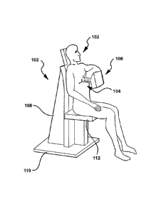
Figures la and lb illustrate a preferred embodiment of the present
invention optimized for cardiac SPECT, showing the overall configuration of
the
system 100 and the positioning of the patient 102. The opening 104 for patient
entry and egress is shown. The imaging section 106 of the system extends as an
arc over the right side of the patient's chest. The imaging section consists
of a lead
shielded housing with internal components as described below. The imaging
section is supported by a stand 108 affixed to a base 110. Together, the rear
portion of the imaging section and the stand fowl the "back" of the patient
support.
The patient is seated upon an adjustable seat 112. The vertical height of this
seat
may be adjusted so as to position the patient's heart within the appropriate
portion
of the imaging device. Such adjustment may be performed by means of electrical
motors, hydraulic devices or other means. The seat is optionally adjustable so
as
to swivel horizontally, thus easing patient entry and egress from the seated
position. The stand and base may also include or support the electronics
necessary
for processing scans, as well as any necessary controls or displays.
As shown, unlike in the prior art systems, the patient is seated generally
upright so that their torso is generally vertical. The lighter weight, simpler
design,
and reduced bulk of the present system cooperate to allow this positioning.
For
definitional purposes, the area surrounded by the imaging section 106 will be
referred to as a field of view. Also for definitional purposes, it may be said
that a
longitudinal axis, generally aligned with the longitudinal axis of the
patient's torso,
extends through the field of view. It may be said that the longitudinal axis
is
generally vertical to distinguish the positioning of the present system from
the
typical systems where the patient is forced into a horizontal position. In
actuality,
the generally vertical longitudinal axis may be reclined somewhat, as shown,
to
increase patient comfort.
As will be clear to those of skill in the art, it is very important to image
the
appropriate portion of the human patient, in order to acquire data about the
portion
of the patient that is of particular interest. For example, the preferred
embodiment
of the present invention is designed to image the patient's heart. Therefore,
it is
CA 02515198 2005-08-04
WO 2004/072679
PCT/US2004/003263
important that the portion or slice being imaged includes the patient's heart.
However, the exact position of the patient's heart within their chest is not
always
easily detennined from an external exam. In prior art systems, the patient is
positioned in front of the detector(s) while the operator views a low-
resolution,
5 two-dimensional display known as a persistence-scope (p-scope). The
persistence
scope image is necessarily of low quality due to its need to be continually
updated
as the patient is repositioned and due to the two-dimensional nature of its
images.
Operator error in patient positioning is not uncommon and, when it occurs,
results
in a useless scan. According to another aspect of the present invention, a
cardiac
10 scan may be preceded by a "quick scan" of the patient's chest so as to
properly
locate the heart so as to adjust the position of the chair so that the heart
is properly
positioned for imaging by the imaging section 106.
The "quick scan" is possible with the present invention for several reasons,
which will become clear after reviewing the entirety of this specification.
Systems
of the present art must partially orbit the patient in order to acquire three-
dimensional imaging. Movement of the large, heavy (typically 450-500 pound)
detectors must be started and stopped within seconds if rapid three-
dimensional
positioning images are to be obtained. This is both mechanically difficult and
may
present a hazard to the patient from the rapid movement of large and heavy
detectors. The present invention requires the movement of only an aperture arc
to
image the portion of the patient in the field of view of the imaging section
106.
The aperture arc is preferably hidden from the patient inside a housing, and
can be
moved much more quickly and safely than can prior art gamma camera. Also, a
full scan requires the arc to move only a short distance, unlike a gamma
camera
where the camera has to move a long distance. In addition, the present
invention
acquires image data more quickly than prior art devices. Therefore, a fast,
low
count, three-dimensional image may be acquired by quickly moving the aperture
arc within the housing. This low count image may be reconstructed almost
instantly with state-of-the-art computers and displayed immediately as slices,
or
preferably, as rotating surface rendered or maximum-intensity-projection
images.
CA 02515198 2005-08-04
WO 2004/072679 PC
T/US2004/003263
11
Such volume-rendered images clearly reveal the underlying patient anatomy and
may be used to reliably detennine the position of the heart prior to the start
of
routine, high-count imaging.
In embodiments of the present invention wherein the seat 112 is adjustable
upwardly and downwardly, the chair position may be optionally adjusted between
two image acquisitions so as to adjust the positions of the slices being
imaged. In
some embodiments, the movement may be very slight, so as to compensate for
effects of the collimators, which are discussed in more detail hereinbelow.
The
chair position may also be adjusted upwardly or downwardly during an image
acquisition.
As known to those of skill in the art, patient movement during imaging is a
significant problem for most imaging systems. Most systems require the patient
to
lie on a narrow horizontal surface, in a rather uncomfortable supine or prone
position. This position is often uncomfortable for patients with back problems
or
for the many cardiac patients that have difficulty breathing when lying flat.
Often,
this results in patient movement during the scan. In order to accommodate the
moving detectors of current art systems, the patient must hold their arms over
their
head for the duration of the imaging procedure. This is quite uncomfortable
for
many patients, particularly those with arthritic shoulders. Many
patients
experience fear or claustrophobia when lying under the large, metal detectors
of
current devices. Patients who are uncomfortable or fearful typically adjust
their
position in an attempt to become more comfortable. Such movement, when it
occurs during an image acquisition, causes image artifacts, which may cause
incorrect findings and subsequent treatment. The problem is exacerbated by
long
scan times. The vertical positioning of a patient enabled by the present
invention,
as illustrated in Figures la and lb, significantly improves patient comfort
and
stability. It is much more comfortable for back and cardiac patients. The anus
do
not need to be held over the head. The open design of the present invention
eliminates claustrophobia. Consequently, patient comfort and security is
increased
and movement is reduced. Also, some embodiments of the present invention
CA 02515198 2005-08-04
WO 2004/072679 PCT/US2004/003263
12
allow significantly reduced scan times, thereby reducing the effects of
patient
motion.
III. General Discussion of 1-Dimensional Solid State Detector Modules
(Strips)
Figure 2 shows one embodiment of an individual detector module 150.
Multiple (typically 64) individual modules are arranged in an arc surrounding
the
patient. The arc may extend over a range of approximately 180 -360 . For
cardiac SPECT, a preferred embodiment is approximately 1800. The embodiment
shown is a solid-state detector module sized for cardiac imaging. Other
detector
module embodiments are discussed below. As shown, the detector module 150 is
an elongated strip. Rectangular regions on the face of detector indicate an
array of
individual solid-state detector elements 152, each comprising one pixel for
data
acquisition. In
this embodiment, the array of detector elements is one-
dimensional, i.e. 1 X N, although two-dimensional arrays may also be employed.
Multiconductor ribbon cable 154 carries electrical signals from the detector
elements to the electronics that process the signals. Alternatively, some of
the
processing circuitry may be integral with or packaged by the detector
elements.
Each detector element 152 is operable to detect if a photon strikes it.
Therefore, the overall detector 150 is operable to detect if a photon strikes
and is
also operable to determine where along its length the photon struck. Each
detector
element includes some semiconductor material, such as cadmium-zinc-telluride,
with an electrode applied to opposing surfaces. An electrical potential is
applied
across the electrodes. As will be clear to those of skill in the art, when a
photon
passes through the front electrode and interacts with the semiconductor
material, a
small current travels between the electrodes. This current is measured to
sense the
impact of photons.
While the present invention is initially described as using the above-
described detector elements, other embodiments of the present invention make
use
of other detector designs, as will be described in more detail herein below.
IV. Aperture Arc ¨ General Discussion
CA 02515198 2005-08-04
WO 2004/072679
PCT/US2004/003263
13
Figure 3 shows the aperture arc 170 for an embodiment of the present
invention optimized for cardiac SPECT. A single radiation detector module 172
is
shown behind the arc to demonstrate relative positioning. As shown, the
detector
module is generally parallel to the longitudinal axis. The arc 170 serves as a
photon-blocking member and may be made of lead or a similar high attenuation
material. The arc 170 is of sufficient height to cover the radiation detection
modules 172 situated behind it. The arc is of sufficient thickness (typically
approximately 3 mm) so as to effect essentially complete absorption of photons
emitted by the patient. The arc is penetrated by a series of vertical aperture
slots
174 which permit photons 176 aligned with the aperture slot to pass from the
patient through the slot to reach the detector modules. The slots are
preferably
generally parallel to the longitudinal axis of the patient.
In Figure 3, the arc 170 is shown as a continuous member with generally
rectangularly shaped slots cut therethrough. In some embodiments, the slots
are
cut straight through, and have sides that are parallel to one another.
Alternatively,
the slots may be cut with angled sides such as shown in Figures 4-6. Each of
these
Figures illustrates a cross-section of the slot taken generally perpendicular
to the
slot. Figure 4 illustrates an embodiment wherein the arc 170 has tapered ends
171.
The arc 170 may be said to have a pair of opposing surfaces. The tapered
points
171 taper from each of these opposing surfaces to a point at approximately the
center plane of the arc. For simplicity, Figures 4-6 illustrate a portion of
the arc as
being generally linear. However, as previously discussed, it is actually
arcuate.
Preferably, the arc 170 blocks substantially all of the photons except those
that pass through the slot 174. A certain thickness of photon blocking
material,
such as lead, is required to adequately block these photons. The tapered
points
171 are thinner than the remainder of the arc. Therefore, it is preferred that
they
are formed out of a material that has even higher photon blocking ability,
such as
tungsten or gold, but could be lead. These tapered points 171 are joined to
the
material that typically forms the remainder of the arc 170. Alternatively, the
arc,
including the edges, could be all one material, such as lead. Figures 5 and 6
CA 02515198 2005-08-04
WO 2004/072679
PCT/US2004/003263
14
illustrate alternative embodiments of tapered points 173 and 175. In these
embodiment, the edges of the slots taper either from the front to the back or
from
the back to the front. As with the embodiment of Figure 4, the points are
preferably formed out of a material with a higher photon blocking ability than
the
remainder of the arc. The pointed edges of the slot are preferred, as they
provide a
more consistent apparent edge of the slot, independent of the angle from which
it
is viewed. That is, a slot with squared-off edges may appear substantially
narrower when viewed from an angle. By tapering the edges of the slot, the
slot
has a more consistent effective width when viewed at a shallow or deeper
angle.
This is especially important in the design of the present invention since
radiation
may enter the aperture at a significant angle. Alternatively, the "points" may
be
rounded.
In some embodiments of the present invention, it is preferred to have slots
with adjustable widths. This allows adjustment in the sensitivity and
resolution of
the imaging system. It may also assist in calibration. Figures 7 and 8
illustrate
one approach to providing slots with adjustable widths. Figure 7 illustrates a
cross-section of a portion of an arc 177 with adjustable slot defining pieces
179
attached thereto. Figure 8 shows a perspective view of one portion of an arc
177
with one adjustable piece 179. By adjusting the positions of the pieces 179
relative to the remainder of the arc 177, the relative position and width of
the slot
178 may be adjusted. As with the embodiments of Figures 4-6, the thinner
portions of the end pieces 179 are preferably formed from a material with a
higher
photon blocking capability than the remainder of the arc 177. The end pieces
179
are illustrated as having a front-to-back taper, but may have any of the
shapes
illustrated in Figures 4-6, or may provide a more squared-off or rounded-off
edge
to the slot. Also, the end pieces 179 are not required to be symmetrical.
Additionally, a single adjustable piece may be provided for each slot, with
the
other side of the slot being defined by a non-moveable edge. As will be clear
to
those of skill in the art, the interconnection between the end pieces 179 and
the arc
177 may be provided in a variety of ways, other than the approach illustrates.
CA 02515198 2005-08-04
WO 2004/072679
PCT/US2004/003263
Adjustment of the slot width may also be achieved in other ways, as will be
clear
to those of skill in the art.
V. Field of View
Figure 9A diagrams (from above) the relative positions of the patient field-
5 of-view area 180, the aperture arc 182 and the detector modules 184. It
may be
seen that the set of detector modules and the aperture arc are situated
concentrically around the patient. One embodiment for cardiac imaging includes
approximately 64 radiation detector modules 184, each consisting of an array
of
individual elements or pixels. In this embodiment, the aperture arc 182 is
10 positioned at a radius, a, of approximately 30 cm and the detector
modules 184 are
positioned at a radius, b, of approximately 40 cm. A patient field-of-view
area
with a diameter, c, of approximately 50 cm fits easily within the arc 182. The
aperture arc 182 and/or the set of detector modules 184 may be arranged in a
true
geometric arc with common arc centers at the longitudinal axis. Alternatively,
15 either or both may be more ovalized or be arcuate with non-shared arc
centers.
For example, the arc centers may be positioned away from the longitudinal axis
so
as to increase the arc radii. It is also possible for the arc 182 and/or the
set of
modules 184 to be non-arcuate. For example, either could be arranged as a
series
of short straight segments, or be partially arcuate and partially non-arcuate.
Another example would be if either had different arc radii at different radial
positions so that the radius of curvature changes along the "arc".
Displacement means is provided for moving the aperture arc 182 relative to
the detectors 184. As will be clear to those of skill in the art, many
different
approaches may be used to move the aperture arc. For example, the aperture arc
182 may be connected by a woini gear or other arrangement to a motor such that
it
can be rotated through a limited angle about the longitudinal patient axis. As
will
be clear to those of skill in the art, the arc may remain stationary with only
the
detectors moving. However, this approach is generally more complicated and
costly. For purposes of' processing the information from the scan, means are
also
provided for accurately determining the position of the arc. As will be clear
to
CA 02515198 2005-08-04
WO 2004/072679
PCT/US2004/003263
16
those of skill in the art, many approaches to providing this means are
available,
including optical encoders and mechanical sensors. The sensing means may also
be used for feedback control of the displacement means. A more detailed
discussion of one approach to moving an aperture arc will be provided
hereinbelow.
VI. Discussion of Sweep due to Aperture Arc movement
Figures 10a-c show overhead views of a single detector 190 and a small
section 192 of the aperture arc. The Figures illustrate the relative position
of the
arc 192 and the detector 190 at three different rotational positions of the
aperture
arc 192. At each position, the position of the aperture slot 194 restricts the
line of
response of the detector to a particular path 196, as shown. It can be seen
that, as
the aperture slot 194 moves in front of the detector 190, the line of sight of
the
detector fans across the patient, generating a multiplicity of lines of
response or
projections.
Since, as diagrammed in Figure 9A, there are a multiplicity of detector
modules 184 and, as shown in Figure 3, a multiplicity of aperture slots 174, a
multiplicity of detector lines of response are fanned at each rotational
position of
the aperture arc. Figure 9B illustrates a small subset of the lines of
response 200
obtained from a few of the detectors 202 as the aperture arc 204 is rotated.
The
aperture slots themselves are not shown in this Figure, for simplicity. A
diagrammatic "slice" 206 through the patient's chest is shown, indicating that
a
full set of projections of the heart, sufficient for tomographic
reconstruction, is
obtained in this manner.
The aperture arc preferably moves continuously, such that the lines of
response "sweep" over the field of view. Alternatively, the aperture arc may
move
in discrete steps, with imaging occurring with the arc stopped at each of the
steps.
VII. Each Detector Illuminated by only a Single Aperture Slot
All detectors preferably "look through" only one slot at all times. Slot
spacing is
CA 02515198 2005-08-04
WO 2004/072679
PCT/US2004/003263
17
determined such that each detector is illuminated by only one slot at a time.
Overall photon detection efficiency is proportional to the number of slots in
the
aperture arcs. The maximum number of slots permissible, nsiotõ is a function
of the
angle 0. , representing the maximum angel of incidence of a usable ray at an
aperture slot, the radius of the detector arc and the minimum length of arc on
the
aperture arc such that a given length of arc BA on the aperture arc such that
a given
detector will only see the patient field-of-view through one slot at a time (
ay):
7.1. = Oarc
7z. = Oarc
27-r 2n-
nslots ¨ n
u A - D 11?
2= -4 Ivo
sin ¨sin
= -1
J?,1)RDJ
where Ro is the radius of the patient, R/4 is the radius of the aperture arc
and RD is
the radius of the detector arc. The aperture arc need only be rotated by the
interval
between slots, nstots, to provide a full set of angular projections.
For one embodiment of the present invention, the radius of the patient Ro,
is assumed to be a maximum of 22 cm, the radius of the aperture arc RA, is 30
cm
and the radius of the detector arc, RD, is 45 cm. The detector arc and
aperture arc
span an angle, (bare, of 180 degrees and the minimum length of the arc, 9,4,
is 36
degrees. For these values, the equation provides that five slots are the
maximum
number of slots to avoid any detector looking through more than one slot at a
time.
Consequently, the aperture arc need only rotate through an angle of 36 degrees
to
provide a full set of angular projections.
The above equation and solution assumes that the slots are equally spaced
along the arc, and separated by an angle of 36 degrees. As will be clear to
those of
skill in the art, the critical issue is actually the angular separation
between the
slots, which determines the number of slots. Referring again to Figure 3, the
arc is
CA 02515198 2005-08-04
WO 2004/072679
PCT/US2004/003263
18
shown with five slots, one which is hidden in the bend, due to the angle of
view in
the Figure.
While the above equation and discussion leads to the conclusion that 5
slots are needed, with a separation of 36 degrees between the slots, the
addition of
a 6th slot is beneficial. Figure 11A diagrammatically illustrates the present
invention with a plurality of detectors 195 disposed in an arc, an aperture
arc 196
with five apertures 197, and a field of view 198. The arc 196 is shown at the
extreme clockwise position. Assuming photons of interest may originate from
anywhere in the field of view, projection rays are drawn to show how the field
of
view is "projected" onto the arc of detectors 196. As shown, some photons are
projected to a position clockwise of the last detector, and therefore do not
contribute to the image.
Likewise, a number of the detectors at the
counterclockwise end are "out of view" of the aperture at the counterclockwise
end of the aperture arc 196, and are therefore unexposed with the arc in this
position. Unexposed detectors represent a less than optimal system efficiency
Figure 11B illustrates the aperture arc at the midpoint of its travel. As
shown, at this position, the projections through all apertures 197 coincide
with the
positions of the detectors 195, so that no photons are wasted and no detectors
are
unexposed.
Figure 11C illustrates the aperture arc 196 at the extreme counterclockwise
position. In this position, detectors at the clockwise end of the detector
assembly
are unexposed, and some photons passing through the apertures at the
counterclockwise end go undetected.
One solution to this problem is to provide a larger number of detectors.
However, the increases the size of the imaging section, and dramatically
increases
the cost of the device. A preferred solution is illustrated in Figure 11D. The
aperture arc 212 now has 6 slots 214 projecting photons onto the detectors
216,
from the field of view 218. The spacing between these slots is unchanged,
however, from that determined by the equation above (36 in this example).
Figure 11D illustrates the arc 212 at the extreme clockwise position. As
shown, all
CA 02515198 2005-08-04
WO 2004/072679
PCT/US2004/003263
19
detectors are illuminated due to the addition of the sixth slot. Figure 11E
illustrates the arc 212 at the midrange of travel, and Figure 11F illustrates
the arc
at the extreme counterclockwise position. Again, all detectors 216 are
illuminated
at all positions, thereby increasing photon collection efficiency. The
addition of
the "extra" slot, results in a perfect match of incoming photons to the length
of the
arc of detectors. In this arrangement, all detectors are illuminated via the
aperture
slots at all times, thereby optimizing photon detection efficiency.
VIII. Diagonal Apertures
Referring again to Figure 3, the slots 174 are shown as generally vertical
slots. That is, they are parallel to the longitudinal axis of the field of the
view.
According to further aspects of the present invention, the slots may be
diagonal as
shown in Figure 12. Figure 12 illustrates an assembly including an aperture
arc
207 with diagonal apertures 208 defined theretlu-ough. The diagonal apertures
are
illustrated as being defined by adjustable side pieces 209, but may
alternatively be
provided by slots cut into the arc 207. Also, as with the earlier embodiments
of
slots, the slot edges may be tapered in a variety of ways, including any of
the
previously disclosed shapes. As will be clear to those of skill in the art,
multiple
apertures are preferred, arranged in intervals along the arc 207. Only two
apertures 208 are illustrated in Figure 12, for simplicity. However,
additional
apertures are preferred. Figure 12 illustrates additional aspects of the
present
invention, which will be discussed hereinbelow with respect to collimator
design.
The angled slots or apertures 208 may be provided at a variety of angles
ranging
from slightly angled from "vertical," to nearly horizontal. As a further
alternative,
the slots may be completely "horizontal" with respect to the patient axis. The
apertures may also be angled in the opposite direction to the angle
illustrated in
Figure 12.
In embodiments of the present invention where the apertures are "vertical"
and the collimators are horizontal, or vice versa, the resolution is different
in the
CA 02515198 2005-08-04
WO 2004/072679
PCT/US2004/003263
vertical and horizontal directions. According to one preferred embodiment of
the
present inventions, the apertures are angled at approximately 45 degrees one
direction, and the collimators are angled at approximately 45 degrees the
other
direction. By angling the apertures and the collimators relative to the
transaxial
5 imaging plane, the overall resolution experienced at the imaging
plane is made
essentially isotropic, i.e. similar in all directions. This is desirable in
some
applications, particularly if the reconstructed date is to be refoilliatted
along
obliquely angled planes.
IX. Collimators
10 Referring again to Figures 3 and 11, the aperture arc and the set of
detectors provide projection data collimated within the transaxial plane, but
not
collimated longitudinally. For this reason, the invention preferably provides
a set
of longitudinal or cross-plane collimators, as shown in Figure 13. As will be
clear
to those of skill in the art, the collimator design illustrated in Figure 13
is designed
15 for use with the "vertical" aperture arc, such as shown in Figure 3.
The
longitudinal collimators consist of a stack-like series of arc-shaped vanes
220
arranged as shown and located concentrically to the arc arrangement of
detectors
222 as shown. The aperture arc is omitted from this figure, but is located
concentrically to the longitudinal collimator vanes. The vanes are preferably
20 mutually parallel and generally perpendicular to the longitudinal
axis of the
patient. The vanes are sheets or panels of lead or similar attenuating
material and
may be separated by spacers of radiolu.cent plastic foam or similar material
(not
shown). The number, size, and thickness of the vanes may be varied depending
on
the application.
Figure 14 is similar to Figure 13 but with the addition of the aperture arc
230. It may be seen that each individual detector element (pixel) of each
detector
232 has a unique line-of-response 234 directed into the patient field-of-view
by the
combined collimating effects of the aperture arc slots 236 and the
longitudinal
collimating vanes 238.
CA 02515198 2005-08-04
WO 2004/072679
PCT/US2004/003263
21
As will be appreciated by those of skill in the art, it is preferred that the
vanes 220 be provided in a plane that is generally perpendicular to the
apertures in
the aperture arc. In the embodiment of Figures 13 and 14, collimators vanes
may
be considered to be "horizontal," since they are perpendicular to the
"vertical"
patient axis. Referring again to Figure 12, it can be seen that the
collimators 210
are angled so as to be generally perpendicular to the angled aperture. Only
five
collimating vanes 210 are illustrated in Figure 12, in order to avoid
cluttering the
drawing. However, it will be appreciated that the vanes are provided along the
entire assembly, as indicated by the arrows. If the apertures are angled at
other
angles, the vanes 210 may also be angled so as to remain perpendicular
thereto.
Alternatively, the collimator vanes 210 and apertures 208 may be at angles to
one
another other than perpendicular.
X. Resolution and Efficiency
The in-plane resolution of a system according to the present invention is
determined by the radii of the detector and aperture arcs, RD and RA, the
distance,
Dist, of the object from the aperture arc, and the widths of the slots and the
detector elements, Wsrot and Wdet respectively:
Dist x (Wslot + Wd et)
resolution flot +
(RD ¨ RA)
Figure 15 plots the resolution at different depths (distance from the
collimator to the point of interest in the patient) of the present invention
versus a
traditional parallel-hole collimator. The slotted arc system is assumed to
have a
slot width of 2.4 mm, a detector width of 4 mm and other parameters as
discussed
with respect to Figure 4. The parallel-hole collimator for which data is
plotted has
a hole diameter of 2.2 mm and a collimator thickness of 3 cm.
CA 02515198 2005-08-04
WO 2004/072679 PCT/US2004/003263
22
The detection efficiency of the slotted aperture system is proportional to
the detector solid angle, 12, for a point source at the center of the field-of-
view and
may be calculated based on Rogers (IEEE TIMI, vol. MI-1, pp. 63-68, 1982) as:
respectively, pdet is the detector packing fraction andfis the fraction of
frontal area
closed by the longitudinal collimating vanes. In the configuration of this
invention, f= vane thickness / vane separation.
As the aperture arc moves to differing positions relative to the detectors,
the apparent width of the aperture slots will vary as a function of the sine
of the
angle between the slot and the detector. Since the apparent width of the
detector
as viewed from the slot also changes according to a similar function, the
overall
detection efficiency will vary as a function of the square of the sine of the
detector-
slot angle. The exact function will depend on the photon cross-section of the
detector element (a function of detector thickness) and on the photon cross-
section
of the slot aperture. This variation of detector sensitivity with slot
position is
easily mapped for a given detector and may be corrected for in software in a
manner similar to the detector uniformity corrections routinely perfoimed in
traditional gamma cameras.
It is to be noted that imaging systems constructed according to the methods
of this disclosure are relatively insensitive to the structured image
artifacts seen in
rotating gamma camera SPECT systems when non-uniformities of detector
sensitivity exist. In the systems described here, the reduced count
sensitivity
caused by a particular, relatively insensitive, detector element is spread
across the
entire image plane, rather than appearing as the structured "ring" or "arc"
artifacts
seen in traditional systems. Such artifacts frequently trouble present
artifact
systems.
XI. Collimator Construction
1 1 11- \ r
Q = ns, ¨ 2= ¨ r 2 __
&ots r, 2 obj D = robjRD RA )J2
¨ 12 I.RArD fP
(let
-11D _ RA
CA 02515198 2005-08-04
WO 2004/072679
PCT/US2004/003263
23
As will be appreciated by those of skill in the art, the construction of lead
collimators presents significant challenges. Lead has a very high density, but
is
not particularly stiff or strong. Therefore, vanes of lead are heavy and
vulnerable
to damage. In traditional parallel hole collimators, the vanes are made very
thin
and define a plurality of small parallel holes. The depth of the holes in the
collimators is somewhat limited by the strength and stiffness of the lead
material.
That is, if a collimator is to be constructed that has more than a particular
depth,
the thin lead vanes may actually sag over time, destroying the usefulness of
the
collimator. Similar considerations apply to the present invention. The
collimating
vanes, such as 220 in Figure 13 and 210 in Figure 12 are large and heavy,
thereby
presenting challenges to how to adequately support the individual vanes.
Additionally, it is important that the individual vanes be accurately
positioned and
aligned.
A further inventive aspect of the present invention is a design providing a
collimator with parallel lead vanes that are supported by being foinied in a
stack
with sheets of radiolucent material disposed between each lead vane. Figure 16
illustrates a portion of a parallel vane collimator constructed according to
this
aspect of the present invention. Figure 16 also illustrates a portion of a
support
structure, including a lower support 240 and an upper support 242.
Figure 17 shows the lower support 240 and upper support 242 in their
entirety, according to one embodiment of the present invention. However,
Figure
17 does not illustrate the collimation assembly inside of the support frame.
Referring to Figure 17, the lower support 240 and upper support 242 foini part
of a
support assembly 244. This support assembly 244 foluis part of the imaging arc
106, as shown in Figures 1A and 1B. It wraps about the patient field of view,
illustrated at 245 in Figure 17. When assembled, the imaging arc includes the
support structure 244, the parallel vane collimator supported therein, single
or
multiple detectors, and the aperture arc. It is also preferably clad in a
housing so
as to protect the internal workings, and provide an aesthetically pleasing
exterior
appearance. One end of the support structure 244 is interconnected with the
chair
CA 02515198 2005-08-04
WO 2004/072679
PCT/US2004/003263
24
base 108 for supporting the imaging arc. This may be accomplished in a variety
of
ways. Alternatively, an additional support may be provided mid-arc.
Referring again to Figure 16, a portion of the parallel vane collimating
assembly is shown at 246. The collimating assembly includes sheets or panels
of
lead 248 with sheets or panels of radiolucent material 250 separating the lead
sheets 248. The collimator assembly may be foimed by stacking a lead sheet,
and
then a radiolucent sheet, and then repeating the process until a sufficiently
tall
stack is formed, as shown. The radiolucent material maintains the relative
positioning of the lead sheets, and prevents any sagging or movement of the
lead
sheets. Preferably, a compression panel 252 is provided on top of the stack of
lead
sheets and radiolucent material, and below the upper support panel 242.
Biasing
devices, such as threaded members 254 are then provided to press downwardly on
the compression panel 252. This compresses and stabilizes the stack 246.
Preferably, a thicker lead sheet, or other photon blocking material 253 is
provided
at the top and bottom of the stack, to block photons from entering the top or
bottom of the collimator.
As will be clear to those of skill in the art, a modified version of this
assembly procedure may be used to construct a collimator assembly such as
shown
in Figure 12. According to a further aspect of the present invention, a
related
approach may be used to form parallel hole collimators. That is, a parallel
hole
collimator may be formed using radiolucent material filling the holes in the
parallel hole collimator, to thereby support the collimator vanes. Parallel
hole
collimators are often damaged in use, because of the fragility of the lead
septae
between the holes. According to the present invention, the holes of the
collimator
may be filled with a radiolucent material as it is constructed. This turns the
parallel hole collimator into substantially a solid block, which is more
resistant to
damage. Also, this allows deeper and/or thinner vanes to be formed and
supported
than would otherwise be practical.
Referring again to Figure 17, an alternative approach to forming a parallel
vane collimator according to the present invention may be provided by allowing
CA 02515198 2005-08-04
WO 2004/072679
PCT/US2004/003263
the upper and lower support members 240 and 242 to be tensioned against each
other, such as by tensioning members 256. That is, the alternating stack of
lead
panels and radiolucent panels may be placed on the lower support member 240,
covered by upper support panel 242, and compressed using compression or
tension
5 members 256. Those of skill in the art will appreciate that the parallel
vane
collimator according to the present invention is very heavy, and therefore the
cantilevered arc bears a substantial load. Figure 18 illustrates that the
support
structure may include a plurality of angled tension members 258, either angled
to
the left as shown, or angled to the right, or both. The tension members act
like
10 bicycle spokes in providing structure and support. They also allow a
substantially
open back to the arc for access to the electronics and for cooling.
Figure 19 provides a cross-sectional view of a portion of the imaging
section of the present invention. It illustrates the bottom support member
240, the
upper support member 242 and the lead sheets 248 positioned therebetween. The
15 radiolucent material is not illustrated in this view. However, an
electronics
package or detector array for detecting incoming photons is illustrated
generally at
260. This detector array will be discussed in more detail hereinbelow.
The design of the present invention provides advantages heretofore
unavailable with respect to collimator design. Traditionally, collimator
designers
20 have limited the depth to width ratio of the collimator holes. That is,
the holes
defined by the collimator may be considered to have a front-to-back depth and
a
side-to-side or top-to-bottom width. (In a parallel hole collimator, a side-to-
side
and top-to-bottom widths are typically the same. In the present invention, the
"side-to-side width" is a function of the size of the aperture in the aperture
arc,
25 while the top-to-bottom width is a function of the spacing between the
parallel
vanes.) In the prior art, a depth-to-width ratio of less than 10:1 has been
considered optimal. In fact, the literature has stated that a 10:1 ratio is
almost
equivalent to an infinitely large ratio. In other words, excepted theory has
taught
against depth to width ratios over 10:1. Additionally, prior art designs for
collimators have made it extraordinarily difficult to create a depth-to-width
ratio
CA 02515198 2005-08-04
WO 2004/072679
PCT/US2004/003263
26
that is very large. Deep collimators suffer from structural integrity issues.
To get
a high depth to weight ratio in prior art designs requires vanes that are too
thin and
tall to be self supporting. So, practicality also taught away from high depth
to
width ratios.
The present invention departs dramatically from the prior art approach. In
one embodiment of the present invention, the lead sheets have a thickness of
approximately 2 mm, as indicated at A in Figure 16. The radiolucent sheets
have a
thickness of approximately 4.5 mm. Therefore, the "gap" between adjacent lead
sheets is approximately 4.5 mm. In this same embodiment, the front-to-back
depth
of the lead vanes 248, as shown at C in Figure 19, is approximately 150 mm. In
this embodiment, the depth-to-width ratio is greater than 33:1. In a more
preferred
embodiment of the present invention, the lead vanes have a thickness of
approximately 1.25 mm. However, the gap remains the same at approximately 4.5
mm. Therefore, the depth-to-width ratio remains the same. According to the
present invention, depth-to-width ratios greater than the prior art maximum of
10:1
are preferred. Depth-to-width ratios greater than 20:1 are more preferred.
Depth-
to-width ratios over 30:1 are even more preferred.
According to the present invention, it is also preferred that the thickness of
the lead vanes be greater than .5 mm. A thickness of greater than .75 mm is
more
preferred, a thickness of 1 mm or more is more preferred, and a thickness of
at
least 1.25 mm is most preferred. These thicknesses also depart dramatically
from
the prior art. Prior art high resolution parallel hole collimators typically
have lead
vanes with a thickness of .2 mm or less, and significant effort has been
expended
to obtain thinner and thinner lead vanes.
The use of substantially greater depth-to-width ratios than used in the prior
art, as well as the use of substantially thicker lead vanes, provides
significant
advantages that have not been recognized or appreciated in the prior art.
In SPECT imaging, it is important to accurately determine the direction
from which a photon is traveling, the energy level of the photon, and the
number
of photons coming from that direction. These photons have sufficient energy to
CA 02515198 2005-08-04
WO 2004/072679
PCT/US2004/003263
27
penetrate lead if it is not sufficiently thick. In prior art, parallel hole
collimators,
the thin lead vanes are typicaAy too thin to stop many of the photons from
passing
therethrough. Therefore, a photon that strikes a particular area cannot be
assumed
to have traveled straight down the hole adjacent that area. Instead, the
photon may
have originated in a different hole and penetrated the lead vane in-between
the
adjacent hole and the hole in which it is sensed. Consequently, accuracy is
sacrificed. This contributes to blur in the resulting image. The depth-to-
width
ratio of the holes in the collimator also has an effect on the resolution of
the
imaging device. If a collimator hole is short and wide, a photon may enter
that
hole at an angle significantly off from the axis of the hole. If the hole is
deeper
and narrower, the range of angles of incoming photons that travel just down
that
hole is much narrower.
In the present invention, the use of substantially thicker vanes and the use
of a collimator with a very high depth-to-width ratio, both lead to
substantially
increased accuracy or resolution. Because the vanes are thick and the depth is
very
high, any photon that reaches the sensor at the back of the collimator can be
assumed to have passed through the aperture in the aperture arc and between
the
adjacent lead vanes. In other words, each photon "count" is a good count.
The prior art also tends towards the use of much smaller gaps than in the
present invention. Experimentation with the present invention have shown that
larger gaps, on the order of 4 or 4.5 mm, along with thicker lead vanes leads
to
higher efficiency and resolution. As a further aspect of the present
invention, the
use of gaps greater than 2 mm is preferred, with gaps with greater than 3 mm
being
more preferred, and gaps of 4 or more mm being most preferred.
Referring again to Figure 19, the sensor array 260 is positioned adjacent
the back of the collimating assembly. In some embodiments, the individual
sensors are positioned immediately adjacent the rearmost end of the vanes,
while
in other embodiments the sensors are spaced from the back of the vanes by a
short
distance. Increasing the gap between the back of the vanes 248 and the sensors
reduces some of the effective dark area caused by the photons that are blocked
by
CA 02515198 2005-08-04
WO 2004/072679
PCT/US2004/003263
28
the vanes. In one preferred embodiment, the sensors are spaced from the back
of
the vanes by 2 to 3 mm.
XII. Extension Flaps
As shown in Figures 1 and 4, for an embodiment optimized for cardiac
imaging, the use of an arc shaped imaging apparatus allows the patient to
easily
enter and leave the imaging system. As the aperture arc rotates however, it
will
extend slightly into the open area of the arc. The invention therefore
optionally
provides for pivoted Extension Flaps to be located at one or both ends of the
aperture arc, as shown in Figures 20A and 20B. This figure shows one end of
the
aperture arc 300 that includes an extension vane 302 extending its length.
Figure
20A shows the aperture arc 300 and vane 302 at one extreme of the arc's
movement and Figure 20B shows them at the other extreme. Extension vane 302
is movably attached to the aperture arc by hinge 304. Pivot rod 306 is located
in
the path of the vane such that, as the extension vane is pushed against it by
the
movement of the aperture arc, the extension vane is caused to pivot away from
the
patient as shown in Figure 20B. This minimizes the extension of the arc or
vane
into the opening while maintaining shielding of the detectors from unwanted
external radiation.
Referring now to Figure 21, one preferred construction of the aperture arc
is illustrated. The aperture arc is shown at 310, being supported on the
support
member 240, which forms the bottom part of the support structure of the
imaging
arc. In this embodiment, the aperture arc 310 is formed from individual
arcuate
panels 312 that are positioned adjacent one another so as to provide an
aperture
314 therebetween. The width of the aperture 314 may be determined by the
relative positioning of the panels 312. The aperture arc 310 is supported in a
track
in the support member 240 and moved by a drive motor 316, which drives a
series
of belts and pulleys.
XIII. Detector Variations
Turning now to detector designs, a variety of approaches may be used with
the present invention. Figures 2 and 3 illustrate strip detectors that may be
CA 02515198 2005-08-04
WO 2004/072679
PCT/US2004/003263
29
considered one-dimensional linear arrays. Two-dimensional arrays are also
provided in this invention. Such arrays may be provided as integral units or
may
be approximated by placing two or more one-dimensional arrays in close
proximity. The overall sensitivity of the imaging system is linearly
proportional to
the detector surface area available.
Referring to Figures 22-24, three views of a preferred embodiment of a
sensor assembly for use with the present invention is generally shown at 320.
As
best shown in Figure 23, the assembly 320 includes three two-dimensional
sensor
arrays 322, 324, and 326. Each sensor array, in turn, is formed of a series of
sensor modules, such as 328 in Figure 24. The sensor modules are solid state
CZT
(Cadmium Zinc Telluride), or alternatively, Cadmium Telluride may also be
used.
Figure 25 illustrates a cross-sectional view of one of the sensor modules 328.
The
module has a central body of CZT 330 with multiple small, thin, square
electrodes
332 on the front face. A larger electrode is provided on the back surface, and
a
chip for processing data signals from the sensor is provided on the back at
336.
Photons strike the front surface of the sensor module 328 and are sensed by
the
module. Figure 26 illustrates an alternative embodiment wherein a chip 338 is
only half covered by sensing materials 340. Figure 26 also illustrates the
configuration of the electrodes 342 on the face of the module.
Figures 22 and 24 illustrate cooling manifolds 346 for the sensing
assemblies.
As known to those of skill in the art, solid state photon sensors are
difficult
to produce without internal flaws. Referring to Figure 25, the body of CZT
material 330 is a crystal that may develop flaws during creation or
manufacturing.
If the body 330 does not have flaws, a photon passing through the front face
and
into the CZT body 330 enables the presence of this photon to be sensed by the
electrodes 332 and 334. As shown in Figure 26, the electrodes 342 define a two-
dimensional grid. Consequently, the location of the photon strike may be
determined by determining which electrode senses the presence of the photon.
If
the CZT is flawed, it may have dead spots, where a photon strike is not
sensed.
CA 02515198 2005-08-04
WO 2004/072679 PCT/US2004/003263
Typically, electrodes on the front of the CZT body are sized and spaced so
that one
electrode is responsible for sensing one "pixel" of information. Typically, a
pixel
size is chosen and equal to the desired resolution of the sensing system. In
cardiac
sensing, it is preferred to have resolution of approximately 4 to 4.5 mm.
5 Therefore, the electrodes would typically be arranged on 4-5 mm centers
such that
one electrode is responsible for each "pixel." If the CZT has a flaw, the flaw
may
cause a dead pixel, which can seriously affect image quality.
According to a further aspect of the present invention, the desired
resolution, in this case, 4 to 4.5 mm, is subdivided into smaller segments and
10 smaller electrodes are used. In Figure 26, box 350 represents an area
that is
approximately 4 to 5 mm wide and tall. However, rather than having a single
electrode in this area, this "macro pixel" is subdivided into four pixels,
each with
its own electrode 352. If the CZT underlying the macro pixel 350 has a flaw,
the
flaw will typically lead to only a single bad pixel associated with one of the
15 electrodes 352. For ,example, one of the four electrodes may be
associated with a
portion of the CZT that has no sensitivity, reduced sensitivity, or, in rare
cases,
increased sensitivity. The sensor module can then be calibrated, and the data
from
the four electrodes 352 processed so as to provide meaningful data from the
macro
pixel 350. For example, if one electrode is associated with a pixel that is
dead, the
20 output from the remaining three pixels may be combined, and multiplied
by % to
obtain an output for the macro pixel 350. In this way, a sensor module with a
CZT
body with some flaws is still useable. In the module of Figure 26, the
electrodes
352 preferably have a side-to-side and top-to-bottom dimension of
approximately
2.46 mm, and a spacing between adjacent electrodes of approximately .04 mm. In
25 another preferred embodiment, especially optimized for cardiac use, the
electrode-
to-electrode pitch is approximately 2.25 mm. Referring again to Figure 19, the
sensor assembly 260 is shown adjacent the rear of the lead vanes 248. Figure
27
illustrates a view of the sensor arrays 360 as viewed through the vanes 362.
In
some embodiments, the pitch between the vanes 362 is not evenly divisible by
the
30 pitch between the electrodes 364. For example, in one embodiment, the
pitch-
CA 02515198 2005-08-04
WO 2004/072679
PCT/US2004/003263
31
between the vanes 362 is approximately 6.5 mm, while the pitch between the
electrodes 364 is approximately 2.5 mm. In order to avoid moire patterns due
to
the alignment between the vanes and the pixels, it is desirable that the
number of
pixels in each gap between the vanes is approximately the same. Because the
vane
pitch is not a multiple of the pixel or electrode pitch in this embodiment,
the sensor
arrays 360 are arranged such that they are centered on the middle vane 366. As
shown in Figure 27, this arrangement prevents an electrode, and hence a pixel,
from lying directly behind one of the vanes 362.
This invention also provides for radiation detectors constructed from
scintillation materials such as sodium iodide or cesium iodide with associated
photomultiplier tubes or other photo-detectors such as solid state
photodiodes.
Figure 28 shows one embodiment of a scintillation-based detector module 400.
This embodiment includes a cylindrical crystal 402 of scintillation material
clad in
a radiolucent, light-reflective covering 404 such as aluminum. The covering
404
is open at both ends of the cylinder. Affixed to each end, via optical
coupling
material, is a light detector such as a photomultiplier tube, photodiode, or
other
photo-detector (not shown). The position of scintillation events occurring
within
the scintillation material is determined by the ratio of outputs of the two
photo-
detectors, thus providing longitudinal position sensing within the detector.
This
embodiment is extremely inexpensive to produce, but has the disadvantage of a
variable photon detection efficiency across its horizontal dimension caused by
the
varying scintillator thickness over its circular cross-section. This causes a
deviation of the detector's response function from a pure rect function, thus
slightly degrading spatial resolution.
Figures 29A-C-c show more efficient embodiments of a scintillator-based
detector, consisting of a rectangular bar 420 of scintillator material clad in
a
radiolucent, light-reflective material 422 such as aluminum. In Figure 29B,
the
cladding is open at the top and bottom so as to permit placement of photo
detectors
424. In the alternative embodiment shown in Figure 29C, the cladding is open
at
the rear of the module so that two or more photo-detectors 426 can be affixed.
In
CA 02515198 2005-08-04
WO 2004/072679
PCT/US2004/003263
32
either case, the photo-detectors are considered to be adjacent the ends of the
scintillation material so that they can locate the position of a scintillation
event.
Figure 30 shows a piece of scintillator material 430 with a trapezoidal cross
section clad in reflecting material 432, similar to the previous Figures. As
with the
embodiments of Figures 29A-C, the photo-detectors may be affixed on either the
top and bottom of the module or at the rear face. The embodiment with the
trapezoidal cross section has the advantage of presenting a more uniform cross-
section to incoming radiation, but is more costly to manufacture. That is,
radiation
coming at an angle to the front face still encounters the full depth of the
scintillator
material.
Axial resolution of the tomography system is directly dependent on
detector width, as described above. Specifically, narrower detectors increase
the
axial resolution of the system. As detector width narrows, however, photon
detection efficiency drops because photons striking the front face of the
narrow
detector may scatter out of the detector material before they have deposited
all of
their energy. According to the present invention, the efficiency of a high
resolution elongated strip of scintillation material may be improved by
masking a
portion of its front face. Figure 31A shows a detector configuration 440 based
on
a rectangular piece of scintillation material. Figure 31B shows a detector
configuration 442 based on a cylindrical piece of scintillation material.
Figure
31C shows a detector configuration 44 based on a piece of scintillation
material
with a trapezoidal cross section. In each of these embodiments, in addition to
the
reflective cladding 446, the scintillator is clad in an additional masking
layer 448
of lead, tungsten or similar high-attenuation material. This outer masking or
shielding layer is configured to have a narrow vertical opening 450 of the
dimensions desired for the detector cross-section. Once photons have passed
through the opening and struck the scintillator, further scattering is more
likely to
occur within the larger volume of scintillator located behind the opening 450
in the
mask 448 rather than scattering outside the scintillator material. If desired,
an
additional layer of low-Z material (not shown) may be interposed between the
CA 02515198 2005-08-04
WO 2004/072679
PCT/US2004/003263
33
cladding and the shielding layers so as to absorb secondary lead x-rays
emitted by
the mask 448. As will be clear to those of skill in the art, the detectors
shown in
Figures 31D have the improved efficiency of wider detectors with the higher
resolution of narrower detectors. Similar masking can be, applied to solid
state
detectors, such as shown in Figure 2, resulting in similar advantages.
Referring to Figure 34, a similar masking approach may be applied to a
two dimensional piece of scintillation material to faun a detector 452 with
the
benefits described above. Specifically, a piece of scintillation material 454
has
mask of lead applied in strips 456 to its face. Narrow vertical openings 458
are
left to allow entrance of photons aligned with the openings. Like with the
embodiment of Figures 14a-14c, this give increased accuracy. Photodetectors
459
are positioned behind the scintillation material 454 and are capable, by means
such
as "Anger logic", of detecting where a pulse of light occurs. Because a
portion of
the face is masked, the electronics "knows" that the photon did not strike in
the
masked areas and can therefore more precisely pinpoint the location of the
strike.
The masking off of certain portions of the detector surface reduces, in
effect, the
positional uncertainty of a given pulse of light, thus permitting its position
to be
determined more accurately and precisely.
Figure 32 shows details of construction of a bar-shaped, masked detector
module 460 as described in the previous Figures but with the photo-detectors
462
attached at the rear face through use of optical coupling material 464. A
similar
masking configuration may be used with solid-state detector modules.
As will be clear to those of skill in the art, photo-detectors of various
types
are somewhat costly. Therefore, it is desirable to reduce the number required.
According to another embodiment of the present invention, a pair or more of
optical fibers may be attached to each of the scintillation based detectors,
with one
fiber connected to each end of the detector. The fiber may be connected to the
top
and bottom and/or to the back face adjacent the top and bottom. The optical
fibers
may then be routed to a photomultiplier of the type have position sensitivity.
These readily available multichannel photomultipliers are capable of providing
CA 02515198 2005-08-04
WO 2004/072679
PCT/US2004/003263
34
distinct outputs for a multiplicity of locations across the face of an
individual tube.
Such a photomultiplier can then sense light pulses from a large number of
optical
fibers running from various detectors. In this way, the total number of photo
detectors is reduced. A similar approach may be applied to two dimensional
scintillation based detectors. Rather than using photodetectors mounted to the
rear
of the material, multiple optical fibers may be used to route the light to
multichannel detectors.
As previously discussed, the pieces of scintillation material that form the
core of a scintillation based detector are clad in a radiolucent, light
reflecting
material such as aluminum. This increases the brightness of the pulse of light
as
perceived by the light detectors. However, in some situations, this
reflectivity may
interfere with the ability of the light detectors to deteimine the
longitudinal
position where the photon struck the scintillation material. Therefore, it may
be
beneficial to reduce the reflectance of one or more surfaces of the
scintillation
material. For this purpose, .the surface may be roughened prior to cladding,
the
cladding may be roughened in certain areas, or a lower reflectance coating may
be
applied to either the scintillation material or the cladding. Alternatively,
it may be
desirable to vary the reflectance along the length of the reflector. For
example, a
roughed strip on one surface of the scintillation material may vary in width
along
the length of the detector. The strip could be narrow in the center, so that
reflectance remains high, and wider near the ends so that reflectance is
reduced.
This increases the likelihood of events near the center being detected at the
ends.
XIV. Detectors and Arc may Both Move
If the spacing of detector modules is sparse, gaps may be seen in the
pattern of angular sampling provided by this system. The importance of such
gaps
depends on the number of angular "bins" of data obtained as the aperture arc
moves. In addition, the significance of any artifacts caused by incomplete
angular
sampling depends on the clinical setting. If such artifacts are objectionable,
this
invention optionally provides for a means (Figure 33) of rotation of the arc
of
detector modules 500 through a limited angular range 502, such motion
occurring
CA 02515198 2005-08-04
WO 2004/072679
PCT/US2004/003263
either continuously or in a limited number of discrete steps. The range of
motion
of the detector arc is equal to the spacing between detectors. At each step of
detector motion, the aperture arc 504 is moved through its range of motion
506. In
this manner, a full set of angular projections may be obtained with even
sparse
5 detector population.
As another alternative, a tomography system according to the present
invention may be provided with a reduced number of detectors to reduce the
cost
of the system. This system would have either reduced resolution or would
require
an increased scan time. Later, the system may be upgraded by adding additional
10 detectors at positions between the existing detectors.
XV. Calibration
As known to those of skill in the art, nuclear medical imaging devices
require regular calibration. With typical parallel hole gamma cameras, a sheet
of
material with radioactive substance on one side is positioned against the face
of the
15 collimator in order to perform a calibration. The present invention
creates
different challenges. A tubular radiation source could be positioned at the
patient
axis. However, calibration would then be very time consuming, since it would
require long exposure times at each arc position over a number of positions.
This
would also lead to unacceptable levels of radiation in the room during the
20 calibration process. Figure 35 presents a preferred calibration
approach. A
portion of an aperture arc is shown at 510 with an aperture at 512. A
calibration
member 514 is shown positioned adjacent the aperture 512. It is arc-shaped,
and
may have a smaller radius and curvature than shown. The inside surface 516 has
a
radioactive material on it, and is positioned such that the radioactive
material
25 causes photons to travel through the aperture 512. This results in
radioactive
material covering the entire field of view of the sensors that can "see" the
aperture.
Obviously, multiple calibration members 514 are used, with one being placed at
each of the apertures. This allows a rapid calibration of the device, allows
for
compact storage of the calibration devices, and minimizes the exposure to
30 radiation.
CA 02515198 2012-08-24
WO 2004/072679
PCT/US2004/003263
36
XVI. Alternative Configurations
The previously described embodiments of the present invention have
specified that the detectors array, the collimators, and the blocking member
each
be arcuate in shape. As will be clear to those of skill in the art, other
shapes are
also possible. For example, the detectors may be laid out in a rectangular or
square arrangement. The blocking member and the collimators could be shaped
likewise. As another example, sets of either strip or two dimensional
detectors
may be arranged in straight rows at various positions around the field of
view.
This approach is shown in Figure 36 using two dimensional detectors 520. Each
row of detectors 520 has a blocking member 522 in the folin of a straight
sheet
positioned in front of it. The blocking member 522 has apertures, such as
slots
524, defined through it and moves as shown by arrows D so that lines or
response
are swept across the field of view. , Collimators, as discussed with other
embodiments herein, may also be provided. As a further alternative, the
detectors,
either strip or two dimensional, may be arranged as shown in Figure 36 and an
arc
or ring shaped blocking member may be used. This arrangement, or the
arrangement of Figure 36 may cover an arc between 180 and 360 degrees. In
these
embodiments, if two dimensional detectors are used, conventional large two
dimensional detectors, as used in gamma cameras may be cut into several,
preferably four, pieces to provide the smaller two dimensional detectors
necessary
for these embodiments. This reduces the total cost of components.
Depending on the application, the system of the present invention may
include other accessories. For example, in cardiac work, it may be desirable
to
stress the heart by having the patient perform an exercise. For this purpose,
the
system may include a bicycle ergometer that is either permanent or detachable.
Also, the system may include an electrocardiogram and/or a built in cardiac
defibrillator. Also, a intravenous infusion pump may be included or be
attachable.
Other variations on the disclosed preferred embodiments will be clear to
those of skill in the art. It is the following claims that define the scope of
the present
invention.
=