Note: Descriptions are shown in the official language in which they were submitted.
CA 02515720 2005-08-10
WO 2004/075729 PCT/US2004/005459
Patent Application
of
Gholam A. Peyman
for
TELEDIOPTIC LENS SYSTEM AND METHOD FOR USING THE SAME
[0001] This application claims the benefit under 35 U.S.C. ~ 119(e) of
provisional patent application Serial No. 60/449,618, filed February 26, 2003,
the
entire content of which is incorporated herein by reference, and is a
continuation in
part of application Serial No. 10/455,788, filed June 6, 2003 and entitled
TELEDIOPTIC LENS SYSTEM AND METHOD FOR US1NG THE SAME, the
entire content of which is incorporated herein by reference.
Cross Reference to Related Applications
[0002] This application is related to U.S. Patent No. 6,197,057 to Peyman et
al. entitled "LENS CONVERSION SYSTEM FOR TELEDIOPTIC OR
DIFFRACTIVE CONFIGURATIONS", and copending U.S. Application Serial No.
10/356,730 entitled "SUBEPITHILIAL IMPLANT AND METHOD OF
TREATMENT OF PRESBYOPIAN AND OTHER REFRACTIVE ERRORS", the
entire contents of both of which are incorporated herein by reference.
CA 02515720 2005-08-10
WO 2004/075729 PCT/US2004/005459
-2-
Field of the Invention
(0003] The present invention generally relates to a lens system for correcting
vision in the eye. More specifically, the present invention generally relates
to a dual
lens system that provides telescopic vision to an eye to correct the vision
thereof.
Background of the Invention
[0004] A normal ametropic eye includes a cornea, lens and retina. The cornea
and lens of the normal eye cooperatively focus light entering the eye from a
far point,
i.e., infinity, onto the retina. However, an eye can have a disease known as
macular
degeneration which can greatly degrade vision.
[0005] Macular degeneration has become one of the leading causes of
blindness in adults. This disease affects the central retinal area knowxn as
the macula
which receives light focused by the cornea and lens and acute vision. Macular
degeneration can lead to a gradual or sudden loss of vision to the level of
20/200 or
less. Commonly, loss of vision only affects the central retinal area of about
0.25 to 4
quare millimeters, and does not usually progress beyond this area, thereby
leaving
95-99% of the retina unaffected. Thus, reading and driving vision can be lost,
while
peripheral vision remains intact.
[0006] U.S. Pat. Nos. 4,666,446 and 4,51,031, both to Koziol and Peyman,
and both of which are incorporated by reference herein, each disclose
intraocular
lenses which are implanted in the eye in place of the natural lens to redirect
the rays
of light to minimize the adverse affect on vision caused by the macular
degeneration
of the eye. For example, U.S. Pat. No. 4,666,446 discloses an intraocular lens
comprising a first portion including a diverging lens and a second portion
including a
converging lens. The converging lens provides the eye with substantially the
same
focusing ability of the natural lens prior to implantation of the intraocular
lens. Thus,
the eye will have decreased visual acuity due to the macular degeneration, but
will
also have unrestricted peripheral vision. The diverging lens, on the other
hand, when
combined with a converging lens positioned outside of the eye (e.g., a
spectacle lens),
provides a magnified image with increased visual acuity but a restricted
visual field.
CA 02515720 2005-08-10
WO 2004/075729 PCT/US2004/005459
-3-
Therefore, this type of intraocular lens creates teledioptic lens system,
which provides
the patient with the choice of unmagnified but peripherally unrestricted
vision or
magnified but peripherally restricted vision.
[0007] U.S. Pat. No. 4,581,031, discloses an intraocular lens including a
convex portion and a prismatic portion. The combined convex/prismatic lens
directs
rays of light away from the center of the retina that has been damaged by
macular
degeneration, and focuses those rays onto an undiseased area of the retina,
thus
providing greater visual acuity.
[0008] As discussed above, U.S. Pat. Nos. 4,666,446 and 4,581,031 clearly
disclose that it is known to use particular types of intraocular lenses in
place of the
natural lens to reduce the adverse affect of macular degeneration on vision.
However,
neither of the patents disclose that it is known to use an intraocular lens to
modify an
existing lens system in the eye, comprising the cornea and a natural or
artificial lens
already present in the eye, to create a lens system having the prismatic or
teledioptic
capabilities discussed above to correct for macular degeneration in the eye.
[0009] U.S. Pat. Nos. 5,098,444, 5,366,502, 5,358,520, and 4,932,971, as well
as world patent application WO 94/07435, each disclose that it is known to
attach a
supplemental intraocular lens to an existing artificial intraocular lens to
correct for
ongoing degradation of vision. That is, if the ability of the eye to focus
grows worse
over time, instead of replacing the entire intraocular lens with a new
intraocular lens
having a different refractive power, a supplemental intraocular lens can be
attached to
the existing intraocular lens. This technique is less invasive and hence, less
traumatic
to the eye.
[0010] U.S. Patent No. 6,197,057, the entire contents of which are herein
incorporated by reference, relates to a lens system that combines a high plus
lens with
a plus and minus intraoculax lens (IOL), so that the lens system works in a
manner
similar to a Galilean telescope. Generally the high plus lens is outside the
eye (i.e. in
glasses or spectacles or in a contact lens) and the plus and minus lens is an
IOL that
replaces or works in conjunction with the natural lens of the patient (See
Figs. 1 and
2).
CA 02515720 2005-08-10
WO 2004/075729 PCT/US2004/005459
-4-
[0011] Additionally, if desired, the plus and minus lens can have a high minus
portion in the center of the eye, while the portions surrounding the minus
portion have
no power, i.e., the surrounding portion can be flat.
[0012] The 'Peyman '057 patent also discloses a supplemental intraocular lens
that can be attached to the natural lens or an existing artificial lens to
make the lens
adaptable to function as a teledioptic or diffractive prismatic lens of the
type
described above.
[0013] Accordingly, a continuing need exists for a supplemental intraocular
and intracorneal lenses that can improve the vision in the eye.
Summary of the Invention
[0014] An obj ect of the invention is to provide a supplemental intraocular
lens
for modifying the natural lens or an existing artificial lens in an eye to
correct for
macular degeneration.
[~O1~] Another object of the present invention is to provide an intraocular
lens
for implantation in the eye to modify the lens system of the eye comprising
the cornea
and the natural or existing artificial lens in the eye, to create a lens
system that
functions as a teledioptic lens system which, when used without an external
lens,
provides unmagnified and peripherally unrestricted vision and which, when used
with
an external lens, provides magnified and peripherally restricted vision to
correct for
macular degeneration.
[0016] A further object of the invention is to provide intraocular lenses of
the
types described above which further include fastening members which enable
those
intraocular lenses to be secured in the eye.
[0017] A still further object of the invention is to provide intraocular
lenses of
the type described above which are capable of being secured directly in front
of the
surface of the natural or existing artificial lens in the eye.
[0018] Another object of the present invention is to provide a Galilean
telescopic lens system to improve vision in the eye.
CA 02515720 2005-08-10
WO 2004/075729 PCT/US2004/005459
-5-
[0019] Yet a further object of the present invention is to provide a
telescopic
lens system for the eye, wherein one of the lenses implanted in the cornea of
the eye,
so that the lens follows the natural direction of the eye.
[0020] The foregoing objects are basically attained by providing an
intraocular lens system for implantation in the eye to modify the lens system
of the
eye comprising the cornea and the natural or existing artificial lens in the
eye,
comprising a high minus lens having an outer perimeter with a diameter of
about 1
millimeter to about 3 millimeters, adapted to be implanted in the eye to
create a lens
system that functions as a teledioptic lens system which, when used without an
external lens, provides unmagnified and peripherally unrestricted vision and
which,
when used with an external lens, provides magnified and peripherally
restricted vision
to correct for macular degeneration.
[0021] The foregoing objects are also basically attained by providing a
method for modifying the lens system of the eye comprising the cornea and the
natural or existing artificial lens in the eye, the method comprising
implanting in the
eye a high minus lens having an outer perimeter with a diameter of about 1
millimeter
to about 3 millimeters, to create a lens system that functions as a
teledioptic lens
system which, when used without an external lens, provides unmagnified and
peripherally unrestricted vision and which, when used with an external lens,
provides
magnified and peripherally restricted vision to correct for macular
degeneration.
[0022] The foregoing objects are also basically attained by providing an
intraocular lens system for implantation in the eye to modify the lens system
of the
eye comprising the cornea and the natural or existing artificial lens in the
eye,
comprising a lens having a high minus portion and an outer portion
substantially
surrounding the high minus portion and being formed as a plus, minus or toric
lens,
adapted to be implanted in the eye to create a lens system that functions as a
teledioptic lens system which, when used without an external lens, provides
unmagnified and peripherally unrestricted vision and which, when used with an
external lens, provides magnified and peripherally restricted vision to
correct for
macular degeneration.
CA 02515720 2005-08-10
WO 2004/075729 PCT/US2004/005459
-6-
[0023] The foregoing objects are also basically attained by providing a
method for modifying the lens system of the eye comprising the cornea and the
natural or existing artificial lens in the eye, the method comprising
implanting in the
eye a lens having a high minus portion and an outer portion substantially
surrounding
the high minus portion and being formed as a plus, minus or toric lens, to
create a lens
system that functions as a teledioptic lens system which, when used without an
external lens, provides unmagnified and peripherally unrestricted vision and
which,
when used with an external lens, provides magnified and peripherally
restricted vision
to correct for macular degeneration.
[0024] Other obj ects, advantages, and salient features of the present
invention
will become apparent to those skilled in the art from the following detailed
description, which, taken in conjunction with the annexed drawings, discloses
preferred embodiments of the invention.
~xief ~e~critati0n 0f the Orawnn~~
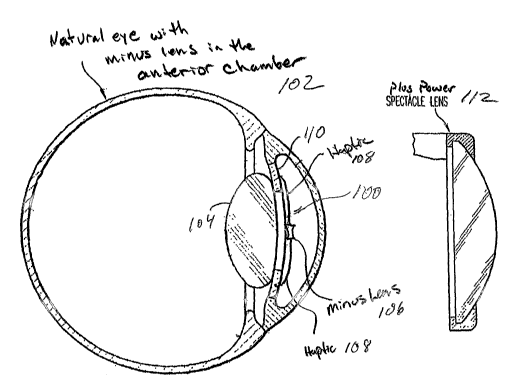
[0025] Referring to the drawings which form a part of this disclosure:
[0026] Fig. 1 illustrates the prior art wherein a plus lens is used outside
the
eye in conjunction with a combination plus and minus intraocular lens;
[002?] Fig. 2 illustrates the prior art wherein a plus lens is used outside
the
eye in conjunction with a lens having a minus portion and a portion with no
refractive
power;
[0028] Fig. 3 illustrates a preferred embodiment of the present invention
including an elevational side view in section of a plus lens outside the eye
and a
minus lens implanted in an anterior portion of the eye;
[0029] Fig. 4 is an elevational side view in section of a plus lens outside
the
eye and a minus lens implanted in a posterior chamber of the eye;
[0030] Fig. 5 is an elevational side view in section of a plus lens implanted
in
the cornea and a minus lens implanted in an anterior chamber of the eye;
[0031] Fig. 6 is an elevational side view in section of a plus lens partially
implanted in the cornea and a minus lens implanted in an anterior chamber of
the eye;
CA 02515720 2005-08-10
WO 2004/075729 PCT/US2004/005459
[0032] Fig. 7 is an elevational side view in section of a plus lens outside
the
eye and a minus lens implanted in the cornea;
(0033] Fig. 8 is an elevational side view in section of two plus lenses, one
implanted in the cornea and one outside the eye in conjunction with a minus
lens
implanted in an anterior chamber of the eye;
[0034] Fig. 9 is an elevational side view in section of two plus lenses, both
outside the eye and two minus lenses, one minus lens in an anterior chamber of
the
eye and one minus lens implanted in the cornea of the eye;
[0035] Fig. 10 is an elevational front view in section of a high minus lens
having a sutured haptic;
[0036] Fig. 11 is an elevational front view in section of a high minus lens in
which the haptic is sutured to the iris;
[0037] Fig. 12 is a front elevational view in section of a minus lens fixed to
an
outer portion of the iris by peripheral iridectomy;
[O~3~] Fig. 13 is an elevational front view of an intraocular lens having a
minus portion in the peripheral part of the lens and a plus portion in the
central part of
the lens; and
[0039] Fig. 14 is a side elevational in section of a lens having a high minus
central portion for the correction of macular degeneration and a plus
peripheral
portion for the correction of hyperopia;
[0040] Fig. 15 is a side elevational in section of a lens having high minus
central portion for the correction of macular degeneration and a minus
peripheral
portion for the correction of myopia; and
[0041] Fig. 16 is a side elevational in section of a lens having a high minus
central portion for the correction of macular degeneration and a toric
peripheral
portion for the correction of astigmatism;
Detailed Description of the Invention
[0042] As illustrated in Figs. 3 and 4, the preferred embodiment of the
present
invention includes a modified or miniaturized telescope 100 for the eye 102.
More
CA 02515720 2005-08-10
WO 2004/075729 PCT/US2004/005459
_g_
specifically, in conjunction with a patient's original lens 104 or in
conjunction with an
IOL, a miniaturized high minus lens 106 is affixed to an interior portion of
the eye
102, the high minus lens 106 having an outer perimeter or free edge, as seen
in Figs. 3
and 4, with a diameter of about 1 millimeter to about 3 millimeters. Although
using a
high minus lens 106 is preferred, the lens 106 can be a minus diopter and not
necessarily a high minus. The minus lens 106 can be affixed using any method
desired, such as haptics 108, adhesive or in any other manner, and can be
affixed to
the iris 110, the angle, the zonulax ligaments, the natural lens 104, or an
IOL, or any
other suitable portion of the eye 102. Additionally, the minus lens can be
affixed in
the posterior chamber or the anterior chamber of the eye.
[0043] Furthermore, a high plus lens or any other suitable lens is placed
outside the eye 102 in spectacles or glasses 112 or as a contact lens and acts
with the
minus lens to produce a telescopic effect.
[0044] In a further embodiment of the present invention, as shown in Fig. 5,
the high plus lens 114 can be inserted into the cornea 116. The high plus lens
can be
implanted in the stroma, the epithelium, or any other portion of the cornea
desired.
[0045] By having the high plus lens 114 implanted in the cornea 116, or a
contact lens, the lens 114 actually moves with the eye 102 and therefore
reduces or
eliminates any distortion. The high plus lens 114 can be inserted into the
cornea in
ally manner desired. For example, the lens can be inserted under a flap or
into a
pocket formed in the cornea. Additionally, the inlay or high plus lens 114-1
can have
a portion embedded in the cornea and a portion exposed and not covered by a
layer of
the cornea (Fig. 6).
[0046] Yet in another embodiment of the present invention, as shown in Figs.
7-9, the minus lens 100-1 can be inserted into the cornea 116, as described
above for
the high plus lens and the high plus lens 114 can be positioned outside the
cornea 116.
For example, the minus lens 100-1 can be inserted under a flap or into a
pocket
formed in the cornea 116. Furthermore a portion of the minus lens 100-1 can be
exposed, as described above, for the high plus lens.
CA 02515720 2005-08-10
WO 2004/075729 PCT/US2004/005459
-9-
[0047] Any of these embodiments can be combined to form a multiple lens
system. For example, two plus lenses 112 and 114 can be used, one lens in the
cornea
116 or partially in the cornea, as described above and a second lens outside
the cornea
in spectacles 112, glasses or contacts (Fig. 9). Additionally, two minus
lenses 100
and 100-1 can be used, one lens 100-1 in the cornea 116, as described above,
and one
lens 100 in the anterior or posterior chamber of the eye 102, as described
above, in
conjunction with a high plus lens outside or inside the eye, as described
above (Figs. 8
and 9). The second minus lens can be affixed to the iris, the angle, the
zonular
ligaments, the natural lens or an IOL, or any other suitable portion of the
eye.
Furthermore, two plus lenses outside the eye in spectacles, glasses or
contacts in any
manner desired, along with the one or two minus lenses described herein.
[0048] Another embodiment is shown in figs. 10-12. As indicated, a lens
structure 100-1 includes a lens 106-2 and a haptic 108-1. A suture 120 may be
used
to fix haptic 108-1 that is connected to lens 106-2 to the eye and, in
particular, to the
iris 110, as shown in Fig. 11. The lens 106-2, which can be a high minus lens
as
discussed above, can thus be positioned in the pupil 122. Alternatively, a
high minus
lens 106 may be inserted in the iris 110 by peripheral iridectomy, in which a
section
124 is removed from the iris 110, as shown in Fig. 12. Preferably, lens 106-2
is used
in conjunction with the telescopic system described above.
[004] Another preferred embodiment shown in Fig. 13 uses an intraocular
lens (IOL) 126 having a minus portion 128 and a plus portion 130. Preferably,
the
minus portion 128 forms the periphery of the lens and surrounds a central plus
portion
130. The plus portion 130 of the lens 126 corrects far vision, while the
peripheral
minus portion 128 acts in conjunction with an outside lens, such as one in
spectacles,
to produce a telescopic effect. The lens 126 may be employed as the lens 106
or 106-
1 discussed above, and thus may be affixed to any suitable portion of the eye,
such as
the iris 110, lens 104, the angle, the zonular ligaments, or piggyback, such
as is shown
in figs. 3-6, 8 and 9 as discussed above. Additionally, the center portion can
be a
minus portion for the correction of myopia or a toric portion for the
correction of an
astigmatism, or any combination of a minus, plus or toric lens if desired.
CA 02515720 2005-08-10
WO 2004/075729 PCT/US2004/005459
- 10-
[0050] Furthermore, Figs. 14-16 illustrate three additional configurations of
v
lens 126, lens 126-1, lens 126-2 and lens 126-3, respectively. In each of
these
configurations, the central portion of each lens can be a high minus portion
130-1 and
the peripheral area or portion can be a plus portion 128-1, a minus portion
128-2 or a
toric portion 128-3. The refractive power of portions 128-1, 128-2 and 128-3
are
generally about plus or minus 2 diopters, but can be any power desired,
depending on
the required correction in the eye. Preferably, the high minus center portion
130-1 is
used to correct for macular degeneration in conjunction with a secondary lens
to
achieve a telescopic effect , as described above, to allow the eye to focus on
a close
object for activities such as reading. The peripheral area or portions 128-1,
128-2 and
128-3 are used to correct a secondary problem, such as hyperopia, myopia
and/or
astigmatism, and are generally used without a secondary lens, although they
can be
used with a secondary lens, if so desired.
[0051] Preferably, lens 126-1 is used in conjunction with the telescopic
system described herein and with the natural lens of the eye, an existing IOL
that
replaces the natural lens in the eye, or an IOL that works in conjunction with
the
natural lens in the eye.
[0052] By forming portions 128-1, 128-2 and 128-3 in the manner described
herein, not only can macular degeneration be corrected, but so can a secondary
type of
vision disorder, such as myopia, hyperopia and/or astigmatism with the same
lens.
This reduces the number of procedures and/or lenses that are implanted in the
eye.
[0053] It is noted that lenses 126 and 126-1 can be any type of lens desired.
For example, lenses 126 and/or 126-1 can be implanted in the eye while
connected to
the iris as shown in Figs. 11 and 12, each can be implanted in the cornea,
similar to
the lenses shown in Figs. 5-9, each can piggyback with existing an IOL, or can
be
coupled to an interior portion of the eye in any conventional manner relative
to the
natural lens in the eye. Additionally, lenses 126 andlor 126-1 can be
positioned in the
eye and replace any lens shown herein. For example, lens 126 and/or 126-1 can
replace lens106, lens114, lens 114-1, and lens 106-1, in Figs. 3-9.
CA 02515720 2005-08-10
WO 2004/075729 PCT/US2004/005459
-11-
[0054) It is further noted that any lens used and described herein can be made
of synthetic material, organic material, or a combination of both synthetic
and organic
material, that permits all or substantially all light having a wavelength in
the visible
spectrum to pass through. Additionally, if desired, the lens can be formed of
material
that absorbs all or substantially all light having a wavelength in a laser
light spectrum.
For example, the lenses described herein can be made of collagen, copolymer
collagen, polyethylene oxide, polypropylene, polyproledine or hydrogel, or
cross-
linked organic material such as collagen, hyaluronic acid, mucopolysacoharide
or
glycoprotein, to name a few. Preferably, each lens is porous to allow oxygen
and
nutrients to pass therethrough. Also, each lens can be made from a donor
cornea of a
human eye, or can be taken from a cultured cornea. However, the lens is not
limited
to those materials, and can be made of any suitable material, such as those
disclosed
in U.S. Patent No. 4,994,058 to Raven et al., U.S. Patent No. 4,718,418 to
L'Esperance, U.S. Patent No. 5,336,261 to Barrett et al., U.S. Patent No.
4,840,175 to
Peyman, and a publication by Jose I. Barraquer, IVLD. entitled "Keratomileusis
and
Keratophakia in the Surgical Correction of Aphakia", the disclosures of which
are
hereby incorporated by reference herein.
[0055) While preferred embodiments have been chosen to illustrate the
invention, it will be understood by those skilled in the art that various
changes and
modifications can be made therein without departing from the scope of the
invention
as defined in the appended claims.