Note: Descriptions are shown in the official language in which they were submitted.
CA 02515827 2005-08-11
WO 2004/080355 PCT/US2004/004352
ARTICULAR DISC PROSTHESIS FOR ANTERIOR-OBLIQUE INSERTION
Cross-Reference to Related Application
This application claims the benefit of U.S. Provisional Application No.
60/446,963
filed on February 12, 2003. U.S. Provisional Application No. 60/446,963 is
herein
incorporated by reference for all legitimate purposes.
Background
The present disclosure relates generally to the field of orthopedics and
spinal
surgery, and in some embodiments, the present disclosure relates to
intemertebral
prosthetic joints for use in the total or partial replacement of a natural
intervertebral disc,
and methods and tools for use therewith.
In the treatment of diseases, injuries or malformations affecting spinal
motion segments,
and especially those affecting disc tissue, it has long been known to remove
some or all of
a degenerated, ruptured or otherswvise failing disc. In cases involving
inter~rcrtebral disc
tissue that has been removed or is otherwise absent from a spinal motion
segment,
corrective measures are taken to ensure the proper spacing of the vertebrae
formerly
separated by the removed disc tissue.
In some instances, the two adjacent vertebrae are fused together using
transplanted
bon a tissue, an artificial fusion component, or ~thcr compositions or
devices. Spinal
fusion procedures, however, have raised concerns in the medical community that
the bio-
mechanical rigidity of intcrvertebral fusion may predispose neighboring spinal
motion
segments to rapid deterioration. More specifically, unlike a natural
intervertebral disc,
spinal fusion prevents the fused vertebrae from pivoting and rotating with
respect to one
another. Such lack of mobility tends to increase stresses on adjacent spinal
motion
segments.
Additionally, several conditions may develop within adjacent spinal motion
segments, including disc degeneration, disc herniation, instability, spinal
stenosis,
spondylolisthesis and facet joint arthritis. Consequently, many patients may
require
additional disc removal and/or another type of surgical procedure as a result
of spinal
fusion. Alternatives to spinal fusion are therefore desirable.
CA 02515827 2005-08-11
WO 2004/080355 PCT/US2004/004352
2
In particular, this disclosure relates to an articulating disc prosthesis that
can be inserted
from the anterior-oblique approach.
Summary
A prosthetic device for anterior-oblique insertion into an intervertebral
space is
provided. The prosthetic device includes a first component having a first
flange for
engaging a first vertebra from an anterior-oblique approach, the first flange
being angled
relative to the longitudinal and transverse axes of the first component, and a
second
component having a second flange for engaging a second vertebra from an
anterior-
oblique approach, the second flange being angled relative to the longitudinal
and
transverse axes of the second component, and wherein the first and second
components
cooperate to permit articulating motion between the first and second
components.
In another embodiment, a prosthetic component for forming a portion of a
prosthetic
device is provided. The prosthetic component includes a first surface having a
flange for
engaging a vertebra from an anterior-oblique approach, the flange being angled
relative to
the longitudinal and transverse axes of the prosthetic component, and a second
surface in
an opposed relation to the first surface, the second surface being adapted to
engage another
prosthetic component.
In yet another embodiment, a prosthetic device for anterior-oblique insertion
into
an intervertebral space is provided. The prosthetic device includes a first
component
having a first articular surface and an opposed first bearing surface, a first
flange
extending from the first bearing surface, the first flange being angled
relative to the
longitudinal and hansverse axes of the first component, and a projection
extending from
the first articular surface'.' The prosthetic device further includes a second
component
having a second articular surface and an opposed second bearing surface, a
second flange
extending from the second bearing surface, the second flange being angled
relative to the
longitudinal and transverse axes of the second component, and a recess formed
in the
second articular surface, the recess being adapted to engage with the
projection to provide
for articulating motion between the first and second components.
In yet another embodiment, a method for inserting a prosthetic device into an
intervertebral space from an anterior-oblique approach is provided. The method
includes
providing a prosthetic device having a first component and a first flange
extending along a
CA 02515827 2005-08-11
WO 2004/080355 PCT/US2004/004352
surface of the first component, the first flange being angled relative to the
longitudinal and
transverse axes of the first component, and a second component and a second
flange
extending along a surface of the second component, the second flange being
angled
relative to the longitudinal aid transverse axes of the second component. The
method
further includes inserting the first component into a first vertebra and
inserting the second
component into a second vertebra from an anterior-oblique approach, whereby
the first
component engages the second component to provide articulating motion
therebetween.
Brief Description of the Drawings
Fig. 1 is a lateral, view of a portion of a spondylosed vertebral column.
Fig. 2 is a lateral view of a pair of adjacent vertebral endplates of Fig. 1.
Fig. 3a is a lateral view of the pair of adjacent vertebral endplates of Fig.
2 with a
rod and screw arrangement.
Fig. 3b is a longitudinal, partial sectional view of the pair of adjacent
vertebral
bodies of Fig. 3a.
Fig. 4a is an isometric view of an articulating prosthetic joint for lateral
insertion
according to one embodiment of the present disclosure.
Fig. 4b is an isometric view of an articulating prosthetic joint for lateral
insertion
according to another embodiment of the present disclosure.
Fig. 4c is a front view of the articulating prosthetic joint for lateral
insertion of Fig.
4b.
Fig. 5 is a longitudinal view of the prosthetic joint of Fig. 4a.
Fig. 6 is a lateral view of the prosthetic joint of Fig. 4a.
Fig. 7 is a lateral; partial sectional view of the prosthetic joint of Fig. 4a
disposed
between a pair of spondylosed vertebral endplates.
Fig. 8 is a lateral, partial sectional view of an alternative articulating
prosthetic
joint disposed between a pair of vertebral endplates.
Fig. 9 is an isometric view of an alternative articulating prosthetic joint
according
to another embodiment of the present disclosure.
Fig. 10 is a lateral, partial sectional view of the prosthetic joint of Fig. 9
disposed
between a pair of spondylosed vertebral endplates.
CA 02515827 2005-08-11
WO 2004/080355 PCT/US2004/004352
4
Fig. 11 is a lateral? partial sectional view of an alternative articulating
prosthetic
joint disposed between a pair of vertebral endplates.
Fig. 12 is an isometric view of a disc prosthesis according to another
embodiment
of the present disclosure.
Fig. 13 is an isometric view of an alternative disc prosthesis according to
another
embodiment of the present disclosure.
Fig. 14 is an isometric view of an alternative articulating prosthetic joint
for
anterior insertion according to another embodiment of the present disclosure.
Fig. 15 is a longitudinal view of the prosthetic joint of Fig. 14,
Fig. 16 is a lateral view of the prosthetic joint of Fig. 14.
Fig. 17 is a lateral view of the prosthetic joint of Fig. 14 disposed between
a pair of
spondylosed vertebral endplates.
Fig. 18 is a longitudinal view of an alternative articulating prosthetic joint
for
anterior insertion according to another embodiment of the present disclosure.
Fig. 19 is a longitudinal view of an alternative articulating prosthetic joint
for
anterior insertion according to yet another embodiment of the present
disclosure.
Fig. 20 is a longitudinal view of an alternative articulating prosthetic joint
fox
anterior insertion according to yet another embodiment of the present
disclosure.
Fig. 21 is a longitudinal view of a pair of verterbral endplates having slots
for
receiving the prosthetic joint of Fig. 18.
Fig. 22 is a longitudinal view of a pair ~f verterbral endplates having sl~ts
f~r
receiving the prosthetic joint of Fig. 19.
Fig. 23 is a longitudinal view of a pair of verterbral endplates having slots
for
receiving the prosthetic joint of Fig. 20.
Fig. 24 is a lateral, partial sectional view of the prosthetic joint of Fig.
14 disposed
between a pair of spondylosed vertebral endplates and an orthopedic implant.
Fig. 25 is a lateral, partial sectional view of the prosthetic joint of Fig.
14 disposed
between a pair of spondylosed vertebral endplates and a lag screw.
Fig. 26 is a schematic top view of the arrangement depicted in Fig. 25.
Fig. 27 is a schematic top view of a vertebral body depicting a path for
transforaminal insertion.
CA 02515827 2005-08-11
WO 2004/080355 PCT/US2004/004352
Fig. 28 is an isometric view of an alternative articulating prosthetic joint
for
transforarninal insertion according to another embodiment of the present
disclosure.
Fig. 29 is a lateral view of the prosthetic joint of Fig. 28.
Fig. 30 is a longitudinal view of the prosthetic joint of Fig. 28.
Fig. 31a is a lateral, partial sectional view of the prosthetic joint of Fig.
28
disposed between a pair of vertebral endplates.
Fig. 31b is a longitudinal, partial sectional view of the prosthetic joint of
Fig. 28
disposed between a pair of vertebral endplates.
Fig. 32 is a schematic top view depicting a transforaminal slot formed in a
vertebral endplate.
Fig. 33 is a schematic top view depicting a milling apparatus shown inserted
above
a vertebral endplate.
Fig. 34a is a lateral view of the milling apparatus of Fig. 33 shown disposed
between a pair of adjacent vertebral endplates.
Fig. 34b is a detailed view of a milling tool of the milling apparatus of Fig.
34a.
Fig. 34c is a detailed view of an alternative milling tool.
Fig. 35 is a schematic view of the milling apparatus of Fig. 33.
Fig. 36 is an isometric view of an alternative articulating prosthetic joint
for
transforaminal insertion according to another embodiment of the present
disclosure.
Fig. 37 is a lateral view of the prosthetic j~int ~f Fig. 36.
Fig. 38 is a longitudinal view of the prosthetic joint Fig. 36.
Fig. 39a is an isometric view of an alternative articulating prosthetic joint
for
anterior-oblique insertion according to another embodiment of the present
disclosure.
Fig. 39b is a top view of the prosthetic joint of Fig. 39a with slight
alterations.
Fig. 39c is a fiont view of the prosthetic joint of Fig. 39b.
Fig. 40 is a longitudinal view of the prosthetic joint of Fig. 39a.
Fig. 41 is a lateral view of the prosthetic joint of Fig. 39a.
Fig. 42 is lateral, partial sectional view of the prosthetic joint of Fig. 39a
disposed
between a pair of vertebral endplates.
Fig. 43 is a longitudinal, partial sectional view of the prosthetic joint of
Fig. 39a
disposed between a pair of vertebral endplates.
CA 02515827 2005-08-11
WO 2004/080355 PCT/US2004/004352
6
Fig. 44a is a top, schematic view depicting a slot formed in a vertebral
endplate for
receiving the prosthetic joint of Fig. 39a.
Fig. 44b is a schematic view depicting an alignment process associated with
the
insertion of the prosthetic joint of Fig. 39a.
Fig. 45 is an exploded view an alternative prosthetic joint according to yet
another
embodiment of the present disclosure.
Fig. 46 is an isometric view of the prosthetic joint of Fig. 45.
Fig. 47 is a longitudinal view of the prosthetic joint of Fig. 46.
Fig. 48 is a longitudinal view of a pair of adjacent vertebral endplates.
Fig. 49a is a plan view of an articular component of the prosthetic joint of
Fig. 45.
Fig. 49b is a sectional view of the articular component of Fig. 49a taken
along the
line 49b-49b.
Fig. SOa is a plan view of a modular projection member of the prosthetic joint
of
Fig. 45.
Fig. SOb is a secti~nal view ~f the modular pr~jection member of Fig. SOa
taken
along the line SOb-SOb.
Fig. 51 is a plan view of the modular projection member of Fig. SOa inserted
into
the articular component of Fig. 49a.
Fig. 52 is a plan view of the modular projection member of Fig. SOa inserted
into the
articular component of Fig. 4~9a depicting the modular projection member in a
different
position relative to Fig. 51.
Description
For the purposes of promoting an understanding ~f the principles of the
invention,
reference will now be made to the embodiments, or examples, illustrated in the
drawings
and specific language will be used to describe the same. It will nevertheless
be understood
that no limitation of the scope of the invention is thereby intended. Any
alterations and
further modifications in the described embodiments, and any further
applications of the
principles of the invention as described herein are contemplated as would
normally occur
to one skilled in the art to which the invention relates. As such, individual
features of
separately described embodiments can be combined to form additional
embodiments. In
addition, examples of deformities such as spondylolisthesis axe discussed;
however, it is
CA 02515827 2005-08-11
WO 2004/080355 PCT/US2004/004352
understood that the various prosthetic devices described herein can be adapted
for use
between not only spondylosed vertebrae, but substantially aligned vertebrae as
well.
I. Lateral Correction
In many cases of deformity, such as spondylolisthesis, one or more vertebral
bodies can be displaced with respect to other vertebrae or the sacrum. In such
a deformity,
it is desirable to reduce the extent of displacement, by re-positioning the
displaced bodies
from their previous position. A spondylolisthesis reduction can be a
technically
demanding procedure requiring great care to prevent neurological impairment
and damage
to surrounding soft tissue.
Referring now to Fig. 1, shown therein is a lateral view of a poxtion of a
spinal
column 10, illustrating a group of adjacent upper and lower vertebrae V1, V2,
V3, V4
separated by natural intervertebral discs D1, D2, D3. The illustration of four
vertebrae is
only intended as an example. Another example would be a sacrum and one
vertebrae.
As shown in the drawing, the vertebrae V2 is dislocated from the vertebrae V 1
in a
direction shown by snow 22. Lilcewise, vertebrae V3 is dislocated in a
direction shown
by snow 23 and vertebrae V4 is dislocated in a direction shown by arrow
direction 24. It
is desired that the position of vertebrae V2, V3, V4 be corrected by moving
them in a
direction opposite to the avows 22, 23, 24, respectively.
Referring now to Fig. 2, for the sake of further example, two of the displaced
vertebrae
will be discussed, designated as the lower vertebrae VL and the upper
vertebrae VU. In
one embodiment, some or all of the natural disc that would have been
positioned between
the two vertebrae VL, VU is typically removed via a discectomy or a similar
surgical
procedure, the details of which would be known to one of ordinary skill in the
art.
Removal of the diseased or degenerated disc results in the formation of an
interventebral
space S between the upper and lower vertebrae VU, VL.
In the present embodiment, it is desired to insert a prosthetic joint into the
intervertebral space S, similar to the prosthetic joint disclosed in U.S. Ser.
No. 10/042,589
filed January 9, 2002, which is incorporated by reference. However, certain
changes are
required of the above-referenced prosthetic joint. For the following
description, the
prosthetic joints discussed and described can be identical to those disclosed
in the above-
referenced patent application, with the exceptions discussed and suggested
below.
CA 02515827 2005-08-11
WO 2004/080355 PCT/US2004/004352
Spondylolisthesis has not heretofore been corrected from the lateral surgical
approach.
However, in some instances, correction of spondylolisthesis may be desirable
from a
lateral approach due to the presence of vessels and/or the nervous plexus. In
some
embodiments, the lateral approach may be particularly pertinent when
correcting
spondylolisthesis in the lumbar region of the spine, although it will be
understood that
other regions of the spine are also contemplated.
Referring to Figs. 3a and 3b, correction of spondylolisthesis can be addressed
from
a lateral approach by, for example, providing a pair of bone screws 30, 32 for
insertion
into the vertebrae VU, VL, respectively. In one embodiment, the bone screws
30, 32 are bi-
cortical. However, it is understood that the bone screws may alternatively be
uni-cortical.
Moreover, the bone screws 30, 32 may be formed of a variety of materials such
as any
resorbable material, titanium, and PEEK. The PEEK embodiment is advantageous
due to
the radiotranslucent properties resulting from the use of PEEK material. It is
further
understood that the bone screws 30, 32 may alternatively be of any other
mechanical
structure, and as such, may take the form of pins or rivets, fox example.
Moreover, the
bone screws 30, 32 are not limited to having threaded portions to engage the
vertebrae VU,
VL.
The bone screws 30, 32 may be linked to one another via a rod 34, Which is
configured to rotate about both of the bone screws. It is understood that a
variety of
comiecting members may be used other than the rod 34~. For example, a non-
uniform
linkage member may be used to link the bone screws 30, 32. A non-uniform
linlcage
member may provide a plurality of slots and/or grooves that can be engaged in
order to aid
in its rotation about the bone screws. The rod 34 may be connected prior to
insertion of
the bone screws 30, 32 into the vertebrae VU, VL, or alternatively, may be
subsequently
connected after placement of the screws. By applying a rotating force to the
rod 34 in the
direction of arrow 36, the upper vertebra VU is encouraged back into a desired
position
relative to the lower vertebra VL. The rotating force can be applied, fox
example, by a
rotatable wrench (not shown) that can be used by a surgeon. It is understood
that the
upper vertebra VU may not reach entirely to a fully corrected position in
relation to the
lower vertebra VL, but the displacement can at least be reduced.
CA 02515827 2005-08-11
WO 2004/080355 PCT/US2004/004352
9
Although not depicted, in another embodiment, it is contemplated that the
spondylosed vertebrae VU, VL can be addressed from both lateral directions.
Thus, a pair
of bone screws substantially identical to the bone screws 30, 32 may be
inserted into the
vertebrae VU, VL on the opposite side from and in the opposite direction to
the bone
screws 30, 32. In such am arrangement, the rod 34 can be replaced with a
ratcheting
system that engages each of the bone screw pairs, and as such, the vertebrae
VU, VL can be
rotated relative to one another to encourage the vertebrae into a desired
position relative to
one another.
Still further, the rod 34 may include any number and type of engagement means
to
receive any number and type of rotating tools used by a surgeon. For example,
a keyed
connection may provide more stability when engaging the rod 34 with a
corresponding
rotating tool. In other examples, a clamping tool may be used and
corresponding
clamping notches may be formed in the rod 34 to receive the clamping tool.
Such an
arrangement may aid in achieving the force necessary for rotation.
Ivloxeover, additional rods 34 and bone screws 30, 32 are contemplated for use
in rotating
the spondylosed vertebrae VU, VL back into a desired position relative to one
another.
Additional rods 34 and bone screws 309 32 may provide additional stability
during the
procedure.
Furthermore, alth~ugh depicted as a substantially lateral insertion, the
insertion of
the bone screws 30, 32 into the veutebrae VU, VL can be slightly angled
relative to the
lateral direction. Such angling of the bone screws 30, 32 during insertion may
provide a
preferred gripping angle from which the surgeon can begin rotation of the
vertebrae VU,
VL relative to one another.
Referring to Figs. 4a, 5, and 6, shown therein is one embodiment of an offset
intervertebral
articulating prosthetic joint 40 for insertion into the intervertebral space S
(Fig. 2) to aid in
the correction of spondylolisthesis. The articulating prosthetic joint 40
extends generally
along a longitudinal axis L and includes a first articular component 42 and a
second
articular component 44. The articular components 42, 44 cooperate to form the
prosthetic
joint 40 which is sized and configured for disposition within the
intervertebral space S
(Fig. 2) between adjacent vertebral bodies VU, VL (Fig. 2).
CA 02515827 2005-08-11
WO 2004/080355 PCT/US2004/004352
The prosthetic joint 40 provides relative pivotal and rotational movement
between
the adjacent vertebral bodies to maintain or restore motion substantially
similar to the
normal bio-mechanical motion provided by a natural intervertebral disc. More
specifically, the articular components 42, 44 are permitted to pivot relative
to one another
about a number of axes, including lateral or side-to-side pivotal movement
about
longitudinal axis L and anterior-posterior pivotal movement about a transverse
axis T. It
should be understood that in one embodiment of the disclosure, the articular
components
42, 44 are permitted to pivot relative to one another about any axes that lies
in a plane that
intersects longitudinal axis L and transverse axis T.
Furthermore, the articular components 42, 44 are permitted to rotate relative
to one
another about a rotational axis R. Although the prosthetic joint 40 has been
illustrated and
described as providing a specific combination of articulating motion, it
should be
understood that other combinations of articulating movement are also possible,
such as,
for example, relative translational or linear motion, and such movement is
contemplated as
falling within the scope of the present disclosure.
Although the articular components 4~2, 44 of prosthetic joint 40 may be formed
from a wide variety of materials, in one embodiment of the disclosure, the
articular
components 42, 44 are formed of a cobalt-chrome-molybdenum metallic alloy
(ASTM F-
799 or F-75). However, in alternative embodiments of the disclosure, the
articular
components 42, 44~ may be formed of other materials such as titanium or
stainless steel, a
polymeric material such as polyethylene, or any other biocompatible material
that would
be apparent to one of ordinary slcill in the art.
The articular components'42, 44 each include a bearing surface 4G, 4~,
respectively, that
may be positioned in direct contact with vertebral bone and is preferably
coated with a
bone-growth promoting substance, such as, for example, a hydroxyapatite
coating formed
of calcium phosphate. Additionally, the bearing surfaces 46, 4~ of the
articular
components 42, 44, respectively, may be roughened prior to being coated with
the bone-
growth promoting substance to further enhance bone on-growth. Such surface
roughening
may be accomplished by way of, for example, acid etching, knurling,
application of a bead
coating, or other methods of roughening that would occur to one of ordinary
skill in the
art.
CA 02515827 2005-08-11
WO 2004/080355 PCT/US2004/004352
11
Articular component 42 includes a support plate 50 having an articular surface
52
and the opposite bearing surface 46. Support plate 50 is sized and shaped to
substantially
correspond to the size and shape of a vertebral endplate of the adjacent
vertebral body VL
(Fig. 2). The support plate 50 may include one or more notches 54 or other
types of
indicia for receiving or engaging with a corresponding portion of a surgical
instrument
(not shown) to aid in the manipulation and insertion of the prosthetic joint
40 within the
intervertebral space S (Fig. 2) between the adjacent vertebral bodies VU, VL
(Fig. 2). The
surgical instrument (not shown) is preferably configured to hold the articular
components
42, 44 at a predetermined orientation and spatial relationship relative to one
another during
manipulation and insertion of the prosthetic joint 40, and to release the
articular
components 42, 44 once properly positioned between the adjacent vertebrae.
In one embodiment of the disclosure, the articular component 42 includes a
pr~jection 56 having a convex shape, which may be configured as a spherical-
shaped ball
(half of which is shown). It should be understood that other configurations of
the
projection 56 are also contemplated, such as, for example, cylindrical,
elliptical or other
arcuate configurations or possibly non-arcuate configurations. It should also
be
understood that the remaining portion of articular component 42 may take on
planar or
non-planar configurations, such as, for example, an angular or conical
configuration
extending about the projecti~n 56.
~ flange member ~r keel 58 extends from the bearing surface 4.6 and is
configured
for disposition within a preformed opening in the adjacent vertebral endplate.
As with the
bearing surface 46, the keel 58 may be coated with a bone-growth promoting
substance,
such as, for example, a hydroxyapatite coating formed of calcium phosphate.
Additi~nally, the keel 58 may be roughened prior to being coated with the bone-
growth
promoting substance to further enhance bone on-growth. In one embodiment, the
keel 58
extends along the transverse axis T and is substantially centered along the
bearing surface
46. However, it should be understood that other positions and orientations of
the keel 58
are also contemplated.
In one embodiment, the keel 58 transversely extends along a substantial
portion of
the articular component 42. Such an embodiment would accommodate insertion of
the
prosthetic joint 40 using a lateral approach as opposed to, for example, an
anterior
CA 02515827 2005-08-11
WO 2004/080355 PCT/US2004/004352
12
approach. In a further embodiment, the keel 58 may be angled, tapered, or
configured in
some other shape to facilitate the functional demands of the keel. In still
another
embodiment, the keel 58 may be configured as a winged keel, including a
lateral portion
(not shown) extending across the main body portion of keel 58.
In one embodiment, the keel 58 includes three openings 60 extending
therethrough
to facilitate bone through-growth to enhance fixation to the adjacent
vertebral bodies VU,
VL (Fig. 2). However, it should be understood that any number of openings 60
may be
defined through the keel 58, including a single opening or two or more
openings. It
should also be understood that the openings 60 need not necessarily extend
entirely
through the keel 58, but may alternatively extend partially therethrough. It
should further
be understood that the keel 58 need not necessarily define any openings 60
extending
either partially or entirely therethrough. Additionally, although the openings
60 are
illustrated as having a circular configuration, it should be understood that
other sizes and
configurations of openings 60 are also contemplated.
In one embodiment, the articular component 44 includes a support plate 70
having
an articular surface 72 and the opposite bearing surface 48. Support plate 70
may be sued
and shaped to substantially correspond to the size and shape of a vertebral
endplate of the
adjacent vertebral body VU. The support plate 70 may include one or more
notches 74 or
other types of indicia for receiving and engaging with a corresponding portion
of a
surgical instrument, such as discussed above with reference to articular
component 4~2.
In one embodiment, the articular surface 72 includes a recess 76. In one
embodiment, the
recess 76 has a concave shape, and is configured as a spherical-shaped socket.
However,
it should be understood that other configuxations of the recess 76 are also
contemplated,
such as, for example, eylindrical, elliptical or other arcuate configurations
or possibly non-
arcuate configurations. The remaining portion of the articular surface 72 can
be angled or
otherwise configured to facilitate the insertion and/or use of the prosthesis.
Although the concave recess 76 is illustrated as having a generally smooth,
uninterrupted articular surface, it should be understood that a surface
depression or cavity
may be defined along a portion of the recess 76 to provide a means for
clearing out matter,
such as particulate debris, that is disposed between the abutting articular
components 42,
44. In such case, the convex articular surface of the projection 56 may
alternatively define
CA 02515827 2005-08-11
WO 2004/080355 PCT/US2004/004352
13
a generally smooth, uninterrupted articular surface. In another embodiment,
each of the
convex projection 56 and the concave recess 76 may define a surface depression
to
facilitate removal of particulate matter disposed between the abutting
articular components
42, 44.
A flange member or keel 68, configured similar to the keel 58 of articular
component 42, extends from the bearing surface 48. In one embodiment, the keel
68
extends along the transverse axis T and is offset from the center of the
bearing surface 48.
Such an embodiment would accommodate insertion of the prosthetic joint 40
using a
lateral approach. However, it should be understood that other shapes,
positions and
orientations of the keel 68 are also contemplated. For example, in Figs. 4b
and 4c, the
keels 58 and 68 may be angled relative to the transverse axis T to aid in the
circumvention
of veins, arteries, bony portions, or other obstacles that may be in place
during insertion of
the prosthetic joint 40. Also, the keel 68 may be angled, tapered, or
configured in some
other shape to facilitate the functional demands of the keel. In still another
embodiment,
the keel 68 may be configuxed as a winged keel, including a transverse portion
extending
across the main body portion of the keel 68.
In one embodiment9 and referring to Fig. 5, the keel 68 also includes three
openings 70 extending therethrough to facilitate bone through-growth to
enhance fixation
to the adjacent vertebra. However, it should be understood that any number of
openings
70 may be defined through keel 70, including a single opening or two or more
openings.
It should also be understood that the openings 70 need not necessarily extend
entirely
through the keel 68, but may alternatively extend partially therethrough. It
should further
be understood that the keel 68 need not necessarily define any openings 70
extending
either partially or entirely therethrough. Additionally, although the openings
70 are
illustrated as having a circular configuration, it should be understood that
other sizes and
configurations of openings 70 are also contemplated. As discussed above, the
bearing
surfaces 46, 48 that are in direct contact with vertebral bone are preferably
coated with a
bone-growth promoting substance. Specifically, the bearing surface 48 and the
surface of
the lceel 68 can be coated with hydroxyapatite to promote bony engagement with
the
adjacent vertebral body VU. As also discussed above, the bearing surface 48
and the
surface of keel 68 can be roughened prior to application of tl~e
hydroxyapatite coating.
CA 02515827 2005-08-11
WO 2004/080355 PCT/US2004/004352
14
In some embodiments, one or both of the keels 58, 68 may include a sharp
forward edge,
illustrated'by edge 68a of Fig. 4. By having such an edge, insertion of the
lceel into the
associated vertebral body is facilitated. Also, the edge 68a can be of
sufficient sharpness
that the adjacent vertebral bodies do not require a slot for receiving the
keel 68, discussed
in greater detail below.
Referring to Fig. 7, to accommodate insertion of the offset prosthetic joint
40
within a spondylosed intervertebral space, the partially corrected upper and
lower
vertebrae VU, VL can be prepared to accept the prosthetic joint 40 (shown in
section in Fig.
7a) therebetween. Specifically, elongate openings or slots 80, 82 may be
formed along the
vertebral endplates of the upper and lower vertebrae VL, VU, respectively, at
a
predetermined width and to a predetermined depth. The slots 80, 82 can be
laterally offset
from each other to accommodate the displaced vertebrae VL and/or VU. In one
embodiment, the elongate slots 80, 82 are rectangular-shaped and extend
laterally through
the vertebrae VL, VU, respectively. In a specific embodiment, the slots 80, 82
are formed
by chiseling or curetting. However, other methods of forming slots 80, 82 are
also
contemplated as would occur to one of ordinary skill in the art, such as, for
example, by
drilling or reaming. Furthermore, for some embodiments of the prosthetic joint
40, the
keels 58 and/or 68 can form their own corresponding slots 80, 82,
respectively.
Referring to Fig. 8, in one embodiment, the upper and lower vertebrae VU, VL
may
be fully corrected, and thus, an alternative articulating prosthetic joint 90
may be used in
correcting spondylolisthesis. The articulating joint 90 may be substantially
similar to the
prosthetic joint 40 with the exception of the orientation of various elements
of the
articulating joint 90. For example, to accommodate insertion into fully
corrected upper
and Iower vertebrae VU, VL, tlae articulating joint 90 may include a laterally-
extending
keel 92 that is substantially centered on an upper articulating component 94
of the
articulating joint and a laterally-extending keel 96 that is substantially
centered on a Iower
articulating component 98. Furthermore, the upper articulating component 94
may include
a recess 100 that is substantially centered to correspond to a substantially
centered
projection 102 extending from the Iower articulating component 98. In one
embodiment,
the upper and lower articulating components 94, 98 are substantially flush
with one
another when disposed between fully corrected upper and lower vertebrae VU,
VL.
CA 02515827 2005-08-11
WO 2004/080355 PCT/US2004/004352
To accommodate insertion of the offset prosthetic joint 90, the fully
corrected
upper and lower vertebrae VU, VL can be prepared to accept the prosthetic
joint 90
therebetween. Specifically, elongate openings or slots 104, 106 may be formed
along the
vertebral endplates of the upper and lower vertebrae VU, VL, respectively, at
a
predetermined width and to a predetermined depth. The slots 104, 106 can be
substantially aligned with each other to accommodate the fully corrected upper
and lower
vertebrae VU, VL. In one embodiment, the elongate slots 104, 106 are
rectangular-shaped
and extend laterally through the vertebrae VU, VL, respectively. In a specific
embodiment,
the slots 104, 106 are formed by chiseling or curetting. However, other
methods of
forming slots 104, 106 are also contemplated as would occur to one of ordinary
skill in the
art, such as, for example, by drilling or reaming. Furthermore, for some
embodiments of
the prosthetic joint, the keels 92 and/or 96 can form their own corresponding
slots 104,
106, respectively.
Referring to Fig. 9, in an alternative embodiment, a slidable prosthetic joint
110
can be used to help with the lateral approach for treating spondylolisthesis.
The sliding
joint 110 extends generally along the longitudinal axis L and includes a first
slidable
component 112 and a second slidable component 114. The slidable components
112, 114
cooperate to form the sliding joint 110 which is sized and configured for
disposition
within an intervertebral space between adjacent vertebral bodies.
The sliding joint 110 provides movement between the adjacent vertebral bodies
to
maintain or restore some of the motion similar to the normal bio-mechanical
motion
provided by a natural intervertebral disc. More specifically, the slidable
components 112,
114 are permitted to translate relative to one another in the axial plane.
Although the slidable components 112, 114 of prosthetic joint 110 may be
formed
from a wide variety of materials, in one embodiment, the slidable components
112, 114
are formed of a cobalt-chrome-molybdenum metallic alloy (ASTM F-799 or F-75).
However, in alternative embodiments, the slidable components 112., 114 may be
forned of
other materials such as titanium or stainless steel, a polymeric material such
as
polyethylene, or any other biocompatible material that would be apparent to
one of
ordinary skill in the art. The surfaces of the slidable components 112, 114
that are
positioned in direct contact with vertebral bone are preferably coated with a
bone-growth
CA 02515827 2005-08-11
WO 2004/080355 PCT/US2004/004352
16
promoting substance, such as, for example, a hydroxyapatite coating formed of
calcium
phosphate. Additionally, the surface of the slidable components 112, 114 that
are
positioned in direct contact with vertebral bone are preferably roughened
prior to being
coated with the bone-growth promoting substance to further enhance bone on-
growth.
Such surface roughening may be accomplished by way of, for example, acid
etching,
knurling, application of a bead coating, or other methods of roughening that
would occur
to one of ordinary skill in the art.
Slidable component 112 includes a support plate 116 having a slidable surface
118
and an opposite bearing surface 120. Support plate 116 is preferably sized and
shaped to
substantially correspond to the size and shape of the vertebral endplate of an
adjacent
vertebra. The support plate 116 can include one or more notches 122 or other
types of
indicia for receiving and engaging with a corresponding portion of a surgical
instrument
(not shown) to aid in the manipulation and insertion of the prosthetic joint
110 within an
intervertebral space between adjacent vertebrae. The surgical instrument (not
shown) is
preferably configured to hold the slidable components 112, 114 at a
predetermined
orientation and spatial relationship relative to one another during
manipulation and
insertion of the prosthetic joint 110, and to release the slidable components
112, 114 once
properly positioned between the adjacent vertebrae.
A flange member or keel 124 extends from the bearing surface 120 and is
configured for disposition within a preforlmed opening in the adjacent
vertebral endplate.
In one embodiment, the keel 124 extends perpendicularly from the bearing
surface 120
and is approximately centrally located along the bearing surface 120. However,
it should
be understood that other positions and orientations of the keel 124 are also
contemplated.
In one embodiment, the keel 124 transversely extends along a substantial
porti~n
of the support plate 114. Such an embodiment would accommodate insertion of
the
prosthetic joint 110 using a lateral approach. In a further embodiment, the
keel 124 may
be angled, tapered, or configured in some other shape to facilitate the
functional demands
of the keel. In still another embodiment, the keel 124 may be configured as a
winged keel,
including a transverse portion extending across the main body portion of keel
124.
The keel 124 also includes openings 126 extending therethrough to facilitate
bone
through-growth to enhance fixation to the adjacent vertebra. However, it
should be
CA 02515827 2005-08-11
WO 2004/080355 PCT/US2004/004352
17
understood that any number of openings 126 may be defined through keel 124,
including a
single opening or three or more openings. It should also be understood that
the openings
104 need not necessarily extend entirely through the keel 124, but may
alternatively
extend partially therethrough. It should further be understood that the keel
124 need not
necessarily define any openings 126 extending either partially or entirely
therethrough.
Additionally, although the openings 126 are illustrated as having a circular
configuration,
it should be understood that other sizes and configurations of openings 126
are also
contemplated. As discussed above, the surfaces of the slidable component 112
that are in
direct contact with vertebral bone are preferably coated with a bone-growth
promoting
substance. Specifically, the bearing surface 120 and the surfaces of the keel
124 can be
coated with hydroxyapatite to promote bony engagement with the adjacent
vertebrae. As
also discussed above, the bearing surface 120 and the surfaces of keel 124 can
be
roughened prior to application of the hydroxyapatite coating.
In one embodiment, the slidable component 114 includes a support plate 128
having a
slidable surface 130 and an opposite bearing surface 132. Support plate 128 is
preferably
sized and shaped to substantially correspond to the size and shape of the
vertebral endplate
of an adjacent vertebra. The support plate 128 can include one or more notches
134 or
other types of indicia for receiving and engaging with a corresponding portion
of a
surgical instrument, such as discussed above with reference to slidable
element 112.
A flange member or keel 136, configured similar to the keel 124 of slidable
component 112, extends from the bearing surface 132. In one embodiment, the
keel 136
extends perpendicularly from the bearing surface I32 and is offset along the
bearing
surface 132 to accommodate spondylosed displacements of the vertebrae. Also,
the offset
position of the keel 136 helps in the circumvention of veins, arteries, bony
portions, or
other obstacles that may be in place during the insertion of the joint 110. It
should be
further understood that other positions, shapes, orientations, and quantities
of the keel 136
are also contemplated. It should also be understood that the keel 136 may also
be
differently positioned, shaped or oriented, or more keels 136 can be used, for
similar or
additional reasons.
In one embodiment, the keel 136 transversely extends along a substantial
portion
of the support plate 128. Such an embodiment Would accommodate insertion of
the
CA 02515827 2005-08-11
WO 2004/080355 PCT/US2004/004352
18
prosthetic joint 110 using a lateral approach as opposed to another approach
such as an
anterior approach. In a further embodiment, the keel 136 may be angled,
tapered, or
configured in some other shape to facilitate the functional demands of the
keel. In still
another embodiment, the keel 136 may be configured as a winged keel, including
a
transverse portion extending across the main body portion of keel 136.
The keel 136 also includes three openings 138 extending therethrough to
facilitate
bone through-growth to enhance fixation to the adjacent vertebra. However, it
should be
understood that any number of openings 138 may be defined through keel 136,
including a
single opening or three or more openings. It should also be understood that
the openings
138 need not necessarily extend entirely through the keel 136, but may
alternatively
extend partially therethrough. It should further be understood that the keel
136 need not
necessarily define any openings 138 extending either partially or entirely
therethrough.
Additionally, although the openings 138 are illustrated as having a circular
configuration,
it should be understood that other sues and configurations of openings 138 are
also
contemplated. As discussed above, the surfaces of the slidable component 114
that are in
direct contact with Vertebral bone are preferably coated with a bone-growth
promoting
substance. Specifically, the bearing surface 132 and the surfaces of the keel
136 can be
coated with hydroxyapatite to promote bony engagement with the adjacent
vertebrae. As
also discussed above, the bearing surface 132 and the surfaces of keel 136 can
be
roughened prior to application of the hydroxyapatite coating.
In some embodiments, one or both of the keels 124, 136 may include a sharp
forward edge, illustrated by edges 124a, 136x. By having such an edge,
insertion of the
keels 124, 136 into the associated vertebral body is facilitated. Also, the
edges 124a, 136a
can be of sufficient sharpness that the vertebral body does not require a slot
for receiving
the keels 124, 136, respectively, discussed in greater detail below.
Referring to Fig. 10, to accommodate insertion of the prosthetic joint 110
within a
spondylosed intervertebral space, the lower and upper vertebrae VL, VU can be
prepared to
accept the prosthetic joint 110 therebetween. Specifically, elongate openings
or slots 142,
144, may be formed along the vertebral endplates of the lower and upper
vertebrae VL,
VU, respectively, at a predetermined width and to a predetermined depth. The
slots 142,
144 can be laterally offset from each other to accommodate the displaced
vertebrae VL
CA 02515827 2005-08-11
WO 2004/080355 PCT/US2004/004352
19
and/or VU. In one embodiment of the disclosure, the elongate slots 142, 144
are
rectangular-shaped and extend laterally through the vertebrae VL, VU. In a
specific
embodiment, the slots 142, 144 are formed by chiseling or curetting. However,
other
methods of forming slots 142, 144 are also contemplated as would occur to one
of
ordinary skill in the art, such as, for example, by drilling or reaming.
Furthermore, for
some embodiments of the prosthetic joint, the keels 124 and/or 136 can form
their own
corresponding slots.
Referring to Fig. 1 I, in one embodiment, the upper and lower vertebrae VU, VL
may be fully corrected, and thus, an alternative articulating joint 150 may be
used in
correcting spondylolisthesis. The articulating joint 150 may be substantially
similar to the
articulating joint 110 with the exception of the orientation of the keel. For
example, to
accommodate insertion into fully corrected upper and lower vertebrae VU, VL,
the
articulating joint 150 may include a keel 152 that is substantially centered
on an upper
articulating component 154 of the articulating joint and a keel 156 that is
substantially
centered on a lower articulating component 158. In one embodiment, the upper
and lower
articulating components 154, 158 are substantially flush with one another when
disposed
between fully corrected upper and lower vertebrae VU, VL.
To accommodate insertion of the offset prosthetic joint 150, the fully
corrected
upper and lower vertebrae VU, VL can be prepared to accept the prosthetic
joint 150
therebetween. Specifically, elongate openings or slots 1609 162 are formed
along the
vertebral endplates of the upper and lower vertebrae VU, VL, at a
predetermined width and
to a predetermined depth. The slots I60, I62 can be substantially aligned with
each other
to accommodate the fully corrected upper and lower vertebrae VU, VL. In one
embodiment, the elongate slots 160, 162 are rectangular-shaped and extend
laterally
through the vertebrae VU, VL, respectively. In a specific embodiment, the
slots 160, 162
are formed by chiseling or curetting. Hovrever, other methods of forming slots
160, 162
are also contemplated as would occur to one of ordinary skill in the art, such
as, for
example, by drilling or reaming. Furthermore, for some embodiments of the
prosthetic
joint, the keels 152 and/or 156 can form their own corresponding slots 160,
162,
respectively.
CA 02515827 2005-08-11
WO 2004/080355 PCT/US2004/004352
Referring to Figs. 12 and 13, fusion plates and cages can also be outfitted
with one
or more keels and laterally inserted, in a manner consistent with the motion-
preserving
embodiments discussed above and superior to conventional fusion arrangements.
Referring specifically to Fig. 12, a lateral prosthesis 170 includes a cage
172, an upper
keel I74, and a lower keel I76. The cage 172 connects to the upper and lower
keels 174,
176 through support plates I78, 180, respectively. The cage 172 can include
many features
of the LT-CAGET"' lumbar tapered fusion device provided by Medtronic Sofamor
Danek
of Memphis, TN, and can be used to contain biological matexial and/or other
bone growth
promoting materials. Also, the lateral keels 174, 176 can help to maintain the
corrected
vertebrae displacement while fusion is occurring.
Referring to Fig. 13, a prosthesis 190 includes a plate 192, an upper keel
194, a
lower keel 196, an upper support plate 198, and a lower support plate 200. The
plate 192
can be used to maintain a desired distance between the two support plates 198,
200 and
promote fusion. Since the plate 192 can be relatively thin, the remainder of
the disc space
can be filled with biological material, bone material, and or other bone
growth promoting
materials.
II. Anteri~r C~rrecti0n
In some instances, correction of spondylolisthesis may be desirable from the
anterior approach. Referring to Figs. 14-16, shown therein is an
intervertebral articulating
prosthetic joint 210 according to an alternative embodiment of the present
disclosure. The
prosthetic joint 2I0 extends generally along a longitudinal axis L and
includes a ~xrst
auticular component 212 and a second articular component 214. The articular
components
212, 214 cooperate to form the articulating joint 210 which is sued and
configured for
disposition within an intervertebral space between a pair of vertebral bodies,
such as the
intervertebral space S between the adjacent vertebral bodies VU, VL.
The prosthetic joint 210 pr~vides relative pivotal and rotational movement
between
the adjacent vertebral bodies VU, VL to maintain or restore motion
substantially similar to
the normal bio-mechanical motion provided by a natural intervertebral disc.
More
specifically, the articular components 212, 214 are permitted to pivot
relative to one
another about a number of axes, including lateral or side-to-side pivotal
movement about
longitudinal axis L and anterior-posterior pivotal movement about a transverse
axis T. It
CA 02515827 2005-08-11
WO 2004/080355 PCT/US2004/004352
21
should be understood that in one embodiment, the articular components 212, 214
are
permitted to pivot relative to one another about any axes that lies in a plane
that intersects
longitudinal axis L and transverse axis T. Additionally, the articular
components 212, 214
are permitted to rotate relative to one another about a rotational axis R.
Although the
prosthetic joint 210 has been illustrated and described as providing a
specific combination
of articulating motion, it should be understood that other combinations of
articulating
movement are also possible, such as, for example, relative translational or
linear motion,
and are contemplated as falling within the scope of the present disclosure.
Although the articular components 212, 214 of prosthetic joint 210 may be
formed
from a wide variety of materials, in one embodiment, the articular components
212, 214
are formed of a cobalt-chrome-molybdenum metallic alloy (AST1VI F-799 or F-
75).
However, in alternative embodiments, the articular components 212, 214 may be
formed
of other.materials such as titanium or stainless steel, a polymeric material
such as
polyethylene, or any ~ther biocompatible material that would be apparent to
one of
ordinauy slcill in the art. The surfaces of the articular components 212, 214
that are
positioned in direct contact with vertebral bone may be coated with a bone-
growth
promoting substance, such as, for example, a hydroxyapatite coating formed of
calcium
phosphate. Additionally, the surface of the articular components 212, 214 that
are
positioned in direct contact with vertebral bone may be roughened prior to
being coated
with the bone-gr~wth promoting substance tc further enhaame b~ne on-gr~wth.
Such
surface roughening may be accomplished by way ~f, for example, acid etching,
knurling,
application ~f a bead coating, or other methods of roughening that would occur
to one of
ordinary skill in the art.
Articular component 212 includes a support plate 216 having an articular
surface
218 and an opposite bearing surface 220. Support plate 216 may be sized and
shaped to
substantially correspond tc the size and shape of the vertebral endplate of an
adjacent
vertebra. The support plate 216 can include one or more n~tches 222 or other
types of
indicia for receiving and engaging with a corresponding portion of a surgical
instrument
(not shown) to aid in the manipulation and insertion of the articulating j
oint 210 within an
intervertebral space between adjacent vertebrae. The surgical instrument (not
shown) is
preferably configured to hold the articular components 212, 214 at a
predetermined
CA 02515827 2005-08-11
WO 2004/080355 PCT/US2004/004352
22
orientation and spatial relationship relative to one another during
manipulation and
insertion of the articulating joint 210, and to release the articular
components 212, 214
once properly positioned between the adjacent vertebrae.
In one embodiment, the articular surface 218 includes a projection 224 having
a
convex shape, which may be configured as a spherical-shaped ball (half of
which is
shown). It should be understood that other configurations of the projection
224 are also
contemplated, such as, for example, cylindrical, elliptical or other arcuate
configurations
or possibly non-arcuate configurations. It should also be understood that the
remaining
portion of articular surface 218 may take on planar or non-planar
configurations, such as,
for example, an angular or conical configuration extending about the
projection 224.
In one embodiment, the convex articular surface of the projection 224 is
interrupted by a
surface depression or cavity 226 extending along the projection 224. In one
embodiment,
the surface depression 226 is configured as a groove. However, it should be
understood
that other types of surface depressions are also contemplated, including no
depression at
all. ~ne purpose of the groove 226 is to facilitate the removal of matter
disposed between
abutting portions of the articular components 212, 214. IVIore specifically,
the groove 226
may aid in clearing out matter such as, for example, particulate material,
that is disposed
between the abutting articular surfaces of components 212, 214.
A flange member or keel 230 extends from the bearing surface 220 and is
configured for disposition within a preformed opening in the adjacent
vertebral endplate.
In one eanbodimcnt, the keel 230 extends perpendicularly from the bearing
surface 220
and is approximately centrally located along the bearing surface 220. However,
it should
be understood that other positions and orientations of the keel 230 are also
contemplated.
In one embodiment, the keel 230 extends along substantially the entire length
of the
support plate 216. Such an embodiment would accommodate insertion of the
articulating
joint 210 using an anterior approach. In a further embodiment, the keel 230
may be
angled, tapered, or configured in some other shape to facilitate the
functional demands of
the keel. In still another embodiment, the keel 230 may be configured as a
winged keel,
including a transverse portion (not shown) extending across the main body
portion of keel
230.
CA 02515827 2005-08-11
WO 2004/080355 PCT/US2004/004352
23
The keel 230 also includes a pair of openings 232 extending therethrough to
facilitate bone through-growth to enhance fixation to the adjacent vertebra.
However, it
should be understood that any number of openings 232 may be defined through
keel 230,
including a single opening or three or more openings. It should also be
understood that the
openings 232 need not necessarily extend entirely through the keel 230, but
may
alternatively extend partially therethrough. It should further be understood
that the keel
230 need not necessarily define any openings 232 extending either partially or
entirely
therethrough. Additionally, although the openings 232 are illustrated as
having a circular
configuration, it should be understood that other sizes and configurations of
the openings
232 are also contemplated. As discussed above, the surfaces of the articular
component
212 that are in direct contact with vertebral bone are preferably coated with
a bone-growth
promoting substance. Specifically, the bearing surface 220 and the suxfaces of
the keel
230 can be coated with hydroxyapatite to promote bony engagement with the
adjacent
vertebrae. As also discussed above, the bearing surface 220 and the surfaces
of keel 230
can be roughened prior to application of the hydroxyapatite coating.
In one embodiment, the articular component 214 includes a support plate 240
having an articular surface 242 and an opposite bearing surface 244. Support
plate 240
may be sized and shaped to substantially correspond to the size and shape of
the vertebral
endplate of an adjacent vertebra. The support plate 240 can include one or
more notches
24~ or other types of indicia for receiving and engaging with a corresponding
portion of a
surgical instrument, such as discussed above with reference to articulax
component 212.
In one embodiment, the articular surface 242 includes a recess 250, which has
a convex
shape, such as that of a spherical-shaped socket. However, it should be
understood that
other configurations of the recess 250 are also contemplated, such as, for
example,
cylindrical, elliptical or other arcuate configurations or possibly non-
arcuate
configurations. The remaining portion of the articular surface 242 can be
angled or
otherwise configured to facilitate the insertion and/or use of the
articulating joint 210.
Although the concave recess 250 is illustrated as having a generally smooth,
uninterrupted articular surface, it should be understood that a surface
depression or cavity
may be defined along a portion of the recess 250 to aid in clearing out
matter, such as
particulate debris, that is disposed between the abutting articular surfaces
of articular
CA 02515827 2005-08-11
WO 2004/080355 PCT/US2004/004352
24
components 212, 214. In such case, the convex articular surface of the ball
224 may
alternatively define a generally smooth, uninterrupted articular surface. In
another
embodiment, each of the convex projection 224 and the concave recess 250 may
define a
surface depression to facilitate removal of particulate matter disposed
between the abutting
articular surfaces.
A flange member or keel 260, configured similar to the keel 230 of articular
component 212, extends from the bearing surface 244. In one embodiment, the
keel 260
extends perpendicularly from the bearing surface 244 and is approximately
centrally
located along bearing surface 244. However, it should be understood that other
positions
and orientations of the keel 260 are also contemplated. It should also be
understood that
the articular component 214 may include two or more keels 260 extending from
the
bearing surface 244.
In one embodiment, the keel 260 extends along substantially the entire length
of
the support plate 240. Such an embodiment would accommodate insertion of the
prosthetic j oint 210 using an anterior approach. In a further embodiment, the
keel 260
may be angled, tapered, or configured in some other shape to facilitate the
functional
demands of the keel. In still another embodiment, the keel 260 may be
configured as a
winged keel, including a transverse portion (not shown) extending across the
main body
portion of keel 260.
The keel 260 also in dudes a pair of openings 262 e~~tending therethrough to
facilitate bone through-growth to enhance fixation to the adjacent vertebra.
however, it
should be understood that any number of openings 262 may be defined through
keel 260,
including a single opening or three or more openings. It should also be
understood that the
openings 262 need not necessarily extend entirely through the keel 260, but
may
alternatively extend partially therethrough. It should further be understood
that the keel
260 need not necessarily define any openings 262 extending either partially or
entirely
therethrough. Additionally, although the openings 262 are illustrated as
having a circular
configuration, it should be understood that other sizes and configurations of
openings 262
are also contemplated. As discussed above, the surfaces of the articular
component 214
that are in direct contact with vertebral bone are preferably coated with a
bone-growth
promoting substance. Specifically, the bearing surface 244 and the surfaces of
the keel
CA 02515827 2005-08-11
WO 2004/080355 PCT/US2004/004352
260 can be coated with hydroxyapatite to promote bony engagement with the
adjacent
vertebrae. As also discussed above, the bearing surface 244 and the surfaces
of keel 260
can be roughened prior to application of the hydroxyapatite coating.
In some embodiments, one or both of the keels 230, 260 may include a sharp
forward edge, illustrated by edge 260a of Fig. 14. By having such an edge,
insertion of the
keel into the associated vertebral body is facilitated. Also, the edge 260a
can be of
sufficient sharpness that the vertebral body does not require a slot for
receiving the keel
260, discussed in greater detail below.
To work with dislocated vertebrae, such as vertebrae Vl-VS of Fig. 1
associated
with spondylolisthesis, it is recognized that the task of fully correcting and
aligning a
spondylosed segment may not be achievable or desirable by the surgeon.
Therefore, the
basic articulation described in co-pending and presently incorporated U.S.
Ser. No.
10/042,589 now has an associated displacement to correspond to the vertebrae
displacement. That is, for the amount of displacement between two adjacent
spondylosed
vertebrae, the articulation of the prosthetic joint 210 is made to correspond
thereto. In
some embodiments, such displacement can be effected by positioning one or more
of the
projection 224 in an offset position on the articular surface 218 of the
articular component
212, and positioning one or more of the recess 250 in an offset position on
the articular
surface 242 of the articular component 214. This allows an uncorrected or
partially
corrected displacement to be mobilized.
Ielore particularly, and referring to Figs. 14 and 17, the projection 224 is
offset
relative to the articular surface 218. For example, when the lower vertebra
(VL of Fig. 17)
is offset in the posterior direction (illustrated by arrow P in Fig. 17), the
articular
component 212 may be configured such that the projection 224 is offset in the
anterior
direction relative to the articular surface 218. Continuing this example, the
upper vertebra
VU is therefore offset from the lower vertebra VL in the anterior direction
(illustrated by
arrow A in Fig. 17), and thus, the articular component 214 may be configured
such that the
recess 250 is offset in the posterior direction relative to the articular
surface 242. In this
manner, the articular components 212, 214 can be configured to engage one
another via
the projection 224 and the recess 250, yet be offset from one another to
accommodate the
spondylosed relationship of the upper and lower vertebrae VU, VL of Fig. 17.
CA 02515827 2005-08-11
WO 2004/080355 PCT/US2004/004352
26
Referring uow to Fig. 16, in another embodiment, the articulating joint 210
may be
modified such that the support plate 216 includes an extended section 270 to
accommodate
a more pronounced displacement relative to Fig. 17 (illustrated by arrow 272)
and/or
provide additional stability against subluxation. The projection 224 may be
positioned on
the extended section 270 to provide for the more pronounced displacement
between
articular components 212, 214.
Referring to Figs. 2 and 17, to accommodate insertion of the prosthetic joint
210
within the intervextebral space S, the upper and lower vertebrae VU, VL can be
prepared to
accept the prosthetic joint 210 therebetween. Specifically, elongate openings
or slots 280,
282 are formed along the vertebxal endplates of the upper and lower vertebrae
VU, VL,
respectively, at a predetermined width and to a predetermined depth. In one
embodiment,
the elongate slots 280, 282 are rectangular-shaped and extend from an anterior
side 284 of
the vertebrae VU, VL toward a posterior side. In a specific embodiment, the
slots 280, 282
are formed by chiseling or curetting. However, other methods of forming the
slots 280,
282 are also contemplated as would occur to one of ordinary skill in the art,
such as, for
example, by drilling or reaming. Furthermore, for some embodiments of the
prosthetic
joint 210, the keels 230 and/or 260 can form their own corresponding slots
280, 282,
respectively. The preparation and example sizes of the slots 280, 282 are
described in
further detail in co-pending and presently incorporated U.S. Ser. No.
10/042,589.
Referring now to Figs. 18-20, in other embodiments, one or both of the
anticular
components 212, 214 may include different numbers of keels and/or modified
keels.
Referring specifically to Fig. 18, two keels, designated 290 and 292, extend
from the
bearing surface 244 and are configured for disposition within preformed
openings in the
adjacent vertebral endplate. In one embodiment, both keels 290, 292 extend
perpendicularly from the bearing surface 244 and are parallel and equally
spaced along a
central portion of the bearing surface 244.
Referring specifically to Fig. 19, two keels, designated 294 and 296, extend
from
the bearing suxface 224 and are configured for disposition within preformed
openings in
the adjacent vertebral endplate. In one embodiment, both keels 294, 296 extend
perpendicularly from the bearing surface 224 and are parallel and equally
spaced along a
CA 02515827 2005-08-11
WO 2004/080355 PCT/US2004/004352
27
central portion of the bearing surface 224. It should be understood that other
positions and
orientations of the keels 290, 292, 294, and 296 are also contemplated.
Referring specifically to Fig. 20, a keel 298 extends from the bearing surface
244
similar to the keel 260 of Fig. 14, except that the keel 298 includes a
laterally-extending or
"winged" portion 300 opposing the bearing surface 244. The winged portion 300
can
provide several functions, including maintaining the bearing surface 244
tightly against
the body Vu, and substantially preventing any longitudinal movement of the
articular
component 214. Similarly, a keel 302 extends from the bearing surface 224 and
includes a
winged portion 304 opposing the bearing surface 224. The winged portion 304
can
provide sevexal functions, including maintaining the bearing surface 224
tightly against
the body VL, and substantially preventing any longitudinal movement of the
articular
component 212.
Referring to Figs. 21-23, to accommodate insertion of the above-described
alternative prosthetic joints 210 within the intervertebral space S, the upper
and lower
vertebrae VU, VL can be prepared to accept each of the articulating joints 210
therebetween. Referring specifically to Fig. 21, for the configuration of the
prosthetic
joint 210 of Fig. 18, multiple slots 310 and 312 are formed along the
vertebral endplate of
the upper vertebrae VU, and a single slot 314 is formed along the vertebral
endplate of the
lower vertebrae V~. Referring specifically to Fig. 22, for the configuration
of the
prosthetic j oint 210 of Fig. 19, multiple slots 316, 318 and 320, 322 are
formed along the
vertebral endplates of the upper vertebrae VU, and lower vertebrae VL,
respectively.
Referring specifically to Fig. 23, for the configuration of the prosthetic
joint 210 of Fig.
20, winged slots 324, 326 are formed along the vertebral endplates of the
upper vertebrae
VU and the lower vertebrae VL, respectively. The preparation of the slots 310,
3 I2, 314,
316, 318, 320, 322, 324, 326 can be accomplished in a similar manner to those
discussed
above with respect to Fig. 17. For the winged slots 324, 326, a standard
chisel can be
used, or alternatively, a unique wing-shaped chisel can be used.
Referring to Fig. 24, in addition to the prosthetic joint 210, a woven
orthopedic
implant 330 can be used to act as an artificial ligament between the two
vertebrae VU, VL.
One embodiment of the woven implant 330 is disclosed in LT.S. Ser. No.
10/082,579,
which is incorporated by reference. The implant 330 functions as a natural
ligament
CA 02515827 2005-08-11
WO 2004/080355 PCT/US2004/004352
28
would function, and helps to stabilize and further secure the two vertebrae
VU, VL
together, and helps to discourage further displacement (or prevent the
displacement from
returning to the way it was pre-surgery).
Referring to Figs. 25 and 26, it is contemplated that a pars fracture, such as
is
illustrated by a fracture in a bony element 332 that connects a posterior
element, such as
an articular process 334 to the vertebra VL, may also be treated during
correction of
spondylolisthesis from the anterior approach. It is understood that the
fractured bony
element 332 is exaggerated in the Fig. 25 for the sake of improved clarity.
The pars
fracture can be repaired by driving a lag screw 336 having a threaded portion
336a and a
non-threaded portion 336b into an opening 338 in the vertebral body VL,
through the bony
element 332, and into the articular process 334. In some embodiments, all or
part of the
opening 338 can be pre-drilled with a drill or chisel (not shown). The lag
screw 336 is
inserted and accessed through the anteriox direction, and multiple screws can
be used to
repair multiple processes. By capturing the fractured posterior element and
tightening the
lag screw 33G, the vertebrae VLis repaired.
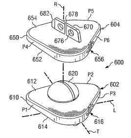
IYI. Transf~raaninal Pr~sthetic .Ioint
In some instances, it is often difficult to appr~ach and clear a defective
intervertebral disc space due to potential damage to important anatomical
structures such
as nerve roots, dura, ligamentum flavum and interspinous ligament. For
example,
preservation of the ligament~us structures is ~f great importance to restore
biomechanical
stability of the segment and its adjacent counterparts. In these situations, a
transforaminal
approach may allow clearance ofthe entire intervertebral disc space by opening
the
neuroforamen on one side. After appropriate clearance, it is possible to
achieve further
enlargement of the cleared intervertebral compartment by posterior tl~ans-
pedicle
distraction. While this approach has been used for fusion techniques, such as
Transforaminal Lumbar Interbody Fusion, or TLIF, it has not heretofore been
used with
motion preserving implants.
Referring to Fig. 27, in a transforaminal approach, the disc V is approached
as
shown by the arrow 400. The approach is between a posterior approach and a
lateral
approach, and in some cases, only one side of the disc needs to be exposed
(right or left) in
order to perform the procedure.
CA 02515827 2005-08-11
WO 2004/080355 PCT/US2004/004352
29
Referring to Figs. 2~-30, shown therein is an intervertebral articulating
prosthetic
joint 410 according to another form of the present disclosure. The
articulating joint 410
extends generally along a longitudinal axis L and includes a first articular
component 412
and a second articular component 414. The articular components 412, 414
cooperate to
form the articulating joint 410 which is sized and configured for disposition
within an
intervertebral space between adjacent vertebral bodies.
The prosthetic joint 410 provides relative pivotal and rotational movement
between
the adjacent vertebral bodies to maintain or restore motion substantially
similar to the
nornial bio-mechanical motion provided by a natural intervertebral disc. More
specifically, the articular components 412, 414 are permitted to pivot
relative to one
another about a number of axes, including lateral or side-to-side pivotal
movement about
longitudinal axis L and anterior-posterior pivotal movement about a transverse
axis T. It
should be understood that in one embodiment, the articular components 412, 414
are
pernzitted to pivot relative to one another about any axes that lies in a
plane that intersects
longitudinal axis L and transverse axis T. Additionally, the articular
components 412, 414
are preferably permitted to rotate relative to one another about a rotational
axis R.
Although the articulating joint 410 has been illustrated and described as
providing a
specific combination of articulating motion, it should be understood that
other
combinations of articulating movement are also possible and are contemplated
as falling
within the scope of the present disclosure. It should also be understood that
other types of
articulating movement are also contemplated, such as, for example, relative
translational
or linear motion.
Although the articular components 412, 414 of prosthetic joint 410 may be
formed
from a wide variety of materials, in one embodiment, the articular components
412, 414
are formed of a cobalt-chrome-molybdenum metallic alloy (ASTM F-799 or F-75).
However, in alternative embodiments, the articular components 412, 414 may be
formed
of other materials such as titanium or stainless steel, a polymeric material
such as
polyethylene, or any other biocompatible material that would be apparent to
one of
ordinary skill in the art. The surfaces of the articular components 412, 414
that are
positioned in direct contact with vertebral bone are preferably coated with a
bone-growth
promoting substance, such as, for example, a hydroxyapatite coating formed of
calcium
CA 02515827 2005-08-11
WO 2004/080355 PCT/US2004/004352
phosphate. Additionally, the suxface of the articular components 412, 414 that
are
positioned in direct contact with vertebral bone are preferably roughened
prior to being
coated with the bone-growth promoting substance to further enhance bone on-
growth.
Such surface roughening may be accomplished by way of, for example, acid
etching,
knurling, application of a bead coating, or other methods of roughening that
would occur
to one of ordinary skill in the art.
Articular component 412 includes a support plate 416 having an articular
surface
418 and an opposite bearing surface 420. Support plate 4I6 may be sized and
shaped to
substantially correspond to the size and shape of the vertebral endplate of an
adjacent
vertebra. In one embodiment, the support plate 416 is shaped to facilitate a
transforaminal
insertion approach. As such, the support plate 416 includes curved side
portions 422a,
422b, which are defined as the generally elongated portions of the support
plate 416
extending between articular surface 418 and the bearing surface 420. Although
not
shown, the suppoxt plate 416 can include one or more notches or other types of
indicia for
receiving and engaging with a corresponding portion of a surgical instrument
(also not
shown) to aid in the manipulation and insertion of the prosthetic joint 410
within an
intervertebral space between adjacent vertebrae. The surgical instrument (not
shown) is
preferably configured to hold the articular components 412, 414 at a
predetermined
orientation and spatial relationship relative to one another during
manipulation and
insertion of the prosthetic joint 4~ 10, and to release the articular
components 4129 4~ 14 once
properly positioned between the adjacent vertebrae.
In one embodiment, the articular surface 418 includes a projection 424 having
a
convex shape, which may be configured as a spherical-shaped ball (half of
which is
shown). It should be understood that other con~guxations of the projection 424
are also
contemplated, such as, for example, cylindrical, elliptical or other arcuate
configurations
or possibly non-arcuate configurations. It should also be understood that the
remaining
portion of articular surface 418 may take on planar or non-planar
configurations, such as,
for example, an angular or conical configuration extending about the
projection 424.
A flange member or keel 426 extends from the bearing surface 410 and is
configured for disposition within a preformed opening in the adjacent
vertebral endplate.
In one embodiment, the keel 426 extends perpendicularly from the bearing
surface 420
CA 02515827 2005-08-11
WO 2004/080355 PCT/US2004/004352
31
and is approximately centrally located along the bearing surface 420. However,
it should
be understood that other positions and orientations of the keel 426 are also
contemplated.
In one embodiment, the keel 426 transversely extends along a substantial
portion
of the support plate 416. The keel 426 is curved, generally in a direction
similar to the
arrow 400 of Fig. 27. The degree of curvature of the keel 426 may be
substantially similar
to and congruous with the degree of curvature of the side portions 422a, 422b.
Such an
embodiment would accommodate insertion of the prosthetic joint 410 using a
transforaminal approach as opposed to the anterior or lateral approaches
discussed above.
In a further embodiment, the keel 426 may be angled, tapered, or configured in
some other
shape to facilitate the functional demands of the keel. In still another
embodiment, the
keel 426 may be configured as a winged keel, including a transverse portion
(not shown)
extending across the main body portion of keel 426.
The keel 426 also includes three openings 428 extending therethrough to
facilitate
bone through-growth to enhance fixation to the adjacent vertebra. However, it
should be
understood that any number of openings 428 may be defined through keel 426,
including a
single opening or three or more openings. It should also be understood that
the openings
428 need not necessarily extend entirely through the keel 426, but may
alternatively
extend partially therethrough. It should further be understood that the keel
426 need not
necessarily define any openings 428 extending either partially or entirely
therethrough.
Additionally, although the openings 428 are illustrated as having ~ circular
configuration,
it should be understood that other sizes and configurations of openings 428
are also
contemplated. As discussed above, the surfaces of the articular component 4I2
that are in
direct contact with vertebral bone are preferably coated with a bone-growth
promoting
substance. Specifically, the bearing surface 420 and the surfaces of the keel
426 can be
coated with hydroxyapatite to promote b~ny engagement with the adjacent
vertebrae. As
also discussed above, the bearing surface 420 and the surfaces of keel 426 can
be
roughened prior to application of the hydroxyapatite coating.
In one embodiment, the articular component 414 includes a support plate 430
having an articular surface 432 and an opposite bearing surface 434. Support
plate 430
may be sized and shaped to substantially correspond to the size and shape of
the vertebral
endplate of an adjacent vertebra. In one embodiment, the support plate 430 is
shaped to
CA 02515827 2005-08-11
WO 2004/080355 PCT/US2004/004352
32
facilitate a transforaminal insertion approach. As such, the support plate 416
includes
curved side portions 436a, 436b, which are defined as the generally elongated
portions of
the support plate 430 extending between articular surface 432 and the bearing
surface 434.
Although not shown, the support plate 430 can include one or more notches or
other types
of indicia for receiving and engaging with a corresponding portion of a
surgical
instrument, such as discussed above with reference to articular element 412.
In one embodiment, the articular surface 432 includes a recess 440 having a
concave shape, which may be configured as a spherical-shaped socket. However,
it
should be understood that other configurations of the recess 440 are also
contemplated,
such as, for example, cylindrical, elliptical or other arcuate configurations
or possibly non-
arcuate configurations. The remaining portion of the articular surface 432 can
be angled
or otherwise configured to facilitate the insertion and/or use of the
prosthesis.
Although the concave recess 440 is illustrated as having a generally smooth,
uninterrupted
articular surface, it should be understood that a surface depression or cavity
may be
defined along a portion of the recess 440 to provide a means for clearing out
matter, such
as particulate debris, that is disposed between the abutting ai-ticular
surfaces of
components 412, 414. In such case, the convex articular surface of the ball
424 may
alternatively define a generally smooth, uninterrupted articular surface. In
another
embodiment, each of the convex projection 424 and the concave recess 440 may
define a
surface depression to facilitate removal of particulate matter disposed
between the abutting
articular surfaces.
A flange member ox keel 450, configured similar to the keel 426 of articular
component 412, extends from the bearing surface 434. In one embodiment, the
keel 450
can be centrally located, and is positioned directly or parallel in-line with
the keel 450.
The keel 450 is curved, in a direction similar to the keel 426 and the arrow
400 of Fig. 27.
The degree of curvature of the keel 450 may be substantially similar to and
congruous
with the degree of curvature of the side portions 436a, 436b. Such an
embodiment would
accommodate insertion of the prosthetic joint 410 using a transforaminal
approach as
opposed to the anterior or lateral approaches discussed above. In some
embodiments, the
position of the keel 450 can be offset to help circumvent veins, arteries,
bony portions, or
other obstacles that may be in place during the insertion of the joint 410.
CA 02515827 2005-08-11
WO 2004/080355 PCT/US2004/004352
33
It should also be understood that the keel 450 may also be differently
positioned,
shaped or oriented, or more keels 450 can be used, for similar or additional
reasons. Also,
the keel 450 may be angled, tapered, or configured in some other shape to
facilitate the
functional demands of the keel. In still another embodiment, the keel 450 may
be
configured as a winged lceel, including a transverse portion (not shown)
extending across
the main body portion of keel 450.
In one embodiment, the keel 450 also includes three openings 452 extending
therethrough to facilitate bone through-growth to enhance fixation to the
adjacent vertebra.
However, it should be understood that any number of openings 452 may be
defined
through keel 450, including a single opening or three or more openings. It
should also be
understood that the openings 452 need not necessarily extend entirely through
the keel
450, but may alternatively extend partially therethrough. It should further be
understood
that the keel 450 need not necessarily define any openings 452 extending
either partially
or entirely therethrough. Additionally, although the openings 452 are
illustrated as having
a circular configuration, it should be understood that other sues and
configurations of
openings 452 are also contemplated. As discussed above, the surfaces of the
articular
component 414 that are in direct contact with vertebral bone are preferably
coated with a
bone-growth promoting substance. Specifically, the bearing surface 434 and the
surfaces
of the keel 450 can be coated with hydroxyapatite to promote bony engagement
with the
adjacent vertebrae. As also discussed above, the bearing surface 434 and the
surfaces of
keel 450 can be roughened prior to application of the hydroxyapatite coating.
In some embodiments, one o~ both of the keels 426, 450 may include a sharp
forward edge, illustrated by edges 460, 462, respectively, of Fig. 28c. By
having such an
edge, insertion of the keel into the associated vertebral body is facilitated.
Also, the edges
460, 462 can be of sufficient sharpness that the vertebral bodies do not
require a slot for
receiving the keels 426, 450, discussed in greater detail below.
Referring to Figs. 31a and 31b, to accommodate insertion of the prosthetic
joint
410 within the intervertebral space, the upper and lower vertebrae VU, VL can
be prepared
to accept the prosthetic joint 410 therebetween. Referring specifically to
Fig. 31a, for the
configuration of the prosthetic joint 410 of Figs. 28-30, multiple slots 470,
472 are formed
along the vertebral endplates of the upper vertebrae VU and the lower
vertebrae VL. The
CA 02515827 2005-08-11
WO 2004/080355 PCT/US2004/004352
34
slots 470, 472 can be created by the keels 426, 450 themselves, or can be
prepared
beforehand.
Referring also to Fig. 32, it may be desirable to prepare one or more of the
slots
470, 472 before the prosthetic joint 410 is inserted between the upper and
lower vertebrae
VU, VL. The slots 470, 472 can be curved, as illustrated by the slot 472, in
accordance
with the curved lceels 426, 450, to facilitate the movement of the prosthetic
joint 410
during insertion.
Referring to Figs. 33-35, as an alternative to chiseling, which provides only
for the
cutting of straight slots, a milling guide 500 may be used in conjunction
with, a milling tool
502 to cut the curved slots 470, 472 (represented by 472 in Fig. 32) in the
upper and lower
vertebral bone VU, VL. The milling guide 500 and milling tool 502 may be
formed of any
material including biocompatible materials such as titanium. The milling guide
500
includes an elongated curved member 503, which defines a curved opening 504 to
correspond to the shape of the desired curve for the slots 470, 472. Of
course, the degree
of curvature of the milling guide 500, and therefore the curved opening 504,
may vary
depending on the desired curve of the slots 470, 472. In one embodiment, the
milling
guide 500 is fox-med of a pliable material that retains a rigid shape upon
reforming such
that the degree of curvature of the curved opening 504 may be altered without
having to
swap out milling guides. The milling guide 500, and therefore the curved
opening 504, is
also of sufficient length so that if the slots 470, 472 need to be continued
through any
posterior elements of the vertebrae such extension of the slots can be
accomplished at the
same time.
Referring specifically to Figs. 34a and 34b, in one embodiment, the milling
tool
502 includes a milling bit 510 that is positioned to be rotated and translated
in the curved
opening 504. In one embodiment, the milling bit 510 is a double fluted routing
bit, that
may extend simultaneously into the upper and lower vertebrae VU, VL.
The milling bit 510 is also adapted to receive a translational force such that
the
milling bit can be moved baclc and forth in the curved opening 504. Referring
to Fig. 34b,
in one embodiment, a milling bit handle 530 is connected in any conventional
manner to a
housing 522 (a portion of which is shown). The handle 530 extends from the
housing 522
and through a slot 532 formed in a proximal end 534 of the milling guide 500
relative to a
CA 02515827 2005-08-11
WO 2004/080355 PCT/US2004/004352
surgeon (not depicted). As such, the handle 530 can be translated by a
surgeon, thereby
translating the milling bit 510 through the curved opening 504. In this
manner, the handle
530 is adapted to impart translational movement to the milling bit 510. To
accommodate
movement of the milling bit 510 within the curved opening 504, a pair of
bearing
assemblies 512, 514 may be positioned adjacent to the housing 522 to guide the
milling bit
510 along the curved opening.
The housing 522 houses a rotation assembly, which, in one embodiment, is a
gear
assembly 524. The gear assembly 524 includes a drive gear 526 connected to and
extending annularly around a rotatable shaft 528. The shaft 528 is rotatable
via an
external source represented by power supply 516 (Fig. 35). In one embodiment,
the shaft
528 is housed within the handle 530.
The gear assembly 524 further includes a bit gear 530, which is connected to
and
extends annularly around the milling bit 510. The bit gear 530 is positioned
on the milling
bit 510 such that the bit gear is orthogonal relative to and in contact with
the drive gear
526. Thus, rotation of the shaft 528 imparts rotation to the milling bit 510
via the gear
assembly 524. A pair of annular shoulders 534, 536 are also connected to the
milling bit
510 such that the milling bit can easily move back and forth through the
curved opening
504 without slippage in the upper or lower directions as viewed in Fig. 34b.
It is
understood that the gear assembly 524 is merely exemplary of an assembly that
rnay be
used to impart rotational motion to the milling bit 510. ~ther types of
rotation-imparting
assemblies are contemplated as falling within the present disclosure such as
pneumatic-
type systems.
Referring to Fig. 34c, in one such embodiment, a pneumatic system 538 rnay be
employed to impart rotation to the milling bit 510. In one embodiment, a
Medtronic
Midas Rex ° LegendTM motor is used to supply power (represented by P)
to the pneumatic
system. A conventional valve 539 is used to control the air flow and pressure
supplied to
rotate the milling bit 510. In still other embodiments, manual or combination
power
supplies are contemplated as being the preferred power supply 516 (Fig. 34b)
and P (Fig.
34c).
Referring again to Figs. 34a and 34b, a guide handle 540 is further provided
such
that the milling guide 500 is independently movable relative to the milling
bit 510. Thus
CA 02515827 2005-08-11
WO 2004/080355 PCT/US2004/004352
36
in one embodiment, the milling guide 500 can be held via the guide handle 540
with one
hand while the milling bit 510 may be moved within the curved opening 504 via
the
handle 530 with the other hand. In some embodiments, the handle 530 may extend
through the guide handle 540 as shown in Fig. 34b. As a result, and referring
to Fig. 35,
the milling bit 510 is adapted to rotate in a direction indicated by arrow Rl,
and is adapted
to be translated through the curved opening 504 in the directions indicated by
arrow R2.
In operation, the milling guide 500 and the milling tool 502 can be used to
cut a
slot, such as the slot 472, to prepare the vertebral body VL to receive the
lower portion of
the prosthetic joint 410.' The surgeon first selects the desired amount of
curvature to
impart to the slot 472 and selects or configures the corresponding milling
guide 500. The
surgeon then approaches the vertebral body VL from the transforaminal approach
to
position the milling guide 500 into the disc space between the upper and lower
vertebrae
VU, VL and to abut the milling bit 510 against the upper and lower vertebrae
VU, VL.
Upon proper positioning, the surgeon may then actuate the milling tool 502 via
the power
supply 516 to begin Butting into the upper and lower vertebrae VU, VL with the
milling bit
510.
The milling guide 500 may be held by the surgeon or via an external instx-
ument
such that the milling guide is stationary during translational movement of the
milling bit
510 through the milling guide. The curvature of the milling guide 500 guides
the milling
bit 510 transforaminally through the upper and lower vertebrae VU, VL to But a
transforaminal slot, such as the slot 472 depicted in the lower vertebra VL
Fig. 32, to
prepare the upper and lower vertebrae to receive the transforaminal prosthetic
joint 410.
In an alternative embodiment, the keels of the prosthetic joint 410 may take
alternative shapes and Bon~gurations to assist in the curved, transforaminal
approach used
in inserting the joint. Referring to Figs. 36-38, the keels, designated 550
and 560, extend
from the bearing surfaces 434 and 420, respectively. The keels 550, 560 are
relatively
short and thus extend along a short portion of the bearing surfaces 434, 420,
respectively,
in comparison to the keels 450, 426 of Figs. 28-30. The relative shortness of
the keels
550, 560 may aid such keels in following the openings 470, 472, respectively.
In addition,
the shortness of the keels 550, 560 and the ease with which such keels follow
the
openings 470, 472, respectively, allows the keels to be configured as either
straight or
CA 02515827 2005-08-11
WO 2004/080355 PCT/US2004/004352
37
curved keels, which increases the design options of the prosthetic joint 410.
The keels
550, 560 may also be tapered to assist in insertion of the keels into the
upper and lower
vertebrae VU, VL.
IV. Anterior-Oblique Prosthetic Joint
Another approach that can be used to avoid potential damage to important
anatomical structures such as nerve roots, dura, ligamentum flavum and
interspinous
ligament is the anterior oblique approach. For example, the straight anterior
approach to
the disc space between vertebra L4 and L5, as well as the superior disc
levels, can present
high surgical risks during the insertion of a total disc replacement implant
because of the
attachment of the major vessels to the anterior aspect of the spine.
Referring to Figs. 39a and 40-41, shown therein is an intervertebral
articulating prosthetic
joint 600 according to another form of the present disclosure. The prosthetic
joint 600
extends generally along a longitudinal axis L and includes a first articular
component 602
and a second articular component 604. The articular components 602, 604
cooperate to
form the prosthetic joint 600 which is sized and configured for disposition
within an
intervertebral space between adjacent vertebral bodies.
The prosthetic joint 600 provides relative pivotal and rotational movement
between
the adjacent vertebral bodies to maintain or restore motion substantially
similar to the
normal bio-mechanical motion provided by a natural intervertebral disc.
I~/lore
specifically, the articular components 602, 604 are permitted to pivot
relative to one
another about a number of axes, including lateral or side-to-side pivotal
movement about
longitudinal axis L and anterior-posterior pivotal movement about a transverse
axis T. It
should be understood that in a preferred embodiment, the articular components
602, 604
are permitted to pivot relative to one another about any axes that lies in a
plane that
intersects longitudinal axis L and transverse axis T. Additionally, the
articular
components 602, 604 may be permitted to rotate relative to one another about a
rotational
axis R. Although the articulating joint 600 has been illustrated and described
as providing
a specific combination of articulating motion, it should be understood that
other
combinations of articulating movement are also possible and are contemplated
as falling
within the scope of the present disclosure. It should also be understood that
other types of
CA 02515827 2005-08-11
WO 2004/080355 PCT/US2004/004352
38
articulating movement are also contemplated, such as, for example, relative
translational
or linear motion.
Although the articular components 602, 604 of prosthetic joint 600 may be
formed
from a wide variety of materials, in one embodiment, the articular components
602, 604
are formed of a cobalt-chrome-molybdenum metallic alloy (ASTM F-799 or F-75).
However, in alternative embodiments of the invention, the articular components
602, 604
may be formed of other materials such as titanium or stainless steel, a
polymeric material
such as polyethylene, or any other biocompatible material that would be
apparent to one of
ordinary skill in the art. The surfaces of the articular components 602, 604
that are
positioned in direct contact with vertebral bone are preferably coated with a
bone-growth
promoting substance, such as, for example, a hydroxyapatite coating formed of
calcium
phosphate. Additionally, the suxface of the articular components 602, 604 that
are
positioned in direct contact with vertebral bone are preferably roughened
prior to being
coated with the bone-growth promoting substance to further enhance bone on-
growth.
Such surface roughening may be accomplished by way of, for example, acid
etching,
knurling, application of a bead coating, or other methods of roughening that
would occur
to one of ordinary skill in the art.
Articular component 602 includes a support plate 610 having an articular
surface
612 and an opposite bearing surface 614. Support plate 610 may be sized and
shaped to
substantially correspond to the size and shape of the vertebral endplate of an
adjacent
vertebra. In one embodiment, the support plate 610 is shaped in a triangular-
like
configuration to facilitate an oblique insertion approach from either the left
or right side of
the spine, and as such, includes side portions P1, P2 and P3. The side
portions P1, P2 and
P3 may take a variety of configurations including curved (illustrated by P2)
or straight
(illustrated by P1 and P3) configurations.
The support plate 610 can include one or mare notches 616 or other types of
indicia for receiving and engaging with a corresponding portion of a surgical
instrument
(also not shown) to aid in the manipulation and insertion of the prosthetic
joint 600 within
an intervertebral space between adjacent vertebrae. The surgical instrument
(not shown) is
preferably configured to hold the articular components 602, 604 at a
predetermined
orientation and spatial relationship relative to one another during
manipulation and
CA 02515827 2005-08-11
WO 2004/080355 PCT/US2004/004352
39
insertion of the prosthetic joint 600, and to release the articular components
602, 604 once
properly positioned between the adjacent vertebrae.
In one embodiment, the articular surface 612 includes a projection 620 having
a convex
shape, which may be configured as a spherical-shaped ball (half of which is
shown). It
should be understood that-other configurations of the projection 620 are also
contemplated, such as, for example, cylindrical, elliptical or other arcuate
configurations
or possibly non-arcuate configurations. It should also be understood that the
remaining
portion of articular surface 612 may take on planar or non-planar
configurations, such as,
for example, an angular or conical configuration extending about the
projection 620.
A flange member or keel 640 extends from the bearing surface 614 and is
configured for disposition within a preformed opening in the adjacent
vertebral endplate.
In one embodiment, the keel 640 extends perpendicularly from the bearing
surface 614
and is approximately centrally located along the bearing surface 614. However,
'it should
be underst~od that other positions and orientations of the keel 640 are also
contemplated.
Furthermore, more keels 640 can be used, for similar or additional reasons.
In one embodiment, the keel 640 extends along a substantial portion of the
support plate
610. The keel 640 is straight, but extends along a direction towards the
notches 616 and is
parallel with one of the side portions P1 of the support plate 610. In the
present example,
the keel 640 is positioned between the transverse axis T and lateral axis L.
Such an
embodiment accommodates insertion of the prosthetic joint 600 using an oblique
approach
as opposed to the anterior, lateral, or transf~raminal approaches discussed
above. In a
further embodiment, the keel 640 may be angled, tapered, or configured in some
other
shape to facilitate the functional demands of the keel. In still another
embodiment, the
keel 640 may be configured as a winged keel, including a transverse portion
(not shown)
extending across the main body portion of keel 640.
The keel 640 also includes a pair of openings 646 extending therethrough to
facilitate bone through-growth t~ enhance fixation to the adjacent vertebra.
Additionally,
a gap 648 may also be formed in the keel 640 to further facilitate bone
through-growth.
The gap 648 also provides a reference point such that an X-ray can be used to
evaluate the
positioning and alignment of the support plate 602 during insertion of the
prosthetic joint
600. It should be understood that any number of openings 646 or gaps 648 rnay
be
CA 02515827 2005-08-11
WO 2004/080355 PCT/US2004/004352
defined through keel 640, including a single opening or gap or several
openings and gaps.
It should also be understood that the openings 646 and gap 648 need not
necessarily
extend entirely through the keel 640, but may alternatively extend partially
therethrough.
It should further be understood that the keel 640 need not necessarily define
any openings
646 or gaps 648 extending either partially or entirely therethrough.
Additionally, although
the openings 646 are illustrated as having a circular configuration, it should
be understood
that other sizes and configurations of openings 646 are also contemplated. As
discussed
above, the surfaces of the articular component 602 that are in direct contact
with vertebral
bone may be coated with a bone-growth promoting substance. Specifically, the
bearing
surface 614 and the surfaces of the keel 640 can be coated with hydroxyapatite
to promote
bony engagement with the adjacent vertebrae. As also discussed above, the
bearing
surface 614 and the surfaces of keel 640 can be roughened prior to application
of the
hydroxyapatite coating.
In one embodiment, the articular component 604 includes a support plate 650
having an articular surface 652 and an opposite bearing surface 654. support
plate 650
may be sized and shaped to substantially correspond to the size and shape of
the vertebral
endplate of an adjacent vertebra. In one embodiment, the support plate 610 is
shaped in a
triangular-like configuration to facilitate an oblique insertion approach from
either the left
or right side of the spine, and as such, includes side portions P4, PS and P6.
The side
portions P4, P5 and P6 may take a variety of configurations including curved
(illustrated
by PS) or straight (illustrated by P4 and P6) configurations.. The support
plate 650 can
include one or more notches 656 or other types of indicia for receiving and
engaging with
a corresponding portion of a surgical instrument, such as discussed above with
reference
to axticular component 602.
In one embodiment, the articular surface 652 includes a recess 660 having a
convex shape, which may be configured as a spherical-shaped socket. However,
it should
be underst~od that other configurations of the recess 660 are also
contemplated, such as,
for example, cylindrical, elliptical or other arcuate configurations or
possibly non-arcuate
configurations. The remaining portion of the articular surface 652 can be
angled or
otherwise configured to facilitate the insertion and/or use of the prosthesis.
CA 02515827 2005-08-11
WO 2004/080355 PCT/US2004/004352
41
Although the concave recess 660 is illustrated as having a generally smooth,
uninterrupted
articular surface, it should be understood that a surface depression or cavity
may be
defined along a portion of the recess 660 to provide a means for clearing out
matter, such
as particulate debris, that is disposed between the abutting articular
surfaces of
components 602, 604. In such case, the convex articular surface of the ball
620 may
alternatively define a generally smooth, uninterrupted articular surface. In
another
embodiment of the invention, each of the convex projection 620 and the concave
recess
660 may define a surface depression to facilitate removal of particulate
matter disposed
between the abutting articular surfaces.
A flange member or keel 670, configured similar to the keel 640 of articular
component 602, extends from the bearing surface 654. In one embodiment, the
keel 670
can be centrally located, and is positioned directly or parallel in-line with
the keel 640.
The keel 640 is straight, but extends along a direction towards the notches
656 and is
parallel with one of the side portions P4 of the support plate 650. such an
embodiment
accommodates insertion of the prosthetic joint 600 using an oblique approach
as opposed
to the anterior, lateral, or transforaminal approaches discussed above. In
some
embodiments, the position of the keel 670 can be offset to help cixcumvent
veins, arteries,
bony portions, or other obstacles that may be in place during the insertion of
the joint 600.
It should be further understood that other positions, shapes, orientations,
and
quantities of the keel 670 are also contemplated. It should also be understood
that more
keels 670 can be used, for similar or additional reasons. Also, the keel 670
may be angled,
tapered, or configured in some other shape to facilitate the functional
demands of the keel.
In still another embodiment, the keel 670 may be configured as a winged keel,
including a
transverse portion (not shown) extending across the main body portion of keel
670.
In one embodiment, the keel 670 also includes a pair of openings 676 extending
therethrough to facilitate bone through-growth to enhance fixation to the
adjacent vertebra.
Additionally, a gap 678 may also be formed in the keel 670 to further
facilitate bone
through-growth. The gap 678 also provides a reference point such that an X-ray
can be
used to evaluate the positioning and alignment of the support plate 604 during
insertion of
the prosthetic joint 600. It should be understood that any number of openings
676 or gaps
678 may be defined through keel 670, including a single opening or gap or
several
CA 02515827 2005-08-11
WO 2004/080355 PCT/US2004/004352
42
openings or gaps. It should also be understood that the openings 676 and gap
678 need
not necessarily extend entirely through the keel 670, but may alternatively
extend partially
therethrough. It should further be understood that the keel 670 need not
necessarily define
any openings 676 or gaps 678 extending either partially or entirely
therethrough.
Additionally, although the openings 676 are illustrated as having a circular
configuration,
it should be understood that other sizes and configurations of openings 676
are also
contemplated. As discussed above, the surfaces of the articular component 602
that are in
direct contact with vertebral bone are preferably coated with a bone-growth
promoting
substance. Specifically, the bearing surface 654 and the surfaces of the keel
670 can be
coated with hydroxyapatite to promote bony engagement with the adjacent
vertebrae. As
also discussed above, the bearing surface 654 and the surfaces of keel 670 can
be
roughened prior to application of the hydroxyapatite coating.
In some embodiments, one ox both of the keels 640, 670 may include a sharp
forward edge, illustrated by edges 680, 682. By having such an edge, insertion
of the
keels 640, 670 into the associated vertebral body is facilitated. Also, the
edges 680, 682
can be of sufficient sharpness that the vertebral body does not require a slot
for receiving
the keel 640, 670, discussed in greater detail below.
Referring to Figs. 42-44a, to accommodate insertion of the prosthetic joint
600
within the intervertebral space, the upper and lower vertebrae VU, VL can be
prepared to
accept the prosthetic joint 600 therebetween. Referring specifically to Fig.
43, fox the
configuration of the prosthetic joint 600 of Figs. 38-40, multiple slots 690,
692 are formed
along the vertebral endplates of the lower vertebrae VL and the upper
vertebrae VU,
respectively. The slots 690, 692 can be created by the keels 640, 670
themselves, or can
be prepared beforehand by one or more of the methods discussed above. It can
be seen
from Figs. 42-44, that if one or more vessels 694 are obstructing a straight
anterior
approach, the oblique approach will allow f~r an anterioxllateral insertion.
The implant
600 design also ensures a sufficient contact surface for contacting the bony
endplates of
the vertebrae VU, VL.
Referring to Fig. 44b, in one embodiment, the prosthetic joint 600 can be
inserted
into the intervertebral space via instrumentation such as the guide disclosed
in co-pending
application U.S. Serial No. 10/430,473, which is herein incorporated by
reference. In one
CA 02515827 2005-08-11
WO 2004/080355 PCT/US2004/004352
43
example of an insertion process for inserting the prosthetic joint 600, the
midline M of the
vertebrae VU, VL is located using imaging equipment and a pin 695 is inserted
into the
upper vertebra VU along the midline. An oblique guide member 696 is then
connected to
the pin 695 via a flange 697 and a handle (not shown) associated with the
oblique guide
member 696 is then adjusted to a proper position. An oblique pin 698 of the
oblique guide
member 696 is then impacted into the upper vertebra VU to fix the oblique
guide member,
thereby indicating the entering reference point and the direction of implant
insertion for
the prosthetic joint 600. The guide (not shown) can then be used to implant
the prosthetic
joint 600 into the intervertebral space from an anterior-oblique approach, the
details of
which are more fully discussed in co-pending application U.S. Serial No.
10/430,473.
Referring to Figs. 39b and 39c, the prosthetic joint 600 may be modified to
include
various notches and/or holes to accommodate insertion or other types of
instruments. For
example, a notch 684 may be formed in the prosthetic joint to receive a
portion of an
insertion instrument (not shown). In some embodiments, the notch 684 may have
a hook
shape. Moreover, a thxough-hole 686 and pin slots 688 may further be formed in
the
prosthetic joint 600, and again, may be sized and configured to receive an
insertion
instrument. It is understood that the notch 684, the through-hole 686, and the
pin slots 688
may take any number of configurations to permit attachment of the prosthetic
joint 600 to
an insertion instrument. Furthermore, provision of the notch 684, through-hole
686, and
the pin sl~ts 688 may aid in preventing rotation of the prosthetic joint 600
during insertion.
V. Mobile-Bearing Prosthetic Joint
In another embodiment, the above-described prosthetic joints can be modified
to
provide for translational movement as well as rotational movement. For
example,
referring to Figs. 45-47, a mobile-bearing prosthetic joint for anterior
insertion is generally
referred to by reference numeral 700. It is understood that the mobile-bearing
pxosthetic
joint 700 is described with respect to anterior insertion for the sake of
clarity only, and
therefore, a variety of insertion directions are contemplated for the mobile-
bearing
prosthetic joint.
CA 02515827 2005-08-11
WO 2004/080355 PCT/US2004/004352
44
The prosthetic joint 700 extends generally along a longitudinal axis L and
includes
a first articular component 702 and a second articular component 704. The
articular
components 702, 704 cooperate to form the prosthetic joint 700 which is sized
and
configured for disposition within an intervertebral space between a pair of
vertebral
bodies, such as an intervertebral space S 1 between adjacent vertebral bodies
Vs, VI (Fig.
48).
The prosthetic joint 700 provides xelative pivotal and rotational movement
between
the adjacent vertebral bodies Vs, VI to maintain or restore motion
substantially similar to
the normal bio-mechanical motion provided by a natural intervertebral disc but
with the
added element of translational motion. More specifically, the articular
components 702,
704 are permitted to pivot relative to one another about a number of axes,
including lateral
or side-to-side pivotal movement about a longitudinal axis L and anterior-
posterior pivotal
movement about a transverse axis T. It should be understood that in one
embodiment, the
articular components 702, 704 are permitted to pivot relative to one another
about any
axes that lies in a plane that intersects longitudinal axis L and transverse
axis T.
Additionally, the articular components 702, 704 are permitted to rotate
relative to one
another about a rotational axis R. In addition, the articular components 702,
704 are
permitted to translate relative to one another as will be further described.
Although the articular components 702, 704 of prosthetic joint 700 may be
formed
from a wide variety of materials, in one embodiment, the articular components
7029 704
axe formed of a cobalt-chrome-molybdenum metallic alloy (ASTM F-7~~ or F-75).
However, in alternative embodiments, the articular components 702, 704 may be
formed
of other materials such as titanium or stainless steel, a polymeric material
such as
polyethylene, or any other biocompatible material that would be apparent to
one of
ordinary shill in the art. The surfaces of the articular components 702, 704
that are
positioned in direct contact with vertebral bone may be coated with a bone-
growth
promoting substance, such as, for example, a hydroxyapatite coating formed of
calcium
phosphate. Additionally, the surface of the articular components 702, 704 that
are
positioned' in direct contact with vertebral bone may be roughened prior to
being coated
with the bone-growth promoting substance to further enhance bone on-growth.
Such
surface roughening may be accomplished by way of, for example, acid etching,
knurling,
CA 02515827 2005-08-11
WO 2004/080355 PCT/US2004/004352
application of a bead coating, or other methods of roughening that would occur
to one of
ordinary skill in the art.
Articular component 702 includes a support plate 706 having an articular
surface
708 and an opposite bearing surface 710. Support plate 706 may be sized and
shaped to
substantially correspond to the size and shape of the vertebral endplate of an
adjacent
vertebra. The support plate 706 can include one or more notches 712 or other
types of
indicia for receiving and engaging with a corresponding portion of a surgical
instrument
(not shown) to aid in the manipulation and insertion of the articulating joint
700 within an
intervertebral space between adjacent vertebrae. The surgical instrument (not
shown) is
preferably configured to hold the articular components 702, 704 at a
predetermined
orientation and spatial xelationship relative to one another during
manipulation and
insertion of the articulating joint 700, and to release the articular
components 702, 704
once properly positioned between the adjacent vertebrae.
In one embodiment, and referring to Figs. 49a and 4.9b, a recess 714 is formed
in the
articular surface 708. A circumferential edge 716 defining the recess 714
along the
articular surface 708 is in a concentric relationship with a recess surface
718, yet has a
smaller diameter relative to the recess surface due to a diverging circular
side 720 (Fig.
48b) of the recess 714. Although described with reference to having a circular
shape, it is
understood that the recess 714 may take any number of shapes such as square,
triangular,
or rectangular shapes.
Deferring to Figs. SOa and SOb, the recess 714 (Fig. 49b) is designed to
receive a
portion of a modular projection member 722. The projection member 722 includes
a
flange portion 724, which is shaped to coiTespond to the shape of the recess
714. As such,
the flange portion 724 includes a diverging circurnferential side 726, which
terminates at a
substantially planar engagement surface 728. The engagement surface 728 is
adapted to
engage the substantially planar recess surface 718 (Fig. 49b). It is
understood, however,
that although depicted as being substantially planar, the engagement surface
728 and the
recess surface 718 may take any number of corresponding shapes. The diameter
of the
engagement surface 728 is smaller than the diameter of the recess surface 718,
thereby
allowing translation of the modular projection member 722 relative to the
articular
component 702.
CA 02515827 2005-08-11
WO 2004/080355 PCT/US2004/004352
46
The remaining portion of the modular projection member 722 is defined by a
projection 730 having a convex shape, which may be configured as a spherical-
shaped ball
(half of which is shown). It should be understood that other configurations of
the
projection 730 are also contemplated, such as, for example, cylindrical,
elliptical or other
arcuate configurations or possibly non-arcuate configurations. It should also
be
understood that the remaining portion of articular surface 708 may take on
planar or non-
planar configurations, such as, fox example, an angular or conical
configuration extending
about the projection 224.
In one embodiment, the convex articular surface of the projection 730 is
interrupted by a surface depression or cavity 732 extending along the
projection 730. In
one embodiment, the surface depression 732 is configured as a groove. However,
it
should be understood that other types of surface depxessions are also
contemplated,
including no depression at all. One purpose of the groove 732 is to facilitate
the removal
of matter disposed between abutting portions of the articular components 702,
704. More
specifically, the groove 732 may aid in clearing out matter such as, for
example,
particulate material, that is disposed between the abutting articular surfaces
of components
702, 704.
Referring to Figs. 45 and 49b, a flange member or keel 740 extends from the
bearing surface 710 and is configured for disposition within a preformed
opening in the
adjacent vertebral endplate (such as ~Iin Fig. 47). In one embodiment, the
keel 740
extends perpendicularly from the bearing surface 710 and is approximately
centrally
located along the bearing surface 710. However, it should be understood that
other
positions and orientations of the keel 740 are also contemplated.
In one embodiment, the keel 740 extends along substantially the entire length
of
the support plate 706. Such an embodiment would accommodate insertion of the
articulating joist 700 using an anterior approach. However, as discussed
above, other
approaches such as lateral, transforaminal, and anterior-oblique approaches
are also
contemplated for insertion of the prosthetic joint 700. In a further
embodiment, the keel
740 may be angled, tapered, or configured in some other shape to facilitate
the functional
demands of the keel. In still another embodiment, the keel 740 may be
configured as a
CA 02515827 2005-08-11
WO 2004/080355 PCT/US2004/004352
47
winged keel, including a transverse portion (not shown) extending across the
main body
portion of keel 740.
The keel 740 also includes a pair of openings 742 extending therethrough to
facilitate bone through-growth to enhance fixation to the adjacent vertebra.
However, it
should be understood that any number of openings 742 may be defined through
keel 740,
including a single opening or three or more openings. It should also be
understood that the
openings 742 need not necessarily extend entixely through the keel 740, but
may
alternatively extend partially therethrough. It should further be understood
that the keel
740 need not necessarily define any openings 742 extending either partially or
entirely
thexethrough. Additionally, although the openings 742 are illustrated as
having a circular
configuration, it should be understood that other sizes and configurations of
the openings
742 are also contemplated. As discussed above, the surfaces of the articular
component
702 that are in direct contact with vertebral bone are preferably coated with
a bone-growth
promoting substance. Specifically, the bearing surface 710 and the surfaces
ofthe keel
740 can be coated with hydroxyapatitc to promote bony engagement with the
adjacent
vertebrae. As also discussed above, the bearing surface 710 and the surfaces
of keel 740
can be roughened prior to application of the hydroxyapatitc coating.
Referring to Figs. 45-47, in one embodiment, the articular component 704
includes
a support plate 750 having an articular surface 752 and an opposite bearing
surface 754.
Support plate 750 may be sized and shaped to substantially correspond to the
size and
shape of the vertebral endplate of an adjacent vertebra. The support plate 750
can include
one or more notches 756 or other types of indicia for receiving and engaging
with a
corresponding portion of a surgical instrument, such as discussed above with
reference to
articular component 702.
In one embodiment, the articular surface 752 includes a recess 758 (Fig. 47),
which has a convex shape, such as that of a spherical-shaped socket. However,
it should
be understood that other configuxations of the recess 758 are also
contemplated, such as,
for example, cylindrical, elliptical or other arcuate configurations or
possibly non-arcuate
configurations. The remaining portion of the axticular surface 752 can be
angled or
otherwise configured to facilitate the insertion and/or use of the
articulating joint 700.
CA 02515827 2005-08-11
WO 2004/080355 PCT/US2004/004352
48
Although the concave recess 758 is illustrated as having a generally smooth,
uninterrupted
articular surface, it should be understood that a surface depression or cavity
may be
defined along a portion of the recess 758 to aid in clearing out matter, such
as particulate
debris, that is disposed between the abutting articular surfaces of articular
components
702, 704. In such case, the convex articular surface of the projection 730 may
alternatively define a generally smooth, uninterrupted articular surface. In
another
embodiment, each of the convex projection 730 and the concave recess 758 may
define a
surface depression to facilitate removal of particulate matter disposed
between the abutting
articular surfaces.
A flange member or keel 760, configured similar to the keel 740 of articular
component 702, extends from the bearing surface 754. In one embodiment, the
keel 760
extends perpendicularly from the bearing surface 754 and is approximately
centrally
located along bearing surface 754. However, it should be understood that other
positions
and orientations of the keel 760 are also contemplated. It should also be
understood that
the articular component 704 may include two or more keels 760 extending from
the
bearing surface 754.
In one embodiment, the keel 760 extends along substantially the entire length
of
the support plate 750. Such an embodiment would accommodate insertion of the
prosthetic joint 700 using an anterior approach. However, as discussed above,
other
approaches such as lateral, transforaminal, and anterior-oblique approaches
are also
contemplated for insertion of the prosthetic joint 700. In a further
embodiment, the keel
760 may be angled, tapered, or configured in some other shape to facilitate
the functional
demands of the keel. In still another embodiment, the keel 760 may be
configured as a
winged keel, including a transverse portion (not shown) extending across the
main body
portion of keel 760.
The lceel 760 also includes a pair of openings 762 extending therethrough to
facilitate bone through-growth to enhance fixation to the adjacent vertebra.
However, it
should be understood that any number of openings 762 may be defined through
keel 760,
including a single opening or three or more openings. It should also be
understood that the
openings 762 need not necessarily extend entirely through the keel 760, but
may
alternatively extend partially therethrough. It should further be understood
that the lceel
CA 02515827 2005-08-11
WO 2004/080355 PCT/US2004/004352
49
760 need not necessarily define any openings 762 extending either partially or
entirely
therethrough. Additionally, although the openings 762 are illustrated as
having a circular
configuration, it should be understood that other sizes and configurations of
openings 762
are also contemplated. As discussed above, the surfaces of the articular
component 704
that are in direct contact with vertebral bone are preferably coated with a
bone-growth
promoting substance. Specifically, the bearing surface 754 and the surfaces of
the keel
760 can be coated with hydroxyapatite to promote bony engagement with the
adjacent
vertebrae. As also discussed above, the bearing surface 754 and the surfaces
of keel 760
can be roughened prior to application of the hydroxyapatite coating.
In some embodiments, one or both of the keels 740, 760 may include a sharp
forward edge, illustrated by edge 760a of Figs. 45 and 46. By having such an
edge,
insertion of the keel 740, 760 into the associated vertebral body is
facilitated. Also, the
edge 760a can be of sufficient sharpness that the vertebral body does not
require a slot for
receiving the keel 760, discussed in greater detail below.
Referring to Fig. 45, the mobile-bearing prosthetic joint 700 is assembled by
inserting the
modular projection 722 member into the recess 714 formed in the articular
surface 708 of
articular component 702. Upon assemblage, the prosthetic joint 700 is ready to
be inserted
into the disc space S 1 between adj acent vertebral bodies Vs, VI (Fig. 48).
Referring to Fig. 48, to accommodate insertion of the prosthetic joint 700
within
the intervertebral space S 1, the adjacent vertebral bodies V s, V I can be
prepared to accept
the prosthetic joint 700 therebetween. For the configuration of the prosthetic
joint 700 of
Figs. 45-47, slots 770, 772 are formed along the vertebral endplates of the
vertebrae Vs
and the vertebrae VI, respectively. The slots 770, 772 can be created by the
keels 740, 760
themselves, or can be prepared beforehand by one or more of the methods
discussed
above.
Upon insertion into the disc space S1, the prosthetic joint 700 allows
translational
movement of the articular component 704 relative to the articular component
702 due to
the engagement of the modular projection 722 with the concave recess 758 of
articular
component 704. For example, in Fig. 51, the modular projection member 722 is
shown in
a posterior position (which would result in movement of the articular
component 704 in
the posterior direction P), while in Fig. 52, the modular projection member
722 is shown
CA 02515827 2005-08-11
WO 2004/080355 PCT/US2004/004352
in an anterior position (which would result in movement of the articular
component 704 in
the anterior direction A). Figs. 51 and 52 are of course only exemplary of the
translational
movement allowed by the implementation of modular projection member 722 and
the
corresponding recess 714, and thus, the amount of translational movement of
the modular
projection member 722, and therefore the articular component 704, relative to
the articular
component 702 can vary, including in directions other than P and A.
Furthermore, the positioning of the modular projection member 722 within the
recess 714 of the articular component 702 allows the modular projection to
spin relative to
the articular component 702. Thus, in such an embodiment, the modular
projection
member 722 adds the benefit of being able to impart rotation to the articular
component
704 (via the engagement with the recess 758) independent of translational
movement
imparted t~ the articular component 704. Such independent relationship between
translational and rotational movement adds to the amount of mobility
experienced at the
prosthetic joint 700 relative to prosthetic joints for which translational
movement is
dependent upon rotational movement and vice versa.
The present disclosure has been described relative to several preferred
embodiments. Improvements or modifications that become apparent to persons of
ordinary skill in the art after reading this disclosure are deemed within the
spirit and scope
of the application. For example, the articulating components of the above-
described
articulating joints may be reversed without departing from certain aspects of
the
disclosure. Accordingly, it is understood that several modifications, changes
and
substitutions are intended in the foregoing disclosure and, in some instances,
some
features of the disclosure will be employed without a corresponding use of
other features.
It is also understood that all spatial references, such as "longitudinal" and
"transverse," are
for illustrative purposes only and can be varied within the scope of the
disclosure.
Accordingly, it is appropriate that the appended claims be construed broadly
and in a
manner consistent with the scope of the disclosure.