Note: Descriptions are shown in the official language in which they were submitted.
CA 02515862 2005-08-12
WO 2004/073563 PCT/US2004/004284
1N-SITU FORMED INTERVERTEBRAL FUSION DEVICE AND METHOD
RELATED APPLICATIONS
This application claims the benefit of U.S. Provisional Application No.
60/448,221, filed on February 14, 2003. The entire teachings of the above
application is incorporated herein by reference.
BACKGROUND OF THE INVENTION
A leading cause of lower back pain arises from lumbar intervertebral disc
pathology, including rupture or degeneration of the disc. Radicular pain in
the lower
extremities may be caused by the compression of spinal nerve roots by a
bulging
disc. Additionally, lower back pain may be caused by collapse of the disc and
the
dysarthrosis of an unstable or degenerative vertebral facet joint. One
proposed
method of managing these problems is to remove the problematic disc and
replace it
>.
with a porous device that restores disc height and allows for bone growth
therethrough for the fusion of the adjacent vertebrae. These devices are
commonly
called "fusion devices."
Intervertebral body fusion devices typically must carry extremely high loads
(on the order of 1-4 kN) for a period of several months, or until fusion
occurs.
Accordingly, a fusion device or bone graft substitute designed for promoting
bony
fusion at another location in the body (such as long bone fusion) may not be
suitable
for use as an intervertebral body fusion device. For example, many bony fusion
devices disclose the use of a gel such as a hydrogel as the structural carrier
for an
osteoinductive or an osteogeneic component. However, such gels typically do
not
posses the stiffness or mechanical strength found to be required for lumbar
intervertebral fusion devices.
In general, delivery of conventional intervertebral fusion devices has
required significantly invasive implantation procedures. Open surgical
implantation
of posterior implants requires excision of stabilizing muscles, ligaments,
tendons,
and bony structures such as the facet joints. The implants must not only
overcome
CA 02515862 2005-08-12
WO 2004/073563 PCT/US2004/004284
-2-
the destabilization caused by the surgical procedure, but must add the extra
stability
needed to promote bony fusion. Open anterior surgery in the lumbar spine is
very
rislcy due to the close proximity of sensitive vascular structures, such as
the aorta
and bifurcation of the aorta. Furthermore, the anterior open procedure can
cause
significant scar formation on the spine, making anterior revision surgery, if
necessary, even more risky.
Minmally invasive procedures have been developed to help mitigate these
problems. However, current techniques require appreciable surgical expertise
and
can significantly increase surgery time. Furthermore, insertion of interbody
fusion
cages through minimally invasive means often requires high insertion forces.
A number of such prosthetic implants have been described for serving as an
intervertebral disc, or nucleus pulposus, replacement, involving the delivery
of
prosthetic materials through a small diameter cannula no larger than is needed
to
perform an adequate discectomy. Therefore, the injectable prosthetic devices
are
typically delivered in a first fluid form and then harden to a second form
once inside
the disc space to span the disc space height and preferably fill the disc
space
following discectomy. However, the requirements for a bone fusion system are
very
different from those of injectable prosthetic devices.
In summary, there is a need for an intervertebral strut injectable into the
disc
space that can create or maintain a preferred spatial relationship between
adjacent
vertebral body endplates (curvature and distraction) and comprises an
osteogenic
component to promote bony fusion between the two adjacent vertebra.
SUMMARY OF THE INVENTION
The present invention relates to a device for intervertebral spinal fusion and
method of malting thereof.
In one embodiment, the present invention is an orthopedic device for
implanting between adjacent vertebrae comprising a generally arcuate balloon
and a
hardenable material within said balloon.
In another embodiment, the present invention is an intervertebral spinal
fusion device comprising at least one arcuate inflatable balloon whereby at
least
partially filling the balloon between two adjacent vertebrae at least
partially restores
CA 02515862 2005-08-12
WO 2004/073563 PCT/US2004/004284
-3-
a natural angle between the adjacent vertebrae, and wherein said arcuate
balloon
contains a load-bearing component within a lumen defined by the balloon.
In another embodiment, the present invention is an intervertebral spinal
fusion device comprising a anterior frame having an upper inflatable rim and a
lower
inflatable rim, and a rigid inflatable posterior frame attached to the upper
and lower
inflatable rims of the anterior frame. The anterior frame is detachably
connected to
the first fluid communication means. The posterior frame is detachably
connected to
the second fluid communication means. Upon at least partially filling the
upper and
lower inflatable rims and the posterior frame between two adjacent vertebrae,
a
natural angle between said vertebrae is at least partially restored.
In another embodiment, the present invention is a ,method of implanting an
intervertebral spinal fusion device, comprising the steps of (a) performing a
discectomy while preserving an outer annular shell; (b) inserting an
inflatable device
that includes a deflated arcuate balloon into an intervertebral space; (c)
directing an
osteobiologic omponent into the deflated arcuate balloon in an amount
sufficient to
inflate the balloon and distract the disc space.
In another embodiment, the present invention is a method of implanting an
intervertebral spinal fusion device, comprising the steps of (a) inserting an
inflatable
device through a cannula into an intervertebral space, said inflatable device
including an arcuate balloon connected to at least one fluid communication
means,
wherein said inflatable device upon expansion between two adjacent vertebrae
at
least partially restores a natural angle between the adjacent vertebrae; (b)
orienting
said inflatable device so that upon expansion a natural angle between
vertebrae will
be at least partially restored; (c) directing a load-bearing component into
the
inflatable device through the fluid communication means.
In another embodiment, the present invention is a method of at least partially
restoring a natural angle between two adjacent vertebrae, comprising the steps
of (a)
inserting an inflatable device through a cannula into an intervextebral space;
(b)
orienting said inflatable device so that upon expansion of the device a
natural angle
between vertebrae will be at least partially restored; and (c) expanding said
inflatable device by directing a load-bearing component into said inflatable
device.
CA 02515862 2005-08-12
WO 2004/073563 PCT/US2004/004284
-4-
In another embodiment, the present invention is a method of delivering an
osteobiologic material comprising (a) inserting an inflatable device into an
intervertebral space wherein at least a portion of the device upon expansion
has a
substantially toroidal shape thereby fozming an open cavity defined by an
outer
surface of the toroidal shape and having an axial dimension and a radial
dimension;
(b) orienting at Ieast a portion of the device so that so that the axial
dimension of the
open cavity is substantially parallel to a major axis of a spinal column of a
patient in
which the device has been implanted; (b) inflating said inflatable device by
directing
a load-bearing component into said inflatable device; (c) directing an
osteobiologic
material into the open cavity, said material including at least one water-
soluble
material; (d) directing an aqueous fluid into into the open cavity defined by
the
inflated device thereby dissolving at least one said water-soluble material,
and
forming a porous matrix; and (e) delivering additional osteobiologic component
into
the porous matrix in the amount sufficient to fill at least 90% of the porous
matrix
by volume.
In another embodiment, the present 'rnvention is a pharmaceutical
composition comprising a pharmaceutically acceptable carrier or diluent and
(a) at
least one polymer flowable between 38 °C and 45 °C selected from
the group
consisting of homopolymers of poly(E-caprolactone), polyp-dioxanone), or
poly(trimethylene carbonate) or copolymers or mixtures thereof, or
copolyesters of
p-dioxanone or trimethylene carbonate and glycolide or lactide or mixtures
thereof,
and in particular, copolymers of p-dioxanone/glycolide, p-dioxanone/lactide,
trimethylene carbonate/glycolide and trimethylene carbonate/lactide, or
copolyesters
of .epsilon.-caprolactone and glycolide or mixtures thereof, or mixtures of
homopolymers of E-caprolactone and lactide; and (b) at least one growth factor
resistant to denaturing at at least about 45 °C selected from the group
consisting of
bone morphogenetic proteins.
In another embodiment, the present invention is an intervertebral fusion
device comprising an ih-situ formed osteobiologic component comprising (a) a
matrix having an internal surface defining an open porosity suitable for bone
growth
therethrough, and (b) an osteogenic component located within the open
porosity.
CA 02515862 2005-08-12
WO 2004/073563 PCT/US2004/004284
-5-
In another embodiment, the present invention is an intervertebral fusion
device for providing bony fusion across a disc space, comprising (a) a strut
having a
upper surface for bearing against the upper endplate and a lower surface for
bearing
against the Iower endplate, and (b) an iya-situ formed osteobiologic
component.
In another embodiment the present invention is an intervertebral fusion
device for providing bony fusion across a disc space, comprising a strut
comprising
(a) an upper surface for bearing against the upper endplate, (b) a lower
surface for
bearing against the lower endplate, and (c) an injectable load bearing
composition
disposed between the upper and lower surfaces.
In another embodiment, the present invention is an intervertebral fusion
device comprising a matrix having an internal surface defining an open
porosity
suitable for bone growth therethrough, wherein the matrix is formed by a
plurality of
ih-situ bonded beads.
In another embodiment, the present invention is an intervertebral fusion
device comprising a strut comprising (a) a first component comprising (i) a
lower
bearing surface adapted for bearing against a lower vertebral endplate, and
(ii) an
upper surface comprising a leading end, an angled middle portion and a
trailing end;
and (b) a second component comprising (i) an upper bearing surface adapted for
bearing against an upper vertebral endplate and (ii) an upper surface
comprising a
leading end, an angled middle portion and a trailing end. The angled poz-tion
of the
first component mates with the angled portion of the second component.
In another embodiment, the present invention is a lit for providing interbody
fusion across an intervertebral disc space, comprising (a) a cannula defining
an inner
diameter; (b) a hardenable material capable of supporting intervertebral Load;
and (c)
a flowable osteobiologic composition.
In another embodiment, the present invention is an intervertebral fusion
device for providing bony fusion across a disc space, comprising (a) a strut
having a
upper surface for bearing against an upper endplate and a lower surface for
bearing
against a lower endplate, the upper surface and lower surface defining a
height
therebetween, and (b) an ira-situ formed osteobiologic component. The height
of the
strut is no greater than the height of the disc space.
CA 02515862 2005-08-12
WO 2004/073563 PCT/US2004/004284
-6-
In another embodiment, the present invention is a method of providing
interbody fusion across an intervertebral disc space, comprising the steps of
(a)
providing a cannula defining an inner diameter; (b) moving a load bearing
composition through the cannula and into the disc space to form a in-situ
formed
load bearing strut; and (c) moving an osteobiologic composition through the
cannula
and into the disc space to form an in-situ formed osteobiologic composition.
In another embodiment, the present invention is an intervertebral fusion
device for providing bony fusion across a disc space, comprising a strut
comprising
(a) an upper surface for bearing agailzst the upper endplate and (b) a lower
surface
for bearing against the lower endplate. The strut comprises an in-situ fomr~ed
load
bearing composition.
In another embodiment, the present invention is an intervertebral fusion
device for providing bony fusion across a disc space, comprising a strut
comprising
(a) an upper surface for bearing against the upper endplate, (b) a lower
surface for
bearing against the lower endplate, and (c) an in-situ formed load bearing
composition disposed between the upper and lower surfaces.
In another embodiment the present invention is an intervertebral fusion
device comprising (a) a strut have a shape memory and comprising (i) an upper
surface for bearing against the upper endplate, (ii) a lower surface for
bearing
against the lower endplate, and (b) an ih-situ formed osteobiologic component.
In another embodiment, the present invention is an intervertebral fusion
device comprising (a) a strut comprising an upper surface for bearing against
the
upper endplate and a lower surface for bearing against the lower endplate, and
(b) an
ira-situ formed osteobiologic component comprising a matrix component having
an
internal surface defining a scaffold having open porosity suitable for bone
growth
therethrough, and an osteogenic component located within the open porosity.
In another embodiment, the present invention is an intervertebral fusion
device comprising a strut comprising an upper surface for bearing against the
upper
endplate and a lower surface for bearing against the lower endplate, and an in-
situ.
formed osteobiologic component comprising an injectable matrix component, an
an
osteoinductve component embedded within the matrix.
CA 02515862 2005-08-12
WO 2004/073563 PCT/US2004/004284
In another embodiment, the present invention is an intervertebral fusion
device comprising a strut comprising an upper surface fox bearing against the
upper
endplate a lower surface for bearing against the lower endplate, and an ih-
situ
formed osteobiologic component comprising an injectable matrix component, and
a
porogen embedded within the matrix.
In another embodiment, the present invention is an intervertebral fusion
device comprising a strut comprising an upper surface for bearing against the
upper
endplate, a lower surface for bearing against the lower endplate, and an iya-
situ
formed osteobiologic component comprising an expandable device defining a
cavity, and an injectable osteobiologic composition located witlv.n the
cavity.
In another embodiment, the present invention is an intervertebral fusion
device comprising a strut comprising an expandable device having a cavity, an
upper
surface for bearing against the upper endplate, a lower surface for bearing
against
the lower endplate, and an inner wall defining a through hole and an
injectable load
bearing composition located within the cavity, and an osteobiologic component
located in the throughhole.
In another embodiment, the present invention is an intervertebral fusion
device comprising a strut comprising an upper surface for bearing against the
upper
endplate, and a lower surface for bearing against the lower endplate; and an
in-situ
formed osteobiologic component comprising an injectable, matrix component
essentially free of monomer.
In another embodiment, the present invention is an intervertebral fusion
device for providing bony fusion across a disc space, comprising a strut
comprising
(a) an upper surface for bearing against the upper endplate, (b) a lower
surface for
bearing against the lower endplate, and (c) an in-situ formed load bearing
composition disposed between the upper and Iower surfaces and made of a
material
comprising a cross-linked resorbable polymer.
The advantages of the present invention are numerous. One advantage is
that the present invention makes possible minimally invasive surgical
procedures to
restore a natural angle and increase disc height between two adjacent
vertebrae .
Furthermore, the same device used used to create distraction/lordosis can
function as
the intervertebral implant needed to maintain height and natural angle.
Another
CA 02515862 2005-08-12
WO 2004/073563 PCT/US2004/004284
_g_
advantage is that the present invention males possible a minimally invasive
procedure to create in situ a structural scaffold filled with osteoinductive
materials.
BRIEF DESCRIPTION OF THE DRAWINGS
FIG. 1 is a plot of strength over time of a resorbable polymer and bone
growth.
FIGs. 2 (a) through 2 (e) are schematic representations of preferred
embodiments of a semicircular, circular, bilateral and generally crescent,
arcuate, or
toroidal shapes of the device of the present invention.
FIGs. 2 (f) and 2 (g) show a perspective and a top view, respectively, of a
preferred embodiment of a device of the present invention.
FIG. 3 (a) and FIG. 3 (b) show a perspective and a top view, respectively, of
a preferred method of the introduction of a cannula into the disc space.
FIG. 4 (a) and FIG. 4 (b) show a perspective and a top view, respectively, of
a preferred method of the deployment of an inflatable device into the disc
space
through the cannula.
FIG. 5 (a) and FIG. 5 (b) show a perspective and a top view, respectively, of
an embodiment of the present invention wherein the device comprises a
generally
toroidal balloon and the osteobiologic component is injected into an open
cavity
defined by the outer surface of the generally toroidal balloon.
FIG. 6 (a) and FIG. 6 (b) show a perspective and a top view, respectively, of
an embodiment of the present invention comprising more than one balloon.
FIG. 7 (a) and FIG. 7 (b) show a perspective and a top view, respectively, of
another embodiment of the present invention comprising more than one balloon.
FIG. 8 (a) and FIG. 8 (b) show an embodiments of the present invention
comprising an arcuate inflatable balloon with reinforced walls
FIGS. 9 (a) through (d) show an embodiment of an inflatable device and a
method of inserting an inflatable device of the present invention into the
disc space,
wherein a pair of semi-circular flexible members is used for guiding the
device.
FIGS. 10 (a) and 10 (b) represent plan and lateral views, respectively, of an
embodiment of an inflatable device of the invention whereby a pair of semi-
circular
CA 02515862 2005-08-12
WO 2004/073563 PCT/US2004/004284
-9-
flexible upper and lower wall components, which can be used for guiding the
device,
are joined by an inflatable balloon.
FIGS. 11 (a) and (b) show an embodiment of the present invention wherein
the device comprises four semi-circular flexible components for guiding the
inflatable device into the disc space.
FIGS. 12 (a) and (b) show another embodiment of device of the present
invention that includes guiding members.
FIGs.l3 (a) through (d) shows a preferred embodiment of the method of the
present invention. FIG. 13 (a) and FIG. 13 (b) show inserting a cannula into
an
intervertebral space, followed by inserting an inflatable balloon of a
generally
toroidal shape into an intervertebral space through the cannula. The balloon
is
expanded by directing a load-bearing component into said balloon. FIG. 13 (c)
shows injecting an osteobiologic component comprising a water-soluble
component
into an open cavity, defined by the outer surface of the balloon, and FIG. 13
(d)
shows dissolving the water-soluble component.
FIGS. 14 (a) and (b) show a top and a lateral view, respectively, of another
embodiment of a device of the present invention employing a ramp.
FIG. 14 (c) is a cross section of the device of FIGS. 14 (a) and (b).
FIG. 14 (d) is a perspective view of the device of FIGS. 14 (a) - (c).
FIG. 15 shows one embodiment of a method of deployment of the device of
FIGS. 14 (a) - (d).
FIG. 16 shows another embodiment of a mthod of deployment of the device
of FIGs. 14 (a) - (d).
FIGs.l7 (a) and (b) show a particularly preferred embodiment of the device
of the present invention in collapsed and expanded configuration,
respectively.
DETAILED DESCRIPTION OF THE INVENTION
The present invention relates to a vertebral fusion device for simultaneously
distracting two adjacent vertebral bodies and delivering a flowable material
into a
disk space. As used herein, the term "vertebral fusion" refers to a medical
procedure
that results in maintaining separation between vertebrae. In one embodiment,
CA 02515862 2005-08-12
WO 2004/073563 PCT/US2004/004284
-10-
vertebral fusion provides for bony ingrowth that fixes two adjacent vertebrae
in a
desired, for example, distracted and/or angulated, position.
In a preferred embodiment, a natural angle between two adjacent vertebral
plates is replicated by fusing the two adj acent vertebrae. As used herein,
the
"natural angle" refers either to natural lordosis or to natural kyphosis. The
angle can
be positive, negative or zero (i.e., when the opposing surfaces of the
adjacent
vertebrae are essntially coplanar). In one embodiment, a natural lordosis is
replicated or restored. As used herein, the term "natural lordosis" refers to
a natural
angle between two adjacent vertebral plates within the lumbar or cervical
spine
segments wherein the distance between the anterior portions of the two
adjacent
vertebral plates is not smaller than the distance between the posterior
portions of the
two adjacent vertebral plates. Tn another embodiment, a natural kyphosis is
replicated or restored. As used herein, the term "natural kyphosis" refers to
a natural
angle between two adjacent vertebral plates within the thoracic spine segment
wherein the distance between the anterior portions of the two adj acent
vertebral
plates is not geater than the distance between the posterior portions of the
two
adj acent vertebral plates.
In another embodiment of vertebral fusion, a fusion means maintains the
separation between the vertebrae. Preferably, the fusion means at least
partially
restore the natural function of nucleus pulposis by permitting relative
freedom of
movement while substantially maintaining the separation between the vertebrae.
The components of the device comprise at least one member selected from
the group consisting of a load-bearing component and an osteobiologic
component.
Preferably, both components are used. In some embodiments, load-bearing
component includes osteobiologic component. As used herein the term "load-
bearing" component or material refers to any material capable of supporting
vertebrae in distracted position. The load-bearing component can include a
hardenable material or a noncompressible fluid contained within an inflatable
balloon. The terms "strut" refers to any part, portion or component of the
device,
including a flowable material, that either alone or in combination with other
parts,
portions or components of the device is capable of supporting vertebrae in
distracted
position. Examples of a strut include a hardened flowable material, a balloon
with
CA 02515862 2005-08-12
WO 2004/073563 PCT/US2004/004284
-11-
rigid walls and an inflatable balloon or bag filled with a hardenable material
or a
noncompressible fluid. The purpose of the strut is to bear the high spinal
loads. In
addition, the strut can be used to increase the disc space height andlor at
least
partially restore or create natural curvature of the spinal region being
fused.
Increasing disc height is often critical for decompressing nerve roots and
restoring or
creating healthy spine curvature is important for preventing accelerated
degeneration
of adjacent intervertebral discs. The term "arcuate" refers to a shape having
curvature roughly corresponding to the perimeter of a vertebral endplate, but
does
not include enclosed rings or generally annular structures.
As used herein, the "osteobiologic" component or material refers to any
material that can induce and/or support existing or new bone growth. In some
embodiments, the load-bearing material includes osteobiologic material. For
example, a material comprising bone growth factors or mesynchemal stem cells
is an
osteobiologic component. Osteobiologic component can further include either
one
or both an osteoinductive component and an osteoconductive component. As used
herein, the "osteoinductive" component or material refers to any material that
can
induce bone growth. Preferably, osteoinductive components includes signal
molecules required to induce the osteoprogenitor cells to form new bone.
Exaanples
of osteoinductive components are bone morphogenetic proteins (BMP's), growth
differentiation factors (GDF's) and transforming growth factors (TGF). As used
herein, the "osteoconductive" component or material refers to any material
that can
provide support for bone growth subsequent to induction. Examples of
osteoconductive components include natural collagen-based materials including
bone, and synthetic porous resorbable polymers and ceramics.
Generally, the present invention relates to in situ formed intervertebral
fusion
devices. Preferably, the components of the in situ formed device can be
delivered
percutaneously (e.g., through a cannula having a diameter of no more than 5
mrn,
preferably no more than 2 mm). However, the precursor components of the in-
situ
formed device can also be delivered in cannulae of much larger dimension (such
as
up to 18 mm, or through a Craig needle). More preferably, the components of
the
in-situ formed device are delivered into the disc space in the form of
injectable
compositions.
CA 02515862 2005-08-12
WO 2004/073563 PCT/US2004/004284
-12-
Fox the purposes of the present invention, the term "ih situ formed" refers to
any material that is delivered into the disc space in a first form and takes
on a
different form after placed in the disc space. In some embodiments, "ih situ
formation" includes delivering a viscous fluid into the disc space and
hardening that
fluid. W some embodiments, "ih. situ formation" includes delivering discrete
components into the disc space and bonding (preferably, heat bonding or by
reaction) together those components. In some embodiments, "in situ formation"
includes delivering discrete components into an opening in an inflatable
device
located in the disc space and preventing their escape from the inflatable
device by
closing off the opening of the inflatable device. In some embodiments, "i~c
situ
formation" includes delivering discrete components into the disc space and
assembling together those components within the disc space.
Ira situ formation" excludes simply packing particles such as autograft or
allograft particles into the disc space, as well as simply delivering a gel
into the disc
space.
Without being limited to any particular theory, it is believed that in
conventional fusion systems, there is often a race between implant degradation
and
bone growth. Now referring to FIG. 1, the hypothetical strength profiles of a
conventional resorbable implant (dotted line) and of the bone that replaces
the
implant (solid line) are provided. For the purpose of explaining FIG. 1, the
strength
of the system is defined as the lesser of the strength of the resorbable
implant and
the strength of the healing bone. It then follows that between the time of the
surgical
procedure (To) and the time for complete bone healing to take place (TF), the
load
applied to the system must never be above the strength.of the system at point
C
(shown as SC). It is known in the art that the maximum ih vivo average daily
living
load on the human lumbax spine is approximately 4,000 N. Assuming that this is
the
maximum load to be experienced by the system, then the system strength should
not
fall below 4,000 N.
Because the strut can be made relatively strong (e.g., capable of supporting
about 151cN in axial compression), even when the load applied to the system is
relatively high, the strength of the system will still be sufficient to
support the disc
space and fusion will occur. Once sufficient bone growth through the
osteobiologic
CA 02515862 2005-08-12
WO 2004/073563 PCT/US2004/004284
-13-
component occurs, the strut may degrade without endangering support of the
disc
space.
To summarize, the strut supports the disc space while the osteobiologic
composition grows bone.
In preferred embodiments, the strut of the present invention acts in a manner
similar to the cortical rim of a vertebral body. Desirable features for the
load bearing
composition of the strut are as follows:
a) sufficient strength to bear the typical loads borne by vertebral bodies;
b) stiffriess similar to that of cortical bone (or, in relatively thick
embodiments,
cortico-cancellous bone);
c) degradation resistance (e.g., capable of bearing at least 15 MPa,
preferably at
least 25 MPa) for at least one year, preferably at least 1 ~ months;
d) resorbability.
Accordingly, in one embodiment, the present invention is an intervertebral
spinal fusion device comprising a resorbable load-bearing material wherein the
combination of the resorbable Ioad-bearing material and the new bone growth
provides a load-carrying capacity that is at least sufficient to support
spinal load.
Preferably, the load-bearing material includes or is supplemented by an
osteobiologic component. In another embodiment, the present invention is a
method
of malting an intervertebral fusion device comprising selecting a resorbable
load-
bearing material wherein the combination of the resorbable load-bearing
material
and the new bone growth provides a load-carrying capacity that is at least
sufficient
to support spinal load.
In one embodiment, the strut should have a size sufficient to provide a
footprint covering between about 3 % and about 40 % of the area of the
corresponding vertebral endplate. Preferably, the strut foot covers between
about
I O% and about 30%, more preferably between about 10% and about 20% of the
corresponding vertebral endplate.
In some embodiments, in which the osteobiologic component contains at
least one of a) a growth factor and b) an osteogenic component, e.g. a source
of cells
(such as stem cells), it is believed that the strut footprint can be in the
range of about
10% to about 20% of the disc space. This is because it is believed that these
CA 02515862 2005-08-12
WO 2004/073563 PCT/US2004/004284
-14-
additives sufficiently shorten the time to fusion so that the danger of strut
subsidence
is sufficiently low. Similarly, in some embodiments, in which the
osteobiologic
component contains bath a) a growth factor and b) stem cells, it is believed
that the
strut footprint can be in the range of about 5% to about 10% of the disc
space.
It is further believed that providing the osteobiologic component with both a)
a growth factor and b) stern cells provides further desirable design options.
These
additives may also reduce or eliminate the need for posterior or supplemental
fixation. Currently posterior fixation is generally thought to be highly
desirable to
achieve a fusion success in the interbody space. In some embodiments, the
provision of effective amounts of such additives can increase the speed for
fusion so
as to render superfluous the posterior or supplemental fixation, and patients
would
no longer need to endure a more invasive pedicle screw procedure to apply the
stability needed for fusion.
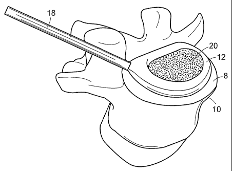
In some embodiments, the device can comprise a balloon of semicircular,
circular, bilateral (comprising more than one balloon) and generally toroidal
shape.
Preferred embodiments and positions of a device of the present invention on an
endplate 8 of a vertebra 10 are shown in FIGS. 2 (a) through (e). Now refernng
to
FIG. 2 (a), this shape allows the balloon 12 to essentially cover at least the
anterior
periphery 14 of the corresponding vertebral endplate 8, and thereby bear a
substantial portion of the spinal load. This shape further allows the surgeon
to first
place the device in place and then fill the remaining portion of the disc
space with,
for example, an osteobiologic component.
In other embodiments, as in FIG. 2 (b), the balloon 12 has a quasi-circular
shape. This device has the advantage of providing even more of a load-bearing
footprint than the embodiment of FIG. 2 (a), and also substantially prevents
unwanted leakage of the osetobiologic component during subsequent filling of
an
open cavity defined by an outer surface of the balloon.
Now referring to FIG. 2 (c), in some embodiments, the device comprises two
balloons 12 that can be used to support the vertebral load. The use of two
balloons
allows a surgeon to evenly support the load on each side of the endplate 8.
Now referring to FIG. 2 (d), in some embodiments, the balloon 12 has a
generally toroidal ("banana") shape. The banana shape allows the surgeon to
put in
CA 02515862 2005-08-12
WO 2004/073563 PCT/US2004/004284
-15-
place a single device preferably on the anterior half 14 of the disc space. In
other
embodiments, the strut has the footprint of a banana cage such as that
described in
Attorney Doclcet #DEP 5012, "Novel Banana Cage", filed December 31, 2002, US
Serial No. 10/334599, the specification of which is incorporated by reference
in its
entirety.
Now refernng to FIG. 2 (e), in some embodiments, the strut 12 is introduced
translaterally so as to form a single ramp stretching essentially transversely
across
the endplate 8. This design in advantageous when used in a posterolateral
approach
of surgery, as this approach takes advantage of the fact that the muscle
planes in the
vicinity of the approach allow the implant to be delivered in a less invasive
manner.
Now referring to FIG. 2 (f), in a preferred embodiment, the device 12 of the
present invention has a substantially semiannular footprint. The device 12 is
placed
on the anterior portion of the endplate 8 of a vertebra 10 so that height D of
a
anterior portion of the device is equal or greater than height h of a
posterior portion
of the device 12. Referring to FIG. 2 (g), the device 12 defines an internal
radius ~Z,
an external radius r~e and thickness t. In one embodiment, illustrated in FIG.
2 (g), ~Z
is approximately about 22 mm, ~e is approximately about 25 mm and t is
approximately about 3 mm.
In preferred embodiments, the height of the strut is at least 90%, and
preferably at least equal to, the height of the natural disc space. This
allows the
surgeon to distract the disc space and restore at least a portion of the disc
height. In
some embodiments, the height of the strut is greater than that of the natural
disc
space.
As used herein the word "distraction" will refer to the separation of joint
surfaces to a desired extent, without rupture of their binding ligaments and
without
displacement. Distraction can be accomplished by any suitable means, for
example
mechanical or hydrostatic means. Mechanical means can include, for instance,
attaching hooks or jacks to the bony endplates and using those hooks or jacks
to
separate the bones. Optionally, the surgeon can employ external traction. In
one
embodiment, an in-situ foaming material is used as a distraction device. Other
means include, for example, hydrostatic means, e.g., by pressurized injection
of the
biomaterial itself. By the use of distraction, the disc space can be
sufficiently re-
CA 02515862 2005-08-12
WO 2004/073563 PCT/US2004/004284
-16-
established to achieve any desired final dimensions and position. Optionally,
acid
preferably, the means used to accomplish distraction also serves the purpose
of
forming one or more barriers (e.g., balloons) for the flowable load bearing
strut
material.
The disc space can be distracted prior to and/or during either a discectomy
itself and/or delivery of a flowable biomaterial. A constricted disc space is
generally
on the order of 3 to 4 mm in height. Suitable distraction means are capable of
providing on the order of about 3 atmospheres to about 4 atmospheres, (or on
the
order of about 40 psi to about 60 psi) in order to distract that space to on
the order of
8 to 12 mm in height.
In one embodiments, the strut has a wedged shape so that the height of the
anterior portion of the expanded device is greater than the height of the
posterior
portion of the expanded device. This allows the surgeon to restore lordosis
when the
interbody fusion device is used in either the lumbar or cervical regions of
the spine.
Preferably, the wedged shape produces an angle of between 5 and 20 degrees,
more
preferably between 5 and 15 degrees.
In another embodiment, the strut has a wedged shape so that the height of the
anterior portion of the expanded device is smaller than the height of the
posterior
portion of the expanded device. This allows the surgeon to restore kyphosis
when
the interbody fusion device is used in thoracic regions of the spine.
Preferably, the
wedged shape produces an angle of between 5 and 20 degrees, more preferably
between 5 and 15 degrees.
In preferred embodiments, the height of the medial portion of the strut is
greater than the height of the lateral portion of the expanded device. This
geometry
more closely mimics the natural doming of the disc space.
With the injectable device of the present invention, there is provided a
"custom" implant formed to the anatomy of the patient's endplates. The
provision
of a conformable implant may provide a faster and more consistent fusion.
In some embodiments, the annulus fibrosus can itself serve as a suitable mold
for the delivery and solidification of either the flowable load-bearing
material (in
one embodiment) or the osteobiologic component (in another embodiment). Free
injection may optimize the extent to which the injectable device conforms to
the
CA 02515862 2005-08-12
WO 2004/073563 PCT/US2004/004284
contour of the disc space, thereby enhancing resistance to retropulsion.
Optionally,
the interior surface of the annulus fibrosus can be treated or covered with a
suitable
material in order to enhance its integrity and use as a mold.
In some embodiments, at least one of the flowable materials is delivered into
an inflatable device (such as a balloon) previously placed in the disc space.
In some embodiments, the load bearing composition is delivered into an
inflatable device (such as a balloon) previously placed in the disc space. Now
referring to FIGs. 3 (a) and (b), in one preferred method, a cannula 18,
having an
inner diameter of no more than 6 mm, is inserted into the disc space. Next,
and now
referring to FIGS. 4 (a) and (b), the inflatable device 12 is deployed through
the exit
opening of the cannula 18 and the flowable load bearing composition is
introduced
into the inflatable device at a pressure and volume suitable to expand the
inflatable
device and distract the disc space.
The fixed shape of the expanded device allows the surgeon to predetermine
the shape of the flowable material and simply fill the device with the
flowable
material. The device substantially prevents unwanted flow of the material. The
prevention of unwanted flow desirably prevents the material from damaging
important surrounding structures such as the spinal cord, aorta and vena cava.
Also,
the inflatable device can be tailored to fill any portion of the disc space.
Further, the present inventors believe that inclusion of an inflatable balloon
in a strut can assure that the opposing trends of degradation of bioabsorbable
materials and new bone growth will result in fusion of the vertebrae in a
position
approximating the natural angle between two adjacent vertebrae. If the balloon
is
made of a resorbable, water-impermeable material, the balloon will effectively
shield the load-bearing composition from water during the initial stages of
fusion
and so delay the onset of hydrolysis and degradation of the Load-bearing
material.
Preferably, the balloon begins to degrade within about 1-2 months after fusion
of the
osteobiologic composition, thereby allowing the Load-bearing material it
contains to
slowly degrade and grow bone.
In some preferred embodiments, the distraction of the disc space is
accomplished by an inflatable, yet rigid, balloon or bladder. The balloon can
be
delivered in deflated form to the interior of the annulus and there inflated
in order to
CA 02515862 2005-08-12
WO 2004/073563 PCT/US2004/004284
-18-
distract the disc space and provide a region for the delivery of biomaterial.
The
balloon is preferably of sufficient strength and of suitable dimensions to
distract the
space to a desired extent and to maintain the space in distracted position for
a period
of time sufficient for the biomaterial to be delivered and, optionally, to
harden.
One of the primary functions of the balloon is to influence or control the
shape of the hardenable material, following injection therein. The implantable
balloon is not normally required to restrain pressure over an extended period
of time.
Thus, a greater design flexibility may be permitted, as compared to
conventional
angioplasty or other dilatation balloons. For example, the balloon may be
porous,
either for drug delivery as has been discussed, or to permit
osteoincorporation and/or
bony ingrowth.
In one particularly preferred embodiment, there is provided a method for
fusing an intervertebral disc space, comprising the steps of:
a) using microsurgical techniques to perform a discectomy while
preserving an outer annular shell;
b) inserting a deflated balloon into the disc space;
c) injecting a flowable load bearing composition into the deflated
balloon (preferably, in an amount sufficient to distract the disc
space), and
d) solidifying the flowable strut material.
In one particularly preferred embodiment, there is provided a method for
fusing an intervertebral disc space, comprising the steps of:
a) using microsurgical techniques to perform a discectomy while
preserving an outer annular shell,
b) inserting a deflated balloon having peripheral struts into the disc
space,
c) injecting an osteobiologic component into the deflated balloon in an
amount sufficient to inflate the balloon and distract the disc space
with the strut component of the balloon.
CA 02515862 2005-08-12
WO 2004/073563 PCT/US2004/004284
-19-
Optionally, and preferably, the space is distracted by the use of one or more
suitable insertable or inflatable devices, e.g., in the form of inflatable
balloons.
When inflated, such balloons provide rigid walls (e.g., fiber supported) that
are
sufficiently strong to distract the space. An inflatable device providing
sufficient
strength and dimensions can be prepared using conventional materials. In one
embodiment, the uninflated balloon can be delivered to the center of the
annular
shell, and there inflated to expand the annular shell and in turn, distract
the space. In
another embodiment, the uninflated balloon can be delivered to the anterior
rim of
the annular shell, and there inflated to provide a cavity for the injection of
the load
bearing flowable material. Preferably, the load bearing composition is
injected in an
amount. sufficient to distract the space.
The inflatable device can be delivered to the disc space by any suitable
means,
e.g., in deflated form retained within or upon the end of a rigid or semi-
rigid rod.
Once positioned within the disc, either centrally within the annular shell or
along the
annular rim, a suitable gas (e.g., nitrogen or carbon dioxide) or the flowable
load-
bearing material can be delivered through the rod in order to inflate the
balloon in
situ, in a substantially radial or longitudinal direction. In some
embodiments, beads
of the load bearing strut material are simply packed into the balloon. The
fact that
the balloon is properly placed can be confirnled by the use of ancillary
means, such
as using a C-arm, or by self effecting means embodied within the balloon
itself or its
delivery apparatus.
In terms of its component parts, in one preferred balloon delivery system of
the
present invention there is provided an inflatable device, a motor drive unit,
with a
remote controller, associated tube sets, a nonscope inflow delivery cannula
having
independent fluid dynamics pressure and flow rate adjustments, attachments for
the
flush, vacuum, waste canister, and overflow jars.
Suitable materials for preparing balloons of the present invention may include
those that are presently used for such purposes as balloon angioplasty.
Suitable
materials provide an optimal combination of such properties as compliance,
biostability and biocompatability, and mechanical characteristics such as
elasticity
and strength. Balloons can be provided in any suitable form, including those
having
a plurality of layers and those having a plurality of compartments when
expanded.
CA 02515862 2005-08-12
WO 2004/073563 PCT/US2004/004284
-20-
A useful balloon apparatus will include the balloon itself, together with a
delivery
catheter (optionally having a plurality of lumen extending longitudinally
therewith),
and fluid or gas pressure means.
Examples of suitable materials (e.g., resins) for making balloons include, but
are not limited to, polyolefin copolymers, polyethylene, polycarbonate,
polyethylene
terephthalate and ether-ketone polymers such as poly(etheretherketone). Such
polymeric materials can be used in either unsupported form, or in supported
form,
e.g., by the integration of DacronTM or other fibers. Preferably, the
materials of
construction of the balloon are resistant to softening or melting at a
temperature of at
least 80 °C, preferably at Ieast I00 °C, more preferably at
least 250 °C. In addition,
the balloon (or balloon-like structure) may be made out of any of a wide
variety of
woven or nonwoven fibers, fabrics, metal mesh such as woven or braided wires,
and
carbon. Biocompatible fabrics or sheet material such as ePTFE and DacronTM may
also be used.
Balloons can also take several forms, depending on the manner in which the
biornaterial is to be delivered and cured. A single, thin walled balloon can
be used,
for instance, to contact and form a barrier along the interior surface of the
remaining
annular material. Once positioned, the flowable load bearing component can be
delivered and solidifted within the balloon to serve as a load bearing strut
of the
present invention. In such an embodiment, the balloon is preferably of a type
that
will allow it to remain in position, without undue detrimental effect, between
the
annular material and the solidified load-bearing component.
Optionally, a balloon can be provided that fills essentially only the central
portion of the disc space. In such an embodiment, the balloon can be, for
instance,
in the shape of a cylinder. Such a balloon can be provided such that its upper
and
lower walls can be positioned to contact the opposing vertebral bodies, and
its side
walls will provide sufficient strength to cause distraction of the space upon
inflation.
Thereafter, the load-bearing component is delivered to the perimeter of the
annulax
space, i.e., the space between the annular material and the balloon, and there
solidified. Optionally, the balloon can be gradually deflated as additional
biomaterial is inserted into the space. Then, once the load bearing material
is stably
CA 02515862 2005-08-12
WO 2004/073563 PCT/US2004/004284
-21-
positioned, the osteobiologic component is introduced into the balloon,
thereby
filling the balloon.
In some embodiments, the balloon has metallic wires or other imageable
means incorporated into it. Any material that can be seen under fluoroscopy
would
be acceptable. Potential materials include any metal, metal alloys, or
ceramics that
could be combined with a polymer. The material can be in the form of wires, a
mesh, or particles incorporated into the balloon or on its surface.
In some embodiments, the balloon has an inner surface that is chemically
active so as to bond with the balloon filler as it polymerizes. As used
herein, a
chemical "bond" is said to exist between two atoms or groups of atoms when the
forces acting between them are strong enough to lead to the formation of an
aggregate with sufficient stability to be regarded as an independent species.
As used
herein, "chemically active" means capable of forming a chemical bond. In one
example, the surface is chemically modified by means such as plasma
polymerization. In this case, the balloon is placed in a vacuum chamber and
plasma
containing a small molecule (an amine for example) is created. The balloon
surface
is bombarded by the small molecule and the small molecule is chemically
attached
to its surface. The balloon's surface with its amine groups can then react
with the
polymer that is injected into the balloon (i.e., an epoxy), forming a device
that would
have greater fatigue properties since the "composite" of balloon and balloon
filler
are chemically bonded to one another.
The desired quantities of the load-bearing and osteobiologic components of
the present invention are delivered by minimally invasive means to the
prepared site.
Prior to delivery, these components can be stored in suitable storage
containers, e.g.,
sterile, teflon-lined metal canisters. The flowable components can be
delivered, as
with a pump, from a storage canister to the delivery cannula on demand. The
components can be delivered in the form of a single composition, or can be
delivered in the form of a plurality of components or ingredients.
In some embodiments, the inflatable device can be filled with a viscous
material that later solidifies to form the strut or osteobiologic component.
The
viscous material can be a heated polymer (such as a composition containing
polycaprolactone), or polymer precursor components (such as the
CA 02515862 2005-08-12
WO 2004/073563 PCT/US2004/004284
-22-
photopolymerizable anlrydrides disclosed by A.K. Burkoth, Biomaterials (2000)
21:2395-2404, the entire teachings of which are incorporated herein by
reference).
In some embodiments, a flowable load bearing composition, such as
polycaprolactone, heated to a temperature yielding a viscosity in the range of
from
about 100 to about 500 cps is injected into the balloon under pressure such as
by
using a pump and pressure within the range of from about 4 ATM to about 10 ATM
or more depending upon viscosity, balloon strength and other design
considerations.
The pump is run for a sufficient duration and under a sufficient pressure to
ensure
that the polycaprolactone wets all of the p-dioxanone fibers. This may range
from
about 10 minutes or more to about an hour, and, in one application where the
pump
was run at about 5 ATM pressure, requires at least about 1 hour. Specific
method
parameters may be optimized depending upon the viscosity of the
polycaprolactone,
infusion pressure, infusion flow rate, density of the packed fibers, and other
variables as will be apparent to those of skill in the art in view of the
disclosure
herein.
It has been reported in the literature that balloons inserted into the disc
space
may be subject to retropulsion. Therefore, in some embodiments of the present
invention, upon expansion, the inflatable device forms an upper surface having
a
first plurality of teeth projecting outwards from the upper surface. Upon
expansion
of the device, these teeth will project in the direction of the upper endplate
and, upon
complete expansion of the device, will engage the endplate to from a secure
interlock with the endplate and resist retropulsion.
Preferably, the teeth are made of a stiff resorbable material, such as
polyetheretherlcetone (PEEK). Preferably, the teeth have a height of between
0.5
and 1.5 mm, and have a triangular cross-section.
In some embodiments of the present invention, upon expansion, the
inflatable device forms an upper surface formed of a material having a high
coefficient of friction. Upon expansion of the device, the high coefficient of
friction
of the upper and lower surfaces will case a drag upon any movement of the
upper
surface and therefore keep the device in place and resist retropulsion.
Preferably, the upper and lower surfaces of the inflatable device are made
from a material selected from a group consisting of polyether block copolymer
CA 02515862 2005-08-12
WO 2004/073563 PCT/US2004/004284
-23-
(PEBAX), ABS (acrylonitrile butadiene styrene); ANS (acrylonitrile styrene);
Delrin~; PVC (polyvinyl chloride); PEN (polyethylene napthalate); PBT
(polybutylene terephthalate); polycarbonate; PEI (polyetherimide); PES
(polyether
sulfone); PET (polyethylene terephthalate); PETG (polyethylene terephthalate
glycol), high and medium melt temperature: polyamides, aromatic polyamides,
polyethers, polyesters, Hytrell~, polymethylinethacrylate, polyurethanes:
copolymers, EVA (ethylene vinyl acetate) or ethylene vinyl alcohol; low,
linear low,
medium and high density polyethylenes, latex rubbers, FEP, TFE, PFA,
polypropylenes, polyolefins; polysiloxanes, liquid crystal polymers, roomers,
Surlins, silicone rubbers, SAN (styrene acrylonitrile), nylons: 6, 6/6, 6/66,
6/9, 6/10,
6/12, 11, all PEBAXs 12; polyether block amides; thermoplastic elastomers and
the
like.
In some embodiments, the vertebral endplates opposing the disc space are
roughened. The roughening provides hills and valleys into which a flowable
polymer can flow and harden, thereby forming a mechanical interlock between
the
device and the bony surface and resisting retropulsion.
The roughening can be provided mechanically (as with a curette), or
chemically (as by an acid), or by an energy-transmitting device (as with an
ablation
unit preferably assisted with hyperconductive fluid, such as hypertonic
saline).
In some embodiments, the flowable polymer forming a mechanical interlock
can be a separate layer. In others, the flowable polymer can be a component of
the
strut. In others, the flowable polymer can be a component of the osteobiologic
composition.
In some embodiments, the strut portion of the device can have an outer layer
of a scaffold material appropriately seeded with osteogenic factors andlor
growth
factors to produce quick bone ingrowth, thereby effectively locking the strut
in
place.
In some embodiments, an outer layer of a scaffold material appropriately
seeded with osteogenic factors and/or growth factors can also be applied to a
balloon
component of the osteobiologic component. The seeding again produces quiclc
bone
ingrowth, thereby effectively loclcing the osteobiologic component in place.
CA 02515862 2005-08-12
WO 2004/073563 PCT/US2004/004284
-24-
Balloons of the present invention can be made using materials and
manufacturing techniques used for balloon angioplasty devices. U.S. Patent No.
5,807,327 by Green, the entire teachings of which are incorporated herein by
reference, (hereinafter "Green") discloses balloons that may be used in the
present
invention. The materials disclosed by Green for the formation of the balloon
include
tough non-compliant layer materials (col. 8, lines 18-36) and high coefficient
of
friction layer materials (col. 8, lines 42-54).
Now referring to FIGS. 5 (a) and (b), in some embodiments, the load-bearing
component is delivered into the disc space through an inflatable balloon 12,
and the
osteobiologic component 20 is freely injected. This embodiment is desirable
because the balloon 12 can act as a barner to hydrolysis of the load-bearing
component, thereby increasing the longevity of the load-bearing component. In
contrast, the absence of the balloon covering the osteobiologic component may
be
desirable in instances in which it is desirable to immediately begin the bone
growth
process.
This embodiment may also be desirable in instances in which the load-
bearing component comprises a cross-linkable composition, and the surgeon
desires
to provide a barrier between the patient's tissue and the precursors during
the
reaction of the precursors.
Now referring to FIGs. 6 (a) and (b), in some embodiments, both the load
bearing and the osteobiologic components are delivered into the disc space
using a
device comprising two separate inflatable balloons 12. This embodiment is
desirable
in instances in which both the annulus fibrosis has been functionally
breached, and
there is a concern that flowable materials would flow from the disc space and
through the breach and into the remainder of the body. In this embodiment, it
is
preferred that the balloon containing the osteobiologic material be at least
semi-
permeable to nutrients and preferably resorbable. As used herein, the term
"semipermeable" refers to a material that is non-permeable to the flowable
materials
described above yet permeable to important water and nutrients to support bone
growth therein. Suitable semi-permeable materials include both porous and non-
porous polymeric constructs such as films, fabrics (woven and non-woven) and
foams.
CA 02515862 2005-08-12
WO 2004/073563 PCT/US2004/004284
-2S-
In some embodiments, both the load bearing and the osteobiologic
components are delivered into the disc space through the same inflatable
device.
Now refernng to FIGS. 7 (a) and (b), another embodiment of the device and
method of the present inevtion is shown wherein the device comprises at least
two
inflatable balloons 12. In this embodiment, the load-bearing component is
delivered
into the disc space through at least two inflatable balloons 12 and the
osteobiologic
component 20 is freely injected into the disk space using the space between
the
ballons.
In some embodiments, the osteobiologic component is delivered into the disc
space through an inflatable device, and the load-bearing component is freely
injected. This embodiment may be desirable in instances in which the
osteobiologic
component comprises an ih situ hardenable composition such as a calcium
containing cement, or a crosslinkable polymer such as polypropylene fumarate),
polyanhydride, or polyoxaester, and the surgeon desires to cordon off the
patient
from the precursors during their reaction. In this embodiment, it is further
preferred
that the balloon containing the osteobiologic material be at least semi-
permeable to
nutrients and preferably resorbable. This embodiment may also be desirable in
instances in which the load-bearing composition comprises growth factors and
the
surgeon desires to immediately begin the bone growth process in the load-
bearing
component.
In some embodiments, the load-bearing component is delivered into the disc
space through an inflatable device, and the osteobiologic component is freely
injected. This embodiment may also be desirable in instances in which the
annulus
fibrosis in essentially intact and the surgeon desires to immediately begin
the bone
growth process in the load-bearing component.
In some embodiments, the inflatable device comprises a single peripheral
wall having an upper and lower surface, upper and lower walls, and a cavity
formed
therebetween. For the purposes of the present invention, this shape of this
embodiment is referred to as a "puclc". The peripheral wall and upper and
lower
walls of the puclc could be designed so as to be percutaneously deliverable
through a
cannula having an inside diameter of between 0.5 and 18 mm, preferably no more
than 4 mm.
CA 02515862 2005-08-12
WO 2004/073563 PCT/US2004/004284
-26-
In one embodiment, the peripheral wall of the puck is designed to be load
bearing when the inflatable device is disposed in its inflated position.
Preferably,
the peripheral wall is made of a shape-memory metal, such as Nitinol, or a
thin film
alloy.
In some embodiments, the periphery of the balloon is reinforced with fibers.
In some embodiments thereof, the peripheral wall comprises polymer fibers.
These
fibers can be made into a weave that is sufficiently flexible (in the
longitudinal
direction of the fiber) to pass through the cannula and expand into the
expanded
state. Typically, these fibers have high tensile strengths so that they can
very
efficiently accommodate the problematic hoop stresses that may be transferred
from
the osteobiologic component contained within the middle annulus of the
balloon.
Various patterns of reinforcement of the periopheral side-walls with the
fibers are contemplated. In one embodiment, the fibers form X-shaped cross-
hatching pattern. In smother embodiment, the fibers form a continuous wave-
like
pattern having peaks and troughs, where said pealcs and troughs approach upper
and
lower surfaces.
In one embodiment, the walls of the device are reinforced by aii internal
frame forming a polygonal structure having sides on the upper, lower and
peripheral
surfaces.
In some embodiments, the peripheral reinforcement is made of a resorbable
polymer fiber.
The upper and lower walls of this puclc embodiment are designed to initially
accept and contain the osteobiologic component that is flowed into the puck
cavity.
Accordingly, the upper and lower walls should be at least semi-permeable so as
to
contain the osteobiologic component. In preferred embodiments, the upper and
lower walls are made of a resorbable material that quickly resorbs, thereby
exposing
the contained osteobiologic material to blood flowing from the decorticated
endplates.
In some embodiments, this absorbable material has an elastomeric quality.
This elastomeric quality allows the resorbable upper and lower walls to be
delivered
through the cannula, and flatten upon device expansion. In preferred
embodiments,
this elastomeric polymer is selected from the materials disclosed in U.S.
Patent No.
CA 02515862 2005-08-12
WO 2004/073563 PCT/US2004/004284
-27-
6,113,624 by Bezwada, the entire teachings of which are incorporated herein by
reference (hereinafter "Bezwada"). In other embodiments, this absorbable
material
is not elastomeric, and is preferably made of a thin film metal alloy or a
braided
metal alloy.
Now refernng to FIGS. 8 (a) and 8 (b) there is provided a device 30 of the
present invention comprising an inflatable portion 32 that includes an arcuate
inflatable balloon.
Now referring to FIG. 8 (a), in its pre-deployed state, the inflatable portion
32 of the device 30 is conveniently repeatedly folded upon itself, thereby
decreasing
the size of the device 30 and allowing for minimally invasive insertion into
the disc
space. During insertion into the disc space, the device 30 is preferably
inserted in
the sandwich orientation as shown in FIG. 8 (a) wherein the structural walls
34 are
disposed essentially parallel to the vertebral endplates. The sandwich
orientation
allows height H of the structural walls 34 to meet or exceed the disc space
height,
while the folded width W does not exceed the disc space height.
Now referring to FIG. 8 (b), after insertion into the disc space, fluid is
flown
into the inflatable portion 32 of the device 30, thereby expanding the device
30 into
the configuration as shown. The height H of the structural walls 34 is
sufficient to
restore the natural height of the disc space. After the device 30 distracts
the disc
space, the cavity, formed by the expanded portion 32, is filled by an
osteobiologic
component. The structural walls 34 of this embodiment are preferably attached
to
the inflatable portion 32 by an adhesive. The structural walls 34 should be
designed
so that the width W and the strength and modulus of the material of
construction
allow for both support of the disc space and bony fusion through the
osteobiologic
component.
In some embodiments, the height H of the structural wall 34 is at least equal
to the height of the natural disc space. This condition desirably restores the
height
of the disc space when the inflatable portion 32 is expanded. In some
embodiments,
the height H of the anterior portion of the wall 34 is greater than the height
h of the
posterior portion of the wall 34. This condition desirably provides a lordotic
effect
upon expansion of the inflatable portion 32.
CA 02515862 2005-08-12
WO 2004/073563 PCT/US2004/004284
_28_
In some embodiments, the walls 34 are made of allograft bone, and
preferably comprise cortical bone. hl others, the walls are made of a
synthetic
resorbable polymeric material. In some embodiments, the walls may be
sufficiently
porous to provide an effective scaffold, thereby allowing bony fusion
therethrough.
In some embodiments, the wall component 34 of this embodiment is made of
bone graft. hl alternative embodiments, component 34 comprises additional
inflatable portions. After insertion into the disc space, a load bearing
composition
may be flowed into the cavities of these additional inflatable portions,
thereby
expanding these additional inflatable portions and eventually producing the
desired
dimensions of the walls 34.
In some embodiments, each wall 34 is translaterally oriented in the expanded
device. In this condition, a first wall supports essentially the anterior
portion of the
opposing cortical rims, while the second wall supports essentially the
posterior
portion of the opposing cortical rims, so that one of these walls will
essentially bear
the entire load during flexion and the other wall will bear essentially the
entire load
during extension. Preferably, these walls have a length L corresponding to the
anterior and posterior aspects of the cortical rim.
The inflatable portion 32 has upper and lower surfaces 36 and 38 for
contacting the adjacent vertebral endplates, a peripheral side surface 40
connecting
the upper and lower surfaces 36 and 38, and an opening 42 in the peripheral
side
surface 40. Upon a flow of fluid through the opening 42 from a cannula 18, the
inflatable portion 32 is expanded and surfaces 36, 38 and 40 are pushed apart
sufficiently to form an internal cavity suitable for containing an
osteobiologic
component. Because the osteobiologic component retained within this cavity is
preferably at least semipermeable in order to provide bony fusion, the upper
and
lower surfaces 36 and 38 of the inflatable portion 32 preferably do not act as
barners to bony fusion. Accordingly, it is preferred that the upper and lower
surfaces 36 and 38 are either porous (preferably, semipermeable) or quickly
resorbable. Preferably, the upper and lower surfaces 36 and 38 are made of a
material that resorbs within 7 days, preferably 3 days, preferably one day.
Examples
of fast-resorbing materials include denatured collagen, polysaccharide-based
materials such as starch and oxidized regenerated cellulose, and hydroxylated
CA 02515862 2005-08-12
WO 2004/073563 PCT/US2004/004284
-29-
lactide-glycolide copolymers. In some embodiments, the opening in the side
surface
40 is formed closely adjacent to the structural wall 34, positioned anteriorly
on a
vertebral endplate.
W some embodiments, the inflatable device 30 of this embodiment has a
configuration designed to match the geometry of the disc space, and is
selected from
1
the group consisting of an anterior lumbar interbody fusion (ALIF)
configuration, a
posterior lumbar interbody fusion (PLIF) configuration, a vertebral body
replacement (VBR) configuration, and an anterior cervical discectomy and
fusion
(ACDF) configuration.
By reducing the effective size of the device 30, this embodiment of the
present invention desirably minimizes the access window required for insertion
of
intervertebral devices. By providing anatomically appropriate structural walls
34, the
device 30 provides a stable environment for the muskuloskeletal growth factors
to
develop.
Now referring to FIGS. 9 (a) and (b), one embodiment of an inflatable
device of the present invention is shown. The device 60 comprises an outer
side
wall component 62, an inner side-wall component 64, and a balloon 66 disposed
between and attached to said inner and outer wall components. The short
cranial
caudal height of the inner and outer walls allows for the device to be
inserted into
the disc space without having to distract the disc space prior to insertion.
Subsequent filling of the balloon with an in-situ hardenable, load-bearing
material
causes the balloon to expand beyond the cranial and caudal margins of the
sidewalls,
thus providing the necessary distraction of the disc space. Furthermore, the
sidewalls prevent expansion of the balloon such that the thickness of the
device is
minimized upon inflation. Minimized wall thiclrness is important for ensuring
maximum area for bone growth (fusion) between the adj acent vertebrae. In some
embodiments, the footprints of the outer and inner side-wall components 62 and
64
represent substantially equal arcs of two concentric circumferences. This
allows
placing device 60 along the periphery of the anterior portion 14 of a
vertebral
endplate 8 and filling a cavity therewithin with a load bearing material.
In some embodiments of device 60, the outer and inner walls 62 and 64 are
made of a flexible plastic such as poly(ethyleneterephthalate), a superelastic
metal
CA 02515862 2005-08-12
WO 2004/073563 PCT/US2004/004284
-30-
such as Nitinol, or a flexible material/geometry combination, whereby each
wall can
be deformed into a relatively elongated shape for delivery to the disc space
through
a cannula 18. The sidewalls are sufficiently rigid to guide the device into
the desired
location in the disc space but sufficiently flexible to allow delivery through
the
cannula. Referring to FIG. 9 (c), during the insertion of the device 60, upon
release
from the cannula 18, components 62 and 64 can then take on the desired arcuate
shape. Referring to FIG. 9 (d), subsequent to insertion, the device 60 is
expanded by
injecting a load-bearing component, an osteobiologic component or a
combination
thereof into a cavity formed by the components 62, 64, and 66. Any suitable
injection means cast be used, for example, a syringe pump 70.
The above characteristics of components 62 and 64 ensure that the cavity
produced between side walls 62, 64 can be filled so that the device 60
distracts the
disc space and can also create a wedge shape for creating or restoring healthy
curvature of the spine.
An alternative embodiment of inflatable device of the present invention is
shown in FIGs. 10 (a) and (b). Device 260 comprises an upper wall component
266
and a lower wall component 268 joined by ail inflatable balloon 270. In some
embodiments, the footprints of the upper and lower wall components 266 and 268
represent substantially equal arcs of two concentric circumferences. Tlus
allows
placing device 260 along the periphery of the anterior portion 14 of a
vertebral
endplate 8 and filling a cavity therewithin with a load bearing material.
In some embodiments of device 260, the upper and lower wall components
266 and 268 are made of a superelastic material such as Nitinol, or a flexible
material/geometry combination, whereby each wall can be deformed into a
relatively
elongated shape for delivery to the disc space through a cannula 18.
Operationally,
device 260 is similar to device 60. Insertion of device 260 can be
accomplished in a
manner depicted in FIG. 9 (c). Upon release from cannula 18, components 266
and
268 can then take on the desired arcuate shape. Subsequent to insertion,
device 260
is inflated by injecting a load-bearing component, an osteobiologic component
or a
combination thereof into balloon 270. Any suitable injection means can be
used, for
example a syringe pump.
CA 02515862 2005-08-12
WO 2004/073563 PCT/US2004/004284
-31-
Preferably, balloon 270 is semi-permeable. In preferred embodiments,
balloon 270 is made of a material that quiclcly resorbs, thereby exposing the
contained osteobiologic material to blood flowing from the decorticated
endplates.
Generally, the strut is deliverable through a cannula having an inside
diameter of between 3 mm and 18 mm, preferably between 4 mm and 12 mm, more
preferably between 5 mm and 10 mm.
In some embodiments in which the surgeon desires to miiumize the size of
the incision, the strut is preferably deliverable through a cannula having an
inside
diameter ofbetween 0.5 mm and 6 mm, preferably between 1 mm and 4 mm, more
preferably between 2 mm and 3 xnm.
Preferably, the upper and lower surfaces of the upper and lower walls,
respectively, have teeth that prevent excessive movement of the strut after
implantation.
Now referring to FIGS. 11 (a) and (b), the device 80 comprises four rail
components 82 wherein the footprints of the rail components 82 represent
substantially equal arcs of two concentric circumferences. Components 82 are
joined by an inflatable balloon 84 such that the device can be inserted in a
collapsed
configuration as shown in FIG. 11 (b) and then expanded as shown in FIG. 11
(a)
once filled with a load-bearing material to increase disc height and provide
thickness
for load bearing support.
In one embodiment of the present invention, device 80 is shown in FIGS. 12
(a) and (b). In this embodiment, device 80 is delivered in a generally diamond-
shaped configuration, shown in plan view on FIG. 12 (a) and in lateral view in
FIG.
12 (b). W this embodiment, the upper and lower rails 82 will cause slight
subsidence
of the vertebral body endplates, thus providing stability of the implanted
device.
Now refernng to FIGS. 13 (a) through (d) a preferred embodiment of the
method of the present invention is shown. As shown in FIG. 13 (a) and 13 (b),
a
cannula 18 is inserted into an intervertebral space. Next an inflatable
balloon 12 of
a generally toroidal shape is inserted through the cannula 18 into the
intervertebral
space. The balloon 12 is expanded by directing a load-bearing component into
said
balloon. Referring to FIG. 13 (c), subsequent to balloon expansion,
osteobiologic
component 20 is injected into the open cavity defined by the outer surface of
the
CA 02515862 2005-08-12
WO 2004/073563 PCT/US2004/004284
-32-
balloon 12. Preferably, the osteobiologic component comprises a water-soluble
component. Next, the water-soluble component is dissolved, thus forming a
porous
matrix shown in FIG. 13 (d).
In one embodiment of the present invention the load-bearing component is
delivered through a balloon, and the osteogenic component is provided in a
hydrogel
phase of the osteobiologic component. Examples of suitable hydrogels are
provided
hereinbelow.
In other embodiments, solid components of the strut are inserted into the
body percutaneously and assembled isa situ to form the strut. In some iya situ
embodiments, the strut is formed by bonding together two bondable components.
Preferably, the bondable materials are selected from the group consisting of
heat
bondable materials such as polycaprolactone, and polymerizable materials such
as
polypropylene furnarate) and polyoxaesters including photo-curable materials
such
as polyanhydrides.
In other embodiments, load-bearing materials in the form of beads is
delivered into the inflatable device and packed into the device so as to
create a
stable strut having an open interstitial porosity. In some embodiments, the
beads
may be packed without subsequent stabilization other than closing off the
opening of
the balloon. In these embodiments, the beads are preferably polyarylether
ketone
(PAEK), more preferably polyetherether ketone (PEEK) with chopped carbon
fiber.
In some embodiments, a bonding material may be subsequently flowed into
the interstitial porosity to further stabilize the packed beads. Preferably,
this bonding
material comprises an aliphatic polyester such as polycaprolactone (PCL). The
bonding material may be resorbable and may include osteogenic additives such
as
growth factors and stem cells.
In some bead embodiments of the device, the beads of the load-bearing
material are made of a heat-bondable material, such as polycaprolactone. When
the
beads axe so constituted, heat may be delivered into the packing and soften
the
contacting surfaces of the beads. Upon subsequent cool down to body
temperature,
the contacting surfaces solidify to further stabilize the paclced structure.
In some
embodiments, the heat is provided exogenously. In other embodiments, the heat
is
provided by the patient's body heat (~37 °C).
CA 02515862 2005-08-12
WO 2004/073563 PCT/US2004/004284
-33-
Now refernng to FIGS. 14 (a) - (d), another embodiment of the device of the
present invention 300 comprises at least two bondable components 310 and 320,
that are delivered into the disc space in unassembled foam, placed closely
adjacent
one another, and then bonded together, preferably by heat bonding.
Referring to FIG. 14 (b), representing a lateral view of the device of FIG. 14
(a) as seen in the direction of arrow A, and to FIG. 14 (d), representing a
perspective
view of device 300, in one embodiment, device 300 comprises first and second
portions 310 and 320. First portion 310 has a lower bearing wall 312, upper
angled
wall 314, and a leading wall 316 and a trailing wall 318. Second portion 320
has an
upper bearing wall 322, lower angled wall 324 and a leading wall 326 and a
trailing
wall 328. The combined height of the assembled portion H exceeds that of the
disc
space. The angled walls form the same angle so that the leading edge of the
second
portion can be ramped up the angled wall of the first portion.
In use, the first portion 310 is placed in the disc space. Because the height
of
the first portion is less than the disc space, the first portion 310 is easily
positionable
anywhere within the disc space. Next, the second portion 320 is introduced
into the
disc space and ramped up the angled wall of the first portion. Corresponding
rails
and groove are provided on the angled walls of the first and second portion so
as to
guide the second portion along the among wall of the first portion (see
below).
Because the second portion only contacts the lower portions of the first
portion, the
upper wall 322 of the second portion 320 does not touch the adjacent endplate
during ramping and so the ramping is easy. Only when the ramping is
essentially
complete does the upper wall of the second portion contact the adjacent upper
endplate. Preferably, the overall height of the ramp H is slightly greater
than that of
the disc space, so that distraction is achieved when the leading edge of the
second
portion reaches the leading edge of the first portion.
Referring to FIG. 14 (c), in one embodiment, a cross-section of the device of
FIG. 14 (a) is shown, tal~en along arrows B. As shown in FIG. 14 (c), angled
wall
314 of first portion 310 includes a grove 330, while angled wall 324 of second
portion 320 includes a ridge 332, designed to fit into a slide against grove
330. In
some embodiments, ridge 332 further includes a metal filament 334.
CA 02515862 2005-08-12
WO 2004/073563 PCT/US2004/004284
-34-
It is understood that the locations of grove 330 and ridge 332 can be
interchanged to between angled walls 314 and 324 of upper and lower portions
310
and 320.
Preferably, the xail and groove feature of the ramps has a Morse taper so as
to lock the ramp in its assembled form when the leading edge of the second
portion
xeaches the leading edge of the first portion.
Once the two ramp portions are in place, an electric current or heat is passed
through wire that passes through the length of the rail of the first portion.
The
resulting localized heating of the contacting areas softens this region
without
changing the dimensions of the ramp. Upon cooling, a highly stable, heat
bonded
ramp results.
Because the ramp of this embodiment is not flowed into the disc space, and
the heating is very localized, extremely strong, high temperature materials
such as
PEEK may be used as the material of construction. In some embodiments, the
ramp
is made of a high temperature resorbable material. In some embodiments, the
high
temperature absorbable material is amorphous and has a glass transition
temperature
of above 100 °C. Preferably, the amorphous absorbable is PLA. In some
embodiments, the high temperature absorbable material is crystalline and has a
melting point of above 100 °C. Preferably, the crystalline absorbable
is p-
dioxanone.
In some ramp embodiments, a guidewire is guided through the center of the
ramp guide. The guidewire would allow the ramps to be inserted over the
guidewire. The guidewire could be remotely steered into place via IGS or
equivalent, and then the ramps could be passed over the guidewire into place.
The
ramps could be semi-rigid which would allow them to follow the guidewire
through
the soft tissue, over the wire.
In other ramp embodiments, an "I"-Beam ramp cage is provided. The ramp
cage discussed above could incorporate or mate with modular tops and bottoms.
These tops and bottoms would have tracks, which would locate on guides fixed
to
the ramps (or the guides could be on the modular top, and tracks on the ramps)
which would aid insertion and ensure the ramps were connected to these modular
tops and bottoms. The surfaces of the modular tops and bottoms would go
between
CA 02515862 2005-08-12
WO 2004/073563 PCT/US2004/004284
-35-
the ramps and the vertebral bodies such that when assembled, a cross-section
of the
rampltop/bottom assembly would resemble an "I"-Beam. This would allow for
thinner ramps to ease insertion via an MIS technique or equivalent, and the
modular
tops and bottoms would provide sufficient surface area to prevent subsidence
of the
implant into the vertebrate bodies. The ramps and modular tops could be shaped
ili
severate configurations, inserted assembled, or assembled within the disk
space.
In other embodiments, there is provided a ribbon-shaped ramp having a
longitudinal through-hole. A threaded rod is inserted through the middle of
the
ribbon so that, as the threaded rod is turned, the ribbon would "accordion"
itsehf,
increasing its height within the disk space. This "accordion"-ing could be
achieved
by other methods, such as a spring, a cable, etc.
Now refernng to FIG. 15, one embodiment of a method of use of device 300
shown in FIGS. 14 (a) - (d) is depicted. In this embodiment, the first and
second
ramp portions 310 and 320 are introduced translaterally so as to form a single
ramp
stretching essentially transversely across the disc space. This design in
advantageous
when used in a posterolateral approach, as this approach tateces advantage of
the fact
that the muscle planes in the vicinity of the approach allow the implant to be
delivered in a less invasive mamler. In some embodiments thereof, the medial
portion of the ramp has a height that is higher than the lateral portions.
This feature
provides the doming that is advantageous in interbody fusions
Now referring to FIG. 16, another embodiment of a method of use of device
300, shown in FIGS. 14 (a) - (c) is depicted. In this embodiment, the ramp of
the
present invention may be advantageously used in a PLIF procedure. In
particular,
two ramps may be constructed in-situ so as to form bilateral struts similar to
the
Steffee struts.
Therefore, in accordance with the present invention, there is provided an
intervertebrate fusion device comprising a strut comprising:
a) a first component comprising:
i) a howerbearing surface adapted for bearing against a lower
vertebral endplate, and,
ii) an upper surface comprising a leading end, an angled middle
portion and a trailing end; and
CA 02515862 2005-08-12
WO 2004/073563 PCT/US2004/004284
-3 6-
b) a second component comprising:
i) an upper bearing surface adapted for bearing against an upper
vertebral endplate, and,
ii) an upper surface comprising a leading end, an angled middle portion
and a trailing end,
wherein the angled portion of the first component mates with the angled
portion of the second component.
In some embodiments of the present invention, the struts are completely
dense. This feature maximizes the strength of the strut, and so is desirable
for the
° 10 load bearing. In other embodiments, the strut has openings sized
to permit bony
fusion therethrough. In some embodiments, the upper and lower walls have
openings
designed to promote bony fusion from the upper endplate to the lower endplate.
In
other embodiments, the sidewalk of the strut also have such openings. In some
embodiments, the openings have a diameter of at least 2 mm. In other
embodiments,
the openings are in the range of from 50-500 um, more preferably between 100
and
300 um, preferably between 100 and 250 um. These preferred opening sizes are
believed to be more conductive to bone growth.
MATERIALS AND COMPOSITIONS SUITABLE FOR USE 1N THE
INVENTION
Provided below is a listing of various attributes of the load bearing
composition and osteobiologic component of the present invention:
Feature Load BearingTypical OsteobiologicTypical
ApplicationLoad Application Osteobiologic
Bearing Application
Application
Resorption>12 months,12-24 months1-3 months 2 months
of
Matrix preferably
beginning
> 12
months
CA 02515862 2005-08-12
WO 2004/073563 PCT/US2004/004284
-37-
Overall High > 50 MPa Moderate 1-SMPa
Strength
Overall Cortico- 0.1-2 GPa Cancellous 0.1-0.5
GPa
Compressioncancellous Bone
Modulus Bone
Second Reinforcementfibers OsteoconductiveNano HA
Phase
particles
Aqueous No no Yes Alginate
phase
OsteogenicNo ~ no - Yes MSCs
component
Growth Yes BMP Yes BMP
factors
Footprint Support 5-40 areal%Bony fusion 60-95 areal%
of disc
space volume
As used herein, the term "second phase" refers to an additive that enhances
the performance of the material, for example carbon fibers enhance the
strength of
the material and calcium phosphate particulates enhance the osteoconductivity
of the
material. As used herein, the term "aqueous phase" refers to a component of
the
material capable of maintaining cell viability, e.g. an alginate hydrogel.
Examples of load-bearing components that satisfy the above Table include at
least one compound selected from the group consisting of poly(lactic acid),
poly(glycolic acid), p-dioxanone fibers, polyarylethyl,
polymethylmethacrylate,
polyurethane, amino-acid-derived polycarbonate, polycaprolactone, aliphatic
polyesters, calcium phosphate, unsaturated linear polyesters, vinyl
pyrrolidone and
polypropylene fumarate diacrylate or mixtures thereof.
Examples of osteobiologic components that satisfy the above Table include
at least one member selected from the group consisting of mesenchymal stem
cells,
a growth factor, cancellous bone chips, hydroxyapatite, tri-calcium phosphate,
polylactic acid, polyglycolic acid, polygalactic acid, polycaprolactone,
polyethylene
CA 02515862 2005-08-12
WO 2004/073563 PCT/US2004/004284
-38-
oxide, polypropylene oxide, polysulfone, polyethylene, polypropylene,
hyaluronic
acid, bioglass, gelatin, collagen and a polymeric fiber.
Because the overall mechanical properties of the load bearing and
osteobiologic components can be significa~ztly varied by the inclusion or
exclusion
of additives such as fibers, particles, cross-linking agents and aqueous
phases, some
matrix components may be used in some instances as the matrix for the load
bearing
component and in other instances as the matrix for the osteobiologia
component.
For example, polycaprolactone may be used in conjunction with p-dioxanone
reinforcing fibers as a matrix for a load bearing component, and may also be
used in
conjunction with polylactic acid and hydroxyapatite as a matrix for an
osteobiologic
component.
For the purposes of the present invention, the term "hardenable" refers to a
material that can be delivered through a cannula into the disc space in a
viscous
form. In one embodiment, material that can be delivered through a cannula,
having
at least about 6 min internal diameter. In another embodiment, a cannula has a
diameter of no more than about 6 mm.
Generally, the flowable load-bearing composition and osteobiologic
component of the present invention are flowable, meaning they are of
sufficient
viscosity to allow their delivery through a cannula of on the order of about 2
mm to
about 6 mm inner diameter, and preferably of about 3 mm to about 5 mm inner
diameter. Such biomaterials are also hardenable, meaning that they can
solidify, in
situ, at the tissue site, in order to retain a desired position and
configuration.
In some instances, the hardenable material is simply a material (such as a
low temperature polymer) having a melting point (for crystalline materials) or
a
glass transition temperature (for amorphous materials) less than 100
°C, and is solid
a body temperature (37 °C). In some embodiments, these low temperature
materials
are simply heated to the point where they are viscous and flowable and then
injected
into the disc space. The subsequent cooling of the viscous material to body
temperature then solidifies them. Because these materials do not need to react
in-
situ, they are desirable for their relative inertness. Accordingly, in some
embodiments, they may be freely injected into the disc space without a
protective
balloon.
CA 02515862 2005-08-12
WO 2004/073563 PCT/US2004/004284
-39-
In some instances, the hardenable material comprises a cross-linlcable
component (or "cross-linking agent"). These materials are desirable because
cross-
linl~ing enhances the strength of the resulting material. Accordingly, in some
embodiments, the load-bearing component comprises, a cross-linking agent. In
such
embodiments, it is desirable that the cross-linking agent be delivered into
the disc
space through a balloon so that the balloon may protect the surrounding tissue
from
the reactive components during the reaction.
In some embodiments, the load-bearing component comprises a cross-
linking agent. In some embodiments, the osteobiologic component comprises a
cross-linl~ing agent.
In some embodiments, the hardenable material comprises a polymer and a
cross-link agent. In some embodiments, the hardenable material may further
comprise a monomer. In some embodiments, the hardenable material may further
comprise an initiator. In some embodiments, the hardenable material may
further
comprise an accelerant.
Preferably, the cross-linking component is made from a two-part
composition comprising a monomer and a crosslinking agent.
Zil some embodiments, the cross-linked composition is flowable at a
temperature of between 37 °C and 40 °C.
In~preferred embodiments, the cross-linkable component is resorbable. For
the purposes of the present invention, a resorbable material loses 50% of its
initial
strength within no more than two years after implantation.
Providing a resorbable cross-linkable component is desirable because it not
only provides the high initial strength required for supporting the disc space
in an
intervertebral fusion application, but also allows for the eventual
replacement by
bone fusion.
In some preferred embodiments, the resorbable cross-linlcable component
comprises those cross-linkable components disclosed by Wise in U.S. Patent No.
6,071,982, the entire teachings of which are incorporated herein by reference.
In preferred embodiments, the cross-linlcable component is UV curable.
Examples of W curable cross-linlcable components are disclosed in
Biornaterialr
CA 02515862 2005-08-12
WO 2004/073563 PCT/US2004/004284
-40-
(2000), 21:2395-2404 and by Shastri in U.S. Patent No. 5,837,752, the entire
teachings of which are incorporated herein by reference.
In some embodiments, the cross-linkable component is water-curable. In
such instances, the resulting body is typically somewhat weak, and so it is
preferred
that the water-curable cross-linkable compound be used as a matrix for the
osteobiologic component.
In some embodiments, the strut is made of a non-resorbable material. Since
the non-resorbable material does not degrade over time, the use of the non-
resorbable material provides the surgeon with a measure of safety and prevents
collapse of the disc space in the event the osetobiologic composition does not
produce a fusion.
Preferably, the non-resorbable material is a polymer. The selection of a
polymer allows the material to be flowed into place.
In some embodiments, the load bearing polymer is a polyarylethyl lcetone
(PAEK). More preferably, the PAEK is selected from the group consisting of
polyetherether ketone PEEK, polyether lcetone ketone PEKK and polyether ketone
PEK. In preferred embodiments, the PAEK is polyetherether ketone.
In general, although they possess high strength, PAEK-type polymers have a
very high melting point (e.g., 250 °C) and so are not amenable to flow
at desirable
temperatures. Accordingly, embodiments of the present invention using PAEK as
the load bearing composition would typically deliver PAEK in a solid fonn,
such as
in bead form or as pre-constructed components, and then assemble and heat bond
the
components in the disc space under very high temperatures (e.g., 250
°C). These
high temperatures would likely require the use of a highly insulated expanded
device.
In some embodiments, the strut is a composite comprising fiber, preferably
carbon fiber. Composite struts comprising carbon fiber are advantageous in
that
they typically have a strength and stiffness that is superior to neat polymer
materials
such as a polyarylethyl ketone PAEK.
In some embodiments, the fiber, preferably, carbon fiber, comprises between
1 percent by volume and 60 percent by volume (vol %). More preferably, the
fiber
comprises between 10 vol % and 50 vol % of the composite. In some embodiments,
CA 02515862 2005-08-12
WO 2004/073563 PCT/US2004/004284
-41-
the polymer and carbon fibers are homogeneously mixed. In others, the
composite
strut is a laminate. In some embodiments, the carbon fiber is present as
chopped
state. Preferably, the chopped carbon fibers have a median length of between 1
mm
and 12 rnm, more preferably between 4.5 nun and 7.5 mm. In some embodiments,
the carbon fiber is present as continuous strands.
In especially preferred embodiments, the composite strut comprises:
a) about 40 % to about 99 % (more preferably, about 60 % to about 80 vol %)
polyarylethyl ketone PAEK, and
b) about 1 % to about 60 % (more preferably, about 20 vol % to about 40 vol%)
carbon fiber,
wherein the polyarylethyl ketone PAEK is selected from the group consisting of
,
polyetherether lcetone PEEK, polyether ketone ketone PEKK and polyether ketone
PEK.
In some embodiments, the composite strut consists essentially of PAEK and
carbon fiber. More preferably, the composite strut comprises about 60 wt % to
about
80 wt % PAEK and about 20 wt % to about 40 wt% carbon fiber. Still more
preferably the composite strut comprises about 65 wt % to about 75 wt % PAEK
and
about 25 wt % to about 35 wt % carbon fiber.
W the context of an arc-shaped inflatable container, for use as a container
for
the load bearing composition of the present invention, the physical
requirements of
the flowable load bearing component will depend upon the length and diameter
of
the arc as well as the physical requirements imposed by the implantation site.
For
certain embodiments, certain load-bearing compositions may or may not exhibit
sufficient physical properties. Physical properties of the load bearing
components
can also be modified through the addition of any of a variety of
reinforcements, such
as carbon fibers, KevlarTM or Titanium Rods, woven or laser etched metallic
tubular
stems, or other strength enhancers as will be understood in the art.
Certain composite materials, such as carbon fibers embedded in a bonding
agent such as a polycaprolactone are believed to be particularly useful in
forming the
load bearing component of the present invention. For example, graphite (carbon
fibers) having a diameter within the range of from about 0.003 to about 0.007
inches
is provided in bundles (tows) composed of from about 3,000 to about 12,000
fibers.
CA 02515862 2005-08-12
WO 2004/073563 PCT/US2004/004284
-42-
One typical fiber useful for this purpose is manufactured by Hexcel Carbon
Fibers,
Salt Lake City, Utah, Part No. HS/CP-5000/IM7-GP 12K. Preferably, the Tow
tensile strength is in the range of from about 5,000 to about 7,000 Mpa. Tow
tensile
modulus is within the range of from about 250 to about 350 Gpa. Within the
range
of from about 30 to about 60 bundles of the carbon fiber described above is
packed
in a deflated balloon, optionally along with a Ni-Ti stent having an 8 mm
diameter
and 8 cm length. Although any of a variety of stents may be utilized, one
useful
structure is similar to the Smart Stent (Cordis), and it helps keep the
structure intact
and also adds structural strength to the implanted structure.
In an alternate embodiment, carbon fibers having within the range of from
about 15 to about 45 degrees of braids are utilized within the inflatable
device to
reinforce the load bearing material. The braid may be in the form of a plain
weave,
and may be obtained, for example, from Composite Structures Technology
(Tehachapi, Calif.). A 0.5 inch diameter of 45 degrees braided carbon fiber
sleeve is
positioned within the center of the balloon. This braided sleeve conforms '
dimensionally to the inside diameter of the balloon. A 0.3 inch diameter
braided
carbon sleeve may also be positioned concentrically within the balloon, within
the
outer braided carbon fiber sleeve. Unidirectional fibers are thereafter
introduced
inside of the m of the inner braided carbon sleeve. Unidirectional fibers are
also
introduced into the annular gap between the two braided sleeves. The volume of
the
fiber per volume of balloon is generally within the range of from about 40% to
about
55%. After placement of the foregoing structure within the portals of the
screws, the
flowable load bearing material of the present invention having a viscosity
within the
range of from about 100 cps to about 500 cps is injected under 10 atmospheres
pressure into the balloon. The use of braided sleeves will produce higher
structural
resistance to sheer stress as a result of torsional loads, plus the ability to
distribute
unidirectional fibers in a homogenous manner within the balloon at all times.
W some embodiments, the polymer comprises polymethylinethacrylate
(PMMA). In preferred embodiments, the matrix comprises a radio-opaque agent. A
blend of diurethane dimethacrylate (DUDMA) and triethylene glycol
dimethacrylate
(TEGDMA) that is suitable for the load bearing strut is disclosed in WO
03/005937,
the entire teachings of which are incorporated herein by reference.
CA 02515862 2005-08-12
WO 2004/073563 PCT/US2004/004284
-43-
In some embodiments, the load bearing composition comprises polyurethane.
In some embodiments, the polyurethane materials disclosed in US Patent No.
6,306,177 by Felt (hereinafter "Felt"), the specification of which is
incorporated by
reference to the extent it is not inconsistent with the remainder of the
specification,
is selected.
Polyurethanes can be tailored to have optimal stiffness by adjusting the ratio
of soft segment to hard segment ratio in the polymer. Furthermore,
polyurethanes
can be prepared as two-part systems that will cure upon mixing. Preferred
polyurethanes, e.g., thermoplastic polyurethanes ("TPU"), are typically
prepared
using three reactants: an isocyanate, a long-chain macrodiol, and a short-
chain diol
extender. The isocyanate and long-chain diol form a "soft" segment, while the
isocyanate and short-chain diol form a "hard" segment. The hard segments form
ordered domains held together by hydrogen bonding. These domains act as cross-
linlcs to the linear chains, making the material similar to a cross-linked
rubber. It is
the interaction of soft and hard segments that determines and provides the
polymer
with rubber-like properties.
In some embodiments, the strut comprises a photocurable material. In some
photocurable embodiments, the material comprises organophosphorous compounds.
These compounds are advantageous because the resulting product is calcium
phosphate based, and so is both biocompatible and resorbable.
In some embodiments the strut has a resorbable matrix material. A
resorbable matrix material is desirable because it is eventually resorbed by
the body,
and may eventually be replaced by bone.
In some embodiments, the resorbable strut is a high temperature material.
For the purposes of the present invention, a high temperature material flows
above
100°C. In these cases, the high temperature absorbable material enters
the disc space
as a plurality of components in a solid form. The components are then
contacted in
the disc space, and heat is applied to bond the components without deforming
the
assembled shape.
In some embodiments, the load bearing composition includes a matrix
comprising an amino-acid derived polycarbonate.
CA 02515862 2005-08-12
WO 2004/073563 PCT/US2004/004284
-44-
In some embodiments, the osteobiologic component comprises a matrix
comprising a biodegradable polyurethane.
In some embodiments, the osteobiologic component comprises a matrix
comprising ail amorphous polymer and has a glass transition temperature of
below
100 °C. Preferably, the amorphous absorbable is D,L-polylactic acid
(PLA).
In general, little modification of polylactic acid polymers is possible
because
there are no other functional groups on the side chain, except the methyl of
the lactic
acid residue. One possibility to modify the properties of these polymers is to
form
copolymers with residues having more diverse side chain structures, e.g.,
lysine.
A poly(lactide-co-lysine) functionalized with peptide containing the
arginine-glycine-aspartate (RGD) sequence was prepared by removal of the
benzyoxycarbonyl protecting group on the lysyl residue and peptide coupling.
The
peptide concentration was found to be approximately 3.1 mmol/g, which could be
translated into a peptide surface density of 310 finol/cm2. A surface density
of as
low as 1 finol/cm2 of an RGD peptide has been previously determined to promote
cell adhesion to an otherwise nonadherent surface (Massia and Hubbell, 1991).
Therefore, by carefully processing the copolymer, biodegradable films with
cell
adhering properties can be prepared from the copolymer of lactide and lysine.
Other strategies have also been employed to widen the properties of
polylactides. For example, polylactic acid (PLA) has also been synthesized as
an
acrylic macromonomer and subsequently copolyrnerized with polar acrylic
monomers (e.g., 2-hydroxyethylinethacrylate) (Barakat et al., 1996). These
polymers
were studied as amphiphilic graft copolymers for drug delivery purposes. The
surface properties of these polymers may be controlled by the ratio of the
polylactic
acid graft length and copolymer content, and can be potentially used to
control the
drug release profile and biodistribution. Other examples of this approach
include
grafting polylactic acid blocks to geraniol and pregnenolone (Kricheldorf and
Kreiser-Saunders, 1996).
In some embodiments, the high temperature resorbable material is semi-
crystalline and has a melting point of above 100 °C. Preferably, the
semi-crystalline
absorbable is selected from the group consisting of p-dioxanone, L-polylactic
acid
and poly(glycolic acid) (PGA), and mixtures thereof.
CA 02515862 2005-08-12
WO 2004/073563 PCT/US2004/004284
-45-
In some embodiments, the strut comprises at last 90 wt % of an aliphatic
polyster. Preferably, the aliphatic polyester is polycaprolactone ("PCL").
Polycaprolactone (PCL) is a linear polyester formed through the ring
opening of the monomer epsilon-caprolactone. Polycaprolactone is a semi-
s crystalline thermoplastic resin, which can be readily molded at moderate
temperatures to yield tough translucent products. Its crystalline melting
point is
about 60 °C, which represents a theoretical upper temperature limit of
use for the
present invention. Above its melting point the material is characterized by a
high
degree of conformability and workability.
Other polymers such as poly(dodecene- 1) and transpolyisoprene are also
useful in this invention. These polymers are characterized by being
crystalline at
room temperature, non-crystalline at about 70 °C and having a
relatively rapid rate
of crystallization when cooled to body temperature. These polymers do not
crystallize like simple compounds so that there is a reasonable time lag after
the
polymer reaches body temperature before crystallization is complete. This
permits
sufficient time for the flowable composition to be positioned in the disc
space while
the polymer is still pliable.
In some embodiments, there is provided an absorbable component
comprising a polymer formed from aliphatic lactone monomers selected from the
group consisting of p-dioxanone, trimethylene carbonate, E-caprolactone,
glycolide,
lactide (l, d, dl, meso), delta-valerolactone, beta-butyrolactone, epsilon-
decalactone,
2,5-diketomorpholine, pivalolactone, alpha, alpha-diethylpropiolactone,
ethylene
carbonate, ethylene oxalate, 3-methyl-1,4-dioxane-2,5-dione, 3,3-diethyl-1,4-
dioxan-2,5-dione, gamma-butyrolactone, 1,4-dioxepan-2-one, 1,5-dioxepan-2-one,
1,4-dioxan-2-one, 6,8-dioxabicycloctane-7-one, and combinations thereof.
In one preferred embodiment, the strut comprises a load bearing composition
consisting essentially of polycaprolactone. According to Walsh, Biomaterials
(2001), 22:1205-1212, the compressive strength of essentially solid
polycaprolactone is about 15 MPa, and its compressive modulus is about 0.5
GPa.
In general, the higher molecular weight polycaprolactones (PCLs) are
preferred, as they tend to have a higher strength and degrade more slowly.
Preferably, the molecular weight of the polycaprolactone is at least 30,000
Daltons.
CA 02515862 2005-08-12
WO 2004/073563 PCT/US2004/004284
-46-
More preferably, the molecular weight of the polycaprolactone is at. least
40,000
Daltons.
In one preferred embodiment, the strut comprises a load bearing composition
of cross-linlced polycaprolactone. The cross-linl~ing of the polycaprolactone
should
enhance its strength. More preferably, the load bearing composition comprises
a
self interpenetrating network (S-IPN) comprising a network of host
polycaprolactone and cross-linked polycaprolactone. According to Hao,
Biomate~ials (2003), 24:1531-39, the entire teachings of which are
incorporated
herein by reference, certain mechanical properties of polycaprolactone
increased by
about 3 fold when it was formed as a S-IPN. When at least 15 wt % HAP was
added, the tensile modulus increased to 6 fold over conventional
polycaprolactone.
If the 3 fold increase in certain mechaucal properties reported by Hao would
also be
realized in compressive strength and compressive modulus, then, the
compressive
strength of the S-IPN of polycaprolactone may be about 45 MPa, and its
compressive modulus may be about 1.5 GPa.
In some embodiments, the polycaprolactone is heat treated to enhance its
crystallinity, and thereby even further enhance its resistance to degradation.
In yet a further aspect of the present invention, the above described polymers
of the present invention may be liquid or low melting temperature, low
molecular
weight polymers, with or without photocurable groups. The liquid or low
melting
polymers are of sufficiently low molecular weight, having an inherent
viscosity of
about 0.05 to about 0.5 dL/g, to yield materials which can easily flow, with
or
without heat being applied, through a small diameter delivery device such as a
syringe or cannula, with or without mechanical assistance, a caullcing gun, a
soft-
sided tube, and the lilce.
The aliphatic polyesters useful in the practice of the present invention will
typically be synthesized by conventional techniques using conventional
processes.
For example, in a ring opening polymerization, the lactone monomers are
polymerized in the presence of an organometallic catalyst and an initiator at
elevated
temperatures. The organometallic catalyst is preferably tin based, e.g.,
stannous
octoate, and is present in the monomer mixture at a molar ratio of monomer to
catalyst ranging from about 10,000/1 to about 100,000/1. The initiator is
typically an
CA 02515862 2005-08-12
WO 2004/073563 PCT/US2004/004284
-47-
allcanol, a glycol, a hydroxyacid, or an amine, and is present in the monomer
mixture
at a molar ratio of monomer to initiator ranging from about 100/1 to about
5000/1.
The polymerization is typically carried out at a temperature range from about
80 °C
to about 220 °C, preferably from about 160 °C to about 200
°C, until the desired
molecular weight and viscosity are achieved.
Under the above described conditions, the homopolymers a~.zd copolymers of
aliphatic polyesters, will typically have a weight average molecular weight of
about
5,000 grams per mole to about 200,000 grams per mole, and more preferably
about
10,000 grams per mole to about 100,000 grams per mole. Polymers of these
molecular weights exhibit inherent viscosities between about 0.05 to about 3.0
deciliters per gram (dL/g), and more preferably about 0.1 to about 2.5 dL/g as
measured in a 0.1 g/dL solution of hexafluoroisopropanol (HFIP) or chloroform
at
25 °C.
Suitable lactone monomers used in the matrices of the present invention may be
selected from the group consisting of glycolide, lactide (l, d, dl, meso), p-
dioxanone,
trimethylene carbonate, E-caprolactone, delta-valerolactone, beta-
butyrolactone,
epsilon-decalactone, 2,5-diketomorpholine, pivalolactone, alpha, alpha-
diethylpropiolactone, ethylene carbonate, ethylene oxalate, 3-methyl-1,4-
dioxane-
2,5-dione, 3,3-diethyl-1,4-dioxan-2,5-dione, gamma-butyrolactone, 1,4-dioxepan-
2-
one, 1,5-dioxepan-2-one, 1,4-dioxan-2-one, 6,8-dioxabicycloctane-7-one and
combinations of two or more thereof. Preferred lactone monomers are selected
from
the group consisting of glycolide, lactide, p-dioxanone, trimethylene
carbonate and
E-caprolactone.
Most preferably, the aliphatic polyesters used in the matrices of the present
invention consist of homopolymers of poly(E-caprolactone), polyp-dioxanone),
or
poly(trimethylene carbonate) or copolymers or mixtures thereof, or
copolyesters of
p-dioxanone or trimethylene carbonate and glycolide or lactide or mixtures
thereof,
and in particular, copolymers of p-dioxanone/glycolide, p-dioxanone/lactide,
trimethylene carbonate/glycolide and trimethylene carbonate/lactide, or
copolyesters
of .epsilon.-caprolactone and glycolide or mixtures thereof, or mixtures of
homopolymers of E-caprolactone and lactide.
CA 02515862 2005-08-12
WO 2004/073563 PCT/US2004/004284
-4~-
In a specific embodiment of the present invention, a biocompatible, non-
absorbable, flowable polymer whose melting point is, from about 45 °C
to about 75
°C and which is a rigid solid at body temperatures below about 42
°C is placed in a
standard Toomeytype disposable syringe with a 35mm diameter and appropriate
capacity of about 50-100 milliliters. The filled syringe is placed in a peel-
apart
paclcage for sterile delivery and sterilized with cobalt radiation or heat,
the former
being preferred. Alternatively, the polymer can be placed in a squeeze bottle
of
suitable capacity and having a slit orifice.
In some embodiments, the strut comprises at least 90 wt % calcium
phosphate. According to Hitchon et al. J. Neurosu~g. (Spifae 2) (2001), 95:215-
220,
the entire teachings of which are incorporated herein by reference, the
compressive
strength of hydroxyapatite is about 65 MPa and the tensile strength of
hydroxyapatite is about 10.6 MPa. The present inventors believe that these
values
should satisfy typical strut load requirements.
In some embodiments, the matrix is made of a cross-linkable compound. In
general, cross-linkable compounds cross-link in-situ and provide higher
compressive
strengths (typically on the order of 20-120 MPa) than heat-flowable polymers
(typically on the order of 1-20 MPa.
In some embodiments, the cross-linkable compound comprises an
unsaturated linear polyester.
In some embodiments, the unsaturated linear polyester comprises a fumarate
double bond, and more preferably comprises polypropylene furnarate.
In some embodiments, the cross-linkable compound is cross-linlced by a
monomer, preferably a vinyl monomer, more preferably vinyl pyrrolidone.
In some embodiments, the links produced by the cross-linking agent are
biodegradable. Preferred embodiments thereof include polypropylene fumarat-
diacrylate.
In some embodiments, the cross-linking reaction is aided by an initiator. In
preferred embodiments, the initiator is benzoyl peroxide. In other, light is
used as
the photoinitiator.
In some embodiments, the cross-linking reaction is aided by an accelerant. In
preferred embodiments, the accelerant is N,N Dimethyl p-toluidine.
CA 02515862 2005-08-12
WO 2004/073563 PCT/US2004/004284
-49-
It is believed that the terminal functional groups affect the strength and
degradation resistance of the cross-linked matrix. In some embodiments, the
cross-
linked compound is terminated by a terminal group selected from the group
consisting of diepoxide, or diacryal functional groups. In preferred
embodiments, the
terminal groups are diepoxide functional groups. These terminal functional
groups
were shown to be more resistant to degradation than divinyl terminated
polypropylene fumarat (Domb 1996).
In some embodiments, a porogen such as NaCl or a foaming agent is added
to the cross-linkable composition. Preferably, the porogen is water soluble,
more
preferably it is a water soluble salt or sucrose.
In some embodiments, a calcium phosphate based compound, such as
hydroxyapatite or tricalcium phosphate, is added to the cross-linkable
composition.
These compounds are desirable because they can provide an osteoconductive
pathway for bone growth, they can neutralize any acid produced from hydrolysis
of
the polymer matrix, and provide reinforcement. Preferably, the calcium
phosphate
is nano high aspect hydroxyapatite.
In some embodiments, the strut of the present invention comprises a load
bearing composition comprising a fumarate-based polymer (such as polypropylene
fumarate) cross-linked with a cross-linking agent containing a polupropylene
fumarate-uut, such as polypropylene fumarate-diacrylate. Exemplary
compositions
are disclosed in Timmer, Biomate~ials (2003) 24:571-577, the entire teachings
of
which are incorporated herein by reference. These compositions are
characterized by
a high initial compressive strength (about 10-30 MPa) that typically increases
over
the first 12 weelcs, high resistance to hydrolytic degradation (about 20-50 at
52
weeks), and an acceptable modulus for use as a strut (0.5-1.2 GPa).
In preferred embodiments, the polypropylene fumarate: polypropylene
furnarate-diacrylate double bond ratio is between about 0.1 and about 3. In
more
preferred embodiments, the polypropylene fumarate-diacrylate double bond ratio
is
between about 0.25 and about 1.5.
In more preferred embodiments, the load bearing composition comprising
polypropylene fumarate cross-linlced by polypropylene furnarate-diacrylate
fluther
comprises tricalcium phosphate (TCP), preferably in an amount of between about
CA 02515862 2005-08-12
WO 2004/073563 PCT/US2004/004284
-50-
0.1 wt % and about 1 wt %. This composition is characterized by a high initial
compressive strength (about 30 MPa) that typically increases over the first 12
weeps
(to about 45 MPa), a high resistance to hydrolytic degradation (about 45 MPa
at 52
weeks), and an acceptable modulus for use as a strut (1.2 GPa at 52 weeps).
In some embodiments, the strut or load bearing composition comprises two
cross-linkable polymer compositions. Upon exposure to appropriate cross-
linking
agents, each of the cross-linkable compositions cross-links with itself, but
not with
the other cross-linked polymer. The result thereof is a matrix comprising two
cross-
linlced polymers. These are called "interpenetrating networks" ("IPN").
In other embodiments, the strut or load bearing composition comprises a first
cross-linkable polymer composition and a second non-cross linlcable polymer
composition. Upon exposure to an appropriate cross-linking agent, the first
cross-
linpable compound cross-links with itself, while the second polymer remains
unaffected. The result thereof is a matrix comprising a first cross linked
polymer
and a second non-cross linked polymers. These are called "semi-
interpenetrating
networks"("S-IPN")
In some embodiments, the S-IPNs comprise a first biodegradable polymer
capable of producing acidic products upon hydrolytic degradation; a second
biodegradable polymer, which, preferably via crosslinking, provides a
biopolymer
scaffolding or internal reinforcement; and optionally a buffering compound
that
buffers the acidic products within a desired pH range. In a preferred
embodiment,
the second biodegradable polymer comprises polypropylene fumarate (PPF) which
is cross-linlced, desirably by a vinyl monomer such as vinyl pyrrolidone (VP)
to
form the biopolymer scaffolding which provides the semi-IPN with the requisite
dimensional and geometric stability. A beneficial end use of this material is
in the
form of internal fixation devices (IFDs) such as bone supports, plates, and
pins,
and/or bone cements for bone repair which are formed from the semi-IPN alloy
disclosed herein.
In some embodiments, the S-IfN comprises a bone cement containing a
biodegradable polymeric semi-IfN alloy comprising a first biodegradable
polymer
(such as PLGA) capable of producing acidic products upon hydrolytic
degradation;
and a second biodegradable polymer (such as polypropylene fumarate), which
CA 02515862 2005-08-12
WO 2004/073563 PCT/US2004/004284
-51-
provides a biopolymer scaffolding or internal reinforcement, wherein the
second
biodegradable polymer is polymerized in vivo to provide a hardened, semi-IPN
alloy
bone cement. Both the bone cement and dimensionally and geometrically stable
IFDs of the disclosure of the invention may advantageously also contain other
agents such as bone repair proteins (BRPs) and antibiotics, to, e.g., actively
promote
bone growth and prevent infection while the bone cement or IFD is in place.
In some embodiments, S-IPNs of the present invention include at least two
components. The first component is a linear, hydrophobic biodegradable
polymer,
preferably a homopolymer or copolymer which includes hydroxy acid and/or
anhydride linkages or a linear, non-biodegradable hydrophilic polymer,
preferably
polyethylene oxide or polyethylene glycol. The second component is one or more
crosslinkable monomers or macromers. At least one of the monomers or macromers
includes a degradable liW~age, preferably an anhydride linkage. The linear
polymer
preferably constituted between 10 and 90% by weight of the composition, more
preferably between 30 and 70% of the composition. The crosslinked polymer
preferably constitutes between about 30 and 70% by weight of the semi-
interpenetrating network composition, more preferably, between 40 and 60
percent
of the composition, with the balance being excipients, therapeutic agents, and
other
components. The compositions form semi-interpenetrating polymer networlcs when
these components are mixed, and the crosslinlcable component is crosslinked.
Semi-
interpenetrating networlcs are defined as compositions that include two
independent
components, where one component is a crosslinlced polymer and the other
component is a non-crosslinked polymer.
These S-IPN compositions can have a viscosity before crosslinl~ing
anywhere between a viscous liquid suitable for injection to a moldable, paste-
like
putty. The viscosity can be adjusted by adding reactive diluents andlor by
adding
appropriate solvents. When crosslinlced, however, the compositions are solid
semi-
interpenetrating networks, which are capable of supporting, bone growth and
repair.
Linear polymers are defined as homopolymers or bloclc copolymers that are
not crosslinked. Hydrophobic polymers are well known to those of skill in the
art.
Biodegradable polymers are those that have a half life under physiological
conditions of between about two hours and one year, preferably less than six
CA 02515862 2005-08-12
WO 2004/073563 PCT/US2004/004284
-52-
months, more preferably, less than three months. Examples of suitable
biodegradable polymers include polyanhydrides, polyorthoesters, polyhydroxy
acids, polydioxanones, polycarbonates, and polyaminocarbonates. Preferred
polymers are polyhydroxy acids and polyanhydrides. Polyanhydrides are the most
preferred polymers.
Linear, hydrophilic polymers are well known to those of skill in the art. Non-
biodegradable polymers are those that have a half life longer than
approximately one
year under physiological conditions. Examples of suitable hydrophilic non-
biodegradable polymers include polyethylene glycol), polyethylene oxide),
partially or fully hydrolyzed polyvinyl alcohol), polyethylene oxide)-co-
polypropylene oxide) block copolymers (poloxamers and meroxapols) and
poloxamines. Preferred polymers are polyethylene glycol), poloxamines,
poloxamers and meroxapols. Polyethylene glycol) is the most preferred polymer.
The composition includes one or more monomers or macromers. However,
at least one of the monomers or macromers includes an anhydride linkage. Other
monomers or macromers that can be used include biocompatible monomers a~Zd
macromers, wluch include at least one free-radical polymerizable group. For
example, polymers including ethylenically unsaturated groups, which can be
photochemically crosslinked, may be used, as disclosed in WO 93/17669 by the
Board of Regents, University of Texas System, the entire teachings of which
are
incorporated by reference.
In some embodiments, the cross-linking polymer of the S-IPN comprises a
fumarate, preferably polypropylene fiunarate.
For the purposes of the present invention, the non-cross-linkable polymer of
an S-IPN may also be referred to as a host polymer. In some embodiments, the
host
polymer for the S-IPN is selected from the group consisting of polylactic
acid,
polyglycolic acid, and their copolymers.
The present inventors have observed that both Hao and Timmer report
significantly greater mechanical properties and resistance to degradation when
the
host polymer is cross linlced by a monomer having the same repeating unit as
the
host polymer.
CA 02515862 2005-08-12
WO 2004/073563 PCT/US2004/004284
-53-
In some embodiments, the cross-linkable compound in the S-IPN is cross-
linked by N-vinyl pyrrolidone, polyethylene glycol dimethacrylate (PEG-DMA),
ethylene dimethacrylate (EDMA), 2-hydroxyethyl methacrylate (HEMA) or
methylinethacrylate (MMA).
In some embodiments, a photopolymerized anhydride is used as the matrix
material. These materials axe characterized as being strong (compressive
strength
30-40 MPa), and relatively stiff (tensile modulus of about 600 MPA to about
1400
MPa).
A.K. Burkoth, Biomate~ials (2000) 21:2395-2404, the entire teaching of
which are incorporated herein by reference, discloses a number of
photopolymerizable anhydrides as suitable for orthopaedic use. The repeating
unit of
these anhydrides comprises a pair of diacid molecules linked by anhydride
bonds
that are susceptible to hydrolysis. Because the diacid molecules are
hydrophobic,
there is a limited diffusion of water into the polymer, and so the polymer is
subject
only to surface degradation (not bulls degradation). This is advantageous
because
the strength of the polymer will essentially correspond to the mass of the
polymer.
In some embodiments, the photopolymerized anhydride is selected from the
group consisting of polymers of methacrylated sebacic acid (MSA),
methacrylated
1,6-bis(p-carboxyphenoxy) hexane (MCPH), 1,3-bis(p-carboxyphenoxy) propane
(CPP), methacrylated cholesterol (MC), methacrylated stearic acid (MstA) and
blends and copolymers therefrom.
In some embodiments, the photopolymerization is carried out by adapting a
light source to the distal end of the delivery cannula that enters the disc
space. In
other embodiments, a photo-optic cable is used to transmit light energy into
the
precursor components that have been deposited in the disc space. In other
embodiments, light is transmitted through the shin (i.e, transcutaneously) or
through
the annulus fibrosus. In some embodiments thereof, a photobleaching initiating
system is used.
In some embodiments, a linear polyanhydride is first dissolved in a
monomer, and then photopolymerized to form a S-IPN of a photopolymerized
anhydride. These are particularly desirable where increased resistance to
hydroysis
CA 02515862 2005-08-12
WO 2004/073563 PCT/US2004/004284
-54-
is desired. Accordingly, in some embodiments, the load bearing composition of
the
present invention comprises a S-IPN comprising a photopolymerized anhydride.
In some embodiments, poly (1,6 bis (p-carboxyphenoxy)hexane (PCPH) is
used. Tlus polymer has a degradation of about 496 days, and so is desirably
used as
the load bearing composition in a strut of the present invention.
Polymerization is preferably initiated using photoinitiators. Photoinitiators
that generate an active species on exposure to UV light are well knowxn to
those of
shill in the art. Active species can also be formed in a relatively mild
masmer from
photon absorption of certain dyes and chemical compounds.
These groups can be polymerized using photoinitiators that generate active
species upon exposure to UV light, or, preferably, using long-wavelength
ultraviolet
light (LWUV) or visible light. LWUV and visible light are preferred because
they
cause less damage to tissue and other biological materials than UV light.
Useful
photoinitiators are those, which can be used to initiate polymerization of the
macromers without cytotoxicity and within a short time frame, minutes at most
and
most preferably seconds.
Exposure of dyes and co-catalysts such as amines to visible or LWUV light
can generate active species. Light absorption by the dye causes the dye to
assume a
triplet state, and the triplet state subsequently reacts with the amine to
form an active
species, which initiates polymerization. Polymerization caxi be initiated by
irradiation with light at a wavelength of between about 200-700 rim, most
preferably
in the long wavelength ultraviolet range or visible range, 320 rim or higher,
and most
preferably between about 365 and 514 nrn.
Numerous dyes can be used for photopolymerization. Suitable dyes are well
l~nown to those of slcill in the art. Preferred dyes include erythrosin,
phloxime, rose
bengal, thonine, camphorquinone, ethyl eosin, eosin, methylene blue,
riboflavin, 2,2-
dimethyl-2-phenylacetophenone, 2-methoxy-2-phenylacetophenone, 2,2-dimethoxy-
2-phenyl acetophenone, other acetophenone derivatives, and camphorquinone.
Suitable cocatalysts include amines such as N-methyl diethanolamine, N,N-
dimethyl
benzylamine, triethanol amine, triethylamine, dibenzyl amine, N-
benzylethanolamine, N-isopropyl benzylamine. Triethanolamine is a preferred
cocatalyst.
CA 02515862 2005-08-12
WO 2004/073563 PCT/US2004/004284
-55-
Photopolymerization of these polymer solutions is based on the discovery
that combinations of polymers and photoinitiators (in a concentration not
toxic to the
cells, less than 0.1% by weight, more preferably between 0.05 and 0.01% by
weight
percent initiator) will crosslink upon exposure to light equivalent to between
one and
three mWatts/cm2 applied to the shin of nude mice.
In some embodiments, the matrix comprises a co-polymer having shape
memory qualities. In preferred embodiments, the shape memory polymer comprises
a first crosslinlcable monomer and a second monomer having shape memory
qualities. Preferably, the linear polyester has a molecular weight of at least
10,000.
Preferably, the first monomer is a linear polyester. Preferably, the second
shape
memory monomer is n-butyl acrylate. Preferably, cross-linking is induced
without
an initiator.
Preferably, the shape memory polymer comprises between about 70 wt
and about 90 wt % of the first crosslinlcable monomer and between 10 and 30
wt%
of the a second monomer having shape memory qualities.
Preferably, the shape memory polymer matrix has a compressive strength of
at least 15 MPa. This would make it a suitable candidate as a load bearing
composition in a strut of the present invention.
Representative shape memory matrices are disclosed in Lendlein, PNAS,
98(3), Jan.30, 2001, pp. 842-7, the entire teachings of which are incorporated
herein
by reference, which discloses polycaprolactone as the first linear polyester.
In other
embodiments, polylactic acid is the first linear polyester. It is believed
that
polylactic acid would provide a strong, stiffer matrix, more suitable for use
as a load
bearing composition in the strut of the present invention.
In one embodiment, the S-1PN comprises:
a) ' a first part comprising a first bioerodible polymer capable of
producing acidic products upon hydrolytic degradation, and
b) a second part comprising a second bioerodible scaffolding polymer,
which upon crosslinl~ing provides a biopolymeric scaffolding or
internal reinforcement for the S-IPN, and a crosslinlcing agent for the
second bioerodible scaffolding polymer.
CA 02515862 2005-08-12
WO 2004/073563 PCT/US2004/004284
-56-
In more preferred embodiments, the S-IPN comprises:
a) a first part comprising a first bioerodible polymer capable of
producing acidic products upon hydrolytic degradation, a
crosslinking initiator, and preferably, a therapeutically effective
amount of a biologically active or therapeutic agent and a
combination of citric acid and sodium bicarbonate; and
b) a second part comprising a second bioerodible scaffolding polymer,
which upon crosslinking provides a biopolymeric scaffolding or
internal reinforcement for the S-IPN, and a crosslinking agent for said
second bioerodible scaffolding polymer.
In general, many of the resorbable materials are believed to have only
moderate strength and stiffness. Therefore, it may be desirable to increase
the
strength and stiffizess of the strut's matrix material by adding
reinforcements to the
matrix. Although the fibers can be made of non-resorbable materials (such as
chopped carbon fibers), preferably the reinforcements are made of materials
that are
also resorbable.
In some embodiments, the fiber comprises carbon fiber. Preferably, carbon
fiber comprises between about 1 vol % and about 60 vol % (more preferably,
between about 10 vol % and about 50 vol%) of the load bearing composition. In
some embodiments, the polymer and carbon fibers are homogeneously mixed. In
others, the material is a laminate. In some embodiments, the carbon fiber is
present
as chopped state. Preferably, the chopped carbon fibers have a median length
of
between 1 mm and 12 mm, more preferably between about 4.5 rnm and about 7.5
rnm. In some embodiments, the carbon fiber is present as continuous strands.
Biodegradable polymers are known, commercially available, or can be
synthesized into fibers using known and published methods. Examples of
polymers
useful in the present invention include poly(L-lactic acid), poly(D,L-lactic
acid),
poly(D L-lactic-co-glycolic acid), poly(glycolic acid), poly(epsilon-
caprolactone),
polyorthoesters, and polyanhydrides. These polymers may be obtained in or
prepared to the molecular weights and molecular weight distribution needed for
service as either the matrix polymer or the pore-forming polymer by processes
CA 02515862 2005-08-12
WO 2004/073563 PCT/US2004/004284
-S7-
lcnown in the art. Preferred polymers are poly(alpha-hydroxy esters). Suitable
solvent systems are known in the art and are published in standard textbooks
and
publications. See, for example, Lange's Handbook of Chemistry, Thirteenth
Edition,
John A. Dean, (Ed.), McGraw-Hill Boolc Co., New York, 1985, the entire
teachings
of which are incorporated herein by reference. These polymers may be formed
into
fibers and webs by standard processing techniques including melt extrusion and
spin
casting, and are commercially available in woven or non-woven form.
In some embodiments, p-dioxanone fibers are used as the reinforcing phase
of the strut. These fibers are advantageous because the high melting point of
p-
dioxanone resists any thermal degradation of the fibers during inj ection into
the disc
space.
In some preferred embodiments, the strut compositions comprise aliphatic
polyesters reinforced with p-dioxanone fibers. In more preferred embodiments,
those
compositions disclosed in U.S. Patent No. 6,147,135 by Yuan (hereinafter
"Yuan"),
the specification of which is incorporated herein by reference in its
entirety, are
selected.
In some embodiments, both the osetobiologic composition and the strut are
bioresorbable. The selection of a bioresorbable strut is advantageous because
it
reduces the amount of foreign materials left in the body.
In some embodiments load-bearing component is used alone.
If desired, the strut material can also include bone growth materials, such as
growth factors and stem cells that promote bone growth upon eventual
resorption of
the resorbable strut. However, since the stem cells must typically be housed
in an
aqueous phase (such as a hydrogel), the inclusion of stem cells likely
requires the
introduction of a porosity into the strut that may significantly degrade the
strength of
the strut. Since the primary purpose of the strut is to support the disc space
while
the osteogeneic composition promotes fusion, adding stem cells to the strut
composition may not be fully desirable in all circumstances. Therefore, in
preferred,
embodiments, only growth factors are added to the strut composition.
In one embodiment, the growth factors are first provided in an aqueous
solution and particles of the resorbable strut material are added to the
solution. The
growth factors cling to the outer surface of the particles. Next, the growth
factor-
CA 02515862 2005-08-12
WO 2004/073563 PCT/US2004/004284
-5 8-
10
laden particles are separated from the growth factor solution. Next, the
growth
factor-laden particles are added to the viscous resorbable material.
In some embodiments, the device of the present invention has at least one of
the following characteristics:
Specification Desired Range of Values Typical Range of Values
Ultimate Load > 5 kN 5-251~N
in Axial Compression
Stiffness > 5 kN/mm 5-25 kN/rnm
in Axial Compression
Ultimate Load > 2 kN 2-6 kN
in Compression Shear
Stiffness > 3 kN/mm 3-91~N/rntn
in Compression Shear
Ultimate Load > 5 N-m 5-20 N-m
in Static Torsion
Stiffness > >1 kN/mm 1-4 kN/mm
in Compression Shear
In some embodiments, the material comprising the strut of the present
invention has at least one of the following intrinsic properties:
Intrinsic Property Preferred Value More Preferred Value
Compression Strength > 11 MPa >25 MPa
Fracture Strength > 20 MPa >40 MPa
Compression Modulus 0.1 -10 GPa 0.5-2 GPa
In some embodiments, the strut device of the present invention has at least
one of the following mechanical performance characteristics:
CA 02515862 2005-08-12
WO 2004/073563 PCT/US2004/004284
-59-
Mechanical Property Preferred Value More Preferred Value
Static Compressive Load >2kN >41~N
Cyclic Comp. Load (106 cycles) >1kN >2ltN
One example of this embodiment is shown in FIGS 2 (f) and (g). The arcuate
shape has a thicl~ness (t) of 3 mm, inner radius (r;) of 22 mm, an outer
radius (r°) of
25 mm and an average height if 15 mm. When this device is produced from a
photopolymerized polyanhydride with an intrinsic compressive strength of 30
MPa
and compressive modulus of 1 GPa, the static compressive load required to fail
the
device is 6.61cN and the compressive stiffness is 15 kN/mm.
In some embodiments, the novel struts of the present invention can be used
with conventional osteobiologic materials, such as platelet-rich plasma (PRP),
allograft particles (such as demineralized bone matrix (DBM) and cancellous
chips)
and autograft.
In preferred embodiments, the osteobiologic component of the present
invention acts in a manner similar to the cancellous core of a vertebral body.
Desirable features for the osteobiologic composition of the strut are as
follows:
a) strength similar to that of cancellous bone;
b) stiffiiess similar to that of cancellous bone (or, in relatively large
footprint
embodiments, cortico-cancellous bone);
c) mild degradation resistance (e.g., degrades in manner that allows bone
growth therethrough; and
d) resorbable.
As noted above, in preferred embodiments, the in-situ formed osteobiologic
composition comprises:
a) a matrix material (preferably, a polymer flowable at between 40 °C
and 80 °C; a linear anhydride, or a fumarate,
b) osteogenenic component (preferably, mesenchymal stem cells present
in a concentrated amount), and
c) an osteoinductive factors (preferably, a bone morphogenetic protein).
CA 02515862 2005-08-12
WO 2004/073563 PCT/US2004/004284
-60-
Examples of matrices that could be used in the osteobiologic component include
ceramics comprising calcium phosphate such as, for example, hydroxyapatite or
tri-calcium
phosphate, polylactic acid, polyglycolic acid, polygalactic acid,
polycaprolactone,
polyethylene oxide, polypropylene oxide, polysulfone, polyethylene, and
polypropylene,
hyaluronic acid, which may be purified with or without crosslinking, bioglass,
gelatin and
collagen.
Preferably, the matrix is a resorbable composition that resorbs within a 2-4
month time period after in-situ formation and comprises:
a) a polymer phase that flows or softens at a temperature of between 40
°C and 80 °C (more preferably, comprising an aliphatic polyester
such as polycaprolactone) and is preferably present in an amount of
between 50 vo1% and 70 vol% of the osteobiologic composition, and
b) an osteoconductive calcium phosphate phase (more preferably
hydroxyapatite) preferably present in an amount of between 10 vol%
and 30 vol% of the osteobiologic composition.
Optionally, a reinforcing phase (preferably, resorbable polymeric chopped
fiber) is also preferably present in an amount of between about 10 vol % and
about
30 vol % of the osteobiologic composition.
Preferably, the osteogenic component comprises an aqueous phase
(preferably a hydrogel phase) having viable osteoprogenitor cells (preferably
mesenchymal stem cells) present therein in a concentrated amount. Preferably,
the
aqueous phase is present as an interconnected phase throughout the
osetobiologic
composition, and is present in an amount of between about 25 vol % and about
35
vol % of the osteobiologic composition and has an average diameter of between
100
and 250 ~.m.
Preferably, the osteoinductive factor is selected from the group consisting of
a bone morphogenetic protein and a transforming growth factor. More
preferably,
the osteoinductive factor is a bone morphogenetic protein. The bone
morphogenetic
protein may be present in any phase of the osteobiologic composition. When
immediate delivery of the bone morphogenetic protein is desirable, the bone
morphogenetic protein is present in the hydrogel phase. When intermediate
delivery
CA 02515862 2005-08-12
WO 2004/073563 PCT/US2004/004284
-61-
of the bone morphogenetic protein is desirable, the bone morphogenetic protein
is
present in the polymer phase. When long term delivery of the bone
morphogenetic
protein is desirable, the bone morphogenetic protein is present in the ceramic
phase.
It is preferable to have at least twice the autologous level of bone
morphogenetic
protein, and more preferably, at least 10 times the autologous level of bone
morphogenetic protein.
In one preferred embodiment, the matrix comprises a material having a
melting point between about 42 °C and about 95 °C , (preferably
between about 42
°C and about 90 °C) which allows it to be flowed into the disc
space without causing
tissue necrosis, and then ira-situ solidified to provide the needed structural
support.
The scaffold material further comprises a porogen that allows it to be made
into a
porous scaffold by conventional leaching techniques. Lastly, growth factors
and
osteoprogenitor cells such as mesenchyrnnal stem cells can be flowed through
the
open porosity of the scaffold to induce bone growth throughout the scaffold.
In another preferred embodiment, mesenchymnal stem cells are isolated from
a bone marrow aspirate taken from the patient and incorporated into
bioabsorbable
particles capable of maintaining cell viability, such as hydrogels. Preferably
the
hydrogels will absorb quickly such that the cells will be released to form
bone. The
particulate are then mixed with a scaffold material in a first liquid form
that will
solidify upon implantation. Preferably, the scaffold material resorbs slowly
such
that bone can be formed throughout the porosity before the scaffold degrades
away.
Preferred scaffold materials are polymers that can be dissolved in a cell-
friendly
solvent such as dimethyl sulfoxide (DMSO), which will leach out once
implanted,
causing the polymer to precipitate out of solution and create a solid
scaffold.
Preferably a growthlnutritive factor cocktail for inducing the osteoprogenitor
cells to
form bone and continue to support the bone formation process is incorporated
into
the cell-seeded hydrogel as well as the scaffold material. Following disc
space
preparation the system is injected to the disc space and no other surgical
steps are
required.
In some aspects of the present invention, there is provided an in-situ formed
(and preferably inj ectable) intervertebral fusion device comprising:
a) a porous scaffold having a porosity suitable for new bone formation,
CA 02515862 2005-08-12
WO 2004/073563 PCT/US2004/004284
-62-
b) viable osteoprogenitor cells, and
c) osteoinductive factors required to signal the osteoprogentor cells to form
new bone.
Porous scaffolds that can form upon injection through a minimally invasive
surgical procedure can be made of a material selected from the group
consisting of
crosslinked natural and synthetic polymers, low melting point polymers,
polymexs
dissolved in biocompatible solvents, and setting ceramics. Porous scaffolds
suitable
for use in the present invention are disclosed in U.S. Patent Nos. 6,280,474
and
6,264,695 (swellable polymers), U.S. Patent No. 5888220
(polycaprolactone/polyurethane), U.S. Patent No. 6,224,894 (absorbable
polyoxaester hydrogels) and U.S. Patent No. 6,071,982, the entire teachings of
the
forgoing U.S. patents are incorporated herein by reference.
In many embodiments, the resorbable polymers, calcium phosphates and
reinforcing phases disclosed above in the description of the strut may be used
to
form the preferred matrix. In general, the matrix is substantially weaker
(owing to
the presence of either open porosity or an interconnected hydrogel phase) than
the
strut, and hydrolyzes quicker.
In one aspect of the present invention, the matrix has a first absorbable
phase
of about 1 weight percent to about 99 weight percent of any of the aliphatic
homopolyesters of E-caprolactone, p-dioxanone, or trimethylene carbonate or
copolymers or mixtures thereof, with the remaining resorbable phase comprising
a
bone osteoconductive or osteoinductive calcium containing, non-fibrous,
powdered
compound, preferably a calcium phosphate such as hydroxyapatite, tri- or tetra-
calcium phosphate, or a bioactive glass, or mixtures thereof.
In a further aspect of the present invention, the matrix has a first
absorbable
phase of about 1 weight percent to about 99 weight percent of aliphatic
copolyesters
of p-dioxanone or trimethylene carbonate, and glycolide or lactide or mixtures
thereof, and in particular, copolymers of p-dioxanone/glycolide, p-
dioxanonellactide, trimethylene carbonate/glycolide and trimethylene
carbonate/lactide, with a remaining resorbable phase comprising a bone
osteoconductive or osteoinductive calcium containing, non-fibrous, powdered
CA 02515862 2005-08-12
WO 2004/073563 PCT/US2004/004284
-63-
compound, preferably a calcium phosphate such as hydroxyapatite, tri- or tetra-
calcium phosphate, or a bioactive glass, or mixtures thereof.
In a further aspect of the present invention, the matrix has a first
absorbable
phase of about 1 weight percent to about 99 weight percent of aliphatic
copolyesters
of E-caprolactone and glycolide or mixtures thereof, or mixtures of
homopolymers
of E-caprolactone and lactide, with a remaining resorbable phase comprising a
bone
osteoconductive or osteoinductive calcium containing, non-fibrous, powdered
compound, preferably a calcium phosphate such as hydroxyapatite, tri- or tetra-
calcium phosphate, or a bioactive glass, or mixtures thereof.
The above-noted matrices will contain sufficient amounts of the absorbable
polymer phase and sufficient amounts of the resorbable second bone
regenerating
phase to -effectively function as bone cements or bone substitutes. Typically,
the
composites will contain about 1 to about 99 weight percent of polymer phase,
and
more preferably about 5 to about 95 weight percent. The composites will
typically
contain about 1 to about 99 weight percent of the bone regenerating phase, and
more
preferably about 5 to about 95 weight percent.
It will be appreciated by those spilled in the art that the relative amounts
of
the first absorbable, polymeric phase to the second resorbable phase in the
above-
noted matrices will depend upon various parameters including, inter alia, the
levels
of strength, stiffness, and other physical and thermal properties, absorption
and
resorption rates, setting and hardening rates, deliverability, etc., which are
required.
The desired properties of the composites of the present invention and their
level of
requirement will depend upon the body structure area where the bone cement or
substitute is needed. Accordingly, the composites of the present invention
will
typically contain about 1 weight percent to about 99 weight percent, and more
preferably about 5 weight percent to about 95 weight percent of aliphatic
polyester
homo- or co-polymers, or blends thereof.
A further aspect of the present invention is a process by which the matrix
component of the osteobiologic composition is prepared. The matrix can be
prepared
by a one-step or a two-step process in which a bone regenerating material is
mixed
in the reaction vessel with a just-formed polymer (one-step process), or mixed
with a
pre-formed polymer in a separate vessel (two-step process).
CA 02515862 2005-08-12
WO 2004/073563 PCT/US2004/004284
-64-
The composites of the present invention can be manufactured in the following
two-step process. The preformed polymers and bone regenerating materials are
individually charged into a conventional mixing vessel having a conventional
mixing device mounted therein such as an impeller. Then, the polymers and bone
substitutes are mixed at a temperature of about 150 °C to about 220
°C, more
preferably about 160 °C to about 200 °C, for about 5 to about 90
minutes, more
preferably for about 10 to about 45 minutes, until a uniformly dispersed
composite is
obtained. Then, the composite is further processed by removing it from the
mixing
device, cooling to room temperature, grinding, and drying under pressures
below
atmospheric at elevated temperatures for a period of time.
In addition to the above manufacturing method, the composites can be
prepared by a one-step process by charging the bone regenerating material to a
reaction vessel which contains the just-formed polymers. Then, the polymers
and
bone substitutes are mixed at a temperature of about 150 °C to about
220 °C, more
preferably about 160 °C to about 200 °C, for about 5 to about 90
minutes, more
preferably for about 10 to about 45 minutes, until a uniformly dispersed
composite is
obtained. Then, the composite is further processed by removing it from the
mixing
vessel, cooling to room temperature, grinding, and drying under pressures
below
atmospheric at elevated temperatures for a period of time.
In other embodiments, the matrix of the present invention includes a bone
implant material, which can be readily molded at a selected temperature at or
below
about 60 °C. The material is formed as a cohesive mixture of hard
filler particles and
a binder composed of a biocompatible, biodegradable thermoplastic polymer
having
fluid-flow properties at the selected temperature at or below about 60
°C.
Any hard biocompatible filler particles, including autogenous bone chips, can
be used in this invention. However hydroxyapatite is a preferred filler for
its
permanence and biological profile. Tricalcium phosphate and glass granules may
also be used alone or in combination with hydroxyapatite, particularly if some
degree of resorption is desired in the filler.
The binder preferably ranges in fluid-flow properties (flowability) between a
highly viscous fluid and a putty-like semi-solid, at the selected temperature.
With
too low a binder viscosity, the implant material suffers the same problems
seen in
CA 02515862 2005-08-12
WO 2004/073563 PCT/US2004/004284
-65-
loose-particle implants: poor shape retention, once molded, and poor
cohesiveness,
leading to exfoliation of particles before or during the tissue ingrowth
period. In a
preferred embodiment, the polymer includes polylactic acid having a molecular
weight between about 400 and about 5,000 daltons.
The binder preferably constitutes no more than about one-third of the total
solid volume of the material, leaving void space in the material, wluch can
accommodate tissue ingrowth. The minimum amount of binder is that necessary to
give easy formability and provide sufficient particle cohesion and shape
retention
during the period of tissue ingrowth.
By similar methods, polylactic acid having progressively greater molecular
weights between about 2,000 and about 5,000 daltons were prepared and tested
for
binder characteristics when formulated with hydroxyapatite particles. Above
about
2,000 daltons, the implant material was quite hard and difficult to mold by
hand at
40 °C, and at 5,000 daltons, temperatures up to about 60 °C were
required to aclueve
moldability.
To form the implant material of the invention, the binder from above is
mixed with hydroxyapatite particles, and the components are thoroughly
blended.
Preferably the material contains some void space, to allow tissue ingrowth
independent of polymer breakdown. Since the void space of a mass of spherical
particles is about one-third that of the particle mass, the implant material
preferably
contains less than about one-third by volume of binder. To optimize the void
space,
the minimum amount of binder needed to produce good particle cohesiveness,
typically between about 5% and 20% of the total solid volume of the material,
is
added. In one embodiment, implant material containing 80% hydroxyapatite
particles (average particle size of about 650 microns), and 20% of polylactic
acid
polymer having average polymer molecular weights of about 1,100 daltons was
prepared. The material was easily moldable by hand at 50 °C, and showed
good
cohesiveness and shape retention at 37 °C.
In practicing the invention, there is provided a moldable hydroxyapatite bone-
implant material. As described above, implant material having a range of
molding
temperatures and biodegradability can be provided, by adjusting the
composition
and amount of binder in the material. Material having a relatively high
molding
CA 02515862 2005-08-12
WO 2004/073563 PCT/US2004/004284
-66-
temperature, e.g., between about 40 °C to about 60 °C, is
generally preferred where
the implant needs to be in a relatively rigid condition during the process of
tissue
ingrowth, for example, to prevent significant shape deformation. Here the
material
is applied and shaped to the bone site in a heated state; after cooling, it
assumes the
desired rigid condition.
The material can be formulated with thermoplastic polymer binders of
various composition and molecular weights, to achieve a selected molding
temperature, rigidity in the bone site, and rate of binder breakdown. By
varying the
relative proportions of binder and particles, selected changes in the void
space and
cohesiveness of the material are possible.
Matrix scaffold polymers can also be produced by first dissolving the
polymer in a biocompatible, water-soluble solvent, injecting the material into
the
disc space, and then allowing the solvent to leach out of the polymer into the
body,
thereby causing the polymer to solidify in vivo. Suitable polymers compatible
with
such solvents include, but are not limited to, poly(lactic acid),
poly(glycolic acid)
and copolymers therefrom. Suitable biocompatible, water-soluble solvents
include
dimethylsulfoxide (DMSO). Preferably, the volume ratio of polymer to solvent
is at
least 1:5, more preferably at least 1:2. By maximizing the amount of polymer
in the
polymerlsolvent injection, the amount of structural material solidified in the
body is
maximized while minimizing the amount of solvent to be excreted by the body.
Injectable ceramics can also serve as components in the matrix of the
osteobiologic component. Preferred injectable, resorbable ceramics axe
amorphous
calcium phosphates or hydroxyapatites. (See U.S. patent 6,214,368 and
6,331,312,
the entire teachings of which are incorporated herein by reference.)
In some embodiments of the present invention, porosity is produced in the
matrix to produce a porous scaffold material. Once in-situ porosity is
produced in
the osetobiologic composition, the surgeon can then inject an osteogenic
component
(such as mesenchymnal stem cells) or an osteoinductive component (such as bone
morphogenetic protein) into the porosity, thereby enhancing the ostobiologic
nature
of the composition.
Providing porosity in-situ allows the matrix of the osteobiologic
composition to comprise materials such as polymers that flow at temperatures
only
CA 02515862 2005-08-12
WO 2004/073563 PCT/US2004/004284
-67-
well above body temperature. For example, many polymers such as
polycaprolactone flow at about 60 °C, a temperature that may well
destroy the
viability of mesenchymnal stem cells contained within the flowable polymer.
Therefore, in some embodiments of the present invention, polymeric
materials that become flowable above 45 °C are first made flowable by
raising their
temperature to at least 45 °C, the flowable polymer is then injected
into the disc
space, the in-situ formed material is then made porous, and porous material is
then
injected with mesenchymnal stem cells.
In some embodiments of the present invention, in-situ porosity is
accomplished by first delivering the matrix material into the disc space as
beads,
then tightly pacl~ing the beads within the disc space, and then bonding the
beads,
preferably by heat bonding, into a stable structure.
In some embodiments of the present invention, porosity is produced in the
matrix to including a foaming agent in the matrix material.
According to mother embodiment, porous injectable graft materials are
optionally made by adding a degradable gas-producing compound. As gas bubbles
are produced from the gas-producing compound, poxes are formed in the bone-
like
materials. The size of the pores are preferably controlled by adjusting the
amount of
gas-producing compound and the viscosity of the mineral matrix in the fluid
used to
mix the materials. In a specific embodiment, sodium bicarbonate and/or calcium
bicarbonate is added to the flowable matrix material and a precise amount of
acid
(e.g. citric acid, formic, acetic, phosphoric acids, hydrochloric acid) is
added to the
mixing fluid. The acidity of the mixing fluid causes carbon dioxide to be
released
from the sodium bicarbonate, wherein the carbon dioxide ultimately forms pores
in
the matrix material. In an alternative embodiment, hydrogen peroxide is
combined
with peroxidase in the graft material. The peroxidase releases oxygen from the
hydrogen peroxide, which has the added advantage of sterilizing the wound
site.
In some embodiments of the present invention, in-situ porosity can be
produced in the matrix material including a porogen with the matrix material,
and
then in-situ leaching out of the porogen. Preferably, a porogen is a water-
soluble
materials. Biodegradable materials can be fabricated into three dimensional
anatomical shapes having load bearing properties similar to or exceeding that
of
CA 02515862 2005-08-12
WO 2004/073563 PCT/US2004/004284
-6~-
natural bone. A matrix component of the osteobiologic component has the
capability of being rendered porous and can serve to foster bony fusion. In
these
embodiments, the osteobiologic composition can be implanted without first
being
rendered to its porous state. Porosity can be achieved after implantation by a
faster
rate of biodegradation of a pore-forming component of the osteobiologic
component
relative to a slower rate of degradation of the matrix component of the
osteobiologic
component. The porous osteobiologic component has sufficient compressive
strength and modulus to serve as a bone replacement prosthesis during that
period
wherein the body regenerates new natural bone within and to the shape of the
osteobiologic component. Ultimately, the osteobiologic component is replaced
by
natural bone as the osteobiologic component biodegrades and by such process is
displaced or eliminated from the body by natural processes.
In such embodiments, the osteobiologic composition comprises at least two
components, a continuous matrix component and an included pore-forming
component. The matrix component comprises 'a biodegradable material having a
rate
of degradation, which at least matches the rate'at which the body regenerates
natural
bone tissue. The pore-forming component is a,material, which differs from the
matrix material such that it may be differentiated from the matrix component
and
ultimately be removed therefrom by differential dissolution or biodegradation
to
provide porosity to the prosthetic template either prior to or after
implantation.
Unless wholly removed from the matrix polymer of the implant before
implantation, the molecular weight, molecular weight distribution and degree
of
crystallinity of the pore-forming polymer is also of significant concern.
Generally,
the pore-forming polymer should biodegrade and/or bioresorb at a rate that is
at least
four times greater than that of the matrix polymer. Further, the pore-forming
polymer should have a polydispersity index of at least 3 to provide for a
controlled
degradation over a period of time that avoids intolerable localized pH
concentrations
due to its degradation by-products.
The osteobiologic compositions of these embodiments can contain a
relatively high ultimate porosity capacity. That is, the osteobiologic
composition is
fabricated in a manner, which results in an osteobiologic component capable of
being rendered highly porous prior to implantation. For example, the matrix
may be
CA 02515862 2005-08-12
WO 2004/073563 PCT/US2004/004284
-69-
formed around included particles or fibers which particles or fibers are
subsequently
removed from the matrix by solvent dissolution or other methods of
degradation,
leaving a highly porous matrix scaffold structure. Alternatively, the
particles or
fibers embedded within the formed matrix may be retained in the osteobiologic
composition for dissolution or degradation in situ after implantation. In
addition,
portions of the pore-creating material may be removed prior to implantation of
the
osteobiologic composition providing a range of actual to ultimate porosities
of the
implantable osteobiologic components.
The ultimate porosity capacity may be defined as the percent porosity of the
matrix after at least 90% of the pore forming material has been removed from
the
template, either in vitro or in vivo. In the present invention, it is
preferred that the
ultimate porosity capacity of the biodegradable/bioresorbable osteobiologic
component be in the range of between about 20% and about 50% volume of the
osteobiologic component.
The biodegradable osteobiologic composition of the present invention, which
has the porogen features described above, including high mechanical strength
necessary for replacement of load bearing bones, high ultimate porosity
capacity to
permit bony fission therethrough, and a rate of degradation approximately
matching
the rate of new tissue growth may, for example, be formed by the methods
described
below. In its simplest embodiment, these osteobiologic compositions of the
present
invention are formed by distributing within a polymeric matrix a pore-creating
substance (or "porogen"). Regardless of the specific methods used to form the
osteobiologic composition, the product will include a three-dimensional,
anatomically-shaped osteobiologic composition having a high ultimate porosity
capacity due to the presence of a pore creating substance dispersed within the
matrix.
The pore creating substance may be formed for example of salts,
polysaccharides, protein, polymers other than the matrix polymers, or other
non-
toxic materials such as gelatin which are, for example, soluble in a solvent
which
does not dissolve the matrix polymer; made fluid at a higher glass transition
temperature (Tg) or melting temperature (Tin) than the matrix polymer; or
otherwise
differentiated from the matrix polymer so as to retain an independent
structure from
CA 02515862 2005-08-12
WO 2004/073563 PCT/US2004/004284
-70-
the polymeric matrix. When subsequently removed, the desired pores are formed
within the matrix.
The temperature required to fluidize polymers is that which permits non-
hindered flow of polymer chains. Fox amorphous polymers, this "flow
temperature"
is the glass transition temperature (Tg). However, for semi-crystalline
polymers this
"flow temperature" is the melting temperature (Tm). As used herein, flow
temperature is meant to be that temperature which permits non-hindered flow of
polymer chains and includes, as appropriate, Tg for amorphous polymers and Tm
for
at least semi-crystalline polymers.
The pore creating substance may be in the form of particles such as salt,
which after forming a matrix~in which the particles have been included, the
particles
are leached out or otherwise removed from the matrix leaving a polymeric
matrix
with high porosity. The pore creating substance may be in the form of fibers
such as
polymeric fibers or webs dispersed within a formed polymeric matrix. The
dispersed
fibers and the surrounding matrix possess differential rates of degradation,
with the
fibers being degraded at a faster rate than the matrix, thereby being removed
from
the osteobiologic composition and creating a highly porous polymeric,
osteobiologic
composition.
The porogen-containing osteobiologic composition may be formed by
dispersing the pore-creating substance in a body of powdered polymer.
Preferably,
the pore-creating substance is a first polymer in fiber or web form dispersed
in a
body of powdered second polymer. The second polymer has a lower flow
temperature (Tf) such that when the dispersion is heated above the flow
temperature
of the powder, the powder is fluid, but the dispersed fibers are not. The
fluid
polymer is next solidified, e.g., by permitting the dispersion to return to
ambient
temperature, resulting in a polymeric matrix having entrapped therein the pore-
forming substance.
In a preferred embodiment, a first polymer is used to form the matrix and a
second polymer is used to form the pore-creating substance dispersed within
the first
polymer. Both first and second polymers are biodegradable but the second
degrades
at a faster rate than the first polymer, e.g., approximately two to eight
times faster,
and preferably about four times faster creating the desired porous body for
ingrowth
CA 02515862 2005-08-12
WO 2004/073563 PCT/US2004/004284
-71-
and proliferation of cells. For example, poly(glycolic acid) (PGA) fiber
meshes may
be dispersed within poly(L-lactic acid) (PLLA). Upon curing of the PLLA
matrix,
the PGA fiber mesh is embedded within the PLLA matrix. The PGA fibers
biodegrade at a more rapid rate than PLLA, thus creating a template having a
high
ultimate porosity capacity.
The pore creating substance may be formed of a low molecular weight
polymer while the matrix is formed of a high molecular weight polymer. Because
the low molecular weight polymers degrade at a faster rate than the lugh
molecular
weight polymers an implant having a desired rate of degradation of each of the
pore
creating substance and the matrix can be formed.
In vivo degradation of the pore-creating substance at a faster rate than the
template matrix, such as PGA fibers which degrade within months of
implantation
in a PLLA matrix which may take more than one year to degrade, permits gradual
replacement of the pore-creating substance with growing bone cells. The
resorbing
pore-creating substance is gradually replaced with newly formed bone tissue,
maintaining a mechanically strong bone prosthesis. The more slowly degrading
polymeric matrix is then resorbed and replaced with bone tissue proliferating
from
the network of growing tissue already present throughout the prosthetic
template.
In some embodiments, the matrix has a sufficient number of pores or
passageways so that the total accessible surface area of the substrate is at
least five
times greater than a solid object having!the same external dimensions. Thus,
the
preferred total surface area can be achieved by using a substrate, which
comprises a
mass of powder, a mass of granules, a mass of fibers, or a highly porous block
of
substrate material. Preferably, the average pore size in the matrix is greater
that 20
~.m, more preferably greater than 50 ~,m, more preferably greater than 100
~,m. In
some embodiments, the pore size is between about 100 ~,m and 250 ~,m.
The osteobiologic compositions of the present invention have a high ultimate
porosity capacity, resulting in a highly porous matrix containing a uniformly
distributed and interconnected pore structure. Pore volume of the porous
osetobiologic composition is approximately 20% to 90%, and the average pore
diameter is approximately 50 to 250 Vim. The pore volume and diameter also
directly
relate to the rate of tissue ingrowth and matrix degradation. The porous
matrix of the
CA 02515862 2005-08-12
WO 2004/073563 PCT/US2004/004284
-72-
present invention accommodates large number of cells adhering to the matrix,
permits cells to be easily distributed throughout the template, and allows an
orgaiuzed networl~ of tissue constituents to be formed. The matrix preferably
promotes cell adhesion and permits the attached cells to retain differentiated
cell
function. In some embodiments, the leachate produces an open porosity having
an
average pore size of between 20 ~m and 500 Vim, preferably 50-250 ~.m. This
range
is preferred for bone growth.
In some embodiments, the matrices of the osteobioloigic component are
fabricated of polymers and by methods which result in implants which are
capable
of being rendered porous for tissue ingrowth while retaining sufficient
mechanical ,
strength to be suitable for supporting a disc space. For example, in their
preporous
state the osteobiologic compositions of the present invention possess a
compressive
strength of approximately 5 MPa to 50 MPa and a compressive modulus of
approximately 50 MPa to 500 MPa as tested by an Instron Materials Testing
Machine according to American Society for Testing and Materials (ASTM)
Standard F451-86. The values of 5 MPa compressive strength and 50 MPa
compressive modulus correspond to the mid-range values for human trabecular
bone.
The biodegradable, bioresorbable matrices of the present invention
preferably are formed of polymeric materials, the matrix polymer having a rate
of
degradation which is matched to the rate of tissue in-growth. The matrix
polymeric
substance preferably ranges in weight average molecular weight from
approximately
50,000 to 200,000. Crystallinity of the matrix polymer of implant is
approximately 0
to 25%. The molecular weight and molecular weight distribution of the matrix
polymer is related to the rate at which the matrix biodegrades. In a matrix of
broad
molecular weight distribution, e.g., having a polydispersity index (Mw/Mn)
greater
than 2 fractions of the material exist in short to long polymeric chains. This
diversity
allows a continuation of degradation over time without sharp changes, e.g., in
pH
due to degradation products, as may occur with a material having a narrow
molecular weight distribution. In the present invention, the polydispersity
index of
the matrix is preferably in the range of 3-6.
CA 02515862 2005-08-12
WO 2004/073563 PCT/US2004/004284
-73-
In some embodiments possessing in-situ created porosity, mesenchymnal
stem cells ("MSCs") are then delivered into the porous matrix.
In some embodiments, the mesenchymnal stem cells are delivered into the
porosity of the scaffold by simply directing an aqueous solution containing
mesenchyrnnal stem cells into the scaffold. In some embodiments, an additional
cannula can be placed near the porous matrix to serve as an exit cannula for
the
fluid.
In some embodiments, a hydrophilic matrix material such as polylactic acid
may be used. In these instances, it has been found that mesenchymnal stem
cells do
not tenaciously adhere to the surface of the polylactic acid. Accordingly, in
some
embodiments, a lining material, such as hydroxyapatite (HA), may be used to
line
the inner surface of the scaffold with a material to which mesenchymnal stem
cells
more tenaciously adhere. In some embodiments, the linings disclosed in U.S.
Patent
No. 5,133,755 by Brel~ke, the entire teachings of which are incorporated
herein by
reference (hereinafter "Brekke"), are selected.
In some embodiments, other cell adhesion molecules may be bound to the
inner surface of the matrix in order to enhance the adhesion of the
mesenchymnal
stem cells to the scaffold. The term "cell adhesion molecules" refers
collectively to
laminins, fibronectin, vitronectin, vascular cell adhesion molecules (V-CAM),
intercellular adhesion molecules (I-CAM), tenascin, thrombospondin,
osteonectin,
osteopontin, bone sialoprotein, and collagens.
Preferably, the mesenchymnal stem cells are delivered into the in-situ
porosity under pressure, such as by injection. In these cases, it is helpful
to surround
the porous osteobiologic composition with a containing envelope in order to
contain
the osteogenic component within the in-situ porosity and prevents its leakage
outside
the osteobiologic composition.
In some embodiments, the envelope is the strut component having a 360
degree span. In other embodiments, the envelope can be an inflatable device
component of the osteobiologic composition.
Although it may be useful to create in-situ porosity, it may sometimes be
problematic to evenly distribute the mesenchymal stem cells throughout the in-
situ
created porosity under normal inj ection pressures. In some embodiments of the
CA 02515862 2005-08-12
WO 2004/073563 PCT/US2004/004284
-74-
present invention, the mesenchymnal stem cells are delivered into the in-situ
porosity under a higher pressure that is sufficient to fill 90 % of the
porosity.
Preferably, the pressure is high enough to completely fill the porosity.
Therefore, in accordance with the present invention, there is provided a
method delivering an osteogenic component, comprising the steps of
a) injecting an osteobiologic composition into a disc space,
b) creating in-situ porosity in the osteobiologic component, and
c) delivering an osteogenic component into the in-situ porosity under a
pressure of at least sufficient to fill at least 90 % of the porosity.
In some embodiments of the present invention, the osteobiologic component
of the present invention further comprises a gelled aqueous phase, wherein
viable
mesenchymnal stem cells are located in the aqueous phase.
Because mesenchymnal stem cells (and many growth factors) are very heat
sensitive, it is desirable to deliver mesenchymnal stem cells and growth
factors at or
near body temperature. However, many of the bioabsorbable polymers are
flowable
at temperatures well in excess of body temperature. Similarly, many cross-
linked
polymers experience an exotherm of over 100 °C. It is not known whether
mesenchylnnal stem cells and growth factors could remain viable after
prolonged
exposure to these temperatures.
Since calcium phosphate can be made flowable at body temperature, it is
desirable to select an osteobiologic composition having a matrix comprising
calcium
phosphate when also choosing to deliver the mesenchyrmial stem cells or growth
factors to the disc space during the delivery of the matrix component of the
osteobiologic composition.
Therefore, in some embodiments there is provided axi osteobiologic
composition that is flowable at body temperature, the composition comprising a
matrix comprising calcium phosphate and an osteogenic component.
Hydrogels are useful in this respect because they can adequately protect bone
growth cells contained therein.
A "hydrogel" is a substance formed when an organic polymer (natural or
synthetic) is set or solidified to create a three- dimensional open-lattice
structure that
CA 02515862 2005-08-12
WO 2004/073563 PCT/US2004/004284
-7S-
entraps molecules of water or other solution to form a gel. The solidification
can
occur, e.g., by aggregation, coagulation, hydrophobic interactions, or cross-
linking.
The hydrogels employed in this invention rapidly solidify to keep the cells at
the
application site, thereby eliminating problems of phagocytosis or cellular
death and
enhancing new cell growth at the application site. The hydrogels are also
biocompatible, e.g., not toxic, to cells suspended in the hydrogel.
A "hydrogel-cell composition" is a suspension of a hydrogel containing
desired tissue precursor cells. These cells can be isolated directly from a
tissue
source or can be obtained from a cell culture. A "tissue" is a collection or
aggregation of particular cells embedded within its natural matrix, wherein
the
natural matrix is produced by the particular living cells.
The hydrogel-cell composition forms a uniform distribution of cells with a
well-defined and precisely controllable density. Moreover, the hydrogel can
support
very large densities of cells, e.g., 50 million cells/ml. These factors
improve the
quality and strength of the new tissue. In addition, the hydrogel allows
diffusion of
nutrients and waste products to, and away from, the cells, which promotes
tissue
growth.
Hydrogels suitable for use in the osteobiologic composition of the present
invention are water-containing gels, i.e., polymers characterized by
hydrophilicity
and insolubility in water. See, for instance, "Hydrogels", pages 45S-459 in
Concise
Encyclopedia of Polymer Science and Engineering, Eds. Mark et al., Wiley and
Sons, 1990, the disclosure of which is incorporated herein by reference.
Although
their use is optional in the present invention, the inclusion of hydrogels is
highly
preferred since they tend to contribute a number of desirable qualities. By
virtue of
their hydrophilic, water-containing nature, hydrogels generally can:
a) house mesenchymal stems cells,
b) assist the cured composite with load bearing capabilities of the cured
composite, and
c) decrease frictional forces on the composite and add thermal elasticity.
Suitable hydrogels generally exhibit an optimal combination of such
properties as compatibility with the matrix polymer of choice, and
biocompatability.
CA 02515862 2005-08-12
WO 2004/073563 PCT/US2004/004284
-76-
Where the osteobiologic composition is delivered in conjunction with a strut
and therefore is no longer required to bear the majority of loads on the
spinal
treatment site, the hydrogel phase is preferably between about 50 and about 90
volume percent of the total volume, more preferably between about 70 and about
85
volume percent.
In some embodiments wherein the osteobiologic component is a stand-alone
component (i.e., there is essentially no strut), the osetobiologic composition
will
preferably contain a hydrogel phase at a concentration of between about 15 and
50
volume percent, and preferably between about 20 and about 30 volume percent of
the osteobiologic composition. The lower levels of the hydrogel phase provide
additional opportunity to use a strong matrix in the osteobiologic component.
Polymer-hydrogel composites demonstrate an optimal combination of
physical/chemical properties, particularly in terms of their conformational
stability,
resorption characteristics, biocompatability, and physical performance, e.g.,
physical
properties such as density, thickness, and surface roughness, and mechanical
properties such as load-bearing strength, tensile strength, static shear
strength,
fatigue of the anchor points, impact absorption, wear characteristics, and
surface
abrasion.
In general, an unsupported hydrogel is not sufficiently stiff or strong to
survive the high spinal loads experienced during the fusion process.
Accordingly, in
many embodiments of the present invention, the hydrogel is supported not only
by
the strut component of the present invention, but also by the matrix component
of
the osteobiologic component. In these cases, the hydrogel is either delivered
into the
disc space along with the matrix component (as is preferred when the matrix
component of the osteobiologic component comprises CaP04), or is delivered
after
in-situ porosity has been produced in the matrix component of the
osteobiologic
component (as with flowable polymers).
However, in some embodiments, the strut component of the present
invention may span a sufficiently large portion of the disc space and have
sufficient
stiffiiess to adequately support and contain the hydrogel phase within the
disc space
without the need of a supplemental matrix in the osetobiologic component. In
these
embodiments, the strut component preferably describes an arc of at least 200
degrees
CA 02515862 2005-08-12
WO 2004/073563 PCT/US2004/004284
_77_
about the disc space, more preferably at least 270 degrees, more preferably at
least
350 degrees, and most preferably is about 360 degrees. Such struts are
exemplified
in FIGs. 2 (a) through (e), FIGS. 4 (a) and (b) and FIGS. 5 (a) and (b).
Therefore, in accordance with the present invention, there is provided an
intervertebral body fusion device, comprising:
a) an in situ produced load bearing strut having a shape that spans at least
200
degrees, and
b) an osteobiologic component consisting essentially of
-an aqueous phase comprising an osteogenic component.
The hydrogel can include any of the following: polysaccharides, proteins,
polyphosphazenes, poly(oxyethylene)-poly(oxypropylene) bloclc polymers,
poly(oxyethylene)-poly(oxypropylene) block polymers of ethylene diamine,
poly(acrylic acids), poly(methacrylic acids), copolymers of acrylic acid and
methacrylic acid, polyvinyl acetate), and sulfonated polymers.
In general, these polymers are at least partially soluble in aqueous
solutions,
e.g., water, or aqueous alcohol solutions that have charged side groups, or a
monovalent ionic salt thereof. There are many examples of polymers with acidic
side groups that can be reacted with cations, e.g., poly(phosphazenes),
poly(acrylic
acids), and poly(methacrylic acids). Examples of acidic groups include
carboxylic
acid groups, sulfonic acid groups, and halogenated (preferably fluorinated)
alcohol
groups. Examples of polymers with basic side groups that can react with anions
are
polyvinyl amines), polyvinyl pyridine), and polyvinyl imidazole).
Water soluble polymers with charged side groups are cross-linked by
reacting the polymer with an aqueous solution containing multivalent ions of
the
opposite charge, either multivalent cations if the polymer has acidic side
groups, or
multivalent anions if the polymer has basic side groups. Cations for cross-
linl~ing the
polymers with acidic side groups to form a hydrogel include divalent and
trivalent
canons such as copper, calcium, aluminum, magnesium, and strontium. Aqueous
solutions of the salts of these cations are added to the polymers to form
soft, highly
swollen hydrogels.
Anions for cross-linking the polymers to form a hydrogel include divalent
and trivalent anions such as low molecular weight dicarboxylate ions,
terepthalate
CA 02515862 2005-08-12
WO 2004/073563 PCT/US2004/004284
_78_
ions, sulfate ions, and carbonate ions. Aqueous solutions of the salts of
these anions
are added to the polymers to form soft, highly swollen hydrogels, as described
with
respect to cations.
For purposes of preventing the passage of antibodies into the hydrogel, but
allowing the entry of nutrients, a useful polymer size in the hydrogel is in
the range
of between 10,000 D and 18,500 D. Smaller polymers result in gels of higher
density with smaller pores.
Ionic polysaccharides, such as alginates or chitosan, can be used to suspend
living cells. In one example, the hydrogel is produced by cross-linking the
asuouc
salt of alginic acid, a carbohydrate polymer isolated from seaweed, with ions,
such
as calcium cations. The strength of the hydrogel increases with either
increasing
concentrations of calcium ions or alginate. For example, U.S. Pat. No.
4,352,883
describes the ionic cross-linking of alginate with divalent cations, in water,
at room
temperature, to form a hydrogel matrix.
Tissue precursor cells are mixed with an alginate solution, the solution is
delivered to an already implanted support structure and then solidifies in a
short time
due to the presence in vivo of physiological concentrations of calcium ions.
Alternatively, the solution is delivered to the support structure prior to
implantation
and solidified in an external solution containing calcium ions.
In some embodiments, the hydrogel comprises alginate. Alginate can be
gelled under mild conditions, allowing cell immobilization with little damage.
Binding of Mg2+ and monovalent ions to alginate does not induce gelation of
alginate in aqueous solution. However, exposure of alginate to soluble calcium
leads to a preferential binding of calcium and subsequent gelling. These
gentle
gelling conditions are in contrast to the large temperature or solvent changes
typically required to induce similar phase changes in most materials.
Alginates have been utilized as immobilization matrices for cell, as an
injectable matrix for engineering cartilaginous tissue to treat vesicoureteral
reflux in
various animal models, and as injectable microcapsules containing islet cells
to treat
animal models of diabetes.
The open lattice structure and wide distribution of pore sizes in calcium
alginate preclude the controlled release of large molecules (e.g. , proteins)
from
CA 02515862 2005-08-12
WO 2004/073563 PCT/US2004/004284
-79-
these materials and limits the use of pure alginate for entrapment of whole
cells or
cell organelles. However, alginate membrane can be modified by incorporating
other polymeric elements (e.g., lysine, polyethylene glycol), polyvinyl
alcohol) or
chitosan). These modified systems have been used to control the release of
proteins
from alginate beads. Haemostatic swabs made of calcium alginate have also been
clinically utilized to reduce blood loss during surgical procedures. The
calcium ions
in alginate may assist the blood clotting process by activating platelets and
clotting
factor VII.
Collagen-polysaccharide-hydroxyapatite compositions suitable for a matrix
of the present invention have been disclosed by Liu in U.S. Patent No.
5,9'72,355,
the entire teachings of which are incorporated herein by reference. A
polysaccharide
I is reacted with an oxidizing agent to open sugar rings on the polysaccharide
to form
aldehyde groups. The aldehyde groups are reacted to form covalent linkages to
collagen.
The type of polysaccharides which can be used include hyaluronic acid,
chondroitin sulfate, dermatan sulfate, keratan sulfate, heparan, heparan
sulfate,
dextran, dextran sulfate, alginate, and other long chain polysaccharides. In a
preferred embodiment, the polysaccharide is hyaluronic acid.
A crosslinked collagen-polysaccharide matrix of the present invention may
be used alone to conduct the growth of tissue; in combination with a growth
factor to
induce the growth of tissue; in combination with fibrin to anchor the matrix
into
sites of tissue defect, or in combination with both growth factor and fibrin.
The method of making a collagen-polysaccharide matrix of the present
invention comprises the steps of oxidizing an exogenous polysaccharide to form
a
modified exogenous polysaccharide having aldehyde groups, and reacting the
modified exogenous polysaccharide with collagen under conditions such that the
aldehyde groups covalently react with collagen to form a crosslinlced matrix.
The
method may further comprise the step of adding a growth factor to the matrix.
A
growth factor can be added before or after the step of reacting the modified
polysaccharide with the collagen.
The fibrin used in a crosslinlced collagen-polysaccharide matrix of the
present invention is prepared by contacting a preformed matrix with a source
of
CA 02515862 2005-08-12
WO 2004/073563 PCT/US2004/004284
-80-
fibrinogen and thrombin or by combining the fibrinogen and thrombin with the
modified exogenous polysaccharide and collagen at the time of reaction.
Alternately,
fibrinogen and thrombin in a collagen polysaccharide matrix may be added to
another preformed collagen polysaccharide matrix. Therefore, the present
invention
also comprises a method for preparing a crosslinked collagen-polysaccharide
matrix
comprising fibrin.
In other embodiments, the hydrogel comprises a microbial polysaccharide.
Microbial polysaccharides are ubiquitous in nature and very abundant
biopolymers.
They are of interest because of their unusual and useful functional
properties. Some
of these properties are summarized as follows: (i) film-forming and gel-
forming
capabilities, (ii) stability over broad temperature ranges, (iii)
biocompatibility
(natural products avoid the release/leaching of toxic metals, residual
chemicals,
catalyst, or additives), (iv) unusual Theological properties, (v)
biodegradability, (vi)
water solubility in the native state or reduced solubility if chemically
modified, and
(vii) thermal processability for some of these polymers. It is worthy to note
that
gellan, one of the microbial polysaccharides, has been investigated as
immobilization materials for enzymes and cells.
In some embodiments, the hydrogel is a synthetic hydrogel. One synthetic
hydrogel is polyphosphazene. Polyphosphazenes contain inorganic backbones
comprised of alternating single and double bonds between nitrogen and
phosphorus
atoms, in contrast to the carbon-carbon backbone in most other polymers. The
uniqueness of polyphosphazenes stems from the combination of this inorganic
backbone with versatile side chain fimctionalities that can be tailored for
different
applications. The degradation of polyphosphazenes results in the release of
phosphate and ammonium ions along with the side groups.
Linear, uncross-linlced polymers such as polyphosphazenes can be prepared
by thermal ring opening polymerization of (NPC12)3 and the chloro group
replaced
by amines, allcoxides or organometallic reagents to form hydrolytically
stable, high
molecular weight poly(organophosphazenes). Depending on the properties of the
~ side groups, the polyphosphazenes can be hydrophobic, hydrophilic or
amphiphilic.
The polymers can be fabricated into films, membranes and hydxogels for
biomedical
applications by cross-linking or grafting. Bioerodible polymers for drug
delivery
CA 02515862 2005-08-12
WO 2004/073563 PCT/US2004/004284
-81-
devices have been prepared by incorporating hydrolytic side chains of
imidazole for
skeletal tissue regeneration.
Polyphosphazenes are polymers with backbones consisting of nitrogen and
phosphorous atoms separated by alternating single and double bonds. Each
phosphorous atom is covalently bonded to two side chains. Polyphosphazenes
that
can be used have a majority of side chains that are acidic and capable of
forming salt
bridges with di- or trivalent cations. Examples of acidic side chains are
carboxylic
acid groups and sulfonic acid groups.
Bioerodible polyphosphazenes have at least two differing types of side
chains, acidic side groups capable of forming salt bridges with multivalent
cations,
and side groups that hydrolyze under in vivo conditions, e.g., imidazole
groups,
amino acid esters, glycerol, and glucosyl. Bioerodible or biodegradable
polymers,
i.e., polymers that dissolve or degrade within a period that is acceptable in
the
desired application (usually in vivo therapy), will degrade in less than about
five
years and most preferably in less than about one year, once exposed to a
physiological solution of pH 6-8 having a temperature of between about 25
°C. and
38 °C. Hydrolysis of the side chain results in erosion of the polymer.
Examples of
hydrolyzing side chains are unsubstituted and substituted imidizoles and amino
acid
esters in which the side chain is bonded to the phosphorous atom through an
amino
linlcage.
Methods for synthesis and the analysis of various types of polyphosphazenes
are described in U.S. Pat. Nos. 4,440,921, 4,495,174, and 4,880,622, the
entire
teachings of which are incorporated herein by reference. Methods for the
synthesis
of the other polymers described above are known to those skilled in the art.
See, for
example Concise Encyclopedia of Polymer Science and Engineering, J. I.
Kroschwitz, editor John Wiley and Sons, New York, N.Y., 1990, the entire
teachings of which are incorporated herein by reference. Many polymers, such
as
poly(acrylic acid), alginates, and PLURONICSTM, are commercially available.
Another synthetic hydrogel is poly (vinyl)alcohol (PVA). PVA is not
synthesized directly but is the deacetylated product of polyvinyl acetate).
Polyvinyl
acetate is usually prepared by radical polymerization of vinyl acetate (bulls,
solution
or emulsion polymerizations). PVA is formed by either alcoholysis, hydrolysis
or
CA 02515862 2005-08-12
WO 2004/073563 PCT/US2004/004284
-82-
aminolysis processes of polyvinyl acetate). The hydrophilicity and water
solubility
of PVA can be readily controlled by the extent of hydrolysis and molecular
weight.
PVA has been widely used as thiclcening and wetting agent.
PVA gels can be prepared by cross-linl~ing with formaldehyde in the
presence of sulfuric acid. These formaldehyde-cross-linked PVA materials have
been used as prosthesis for a variety of plastic surgery applications
including breast
augmentation, diaphragm replacement and bone replacement. However, a variety
of
complications were found after long term implantation, including calcification
of the
PVA.
More recently, PVA was made into an insoluble gel using a physical cross-
linlcing process. These gels were prepared with a repeated freezing-thawing
process.
This causes structural densification of the hydrogel due to the formation of
semicrystalline structures. The use of this gel in drug delivery applications
has been
reported. However, PVA is not truly biodegradable due to the lack of labile
bonds
within the polymer bond. Only low molecular weight materials are advisable to
be
used as implant materials.
Another synthetic hydrogel is polyethylene oxide (PEO). PEO can be
produced by the anionic or cationic polymerization of ethylene oxide using a
variety
of initiators. PEO is highly hydrophilic and biocompatible, and has been
utilized in
a variety of biomedical applications including preparation of biologically
relevant
conjugates, induction of cell membrane fusion aizd surface modification of
biomateriahs. Different polymer architectures have been synthesized and some
of
their applications in medicine have been recently reviewed. For example, PEO
can
be made into hydrogehs by y-ray or electron beam irradiation and chemical
crosslinlcing. These hydrogels have been used as matrices for drug delivery
and cell
adhesion studies.
Pluronic polyols or polyoxamers are block copolymers of PEO and
polypropylene oxide) and are usually synthesized by anionic polymerization in
the
form of an ABA tribhoclc using a difunctional initiator. Pluronics F 127,
which
contains 70% ethylene oxide and 30% propylene oxide by weight with an average
molecular weight of 11,500, is the most commonly used gel-forming polymer
matrix
to deliver proteins.
CA 02515862 2005-08-12
WO 2004/073563 PCT/US2004/004284
-83-
This polymer exhibits a reversible thermal gelation in aqueous solutions at a
concentration of 20% or more. Thus, the polymer solution is a liquid at room
temperature but gels rapidly in the body. Although the polymer is not degraded
by
the body, the gels dissolve slowly and the polymer is eventually cleared. This
polymer has been utilized in protein delivery and skin burn treatments.
Although PGA is not water soluble, bioerodible hydrogels based on
photopolymerized PGA-PEO copolymers have been synthesized and their biological
activities investigated. Macromonomers having a polyethylene glycol) central
bloclc, extended with oligomers of .alpha.-hydroxy acids (e.g., oligo(dl-
lactic acid)
or oligo(glycolic acid)) and terminated with acrylate groups were synthesized.
These
hydrogels were designed to form direct contacts with tissues or proteins
following
photopolymerization, and act as a barrier.
These gels degrade upon hydrolysis of the oligo(a-hydroxy acid) regions
into polyethylene glycol), the .alpha.-hydroxy acid, and oligo(acrylic acid).
The
degradation rate of these gels could be tailored from less than 1 day to 4
months by
appropriate choice of the oligo(.alpha.-hydroxy acid). The macromonomer could
be
polymerized using non-toxic photoinitiators with visible light without excess
heating
or local toxicity. The hydrogels polymerized in contact with tissue adhere
tightly to
the underlying tissue. In contrast, the gels were nonadhesive if they were
polymerized prior to contact with tissue. These hydrogels have been utilized
in
animal models to prevent post-surgical adhesion and thrombosis of blood
vessels
and initimal thickening following balloon catheterization.
It can thus be seen that there are a large number of synthetic biodegradable
polymers that may be used in the spinal tissue engineering invention described
herein. Established polymer chemistries enable one to tailor properties of the
synthetic polymers by using different i) functional groups (either on the
backbone or
side chain), ii) polymer architectures (linear, branched, comb or star), and
iii)
combinations of polymer species physically mixed (polymer blends or
interpenetrating networks) or chemically bonded (copolymers). The current
preference for PGA and related polyesters is partially due to their
established safety
in human applications, and the projected approval of the Food and Drug
Administration. PLGA can also be used with specific peptide sequences
CA 02515862 2005-08-12
WO 2004/073563 PCT/US2004/004284
-84-
incorporated into the polymer. Polymers constituted of building blocks similar
to
components of ECM, e.g., carbohydrates and peptides, may also be used.
Other hydrogels that can be used in the methods of the invention are
solidified by either visible or ultraviolet light. These hydrogels are made of
macromers including a water soluble region, a biodegradable region, and at
least two
polymerizable regions as described in U.S. Pat. No. 5,410,016, the entire
teachings
of which are incorporated herein by reference. For example, the hydrogel can
begin
with a biodegradable, polymerizable macromer including a core, an extension on
each end of the core, and an end cap on each extension. The core is a
hydrophilic
polymer, the extensions are biodegradable polymers, and the end caps are
oligomers
capable of cross-linking the macromers upon exposure to visible or ultraviolet
light,
e.g., long wavelength ultraviolet light.
Examples of such light solidified hydrogels include polyethylene oxide block
copolymers, polyethylene glycol polylactic acid copolymers with acrylate end
groups, and 10 K polyethylene glycol-glycolide copolymer capped by an acrylate
at
both ends. As with the PLURONICTM hydrogels, the copolymers comprising these
hydrogels can be manipulated by standard techniques to modify their physical
properties such as rate of degradation, differences in crystallinity, and
degree of
rigidity.
It is known that stem cells are fairly sensitive to temperatures greatly in
excess of body temperature. Therefore, in some embodiments of the present
invention, the osteobiologic composition is delivered into the disc space at a
temperature of between about 37 °C and about 60 °C, preferably
between about 40
°C and about 50 °C, more preferably between about 40 °C
and about 45 °C.
In some embodiments, a semipermeable membrane is formed around the
hydrogel to protect the cells inside. In these instances, the techniques
disclosed in
U.S. Patent No. 4,352,883 by Lin, the entire teachings of which are
incorporated
herein by reference (hereinafter "Lin"), are used.
In one aspect, the instant invention provides a method of encapsulating bone
growth cells or growth factors in a semipermeable membrane. The basic approach
involves suspending the bone growth cells or growth factors to be encapsulated
in a
physiologically compatible medium containing a water-soluble substance that
can be
CA 02515862 2005-08-12
WO 2004/073563 PCT/US2004/004284
-85-
made insoluble in water, that is, gelled, to provide a temporary protective
environment for the tissue. The medium is next formed into droplets containing
the
bone growth cells or growth factors and gelled, for example, by changing
conditions
of temperature, pH, or ionic environment. The "temporary capsules" thereby
produced are then subjected to a treatment, which can be a known treatment,
that
results in the production of membrazies of a controlled permeability
(including
impermeability) about the shape-retaining temporary capsules.
The temporary capsules can be fabricated from any nontoxic, water soluble
substance that can be gelled to form a shape retaining mass by a change of
conditions in the medium in which it is placed, and also may comprise plural
groups
that are readily ionized to form anionic or cationic groups. The presence of
such
groups in the polymer enables surface layers of the capsule to be cross-linked
to
produce a "permanent" membrane when exposed to polymers containing multiple
functionalities of the opposite chaxge.
The presently preferred material for forming the temporary capsules is
polysaccharide gums, either natural or synthetic, of the type which can be (a)
gelled
to form a shape retaining mass by being exposed to a change in conditions such
as a
pH change or by being exposed to multivalent cations such as Cap; and (b)
permanently "crosslinked" or hardened by polymers containing reactive groups
such
as amine or imine groups which can react with acidic polysaccharide
constituents.
The presently preferred gum is alkali metal alginate. Other water soluble gums
which may be used include guar gum, gum arabic, carrageenan, pectin,
tragacanth
gum, xanthan gum or acidic fractions thereof. When encapsulating thermally
refractory materials, gelatin or agax may be used in place of the gums.
The preferred method of formation of the droplets is to force the gum-
nutrient-tissue suspension through a vibrating capillary tube placed within
the center
of the vortex created by rapidly stirring a solution of a multivalent cation.
Droplets
ejected from the tip of the capillary immediately contact the solution and gel
as
spheroidal shaped bodies.
The preferred method of forming a permanent semipermeable membrane
about the temporary capsules is to "crosslink" surface layers of a gelled gum
of the
type having free acid groups with polymers containing acid reactive groups
such as
CA 02515862 2005-08-12
WO 2004/073563 PCT/US2004/004284
-86-
amine or imine groups. This is typically done in a dilute solution of the
selected
polymer. Generally, the lower the molecular weight of the polymer, the greater
the
penetration into the surface of the temporary capsule, and the greater the
penetration,
the less permeable the resulting membrane. Permanent crosslinlcs are produced
as a
consequence of salt formation between the acid reactive groups of the
crosslinlcing
polymer and the acid groups of the polysaccharide gum. Within limits,
semipermeability can be controlled by setting the molecular weight of the
crosslinking polymer, its concentration, and the duration of reaction.
Crosslinking
polymers which have been used with success include polyethylenimine and
polylysine. Molecular weight can vary, depending on the degree of permeability
required, between about 3,000 and about 100,000 or more. Good results have
been
obtained using polymers having an average molecular weight on the order of
about
35,000.
The capsules can be engineered to have a selected in vivo useful life by
astute selection of the cross-linking polymer. Proteins or polypeptide
crosslinkers,
e.g., polylysine, are readily attached in vivo resulting in relatively rapid
destruction
of the membrane. Cross-linkers not readily degradable in mammalian bodies,
e.g.,
polyethyleneimine, result in longer lasting membranes. By selecting the
crosslinlcing
polymer or by cross-linking simultaneously or sequentially with two or more
such
materials, it is possible to preselect the length of time the implanted tissue
remains
protected.
Optionally, with certain materials used to form the temporary capsules, it is
possible to improve mass transfer within the capsule after formation of the
permanent membrane by re-establishing the conditions under which the material
is
liquid, e.g., removing the multivalent cation. This can be done by ion
exchange, e.g.,
immersion in phosphate buffered saline or citrate buffer. In some situations,
such as
where it is desired to preserve the encapsulated tissue, or where the
temporary gelled
capsule is permeable, it may be preferable to leave the encapsulated gum in
the
crosslinked, gelled state.
An alternative method of membrane formation involves an interfacial
polycondensation of polyaddition. This approach involves preparing a
suspension of
temporary capsules in an aqueous solution of the water soluble reactant of a
pair of
CA 02515862 2005-08-12
WO 2004/073563 PCT/US2004/004284
_$7_
complementary monomers which can form a polymer. Thereafter, the aqueous phase
is suspended in a hydrophobic liquid in which the complementary reactant is
soluble. When the second reactant is added to the two-phase system,
polymerization
tales place at the interface. Permeability can be controlled by controlling
the
makeup of the hydrophobic solvent and the concentration of the reactants.
Still
another way to form a semipermeable membrane is to include a quantity of
protein
in the temporary capsule which can thereafter be crosslinked in surface layers
by
exposure to a solution of a crosslinking agent such as gluteraldehyde.
The foregoing process has been used to encapsulate viable mesenchymal
stems cells which, in a medium containing the nutrients and other materials
necessary to maintain viability and support in vitro metabolism of the tissue,
provide
bone growth.
In another aspect, the instant invention provides a tissue implantation method
which does not require surgery and which overcomes many of the problems of
immune rejection. In accordance with the invention, the capsules are injected
into a
suitable site in a mammalian body, and function normally until the tissue
expires, or
until natural body processes succeed in isolating the capsules so that
substances
required for viability of the tissue are no longer available. At this point,
because
surgery is not required for the implant, fresh tissue may be readily provided
by
another injection. The mammalian body may accordingly be provided with the
specialized function of the tissue as long as desired.
In a preferred embodiment of the invention, marmnalian mesenchymal stems
cells are encapsulated in polylysine and polyethyleneirnine cross-linked
alginate
membranes. These may be inj ected into the polymer matrix of the osteobiologic
component.
Accordingly, it is a primary obj ect of the invention to provide a method of
encapsulating living a osteogenic component or growth factors within a
membrane
permeable to the nutrients and other substances needed for maintenance and
metabolism and to metabolic products, but impermeable to the polymer matrix
material having a molecular weight above a selected level.
Other objects of the invention are to provide a method of implanting living
tissue in mammalian bodies and to provide a non-surgical tissue implantation
CA 02515862 2005-08-12
WO 2004/073563 PCT/US2004/004284
_$g_
technique. Still another object is to provide a method of encapsulating living
tissue
which allows the production of capsule's having a high surface area to volume
ratio
and membranes with a preselected in vivo residence time.
Each of IPNs and S-IPNs are extremely desirable because cells can be
suspended in the polymer solutions which can be cross-linked by a non-toxic
active
species, such as by photoinitiation. In some embodiments, both the active
species
and the initiator are present in an amount that is non-toxic to cells. In
other
embodiments, the active species is present in an amount that is non-toxic to
cells and
the initiation occurs via photoinitiation.
Cells can be obtained directed from a donor, from cell culture of cells from a
donor, or from established cell culture lines. In the preferred embodiments,
cells are
obtained directly from a donor, washed and implanted directly in combination
with
the polymeric material. The cells are cultured using techniques known to those
spilled in the art of tissue culture.
Cell attachment and viability can be assessed using scanning electron
microscopy, histology, and quantitative assessment with radioisotopes. The
function
of the implanted cells can be determined using a combination of the above-
techniques and functional assays. For example, in the case of hepatocytes, in
vivo
liver function studies can be performed by placing a cannula into the
recipient's
common bile duct. Bile can then be collected in increments. Bile pigments can
be
analyzed by high pressure liquid chromatography looking for underivatized
tetrapyrroles or by thin layer chromatography after being converted to
azodipyrroles
by reaction with diazotized azodipyrroles ethylanthranilate either with or
without
treatment with P-glucuronidase. Diconjugated and monoconjugated bilirubin can
also be determined by thin layer chromatography after alkalinemethanolysis of
conjugated bile pigments. In general, as the number of functioning
transplanted
hepatocytes increases, the levels of conjugated bilirubin will increase.
Simple liver
function tests can also be done on blood samples, such as albumin production.
Analogous organ function studies can be conducted using techniques known to
those
skilled in the art, as required to determine the extent of cell function after
implantation. For example, islet cells of the pancreas may be delivered in a
similar
fashion to that specifically used to implant hepatocytes, to achieve glucose
CA 02515862 2005-08-12
WO 2004/073563 PCT/US2004/004284
-S9-
regulation by appropriate secretion of insulin to cure diabetes. Other
endocrine
tissues can also be implanted. Studies using labeled glucose as well as
studies using
protein assays can be performed to quantitate cell mass on the polymer
scaffolds.
These studies of cell mass can then be correlated with cell functional studies
to
determine what the appropriate cell mass is. In the case of chondrocytes,
function is
defined as providing appropriate structural support for the surrounding
attached
tissues.
This tecluuque caxl be used to provide multiple cell types, including
genetically altered cells, within a three-dimensional scaffolding for the
efficient
transfer of large number of cells and the promotion of transplant engraftment
for the
purpose of creating a new tissue or tissue equivalent. It can also be used for
immunoprotection of cell transplants while a new tissue or tissue equivalent
is
growing by excluding the host irntnune system.
Examples of cells which can be implanted as described herein include
chondrocytes and other cells that form cartilage, osteoblasts and other cells
that form
bone, muscle cells, fibroblasts, and organ cells. As used herein, "organ
cells"
includes hepatocytes, islet cells, cells of intestinal origin, cells derived
from the
kidney, and other cells acting primarily to synthesize and secret, or to
metabolize
materials.
In some embodiments, the osteobiologic component comprises an
osteoconductive phase. In some embodiments, the osteoconductive phase
comprises
a particulate phase comprising a hard tissue, osteoconductive or
osteoinductive
calcium containing, non-fibrous, powdered compound, wherein the calcium
containing compound comprises a material having the formula:
MZ~yo-n) Nl~zn (Z0~3-)s mY"_
where n=1-10, and m=2 when x=1, and/or m=1 when x=2
where M and N are alkali or alkaline earth metals, preferably calcium,
magnesium,
sodium, zinc and potassium. Z04 is an acid radical, where Z is preferably
CA 02515862 2005-08-12
WO 2004/073563 PCT/US2004/004284
-90-
phosphorus, arsenic, vanadium, sulfur or silicon, or is substituted in whole
or part
with carbonate (C032-. Y is an anion, preferably halide, hydroxide, or
carbonate.
Most preferably, the calcium containing compound comprises mono-, di-,
octa-, .alpha.-tri-, .beta.-tri-, or tetra-calcium phosphate, hydroxyapatite,
fluorapatite,
calcium sulfate, calcium fluoride and mixtures thereof.
The calcium containing bone regenerating compound can also contain a
bioactive glass comprising metal oxides such as calcium oxide, silicon
dioxide,
sodium oxide, phosphorus pentoxide, and mixtures thereof, and the like.
Preferably, the calcium containing compound used in the composites of the
present invention will have a particle size of about 10 microns to about 1000
microns, and most preferably about 100 microns to about 500 microns. The
particles
are prepared by conventional processes such as pulverizing, milling, and the
like.
In some embodiments, hydroxyapatite particles are preferably the type of dry
free-flowing hydroxyapatite particles supplied for use in forming wetted,
loose-mass
implants, and can be obtained commercially from Orthomatrix Corporation
(Dublin, '
CA) or Calcitek (San Diego, CA). Particle sizes of between about 250 and 2000
microns are preferred, smaller particles showing increased difficulty in
allowing
tissue ingrowth and larger particles requiring increased quantities of binder
for ease
of application.
In some embodiments, the material comprising the osteobiologic component
of the present invention has at least one of the following intrinsic
properties:
Intrinsic Property Preferred Value More Preferred Value
Compression Strength > 1 MPa >10 MPa
Fracture Strength > 1 MPa >10 MPa
Compression Modulus 0.1-2 GPa 0.2-0.7 GPa
In some embodiments, the osteobiologic component of the present invention
has the following mechanical performance characteristics:
Mechanical Property Preferred Value More Preferred Value
Static Compressive Load >2kN >4lcN
CA 02515862 2005-08-12
WO 2004/073563 PCT/US2004/004284
-91-
One example of such an osteobiologic composition comprises an in situ
formed, porous, polyoxaester scaffold that occupies the space created by the
strut
described in FIGs 2 (f) and 2 (g).
In some embodiments, the polymer has a Tm of no more than about 80
°C.
This allows the use of water or steam as the heating fluid. In some
embodiments, the
polymer has a Tm of less than 100 °C, and so is less likely to damage
surrounding
tissue.
In particular, preferred embodiments, in the solidifed form, exhibit
mechanical properties approximating those of the cancellous bone. For
instance,
preferred embodiments of the osetobiologic composition exhibit a load bearing
strength of between about 50 and about 200 psi (pounds per square inch), and
preferably between about 100 and about 150 psi. Such composites also exhibit a
shear stress of between about 10 and 100 psi, and preferably between about 30
and
50 psi, as such units are typically determined in the evaluation of natural
tissue and
joints.
As used herein, the term "growth factors" encompasses any cellular product
that modulates the growth or differentiation of other cells, particularly
connective
tissue progenitor cells. The growth factors that may be used in accordance
with the
present invention include, but are not limited to, FGF-l, FGF-2, FGF-4, PDGFs,
EGFs, IGFs, PDGF-bb, bone morphogenetic protein-1, bone morphogenetic protein-
2, OP-1, transforming growth factor-[3, osteoid-inducing factor (OIF),
angiogenin(s),
endothelins, hepatocyte growth factor and keratinocyte growth factor,
osteogenin
(bone morphogenetic protein-3); bone morphogenetic protein-2; OP-1; bone
morphogenetic protein-2A, -2B, and -7; trmsforming growth factor-(3, FiBGF-1
and
-2; isoforms of platelet-derived growth factors (PDGF), fibroblast growth
factors,
epithelial growth factors, isoforms of transforming growth factor-(3, insulin-
like
growth factors, and bone morphogenic proteins, and FGF-1 and 4.
Growth factors which can be used with a matrix of the present invention
include, but are not limited to, members of the transforming growth factor-(3
superfamily, including transforming growth factor-(31,2 and 3, the bone
morphogenetic proteins (BMP's), the growth differentiation factors(GDF's), and
CA 02515862 2005-08-12
WO 2004/073563 PCT/US2004/004284
-92-
ADMP-1; members of the fibroblast growth factor family, including acidic and
basic
fibroblast growth factor (FGF-1 and -2); members of the hedgehog family of
proteins, including Indian, sonic and desert hedgehog; members of the insulin-
like
growth factor (IGF) family, including IGF-I and -II; members of the platelet-
derived
growth factor (PDGF) family, including PDGF-AB, PDGF-BB and PDGF-AA;
members of the interleulcin (IL) family, including IL-1 thru -6; and members
of the
colony-stimulating factor (CSF) family, including CSF-1, G-CSF, and GM-CSF.
As noted above, there is a concern that including osteogeivc components and
osteoinductive components in a heated polymer matrix may render nonviable or
denature these components. However, there are some growth factors known to
those
skilled in the art that are more heat resistant than the majority of growth
factors. It is
believed that these high temperature growth factors in osteobiologic
compositions
may be included in osteobiologic compositions that are to be flowed into the
disc
space at temperatures between body temperature and about 45 °C.
Accordingly, in one embodiment, the present invention is a pharmaceutical
composition comprising a pharmaceutically acceptable carrier or diluent and
(a) at
least one polymer flowable between 38 °C and 45 °C selected from
the group
consisting of homopolymers of poly(E-caprolactone), polyp-dioxanone), or
poly(trimethylene carbonate) or copolymers or mixtures thereof, or
copolyesters of
p-dioxanone or trimethylene carbonate and glycolide or lactide or mixtures
thereof,
and in particular, copolymers of p-dioxanone/glycolide, p-dioxanone/lactide,
trimethylene carbonate/glycolide and trimethylene carbonate/lactide, or
copolyesters
of .epsilon.-caprolactone and glycolide or mixtures thereof, or mixtures of
homopolymers of E-caprolactone and lactide, and (b) at least one growth factor
resistant to denaturing at at least about 45 °C selected from the group
consisting of
bone morphogenetic proteins .
As used herein, a "pharmaceutical composition" is a formulation comprising
the disclosed compounds and a pharmaceutically acceptable diluent or Garner,
in a
form suitable for administration to a subject. The quantity of active
ingredient (e.g. a
growth factor) in a unit dose of composition is an effective amount and may be
varied according to the particular treatment involved. As used herein, an
"effective
amount" of a compound is the quantity which, when administered to a subject in
CA 02515862 2005-08-12
WO 2004/073563 PCT/US2004/004284
-93-
need of treatment, improves the prognosis of the subject, e.g. reduces the
severity of
one or more of the subject's symptoms associated with a spinal injury. It may
be
appreciated that it may be necessary to make routine variations to the dosage
depending on the age and condition of the patient. The amount of the active
ingredient to be administered to a subject will depend on the type of injury
and the
characteristics of the subj ect, such as general health, other diseases, age,
sex,
genotype, body weight and tolerance to drugs. The skilled artisan will be able
to
determine appropriate dosages depending on these and other factors.
The compounds described herein can be used in pharmaceutical preparations
in combination with a pharmaceutically acceptable carrier or diluent. Suitable
pharmaceutically acceptable carriers include inert solid fillers or diluents
and sterile
aqueous or organic solutions. The compounds will be present in such
pharmaceutical compositions in amounts sufficient to provide the desired
dosage
amount in the range described herein. Techniques for formulation and
administration of the compounds of the instant invention can be found in
Remihgtoh: the Science and Practice of Pharmacy, 19th edition, Maclc
Publishing
Co., Easton, PA (1995).
Preferably, the matrix material becomes flowable in the temperature range of
at least 40 °C and 55 °C, more preferably in the temperature
range of at least 45 °C
and 50 °C.
Preferably, the growth factor is resistant to denaturing at a temperature of
at
least 40 °C. In some embodiments, the growth factor is a dimer. In some
embodiments, the growth factor is a bone morphogenetic protein dimer.
If desired, substances such as antibiotics, antibacterial agents, and
antifungal
agents may also be admixed with the polymer. Examples of antimicrobial agents
which may be employed include tetracycline, oxytetracycline,
chlorotetracycline,
neomycin, erithromycin, and its derivative, bacitracin, streptomycin,
rifampicin and
its derivatives such as N-dimethylrifampicin, kanamycin and chloromycetin.
Useful
antifungal agents include griseofulvin, mycostatin, micona,zole and its
derivatives as
described in U.S. Pat. No. 3,717,655, the entire teachings of which are
incorporated
herein by reference; bisdiguanides such as chlorhexidine; and more
particularly
quaternary ammonium compounds such as domiphen bromide, domiphen chloride,
CA 02515862 2005-08-12
WO 2004/073563 PCT/US2004/004284
-94-
domiphen fluoride, benzalkonium chloride, cetyl pyridinium chloride,
dequalinium
chloride, the cis isomer of 1-(3-chlorallyl)-3,5,7-triaza-1-azoniaadamantane
chloride
(available commercially from the Dow Chemical Company under the trademarlc
Dowicil 200) and its analogues as described in U.S. Pat. No. 3,228,828, the
entire
teaclungs of which are incorporated herein by reference, cetyl trimethyl
ammonium
bromide as well as benzethonium chloride and methylbenzethonium chloride such
as
described in U.S. Pat. Nos. 2,170,111, 2,115,250 and 2,229,024, the entire
teachings
of which are incorporated herein by reference; the carbanilides and
salicylanilides
such 3,4,4'-trichlorocarbanilide, and 3,4'5-tribromosalicylanilide; the
hydroxydiphenyls such as dichlorophene, tetrachlorophene, hexachlorophene, and
2,4,4'-trichloro- 2'-hydroxydiphenylether; and organometallic and halogen
antiseptics such as sine pyrithione, silver sulfadiazone, silver uracil,
iodine, and the
iodophores derived from non-ionic surface active agents such as are described
in
U.S. Pat. Nos. 2,710,277 and 2,977,315, the entire teachings of which are
incorporated herein by reference, and from polyvinylpyrrolidone such as
described
in U.S. Pat. Nos. 2,706,701, 2,826,532 and 2,900,305, the entire teachings of
which
are incorporated herein by reference.
Optionally, the matrix has antibodies that have affinity for connective tissue
progenitor stem cells bound to the surface thereof. Suitable antibodies,
include by
way of example, STRO-1, SH-2, SH-3, SH-4, SB-10, SB-20, and antibodies to
allcaline phosphatase. Such antibodies are described in Haynesworth et al.,
Bone
(1992),13:69-80; Bruder, S. et al., Trans O~tho Res Soc (1996), 21:574;
Haynesworth, S. E., et al., Bone (1992),13:69-80; Stewart, K., et al, JBone
Miner
Res (1996), 11 (Suppl.):5142; Flemming J E, et al., in "Embryonic Human Skin.
Developmental Dynamics," 212:119-132, (1998); and Bruder S P, et al., Bone
(1997), 21(3): 225-235, the entire teachings of which are incorporated herein
by
reference.
In U.S. Patent No. 6,197,325, the entire teachings of which are incorporated
herein by reference, Mac Phee discloses that drugs, polyclonal and monoclonal
antibodies and other compounds, including, but not limited to, DBM and bone
morphogenetic proteins may be added to the matrix, such as a matrix of the
present
invention. They accelerate wound healing, combat infection, neoplasia, and/or
other
CA 02515862 2005-08-12
WO 2004/073563 PCT/US2004/004284
-95-
disease processes, mediate or enhance the activity of the growth factor in the
matrix,
and/or interfere with matrix components which inhibit the activities of the
growth
factor in the matrix. These drugs rnay include, but are not limited to:
antibiotics,
such as tetracycline and ciprofloxacin; antiproliferative/cytotoxic drugs,
such as 5-
fluorouracil (5-FLT, taxol and/or taxotere; antivirals, such as gangcyclovir,
zidovudine, amantidine, vidarabine, ribaravin, trifluridine, acyclovir,
dideoxyuridine
and antibodies to viral components or gene products; cytokines, such as a- or
(3- or
y-Interferon, a- or (3-tumor necrosis factor, and interleukins; colony
stimulating
factors; erythropoietin; antifungals, such as diflucan, ketaconizole and
nystatin;
antiparasitic agents, such as pentamidine; anti-inflammatory agents, such as a-
1-
anti-trypsin and a-1-antichymotrypsin; steroids; anesthetics; analgesics; and
hormones. Other compounds which may be added to the matrix include, but are
not
limited to: vitamins and other nutritional supplements; hormones;
glycoproteins;
fibronectin; peptides and proteins; carbohydrates (both simple and/or
complex);
proteoglycans; antiangiogenins; antigens; oligonucleotides (sense and/or
antisense
DNA and/or RNA); bone morphogenetic proteins; DBM; antibodies (for example, to
infectious agents, tumors, drugs or hormones); and gene therapy reagents.
Genetically altered cells and/or other cells may also be included in the
matrix of this
invention.
If desired, substances such as pain killers and narcotics may also be admixed
with the polymer for delivery and release to the disc space.
In some embodiments of the resent invention, the additive is embedded
within the matrix material of the scaffold. In other embodiments, the additive
resides on the inner surface of the open porosity created by the leaching of
the
leachate. In other embodiments, the additive resides within the hydrogel
phase.
When the osteobiologic composition comprises one or more bone
morphogenetic proteins, they are preferably located on the inner surface of
the open
porosity in the case where the scaffold is formed prior to being populated
with cells,
and are preferably located within the hydrogel phase in the case where a
hydrogel is
used to deliver cells at the same time the scaffold is delivered. This is done
so that
the cells will contact the bone morphogenetic proteins as soon as possible
following
implantation in order to initiate the bone forming process. Furthermore, since
the
CA 02515862 2005-08-12
WO 2004/073563 PCT/US2004/004284
-96-
bone morphogenetic proteins have a limited time in which they are active in
inducing cells to form bone, it is important to expose the cells to the bone
morphogenetic proteins as soon as possible to take maximum advantage of their
potency.
When osteoprogenitor cells are seeded onto the scaffold following in situ
formation of the scaffold, they preferably adhere to the surface of the inner
porosity
of the scaffold. This is important because osteoprogenitor cells must attach
to a
substrate in order to begin forming bone. Likewise when osteoprogenitor cells
are
delivered to the fusion site while encapsulated in a hydrogel,~ they
preferably attach
to the inner porosity of the hydrogel.
In some embodiments, bone morphogenetic protein is present in the scaffold
in a concentration of at least 2 times the atologus concentration. More
preferably,
the bone morphogenetic protein is present in the scaffold in a concentration
of at
least 100 times the autologous concentration.
In some embodiments, mesenchymnal stem cells are present in the scaffold
in a concentration of at least 2 times the autologous concentration. More
preferably
the mesenchyrmal stem cells are present in the scaffolding in a concentration
of 10
times the autologous concentration, and most preferably they are present in a
concentration of 100 times the autologous concentration.
In some embodiments, the osteobiologic composition has a sufficiently high
osteobiologic nature and a matrix that is sufficiently resistant to
degradation that the
bone growth essentially fills the entire porosity of the scaffold of the
osteobiologic
composition before there is any significant degradation of the matrix. In such
a case,
the new bone can begin to significiantly share the compressive load
experienced by
the device before the device undergoes a significant loss ~in strength.
In preferred embodiments, bony ingrowth penetrates at least 50% of the
distance to the center of the implant before the matrix loses 50% of its
weight. In
more preferred embodiments, bony ingrowth penetrates at least 75% of the
distance
to the center of the implant before the matrix loses 25% of its weight. In
more
preferred embodiments, bony ingrowth penetrates at least 90% of the distance
to the
center of the implant before the matrix loses 10% of its weight.
CA 02515862 2005-08-12
WO 2004/073563 PCT/US2004/004284
-97-
In preferred embodiments, bony ingrowth penetrates at least 50% of the
distance to the center of the implant before the matrix loses 50% of its
compressive
strength. In more preferred embodiments, bony ingrowth penetrates at least 75%
of
the distance to the center of the implant before the matrix loses 25% of its
compressive strength. In more preferred embodiments, bony ingrowth penetrates
at
least 90% of the distance to the center of the implant before the matrix loses
10% of
its compressive strength.
In accord with the present invention, the injectable implants of the invention
can be used to fuse facets and to fuse the interspinous region. In some
embodiments, the implants of the present invention use an elastomer to tension
the
interspinous region to correct lordotic angle.
It is further believed that the above noted osteobiologic compositions can be
advantageously used in vertebroplasty procedures, particularly when delivered
into
the porosity of a sl~eleton created in the vertebral body, as disclosed in
U.S. Patent
Application by Martin Reynolds entitled "Method of Performing Embolism Free
Vertebroplasty and Devise Therefore," which was filed on November 21, 2002,
the
entire teachings of which are incorpoxated herein by reference.
ADDITIONAL EMBODIMENTS
In one embodiment, the present invention is an intervertebral fusion device
for providing bony fusion across a disc space. The device comprises a strut.
The
strut includes an upper surface for bearing against the upper endplate, a
lower
surface for bearing against the lower endplate, and an in-situ formed load
bearing
composition disposed between the upper and lower surfaces.
In another embodiment, the present invention is an intervertebral fusion
device comprising a strut, having a shape memory, and an in-situ formed
osteobiologic component. The strut further includes (i) an upper surface for
bearing
against the upper endplate and (ii) a lower surface for bearing against the
lower
endplate.
In another embodiment, the rpesent invention is an intervertebral fusion
device comprising a strut and an in-situ formed osteobiologic component. The
strut
includes an upper surface for bearing against the upper endplate, and a lower
surface
CA 02515862 2005-08-12
WO 2004/073563 PCT/US2004/004284
-98-
for bearing against the lower endplate. The in-situ formed osteobiologic
component
includes a matrix component having an internal surface defining a scaffold
having
open porosity suitable for bone growth therethrough, and an osteogenic
component
located within the open porosity.
In another embodiment, the present invention is an intervertebral fusion
device comprising a strut and an ih-situ formed osteobiologic component. The
strut
includes an upper surface for bearing against the upper endplate, and a lower
surface
for bearing against the lower endplate. The in-situ formed osteobiologic
component
includes an injectable matrix component, and an osteoinductive component
embedded within the matrix.
In another embodiment, the present invention is an intervertebral fusion
device comprising a stmt and an in-situ formed osteobiologic component. The
strut
includes an upper surface for bearing against the upper endplate, and a lower
surface
for bearing against the lower endplate. The in-situ formed osteobiologic
component
includes an injectable matrix component, and a porogen embedded within the
matrix.
In another embodiment, the present invention is an intervertebral fusion
device comprising a strut and an ifZ-situ formed osteobiologic component. The
strut
includes an upper surface for bearing against the upper endplate, and a lower
surface
for bearing against the lower endplate. The in.-situ formed osteobiologic
component
includes an expandable device defining a cavity, and an injectable
osteobiologic
composition located within the cavity.
In another embodiment, the present invention is an intervertebral fusion
device comprising a strut that includes an expandable device having a cavity,
an
upper surface for bearing against the upper endplate, a lower surface for
bearing
against the lower endplate, and an inner wall defining a through hole. The
strut
fiu~ther includes an injectable load bearing composition located within the
cavity.
The fusuion device further includes an osteobiologic component located in the
throughhole.
In another embodiment, the present invention is an intervertebral fusion
device comprising a strut and an in-situ formed osteobiologic component. The
strut
includes an upper surface for bearing against the upper endplate a lower
surface for
CA 02515862 2005-08-12
WO 2004/073563 PCT/US2004/004284
-99-
bearing against the lower endplate. Preferably, the ih-situ formed
osteobiologic
component includes an injectable, matrix component essentially free of
monomer.
In another embodiment, the present invention is an intervertebral fusion
device for providing bony fusion across a disc space, comprising a strut. The
strut
includes an upper surface for bearing against the upper endplate, a lower
surface for
bearing against the lower endplate, and an in-situ formed load bearing
composition
disposed between the upper and lower surfaces and made of a material
comprising a
cross-linked resorbable polymer.
THE PREFERRED EMBODIMENTS
As used herein, the term "torpid" refers to a surface obtained by at least
partially rotating a closed curve, which lies in a plane, about an axis
parallel to the
plane and which does not intersect the curve. A~i example of an "open cavity
defined by an outer surface of a torpid" is a hole.
In one preferred embodiment, the present invention is an intervertebral spinal
fusion device comprising at least one arcuate inflatable balloon that upon
expansion
between two adjacent vertebrae at least partially restores natural a natural
angle
between two adjacent vertebrae, said device having a footprint that
substantially
corresppnds to a perimeter of a vertebral endplate.
Preferably, the intervertebral spinal fusion device has an upper area, a lower
area, an anterior area and a posterior area. Upon inflation, said device can
have a
footprint that substantially corresponds to a rim of a vertebral endplate and
said
anterior area height being greater than said posterior area height. More
preferably,
upon expansion, at least a portion of the device has a generally toroidal
shape
thereby defining an open cavity having an axial dimension and a radial
dimension.
In one embpdiment, the device comprises at least one exandable balloon that
contains a plurality of lumena.
In a particularly preferred embodiment, the device comprises at least one
said balloon including a resorbable, semi-permeable material selected from the
group consisting of polyolefin copolymers, polyethylene, polycarbonate,
polyethylene terephthalate, ether-ketone polymers, woven fibers, nonwoven
fibers,
fabrics and metal mesh.
CA 02515862 2005-08-12
WO 2004/073563 PCT/US2004/004284
-100-
In a preferred embodiment, the balloon defines at least one opening.
In another embodimemt, the upper and lower areas of the device have a
plurality of outward proj ections. The outward proj ections preferably include
polyetherether ketone (PEEK).
The upper area of the device can include at least one material selected from
the group consisting of polyether block copolymer (PEBAX), ABS (acryloutrile
butadiene styrene), ANS (acrylonitrile styrene), delrin acetal; PVC (polyvinyl
chloride), PEN (polyethylene napthalate), PBT (polybutylene terephthalate),
polycarbonate, PEI (polyetherimide), PES (polyether sulfone), FET
(polyethylene
terephthalate), PETG (polyethylene terephthalate glycol), polyamide, aromatic
polyamide, polyether, polyester, polymethylmethacrylate, polyurethane
copolymer,
ethylene vinyl acetate (EVA), ethylene vinyl alcohol, polyethylene, latex
rubber,
FEP (fluorinated ethylene polymer), PTFE (polytetrafluoroethylene), PFA
(perfluoro-alkoxyallcane), polypropylene, polyolefn, polysiloxane, liquid
crystal
polymer, ionomer, polyethylene-co-methacrylic) acid, silicone rubber, SAN
(styrene acrylonitrile), nylon, polyether block amide and thermoplastic
elastomer.
In one preferred embodiment, the device of the present invention has at least
one balloon that contains at least one member of the group consisting of a
load
bearing component and an osteobiologic component. The load-bearing and the
osteobiologic components can be used alone or in combination. Combination is
preferred. In a particularly preferred embodiment, the load-bearing and the
osteobiological components are resorbable.
The the load-bearing component can comprise at least one compound
selected from the group consisting of poly(lactic acid), poly(glycolic acid),
p-
dioxanone fibers, polyarylethyl, polymethylinethacrylate, polyurethane, amino-
acid-
derived polycarbonate, polycaprolactone, aliphatic polyesters, calcium
phosphate,
unsaturated linear polyesters, vinyl pyrrolidone and polypropylene famerete
diacrylate, or mixtures thereof.
The osteobiologic component can include at least one element selected from
the group consisting of mesenchymal stem cells, growth factors, cancellous
bone
chips , hydroxyapatite, tri-calcium phosphate, polylactic acid, polyglycolic
acid,
polygalactic acid, polycaprolactone, polyethylene oxide, polypropylene oxide,
CA 02515862 2005-08-12
WO 2004/073563 PCT/US2004/004284
-101-
polysulfone, polyethylene, polypropylene, hyaluronic acid, bioglass, gelatin,
collagen and chopped polymeric fibers or mixtures thereof.
As used herein, the term "cancellous" refers to a bone having a porous
structure. The normal type of adult mammalian bone, whether cancellous or
compact, is composed of parallel lamellae in the former and concentric
lamellae in
the latter; lamellar organization reflects a repeating pattern of collagen
fibroarchitecture. Adult bone consisting of mineralised regularly ordered
parallel
collagen fibres more loosely organised than the lamellar bone of the shaft of
adult
long bones, such as bone found in the end of long bones, is known as
"cancellous
bone".
In another preferred embodiment, the device can further comprise an
osteoinductive component and an osteoconductive component. .
The osteoinductive component can include at least one compound selected
from the group consisting of fibroblast growth factors, such as (FGFs) FGF-1,
FGF-
2 and FGF-4; platelet-derived growth factors (PDGFs), such as PDGF-AB, PDGF-
BB, PDGF-AA; epithelial growth factors EGFs; insulin-like growth factors
(IGF),
such as IGF-I, IGF-II; osteogenic protein-1 (OP-1); transforming growth
factors
(TGFs), such as transforming growth factor-(3, transforming growth factor-(31,
transforming growth factor-(32, transforming growth factor-(33; osteoid-
inducing
factor (OIF); angiogenin(s); endothelins; hepatocyte growth factor and
lceratinocyte
growth factor; bone morphogenetic proteins (BMPs), such as osteogenin (bone
morphogenetic protein-3), bone morphogenetic protein-2; bone morphogenetic
protein-2A, bone morphogenetic protein-2B, bone morphogenetic protein-7;
heparin-binding growth factors (HBGFs), such as HBGF-1, HBGF-2; isoforms of
platelet-derived growth factors, fibroblast growth factors, epithelial growth
factors
transforming growth factor-Vii, insulin-lilce growth factors, bone morphogenic
proteins, the bone morphogenetic proteins and the growth differentiation
factors
(GDF's); Indian hedgehog, sonic hedgehog, desert hedgehog; cytokines, such as
IL-
1, IL-2, IL-3, IL-4, IL-5, IL-6; colony-stimulating factors (CSFs), such as
CSF-1, G-
CSF and GM-CSF or mixtures thereof.
The osteoconductive component can include at least one compound selected
from the group consisting of a material having the formula:
CA 02515862 2005-08-12
WO 2004/073563 PCT/US2004/004284
-102-
M2+(10-n) N1+2n (ZOa3 )6 mY"
where
n=1-10, and m=2 when x=1, and/or m=1 when x=2;
M and N are all~ali or all~aline earth metals;
Z04 is an acid radical, where Z is phosphorus, arsenic, vanadium, sulfur or
silicon;
and
Y is an anion, preferably halide, hydroxide, or carbonate.
The osteoconductive component can further include at least one of material
selected from the group consisting of mono-calcium phosphate, di-calcium
phosphate, octa-calcium phosphate, alpha-tri-calcium phosphate, beta-tri-
calcium
phosphate, or tetra-calcium phosphate, hydroxyapatite, fluorapatite, calcium
sulfate,
calcium fluoride, calcium oxide, silicon dioxide, sodium oxide, and phosphorus
pentoxide or mixtures thereof.
In another preferred embodiment, the osteobiologic component can further
include at least one water-soluble materials selected from the group
consisting of
gelatin, salts, polysaccharides and proteins.
A particularly preferred embodiment of the present invention is an
intervertebral spinal fusion device shown in FIGS. 17 (a) (collapsed) and (b)
(expanded). The device 100 comprises (a) a partially rigid anterior frame 110
detachably connected to a first fluid communication means 120, said frame
having
an upper inflatable rim 130 and a Iower inflatable rim 140; and (b) a rigid
posterior
expandable frame 150, detachably connected to a second fluid communication
means 122, said frame having a rigid upper rim 160 and a rigid lower rim 170,
connected respectively to the upper inflatable rim 130 and lower inflatable
rim 140
of the anterior frame 110.
Preferably, the device further comprises at least one mesh element 180
connected to the upper and the lower inflatable rims 130 and 140 of the
anterior
frame 110. At least one of the upper and the lower inflatable rims 130 and 140
of
the anterior frame 110 of the device 100 can have a plurality of outward
projections
190.
CA 02515862 2005-08-12
WO 2004/073563 PCT/US2004/004284
-103-
Most preferably, the posterior frame 150 of the device further includes at
least one telescopically expandable supporting element 200, each said
supporting
element being connected to the upper and the lower rigid rims 160 and 170 of
the
posterior frame 150.
Device 100 can be inserted into an intervertebral space in a collapsed state
210. Device 100 can next be oriented so that the anterior frame 110 of the
device is
oriented to face an anterior aspect of a vertebra, the posterior frame 150 of
the
device is oriented to face a posterior aspect of the vertebra and the upper
and lower
rims 130, 140, 160 and 170 of each frame face upper and lower vertebral
endplates
endplates, respectively.
In a preferred embodiment, at least one of the load-bearing component and
the osteobiologic component is directed into the device by directing at least
one
component under pressure through at least one of the first and the second
fluid
communication means 120 and 122, thereby causing the device to expand and
directing the upper inflatable rim 130 and the lower inflatable rim 140 of the
anterior
frame and a posterior frame 150 of the device against the respective vertebral
endplates, thereby at least partially restoring a natural angle between two
adjacent
vertebrae.
In a particularly preferred embodiment, upon at least partially filling the
upper and lower inflatable rims 130 and 140 and the posterior frame 150
between
two adjacent vertebrae (not shown), natural angle between said two vertebrae
is at
least partially restored. Preferably, upon filling the upper and the lower
inflatable
rims 130 and 140 and the posterior frame 150, the distance D between the upper
and
the lower inflatable rims is different from the height h of the posterior
frame. In one
embodiment, upon at least partially filling the upper and the lower inflatable
rims
130 and 140, said rims each have a footprint substantially corresponding to a
rim of
a vertebral endplate. Preferably, upon at least partially filling the upper
and the
lower inflatable rims 130 and 140 and the posterior frame 150, the device
defines an
open cavity 205 having an axial and a radial dimensions.
In another preferred embodiment, the present invention is a method of
malting an intervertebral spinal fusion device comprising (a) inserting an
inflatable
device through a cannula into an intervertebral space; (b) orienting said
inflatable
CA 02515862 2005-08-12
WO 2004/073563 PCT/US2004/004284
-104-
device so that upon expansion a natural angle between two adjacent vertebrae
will
be at least partially restored; and (c) directing at least one member of the
group
consisting of a load-bearing component and an osteobiologic component into the
inflatable device through the fluid communication means. Most preferably the
method of the present invention further includes the step of hardening the
load-
bearing component. In one embodiment, said inflatable device includes an
arcuate
balloon connected to at least one fluid communication means, wherein said
inflatable device upon expansion between two adjacent vertebrae has a
footprint that
substantially corresponds to a perimeter of a vertebral endplate and at least
partially
restores a natural angle between two adjacent vertebrae. In another
embodiment,
said inflatable device includes at least one inflatable balloon, said device
having an
upper area, a lower area, an anterior area and a posterior area, and where
upon
expansion of the upper and the lower areas against the respective vertebral
endplates, said anterior area is unequal to than said posterior area height,
and a
footprint of the device substantially corresponds to a rim of a vertebral
endplate.
The at least one balloon can contain a plurality of lumena.
Preferably, the anterior area of the inflatable device is oriented to face an
anterior aspect of a vertebra and the posterior area of the device is oriented
to face a
posterior aspect of the vertebra.
Most preferably, at least one of the load-bearing component and the
osteobiologic component is directed into the balloon by directing at least one
component under pressure through the fluid communication means, thereby
causing
the balloon to expand and directing the upper area and the lower area of the
device
against the respective vertebral endplates, thereby at least partially
restoring a
natural angle between two adjacent vertebrae.
In another preferred embodiment, at least a portion of the device used to
practice the method of the present invention, upon expansion, has a generally
toroidal shape thereby forming an open cavity defined by an outer surface of
the
toroidal shape having an axial dimension and a radial dimension. Preferably,
the at
least a portion of the device is oriented so that the axial dimension of the
open cavity
is substantially parallel to a major axis of a spinal column of a patient in
which the
device has been implanted.
CA 02515862 2005-08-12
WO 2004/073563 PCT/US2004/004284
-105-
In one embodiment, at least one of a load-bearing component and an
osteobiologic component can be directed into the open cavity defined by the
expanded device.
The method of the present invention can further include the step of
dissolving at least one water-soluble material, thereby forming a porous
matrix.
Preferably, the method of the present invention further includes the step of
directing into the inflatable device osteoinductive and/or osteoconductive
components.
In one preferred embodiment, the present invention is a method of at least
partially restoring a natural angle between two adj acent vertebrae
comprising: (a)
inserting an inflatable device through a cannula into an intervertebral space;
(b)
orienting said inflatable device so that upon expansion a natural angle
between two
adjacent vertebrae will be at least partially restored; and (c) expanding said
inflatable device by directing at least one of a load-bearing component and an
osteobiologic component, into said inflatable device. The inflatable devices
suitable
for practicing the method of the present invention are described above.
Preferably, the method of the present invention includes the step of inflating
said inflatable device. Inflating includes introducing at least one of a load-
bearing
component and an osteobiologic component into said device by directing at
least one
component through the fluid communication means, thereby allowing the lower
area
and the upper area to engage the respective endplates and the anterior area
height of
said inflatable device to be greater than the posterior area height, thereby
at least
partially restoring or creating a natural angle between two adjacent
vertebrae. Most
preferably, the method further includes the step of hardening at least one of
the load-
bearing component and an osteobiologic component.
In a preferred embodiment, the device upon expansion has a generally
toroidal shape thereby forming an open cavity defined by an outer surface of
the
toroidal shape and having an axial dimension and a radial dimension and the
step of
orienting said inflatable device includes orienting at least a portion of the
device so
that so that the axial dimension of the open cavity is substantially parallel
to a major
axis of a spinal column of a patient in which the device has been implanted.
In one
embodiment, the method further includes the step of introducing at least one
of the
CA 02515862 2005-08-12
WO 2004/073563 PCT/US2004/004284
-106-
load-bearing component and the osteobiologic component into the cavity and the
step of hardening at least one of the load-bearing component and an
osteobiologic
component. The method can further includes the step of dissolving at least one
water-soluble material, thereby forming a porous matrix.
The invention will now be further and secificaly described by the following
examples that are nopt intended to be limiting in any way.
EXEMPLIFICATION
Example 1 EmployingLa Method of the Present Invention
In performing a preferred method of the present invention, the patient is
brought to the pre-surgical area and prepped. Anesthesia is then induced and
the area
of the spine is fw-ther prepped. A small incision through the muscles is
opened under
dissecting microscopic visualization. The incision is made as small as
possible and
is longitudinal in the plane of the spine. The paravertebral muscles are
separated by
blunt dissection and held apart with forceps and dividers. The intervertebral
disc
area is visualized, with initial exposure down to the lamina. The axea below
the
lamina, at the point of the intervertebral foramina, can also be exposed.
The disc is examined for extruded material and any extruded material is
removed. Magnetic resonance imaging ("MRI") data can be used to determine the
integrity of the annulus fibrosis at this point. An arthroscope is inserted
into the disc
and used to examine the inside of the annulus. Optionally, an intraoperative
discogram can be performed, in which a dye material is inserted and visualized
in
order to substantiate the integrity of the annulus fibrosis. Points of
weal~ness, or
rents, in the annulus fibrosis are identified and located and suitable means,
e.g., a
bioabsorbable glue is employed to block these rents. If balloons are used to
deliver
all of the flowable materials used in the present invention, then the rents
need not be
patched.
Distraction of the intervertebral disc space can then be accomplished, as
described above, by inserting a deflated balloon into the disc space and
delivering a
fluid (preferably, the flowable load bearing component of the present
invention) into
the balloon cavity.
CA 02515862 2005-08-12
WO 2004/073563 PCT/US2004/004284
-107-
Next, the endplates of the opposing vertebral body are partially decorticated
,
typically through the use of a curette, in order to allow blood flow into the
disc
space.
After endplate decortication, the application cannula is inserted into the
joint
or disc space and under visualization from the fiberoptic scope the
biomaterial is
delivered. The flow of the biomaterial is controlled by the operator via a
foot pedal
connected to the pumping mechanism on the polymer canister. The biomaterial
flows from the tip of the application catheter to fill the space provided.
If the load bearing component has a flowable component, the flowable
component is preferably solidified within 3 to 5 minutes, and preferably
within 1 to
2 minutes. Once the disc space is suitably distracted, the osteobiologic
component
of the present invention is introduced to the distracted space, thereby
filling the
remainder of the disc space. The arthroscopic cammla and the application
cannula
are removed. The flowable materials are further allowed to harden over 15 to
20
minutes.
The delivered biomaterial is allowed to cure, or cured by minimally invasive
means and in such a manner that the cured biomaterial is retained in
apposition to
the prepared site. As described herein, the biornaterial can be cured by any
suitable
means, either in a single step or in stages as it is delivered. Once cured,
the
biomaterial surface can be contoured as needed by other suitable, e.g.,
endoscopic or
arthroscopic, instruments. The joint is irrigated and the instruments removed
from
the portals.
At that point, interoperative x-rays are obtained to substantiate the
preservation of the intervertebral disc space. Direct observation of the
intervertebral
foramina for free cursing of the nerve rootlet is substantiated by
visualization. The
retracted muscles are replaced and the local fascia is closed with interrupted
absorbable suture. The subcutaneous fascia and skin are then closed in the
usual
fashion. The wound is then dressed.
CA 02515862 2005-08-12
WO 2004/073563 PCT/US2004/004284
-10~-
Example 2 A Surgical Procedure That Employs Methods and Devices of the Present
Invention
A surgical procedure to fuse the vertebrae using methods and devices of the
present invention can comprise the following steps:
i. Puncture or cut a flap in the annulus fibrosus and insert a small diameter
tube
into the slit,
ii. Perform a conventional discectomy to remove the nucleus pulposus,
iii. Insert a small diameter tube, e.g. a cannula, into the disc space through
the
slit,
iv. Insert a strut, e.g. a balloon or a ramp having a partially annular shape,
into
the disc space through the tube,
v. Flow glucose-containing polycaprolactone into the disc space including the
volume defined by the outer surface of the partially annular balloon or a
ramp, through the tube at about 70 °C. Upon cooling to 37 °C,
the
polycaprolactone should become solid, thereby supplementing the
mechanical attributes of the shut,
vi. Leach out the glucose, therby forming a porous matrix.
vii. Flow solutions laden with osteobiologic materials through the porous
matrix,
so that the osteobiologic materials collect in the pores. The tube can also
have a vacuum port to collect the eluted solution.
viii. remove the tube(s), seal the flap, and wait a month for bone growth.
The result of this procedure is a formation of a fusion cage. This procedure
has numerous advantages. First, the resulting cage fills and supports the
entire disc
space, and so it is stable and is not prone to subsidence. Second, the
minimally
invasive treatment of the annulus fiborsus allows the resulting cage to be
held in
place by the retained annulus fibrosus. Third, the in situ formation of a
scaffold
eliminates the need in high impaction forces. Four, by the very nature of an
inflatable device, it is adjusted to fit the desired disc height.
CA 02515862 2005-08-12
WO 2004/073563 PCT/US2004/004284
-109-
Example 3 Harvesting Progenitor Cells for Use in Osteobiolo~ic Material
Prior to performing spinal surgery, approximately Scc of bone marrow is
aspirated from the iliac crest of the patient into a heparinized syringe tube.
The
heparinized marrow is then passed through a selective cell attachment filter.
The
filter is designed for selective attachment of osteoprogenitor cells such as
mesenchymal stem cells and osteoblasts. Following selective cell attachment,
the
cells are tripsinized off of the filter and collected in a flask. The flask is
then
centrifuged to precipitate a cell pellet on the bottom of the flaslc and the
supernatant
is poured off. The cells are then mixed with the injectable precursor form of
the
hydrogel. The precursor hydrogel is then poured into molds that are between 50
-
250 um in any dimension. The precursor hydrogel is then cured, for example
with a
photoinitiator, to yield cell loaded hydrogel particles. These cell-hydrogel
particles
axe then mixed with the viscous form of the hardenable material and injected
as the
osteobiologic composition.
Example 4 Desirable Specifications for Lumbar Fusion Device
Specifications for lumbar interbody fusion devices are often formulated
assuming the following characteristics:
a) each vertebral endplate of a patient has a 1500 mrn2 cross-sectional area,
b) the maximum in vivo load experienced by a patient is 3.4 kN;
c) the ultimate strength of a vertebral body is about 8.2 kN;
d) the device should initially be able resist the maximum in vivo load;
e) after one year, the device should be able to resist half the maximum in
vivo load,
f) the strut portion of the device will have a footprint of 20 areal% of the
disc space.
Accordingly, the following criteria for the device can be obtained:
Strength of a Load-Bearing Component
Ultimate Strength - 8.21cN = 27 MPa
(1)
300 mm2
CA 02515862 2005-08-12
WO 2004/073563 PCT/US2004/004284
-110-
Max in Vivo Load - 3.4 kN = 11.3 MPa
(2)
300 mm2
Because both the strut and osteobiologic components will initially share the
axial compressive load of the spine, the initial minimum strength required by
the
device may be decreased. If the OB composition is chosen to provide a 5 MPa
strength and a 0.05 GPa modulus for at least 6-12 weeks in order to mimic
cancellous bone, then the OB composition may share about 10% of the applied
compressive load when the modulus of the strut is 2 GPa (assuming no annulus
fibrosis). Therefore, the strength of the strut may be about 10% lower.
Modulus of a Load-Bearing Component
It is preferred that the devices of the present invention have a stiffiless of
at
least 0.51~N/mrn. This lower preferred limit corresponds to the stiffness of
conventional allograft cages. However, it is believed by some that the low
stiffness
of the allograft cages may sometime cause too much microfracture in the
remodeling
process. Therefore, in some embodiments, the stiffness of the devices of the
present
invention is preferably at least 5 kN/mm. Because it is believed that
excessive
device stiffness may undesirably cause stress shielding of the osteobiologic
composition (and bone resorption), the stiffness of the device of the present
invention is desirably no more than 50 kN/mm.
In many embodiments of the present invention, the stiffness of the device of
the present invention is between 10 and 20 kN/mm. This range of values is
comfortably between the range of stiffiiesses found in conventional allograft
cages
(0.6-2.6 kN/mm) and CFRP cages (20-30 kN/mm). Accordingly, it is believed that
the devices of the present invention will have stiffiiess appropriate for the
support of
bony fusion through the disc space.
By way of non-limiting explanation, the stiffiiess of a component can be
calculated as follows:
CA 02515862 2005-08-12
WO 2004/073563 PCT/US2004/004284
-111-
Comp. Modulus (GPa) x Area (mm2) / Disc Space Depth (mm) = Stiffness (kN/mm)
(7).
Assuming a disc space depth of 10 mm and area of 300 mm2, the following
table can be constructed:
Table I
Intrinsic Material Device
Compressive Modulus (GPa) Stiffness (kN/mm)
0.1 3
0.5 15
1.0 30
1.5 45
Because both the strut and osteobiologic components will initially share the
axial compressive load of the spine, the initial minimum modulus required by
the
device may be decreased. If the OB composition is chosen to provide a 5 MPa
strength and a 0.05 GPa modulus for at least 6-12 weeks in order to mimic
cancellous bone, then the OB composition may share about 10% of the applied
compressive load when the modulus of the strut is 2 GPa (assuming no annulus
fibrosis). Therefore, the modulus of the strut may be about 10% lower.
Similarly, if an initial device stiffness of 15 kN/mm is desired, then the
strut
stiffiiess should be about 1 GPa. As noted above, the material reported by
Timmer
meets this requirement.
Example 5 Combinations of Materials and Devices
By way of introduction, the compositions and materials suitable for use in
the present invention will be described below.
Exemplary compositions suitable for use as load-bearing component of the
present invention that include a fumarate-based polymer (such as polypropylene
CA 02515862 2005-08-12
WO 2004/073563 PCT/US2004/004284
-112-
fumarate) cross-linked with a cross-linking agent containing a polypropylene
fumarate-unit, such as polypropylene fumarate-diacrylate are disclosed in
Timmer,
Biofnate~ials (2003) 24:571-577 (hereinafter, "Timmer"), the entire teachings
of
which are incorporated herein by reference. These compositions are
characterized
by a high initial compressive strength (about 10-30 MPa) that typically
increases
over the first 12 weelcs, high resistance to hydrolytic degradation (about 20-
50 at 52
weeks), and an acceptable modulus for use as a strut (0.5-1.2 GPa).
Exemplary compositions suitable for use as resorbable cross-linkable
component comprises those cross-linkable components disclosed by Wise in U.S.
Patent No. 6,071,982 (hereinafter, "Wise"), the entire teaching of which are
herein
incorporated by reference.
Exemplary absorbable elastomeric materials that allows resorbale devices to
be delivered through the cannula are disclosed in U.S. Patent No. 6,113,624 by
Bezwada (hereinafter, "Bezwada), the entire teachings of which are
incorporated
herein by reference.
Exemplary injectable osteobiologic polymer-based compositions suitable for
use in the present invention are described in the U.S. Patent No. 5,679,723 by
Cooper (herein after, "Cooper"), the entire teachings of which are
incorporated
herein by reference.
Exemplary osteobiologic compositions in which porosity is produced in situ,
are described in the U.S. Patent No. 5,522,895 by Milcos (hereinafter,
"Milcos"), the
entire teachings of which are incorporated herein by reference.
As used herein, "PCL" is polycaprolactone, "PLA" is poly(lactic acid),
"PPF" is polypropylene fumarate and "PMMA" is polymethylmethacrylate.
As used herein, "IPN" or "interpenetrating networks" is a composition
comprising two cross-linlcable polymers, wherein two cross-linkable polymers,
upon
exposure to appropriate cross-linking agents, cross-links with itself, but not
with the
other cross-linked polymer. "S-IPN" or "Semi-interpenetrating networks" is a
composition comprising a first cross-linkable polymer and a second non-cross-
linkable polymer whrein, upon exposure to an appropriate cross-linlcing agent,
the
first cross-linlcable polymer cross-links with itself, while the second
polymer
remains unaffected.
CA 02515862 2005-08-12
WO 2004/073563 PCT/US2004/004284
-113-
According to Hao, Biomaterials (2003), 24:1531-39, (hereinafter, "Hao") the
entire teachings of which are incorporated herein by reference, certain
mechanical
properties of polycaprolactone increased by about 3 fold when it was formed as
a S-
IPN. When at least 15 wt % HAP was added, the tensile modulus increased to 6
fold
over conventional polycaprolactone.
Using the assumptions and criteria presented in Example 4, the combinations
of materials and devices of the present invention, provided below in Table II,
were
selected:
Table II
LOAD-BEARING OSTEOBIOLOGIC
CombinationComponent Component
CompositionBalloon Composition Balloon
1. Tinnner Short livedWise-foam lock Bezwada
PPF
2. PCL Short livedCooper None
3. PCL S-IPN Short livedWise Bezwada
4. CaP04 Long lived CaP04 None
5. Wise PPF Long lived Mikos porogen None
6. PMMA- Permanent TBD TBD
PCL
7. none none PLA beads Non-
compliant
8. none none Timmer w/porogenReinforced
stiff sidewalk
Combination 1
In this example, the Timmer IPN composition is chosen as the load bearing
composition in the strut because it has sufficient initial and long term
strength,
acceptable modulus, and is resorbable.
Since the Timmer composition contains monomers, it is desirable to contain
the composition in an inflatable device during curing. Since the Timmer
CA 02515862 2005-08-12
WO 2004/073563 PCT/US2004/004284
-114-
composition is relatively resistant to degradation , the inflatable device can
be made
of a resorbable material having a short half life. Since the strut should also
act as the
distractor, the balloon should be non-compliant.
The Wise composition is chosen as the osteobiologic composition because is
forms a scaffold having a strength and modulus essentially similar to that of
cancellous bone. It can be infiltrated in-situ with a hydrogel containing
osteogeiuc
cells and osteoinductive growth factors.
Since the Wise composition contains monomers, it is desirable to contain the
composition in an inflatable device during curing. Since bone in-growth is
desirable
through the region occupied by the balloon, the inflatable device should be
made of
a resorbable material having a very short half life (such as one day). Since
the Wise
composition a 25% expansion during pore formation, it would be desirable for
the
balloon to be compliant to allow the Wise composition conform to the disc
space
contour.
Combination 2
In this example, solid neat polycaprolactone is chosen as the load bearing
composition in the strut because it has sufficient initial strength (15 MPa),
is very
resistant to degradation, has an acceptable modulus (0.5 GPa), and is
resorbable.
Since solid polycaprolactone is relatively resistant to regradation, the
inflatable device need not be relatively resistant to degradation, and so can
be made
of a resorbable material having a short half life. Since the strut should also
act as the
distractor, the balloon should be non-compliant.
The Cooper composition is chosen as the osteobiologic composition because
it is flowable at 40°C, and degrades sufficiently within a few months
to form an
hydroxyapatite based-scaffold. Because of its low delivery temperature,
certain
dimer bone morphogenetic proteins can also be delivered during the injection
of this
composition.
Since the Cooper composition is fully biodegrable, there is no real need to
contain the composition in an inflatable device.
However, if it would be desirable to inject enough of the Cooper composition
to conform it to the disc space contour, then it may be desirable to contain
it in an
CA 02515862 2005-08-12
WO 2004/073563 PCT/US2004/004284
-115-
inflatable device. Since bone in-growth is desirable through the osteobiologic
composition, the inflatable device should be made of a resorbable material
having a
very short half life (such as one day).
Combination 3
In this example, the polycaprolactone S-IPN composition (as reported in
Hao) is chosen as the load bearing composition in the strut because it may
have
mechanical properties about 3-6 fold greater than neat polycaprolactone, and
is
resorbable.
Since the polycaprolactone-polycaprolactone composition contains
monomers, it is desirable to contain the composition in an inflatable device
during
curing. Since the polycaprolactone composition is relatively resistant to
degradation,
the inflatable device can be made of a resorbable material having a short half
life.
Since the strut should also act as the distractor, the balloon should be non-
compliant.
Combination 4
In this example, CaP04 is chosen as the load bearing composition in the strut
because it has sufficient initial and long term strength, acceptable rnodulus,
and is
resorbable.
Since CaP04 is very susceptable to degradation, the inflatable device need be
relatively resistant to degradation, and so should be made of a resistant
material that
can contain the CaP04 for at least one year. Since the strut should also act
as the
distractor, the balloon should be non-compliant. One material that is
resistant and
non-compliant is polyetherether lcetone.
The CaT'04 composition is chosen as the osteobiologic composition because
it is flowable at body temperature, and degrades sufficiently within a few
months to
form an hydroxyapatite based-scaffold. Because of its delivery at body
temperature,
hydrogels containing temperature sensitive additives, such as osteogenic cells
and
osteoinductive components (such as bone morphogenetic proteins), can also be
delivered during the injection of this composition.
Since the Ca1'04 composition is fully biodegradable, there is no real need to
contain the composition in an inflatable device.
CA 02515862 2005-08-12
WO 2004/073563 PCT/US2004/004284
-116-
Combination 5
hi this example, the Wise composition is chosen as the load bearing
composition in the strut because it has sufficient initial strength,
acceptable modulus,
and is resorbable.
Since the Wise composition is very susceptable to degradation, the inflatable
device need be relatively resistant to degradation, and so should be made of a
resistant material that can contain the Wise Composition for at least one
year. Since
the strut should also act as the distractor, the balloon should be non-
compliant. One
material that is resistant and non-compliant is polyetherether ketone.
The Mikos composition is chosen as the osteobiologic composition because
it is injectable at body temperature, forms an in-situ scaffold in which
hydrogels
containing temperature sensitive additives, such as osteogenic cells and
osteoinductive components (such as bone morphogenetic proteins), can be
delivered.
, Since a hydrogel should be injected into the Mikos composition during
surgery, it is desirable to inect the Mikos composition without the aid of a
balloon.
Combination 6
In this example, which is disclosed in Mendez, JBMR (2002), 61:66-74, the
entire teachings of which are incorporated herein by reference, CaPO~ is
chosen as
the load bearing composition in the strut because it has sufficient initial
and long
term strength, acceptable modulus, and is resorbable.
Since CaP04 is very susceptable to degradation, the inflatable device need be
relatively resistant to degradation, and so should be made of a resistant
material that
can contain the CaP04 for at least one year. Since the strut should also act
as the
distractor, the balloon should be non-compliant. One material that is
resistant and
non-compliant is polyetherether lcetone.
Combination 7
The polylactic acid beads are chosen as the matrix of the osteobiologic
composition because they can be paclced into the disc space at body
temperature and
heat bonded with hot water to form an in-situ formed scaffold. If the beads
are
CA 02515862 2005-08-12
WO 2004/073563 PCT/US2004/004284
-117-
selected to have a 2 mm diameter, the porosity of the resulting scaffold will
be about
40 vol% with a pore size of about 500 um. Hydrogels containing temperature
sensitive additives, such as osteogenic cells and osteoinductive components
(such as
bone morphogenetic proteins), can then be delivered the in-situ scaffold.
Since the packed beads must be packed into the disc space and then heat
bonded with a high temperature fluid, it may be desirable to contain both the
beads
and the hot fluid in a balloon.
The Nitonol reinforced balloon is desirable because the reinforcements can
help the balloon withstand the high pressures needed during packing.
Since the polylactic acid beads have sufficient initial and long term
strength,
acceptable modulus, there is no need for a strut.
Combination 8
Timrner polypropylene fumarate-polypropylene fumarate-diacrylate with
tricalcium phosphate (Embodiment B) with 50 vol% porosity (or seeded hydrogel
phase) will still have a 25 MPa compressive strength after one year. If it
takes the
whole disc space, only 11.3 MPa is required. 2X safety factor.
EQUIVALENTS
While this invention has been particularly shown and described with
references to preferred embodiments thereof, it will be understood by those
spilled
in the art that various changes in form and details may be made therein
without
departing from the scope of the invention encompassed by the appended claims.