Note: Descriptions are shown in the official language in which they were submitted.
CA 02515921 2005-08-12
WO 2004/071309 PCT/JP2004/001440
DESCRIPTION
METHOD, MEMBERS, SYSTEM AND PROGRAM
FOR BONE CORRECTION
TECHNICAL FIELD
The present invention relates to an osteotomy
assisting member and the like preferably used for correcting
a bone deformed by fracture or the like into a normal form.
BACKGRO'IJND ART
The following description includes information
which is considered to be useful for understanding the present
invention. The information presented herein isnot admitted
as prior art for the present invention, or any publications
explicitly or implicitly referred to herein is not admitted
as prior art for the present invention.
~0
Conventionally, a bone deformity cause. by fracture
is healed. by: first performing preoperative pls.nning using
x-rays, CT (computed tomography) images, perspective
drawings which are two-dimensional images, and then cutting
and correcting bones. Bone is deformed three-dimensionally,
and it is difficult to accurately simulate three-dimensional
correction osteotomy surgical operations. Currently,
actual surgical operations have many defects that, for
example, the bone is cut at an inappropriate position, and
correction is insufficient.
The present invention has an objective of providing
simple and accurate treatment for a bone deformity.
CA 02515921 2005-08-12
WO 2004/071309 PCT/JP2004/001440
- 2 -
DISCLOSURE OF THE INVENTION
The present invention unexpectedly has realized
accurate and easy correction of a deformed bone by creating
a bone model directly using three-dimensional data and
directly using a difference between a target bone model,
representatively showing a shape of a normal bone, and a
bone model which is a model of a bone as a subject of treatment
(e.g.,a malunited bone). According to the present invention,
correction isaccurately simulated three-dimensionally, and
an assisting member for realizing the correction is designed
as necessary. It has been found that accurate correction
osteotomy surgical operations which were conventionally
impossible is realized in this ms.nner.
In more detail, one feature of the present invention
is that a difference between a target bone model,
representatively showing a shape of a normal bone, and a
bone model which is a model of a bone as a subject of treatment
(e. g., a malunited bone) is calculated by using th.e Screw
Displacement-~3~~is method or the affine transformation. meth~d,
and the difference is compensated for by, for example,
rotation, graft insertion, or excision. It has been
demonstrated that by performing calculations for rotation,
graft insertion, excision or the like directly based on the
target bone model and the bone model and executing the
treatment according to the present invention, the corrected
state of the bone is unexpectedly maintained several weeks
or even several months later. Thus, the present invention
provides a simple and more accurate method for performing
a surgical operation on a body. The present invention
provides technology for correcting an abnormal bone such
CA 02515921 2005-08-12
WO 2004/071309 PCT/JP2004/001440
- 3 -
as a deformed bone into a normal shape by cutting the bone
substantially once.
The present invention provides the following.
1. A method for treating a bone, the method comprising the
steps of
(A) obtaining a bone model representing a bone which
is a subject of treatment;
( B ) obtaining a target bone model to which treatment
aims;
( C ) determining a treatment process which. is to be
performed on the bone based on the bone model and the target
bone models and
(I?) performing a surgical operation using the
determined treatment process.
~ . A method according to claim 1 ~ wherein the treatment uses
an assisting member.
~0
3. A method. according to claim 1, wherein at least one of
the grr~up consisting ~f the bone model and the target b~ne
model is obtained by directly obtaining three-dimensional
data.
~:. A method according to claim 1, wherein the step of
determining directly or indirectly uses parameters in all
three-dimensional directions of the bone.
5. A method according to claim 1, wherein the step of
determining includes the step of determining a rotation axis
for the bone.
CA 02515921 2005-08-12
WO 2004/071309 PCT/JP2004/001440
- 4 -
6 . A method according to claim 5 , wherein the rotation axis
for the bone is determined using a Screw Displacement-Axis
method.
7. A method according to claim 1, wherein the treatment
process includes at least one selected from the group
consisting of bone rotation, bone excision, insertion of
a graft, and bone distraction.
8 . Amethod according to claim 2, wherein the assistingmember
includes a template assisting member.
9. A method according to claim 8, wherein the template
assisting member includes at least one element selected from
1~ the group consisting of a positioning element for indicating
a position of the template assisting member which is to be
attached to the bone ~ a cutting section indicating element
for indicating a cutting section along which the bone is
to be cut~ and an attachment position indicating element
for indicating a position at which a correction position
determination assisting m~;mber is to be ~.ttacl-~ed.
10. A method according to claim 3, wherein the template
assisting member includes the positioning element, the
cutting section indicating element, and an attachment
position indicating element.
11. A method according to claim 2, wherein the assisting
member includes an external fixation de~aice.
12 . A method according to claim 1, wherein the step of
performing a surgical operation includes the steps of:
(A) cutting the bone at at least one position into
CA 02515921 2005-08-12
WO 2004/071309 PCT/JP2004/001440
- 5 -
bone fragments;
( B ) performing at least one selected from the group
consisting of:
(i) performing the bone rotation when
necessary,
(ii) performing the insertion of a graft when
necessary, and
(111) performing the bone excision when
necessary; and
(C) joining the bone fragments.
13 . A method according to claim 12 , wherein the bone rotation
the insertion of a graft and the bone excision are defined
by the Screw Displacement-Axis method or an affine
transformation method.
1~. . A method according to claim 12 ~ wherein the bone excision
is defined by a template assisting member.
15. A method acc~rding to claim 12~ wherein the step of
performing the bone r~t~.tion includes the stew of arranging
ccarrecti~n assisting members p~.ssi~ag through at least ~. pair
of ~penings which define a deformity amount, the pair of
openings being on the template assisting member.
16. A method according to claim 12, wherein the graft is
defined and produced by the Screw Displacement-Axis method
or an affine transformation method.
17 . Amethod according to claim 12 , wherein the step of cutting
the bone includes the step of cutting the bone at one position
into a proximal bone fragment and a distal bone fragment,
and the step of performing a surgical operation includes
CA 02515921 2005-08-12
WO 2004/071309 PCT/JP2004/001440
- 6 -
fixing either the proximal bone fragment or the distal bone
fragment of the bone.
18 . A method according to claim 1, wherein the target bone
model is created based on a proximal portion and a distal
portion of the bone.
19. A method according to claim 1, wherein the treatment
process includes bone distraction, which is performed by
1~ a callus distraction method.
2 ~ . A method according to claim 1, wherein the bone includes
a bone of a limb .
21. A method s.ccording to claim 1, wherein the bone is
malunited.
22. A method according to claim 1~ wherein the treatment
process includes the insertion of a graft, and the graft
2~ is a natural bone or an artificial bone.
28. t~ method according t~ claim 22, w~aerei~ the natural bone
is selected from the group consisting of autobone, homogenous
bone, and heterologous bone.
2~.. A method according to claim 22~ wherein the graft is
the autobone.
25. A method according to claim 22, wherein the graft is
the artificial bone.
26. A method according to claim 25, wherein the artificial
bone contains calcium phosphate.
CA 02515921 2005-08-12
WO 2004/071309 PCT/JP2004/001440
_ 7 _
27. A method according to claim 26, wherein the calcium
phosphate contains hydroxyapatite.
28. A method according to claim 1, further comprising the
step of checking whether the treatment which has been
performed was performed properly or not after the step of
performing a surgical operation.
29. A method according to claim 1, further comprising the
step of firing the treated bone after the step of performing
a surgical operation.
30 . A method according to claim 1, wherein the target bone
model is defin~:d based on a partner of a pair of bones including
the bone represented by the bone model.
31. A method according to claim 1, wherein the target bone
model is defined based on a standard of a patient haying
the bone to be treated.
32. A method according to claim 1, wherein the tre~.tment
process includes bone rotation, and the bone rotation is
a rotation about a single rotation axis.
33 . A method for simulating bone treatment ~ comprising the
steps of
(A) obtaining a bone model representing a bone which
is a subject of treatment;
(B) obtaining a target bone model to which the
treatment aims;
( C ) determining a treatment process which is to be
performed on the bone based on the bone model and the target
CA 02515921 2005-08-12
WO 2004/071309 PCT/JP2004/001440
_ g _
bone model; and
(D) creating a production model based on the bone
model and performing a simulation of the bone treatment based
on the determined treatment process.
34 . A bone treatment kit used for treating a bone, comprising:
(A) a template assisting member including a
positioning element for indicating a position of the template
assisting member which is to be attached to the bone, a cutting
section indicating element for indicating a cutting section
along which the bone is to be cut , and at least one opening
through which. the correction assisting member for rotation
is to be inserted; and
(D) a~correction position determination assisting
member.
35 . A bone treatment kit according to claim 34 ~ wherein the
template assisting member includes at least two openings.
36. ~ bone treatment kit according to claim 34 , wherein the
correction position determine.tion assisting member is ~. wire
haying a translation assisting functi~n.
37 . A bone treatment kit according to claim 36 , wherein the
~5 wire is formed of stainless steel.
38. A bone treatment kit according to claim 3~., wherein the
correction position determination assisting member has at
least one function selected from the group consisting of
a translation assisting function for translation and a
rotation assisting function for rotation.
39. A bone treatment kit according to claim 34, wherein the
CA 02515921 2005-08-12
WO 2004/071309 PCT/JP2004/001440
_ g _
correction position determination assisting member has a
translation assistingfunctionfor translation and a rotation
assisting function for rotation.
40. A bone treatment kit according to claim 34, further
comprising a fixation assisting member for fixing the bone
after a surgical operation.
41. A template assisting member used for cutting and dividing
a bone into bone fragments and correcting the bone fragments
into a normal positional relationship,the template assisting
member comprising:
(A) a positioning element for. positioning and
attaching the bone at a prescribed position;
(~) a cutting section indicating element for
indicating a cutting section along which the bone is to be
cute
(C) an attachment position indicating element for
indicating positions of bone fragments of the bone at which
~0 eorrection position determination assisting members are to
be attached res~aectivel~, the position determination
assisti~ag members to be s.tts.c~a.e~. to the; respective bone
fragments so that it can be determined whether the bone
fragments are in a normal positional relateonship or not
from a positional relationship of the correction position
determination assisting members.
42. A template assisting member according to claim 41,
wherein the attachment position indicating element is a guide
hole for indicating a position and an angle at which an
attachment hole is formed in each of the bone fragments for
attaching each of the correction position determination
assisting members to the respective bone fragment.
CA 02515921 2005-08-12
WO 2004/071309 PCT/JP2004/001440
- 10 -
43. A template assisting member according to claim 41,
wherein the positioning element is a fitting surface capable
of fitting a surface feature portion of the bone.
44. A template assisting member according to claim 41,
wherein the surface section indicating element is a slit
provided so as to be along a line along which the bone is
to be cut.
45. Correction position determination assisting members
used for cutting and dividing a bone into bone fragments
and correcting the bone fragments into a normal positional
relationship, wherein the correction position determination
assisting members are to be attached to the respective bone
fragments so that it can be determined whether the bone
fragments are in a normal positional relationship or not
from a positional relationship of the correction position
determination assisting members.
~0
4E . Correcti~an position determins.tion assisting members
a.cc~rding t~ claim a.5 , which ~.re arranged to indicate that
the bone fragments are in a normal positional relateonship
b~ being coupled to each. other directly or via an intermediate
member.
47. Correction position determination assisting members
according to claim 45, each. of which is provided with an
engaging element engageable with the respective correction
position determination assisting member when the correction
position determination assisting members are in the normal
positional relationship.
CA 02515921 2005-08-12
WO 2004/071309 PCT/JP2004/001440
- 11 -
48. A graft used for treating a bone, wherein the graft has
a shape substantially represents a difference between a bone
model representing a bone which is a subject of treatment
and a target bone model and a target bone model to which
the treatment aims.
49. A graft according to claim 48, which is defined by a
Screw Displacement-Axis method or an affine transformation
method.
50. A system for treating a bone, comprising:
(A) means for obtaining a bone model representing
a. bone which is a subject of treatment~
B ) means for obtaining a target bone model to which
treatment aimsa
( C ) means for determining a treatment process which.
is to be performed on the bone based on the bone model and
the target bone model~ and
(D) means for performing a surgical operation using
the determined treatment process.
51. A system acc~rding to claim 50 ~ wherein. the means for
determining is means for determining an assisting member.
52. A system for simulating bone treatment, comprising:
(A) means for obtaining a bone model representing
a bone which is a subject of treatment~
B ) means for obtaining a target bone model to which
the treatment aims;
( C ) means for determining a treatment process which
is to be performed on the bone based on the bone model and
the target bone model; and
(D) means for performing a simulation of the bone
CA 02515921 2005-08-12
WO 2004/071309 PCT/JP2004/001440
- 12 -
treatment based on the determined treatment process.
53. A system according to claim 52, wherein the means for
performing a simulation uses an assisting member.
54. A program for making a computer execute processing for
determining a treatment process to be performed on a bone,
the processing comprising the steps of:
(A) obtaining a bone model representing a bone which
is a subject of treatment;
( B ) obtaining a target bone model to which treatment
aims; and
(C) determining a treatment process to be performed
on the bone based on the bone model and the target bone model.
55. A program according to claim 54, wherein the step of
determining a treatment process which is to be performed
on the bone includes the step of determining an assisting
member necessary for the treatment process.
5~. ~. program according to claim 54, wherein each of the
bone model and. the target bone model is represented b~%
three-dimensional model.
57. A program according to claim 54., wherein the step (C)
includes the steps of:
( C1 ) defining aproximal portion and a distal portion
from the bone model;
(C2) determining a direction and an amount of
movement of the distal portion with respect to the proximal
portion; and
( C3 ) determining a cutting section of the bone model .
CA 02515921 2005-08-12
WO 2004/071309 PCT/JP2004/001440
- 13 -
58. A program according to claim 57, wherein the step (C)
further includes the step of:
(C4) creating a model representing the assisting
member based on the direction and the amount of movement
of the distal portion with respect to the proximal portion
and the cutting section of the bone model.
59 . A program according to claim 57 , wherein the step ( C2 )
includes the steps of:
(C~1) calculating proximal movement information
representing a direction and an amount of movement of the
proximal portion which are necessary for matching the
proximal portion to a proximal portion of the target 'bone
model;
(C2~) calculating distal movement information
representing a direction and an amount of movement of the
distal portion which are necessary for matching the distal
portion to a distal portion of the target bone modela and
(C~3) calculating rels.tive movement information
~0 representing a direction and an amount of movement of the
distal portion with respect to the proximal portion based
on a difference between the pro~ims.l movement information
and the distal movement information.
50. A program according to claim 59, wherein:
the proximal movement information is represented by
a first matrix in compliance with a representation of an
affine transformation method, and the distal movement
information is represented by a second matrix in compliance
with the representation of the affine transformation method;
and
the step (C23) includes the steps of:
calculating a relative matrix by finding a difference
CA 02515921 2005-08-12
WO 2004/071309 PCT/JP2004/001440
- 14 -
between the first matrix and the second matrix; and
transforming the relative matrix into a
representation of a Screw Displacement-Axis method.
61. A program according to claim 59, wherein:
the relative movement information is represented by
an axis L, a rotation amount ~ about the axis L, and a movement
amount t along the axis L in compliance with a representation
of a Screw Displacement-Axis method; and
the step (C3) includes the step of:
determining a surface vertical to the axis L as the
cutting section of the bone model when the axis L is
substantially parallel to a longer axis of the bone model,
and determining a surface parallel to the axis L as the cutting
section of the bone model when the a~.is L is substaa~tially
vertical to the longer a~~is of the bone model.
6~. A program according to claim 5~, wherein in the step
(C4) a as a model representing the assisting member~ at least
~~ one of a model representing a template assisting member~
s.model representing the correcti~n position determination
assistingmembere andamodelreprese~.tingagraftiscreated.
63. A method for determining a treatment process to be
~5 performed on a bone using a computers the method comprising
the steps of:
(A) obtaining a bone model representing a bone which
is a subject of treatment;
( B ) obtaining a target bone model to which treatment
30 aims; and
(C) determining the treatment process which is to
be performed on the bone based on the bone model and the
target bone model.
CA 02515921 2005-08-12
WO 2004/071309 PCT/JP2004/001440
- 15 -
64. An apparatus for determining a treatment process to be
performed on a bone, the apparatus comprising the steps of
(A) means for obtaining a bone model representing
a bone which is a subject of treatment;
( B ) means for obtaining a target bone model to which
treatment aims; and
(C) means for determining the treatment process
which is to be performed on the bone based on the bone model
and the target bone model.
HRIEF I2ESCRIPTI~1V ~F THE DRAWI~dC~
Figure 1 is a front view of an osteotomy assisting
member according in an example according to the present
inventi~n.
Figure ~ is a rear view of the osteotomy assisting
member in the e~~ample .
Figure 3 is s. side view of ~. rod in the example.
Figures 4: through 11 illustrate amethodforproducing
~.n osteotomy assisting member in the example.
~5
Figure 1~ illustrates how to produce a block in the
example.
Figures 13 through 17 illustrate a procedure for
performing a correction osteotomy surgical operation using
the osteotomy assisting member in the example.
Figure 18 shows a state of a bone after the correction
CA 02515921 2005-08-12
WO 2004/071309 PCT/JP2004/001440
- 16 -
osteotomy surgical operation performed using the osteotomy
assisting member in the example.
Figure l9 shows a state of abone before the correction
osteotomy surgical operation performed using the osteotomy
assisting member in the example.
Figure 20 shows a simulation for finding a correction
position of a bone in another example according to the present
invention.
Figure 21 shows an example of a preoperative computer
simulation.
Figure 22~ shows two-dimensional data of a CF or BFI
image of both forearm.
Figure 22~ shows an exemplary procedure for
semi-automatically marking and extracting a bone as a subject
of treatment.
Figure 22~ shows a segmented model.
Figure 22D shows an example of a created
three-dimensional surface model of the bone.
Figure 23 shows an exemplary optimum cutting position
of the bone and correction amount which have been determined.
Figure 24 shows an exemplary procedure performed with
reference to the site and amount of deformity obtained by
the simulation and computer images.
CA 02515921 2005-08-12
WO 2004/071309 PCT/JP2004/001440
- 17 -
Figure 25A shows an exemplary cutting position of
the bone and correction amount which have been determined
based on CT data.
Figure 25B shows designing of the osteotomy template .
Figure 25C shows a photo printout model.
Figure 26 is a table summarizing the results of group
A and group B in Example 1.
Figure 27 shows x-rays of the affected site of a
patient in Example 2 (Left photo shows a front view, and
right photo shows a side view.)
figure 25 shows a progress of hyper-extension in the
cast.
Figure 2~ shows the state of the patient in Example
~ years after the initial treatment.
Figure 3~ s~~.ows an aftereffect ~f hyper-e~~tension
and varus deformity.
Figure 35. shows an exemplary surgical operation
performed on the patient in Example 2 using a
three-dimensional model produced based on M1~I.
Figure 32 shows a photo printout model produced in
Example 2.
Figure 33 shows intraoperative photos of the
malunited bone performed on the patient in Example 2.
CA 02515921 2005-08-12
WO 2004/071309 PCT/JP2004/001440
- 18 -
Figure 34 shows bone cutting performed by a bone saw.
Figure 35 shows a state after the bone cutting.
Figure 36 shows excision of an extra bone portion .
Figure 37 shows bone correction in Example 2.
Figure 38 shows x-rays immediately after the surgical
operation in Example ~.
Figure 39 shows a movable range of the elbow
lmmedl.ately after the surgical operation in Example 2.
Figure 4 ~ shows an external appearance immediately
after the surgical operation in Example
Figure ~~1 shows ~~-rays 1 year after the surgical
~0 operation in Example 2.
Figure ~3 shows e~~ternal appearances of the patient
in Example ~.
~5 Figure 4~3 shows an affected site of a 21-~°ear-old
male patient having a fraction malunion on the forearm in
Example 3.
Figure 44 shows a curve and restriction of supination
30 of forearm of the patient in Example 3.
Figure 45 shows a computer simulation in Example 3.
CA 02515921 2005-08-12
WO 2004/071309 PCT/JP2004/001440
- 19 -
Figure 46 shows designing of a template in Example
3.
Figure 47 shows the malunited bone which i.s exposed
in Example 3.
Figure 48 shows attachment of the template in Example
3.
Figure 49 shows bone cutting in Example 3.
Figure 5~ shows remo~a°al of the template in Example
3.
Figure 51 shows bone correction by a correction guide
~.n E~aample 3 .
Figure 5~ is an x-ray showing the correctioa~ result
in Example 3.
Figure 53 shows implantation of a graft in E~~ample
3.
Figure 54 shows fixation of a bone and a graft using
~5 a template in E~~ample 3.
Figure 55 shows removal of Kirschner wires from the
patient in Example 3.
Figure 56 shows x-rays immediately after the surgical
operation in Example 3.
Figure 57 shows x-rays 9 months after the surgical
CA 02515921 2005-08-12
WO 2004/071309 PCT/JP2004/001440
- ~0 -
operation in Example 3.
Figure 58 shows x-rays 1 year and 1 month after the
surgical operation in Example 3.
Figure 59 shows external appearances of the patient
1 year and 1 month after the surgical operation in Example
3.
Figure 60 shows x-rays of a 48-year-old female patient
having a fracture malunion on the distal end of left radius
in Example 4.
Figure 61 shows an external deformity and a disorder
in the movable range of the left wrist joint of the patient
in E~~ample 4.
Figure 6~ shows a three-dimensional simulation on
the bone in Example 4.
~0
Figure 63 shows an osteotomy template designed in
Example ~..
Figure 64 shows a photo printout model in Example
4.
Figure 65 shows exposure of the malunited bone in
Example 4.
Figure 66 shows fixation of Kirschnerwires in Example
4.
Figure 67 shows bone correction in Example 4.
CA 02515921 2005-08-12
WO 2004/071309 PCT/JP2004/001440
- 21 -
Figure 68 shows a graft obtained by shaping
hydroxyapatite using CAD used in Example 4.
Figure 69 shows postoperative x-rays in Example 4.
Figure 70 shows x-rays 4 months after the surgical
operation in Example 4.
Figure 71 shows external appearances of the patient
4 months after the surgical operation in Example 4.
Figure 72 shows x-rays of a 67-year-old female patient
having a fracture malunion on the distal end of the radius
in Example 5.
Figure 73 shows an external appearance of the patient
in Example 5.
Figure 7~. shows the disorder of the patient in Example
5.
Figure 75 shows a three-dimensional oatsotomy
simulation in Example 5.
Figure 7~ shows designing of an osteotomy template
in Example 5.
Figure 77 shows designing of a correction guide in
Example 5.
Figure 78 shows a model of the corrected state in
Example 5.
CA 02515921 2005-08-12
WO 2004/071309 PCT/JP2004/001440
- 22 -
Figure 79 shows exposure of the malunited bone in
Example 5.
Figures 80 and 81 show attachment of the template
in Example 5.
Figure 82 shows bone cutting in Example 5.
Figures ~3 and 84 show graft implantation in Example
5.
Figure ~5 shows x-rays after the template is fixed
in Example 5.
Figure ~~ shows x-rays ~ months after the surgical
operation in Example 5.
Figure ~7 shows an imaa~e of a normal wrist joint in
Example 5.
Figure ~~ shows ~~.-rays of e. patient in E~.,ample 512.e.~ing
a fracture on the distal end of the radius as a result of
a motorbike accident.
Figure ~9 shows a deformity of the wrist joint of
the patient in Example 6.
Figure 90 shows a restricted forearm rotation of the
patient in Example 6.
Figure 91 shows a simulation of treatment of the
patient in Example 6.
CA 02515921 2005-08-12
WO 2004/071309 PCT/JP2004/001440
- 23 -
Figure 92 shows a method for finding a screw axis
in the simulation of treatment of the patient in Example
6.
Figure 93 shows osteotomy accompanying distraction
in Example 6.
Figure 94 shows the states before and after the
correction in Example 6.
Figure 95 shows designing of a template in Example
6.
Figure 9~ shows designing of a correction guide in
Example 6.
Figure 97 shows a photo printout model produced in
Example 6.
Figure 9~ shows exposure of the malunited bone in
Exar~t~ale ~ .
Figure 99 shows fixation of the template to the bone
in Example 6.
Figure 100 shows bone cutting (left photo) and
fixation with the correction guide (right photo) in Example
6.
Figure 101 shows molding of a graft in Example 6.
Figure 102 shows insertion ( left photo ) and fixation
CA 02515921 2005-08-12
WO 2004/071309 PCT/JP2004/001440
- 24 -
( right photo ) of the graft a.n Example 6 .
Figure 103 shows x-rays immediately after the
surgical operation in Example 6.
Figure 104 shows x-rays 9 months after the surgical
operation in Example 6.
Figure 105 shows external appearances 1 year after
the surgical operation in Example 6.
Figure 105 shows recovery of the movable range 1 year
after the surgical operation in Example 6.
Figure 107 shows x-rays of a 13-year-old male patient
haying a fracture malunion on the left radius diaphysis in
Example 7.
Figure 103 shows a pronation disorder of the patient
in Example 7.
Figure 10~ shows an osteotomy simulation in E~am~ale
7.
Figure 110 shows a rotational correction plan in
Example 7.
Figures 111 through 114 show designing of a guide
for rotational osteotomy in Example 7.
Figure 115 shows external appearances of an osteotomy
assisting member on a computer in Example 7.
CA 02515921 2005-08-12
WO 2004/071309 PCT/JP2004/001440
7.
- 25 -
Figure 116 shows a photo printout produced in Example
Figure 117 shows exposure of a malunited bone in
Example 7.
Figure 118 shows fixation of the template in Example
7.
Figure 119 shows the state after the template is
removed in Example 7.
Figure 120 shows x-rays after the correction guide
is attached in Example 7.
Figure 121 shows fi~~ation of the template in Example
7.
Figure 122 shows x-rays immediately after the
surgical operation in Example 7.
Figure 123 shows ~y-rays 6 months after the surgical
operation in Example 7.
Figure 12~ shows the symptom of a patient in E~~ample
8.
8.
8.
Figure 125 shows x-rays of the patient in Example
Figure 126 shows an osteotomy simulation in Example
CA 02515921 2005-08-12
WO 2004/071309 PCT/JP2004/001440
- 26 -
Figure 127 shows designing of a template in Example
8.
Figures 128 and 129 show designing of a correction
guide in Example 8.
Figure 130 shows designing of osteotomy in Example
8.
Figure 131 shows a photo printout model produced by
in Example 8.
Figure 132 shows a correction guide in Example 8.
Figure 133 shows exposure of ~. malunited bone in
Example 8.
Figure 13~ shows attachment of the template in Example
8.
Figure 135 shows bone cutting in Example 8.
Figure 13~ shows a xcision of a wedge-shaped bone in
Example 8.
Figure 137 shows restoration after the bone is excised
1ri Example 8.
Figure 138 shows the postoperative correction state
in Example 8.
Figure 139 shows the correction state immediately
after the surgical operation, 2 month after, and 6 months
CA 02515921 2005-08-12
WO 2004/071309 PCT/JP2004/001440
- 27 -
after the surgical operation in Example 8.
Figure 140 shows a 7-year-old male patient having
a fracture of the distal end of the radius in Example 9.
Figure 141 shows the progress of the bone dislocation
of the patient in Example 9.
Figure 142 shows the state 1 month after the surgical
operation in Example 9.
Figure 143 shows an x-ray 8 months after the injury
in Example 9.
Figure 1~~ shows x-rays 5 years after the injury .in
Example 9.
Figure 14.5 shows external appearances of the patient
in Example ~.
Fig~.re 14.~ shows the volar fle~~ion of the wrist joint
of the patient in Example
Figure 147 shows a simulation in Example 9.
Figure 148 shows an osteotomy model in Example ~.
Figure 14~ shows an external fixation device used
in Example 9.
Figure 150 shows parts of the external fixation device
used in Example 9. The upper left photo shows a top part
of the device, the lower left view is of a bone, and the
CA 02515921 2005-08-12
WO 2004/071309 PCT/JP2004/001440
- 28 -
right photo shows the bone fixed by wires.
Figure 151 shows the external fixation device used
in Example 9. The left photo shows the state before
correction, and the right photo shows the state after
correction.
Figure 152 shows a surgical operation of the surgical
operation in Example 9.
Figure 153 shows the state immediately after the
surgical operation in Example 9.
Figure 154 shows an x-ray after the surgical operation
in Example 9.
Figure 155 shows the state immediately after the
distraction started in Example 9.
Figure 15~ shows the state immediately after the
distraction finished. in E~~ample ~ .
Figure 15% shows an x-ray 2.5 months after the
distraction started in Example 9.
Figure 158 shows removal of the ex ternal fixation
device 3.5 months after the surgical operation in Example
9.
Figure 159 show comparison of x-rays 4 months after
the surgical operation and before the surgical operation
in Example 9.
CA 02515921 2005-08-12
WO 2004/071309 PCT/JP2004/001440
- 29 -
Figure 160 shows comparison of external appearances
before the surgical operation and 5 months after the surgical
operation in Example 9.
Figure 161 shows improvement of volar flexion of the
wrist joint in Example 9, and also shows that the patient
has recovered from the restriction of the movable range.
Figure 162A shows a three-dimensional surface model
of the schaphoid re-constructed on a computer in case 3 in
Example 10. The left view shows a frame model, and the right
view shows a surface model.
Figure 162B shows a surface image obtainedb~matching
the distal portion and the pro~yimal portion of the nonunion
model ( left ) to the opposite normal scaphoid model ( right
for simulating restoration of the deformed scaphoid in
Example 10. The deformity to be restored is represented as
rotation about the screw Displacement-Axis (center).
Figures 162 and 163D show a simulation of estimation
of the ~aone ~.efect (arrow) and. screw insertion (arrow) in
Example 10. An appropriate site and direction of screw
insertion were determined by observing the restoredscaphoid
model b~ a transparent mode from various angles.
Figure 163A shows a photo printout model ( hard model )
of the scaphoid. The left model is a scaphoid nonunion model
the central model shows a restoration model obtained by~
appropriate insertion model, and the right model is an mirror
image model of the normal scaphoid on the opposite side.
Figure 163B shows a model of an estimated bone defect .
CA 02515921 2005-08-12
WO 2004/071309 PCT/JP2004/001440
- 30 -
Figure 164A shows the scaphoid nonunion of the carpus
in the surgical operation in case 7 in Example 10.
Figure 164B shows an image of the dorsal rotation
of the lunate bone seen from the side of the three-dimensional
model in the surgical operation in Example 10.
Figure 1640 shows comparison of the nonunited site
with that of the hard model regarding the surgical operation
in Example 10.
Figures 164D and 164E show molding of iliac bone graft
performed using a hard model as a reference for the surgical
operation in E~~ample 10.
Figure 16~'.~' shows that after the insertion of the
iliac bone graft ~ the site and direction of screw insertion
were determined using a hard model for the surgical operation
~0 in Example 10.
Figure 16~~~ shows a~a GK-ra.~ imanediatel~ after the
surgical operation in E~~ample 10.
Figures 164. and 164. show x-rays of anteroposterior
and side ~aiews 6 wee3~s after the surgical operation in Example
10.
Figure 165 shows a screw axis of the deformed scaphoid
in the case shown in Figures 162A and 162B ( case 3 in Example
10).
Figure 166A shows that, in case 4 in Example 10,
CA 02515921 2005-08-12
WO 2004/071309 PCT/JP2004/001440
- 31 -
osteophyte was observed on the dorsal side of the scaphoid
in the three-dimensional model but was not clear in the simple
x-ray before the surgical operation.
Figure 166B shows that, in case 4 in Example 10,
osteophyte was observed on the dorsal side of the scaphoid
in the three-dimensional model but was not clear in the simple
x-ray after the surgical operation.
Figure 167 shows the three-dimensional images used
for case 4 in Example 10.
Figure 16~ shows an x-ray of the left forearm, in
which the ulna is internally curved than in the normal state
(arrow).
Figure 16~ shows an x-ray of the normal side.
Figure 1'7~ is a photo of a patient of Example 11,
~0 showing the supination of the left forearm is impossible.
Figure ~.~'1 is a photo showing th~.t the prone.tion of
the patient is slightly restricted.
Figure 17~ shows a three-dimensional model of the
affected side in Example 11.
Figure 173 shows a mirror image model of the healthy
side in Example 11.
Figure 174A shows closed wedge osteotomy planned for
the ulna in Example 11. The dot represents the screw axis .
CA 02515921 2005-08-12
WO 2004/071309 PCT/JP2004/001440
- 32 -
Figure 174B shows the step of cutting the bone along
the plane passing through the screw axis and excising a
wedge-shaped bone portion having an angle of 13 degrees in
the closed wedge osteotomy in Example 11.
Figure 174C shows the correction osteotomy.
Figures 174D and 1748 show the step of implanting
a wedge-shaped graft in the defect which is generated after
the correction in the closed wedge osteotomy in Example 11.
Figure 175A shows the osteotomy design by which the
bone is cut along an appropriate arch having the screw axis
at the center to provide a dome-shaped bone fragment.
Figure 175E shows that the upper bone fragment is
rotated at 13 degrees~ and thus correction is completed.
Figure 17~~'~ shows an osteotomy template seen from
the dorsal side in E~~ample 11.
Figure 17~~ shoes an osteotomy template seen from
the volar side in Example 11.
Figure 17~C shows a correction guide seen from the
dorsal side in Example 11.
Figure 176D shows the correction guide seen from the
volar side in Example 11.
Figure 177A shows the intraoperative step of exposing
the deformed site in an osteotomy operation performed on
the ulna.
CA 02515921 2005-08-12
WO 2004/071309 PCT/JP2004/001440
- 33 -
Figure 177B shows the step of applying an osteotomy
template to the site and fixing the template with Kirschner
wires angled at 13 degrees in the osteotomy operation
performed on the ulna.
Figure 177C shows the step of arranging the Kirschner
wires to be parallel to each other after the cutting in the
osteotomy operation performed on the ulna.
Figure 177D shows the step of fixing the template
in the state shown in Figure 1770 and filling a bone defect
generated on the side closer to the operator with a
wedge-shaped bone excised from the deeper side in the
osteotomy operation performed on the ulna.
Figure 178 shows an x-ray after osteotomy was
performed by a combination of closed wedge osteotomy and
open wedge osteotomy.
Figure 17~ shows an e~~emplary structure of a computer
1~00 for executing processing fear determini~e~ a treatment
process to be performed on a bone.
Figure 180 shows an exemplaryp.rocedure of processing
for determining the treatment process to be performed on
the bone.
Figure 181 shows an exemplary procedure of processing
for performing the step 2030 shown in Figure 180.
Figure 182 shows an exemplary procedure of processing
for determining a direction and an amount of movement of
CA 02515921 2005-08-12
WO 2004/071309 PCT/JP2004/001440
- 34 -
a distal bone fragment model with reference to a proximal
bone fragment model.
BEST MODE FOR CARRYING OUT THE INVENTION
The present invention will be described below. It
should be understood throughout the present specification
that articles for singular forms include the concept of their
plurality unless otherwise mentioned. Therefore, articles
or adjectives for singular forms (e.g., "a", "an", "the",
and the like in English. ) include the concept of their plurality
unless otherwise specified. Also, it should be also
understood that terms as used herein have definitions
ordinarily used in the art unless otherwise mentioned.
Therefore~ all technical and scientific terms used herein
have the same meanings as commonly understood by those skilled
in the relevant art. Otherwise, the usage in this
specification (including definitions) talges precedence.
~0 ( Definitions
Hereinafter~ definitions of the terms specifically
used ia~ this specification will be described.
As used herein, the term "bone model" used for bones
refers to a model representing a current state of a bone
as a subject of treatment. A bone model is usually
represented three-dimensionally.
As used herein, the term "target bone model" refers
to an image showing a shape which should be obtained after
the treatment performed by the method for treating a bone
according to the present invention . A target bone model is ,
for example, a normal bone model, but is not limited to this .
CA 02515921 2005-08-12
WO 2004/071309 PCT/JP2004/001440
- 35 -
A target bone model a.s usually represented
three-dimensionally by the same representation technique
as that of the bone model. An existing model a.s usable as
a target bone model . Alternatively, a target bone model may
be manually created, but usually created using a computer.
In this specification, the "three-dimensional
representation" is usually performed using an orthogonal
system, but any system is usable as long as three-dimensional
representation is possible.
As used herein, the term "bone" refers to a supportive
organ of vertebrate which is an individual component of an
endoskeleton. Bones of vertebrate are mainly composed of
bones except for marsipobranch and cartilage fish. Herein,
the term "bone" encompasses cartilage. Herein~ a hard
connective tissue which forms the majority of the s3celeton
of vertebrate is specifically referred to as a "hard bone" .
This specification describes bones as an example, but it
should be understood that treatment may be designed and
carried out by the present invention for other parts of the
body than bones.
The bone used as a subject of treatment according
~5 to the present invention is often an abnormal bone, and
representatively is a bone which has e~aperienced fracture .
Fracture is classified into complete fracture and incomplete
fracture, or into open fracture which accompanies skin damage
and closed fracture . Abnormal bones which have been healed
from such a fracture can all be subjects of treatment according
to the present invention . A bone is usually healed in 6 weeks
to 6 months after fracture, during which time the bone is
healed by formation of new bone in the crevice or a bone
CA 02515921 2005-08-12
WO 2004/071309 PCT/JP2004/001440
- 36 -
deformity. A bone which is healed by first intention has
broken bone fragments tightly.fit each other and is difficult
to be deformed, and therefore is rarely a subject of the
present invention. When broken bone fragments do not fit
each other, ends of broken bone fragments are stimulated
and the bone fragments are gradually joined through a
cartilage-type callus . This is referred to as "healing by
second intention" . A bone is healed by second intention is
often deformed when healed, and is often a subject of the
present invention.
As used herein, the expression "meth.od for treating
a bone" involves treatment processes t~ be performed ~n a
bone, which is a subject of bone treatment and for which
a bone m~del has been created, in order to guide the bone
to the target bone model (e. g., cutting~ insertion and/or
excision~ translation, rotation) and the shape of an
assisting member required for the method (e. g., template
assisting member, correction assisting member, graft to be
inserted).
As used. hex°ein, the term "a~;sisting member" refers
to a member used for surgical ~perati~ns of a bone performed
according to the present invention. examples of the
assisting member include, f~r example, a template assisting
member, a graft, a c~rrection positi~n determinateon
assisting member, a fixation assisting member, a correction
assisting member, and a translation assisting member, but
.is not limited to these.
As used herein, the term "parameter" in the
three-dimensional direction regarding a bone refers to a
factor which represents each of the dimensions in the
CA 02515921 2005-08-12
WO 2004/071309 PCT/JP2004/001440
- 37 -
three-dimensional representation. For example, when a
space a.s represented by a normal orthogonal system, the
parameter is a factor regarding each of x, y and z axes ( for
example, a vector). The space which is represented by x,
y and z axes may be alternatively represented by rotation
axis, rotation angle and distance.
As used herein, the term "rotation axis" refers to
one of the symmetric factors of a point group. For example,
when a rigid body ( a . g. , a bone ) is rotated, a set of points
which. does not move during the rotation is the rotation axis .
It is assumed that a construct is entirely rotated at a certain
angle using a straight line as an axis . If the post-rotation
construct matches the pre-rotation construct, this axis is
the rotation axis. In the surgical operation. method
according to the present invention, the term "rotation axis"
specifically refers to a rotation axis when a portion of
a bone model needs to be rotated in order to realize a target
bone model.
~0
~.s used herein, the term "screw displs.cement" refers
to a dis~alacem~;nt such. th~.t a rotation of a rigid bo~.y about
an axis accompanies a translation along the axis.
~s used herein, the term "Screw Displacement-Axis"
is also referred to as a "screw axis". The "Screw
Displacement-Axis" refers to a rotation axis ~f the screw
displacement . Such a rotation axis is one of the symmetric
factors of a space group. It is assumed that a construct
is entirely rotated about a certain straight line as an axis
at a certain angle and then is translated parallel to the
axis, and this series of operations is repeated. If the
resultant construct matchesthe original construct, the axis
CA 02515921 2005-08-12
WO 2004/071309 PCT/JP2004/001440
- 38 -
is the screw axis . Processing performed using this rotation
axis is referred to as the "Screw Displacement-Axis" method.
According to the Screw Displacement-Axis method, the
displacement can be represented using the vector
representation of the rotation axis, the moving distance
of the vector, and the like. The Screw Displacement-Axis
method, which is one method of representing an obj ect movement ,
is used in geometry, but is never used for surgical operations .
As used herein, the term "affine transformation
method" means a method of representing a displacement, by
which translation for moving the origin is added to the linear
transformation of a linear space. According to the affine
transformation method,a displacementisusually represented
by the sum of normal linear transformation and definite vector.
In more detail, refer to, for ec~ample~
http://mailsrv.nara-edu.ac.jp/ asait/open_gl/linear.htm.
~Tf.en a translation and a rotation are synthesised
~0 together and the coordinate of a point is specified in a
plane, the following is obtained in the original coordinate
sys t can
1 L1~ ~~~' -sink ~ x
f ~olb~~~inBeos~ D~~~.P~
poi a o 1 i ;
......... . . .. ....,............ . . ,...,......... ..,.,.,.."..-
........~~.....~......~. ~...~.,.......~..~._... . .. ............"e.
............... .~...,......,.........~.........E
The translation, rotation and the like are more easily
understood when interpreted as a movement of the coordinate
system. In fact, where the unit vector of the original
CA 02515921 2005-08-12
WO 2004/071309 PCT/JP2004/001440
- 39 -
coordinate system is {e1, e2}, the following calculations
can be carried out:
1 Oa casB -sin8 0 x
lei, era 0~ ~ 0 1 b ) ( sing cosB 0 ) ~ ,~ ~
001 0 0 1 1
cas8 -~in8 0
_ (ear era ae~ + beg) ( sin8 cas~7 0 ) f ,v ~
C
0 - 0 1 1
i x
_ ~cas~e! + siaa~'e~a -~in8et + cas~'e~a a et + b e~) ~ ~' ~ f
i
1
= fcas~'e~ + sin~e~?x + ~-sinBe~ + ca~~e4~,~ + ~a e~ + b e~)
The result can be interpreted as f~llows:
cou ~.~~- ,siwc ~ ~~
-.°sin f.~ ~:~-~- ~ccxs ' ~:~
'~ y .,,~~s
l, yi.~ ..~--'~.,,.~~...~""
By giving the coordinate system in the affine space by the
group of ( unit vector on x axis , unit vector on y axis , position
CA 02515921 2005-08-12
WO 2004/071309 PCT/JP2004/001440
- 40 -
of the origin), it is understood that the movement of the
point can be interpreted as the movement of the coordinate
system based on the above-shown calculation result.
As used herein, the expression "treatment" of a bone
refers to physically acting on the bone, and refers to, for
example, rotation, excision, cutting, insertion of a graft,
distraction and fixation, but is not limited to these.
As used herein, the term "graft" refer to ahomogenous
or heterologous tissue, cell group or artifact which should
be inserted to a specific site of the body (for example,
calcium phosphate construct ) and becomes a part of the 'body
after insertion. A graft is, for example, a part of a 'bone
(a. g., natural bore (a. g., autobone, homogenous bone,
heterologous bone ) or a calcium phosphs.te construct ( a . g. ,
hydroxyapatite ) ) ~ but is not limited t~ these . Therefore
the term a "graft" encompasses all which is inserted to a
defective site of a portion and used to compensate for the
~0 defect. Preferably, a graft which does not cause an
immunological rejection reaction is used. As a graft,
autograft, allograft, ~r heterograft (e.g. , c~ral t~ human)
is used in accordance with the type of donor, but usable
grafts are not limited to these.
~5
As used herein, the term "autograft (bone, tissue,
cell, organ, etc.)" used regarding a certain individual
refers to a graft derived from that individual (bone, tissue,
cell, organ, etc. ) . As usedherein, the term "autograft (bone,
30 tissue, cell, organ, etc.)" may encompass a graft ('bone,
tissue, cell, organ, etc.) from another individual which
is genetically the same ( a . g . , identical twins ) in a broad
sense. As used herein, the expression "auto" is used
CA 02515921 2005-08-12
WO 2004/071309 PCT/JP2004/001440
- 41 -
interchangeably with "derived from the test subject".
Accordingly, in this specification, the expression "not
derived from the subject" has the same meaning as that of
"non-auto".
As used herein, the term "allograft (bone, tissue,
cell, organ, etc. )" refers to a graft (bone, tissue, cell,
organ, etc.) implanted from another individual which is
homogenous but genetically heterologous. Since being
genetically heterologous, an allograft (bone, tissue, cell,
organ, etc.) can cause an immunological reaction in an
individual to which the allograf t is implanted ( recipient ) .
Such a graft is, for example, a graft (bone, tissue, cell,
organ, etc.) derived from a parent, but is not limited to
this.
As used herein, the term "heterograft (bone, tissue,
cell, organ, etc. ) " refers to a graft (bone, tissue, cell,
organ, etc.) implanted from a hater~logous individual.
Accordingly, when~ for example, a human is a recipient, a
graft (bone, tissue, cell, organ, etc. ) from a pig or a coral
comp~ne~.t is referred to as the heterogrs.ft (bone, tissue,
cell, organ, etc.).
~5 As used herein, the term "artificial" graft (bone,
tissue, cell, organ, etc.) refer to a graft which is not
derived from a natural organism ( a . g . , a construct including
artificial calcium phosphate) . When the artificial graft
is an artificial bone, it is preferable that calciumphosphate
is contained. More preferably, the calcium phosphate
contains hydroxyapatite.
As used herein, the term "recipient" refers to an
CA 02515921 2005-08-12
WO 2004/071309 PCT/JP2004/001440
- 42 -
individual receiving the graft (bone, tissue, cell, organ,
etc.) or a graft body (bone, tissue, cell, organ, etc.),
and is also referred to as an "implant". An individual
providing the graft (bone, tissue, cell, organ, etc.) or
the graft body (bone, tissue, cell, organ, etc. ) is referred
to as a "donor".
As usedherein, the term "test subject " is an organism
to which treatment of the present invention is to be applied,
and is also referred to as a "patient" . The patient or test
subject may preferably be a human.
The graft used in the present invention may be
autograft, allograft or heterograft. An autograft is
preferable since an immunological rejection reaction may
possibly ~ccur with the other types of graft. Vdhere the
immunological rejection reaction is not a problem, allograft
is usable. Even a graft which causes an immunological
rejects~n reacts~n can be made usable by performing a process
for solving the rejection reaction as necessary. Etethods
f~r svoiding the rejection reacts~n are E.n~wn in the art
and are ~.escribed ira, for example, ,f.Faan ~~~~~~a~nza 2'aa~~a,
'Fol. 12, "~ol~i Isho3~u (hea-rt and lung transplantati~n: from
technological and ethical preparations to practice)" (3rd
revised edits~n) (published by Na~ayama ~hoten). such
methods include, for example~ use of iunosuppressant or
steroid. Currently available immunosuppressants include
cyclosporine (Sandimmun°/Neoral°), Tacrolimus (Prograph),
azathioprine (Imuran), steroid hormone (prednine,
methylprednine), and T-cell antibodies (OKT3, ATG). A
method used in many facilities throughout the world for
prophylactic immunosuppression is to use the three agents
cyclosporine, azathioprine and steroid hormone. An
CA 02515921 2005-08-12
WO 2004/071309 PCT/JP2004/001440
- 43 -
immunosuppressant is preferably administered concurrently
with the graft of the present invention , but it is not necessary.
Accordingly, the immunosuppressant may be administered
before or after the treatment according to the present
invention as long as the immunosuppression effect is
provided.
As used herein, the term "reinforce" refers to
improving the function of an intended part of the organism.
As used herein, the term "cutting" of a bone refers
to a treatment process for dividing a bone into two or more
portions. Representatively, bone cutting is performed
using a bone cutting device (e.g. , bone saw) . In this
specifice.tion ~ a portion of the bone subjected to b~ne cutting
and the portion which has been cut are referred to as a "cutting
portion".
As used herein ~ the term "rotation" of a bone refers
~0 to a treatment process of a. bone of , after dividing a bone
into two ~r more portions g rotating the portions with respect
t~ each other about ~. rotation axis . P~cc~r ding to the present
invention, the rotation is carried out by, for example,
arranging parallel a pair of holes on a template assisting
~5 member attached to the bone to be treated (e. g. , a hole formed
in a distal portion and a hole formed in a proximal portion
of the template assisting member) . The pair of holes define
the rotation axis. In this specification, a bone may be
rotated at 0 degrees.
As used herein, the term "excision" of a bone refers
to removing a part of the bone. Representatively, excision
is carried out by, after the bone is cut once, cutting the
CA 02515921 2005-08-12
WO 2004/071309 PCT/JP2004/001440
- 44 -
bone at least once more at a different position. The excision
of a bone is not limited to. this.
As used herein, the term "distraction" of a bone means
to increasing the length of the bone in at least one direction
(representatively, in the direction of a longer axis of the
bone). The distraction of the bone is representatively
performed by a callus distraction method. According to the
callus distraction method, the cutting position isgradually
stretched using an external fixation device. More
specifically, the bone is distracted by stretching the callus
generated at the cutting p~s1t10n. An optimum stretching
rate is considered to be about 1 mm per day. The bone is
distracted to a target length at this rate over several tens
of days ~ a~.d then the bone is kept fixed using the ee~ternal
fixation device, ~ra~.iting for the callus to be matured into
a bone. Elgin, muscle, nervous tissue and the lilce also
elongate by being subjected to a tensile strength. Thus,
legs or limbs can be distracted by distracting both the bone
~0 and the soft tissue. The external fixation device is
available in a single post type and a ring type. t~fter the
external fixation device is ~.ttached during s. surgical
operation, the pos.itlon of the frame is moved little by little
every day. Thus, the position of the bone can be precisely
moved and controlled.
As used herein, the term "fixation" of a bone refers
to, after a certain treatment process is performed on a bone,
substantially keeping the post-treatmentstate. Generally,
only the bone as a subject of the treatment is fixed.
As used herein, the term "template assisting member"
or "template" refer to an assisting member used a.n the method
CA 02515921 2005-08-12
WO 2004/071309 PCT/JP2004/001440
- 45 -
for treating a bone according to the present invention. The
template assisting member, or the template, specifically
indicates the cutting section of the bone, rotation axis,
and distance of translation. A template assisting member
representatively includes a positioning element to be
positioned and attached to a prescribed position of the bone,
a cutting section indicating element for indicating an
appropriate cutting section of the bone, and an attachment
position indicating element. The attachment position
indicating element shows a position of each of the bone
fragments of the bone at which each ~f the position
determinati~n assisting members is attached. The position
determination assisting members are respectively attached
to the bone fragments of the bone and can allow for
determination on whether the bone fragments of the bone are
in a normal positional relationship or not, based on the
relative positions ~f the position determination assisting
members. Acc~rdingly, the template assisting member
enc~mpasses an osteotomy assisting member.
As used herein, the term "pr~a<~imal" refers to one
~f tw~ p~rti~ns or positi~ns ~f a bone or the like which
is closer to the heart.
As used herein, the term "distal" refers t~ one of
two portions or positions of a bone or the like which is
farther from the heart.
As used herein, the term "malunion" refers to a
post-healing state or shape which is not normal (i.e.,
deformed) resultant from fracture or other bone disorders .
As used herein, the expression "partner of a pair
CA 02515921 2005-08-12
WO 2004/071309 PCT/JP2004/001440
- 46 -
of bones" refers to, in a pair of bones including a bone
which is a subject of treatment, a bone which is different
from bone as the sub j ect . Such a pair of bones are , for example ,
bones of limbs, but not limited to these.
As used herein, the term "standard" refer to a normal
range of a certain group. Representatively, standard means
a range which is within ~1 SD (Standard Deviation) from the
average where statistics of a certain group are taken. In
the present invention, where one of a pair of bones is to
be treated, the other bone of the pair may alternatively
be used as the standard.
As usedherein, the term "kit" refer to a set of members
or devices to be used for a certain purpose . A kit includes ,
for example, various assisting members (e. g., a template
assisting member,a position determination assisting member,
a translation.assisting member,a correction assisting member,
a correction position determination assisting member). A
kit may include an instruction sheet which describes how
to use the members.
In this specification, the "instruction" describes
a method of using a device, member, kit or the like according
to the present invention or a method of surgical operation
performed using the device, member, lit or the lil~.e according
to the present invention, such that the users such as a
physician, medical practitioner, patient or the like
understands such a method, member, kit or the like. The
instruction sheet is prepared in conformity with the manner
defined by the authoritative governmental office of the
country in which the present invention is carried out ( a . g. ,
Ministry of Health, Labour and Welfare in Japan, Food and
CA 02515921 2005-08-12
WO 2004/071309 PCT/JP2004/001440
- 47 -
Drug Administration (FDA) in the U.S.A.), and explicitly
states that an approval by the authoritative governmental
office has been obtained. The instruction sheet is a
so-called package insert, and is usually provided in the
form of a sheet of paper but is not limited to this. The
instruction sheet may be provided in the form of, for example,
electronic mediums(e.g.,anInternet website, an electronic
document in the form of PDF, an electronic mail).
As usedherein, the term "positioningelement" refers
to a portion of an assisting member according to the present
invention which is to be attached to a prescribed position
of the bone. The positioning element can be represented by
molding, or labeling the assisting member with a stain or
the like.
~~s used herein, the term "cutting section indicating
element" refers to a portion which indicates a cutting section
along which the bone is to be cut in a surgical operation
~0 performed using an assisting member according to the present
invention. The cutting section indicating element can be
represented by , for e~:ample, f~rmino~ a slit-shaped opening
in the assisting member or labeling the assisting member
with a stain or the like.
~5
As used herein, the term "opening" for rotation is
used interchangeably with the term "hole". An opening or
hole is formed a.n an assisting member (e. g., a template
assisting member) to guarantee that the treatment is
30 performed without fail. The opening or hole has a shape
allowing a correction assisting member to be placed therein.
In this specification, an "assisting member" may be
CA 02515921 2005-08-12
WO 2004/071309 PCT/JP2004/001440
- 48 -
formed of any material. For example, the assisting member
may be formed of metal (e. g., stainless steel, titanium),
plastics, biocompatible polymers, or biodegradable
polymers.
In this specification, an "assisting member" may be
provided in variousforms. For example, the assisting member
may be provided as a template assisting member, a correction
position determination assisting member, a correction
assisting member, an osteotomy assisting member, a
translation assisting member, or the like, but is not limited
to these.
As used herein, the term "correction position
determination assisting member" refers to an optional
assisting member used for determining a position to be
corrected.
As used herein, the term '°correction assisting
member" refers to an assisting member used for correction
tree.tment . The correction e.ssisting member is usually used.
with e. templ~.te a.ssisting~ anember, b~.t is not limited to this .
As used herein, the term "translateon assisting
member" refers to an assisting member for assisting
translation.
As used herein, the term "biocompatible" refers to
a property by which. a substance can exist in an organism
without causing any disorder owing to lack of toxicity or
immunological rejection.
As used herein, the term "biocompatible polymer"
CA 02515921 2005-08-12
WO 2004/071309 PCT/JP2004/001440
- 49 -
refers to a polymer having good biocompatibility.
Specifically, a biocompatible polymer does not cause toxicity
if remaining in an organism. In this specification, whether
a polymer a.s biocompatible or not is determined by use of
a test, for example, an implantation test of implanting the
polymer subcutaneously in laboratory animals such as rats,
rabbits, dogs or the like. When a relatively acute
immunoreaction or allergic reaction or the like occurs to
cause a swell, flare or fever as a result of implantation,
it is visually appreciated that the polymer is low in
biocompatibility. When a biocompatible polymer is
implanted into a specific affected site, for example, an
animal blood vessel, the site of implantation is observed
several days or several months later. What are observed are
whether there is a take of tissue, and the d~:gree ~f
inflammation, adhesion, thrombosis formati~n, and the like
caused by bl~od coagulation in the vicinity of the implanted
biocompatible polymer. Thusg biocompatibility is
determined. Another method of determining tissue
compatibility is performed as f~llows: A tissue piece of
the implanted site is created and the cells are stained by
la.emato~~ylin and. eosin stain ~r other stain techniques and
observed. It is determined whether or not many granular cells
in charge of the immune system have invaded, or whether or
~5 not a cicatrix tissue has been f~rmed between a tissue existent
before the implantation and the implanted biocompatible
polymer s~ as to separate the two types of tissues. When
many granular cells have invaded, or the cicatrix tissue
has been formed, the biocompatibility is low.
As used herein, the term "biodegradable" refers to
aproperty of a substance, by which the substance is degradable
in an organism or by the action of microorganisms. A
CA 02515921 2005-08-12
WO 2004/071309 PCT/JP2004/001440
- 50 -
biodegradable polymer may be degraded, for example, into
water, carbon dioxide, methane and the like by hydrolysis.
In this specification, whether a substance is biodegradable
or not is determined as follows. Bioabsorbability, which
a.s a part of biodegradability, of the substance is determined
by performing an implantation test using rats, rabbits, dogs
or other laboratory animals for several days to several years .
Degradability by microorganisms of the substance a.s
determined by, for example, performing a placing and
destruction of a sheet-type polymer in the soil for several
days to several years.
In this specification, a "wire" may be formed of any
material, for example, metal (e. g., stainless steel,
titanium), plastics or the like,
In this specification, "means for obtaining a model"
may be any means which can obtain a three-dimensional model
of a subj ect . The means for obtaining a model is , for example ,
a CCD camera, an optical camera, x-rays, CT, or i~IRI (magnetic
resonance imaging), but is not limited to these.
In this specification, "means for obtaining a bone
model" may be any means which can obtain a three-dimensional
model of a subject. The means for obtaining a 'bone model
is, for example, software of, for example, computer graphics,
but is not limited to this.
~s used herein, the expression "determine an
assisting member necessary for treatment process to be
performed on a bone" refers to determining an assisting member
necessary to perform the treatment process which is to be
determined according to the present invention. For this
CA 02515921 2005-08-12
WO 2004/071309 PCT/JP2004/001440
- 51 -
determination, any means which can perform mathematical
processing of a three-dimensional model is usable.
As used herein, the term "means for performing a
surgical operation" may be any means usually used by those
skilled in the art for surgical operations of bones. The
operating means is , for example, a bone chisel ( and a hammer
( a . g . , wooden hammer or metal hammer ) ) , an electric or air
motion bone saw, but is not limited to these.
Hereinafter, preferred embodiments of the present
invention will be described. The following embodiments are
provided for a better understanding of the present invention
and the scope of the present invention should not be limited
tothefollowing description. It will be clearly appreciated
by those skilled in the art that variations and modific~.tions
can be made without departing from the scope of the present
invention with. reference to the specification.
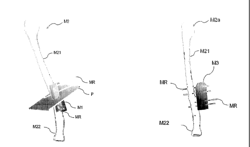
Figures 1 through3 show anosteotomyassistingmember
1 as a template assisting member and a rod. ~ as ~. correction
positi~n d~aermina.tion assisting member ~ accor~.ing to the
present invention. The osteotomy assisting member 1 and the
rod 2 are used as a pair for osteotomy performed for the
purpose of correcting a malunited bone.
In more detail, as shown in Figures 1 and ~, the
osteotomy assisting member 1, which is formed of a resin
block, is produced by rapid prototyping such as photo printout
based on three-dimensional model data obtained or created
on a computer. The osteotomy assisting member 1 includes
a fitting surface 11 ( not shown in Figures 1 through 3 ) as
a positioning element for indicating a position of the
CA 02515921 2005-08-12
WO 2004/071309 PCT/JP2004/001440
- 52 -
osteotomy assisting member 1 which is to be attached to the
bone; a slit 12 as a cutting section indicating element for
indicating a cutting section along which the bone is to be
cut and divided; and guide holes 13 each as an attachment
position indicating element for indicating an attachment
position of the rods 2.
The fitting surface 11 is formed to fit a surface
feature portion of the bone.
The slit 12 is provided so as to correspond to the
cutting section of the bone. The cutting section is defined
so as to be such a position that a post-correction bone shape
and the shape of a normal bone are closest to each other.
The post-c~rrection bone shape is obtained by dividing the
bone along the cutting section into a proximal portion and
a distal portion and moving and/or rotating the distal
portion.
~s described above a the guide holes 13 are provided
for in~.icating the attachment positions of the rods 2. The
thickness of the oats~tom~% assisting member 1 is set such
that each guide hole 13 has a sufficient length to have a
rod positioning function.
~s shown in rigors 3, each rod ~ is formed of metal
and is flexible to some e~mtent . The rod 2 is formed to have
for example, a pointed end so as to pierce a bone. The rod
2 has a diameter which is slightly smaller (for example,
0.1 to 0.2 mm) than that of the guide holes 13. The rod ~
can be inserted into each guide hole 13 and pierce the bone,
so as to be attached to the bone at a desired position in
a desired direction while making a hole in the bone . In this
CA 02515921 2005-08-12
WO 2004/071309 PCT/JP2004/001440
- 53 -
example, it is determined that two bone fragments are in
a normal positional relationship in the following state:
the two bone fragments each having two rods 2 piercing therein
are moved until the rods 2 in the two bone fragments are
in a prescribed positional relationship (for example,
parallel to each other ) . More specifically, it is determined
that the two bone fragments are in a normal positional
relationship in the following state : where a block 3 ( Figure
4) is provided as an intermediate member having insertion
holes 31 as engaging elements , each of which is capable of
receiving the rod ~, the rods 2 are insertable into each
insertion hole 31.
Hereinafter, a method for producing the osteotomy
assisting member 1 will be described. The osteotomy
assisting member ~. is produced by rapid. prototypia~g such
as photo printout based on three-dimensional model data ~~3.
Figure 7.1 ) obtained or created on a computer as described
below.
~0
First, as shown in Figure 5, a three-dimensional
surface bo~.e m~d~;l ~~~ is crc~~.ted. by a computer based ~~ data
obtained by, for example, CT o-r ~IRI. ~ three-dimensional
surface bone model biT of a normal bone is also created. The
~ 5 proximal portions of the bone models ~~~ and HT are overlapped .
since this example uses arm bones, the normal bone model
~iT is produced using a mirror image of a bone model of a
normal arm. l~lternatively, the normal bone model ~iT may be
produced using past data of the target bone when it was normal
30 or using various other techniques.
Then, a cutting section P (Figure 7) necessary for
correction and a moving amount of a distal bone fragment
CA 02515921 2005-08-12
WO 2004/071309 PCT/JP2004/001440
- 54 -
model M21 obtained by dividing the bone model M2 into two
portions are set. Thus, as shown in Figure 6, a
post-correction bone model M2a is determined. Briefly, the
cutting section P and the moving amount of the distal bone
fragment model M21 are set by arithmetic operations such
that the difference in shape between the post-correction
bone model M2a and the normal bone model MT is smallest.
This will be specifically described. In this
example, a surface matching method referred to as an "ICP
(Iterative Closest Point) algorithm" is used. This is
performed as follows . As shown in Figure 7 ~ the bone model
M2 is divided along a surface perpendicular to a virtual
screw axis I, appropriately set into the distal bone fragment
model ~1'~1 and the proximal bone fragment model ~~~~ . The
distal bone fragment model ~~~~. is translated linearly
parallel to the screw axis line ~ and/or rotated about the
screw axis line h. A difference between the post-correction
bone model ~~~a after the translation/rotation and the normal
bone model E~~T is converted into a numerical value by, for
example, squaring the data. on the corresponding p~sition
pan the surface of the mo~.el. The distal ~aone fragment model
h~~~3. is translated and/or rotated such that the above-obtained
numerical value is minimal.
~5
For defining the screw axis ~, the Screw
Displacement-Axis methods which is one method for
representing movement of an object, is used. The principle
of the Screw Displacement-Axis method will be described with
reference to Figure 8. There is a unique axis, for one
movement of an object in space, for which the following is
true: When an object is rotated about the unique axis by
an angle c~ and moved parallel to the axis by distance t, the
CA 02515921 2005-08-12
WO 2004/071309 PCT/JP2004/001440
- 55 -
object can be moved to any position. For correcting a
deformed bone , the bone is cut at a position into two portions ,
and the portions are moved with respect to each other into
a normal positional relationship. The Screw
Displacement-Axis for the movement is obtained. By
respectively matching characteristic shapes of the proximal
portion and the distal portion of a deformed bone with those
of a normal bone, it is found how much the deformed portion
(proximal or distal) needs to be moved to realize correction.
Thus, the Screw Displacement-Axis for moving the deformed
portion by the found amount is obtained by calculation. In
many of the clinical uses, the translation component is
sufficiently small to be negligible (e. g., within 1 mm),
and therefore only a rotation component needs to be considered.
Thus, it can be considered that the Screw Displacement-A~~as
- screw a~yis .
Then the screw axis L is parallel to, or substantially
parallel to , the longer axis of the 'bone as shown in Figure
a0 5, the bone is cut along a plane vertical to the screw axis
L at a position where the screw a~Wis L is closest to the
center o~ the bone, anal the distal bone fr~.gmer~t is rot~.te~.
about the screw axis L . Thus , the bone can be corrected with
the contact area of the bone fragments being maximal. Thus,
this cutting section can be set as an optimum cutting section.
The screw axis L varies in accordance with the form
of the bone. For example, as shown in Figure ~0, the
simulation results in the bone fragments being rotated about
the screw axis L substantially vertical to the longer axis
of the bone. In this case, a defect is created in the bone,
and the bone correction is completed by filling in this defect
by bone implantation or insertion of a bone filing material.
CA 02515921 2005-08-12
WO 2004/071309 PCT/JP2004/001440
- 56 -
There are cases where the bone needs to be partially
excised, but in many cases, correction simulation using the
Screw Displacement-Axis method ( = screw axis L ) is useful .
The above-described simulation is not always used.
When, for example, the screw axis L passes through a joint,
correction osteotomy is not performed unless the joint is
cut. Therefore, the above-described method is not usable.
In this case, the bone is cut at a convenient position to
the operator for simulation.
As shown in Figure 9 , rod models MR are respectively
attached to each of the bone fragment models M21 and M2~
of the p~st-correction bone model ~~~a such that the rod models
~~~ are parallel to each other. As shown in Figure 1~, the
attachment positions of the rod models ~~~ on the bone fragment
models M~1 and M~~ of the bone model M~ are obtained by
calculation.
~0
As shown in Figure 1~. ~ the three-dimensional model
~~1 of the osteotomy assisting member 1 17.a~ing the fitting
surface which can fit the surface feature portion of the
bone model is created so as to include the cutting section
~5 P determined as described above . At this point , a slit is
formed in the model a~7. at a position corresponding to the
cutting section P, and guide holes are formed in the model
M7. at positions corresponding to the rod models MR.
30 In this manner, the three-dimensional model M1 of
the osteotomy assisting member 1 is created having the fitting
surface, the slit and guide holes . Based on the data of the
model M1, the actual osteotomy assisting member 7. is produced.
CA 02515921 2005-08-12
WO 2004/071309 PCT/JP2004/001440
- 57 -
As shown in Figure 12, while the rod models MR are
attached to the bone fragment models M21 and M22 of the
post-correction bone model M2a, a three-dimensional block
model M3 of the block 3 having insertion holes through which
the rod models MR can pass is created on the computer.
Similarly to the osteotomy assisting member 1, the actual
block 3 is produced based on the three-dimensional block
model M3 by rapid prototyping such as photo printout for
the' like .
A method for performing a surgical operation using
the osteotomy assisting member 1, the block 3 and the like
thus produced will be described.
As shown in Figure 13, an affected site i.s incised,
so as to expose a necessary portion of a bone 5.
Then, as shown in Figure 1~, the fitting surface of
~0 the osteotomy assisting member 1 and the surface feature
portion of the bone 5 are made to fit each other, so as to
fi~a the osteotomy assisting memb~cr 1 ia~ a closely fit manner.
Thus , the osteotomy assisting member 1 is uniquely positioned
and attached to the bone 5. The rods 2 are inserted into
~5 the guide holes 13, and the bone 5 is pierced with the end
of each rod 2 so as to attach the rods 2 to the bone 5.
Next, the bone 5 is cut by moving a cutting jig such
as a saw or the like along the slit 12. Then, as shown in
30 Figure 15, the osteotomy assisting member 1 is removed while
leaving the rods 2 in place.
As shown in Figure 16, the distal bone fragment and
CA 02515921 2005-08-12
WO 2004/071309 PCT/JP2004/001440
- 58 -
the accompanying tissues are moved to a position at which
the other end of each rod 2 is insertable into the respective
insertion hole 31 of the block 3 , and then the rod 2 is inserted
thereinto. This step puts the bone fragments into a normal
positional relationship.
Finally, as shown in Figure 17, the bone fragments
are joined to each other in this state using plate and screws
or other fixing devices.
In this type of osteotomy surgical operation for bone
correction, the osteotomy assisting member 1 and the rod
~ in this example having the above-described structure allow
the bone to be easily cut along an appropriate cutting section
which. is pre-calculated by computer simulation and also allow
the post-cut bone frs.gments t~ be e~.sily moved to an
appropriate position which isalso pre-calculatedby computer
simulation. ~.ccordingly, accurate and easy osteotomy
surgical operations for bone correction are realised with~ut
relying on the physician's experience or skill.
specifically, the pre-correction bone shown in the ~~-rays
in Figure 1~ was superbly c~rrected to the b~ne shown in
Figure 1~.
~5 The present inventi~n is not limited to the
above-described example.
For example, one rod may be inserted into each bone
fragment. The correction position determination assisting
member is not limited to the rod but may be some other member.
For example, a member for fixing the bone while holding the
bone between two portions thereof may be more preferable
depending on the site of operation.
CA 02515921 2005-08-12
WO 2004/071309 PCT/JP2004/001440
- 59 -
A plurality of slits may be formed instead of one
in the case where the bone needs to be divided into three
or more fragments needs to be partially excised.
The assisting members according to the present
invention are not limited to those shown in the
above-mentioned figures but may be modified in various
manners as long as the members can act as intended.
Alternatively, the following procedure may be used.
( i ) A proximal portion of a deformed bone is matched
to a proximal portion of a normal bone, and the position
information thereof (matri x in the sense of affine
transformation) i.s obtained.
( ii ) Next ~ distal portions of the deformed bone and
the normal bone, and the matrix thereof is obtained. ror
~0 the matching, an ICP algorithm may be used ( it is not necessary
t~ use the ICP algorithm).
(iii) ~ difference between the matrices of the
proximal portions and the distal portions is obtained. This
is called by the present inventor as "relative matrix".
(iv) The relative matrix is transformed into the
Screw Displacement-Axis method. Thus, the Screw
Displacement-Axis (rotation axis) and the rotation amount
about the Screw Displacement-Axis(andthe translation amount
along the Screw Displacement-Axis) are found.
After the Screw Displacement-Axis is found, it is
CA 02515921 2005-08-12
WO 2004/071309 PCT/JP2004/001440
- 60 -
determined whether rotational osteotomy or closed/open wedge
osteotomy is to be used in accordance with whether the Screw
Displacement-Axis is parallel or vertical to the longer axis
of the bone . Thus , the cutting section of the bone is set .
One example of this procedure will be described in Example
12.
(Surgical operation method)
Hereinafter, a specific operation method will be
described in detail.
The present invention, in one aspect thereof,
provides a method for treating a bone ( a . g . , performing a
surgical operation on the bone). The method includes the
steps of: (A) obtaining a bone model representing a bone
which is a sub j ect of treatment ~ ( B ) obtaining a target bone
model to which tr eatment aims ; ( C ) determining a treatment
process which is to be performed on the bone ( a . g . a determining
an assisting member necessary for the treatment process to
be performed on the bone) based on the bone model and the
target bone model ~ and ( D ) performing a surgical operation
using the ~.etermine~ treatment process (a.g., using the
assisting member). Conventionally, a malunited bone or the
like was gradually corrected by performing surgical
operations within a possible range with trial and. error.
Ey contrast, it has been unexpectedly found that the present
invention allows a bone to be healed to a substantially normal
state by performing, on principle, one treatment process
(for example, cutting, insertion or rotation).
The step of obtaining a bone model representing a
bone which is a subject of treatment is performed by using
any imaging device, and a data storage and/or processing
CA 02515921 2005-08-12
WO 2004/071309 PCT/JP2004/001440
- 61 -
device usable for the data obtained by the imaging device.
Examples of the imaging device include optical imaging
devices (e. g., cameras), and CT and MRI imaging devices,
but are not limited to these . Examples of the data storage
and/or processing device include computers in which software
for extracting CT or MRI data on the bone and obtaining or
creating a surface model is installed, but are not limited
to these. A surface model is representatively stored in the
format of VTK or STL, but may be stored in other formats.
Examples of software for creating such a surface model include
Virtual Place M (Medical School and marketed by Medical
Imaging Laboratory, Ltd., Japan), Mimics (Materialise,
Belgium) , ZviewT~ (view, Inc. , Huntington Beach, CA, USA) ,
Realize (Mayo Clinic), but not limited to these.
The step of obtaining a target bone model to which
treatment aims can be performed by using any image creating
device. The image may be manually created. Usually, the
image is created by introducing an image of the subject of
~0 the model (e. g. , where one of the arms or legs is to be treated,
the other arm or leg) or by using, for example, a model of
a normal bone created day a computer (using C~~D of other ty~aes
of software) or stored by the computer.
~5 The step of determining a treatment process which
is to be performed on the bone based ~n the bone model and
the target bone model usually includes the step of determining
an assisting member necessary for the treatment which is
to be performed on the bone. This step can be performed by
30 using any device capable of processing a model. Examples
of such a device include computers having image processing
software installed therein, but are not limited to these.
Such software may have functions of, for example, performing
CA 02515921 2005-08-12
WO 2004/071309 PCT/JP2004/001440
- 62 -
surface matching, deriving movement information, orcreating
a template. In this example, a program for controlling and
managing position information regarding the surface model
on a computer (easily created using, for example, an open
source referred to as "VTK"
(http://public.kitware.com/VTK/)) is usable when
necessary.
Surface matching may be performed using, for example,
an algorithm suitable to matching such as an ICF ( Iterative
Closest Point ) algorithm, but other techniques may also be
used. In surface matching, accurately matching two models
(the target bone model and the bone model) is attempted.
When a program for controlling and managing position
information rega.rc~ing the surface model on a computer is
used, the two models are first manually matched to each other
to some extent when necessary, and a.re then matched using
the alg~rithm as mentioned ab~ve . When the bone is deformed,
the tw~ models cann~t, on principle~ match perfectly.
Therefore, it is acceptable to ignore the portions which
are distanced fr om each other by an arbitr ary threshold ( a . g . ,
1 mm) or m~re anc~ match. the p~rti~ns which are ~~ithin the
distance effectively within the threshold. Since there are
often excessive errors, the two model may be put closer to
each other manually and visually, rather than relying on
the algorithm.
The movement information can be found through
calculation by deriving relative movement information from
information on two positions (the positions of the target
bone model and the bone model). First, a proximal bone
fragment and a distal bone fragment of the bone to be treated
are matched to those of the target bone model ( a . g. , a mirror
CA 02515921 2005-08-12
WO 2004/071309 PCT/JP2004/001440
- 63 -
image of the normal arm or leg). Then, information on a
relative deformity can be obtained from the information on
the two positions. Here, the Screw Displacement-Axis
(rotation axis) and how much the distal bone fragment (or
the proximal bone fragment) needs to be rotated about the
rotation axis and translated along the rotation axis can
be calculated using a Screw Displacement-Axis method.
Instead of using the Screw Displacement-Axis method, the
movement information on any coordinate axis can be
represented using an affine transformation method.
A template model can usually be created using a
Boolean operation. The created model can be stored in the
STI~ format (used in photo printout for the like; the model
is represented ~.s a surface model filled with triangles ) ,
but may be stored in other formats . The Boolean operation
is one o~ the 3D graphic modeling technigues ~ and performs
modeling by an operation referred to as set operation. With
the Boolean operations modeling can be performed by
~0 arithmetic operation of : forming a plurality of overlapped
models into one mass ( sum) ~ deleting the overl~.ppe~. models
(difference) , and retrieving only the ~veilapped porti~n.s
( logical pro~.uct ) . The Boolean operation is adopted in, for
example, formal (available from autodessys). for creation
~5 of the template, formal or Magics 1~P (Materialise) is usable,
but other types of software is also usable. Such software
is developed for the industrial uses, and is not
conventionally used for treating deformed bones.
30 In a preferred embodiment of the method for treating
a bone according to the present invention, the step of
determining a treatment process (usually, the step of
determining an assisting member necessary for the treatment
CA 02515921 2005-08-12
WO 2004/071309 PCT/JP2004/001440
- 64 -
process to be performed on the bone) directly or indirectly
uses parameters in all three-dimensional directions of the
bone . In the prior art , the two-dimensional directions are
usually considered, and there is no example of considering
the three-dimensional directions. The present invention,
which considers the parameters in the three-dimensional
directions, is highly useful in achieving more accurate
surgical operations and more satisfactory postoperative
progresses.
In a preferred embodiment of the method for treating
a bone according to the present invention, the step of
determining an assisting member necessary for the treatment
process to be performed on the bone comprises the step of
determining a rotation axis of the bone.
Preferably in the case where the bone to be treated
is deformed, the rotation axis of the bone is found as follows .
The bone model is divided into a distal portion and a proximal
portion. Then, it is determined by calculation where either
the distal portion or the ~aro~~imal portion overlaps with
the target b~ne model fear com~aa.ris~n, ~ah.eth.ea~ or not the
target bone model is created by rotating the other portion
(i.e., by rotating the proximal portion when the distal
portion overlaps with. the target bone model, and by rotating
the distal portion when the pr~~ylmal portion overlaps with
the target bone model ) . The rotation axis can be found by
using, for example, the Screw Displacement-Axis method.
Actual calculation may be performed manually, but is usually
performed using a computer having a program for executing
the Screw Displacement-Axis method installed therein.
Accordingly, in a preferred embodiment of the method
CA 02515921 2005-08-12
WO 2004/071309 PCT/JP2004/001440
- 65 -
for treating a bone according to the present invention, the
rotation axisisdetermined using the Screw Displacement-Axis
method. The Screw Displacement-Axismethod isdescribed in,
for example, S. Ohwovoriole and B. Roth: "An extension of
screw theory", Journal of Mechanical Design, Vol. 103,
pp. 725-735, Oct. 1981. Using the Screw Displacement-Axis
method, bone correction can be realized substantially by
cutting the bone once and performing at least one selected
from the group consisting of bone rotation, bone excision,
bone distraction, and insertion of a graft. According to
the presentinvention,it hasbeen unexpectedly demonstrated
that the postoperative progress was favorable even several
months, or one year or more after the operation. As can be
appreciated from this, the present invention provides a
simple and efficient surgical o~aeration method for bone
correction which was une~~pected from the prior art.
Accordingly, a preferred embodiment of the method
for treating a bone according to the present invention
includes at least one selected from the group consisting
of bone rotation, bone e~~cision~ bone distraction, and
insertion of a graft. ~aThich one or ones ~f the bone rotations
bone excision, bone distraction, and insertion of a graft
should be performed can be easily determined by those skilled
in the art by the degree of deformity (i.e., a difference
between the target bone model and the bone model). The
difference can be found by calculation using any mathematical
technique well known in the art . Each of bone rotation, bone
excision, bone distraction, and insertion of a graft is
preferably performed once. In the prior art, there is no
example in which it is necessary to perform each of the rotation,
excision, insertion, implantation and the like only once.
Thus, the present invention has realized the simplicity,
CA 02515921 2005-08-12
WO 2004/071309 PCT/JP2004/001440
- 66 -
which was conventionally impossible.
In a preferred embodiment of the method for treating
a bone according to the present invention, a template
assisting member is used. In a conventional treatment of
an abnormal bone, such as a deformed bone, the bone is cut,
at an approximately proper position determined by visual
inspection, into bone fragments and then the bone fragments
were joined in an approximately proper manner determined
by visual inspection. Use of a template assisting member
realises simple and accurate correction. The template
assisting member can be produced by creating a model by,
for example, a computer-assisted image creation method
described below in this specification and then molding a
material ~e.g., metal, plastics, ceramics) by any molding
method such as photo printout.
~ptical formation is a technique for producing a
precise object substantially the same as that of ~D data
~0 in a short time. By photo printout, a liquid ultraviolet
curable resin ~a liquid which is eure~. by reacting to
ultraviolet rays ) is cured by ultraviolet laser light from
an photo printout apparatus and laminating the cured layers .
Specifically, data of a three-dimensional model
designed by, for example, Ct~D is output in the STL format
and the three-dimensional model is sliced at a pitch of 0.1
to 0 . ~ mm. Thus , contour data for photo printout is obtained.
Based on the contour data, the ultraviolet laser light from
a semiconductor laser device draws a shape of a cross-section
on a surface of a liquid resin in a tank. The portion
irradiated with the laser light is chemically reacted and
is cured. When one layer is formed, the substrate bearing
CA 02515921 2005-08-12
WO 2004/071309 PCT/JP2004/001440
- 67 -
the formed laminate sinks by the slicing pitch, and a shape
of the next cross-section is drawn and formed into a layer
by the laser scanning, and laminated on the first layer.
By laminating such thin cross-sections sequentially in this
manner, a three-dimensional model is formed below the liquid
surface. Then, the three-dimensional model is washed or
subjected to other processing, and the template assisting
member is completed. The service of producing such template
assisting members is available from many various providers
including PLAMQS of the Daicel Group, Kida Industry Co.,
Ltd., and CMET Co., Ltd.
The template assisting member used in the present
invention preferably includes at least one element selected
from the group consisting of a positioning element for
indicating a position at which the template assisting member
is to be located; a cutting secti~n indicating element for
indicating a cutting section along which the bone is to be
cut; and an attachment p~sition indicating element for
~0 indicating a position at which a correction position
determinate~n assisting member is to be attached. ~~re
~areferably, the template assisting member includes at least
the cuttingsection indicating element. Owing to the cutting
section indicating element , the cutting position of the bone
can be easily identified so that the bone can be cut accurately.
Stillm~repreferably, the templateassistingmemberincludes
the attachment position indicating element. ~wing to the
attachment position indicating element, the template
assisting member can be more smoothly and more accurately
moved after the bone is cut. The positioning element can
be easily realized by molding, or labeling the template
assisting member with a label (e.g., a stain), but may be
realized by other methods. The cutting section indicating
CA 02515921 2005-08-12
WO 2004/071309 PCT/JP2004/001440
- 68 -
element can also be easily realized by molding, or labeling
the template assisting member with a label (e.g. , a stain) ,
but may be realized by other methods . Advantageously, the
cuttingsection indicating element is realized by physically
forming a slit, since the slit allows the cutting device
such as a bone saw to be operated more easily. The attachment
position indicating element can also be easily realized by
molding, or labeling the template assisting member with a
label ( a . g . , a stain ) , but may be realized by other methods .
The attachment position indicating element is preferably
an opening, since the correction position determination
assisting member is preferably a wire.
More preferably, the template assisting member used
in the present invention includes all of the positioning
element~the cuttingsection indicating element(preferably~
a slit), and the attachment position indicating element
preferably ~ an opening or a hole ) . Vdhen including all of
these elements, a single template assisting member can
~0 perform all of positioning of the template assisting member,
bone cutting, and bone correction.
In a preferred. embodiment of the method for treating
a bone according to the present invention, the step of
performing a surgical operation includes the steps of:
(A) cutting the bone at at least one position into bone
fragments; (~) performing at least one selected from the
group consisting of : ( i ) performing the bone rotation when
necessary, (ii) performing the insertion of a graft when
necessary, and (iii) performing the bone excision when
necessary; and (C) joining the bone fragments.
The step of cutting the bone can be performed using
CA 02515921 2005-08-12
WO 2004/071309 PCT/JP2004/001440
- 69 -
any cutting device. Preferably, an assisting. member (e. g.,
a template assisting member of the present invention) is
used. The bone may be cut an unlimited number of times, but
preferably once. The present invention realizes correction
of an abnormal bone by cutting the bone only once and performing
the subsequent treatment processes.
The step of performing at least one treatment process
can be performed using any device and the hands of a physician
or other medical practitioners. Preferably, an assisting
member (e. g., a template assisting member of the present
invention ) is used. Each of the treatment processes ( i . a . ,
bone rotation, insertion of a graft , and bone ) maybe performed
an unlimited number of times, but preferably once. The
present invention realizes correction of an abnormal bone
by performing each of at least one of the treatment processes
only once. Thus, the present invention realizes simple and
accurate bone correction. ~ graft to be inserted can be
designed by a computer. ~n actual graft is produced based
on the computer-designed graft model by molding a material
(e. g., metal, plastics, ceramics) using any molding method
such as 3D cutting, FTC m~.c~a.iaaing or the li3~e. The cutting
can be performed using a commercially available cutting tool
such as , for example, the 3D cutting tool by Poland DG ( a . g. ,
~5 ~D.~ series including T~D.~-20, P~dC-3200) .
In a preferred embodiment of the method for treating
a bone according to the present invention, the treatment
process includes bone rotation. Advantageously, the bone
is rotated about one rotation axis . There is no conventional
technique for healing an abnormal bone by rotating the bone
about a rotation axis . It was not conventionally expected
that a deformed bone can be corrected by using rotation
CA 02515921 2005-08-12
WO 2004/071309 PCT/JP2004/001440
- 70 -
technique in a surgical operation. The present invention,
which demonstrates that simple and accurate correction can
be realized by rotation, is useful for various types of bone
correction.
The step of joining the cut bone fragments can be
performed using any device well known in the art . The bone
fragments can be joined using, for example, an internal
fixation device such as aplate, screw, wire, intramedullary
nail or the like . A defect of the bone may be f filled i.n by
calcium phosphate (e. g., hydroxyapatite, (3TCP, calcium
phosphate paste) or other biocompatible materials.
Alternatively, the defect of the bone may be filled in by
'bone implantation.
defter the bone fragments are joined, it is preferable
to maintain the state for a long time. In this manner, the
corrected state is retained and the effect of correction
is guaranteed. When appropriate, the bone may be distracted
rather than joined. Such distraction may be performed using
a callus distraction method.
In a preferred embodiment of the method for treating
a bone according to the present invention, the bone rotation,
~5 the insertion of a graft and the bone excision are defined
by the Screw Displacement-Axis method or an affine
transformation method. Surprisingly, it has been
demonstrated by the present invention that bone correction
is realized by simply performing a surgical operation with
a difference calculated by such a mathematical technique
being reflected on the bone rotation, insertion of a graft,
bone excision, bone distraction of the like, and that the
effect of correction is maintained for one year or longer
CA 02515921 2005-08-12
WO 2004/071309 PCT/JP2004/001440
- 71 -
for the first time in history. In order to closely follow
the difference calculated by the above mathematical technique ,
it is preferable to use a template assisting member. Using
the template assisting member, the operator can simply follow
the instruction line on the template assisting member to
perform the cutting, rotation, or other processes . Such a
method of surgical operation brings an epoch-making change
in the field of surgery, in which many of the operation
procedures conventionally needed to be conducted manually
relying on only the experience of the operator.
In one embodiment of the method for treating a bone
according to the present invention, the bone excision is
defined by a template assisting member, but the present
1a invention is not limited to this. The template assisting
member may be attached after the bone is cut.
In one embodiment of the method for treating a bone
according to the present invention ~ the step of performing
the bone rotation representatively includes the step of
arranging correction assisting members passing through. at
least a pair of ~penings which ~.efine a deformity amoua~t,
the pair of openings being on the template assisting member.
In this case~ the template assisting member is designed such
~5 that a target rotation is achieved by arranging the correction
assisting members in parallel . Such a design can be realised
by, for example, the Screw Displacement-Axis method or the
affine transformation method.
The graft to be inserted in the method for treating
a bone according to the present invention is preferably
defined and produced by the Screw Displacement-Axis method
or the affine transformation method. By such methods, a
CA 02515921 2005-08-12
WO 2004/071309 PCT/JP2004/001440
- 72 -
difference between the bone model and the target bone model
can be obtained and a model of the graft can be created from
the difference. Based on the created model, an actual graft
can be produced by, for example, 3D shaving.
In a preferred embodiment of the method for treating
bone according to the present invention, the step of cutting
the bone includes the step of cutting the bone at one position
into a proximal bone fragment and a distal bone fragment,
and the step of performing a surgical operation includes
fixing either the proximal bone fragment or the distal bone
fragment of the bone. The target bone model is preferably
created based on a proximal portion and a distal portion
of the bone, since one of the bone fragments usually has
a normal shape.
The bone to be treated by the present inventson is
usually a bone of a limb, but is not limited to this . Any
bone can be treated by the present invention. For example,
200 bones in a human body are all subjects of the present
invention. These bones include, fore~~~ample, long bones (for
example , bones of limbs ) , short bones , f la.t bones ( a . g . ,
sterna, costae, scapulae, ilia), sesamoid bones (e. g.,
patellae), and irregular bones(viscerocranium,vertebrae),
but also include other types of bones. ror the present
invention ~ correction of long bones ( for example, bones of
limbs) is suitable. ~ subject of correction according to
the present invention is representatively a malunited bone.
In one embodiment of the method for treating a bone
according to the present invention, a graft to be inserted
is produced using a natural bone or an artificial bone . The
natural bone may be, for example, autobone, homogenous bone,
CA 02515921 2005-08-12
WO 2004/071309 PCT/JP2004/001440
- 73 -
or heterologous bone, but is not limited to these. Autobone
is preferable . As the autobone, ilia or the like is usable .
When autobone is not easily available, homogenous bone is
used. The homogenous bone is available from, for example,
the bone bank.
The artificial bone may be formed of any biocompatible
material, but preferably of a material compatible to bone,
and more preferably of a material which can be a part of
the bone. Alternatively, the artificial bone may be formed
of a biodegradable material. As a biodegradable material,
a material which is gradually degraded ( a . g . , over several
months ) is usable, not a material which is rapidly degraded.
Preferable materials include, for example, bone-affinitive
materials such as bone ceramics , and calcium phosphate ~ and
hydroxyapatite. Hydroxyapatite is especially preferable.
Such artificial bone may be, for example~ ~1~~B~NS (T Co. ,
LTD., ~saka), but is not limited to this.
A preferred embodiment of the method for treating
a bone according to the present invention includes the step
of chec3cing whether treatment was ~aerfora~e~. properly or got
after the step of performing a surgical operation. Such a
checking can be performed using any device or technique well
~5 known in the art~ for e~:ample, by using a correction position
determination assisting member of the present invention.
Alternatively, the checking can be performed by taking an
x-ray of the post-correction bone, obtaining a model of the
bone, and comparing the target bone model and the model of
the post-correction bone to determine the difference . Such
a comparison can be performed using any program known in
the art.
CA 02515921 2005-08-12
WO 2004/071309 PCT/JP2004/001440
- 74 -
The target bone model used in the present invention
is defined based on a partner of a pair of bones including
the bone represented by the bone model. Such a pair of bones
are, for example, bones of limbs, but not limited to these.
Alternatively, the target bone model used in the
present invention is defined based on a standard of patients
having the bone to be treated. The standard can be found
by investigating a set of patients, processing the results
statistically, and setting a range of the average ~ 1 SD
as the standard.
(Simulation)
The present invention, in another aspect thereof,
provides a method for simulating bone treatment (e. g.,
surgical operation). The method includes the steps of:
(A) obtaining a bone model representing a bone which is a
subject of treatments (D) obtaining a target bone model to
which the treatment aims;(C) determining a treatment process
~0 which is to be performed on the bone (e.g. , determining an
assisting member necessary for the treatment process t~ be
perf~rmed ~n the b~ne) based. ~n the bone model and the target
bone model; and (D) creating a production model based on
the bone model and performing a simulation of the bone
~5 treatment based on the determined treatment process (e. g.,
using the assisting member). Conventionally, a malunited
bone or the lilee was gradually corrected by performing
surgical operations within a possible range with trial and
error. By contrast, the present invention realizes a
30 simulation of the treatment process to be performed. Thus,
even a beginner can easily perform the treatment processes
and become familiar with the technique. Any of the steps
included in the method for simulating bone treatment
CA 02515921 2005-08-12
WO 2004/071309 PCT/JP2004/001440
- 75 -
according to the present invention can be performed using
any of the devices or techniques described in the "Surgical
operation method" section above.
For simulation, it is possible to actually produce
a production model (of, for example, hydroxyapatite) and
an assisting member when necessary, and perform a simulation
of the surgical operation in accordance with the determined
treatment process.
(Treatment kit)
The present invention, in still another aspect
thereof, provides a bone treatment kit used for treating
a bone. The kit includes: (A) a template assisting member
including a positioning element for indicating a position
of the template assisting member which is to be attached
to the bone, a cutting section indicating element for
l.nd.l.~atlng a cutting section along which the bone is to be
cut , and at least one opening through which the correction
aS~l~tl.n~ member f~r rotation iS t~ be 3.n~erted: and (~) a
correction position determination ~.ssisting member. The
template assisting member used here may be in any form or
material which is described above in detail in this
specification. The correction position determination
assisting member may also be in any form described above.
Preferably, the template assisting member includes
at least two openings. With two openings, it is easily
determined whether or not the correction position
determination assisting members are parallel to each other.
More preferably, the template assisting member includes three
openings , and still more preferably four openings . With four
openings, it is more easily determined by visual inspection
CA 02515921 2005-08-12
WO 2004/071309 PCT/JP2004/001440
- 76 -
whether or not the correction position determination
assisting membersare parallel to each other. The correction
position determination assisting member is preferably a wire
having a translation assisting function, but is not limited
to this as long as the member has a translation assisting
function. The wire may be formed of any material, preferably
stainless steel.
In a preferred embodiment of the bone treatment kit
according to the present invention, the correction position
determination assisting member has at least one function
selected from the group consisting of a translation assisting
function for translation and a rotation assisting function
for rotation. Alternatively, the bone treatment kit may
include a plurality of correction position determinatioa2
assisting members, each of which has both the translation
assisting function and the rotation assisting function.
It is advantageous that the bone treatment kit
~~ according to the present invention has both. the translation
assisting function and the rotation assisting funetio~.. The
translation ~.ssistin.g function can be reali~ec~. b;~ mar~~.ing
a member extending in a direction of a longer axis of the
bone, for example, a wire. The rotation assisting function
may be realised using a linearly extending member, for example,
a wire.
Preferably, the bone treatment kit according to the
present invention further includes a fixation assisting
member for fixing the bone after a surgical operation. Such
a fixation assisting member may be any member. For example,
a conventionally used device such as an external fixation
device or the like may be used.
CA 02515921 2005-08-12
WO 2004/071309 PCT/JP2004/001440
_ 77 _
(Simulation kit or system)
The present invention, in still another aspect
thereof, provides a kit or system for simulating bone
treatment (e. g., surgical operation). The kit includes
(A) means for obtaining a bone model representing a bone
which is a subject of treatment; (B) means for obtaining
a target bone model to which the treatment aims ; ( C ) means
for determining a treatment process which is to be performed
on the bone (e. g. , determining an assisting member necessary
for the treatment process to be performed on the bone ) based
on the bone model and the target bone model; and ( D ) means
for performing a simulation of the bone treatment based on
the determined treatment process (e. g., using the assisting
member). The kit according to the present invention may
include a production model of the bone, an assisting member
when necessary, and an instruction sheet which indicates
the determined treatment process. Conventionally, a
malunited bone or the like was gradually corrected by
performing surgical operations within a possible range with
trial and error. D~° contrast ~ the present invention reali~~a
a simulati~n of the treatment process t~ be performed. Thus,
even a 'beginner can easily perform the treatment processes
and become familiar with the tecl7.nie~ue. any of the means
included in the kit for simulating bone treatment according
to the present invention can be in any form described in
the "Treatment kit" section above.
For simulation, it is possible to actually produce
a production model (of, for example, hydroxyapatite) and
an assisting member when necessary, and perform a simulation
of the surgical operation in accordance with the determined
treatment process.
CA 02515921 2005-08-12
WO 2004/071309 PCT/JP2004/001440
_ 78 _
( Graft )
The present invention, in still another aspect
thereof, provides a graft used for treating a bone. The graft
has a shape substantially representing a difference between
a bone model representing a bone which is a sub j ect of treatment
and a target bone model and a target bone model to which
the treatment aims . The shape representing such a difference
is realized by any molding method. The molding method may
be 3D shaving, RP, or NC processing, but not limited to these.
3D shaving is preferable since 3D shaving can realize
substantially any shape . Preferably, the shape of the graft
according to the present invention is determined by a
difference between the target bone model and the bone model
defined by the Screw Disple.cement-t~~.is method or the affine
transform~.tion method.
(Treatment system)
The present invention, in still an~ther aspect
thereof, provides a system for treating a bone. The system
includes (~1.) means for obtaining a bone model representing
a bone which is a subj ect ~f trr~atment ~ ( D ) means for obt~.i~aing
a target bone model to which treatment aims ~ ( C ) means for
determining a treatment process which is to be performed
~5 on the bone (e.g. ~ determining an assisting member necess~.ry
for the treatment process to be performed on the bone) based
on the bone model and the target bone model ~ and ( D ) means
for performing a surgical operation using the determined
treatment process (e. g., using the assisting member).
The means for obtaining a bone model, the means for
obtaining a target bone model, and the means for determining
a treatment process, and the means for performing a surgical
CA 02515921 2005-08-12
WO 2004/071309 PCT/JP2004/001440
_ 79 _
operation may be in any means described in the "Surgical
operation method" section above.
(Simulation system)
The present invention, in still another aspect
thereof,providesasystemforsimulating bone treatment(e.g.,
surgical operation). The system includes: (A) means for
obtaining a bone model representing a bone which is a subject
of treatment; (B) means for obtaining a target bone model
to which the treatment aims; (C) means for determining a
treatment process which is to be performed on the bone (e.g. ,
determining an assisting member necessary for the treatment
process to be performed on the bone) based on the bone model
and the target bone model; and ( D ) means for performing a
simulation of the bone treatment based on the determined
treatment process (e. g., using the assisting member).
Conventionally, a malunited bone or the lilce was gradually
corrected byperf~rming surgical operations within apossible
range with trial and error. By contrast, the present
~0 invention realises a simulation of the treatment process
to be performed. Thus ~ even a beginner can easily perf~rm
the treatment ~arocesses ~.nd become familiar with the
technique. Any ~f the means included in the system f~r
simulating bone treatment according to the present inventi~n
~5 can be in any f~rm described in the "Treatment system" section
ab~ve.
For simulation, it is possible to actually produce
a production model (of, for example, hydroxyapatite) and
30 an assisting member when necessary, and perform a simulation
of the surgical operation in accordance with the determined
treatment process.
CA 02515921 2005-08-12
WO 2004/071309 PCT/JP2004/001440
- 80 -
(Computer program)
The present invention, in still another aspect
thereof, provides a program for making a computer execute
processing for determining a treatment process to be
performed on a bone . The processing includes the steps of
(A) obtaining a bone model representing a bone which is a
sub j ect of treatment ; ( B ) obtaining a target bone model to
which treatment aims;and(C) determining a treatment process
to be performed on the bone based on the bone model and the
target bone model. The program according to the present
invention can allow the treatment process to be performed
on the bone ( a . g . , bone cutting, movement of bone fragments )
to be determined in order to achieve a desired purpose ( a . g. ,
bone correction). Thus, even a physician with little
experience can perform the treatment appropriately.
By executing the program for making a computer execute
processing for determining a treatment process to be
performed on a bone, the computer provie~es a method for
~0 determining the treatment process . By e~9ecuting the program
for making ~. computer e~~ecute processing for determining
a treatment process to be performed on a bone, the c~mputer
acts as an apparatus for determining the treatment process .
~5 The above description is provided regarding bones,
but it should be understood that the present invention is
applicable to portions of a body other than bones (e. g.,
soft portions such as skin).
30 Hereinafter, the present invention will be described
by way of examples . The following examples are provided for
the purpose of illustration, and the present invention is
not limited to the following example but is limited only
CA 02515921 2005-08-12
WO 2004/071309 PCT/JP2004/001440
- 81 -
by the claims.
EXAMPLES
In thefollowing examples,experiments on humans were
performed after obtaining informed consent in compliance
with the criteria defined by the Ministry of Health, Labour
and Welfare in Japan or equivalent authoritative governmental
offices .
(Example 1: Production of template assisting members and
application thereof)
In this example, template assisting members were
produced after performing a preoperative simulation (Figure
21 ) using actual deformed bones as a model . In the case of
a fracture malunion on the forearm, the degree of deformity
of the radius and the ulna is one of the main factors of
restriction of pronation and supination. Pronation refers
to rotating the forearm such. that the palm is directed downward
and the supination refers to rotating the forearm such that
the palm is directed upward ( such that the palm is to receive
s~mething). It is desir~.ble to c~rrect the def~rmity as
accurately as possible. The present inventor performed
accurate correction osteotomy through. a preoperative
simulation using Cl~ or L~1RI three-dimensional images . The
methods and results will be described below.
There were 6 cases , arid the patients were all male
in the age range of 12 to 31 years with the average age of
19.2 years. The correction osteotomy was performed on a
deformity in radius diaphysis in 5 cases and radius distal
in 1 case. The time period from injury to osteotomy was 11. 6
months on average . The preoperative angle of deformity on
CA 02515921 2005-08-12
WO 2004/071309 PCT/JP2004/001440
- 82 -
x-ray was 22.8 degrees, the preoperative movable range of
the forearm was limited to 62 degrees for pronation and 25
degrees for supination. The average period of follow-up
survey was 6.6 months.
(Creation of a model)
For each of these cases, a subject bone model was
extracted by semi-automatic marking (Figure 22B) from CT
or MRI two-dimensional image data of both forearm (Figure
22A) using specialized software , and segmented (Figure 22C) .
Thus, a three-dimensional bone surface model was created
(Figure 22D).
The forearm bone model of each patient was overlapped
with. the proximal portion and the distal portion of the mirror
image of the healthy arm, so that the screw axis of the
deformity and the deformity amount were calculated, and an
optimal site for osteotomy and correction amount were
determined (e. g., Figure 23).
For the first 3 cases, the surgical operation was
performed. with reference tea the deformed site and. t~.~.e
deformity amount obtaine~.by the simulation, the computer
images and the like. These cases were grouped as group A
( Figure 2~: ) .
For the second 3 cases, the site for osteotomy and
the correction amount were determined based on the CT data
(Figure 25A) , and an osteotomy template was designed on the
computer. The osteotomy template designed was such that it
fits snugly the bone surface of the malunited site and has
a guide hole, through which Kirschner wires (also referred
to as the "K-wires" ) which would be parallel to each other
CA 02515921 2005-08-12
WO 2004/071309 PCT/JP2004/001440
- 83 -
when the bone is corrected, can be inserted (Figure 25B).
The template also has a bone cutting Clit. A guide for
retaining the restored state with a K-wire passing
therethrough was also designed, and an actual production
model of the guide was produced by photo printout and used
for the surgical operation (Figure 25C) . These cases were
grouped as group B.
(Results)
Both groups were substantially the same in age, time
period from injury to the surgical operation, and the
preoperative degree of deformity. After the operation, the
deformity angle of group A, was insufficient at 9.3 degrees,
whereas the deformity angle of group B was 0.3 degrees, which
means almost complete correction. The movable range for
pronation and supin~.tion was changed from 73 degrees
(preoperative) to 1~6 degrees (postoperative) in group ~.
In group B ~ the movable range for pronation and supination
was changed from 120 degrees (preoperative) to 180 degrees
(postoperative) although the result was evaluated in only
one case 6 months after the surgical operatioa~. The results
are sb.own in Figure 2~.
(Example 2: 8-year-old male child; closed wedge osteotomy
for supracondylar fracture of the humerus)
In this example, it will be demonstrated that the
closed wedge osteotomy is applicable to the present invention.
A surgical operation was performed by the method according
to the present invention on an 8-year-old male patient having
a supracondylar fracture of the humerus, which is a
representative fracture around the elbow joint of children.
This case is known as easily resulting in cubitus varus.
Figure 27 shows x-rays illustrating the affected site ( the
CA 02515921 2005-08-12
WO 2004/071309 PCT/JP2004/001440
_. g4 _
left x-ray shows a front view, and the right x-ray shows
a side view thereof).
Figure 28 shows the progress of hyper-extension in
the cast caused by the initial treatment . It is shown that
dorsiflexion occurs since no appropriate treatment was
performed.
Figure ~9 shows the state of this patient 2 years
after the initial treatment . It is shown that varus deformity
and hyper-extension is left and natural correction is not
sufficient even 2 years after the bone union. In the
front-view x-ray (left in Figure ~9), the elbow exhibits
about 10° of varus ( the forearm is slightly internally flexed;
normally, the forearm should be externally flexed at about
10 degrees ) . In the side-view x-ray ( right in Figure
the distal humerus exhibits about 20° dorsifle xion with
respect to the axis of the humerus. Normally, the distal
humerus should e~ghibit about 20° anterior flexion.
Figure 3~ shows an aftereffect ~f hyper-e~~.tension
and varus def~rmity . The upper left photo shows a fle~~ion
disorder, by which the arm can bend at up to only 90 degrees.
Normally, the arm should bend at up to 130 to 1~0 degrees .
~5 The lower left photo shows 30° hyper-extension . Normally,
the arm can only extend at about 10 degrees . The right photo
shows cubitus varus.
(Correction osteotomy simulation using an MRI model)
On this patient , a surgical operation was performed
using a three-dimensional model created from MRI data ( see
Figure 31 ) . The three-dimensional model was matched to the
mirror image of the healthy arm as a target bone model, so
CA 02515921 2005-08-12
WO 2004/071309 PCT/JP2004/001440
- 85 -
that the screw axis and angle of deformity were found. The
deformity can be accurately corrected by excising a
wedge-shaped bone having the screw axis as an apex and closing
the cutting portion. It is impossible for the operator to
accurately determine the direction and angle of osteotomy
by visual inspection during the surgical operation.
The matching may be performed either manually or by
use of a computer using an ICP (Iterative Closest Point)
algorithm.
(Photo printout)
Sased on a template produced by matching, a photo
printout model was produced. The model was described in the
STL format. (The STL format is one format of
three-dimensional CAD data. Sy the STL format, the data
representing the surface is polygoni~ed using triangles.
"STL" represents Standard Triangulation Language. The STL
format is especially used in photo printout as a standard
~0 file format.) The STL design was converted into
cross-sectional data at a slicing pitch of 0.1 anmm. Sased
on the slice. data , the s~arfa.ce of the photocurable resin
was scanned and thus cured by an LD excited tT~ solid-state
laser light via a galvanometer mirror. Layers each having
~5 a thickness of 0.1 mm were laminated, so as to produce a
three-dimensional model. The model was washed and
completely cured by a post-cure device . ~s the photo printout
apparatus , Fapid ~Ieister 3000 available from CIf~ET Co . , Ltd.
was used.
Figure 32 shows a photo printout procedure.
CA 02515921 2005-08-12
WO 2004/071309 PCT/JP2004/001440
- 86 -
(Surgical operation)
Using the template, the surgical operation was
performed to correct the deformity. Figure 33 shows the outer
side of the left arm during the surgical operation. The
shoulder is on the right and the hand is on the left of the
photos . The template was fit to the lateral condyle of the
humerus. The left photo of Figure 33 shows the state
immediately after the operation site ( arm) was exposed by
incision and retraction. The right photo of Figure 33 shows
the state after the template and wires were set after the
site was exposed.
After setting the template and the like, the bone
was cut using a bone saw (Figure 34) . Figure 35 shows the
state after the bone was cut . ~s showa~ here, it is appreciated
that the method of the present invention allows the bone
to be accurately cut at the first trial, which is impossible
by an operation performed merely manually.
~0 P~fter the bone was cut~ an excessive 'bone portion
was e«cised as shown in Figure 3~. after the bone portion
was excised, the positions ~f the bone fragments were
corrected such that the wires were parallel to each other
Figure 37 ) . Figure 3~ shows x-rays immediately after the
surgical operation. The bone is fixed by the K-wires. P~s
shown here, fine correction and fixation are realized by
the treatment according to the present invention. The left
photo of Figure 38 shows 5° valgus , and the right photo shows
20° anteversion of the distal humerus. The movable range
of the elbow was normalized in both flexion and extension
immediately after the surgical operation. Even during the
surgical operation, it was shown that the flexion was at
130 degrees and the extension was at 10 degrees ( Figure 39 ) .
CA 02515921 2005-08-12
WO 2004/071309 PCT/JP2004/001440
_ 87 _
Figure 40 shows an external appearance immediately after
the surgical operation. The values of flexion and extension
are close to normal values. It was demonstrated that the
method for treating a bone according to the present invention
provides the effect of recovering substantially normal
functions.
Figure 41 shows x-rays 1 year after the surgical
operation, and Figure 42 shows an external appearance of
the patient 1 year after the surgical operation.
Surprisingly, it is understood that the normal state is kept
even 1 year after the surgical operation ~ or rather the patient
has almost completely recovered with the postoperative
unevenness disappearing.
This example is directed to the case where excision
and rotation are necessary.
The above-described effect was not e~spected from the
prior art, and it is demonstrated a.nd understood that the
present invention is highly useful in medical practice.
(F~~ample 3: 21-year-old melee open wedge osteotomy for a
left forearm fracture malunion)
7Ln this example ~ it will be demonstrated that open
wedge osteotomy is applicable to the present invention. ~s
an e.x.ample ~ a case of a 21-year-old male patient with a left
forearm fracture malunion was used. This patient showed an
excessive deformity of the radius (Figure 43) . An forearm
fracture is often seen among men in early youth to youth,
and is considered to often cause a malunion when treated
in a cast.
CA 02515921 2005-08-12
WO 2004/071309 PCT/JP2004/001440
_ 88 _
As shown in Figure 44, this patient complained of
a deformity and restriction of supination of forearm (i.e. ,
such that the palm is to receive something) . The supination
was restricted to about 40 degrees (normally, 80 to 90
degrees).
It was found by computer simulation that the deformed
bone was matched to the healthy bone by rotating the bone
at 30 degrees about the screw axis (Figure 45) . A template
was designed based on this information. It was found that
by subtracting the bone and a pre-produced rectangular
parallelepiped shape and cylindrical shape from the block
(Figure 4~, left) (Boolean operation) , a template which fits
the bone surface showing the feature of the malunitedportion,
which has a cutting section indicating element and which
allows wires to be inserted thereto (Figure ~.~, right) can
be designed. A correction guide was designed in a similar
manner.
~0 (surgical operation)
The p~.tierat was treated using this template. As
shown i~ figure ~~~, the m~.lunited site of the bone was e~~.posed
by incision and retraction. Then, the osteotomy template
was applied to the malunited site and it was confirmed that
the osteotomy template completely fitted the malunited site
( Figure 45 ) . Then , the template was pierced with ~lrschner
wires(stainlesssteel wiresgenerally used in orthopaedics)
and fixed. Then, as shown in Figure 4.9, the bone was cut
along the slit of the template . As shown in Figure 50 , the
osteotomy was completed and the template was removed.
As shown in Figure 51, the correction guide was
outserted into the wires to perform correction . The cutting
CA 02515921 2005-08-12
WO 2004/071309 PCT/JP2004/001440
- 89 -
portion was opened. Although it was not clear whether the
deformity was corrected or not by a visual inspection of
the site of operation (the surface is seen as extremely
projecting forward), it was confirmed by an x-ray during
the operation that the deformity was accurately corrected
(Figure 52).
Then, as shown in Figure 53, the iliac bone was shaped
into a wedge-shape and implanted. After this, the bone and
the bone graft were fixed with a titanium plate and screws
( Figure 54 ) . The Kirschner wires were then removed ( Figure
55 ) . A postoperative x-ray shows that fine correction was
realised (Figure 56).
(Postoperative progress)
This patient was exami~.ed 9 months after the surgical
operation. As shown in Figure 57 ~ the bone was united while
the deformity of the radius was corrected to an almost normal
state. 1 year and 1 month after the so.rgical operation, the
~0 plate was removed (Figure 58) . The a x.ternal deformity had
disappeared, and the forearm had been recovered from the
rot~.tio~, disorder (Figure 5~ 6 left ) . The supin~.tion w~.s
possible to up to 90 degrees (Figure 59, center) . The
pronation was normal (Figure 59, right).
Fxample ~ : ~.8-year-old female a a fracture malunion on the
distal end of the left radius)
It will be demonstrated that the deformity can be
corrected even for a middle-aged woman. As an example, a
48-year-old female patient having a fracture malunion on
the distal end of left radius was used.
This patient had a fracture malunion on the distal
CA 02515921 2005-08-12
WO 2004/071309 PCT/JP2004/001440
- 90 -
end of left radius as shown in Figure 60. The left photo
of Figure 60 shows a left view thereof. As shown, the
articular surface shows dorsiflexion. The solid line
represents a tilt of the articular surface, which shows about
20° dorsiflexion. In a normal state, as represented with
the dotted line in Figure 60, lower center, the articular
surface shows 10 to 20° volar flexion. The upper center photo
of Figure 60 shows a front view of the affected side . The
radius is shortened by about 4 mm as represented with the
arrow, and the tilt of the articular surface of about 10
degrees is smaller than the normal tilt of 20 to 30 degrees .
The left photo of Figure 60 is a reversed photo of the healthy
side as reference. The line represents a tilt of the
artificial surface of 25 degrees .
The fracture of the distal end of the radius suffered
by this patient is a representative fracture among middle-
and senior-aged women ~ and is lilcely to result in a malunion .
~ slight malunion is often left without treatment since the
bone is united in a fine manner, but patients of or under
50 years of age, and older patients actively doing physical
ex~rcise ~ often complain of a disorder ia~ the movable range
and pain during physical exercise.
This patient has an external deformity and disorder
of the movable range of the left wrist joint as shown in
Figure 61. Such a deformity of the distal end of the radius
is referred to as a fork-type deformity since it looks like
a fork seen from the projecting side. ~s shown in Figure
60 , right , the volar flexion is restricted to about 35 degrees .
The normal angle of volar flexion is 60 to 80 degrees . This
patient also complained of pain of the wrist joint during
physical exercise.
CA 02515921 2005-08-12
WO 2004/071309 PCT/JP2004/001440
- 91 -
(Three-dimensional simulation)
A three-dimensional simulation of the bone of this
patient was performed (Figure 62). As a result, it was
calculated that 30° open wedge osteotomy was necessary.
Based on the calculation result, an osteotomy template, a
correction guide and a bone graft were designed ( Figure 63 ) .
Figure 64 shows the procedure for producing the template
or the like by photo printout.
(Surgical operation)
A surgical operation was performed on the patient
using the template and the like. The left photo of Figure
65 shows the malunited bone was exposed by incision and
retraction . As shown in Figure 65 , right , the template was
fit to the site. ~~s shown in Figure 66, left, the template
was fixed with ~.irschner wires and, and as shown in Figure
66 ~ right , the bone was cut with a bone saw. As a correction
guide, a stainless steel guide (cylindrical guide having
slits) was used so as not to damage the resin. The deformity
was corrected as shown in Figure 67 using the guide. A graft
~ahiclz. w~.s obtained. b~ sh.a~aing h~ droi~~~.~a~.titP using C~~I2 before
the surgical operation was used (Figure 63). Such an
artificial graft contributes t~ simplification and higher
~5 accuracy of the surgical operation.
Then, a postoperative x-ray was taken after the bone
was fixed by the template (Figure 69). As shown, the
correction is finely done. The central photo of Figure 69
is a reversed image of the healthy side. Through the
comparison, it was confirmed that the deformity was corrected
to the state which is substantially the same as that of the
healthy side.
CA 02515921 2005-08-12
WO 2004/071309 PCT/JP2004/001440
- 92 -
Figure 70 shows a state 4 months after the surgical
operation. As is clear from the photo, the corrected state
was maintained. The artificial bone and the surrounding
bones were being united to each other.
Figure 71 shows the patient 4 months after the
surgical operation. The restrictedvolar flexion of the left
wrist joint had been recovered to 80 degrees. The external
deformity and the pain at the wrist joint disappeared, and
the patient was highly satisfied.
It was demonstrated that the present invention
provides the effect of recovering the external appearance
and mobility to the patient's satisfaction.
(Fxample 5: 67-year-old females a fracture malunion on the
distal end of the radius)
Here~ it will be demonstrated that a similar case
2~ of a fracture malunion on the distal end of the radius can
be corrected for an older patient (~7-year-old woman) . In
Figu~°e ~2, the two lower photos each show ~. :~e~hersed photo
of the healthy side. A side view of the affected side shows
dorsiflexion of the articular surface, although a front view
does not show an extreme deformity. Figure 7~ shows the
external deformity of the patient . Figure 7~~.. shows amobility
disorder. As shown here, the left hand has a pronation
disorder (upper left photo) of the left hand and a volar
flexion disorder (lower right photo) of the left wrist joint.
This patient also complained of numbness of the hand due
to nerve disturbance . In this type of case, nerve disturbance
may be caused by deformity.
CA 02515921 2005-08-12
WO 2004/071309 PCT/JP2004/001440
- 93 -
(Simulation)
A model was created from this patient, and a
three-dimensional simulation of osteotomy was performed as
shown in Figure 75. It was found that by rotating the bone
fragment about the axis shown in Figure 75, the deformity
can be corrected. Based on the model, an osteotomy template
was designed (Figure 76 ) , and a correction guide was designed
( Figure 77 ) . Figure 78 shows the corrected state which is
expected by this correction method.
(Surgical operation)
surgical operation was performed on the patient
using the template and the like . ~s shown in Figure 79 , the
malunited bone was exposed by incision and retraction. As
shown in Figures 8~ and 81, the template was set. Then, the
bone was cut, and the osteotomy was finished as shown in
Figure ~~.
Then~ a bone graft was implanted in this patient.
~0 Since the patient was relatively old and was considered to
be poor in bone union capability ~ an autobone ( her own iliac
b~ne) , which is more advantageous for bone uni~n, was used
( Figure 83 ) . Figures 83 and 8~: show the bone implantation .
Figure 85 shows x-rays taken after the bone was fixed with
the titanium plate.
Figure 86 shows x-rays taken ~!. months after the
surgical operation. ~s is clear, the bone was united while
the corrected state was maintained. The numbness and ache
were alleviated, and the movable range of the wrist joint
was normalized as demonstrated by the volar flexion of 65
degrees, the dorsiflexion of 64 degrees, the pronation of
80 degrees , and the supination of 90 degrees . Figure 87 shows
CA 02515921 2005-08-12
WO 2004/071309 PCT/JP2004/001440
- 94 -
a front view of a normal wrist joint.
Thus, it was demonstrated that the present invention
is effective even to correction of malunited bones of old
patients. Accordingly, it was found thatthe bone correction
can be performed on the middle-aged and senior-aged people,
for whom bone correction conventionally had been abandoned.
( Example 6 : Radius distal was fractured by a motorbike
accident received conservative treatment by a previous
physician, resulting in a malunion; open wedge osteotomy
accompanying distraction)
It will be demonstrated that the present invention
is effective even to osteotomy accompanying distraction.
l5 The patient in tb.is example heal a fractured distal end of
- the rs.dius in a motorbike accident , and received conservative
treatment by a previous physician. As a result, the bone
was malunited ( Figure ~~ ) . This patient had a deformity at
the wrist joint (Figure ~9) and a rotational disorder of
~~ the right forearm (Figure 9~). Tl2e supination is normal,
but the pronation is restricted to 5~ degrees (Figure ~~) .
The patient complai~aed of pain in the wrist joint during
physical exercise.
~5 (Simulation)
A treatment simulation of this patient was performed
(Figure 91). A three-dimensional model of the radius was
matched to the mirror image of the healthy side. It is
observed that the distal end is shortened and the articular
30 surface is tilted. Thus , the screw axis was obtained ( Figure
92). Since the axis was distanced from the bone, the
correction was to be performed while distracting the bone.
In the case where the screw axis is distanced from the bone
CA 02515921 2005-08-12
WO 2004/071309 PCT/JP2004/001440
- 95 -
like this, osteotomy accompanies distraction (or may
accompany shortening) (Figure 93). Figure 94 shows the
pre-correction state and the post-correction state. The
hatched portion (in the left figure; the left side of the
overlapping portion, and in the right figure, the right side
of the overlapping portion) shows the pre-correction state.
The vertical-lined portion (in the left figure; the right
side of the overlapping portion, and in the right figure,
the left side of the overlapping portion) shows the
post-correction state.
Based on this information, an osteotomy template was
designed ( Figure 95 ) . Next , a correction guide was designed
(Figure 96) . The oblique-lined portion shows the shape of
the bone graft to be implanted. The guide and the like were
produced by photo printout (Figure 9f).
(Surgical operation)
A surgical operation was performed on the patient
~0 using the template and the like. As shown in Figure 9~, the
malunited portion of the bone was e~jposed by incision and
retraction. The t~m~ale.te was fit to the site and fibbed with
~irschner wires ( Figure 99 ) . Then, the bone was cut as shown
in Figure 100. The cutting portion was opened and the bone
fragments were arranged such that the ~irschner wires were
parallel to each other. The T~irschner wires were fixed with
the correction guide . As shown in Figure 101 ~ a bone graft
( iliac bone) was shaped with reference to the estimated bone
defect. As shown in Figure 10~, left, the bone graft was
inserted, and as shown in Figure 102, right , the bone fragments
were fixed with a titanium plate. Figure 103 shows the state
immediately after the surgical operation. Although the
portion aroundthe cutting portion appearsslightly abnormal
CA 02515921 2005-08-12
WO 2004/071309 PCT/JP2004/001440
- 96 -
due to the callus which had been generated before the surgical
operation, the position and tilt of the articular surface
are normal.
The postoperative progress was fine. As shown in
Figure 104, 9 months after the surgical operation, the bone
union was realized and the portion around the bone graft
was progressively remodeled for fine correction (see the
arrow). The external deformity was finely corrected(Figure
105). The movable range was completely normalized (Figure
10f).
(Example 7: 13-year-old male; rotational osteotomy for a
fracture malunion on left radius diaphysis)
It was investigated whether or not treatme~.t would
be possible using rotational osteotomy. ~.s an example, a
13-year-old boy having a fracture malunion on the left radius
diaphysis was used (Figure 107). About 30° deformity was
seen in radius diaphysis. It was also seen that the left
forearm had a supination disorder (Figure 10~). The
pronation was restricted to 60 degrees , and the supination
wa.s :~°PStricted to -~0 degrees. Pronatio~a refers to rotating
the forearm such that the palm is directed downward, and
the supination refers to rotating the forearm such that the
palm is directed upward (such that the palm is to receive
something). Normally,the pronation andsupination both are
possible up to 50 to 90 degrees . This case is seen as a simple
angular deformity by x-rays, but by measurement using
three-dimensional models , it is understood as in Figure 109
as a 45° rotation deformity about an oblique line.
As shown in Figure 110, it was planned to cut the
bone along one plane and rotating the bone about the plane .
CA 02515921 2005-08-12
WO 2004/071309 PCT/JP2004/001440
- 97 -
This corrects the rotation deformity as shown above in the
x-ray.
Next, a first guide for rotational osteotomy was
designed as follows. First, a cross-section of the bone,
which is vertical to the screw axis at the position where
the screw axis is closest to the center of the bone, was
set as the cutting section ( Figure 111 ) . Next , as shown in
Figure 112, four cylindrical wires parallel to each other
were set in a corrected state. Then, as shown in Figure 113,
the corrected state was returned to the initial deformed
state. The cylindrical wires Were axially offset. As shown
in Figure 113 , right , the block was applied to the bone in
this state, and the bone, the cutting section, and the
cylindrical model were subtracted from the block (Foolean
operati~n). Then, as shown in Figure 11~., by ctatting the
bone as indicated by the template and arranging the wires
cylindrical wires ) parallel to each other, the deformity
is corrected. Figure 11~. ~ right ~ shows a guide for keeping
the wires parallel to each other.
Fig~.re 11~ s~.~ows general ~~iews of tb.e osteotomy
template shown on the computer. It is clearly seen that the
osteotomy template has openings and a slit . The production
~5 model of the osteotomy template was produced by photo printout
as shown in Figure 11~.
(Surgical operation)
~s shown in Figure 117, the cutting portion was
exposed by incision and retraction . The template was fixed
with two wires being inserted into each of two portions of
the screwed bone ( Figure 118 ) . As shown in Figure 119 , the
osteotomy was finished and the template was removed. Then,
CA 02515921 2005-08-12
WO 2004/071309 PCT/JP2004/001440
- 98 -
the correction was performed with the correction guide, and
the corrected state was confirmed with an x-ray ( Figure 120 ) .
The rotational deformity was finely corrected merely by
rotation. As shown in Figure 121, the bone fragments were
well fixed with a titanium plate . Figure 122 shows x-rays
immediately after the surgical operation. It is shown that
the bone is fixed in the corrected state. X-rays taken 6
months after the surgical operation ( Figure 123 ) show that
fine bone union was realized while the corrected state was
maintained. Clinically, both the pronation and the
supination were improved respectively to 70 degrees. Thus,
a.t was demonstrated that the present invention provides the
effect even when the distraction is necessary.
( Example 8 : Case where comparison with the healthy side is
imp~ssible; affected on both sides of the body)
~iText, it will be demonstrated that the present
invention is also effective in the case where comparison
with the healthy side is impossible since both sides of the
body are affected. In the case where both sides of the body
are deformed or where the degree of deformity is e~~cessive
and complete c~rrection is n~t intended, a m~del created
on a computer is manually cut to simulate appropriate
correction. A template can be produced based on the
simulation. It was demonstrated that the principle of screw
axis does not need to be used.
As an example, the patient shown in Figure 12~ was
used. This patient has cubitus varus on both sides. In such
a case, it is impossible to obtain a screw axis by matching
the affected side to the healthy side. Figure 125 shows
x-rays of the cubitus varus.
CA 02515921 2005-08-12
WO 2004/071309 PCT/JP2004/001440
- 99 -
For this patient, a cutting section, a correction
direction, and a correction amount were arbitrarily
determined and planned as shown in Figure 126. A screw axis
was set to be an intersection line of two planes in Figure
126. It was determined that the bone was cut along the screw
axis at 30 degrees.
Based on this information, a template was designed
(Figure 12T) . A correction guide was also designed (Figures
128 and 129 ) . Then, the cutting section was designed ( Figure
130 ) . Based on this model, an actual template ( Figure 131 )
and a correction guide ( Figure 132 ) were produced by photo
printout.
(Surgical operation)
A surgice.l operation was performed using the templ~.te
and the life. First, the cutting portion was e~gposed by
incision and retraction ( Figure 1.33 ) . The template was fit
to the site as shown in Figure 13~ . The bone was cut as shown
in Figure 135 ~ and a wedge-shaped bone portion was excised
as shown in Figure 136. Figure 137 shows restoration.
t~fter the operation, the correction was performed
as e'pected as shown in Figure 138. It was demonstrated that
osteotomy using a template is possible without a screw axis ,
as long as the pre-correction and post-correction position
information is available. As shown in Figure 139, the bone
of this patient was perfectly corrected by the surgical
operation. As shown in Figure 139, right, a slight
dislocation occurred due to the lack of an internal fixation
force and a slight deformity was left when the bone was
completed united. However, the deformity caused almost no
functional problem.
CA 02515921 2005-08-12
WO 2004/071309 PCT/JP2004/001440
- 100 -
(Example 9: Use of an external fixation device)
The present invention was applied to a technique for
gradually correcting a forearm deformity using an Ilizalov
external fixation device and by use of a computer simulation.
As an example, a 7-year-old boy having a fracture of the
distal end of the radius ( one representative forearm fracture
of children) was used (Figure 140). As shown here, the
dislocation was slight. After this stage, the dislocation
increased and deformity progressed in the cast as shown in
Figure 141. This is especially clear from the right photo
of Figure 141.
1 month after the injury, a surgical operation was
performed (Figure 1~~). 3 months after the surgical
operation, deformity was observed in an x-ray ( Figure 1~.3 ) .
It was a failure to thrive in the radius caused by an epiphysial
line disorder. 5 years after the surgical operation, the
radius was ee~cessively shortened, and the tilt of the
articular surface was abnormal as shown in Figure 1.~ . The
left photo of Figure 1~.~ shows a front view of the wrist
joint, and. the ric~h.t photo of Figure 1~.~, shows a side view
thereof. As shown in the front view, the radius is shortened
and the articular surface is tilted in the opposite direction
to the normal tilt . As shown in the side view, the articular
surface exhibits dorsiflexion. The central photo of Figure
14.x: shows a front view of the normal side . Figure 145 shows
an external deformity.
As shown in Figure 146, this patient had a volar
flexion disorder at the wrist joint (Figure 146, right).
CA 02515921 2005-08-12
WO 2004/071309 PCT/JP2004/001440
- 101 -
(Simulation)
Based on the information on this patient, a surgical
operation was simulated (Figure 147) . It was found that the
articular surface of the radius was transferred to a
substantially normal position by rotating the oblique-lined
portion of the radius about the axis at 40 degrees as shown
in Figure 147.
In an actual operation, the screw axis is determined
by moving the oblique-lined portion to the normal position
and converting the position information at that point (Figure
14~). However, it is impossible to move the bone by that
distance in one surgical operation. Thus, gradual
correction using an Ilizalov external fixation device was
used (Figure 1~.~).
~ full-sire photo printout model of the forearm of
the patient was produced from the CT data, and a mock surgical
operation was performed (Figure 1~~) . The radius was fibbed
to two rings via the wires . The rings were fi~aed by three
supports . The rings were fixed with two supports among the
three su~aports via hinges . The line passing through the two
hinges was matched to the screw a~.is mentioned above . The
radius was cut between the two rings.
t~s shown in Figure 1~1, the third support was made
to be extended using a nut. First, the two rings were tilted
at ~:0 degrees , such that gradual correction and distraction
could be performed about the screw axis by rotating the nut .
The right photo of Figure 151 shows the state when the
correction was finished. In actuality, the cut portion is
distracted by 1 mm per day by callus being generated, and
the distracted portion is buried with bone.
CA 02515921 2005-08-12
WO 2004/071309 PCT/JP2004/001440
- 102 -
(Surgical operation)
Using the above method, an actual surgical operation
was performed as shown in Figure 15~ . Figure 153 shows the
state immediately after the surgical operation. An x-ray
was taken after the surgical operation (Figure 154). The
state is relatively fine . 1 week after the surgical operation,
the distraction was started (Figure 155) . It is shown that
a thin layer of 'bone is generated.
After the distraction was finished, no action was
taken until the amount of callus became sufficient (Figure
156). The distraction was finished 2.5 months after the
surgical operation (Figure 157). As shown in Figure 158,
the external fi~a~.tion device was removed 3.5 months after
the surgical operation. The corrected state was fine.
An x-ray was taken ~: months after the surgical
operation, and compared with the x-ray before the surgical
operation (Figure 15~). The preoperative and postoperative
external appearances were compared ( Figure 16~ ) . ~~s shown
in Figure 161, the state ~f the v~la.r fle~~ion of the wrist
joint of the patient was improved (upper right photo of Figure
161) and the movable range of other portions were not
substantially deteriorated (Figure 161).
Thus , it was found that the method according to the
present invention can be performed without a template. The
Ilizalov method and the method of the present invention are
different in that the present invention uses a difference
between the target bone model and the bone model. The
Ilizalov method corrects the deformity based on a difference
between the deformed side and the healthy side but based
CA 02515921 2005-08-12
WO 2004/071309 PCT/JP2004/001440
- 103 -
on a two-dimensional image. The present invention has
advantages in that correction can be performed accurately
based on three-dimensional information; three-dimensional
visualization is possible; and a mock operation can be
performed using a three-dimensional model (produced by, for
example, photo printout) before an actual operation. It was
demonstrated for the first time by using the present invention
that a surgical operation method directly using
three-dimensional data like in this example can provide a
satisfactory effect .
(Example 10: scaphoid nonunion)
The present inventor developed a system for
simulating a surgical operation of a deformed scaphoid
nonunion using a three-dimensional image, and e.pplied the
system t~ 3 clinical cases . ~ surface m~del of the scaphoid
was re-constructed on a computer from CT data of both wrist
joints, and a distal portion and a proximal portion of the
nonunion model were matched to those of a mirror image of
~0 the healthy, opposite side . Thus , an appropriate site and
direction of the estimated defect of the bone and screw
insertion were ~btainec~ b~ s. sim~alati~a~a. E~.se~. on t~~.e
resultant CAD data, a full-size photo printout model of the
scaphoid was produced. In an actual surgical operation,
restoration, bone implantation and screw insertion were
performed using the model as a guide . In all the 3 cases ,
fine bone union and significant improvement in the clinical
symptoms were obtained. The postoperative SL angle and RL
angle were normalized. It wasfound that three-dimensional
computer simulation is effective to perform accurate
restoration and fixation of a scaphoid nonunion and to
maintain the normal arrangement of the hand bone.
CA 02515921 2005-08-12
WO 2004/071309 PCT/JP2004/001440
- 104 -
(Introduction)
In the scaphoid nonunion, the distal portion is
usually volarly rotated and the proximal portion is usually
dorsally rotated. As a result, a scaphoid bone causes volar
flexion, and as a result, instability of carpus referred
to as a DISI (Demonstrable Dorsal Intercalated Segment
Instability) pattern. When this deformity is left without
being treated, osteoarthritis starts with scaphoid joint
of the radius and is finally expanded to the mid-carpal joint .
Main causes of progress of osteoarthritis are
considered to be incongruity of the articular surface
generated as a result of deformity of the scaphoid nonunion
and the instability of the carpus. For the purpose of
c~rrecting the carpusmalalignment and restoring thescaphoid
int~ a normal anatomy to prevent the scaph~id nonunion from
developing t~ the wrist arthropathy, it has been recommended
to implant a volar wedge-shaped bone since the 170' s . In
1984~ Fernande~ reported a preoperative plan using simple
~0 x-rays of the front view and the side view (Fernande~ DI~,
J~ Hand Surg 1~8~. a 9A: 733-7 ) . His method is a simple meth.~d
based on tea~-c~ime~si~nal information. An actual scaphoid
nonuni~n has a c~mplicated three-dimensi~nal shape. x~s
conventionally described, usual deformity patterns are
various degrees ~f shortening, flex°i~n, ulnar deviati~n,
and pr~nation of a distal portion with respect to a proximal
p~rtion. The scaphoid is occasionally malunited, but when
this occurs ~ the movable range ~f the wrist joint is reduced
and the functions of the radiocarpal joint are spoiled.
For the last 10 years , CT apparatuses and computer
technologieshave progressed remarkably. Today,orthpaedic
simulation using a three-dimensional model has become
CA 02515921 2005-08-12
WO 2004/071309 PCT/JP2004/001440
- 105 -
possible. The present inventor produced a
three-dimensional model of carpal bones using CT data in
order to simulate a'surgical operation on the scaphoid
nonunion. In this example, an attempt to simulate
restoration of a deformed scaphoid nonunion using a
three-dimensional image and application of this technique
to 8 clinical cases will be described.
(Cases and methods)
For 8 patients with an average age of 24 . 3 years ( 18
to 4~ years of age) , a three-dimensional computer simulation
was performed and then an actual surgical operation was
performed. A follow-up survey was performed for 6 months
or longer (Table I).
CA 02515921 2005-08-12
WO 2004/071309 PCT/JP2004/001440
- 106 -
,.
"' o ~ ~. ~. v .. ..,
~ o '~' N' ~ v
:
~ 0 ~ v-i t N O
n
0 ~ _ _
0
p U w ~ '-i i N N
C~.
G s v v N M
M
U N W t<
O ~ ~ N ~ ~ ~
. 0 ~ o
~0 3 ,-~ ~' o
0
b
o ,~ ~ s.
p o ~ a v ~ ~ v v v
v v ~ v ~ v
O ~ ~1 O V~ t
N O P~ V~
~a co co tw o Tw o
0 0 0 a
e 0 o o
a , , ,
~ G C ~
.~ ~ G G
G ~ G ~ ~ C
b~
P. ~ 0 O
N ~ ~ ~ e ~ O
~ ea p
N ~ ue '
' d M ~ iW d N
d' '
v ~
D
a>
r r~ s~
r~ ,.~ s~ n s~ s~
O O O
y d M ~ ~
~~ ~~ p i t
. ~ 0 'a si si O ~i
.
_ C'~ N N H N N N
U "~ O
~
. n O O en ~n O n
lm e'a vD ~'a ~D l~ lW O
O t~'~1O ~ O p ~ s~
by :o- ~ O
m ~ ~ ~ ~ ~ v
v ~ v
'~-' ~ M ~ ~ 00 ~~' '~
~ x
, u--1 -i r-1 -1 e-i . c r1
r-I 7
r1
O .o "'
~
as ~p ~ N N O N
~
as a-o H ~ r,
3
,fl
3 ~ ~t -~
o ~ ; , ,-~ c~ ~n r-,
~ a> o o, o, ~ t~ a, ~ o<
w
~
.
o ' .
~ y
0
o a\ M ~ ~ ~ M ~ a,
~
A ~
0
yo
x' a x a a a a a
U ~n
"
~
N
O
w .
A c~ ~ y~
.
d
.o
'o
N N Oy d: ~t N M
~
~ N N ~ N
~' ~ N ~ N N is
d ~,
w
~
U N M dw 1 ~ l~ oo
E
CA 02515921 2005-08-12
WO 2004/071309 PCT/JP2004/001440
- 107 -
The patients were 7 males and 1 female. The injury
was on the right side in 2 cases and on the left side in
6 cases . The period from the injury to the surgical operation
was 28 months on average ( 3 to 110 months ) . In 4 cases , no
initial treatment was performed. In 2 cases, the scaphoid
nonunion was overlooked at the first examination. In 1 case,
the initial treatment was discontinued by the patient's own
W111.
The site of the nonunion was a 1/3 portion at the
center of the bone in all 8 cases . ~steoarthritis Was not
found in the preoperative simple x-ray. In all 8 cases ~ the
patients complained of moderate pain When using the affected
limb before the surgical operation.
(Three-dimensional simulation for a surgical operation on
the scaphoid nonunion using CT data)
The patient Was laid face-down and both arms Were
raised over the head to take a CT scan ir~~.ge of both. the
Wrist joints at a slice pitch of 0.625 to 1.0 mm (High Speed
Ad~aance or LightSpeed Ultra 16 available from General
~lc~ctracs) . The image Was storP~. as I~ICGh~ (Digital Ima~Cing
and Communications in Medicine) data. In order to maintain
the wrist joint at an intermediate position, a splint formed
of a radioparent material was attached. during the imaging'.
The profile of the scaphoid Was semi-automatically segmented
using image analysis software (t7irtual Place M~,Medical
Imaging Laboratory, Tokyo). A three-dimensional bone
surface model was constructed from the surface of the cortical
bone using a Marching Cubes algorithm. A three-dimensional
model of the scaphoid was visualised using a computer program
developed by the present inventor (Figure 162A).
CA 02515921 2005-08-12
WO 2004/071309 PCT/JP2004/001440
- 108 -
A distal portion and a proximal portion of the
nonunion model were matched to an mirror image model of the
model of the opposite scaphoid on a graphic workstation using
an ICP alrorithm, which is one of the most advanced algorithms
for surface matching. In this method, position matching is
performed as follows: calculations are started from the
initial position at which a three-dimensional model and a
set of three-dimensional points are roughly matched manually,
and a parameter by which. the sum of the distances from each
three-dimensional points to the surface is minimal. The
distal portion and the proximal portion of the nonunion model
were matched to the distal portion and the proximal portion
of the mirror image of the scaphoid of the healthy side.
The rotation of the distal portion with respect to the proximal
portion was calculated using the screw Displacement-Axis
method. Thus, the present inventor simulated restoration
of the deformed scaphoid nonunion (rigors 1.~~D).
The estimated defect was calculated by subtracting
the restored nonunion model from the mirror image of the
scaphoid on the opposite, healthy side by the Doolean
opers.tioa~ using commercial available computer software
(~tagics RP'~~ ~taterialise~ Delgium) . Thus, an appropriate
site and direction of screw insertion were obtained by a
~5 slmulatlon. The simulation was performed by attempting
screw insertion on the computer screen and then malting the
model semi-transparent or visualizing the cross-section of
the nonunion model (Figures 7.~~C and 1.6~D).
In order to easily reproduce the computer simulation
in an actual surgical operation, full-size photo printout
models of the bones (hard models) were produced based on
the CAD data and used as guides during the surgical operation
CA 02515921 2005-08-12
WO 2004/071309 PCT/JP2004/001440
- 109 -
(Figures 163A and 163B) . The produced photo printout models
were a deformed scaphoid nonunion model, a mirror image model
of the normal scaphoid, a post-restoration nonunion model
after the screw insertion was simulated, and a model of an
estimated bone graft. These hard models were produced of
an epoxy resin at an accuracy of 0.01 mm (CMET, Co., Ltd.,
Yokohama, Japan).
(Procedure of surgical operation)
A surgical operation was performed by a volar approach
as follows . The articular capsule of the radiocarpal joint
was incised in the direction of a longer axis of the bone
to expose the volar surface of the scaphoid. The site of
nonunion was compared with the hard model (Figures 164A
through 16~C). By extending the wrist joint, the profile
of the volar cortical bone of the scaph~id n~nunion was matchedl
to the shape of the phot~ printout model mimicking the
restoration. Thus, restoration was performed. The cured
bone at the site of nonunion was excised until bleeding was
confirmed. Then, a bone graft was sampled from the iliac
bone . In consideration of that the defect is larger by the
si~~; of the cured bo~.e than the sire of the defect calculated
based on the simulation, an actual bone graft was m~lded
to be slightly larger than the hard model reproducing the
shape of the defect (Figures 164D and 164.E) . The bone graft
was trimmed and inserted into the defect ~ and then a trial
Kirschner wire having a diameter of 1.2 mm was inserted at
an appropriate position and in an appropriate direction with
reference to the screw insertion positions and directions
for the hard model (Figure 164F). The position of the
Kirschner wire was confirmed, and then a second Kirschner
wire having a diameter of 1.0 to 1.2 mm was inserted along
the first Kirschner wire. The first Kirschner wire was
CA 02515921 2005-08-12
WO 2004/071309 PCT/JP2004/001440
- 110 -
removed and a double threaded screw was inserted into the
hole made by the first Kirschner wire to realize internal
fixation (Figure 164G).
The wrist joint was fixed for 4 weeks after the
surgical operation, and a wrist joint split was attached
until the bone union was confirmed on the x-ray (Figures
164H and 164I).
(Evaluation of x-rays)
In order to evaluate the carpal alignment, the
radio-lunate angle (RLA), scapho-lunate angle (SLA), and
capito-lunate angle (GLA) were measured before and after
the surgical operation and at the time of the final examination .
If the fracture line dise.ppeared and the bone trabeclae
continuitywe.s confirmed, the bone w~.s ev~.lue.ted as "united" .
The change in the arthrosis was evaluated before the surgical
operation and at the most recent follow-up survey. If the
gap of the articulatio became slightly narrower and/or
styloid prominence on the radius side was pointed, the
e.rthrosis was classified as "mild". If the gap of the
mid.-carpal joi~.t we.s narrcawed ~.n~. ~aointec~., the e.rthrosis
was classified as "moderate'° . If a screw was inserted along
the longer axis of the scaphoid in the postoperative x-ray,
it was evaluated that "screw insertion is appropriate".
(Clinical evaluation)
An evaluation system by Gooney et al. was adopted
(Gooney, WP, Linscheid RL., Dobyns JH., Wood MB., J Hand
Surg 1988; 13A: 635-650). The ache, function, movable range
of the wrist joint, and the grip strength of each case were
scored with the percentage thereof with respect to the normal
values (score of 0 to 25 points). The scores in the four
CA 02515921 2005-08-12
WO 2004/071309 PCT/JP2004/001440
- 111 -
categories were added together. If the sum of the scores
is 65 or higher, the result is evaluated as satisfactory.
The result of x-ray examination was not included in the scores .
(Results)
(Preoperative three-dimensional analysis)
The distal portion of the scaphoid was rotated with
respect to the proximal portion at an average of 39 . 7° ( 18 .
7°
to 73.9°). In all the cases, the Screw Displacement-Axis
about which the distal portion of the scaphoid was rotated
with respect to the proximal portion passed through the head
of the capitate and ran from the ulnar side to the radius
side ( Figure 165 ) . The estimated bone defect had a prismatic
shape having a base on the volar side and the apex on the
dorsal side . The defect b.ad an anterior thickness of 5 . 5 mm
on average (3.8 to 7.3 mm)~ a depth. of 9.7 man on average
(7.5 to 1~.1 man), and a width of 10.7 mm on average (9.1
to 12.8 mm) as summarised in Table II.
CA 02515921 2005-08-12
WO 2004/071309 PCT/JP2004/001440
- 112 -
Table II: Results of three-dimensional computer simulation
Table II. Results of preoperative 3D computer
Estimated bone defect
Volar rotation (mm)
Case of the distal
fragment
(degrees) anterior de th width
thickness
1 24.5 4 8.9 9.1
2 70.'2 6.7 8.6 12.8
3 28.6 4.3 8.9 10.7
4~ 36.9 6 11.3 9.2
40.8 4 8.1 10
6 23.9 6.7 12.1 1'?.7
7 18.7 5.3 11.9 11.3
8 73.9 7.3 7.5 9.6
The appropriatesiteforscrew insertion wasslightl~
to the ulnar side and to the dorsal side with respect to
5 the: center of the tubercle of the scaphoid. The photo
printout models were ~er~ useful during the surgical
operation as good guides for the present inventor. Using
the hard models, the present inventor could restore the
nonunion, place the graft, and insert the screw during the
surgical operation in accordance with the preoperative plan.
CA 02515921 2005-08-12
WO 2004/071309 PCT/JP2004/001440
- 113 -
(Result of radiographic examination)
In all the cases , fine bone union was obtained before
9.6 weeks on average (8 to 12 weeks) after the surgical
operation. Before the surgical operation, the SL angle, RL
angle, and CL angle were respectively 69.4°, -6.0°, and -
3.8°
on average. After the surgical operation, the SL angle, RL
angle, and CL angle were respectively 49.1°, 2.8°, and
3.9°
( Table I ) . The SL angle, RL angle, and CL angle of the opposite,
normal side were respectively 49.9°, 5.0° and 8.6°(Table
I) .
The parameters of the x-rays did not change even at the most
recent follow-up survey. The postoperative x-rays did not
show progressive osteoarthritis. In all the cases,thescrew
was appropriately inserted as planned before the surgical
operation.
(Clinical results)
The post~perative score for the wrist joint was 65
to 100, with an average of 89. The movable range of flexion
and e~aension was 138 degrees on average ( 115 to 160 degrees )
and the movable range of the radius and the ulna was 64 degrees
on average ( 50 to 75 degrees ) . The grip strength was 80 to
100 ~ , with. the average being 90 ~ , of that of the ~.~.ealthy
side (Table I). In 1 case (case 9:)~ fine bone union was
observed at the most recent follow-up survey but the patient
complained of the ache when extending the wrist joint and
the restriction of the movable range. In this case,
osteophyte was observed on the dorsal side of the scaphoid
in the three-dimensional model but was not Blear in the simple
x-ray ( Figures 1~6A and 1f 6B and Figure 1f 7 ) . This osteophyte
was considered to be the cause for the slightly lower clinical
score (65) of this case. In the other cases, no ache or
functional disorder was observed at the most recent follow-up
survey.
CA 02515921 2005-08-12
WO 2004/071309 PCT/JP2004/001440
- 114 -
(Analysis)
Treatment of deformed scaphoid nonunion is still
difficult. It is important to firmly maintain bone union
and normal carpal alignment in order to realize good
postoperative functions. Conventionally, importance of
anatomical restoration has been strongly discussed.
Patients having a facture malunion of the scaphoid have a
higher incidence of experiencing osteoarthritis and
functional disorder of the wrist joint than patients having
anatomical union of the scaphoid. It is considered that the
radiocarpal joint is made to easily develop into
osteoarthritis due to the change in the stress applied on
the articulatio and the incongruity of the artieular surface .
Tsuyuguchi et al . studied patients who e~~perienced a surgical
operation of scaphoid nonunion, and reported that the
patients having normal carpal alignment after the surgical
operation exhibited significantly better functional scores
than the patients , whose postoperative SL angle was increased
to show DISI deformity (Tsuyuguchi y, ~iurase T, Hida~a ~T~
Qhno H, ~~awai H, J Hand Surg 1995; 20B: 194-200) . ~mac~.io
et al. report~:d that f~r successf~.l t:~e~.tment of a fracture
of the scaphoid, mere bone union was not sufficient but
anatomical restoration of thescaphoid wasimportant (Amadio
PC. , Berquist TH. , Smith I2I~. , Ilstrup Due. , Gooney ATP. ,
I~inscheidRL., JHandSurg1989; 1~.A: 679-687). Insimulation
studies using cadaver samples, the extension of the wrist
joint was reduced in proportion to the volar deformity of
the scaphoid; and the extension of the wrist joint became
0 degrees when the volar deformity of the scaphoid was 30
degrees (Burgress RC, The effect of simulated scaphoid
malunion on wrist motion, J Hand Surg 1987; 12A: 774-6).
CA 02515921 2005-08-12
WO 2004/071309 PCT/JP2004/001440
- 115 -
Comprehensively considering the knowledge obtained
by these studies , it is desirable to performamaximumpossible
anatomically accurate restoration for treatment of scaphoid
nonunion.
In 1970, Fisk advocated sampling a wedge-shaped bone
graft having a volar projection from the styloid prominence
using a lateral approach (Fisk GR. , Ann R. , Surg Engl 1970;
46 : 63-76 ) . Fernandez et al. improved the surgical method
of Fisk and recommended inserting a wedge-shaped or
trapezoidal iliac bone graft by a volar approach for internal
flxatl~n (Fernandez DL., J Hand Surg 1934; 9A: 733-737).
They planned the size of the graft using x-rays of a front
view and a s ide view thereof . Nakamura et al . ( Nakamura R . ,
Hori ~. , H~rii E~ ~tiura T. ~ J Hand Surg 1937; 12A: 1000-1005)
and Tomanio et al. (Toms.nio . , ~~ing J. B Pizillo fir. , J' Hand
Surg X000; ~5A: 32~-3~9 ) piereed the lunate with a ~~irschner
wire and filled the defect of the scaphoid, generated by
correction of the DISI deformity, with the anterior
~0 wedge-shaped bone graft.
Th~ method of Fern.an~.ez using the preoperative ~~-r ays
is simple, but it is difficult to rely on two-dimensional
images to plan a surgical operation on a scaphoid nonunion
~5 having a complicated three-dimensional shape. It is
reasonable to evaluate the deformity using three-dimensional
information in order to plan a surgical operation on scaphoid
nonunion.
30 In 1991, Nakamura et al. reported that
three-dimensional CT is useful for evaluating the deformity
of a scaphoid nonunion and classifying the deformity patterns
into two categories (volar and dorsal) (Nakamura R. , Imaeda
CA 02515921 2005-08-12
WO 2004/071309 PCT/JP2004/001440
- 116 -
T., Miura T., Scaphoid malunion., J Bone Joint Surg [Bg]
1991; 73B: 134-137) . Morimoto et al. further investigated
the relationship among the fracture site, deformity pattern,
generation of DISI deformity, and change in the contact area
of the articulatio in the radiocarpal joint using
three-dimensional models, and clarified the mechanism of
deformed scaphoid nonunion. These researchers
qualitatively visualized the fracture pattern and the
dislocation of bone fragments using three-dimensional images
or three-dimensional models.
Belsole et al. calculated the deformity angle and
the volume of the bone defect by overlapping computer images
of a normal scaphoid and a fractured scaphoid, and reported
that the proximal portion exhibited extension, radial
deviation, and supination with respect to the distal portion
(Belsole HJ. , Hilberlink DH. , I~lewellyn Jt~. , Dale i~. , ~reene
TL . , Rayhack JM . , J Hand Surg 19 91; 16A : 8 9 9 - 9 0 6 ) . The amount
of the bone defect was 6 to 15 ~ of the volume of the scaphoid,
~0 and the bone defect had a prismatic shape having a base on
the volar side. The three-dimensional technology used by
the present invent~r in this e~~~am~ale fundamentally uses the
basic technology used by Belsole et al. Gdhereas the present
inventor used the calculation results for the purpose of
?5 treatment, Belsole et al. used the calculation results for
understanding the pathologic mechanism of the scaphoid
nonunion. Belsole et al. does not suggest the present
invention ~ since Belsole et al . provides no description on
the technique for finding the bone defect and does not simulate
30 screw insertion or the like. Accordingly, the present
invention would not have been obvious to those skilled in
the art based on Belsole et al.
CA 02515921 2005-08-12
WO 2004/071309 PCT/JP2004/001440
- 117 -
According to the method of the present invention,
the deformity could be measured three-dimensionally and
accurately. The amount of displacement of the distal portion
with respect to the proximal portion of the scaphoid nonunion
was represented as the rotation about the screw axis, and
visualized as a three-dimensional image. The present
inventor could know the estimated bone defect and the
appropriate site and direction of screw insertion before
the surgical operation. The estimated bone defect had a
triangular shape having a base on the volar side and a width
of 4 to 7 mm. In order to reflect the result of the
preoperative simulation on the actual surgical operation~
a full-size plastic model produced by photo printout was
used as a guide. This guide was found to be useful to obtain
the orientation during the surgical operation~ restore the
deformed nonunion, mold a bone graft ~ arid determine an
appropriate position and direction forscrew insertion. The
carpal alignment in the postoperative x-ray was good in 7
cases . The clinical result was excellent or good eg~cept for
~0 1 case (in which slight osteoarthritis was observed in a
three-dimensi~nal image).
Another advantage ~f using a three-dimensional image
is that a very small morphological change such as a small
osteophyte, which cannot be detected by a simple x-ray, can
be observed. In a three-dimensional image in 1 case used
in this study, an osteophyte on the dorsal side was observed
which cannot be found in a simple x-ray. In this case, the
patient complained of a lasting pain at the wrist joint after
the surgical operation, despite the DISI deformity shown
in an x-ray indicating sufficient correction by the surgical
operation. The reason that the symptom was left is considered
to be that the radiocarpal joint had already had
CA 02515921 2005-08-12
WO 2004/071309 PCT/JP2004/001440
- 118 -
osteoarthritis before the operation.
A three-dimensional computer simulation was found
to be useful to realize correct correction of scaphoid
nonunion and maintain the normal carpal alignment in order
to obtain good clinical results.
(Example 11: Combination of closed wedge osteotomy and open
wedge osteotomy)
In this example, it will be demonstrated that the
present invention is applicable even to a combination of
closed wedge osteotomy and open wedge osteotomy. As an
example, a 15-year-old woman is used. This patient had a
fracture on the left forearm at the age of 7. She complained
of the restriction of the supination of the forearm and
received an e~aamination. Figure 1~~ shows an x-ray of the
left forearm. As shown here, the ulna is internally curved
as compared to the normal state ( see the arrow) . Figure 16~
shows an x-ray of the normal side. As shown in figure ~.~~~
~0 this patient could not perform supination of the left forearm.
As shown in Figure x.71, the pronatio~ of the patient was
~lif1'a..tl~' rc:~tr~~tee~..
(Three-dimensional computer simulation)
~5 A three-dimensional computer simulation was
performed in the same manner as shown. in the above examples .
As a result, it was found that the screw axis of the ulna
was substantially vertical to the ulna and passed through
the center of the ulna. A distal portion of the ulna exhibited
30 radial deviation about the screw axis at 13 degrees. The
screw axis of the radius was substantially parallel to the
longer axis of the radius. A distal portion of the radius
was internally deformed about the screw axis at 46 degrees .
CA 02515921 2005-08-12
WO 2004/071309 PCT/JP2004/001440
- 119 -
Figure 172 shows a three-dimensional model and the radius
of the affected side. The screw axis of the ulna is shown
with the substantially horizontal line. Figure 173 shows
a mirror image model of the healthy side.
(Plan for correction osteotomy for ulna)
Next, a plan for correction osteotomy was made in
the same manner as shown in the above examples . In the case
where the screw axis passed through the ulna and was
substantially vertical to the axis of the ulna, a combination
of closed wedge osteotomy and open wedge osteotomywas planned.
Figures 174A through 1T~E show the plan. As shown in Figure
1'14A, the closed wedge osteotomy was planned. The point
represents the screw axis seen from the side thereof. As
shown in Figure 1%~.Be the bone was cut along the plane passing
through the screw a~~is ~.nd a wedge-shaped bone having an
angle of 13 degrees was e~~cised. As shown in Figure 17~~
the correction osteotomy was performed. As in Figures 17~.D
and 1~~~8 a wedge-shaped graft was implanted in the defect
which is generated after the correction.
AltT~.o~agh not performo,~. in this e<~ample ~ similar
correction osteotomy can be realized by cutting the bone
along an arch having the screw axis at the center as shown
in Figures 175A anc~ 1758 to provide a dome-shaped bone and
rotating the bone fragment. Figure 175 schematically shows
such a procedure . As shown in Figure 175A ~ the bone is cut
along an arch of an appropriate radius having the screw axis
at the center. As shown in Figure 175B, the upper bone
fragment is rotated at 13 degrees. It is understood that
correction is realized in this manner.
( Designing of an osteotomy template and a correction guide )
CA 02515921 2005-08-12
WO 2004/071309 PCT/JP2004/001440
- 120 -
The radius was planned to be treated with rotational
osteotomy as in Example 7, and a template was designed.
Figures 176A through 176D schematically show the procedure.
Figures 176A and 176B, respectively show the osteotomy
template seen from the dorsal side and from the volar side .
Figures 176C and 176D, respectively show the correction guide
seen from the dorsal side and from the volar side.
Figures 177A through 177D show photos of correction
osteotomy of ulna during the surgical operation. As shown
in Figure 177A, the deformed site of the ulna was developed.
As shown in Figure 177B, the osteotomy template was applied
to the site and fixed with I~irschner wires . ~lotably, the
wires are angled at 13 degrees. As shown in Figure 1770,
after the bone was cut, the correction guide was outserted
into the ~irschner wires for performing correction. The
T~irschner wires were arranged to be parallel t~ each other.
The bone fragments were fixed with a metal plate in the state
shown Figure 177D. A bone defect generated on the side
closer to the operator was filled with a wedge-shaped bone
excised from the deeper side.
(Results)
As shown in Figure 17~, the correction was performed
as shown in the postoperative n-ray and also as simulated
by a combination of closed wedge oats~tomy and open wedge
osteotomy.
(Example 12: Computer and computer program)
Figure 179 shows an exemplary structure of a computer
1000 for executing processing for determining a treatment
process to be performed on a bone.
CA 02515921 2005-08-12
WO 2004/071309 PCT/JP2004/001440
- 121 -
The computer 1000 includes a CPU 1010 , a memory 1020 ,
an input interface 1030, an output interface 1040, a user
interface 1050, and a bus 1060.
The CPU 1010 executes a program.
The memory 1020 stores a program and other data
required for executing the program.
The input interface 1030 acts as an interface for
receiving datafrom an imaging apparatus(external apparatus)
such as, for example a CT apparatus or an I~IRI apparatus.
The output interface 1040 acts as an interface for
outputting data to an assisting member molding apparatus
( a eternal apparatus ) such as a resin blocl~ molding apparatus .
The user interface 1050 acts as an interface for
controlling interaction with. the user. The user interface
2~ 1050 can be connected to an input device such as ~ for a«ample,
a ~.eyboard or a mouse or an output device such as, for example,
a dis~ala.y device or a. printing device.
The bus 1060 is used for connecting the CPU 1010,
the memory 1020, the input interface 1030, the output
interface 10~~...0 ~ and the user interface 1050 to each other.
rigure 1~0 shows an exemplary procedure of processing
for determining a treatment process to be performed on a
bone. This processing a.s provided in the form of a program.
The program is executed by the CPU 1010.
In step 2010, a bone model representing a bone which
CA 02515921 2005-08-12
WO 2004/071309 PCT/JP2004/001440
- 122 -
is a subject of treatment is obtained.
The bone model can be represented by a
three-dimensional model which represents a
three-dimensional structure of the bone . The bone which is
a subject of treatment is representatively an affected bone,
but is not limited to this . The bone model is obtained, for
example, as follows. Data which is output from an imaging
apparatus such as a CT apparatus or an MRI apparatus through
the input interface 1030, extracting data representing the
bone from the received data, and generating a surface model
of the bone based on the extracted data. The bone model may
be obtained by other methods, and may be obtained by any
appropriate method . For example , a bone model may be obtained
by reading a surface model of the bone recorded on any type
of recording medium, or by reading a surface model of the
bone stored in the memory 1020.
In step 2020, a target bone model to which treatment
aims is obtained.
The target bone model can be represented by ~
three-dimensional model which represents a
three-dimensional structure of the bone. The target bone
is representatively a normal bone, but is not limited to
this. The target bone model is obtained by, for example,
generating a surface model of the bone using a mirror image
of the bone model on the healthy side . The target bone model
may be obtained i.n other methods, and may be obtained in
any appropriate method. For example, a target bone model
may be obtained by reading a surface model of the target
bone recorded on any type of recording medium, or by reading
a surface model of the target bone stored in the memory 1020.
CA 02515921 2005-08-12
WO 2004/071309 PCT/JP2004/001440
- 123 -
Alternatively, a model arbitrarily created by the user ( for
example, the operator) may be used as a target bone model.
In steps 2010 and 2020 , software usable for generating
a surface model of the bone, is for example, Virtual Place
M (Medical Imaging Laboratory, Ltd., Tokyo), Mimics
(Materalise,Belgium),ZviewTM(ZviewInc.,Huntington Beach,
CA. , USA) , or Realize (Mayo Clinic) , but is not limited to
these . The surface model of the bone may be stored in the
memory 1020 in the format of VTK, STL or the like.
In step 2030, a treatment process to be performed
on the 'bone is determined based on the bone model and the
target bone model.
The treatment process to be performed on the bone
includes, for example, cutting the bone along a cutting
section, and moving one of a proximal bone fragment and a
distal bone fragment obt~.ined by cutting the bone in a certain
direction by a certain amount. In this case, the treatment
process to be performed on the bone is ~.etermined by
determining the cutting section of the bo~ae m~~el and.
determining the direction and amount of moving ~ne of the
proximal bone fragment and the distal bone fragment.
The treatment process to be performed on the bone
may or may not require an assisting member. In the case where
the treatment process requires an assisting member, the step
of determining a treatment process which is to be performed
on the bone includes the step of determining the assisting
member required for the treatment process. The required
assisting member may be, for example, at least one of a template
assisting member, a correction position determination
CA 02515921 2005-08-12
WO 2004/071309 PCT/JP2004/001440
- 124 -
assisting member and a graft. As the assisting member
required for the treatment process, an external fixation
device may be used.
The computer 1000 may provide amethod for determining
a treatment process to be performed on the bone by the CPU
1010 executing the program for performing the processing
for determining the treatment process to be performed on
the bone.
The computer 1000 may act as an apparatus for
determining a treatment process to be performed on the bone
by the CPU 1010 executing the program for performing the
processing for determining the treatment process to be
performed on the bone. In the above e~~ample, the steps 2010
through 2030 are implemented by software~ but the present
invention is not limited to this. The functions provided
by steps 2010 through 2030 may be implemented by hardware
(for e~~ample, circuits, boards, semiconductor chips), or
by a combination of software and hardware. accordingly, any
apparatus including (~) means for obtaining a bone model
representing a bone which is a subject of treatment a ( ~ ) means
for obtaining a target bone model to which treatment aims;
(C) means for determining a treatment process which is to
be performed on the bone based on the bone model and the
target bone model is encompassed in the scope of the present
invention.
The program for executing the processing for
determining the treatment process to be performed on the
bone may be provided to the user in any form. For example,
the program may be provided to the user by distributing a
recording medium having the program recorded thereon or by
CA 02515921 2005-08-12
WO 2004/071309 PCT/JP2004/001440
- 125 -
the user downloading the program from a server to a terminal
through a network. The program may be provided with or
without charge . As the recording medium having the program
recorded thereon, any recording medium such as a flexible
disc, an MO disc, a DVD or the like is usable. As the network
any network such as, for example, the Internet is usable.
Figure 181 shows an exemplary procedure for
performing step 2030 shown in Figure 180. This procedure
is performed by a program which is executed by the CPU 1010.
In step 200, a proximal bone fragment model and a
distal bone fragment model are defined from the bone model.
This is performed as follows. For example, a
pro~aimal portion of the bone model is overlapped on a proximal
portion of the target bone model. A portion in which the
proximal portions of the two bone models are distanced from
each other by a prescribed distance (e.g. , 1 mm) is ignored.
A portion of the proximal portion of the bone model which.
matches the proximal portion of the target bone model within
an error ~f a prescribed distance (e. g., 1 mm) is define.
as a "proximal bone model".
For example, a distal portion of the bone model is
overlapped on a distal portion of the target bone model.
A portion in which the distal portions of the two bone models
are distanced from each other by a prescribed distance (e. g. ,
1 mm) is ignored. A portion of the distal portion of the
bone model which matches the distal portion of the target
bone model within an error of a prescribed distance ( a . g. ,
1 mm) is defined as a "distal bone model".
CA 02515921 2005-08-12
WO 2004/071309 PCT/JP2004/001440
- 126 -
The proximal portion of the bone model and the
proximal portion of the target bone model (or the distal
portion of the bone model and the distal portion of the target
bone model ) can be overlapped by, for example, using a program
for controlling and managing position information of the
surface model. Such a program can be easily created based
on an open source of, for example, VTK (The Visualisation
ToolKit) (http://public.kitware.com/VTK/).
In order to accurately match the two models, for
example, a program referred to as an ICP ( Iterative Closest
Point ) algorithm using a surface matching technique is used.
In order to execute this program for the bone model and the
target bone model, it is preferable to set parameters for
the program such that portions in which. the bone model and
the target bone model are distanced from each other by a
prescribed distance (e. g., 1 mm) are ignored and portions
in which the bone model and the target bone model are within
the prescribed distance are made effective. The reason is
that where the bone is deformed, the bone model and the target
bone model do not substantially match each other.
In step 2~50, the relative moving direction and the
moving amount of the distal bone fragment model with respect
to the proximal bone fragment model are determined.
The relative movement of the distal bone fragment
model with respect to the proximal bone fragment model is
represented in accordance with, for example, the Screw
Displacement-Axis method.
The principle of the Screw Displacement-Axis method
will be described with reference to Figure ~. There is a
CA 02515921 2005-08-12
WO 2004/071309 PCT/JP2004/001440
- 127 -
unique axis, for one movement of an object in space, for
which the following is true: When an object is rotated about
the unique axis by an angle ~ and moved parallel to the axis
by distance t, the object can be moved to any position.
Therefore, the relative movement of the distal bone fragment
model with respect to the proximal bone fragment model can
be represented by determining one axis , the rotating angle
~ about the axis and the moving distance t along the axis .
In many of the actual clinical uses, the moving distance
t along the axis is sufficiently small to be negligible (e. g. ,
within 1 mm) . In such a case, it is sufficient to consider
the rotating angle c~ about the axis.
The relative movement of the distal bone fragment
model with respect to the proximal bone fragment model may
be re~aresented in accordance with. the affix~e transformation
method.
In step 200, the cutting section of the bone model
2~ is determined.
The cutting section. of the b~ne model mad tae
determined by the computer 3.000 by executing a program, or
by an instruction of the user ( a . g. , a physician ) . such an
a.nstruction is, for example, input to the CP~CJ 3.03.0 via the
user interface 1050 by the user operating the mouse. The
instruction may be of any type or any manner. ror example,
the user may instruct the position of the cutting section
of the bone to the computer 1000 in any manner well known
in three-dimensional graphics.
Alternatively, the computer 1000 may support the user
instructing a cutting section of the bone model as follows .
CA 02515921 2005-08-12
WO 2004/071309 PCT/JP2004/001440
- 128 -
One or more candidates for the cutting section of the bone
model are displayed on a display device together with the
bone model and the target bone model, and the user is permitted
to select one of the candidates (or the user is permitted
to modify one of the candidates ) . Such a candidate or
candidates can be displayed by storing the history of
instructions issued by the user in the memory 100.
For example, it is assumed that the relative movement
of the distal bone fragment model with respect to the proximal
bone fragment model is represented by the screw axis L, the
rotating angle ~ about the screw axis Ia , and the moving distance
t in accordance with the Screw Displacement-Axis method.
In this case, it is desirable to determine the cutting section
of the bone model in accordance with whether the screw axis
~ is substantially parallel or vertical to the longer ax is
of the bone model. The cutting section of the bone model
may be determined by the computer 1000 by executing a program
or in accordance with an instruction from the user (e. g.,
a physician) under the support of the computer 1000.
for e~sample ~ when the screw a~~is ~ is substantially
parallel to the longer a~~is of the bone model, it is preferable
to adopt rotational osteotomy and determine a plane vertical
to the screw axis T~ as the cutting section of the bone model.
For a sample ~ when the screw axis L is substantially vertical
to the longer axis of the bone model, it is preferable to
adopt closed/operation wedge: osteotomy and determine a
plane parallel to the screw axis L as the cutting section
of the bone model.
Alternatively, one or more candidates for the cutting
section of the bone model may be displayed together with
CA 02515921 2005-08-12
WO 2004/071309 PCT/JP2004/001440
- 129 -
the bone model and the target bone model in accordance with
whether the screw axis L is substantially parallel or vertical
to the longer axis of the bone model. By displaying the
candidate or candidates for the cutting section of the bone
model, the user can more easily determine the cutting section
of the bone model. For example, when the screw axis L is
substantially parallel to the longer axis of the bone model,
it is conceivable to adopt rotational osteotomy and display
one or more planes vertical to the screw axis L as the
candidate ( s ) for the cutting section of the bone model . For
example, when the screw axis L is substantially vertical
to the longer axis of the bone model, it is conceivable to
adopt closed/operation wedge osteotomy and display one or
more planes parallel to the screw axis L as the candidate ( s )
for the cutting section of the bone model.
~~s described above, in step 200, the proximal bone
fragment model and the distal bone fragment model are defined.
In step 200, the direction and the amount (distance) of
movement of the distal bone fragment model with. respect to
the proximal bone fragment model are determined. In step
200, the cutting section of the ~aone mo~.el is determine..
Thus , the treatment process to be performed on the bone ( bone
cutting, movement of a bone fragment , etc . ) can be determined.
In step 2070 , amodel representing an assisting member
(assisting member model) is created based on the direction
and the amount of movement of the distal bone fragment model
with respect to the proximal bone fragment model, and the
cutting section of the bone model . When the assisting member
is not required for the treatment process to be performed
on the bone, the step 2070 may be omitted.
CA 02515921 2005-08-12
WO 2004/071309 PCT/JP2004/001440
- 130 -
The assisting member model may be represented by
three-dimensional data which represents a three-dimensional
structure of the assisting member. The assisting member
model may be stored in the memory 100 in a format of , for
example, STL. The STL format is commonly used for rapid
prototyping such as photo printout or the like . By the STL
format, the data representing the surface is polygonized
using triangles. The assisting member model is output to
an assisting member molding apparatus via the output
interface 1040. The assisting member molding apparatus
produces an assisting member corresponding to the assisting
member model by molding a material ( a . g . , metal , plastics ,
ceramics) by any molding method such as, for example, photo
printout.
The assisting member model mama be, for example, a
model representing a template assisting member (template
assisting member model). The template assisting' member
model includes a fitting surface to be fit to the bone model,
~0 a cutting section guide for guiding the cutting section of
the bone model , and a plurality of insertion guides for guiding
a plurality of mo~.els ( a plurality of rock models ) representing
a plurality of rods into the bone model. ~ne, example of the
template assisting member model is shown in Figure 11 as
~5 the three-dimensional model X11 of the osteotomy assisting
member 1. alternatively, the template assisting member
model may include at least one of the fitting surface, the
cutting section guide and a plurality of insertion guides .
30 The cutting section guide is preferably a slit formed
at such a position that corresponds to the cutting section
of the bone model. The plurality of insertion guides are
preferably a plurality of guide holes, into which the
CA 02515921 2005-08-12
WO 2004/071309 PCT/JP2004/001440
- 131 -
plurality of rod models can be inserted. More specifically,
the plurality of insertion guides are preferably a plurality
of guide holes formed such that the plurality of rod models
are substantially parallel to each other when the proximal
bone fragment model and the distal bone fragment model are
in a normal positional relationship.
For example, an initial model of the template
assisting member is created so as to include a cutting section .
By performing a Boolean operation on the initial model and
the bone model (by subtracting the overlapping portion of
the initial model and the bone model from the initial m~del) ,
a template assisting member model having a fitting surface
can be created.
For example, an initial model of the template
assisting member is created so as to include a cutting section.
By performing a Boolean operation on the initial model and
the cutting surfs.ce (by subtracting the overlapping portion
of the initial model and the cutting section from the initial
model) , a template s.ssisting member m~d.el h.~.ving a slit at
such a position as to correspond to the cutting secti~n can
be created.
For example, an initial model of the template
assisting member is created so as to include a cutting section .
By performing a Boolean operation on the initial model and
the plurality of rod models (by subtracting the overlapping
portion of the initial model and the plurality of rod models
from the initial model) , a template assisting member model
having a plurality of guide holes into which the plurality
of rod models can be inserted can be created.
CA 02515921 2005-08-12
WO 2004/071309 PCT/JP2004/001440
- 132 -
As describe above, a template assisting member model
having at least one of a slit or a plurality of guide holes
can be created by performing a Boolean operation on the initial
model of the template assisting mode and a prescribed model
( a bone model, a cutting section, or a plurality of rodmodels ) .
The Boolean operation is one modeling technique in
three-dimensional graphics. Usable softwarefor performing
a Boolean operation may be, for example, Magics RP
(Materialise) which a.s commercially available, but examples
are not limited to this . The initial model of the template
may be created manually by instruction from the user, or
semi-automatically by instructionfrom the user(for example,
creating an initial model of a certain thickness including
an area designated by the user as a fitting surface).
The assisting member model may be, for example, a
model representing a correction position determination
assisting member (position confirmation assisting member
model). The correction position determination assisting
member model is used for, after the bone model is corrected
into the target bone model, co~.firming that the pro~~imal
bone fragment model ~.nd the ~,istal bone fr~.gment model ~.re
in a normal positional relationship. The correction
position determination assisting member model has, for
example, a plurality of guide holes which are formed such
that , where the proximal bone fragment model and the distal
bone fragment model are in a normal positional relationship,
the plurality of rod models are substantially parallel to
each other. Qne example of the correction position
determination assisting member model is shown in Figure 12
as the three-dimensional block model M3.
For example, an initial model of the correction
CA 02515921 2005-08-12
WO 2004/071309 PCT/JP2004/001440
- 133 -
position determination assisting member is created at a
position distanced from the bone model and the target bone
model, and a Boolean operation is performed (i.e., an
overlapping portion of the initial model and the plurality
of rod members is subtracted from the initial model ) . Thus ,
a correction position determination assisting member model
having a plurality of guide holes into which the plurality
of rod models can be inserted can be created.
The assisting member model may be, for example, a
model representing a graft (graft model) . For example, the
graft model can be created by obtaining a difference between
the bone model and the target bone model.
In step 2070, the meth~~ for using the assisting
member may be determined instead of creating the assisting
member model . For e~~ample, when the external fixation device
is used as the assisting member, the method for using the
external fixation device (e. g.~ the position of the axis
of the external fi~$ation device, the position of the hinge)
may be determined.
Figure 1~2 shows an exemplary procedure of the
processing for determining the direction and the amount of
movement of the distal bone fragment model with respect to
the proximal bone fragment model. This processing is one
example for performing the step 2050 shown in Figure 151.
The processing may be implemented by executing a program.
The program is executed by the CPU 1010.
In step 2052, proximal movement information
representing a direction and an amount of relative movement
of the proximal fragment bone model which is required to
CA 02515921 2005-08-12
WO 2004/071309 PCT/JP2004/001440
- 134 -
match the proximal portion of the bone model to the proximal
portion of the target bone model is found by calculation.
The proximal movement information can be found by,
for example , matching the proximal bone fragment model of
the bone model with the corresponding portion of the target
bone model . Such a matching can be performed using a program
adopting a surface matching technique referred to the ICP
(Iterative Closest Point) algorithm mentioned above. The
proximal movement information may be represented by a matrix
(hereinafter, referred to as the "first matrix") in
accordance with the affine transformation method.
In step 20548 distal movement information
representing a direction and an amount of relative movement
of the distal fragment bone model which is require. to match
the distal portion of the bone model to the distal portion
of the target bone model is found by calculation.
~~ The distal movement information can be found by, for
example ~ matching the distal bone fragment model of the bone
mo~.el with. the correslaona~ing gortion of the target bone model .
Such a matching can be performed using a program adopting
a surface matching technique referred to the ICP ( Iterative
Closest Point) algorithm mentioned above. The distal
movement information may be represented by a matrix
(hereinafter, referred to as the "second matrix") in
accordance with the affine transformation method.
In step 2056, relative movement information
representing the direction and the amount of movement of
the proximal bond fragment model with respect to the distal
bond fragment model is found by calculation through a
CA 02515921 2005-08-12
WO 2004/071309 PCT/JP2004/001440
- 135 -
difference between the proximal movement information and
the distal movement information.
For example, in the case where the proximal movement
information is represented by the first matrix in accordance
with the affine transformation method and the distal movement
information is represented by the second matrix in accordance
with the affine transformation method, the relative movement
information can be found by first finding a relative matrix
by obtaining a difference between the first matrix and the
second matrix and then transforming the relative matrix to
the representation by the Screw Displacement-Axis method
( i. a . , representation by the screw axis L, the rotating angle
c~, and the moving distance t along the screw axis L).
The relative matrix represents the direction and the
amount of movement of the distal bone fragment model with
respect to the proximal bone fragment model in accordance
with the affine transformation method. The screw axis ~,
the rotating angle ~, and the moving distance t along the
screw a~mis ~ represent the direction anal the amount of movement
of the distal bone fb~.gment model with respect to the prop>imal
bone fragment model in accordance with the Screw
Displacement-Axis method. Accordingly, transformation of
~5 the relative matrix into the representation by the Screw
Displacement-Axis method means transforming the relative
movement information by the representation of the affine
transformation method into the relative movement information
by the representation of the Screw Displacement-Axis. Such
transformation of the relative movement information is
equivalent to representing a movement of an object in a space
by a different coordinate system.
CA 02515921 2005-08-12
WO 2004/071309 PCT/JP2004/001440
- 136 -
Thus, by the program according to the present
invention, the treatment process (e. g., bone cutting,
movement of bone fragments ) to be performed on a bone for
achieving a desired purpose (e. g., correction of a bone)
can be determined. Thus, even a physician with little
experience can perform the treatment appropriately.
Although certain preferred embodiments have been
described herein, it is not intended that such embodiments
be construed as limitations on the scope of the invention
except as set forth in the appended claims . Various other
modifications and equivalents will be apparent to and can
be readily made by those skilled in the art, after reading
the description herein, without departing from the scope
andspirit of this invention. All patents, published patent
applications and publications cited herein are incorporated
by reference as if set forth fully herein.
I1~DL~'STRIAL t~PPLICA~IhITS~'
~0
~-~s ~.escribe~. so far B the present invention ~ when used.
for c~rrection osteotomy snbgical operati~ns, significantly
improves the accuracy and ease of the surgical operations
and contributes to the remarkable technological development
in the art.