Note: Descriptions are shown in the official language in which they were submitted.
CA 02516116 2005-07-15
WO 2004/064751 PCT/US2004/001472
-1-
TITLE OF THE INVENTION
Nanoparticle Based Stabilization of IR Fluorescent Dyes
TECHNICAL FIELD OF THE INVENTION
(0001 ] This invention relates to stabilization of dyes, nanoparticles
and nanoparticle-entrapped dyes, and methods of making them. The
nanoparticles of the invention protect dyes, particularly near-infrared (near-
IR) fluorescent dyes, from degradation and aggregation in vitro and in vivo,
thereby significantly enhancing their half-life and utility for a broad
variety of
applications. This invention further provides nanoparticles comprised of
biodegradable polymers such as poly(dl-lactide-co-glycolide) (PLGA). This
invention also provides nanoparticles for use as biomarkers, targeting and
photodynamic agents in biomedical applications.
BACKGROUND OF THE INVENTION
[0002] Recent studies of near-IR cyanine dyes have proven their
usefulness in numerous analytical applications. Near-IR dyes are known to
have strong absorption bands in the long wavelength region of the
spectrum, and many have large molar absorptivities. The near-IR dyes are
particularly useful as biomarkers for in vivo imaging due to their absorption
and emission properties in the near-IR region of the spectrum from about
600 to 1000 nm. Most biomolecules do not absorb and fluoresce in this
region; therefore, the dye is relatively free from body's intrinsic background
interference, greatly enhancing the dye's selectivity.
[0003] The tricarbocyanine dye, indocyanine green (ICG), is an
example of an infrared dye widely used in clinical applications that has been
approved by the United States Food and Drug Administration (FDA). One
important characteristics of ICG, however, has proven to be a handicap for
clinical applications: the poor stability of the dye in solution. Instability
of
CA 02516116 2005-07-15
WO 2004/064751 PCT/US2004/001472
-2-
ICG solutions has been shown to depend upon the nature of the solvent,
the concentration of the dye, the ionic content of the solution, and its
temperature and light exposure during storage. In aqueous solution and
blood plasma, ICG has been observed to undergo physicochemical
transformations attributed to aggregation and irreversible degradation.
Such changes have been shown to result in decreased light absorption,
decreased fluorescence, and a shift of the wavelength of maximum
absorption.
(0004] In addition to its instability in aqueous solutions, ICG
fluorescence demonstrates a complex dependence on dye concentration.
Dye fluorescence increases as a function of concentration to a maximum
beyond which addition of more dye results in a decrease of the fluorescence
intensity. Some factors affecting the fluorescence of ICG as a function of
concentration include the formation of weakly fluorescent aggregates at high
concentration, concentration quenching (i.e. self-quenching), and overlap of
the absorption and emission spectra of the dye which results in reabsorption
of the emitted fluorescence by dye molecules.
[0005] Furthermore, ICG has an elimination half-life of 2-4 minutes in
the human body when administered intravenously, due to the body's own
natural elimination mechanisms.
[0006] Therefore, due to dyes such as ICG's susceptibility to
degrade in solution and to form aggregates with increased concentration, a
delivery system that would provide stability in aqueous solution and prevent
aggregate formation is of therapeutic interest.
[0007] Earlier work for stabilization of ICG has centered on the
addition of proteins as stabilizing agents (See, for example, Moody, E.D.,
Viskari, P.J. and Colyer, C.L., Non-covalent labeling of human serum
albumin with indocyanine green: a study by capillary electrophoresis with
CA 02516116 2005-07-15
WO 2004/064751 PCT/US2004/001472
-3-
diode laser-induced fluorescence detection. J. Chrom. 8: Biomed. Sci. App.
729 1-2 (1999), pp. 55-64; Maarek, J.-M.I. et al. Fluorescence of
indocyanine green in blood: intensity depedence on concentration and
stabilization with sodium polyaspartate. J. Photochem. Photobiol. 8. Biol.,
65 (2001), pp. 157-164.).
[0008] Alternative approaches involve delivery systems based upon
biodegradable colloidal carriers. In recent years, polymer nanoparticles
(solid colloidal particles ranging from 1 to 1000 nm in size) have been used
as colloidal drug carriers for controlled drug delivery via intravenous,
ocular
and oral administration routes. Polymers such as poly(dl-lactide-co-
glycolide) (PGLA) are widely used in pharmaceutical applications due to
their biocompatibility and biodegradability (See, for example, U.S. Patent
Nos. 6,447,796 B1 and 6,312,732).
[0009] Therefore, an object of the present invention is the
development of a nanoparticle system made of polymeric materials that
protect dyes such as near-IR dyes from degradation and aggregation in
aqueous solution.
[0010] Yet another object of the invention is the preparation of
polymeric nanoparticles that efficiently entrap IR fluorescent dyes.
[0011 ] A further object of the present invention is the use of
compositions comprising the nanoparticle-dye system in bioimaging,
diagnosis, and treatment of disease.
[0012] Yet another object of the invention is an injectable delivery
system providing stability of the IR dye in aqueous solution and prevention
of aggregate formation in vivo.
[0013] Another object of the present invention is the production of kits
containing the nanoparticle-dye system of the invention.
CA 02516116 2005-07-15
WO 2004/064751 PCT/US2004/001472
-4-
SUMMARY OF THE INVENTION
[0014] This invention relates to the use of polymer nanoparticles to
entrap fluorescent dyes and increase their stability in vitro and in vivo. In
a
preferred embodiment the nanoparticles are comprised of the biodegradable
colloidal polymer, PLGA.
[0015] The polymeric nanoparticles of the present invention have a
diameter of about 1 nm to about 1000 nm. Preferably, the nanoparticle
diameters range in size from about 50 to 800 nm, and more preferably from
about 100 to 350 nm. The nanoparticles of the invention are of optimal size
for in vivo applications and for reduction of degradation and aggregation of
IR dyes.
[0016] The present invention further relates to nanoparticles made of
biocompatible and biodegradable polymeric materials such as PLGA. The
invention also contemplates that other dye entrapping polymeric materials
having similar biocompatible properties would work equally as well, among
which, illustratively, are polylactic acid (PLA) and polyglycolic acid (PGA).
[0017] The present invention further provides that the nanoparticles
entrap fluorescent dyes, particularly, near-IR fluorescent dyes. Preferred
near-IR dyes include, but are not limited to, the tricarbocyanine dye, ICG.
[0018] The present invention also relates to a nanoparticle-dye
complex further comprising targeting molecules or agents which facilitate
the targeted delivery of the nanoparticle-dye complexes to a specific tissue
or site in vivo.
[0019] The invention also relates to nanoparticles which are coated
with agents such as polyethylene glycol (PEG) to further increase the
stability of the nanoparticle-dye complex in vivo for imaging and
photodynamic therapy applications, among others.
CA 02516116 2005-07-15
WO 2004/064751 PCT/US2004/001472
-5-
[0020] The present invention further relates to methods of preparing
the nanoparticles containing substantive amounts of dye and/or an imaging
substance, as high as about 10 to about 75%. The methods disclosed
herein optimize entrapment of the dye or imaging substance, from about 2%
to about 74%, and produce nanoparticle-dye complexes that maintain the
activity of co-incorporated molecules, are structurally stable, and are less
than 1000 nm in diameter.
[0021 ] The present invention further relates to methods of using the
nanoparticle-dye system in diagnosis and bioimaging.
[0022] The present invention also relates to methods of treating
diseases, ailments and conditions based upon the nanoparticle-facilitated
delivery of IR-dyes. For example, the present invention provides
pharmaceutical compositions and methods for killing tumor cells in vivo.
The invention also relates to co- entrapment of additional therapeutic agents
that augment the therapeutic effect.
[0023] The present invention further provides pharmaceutical
compositions comprising the nanoparticle-dye complexes, and a
pharmaceutically acceptable carrier.
[0024] The present invention also relates to kits containing the
nanoparticle-dye complexes of the invention for a variety of clinical
applications.
BRIEF DESCRIPTION OF THE FIGURES
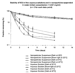
[0025] Figure 1: Relative stabilities of Indocyanine green (IR-125)
loaded nanoparticles as compared with Indocyanine green aqueous
solutions under various temperature and light exposure conditions.
[0026] Figure 2: Atomic Force Microscopic images of ICG (IR-125)
loaded PLGA nanoparticles.
CA 02516116 2005-07-15
WO 2004/064751 PCT/US2004/001472
-6-
[0027] Figure 3: Evaluation of particle size through Atomic Force
Microscopy of ICG (IR-125) loaded PLGA nanoparticles.
[0028] Figure 4: Intracellular uptake of Indocyanine green (ICG), by
C-33A cancer cell line, when incubated with ICG solution and ICG loaded
nanoparticle suspension.
[0029] Figure 5: Relative intracellular uptake of Indocyanine green
(ICG), by C-33A and B16-F10 cancer cell lines, when incubated with ICG
loaded nanoparticle suspension.
[0030] Figure 6: Effect of initial PEG concentration used for
nanoparticle coating on the amount of PEG coated on the nanoparticles.
DETAILED DESCRIPTION OF THE INVENTION
[0031 ] The present invention relates to the discovery that polymeric
nanoparticles ranging in diameter from about 1 to 1000 nm efficiently entrap
imaging substances such as dyes, particularly, near-IR dyes, and
substantially enhance their half-life and stability in vitro and in vivo. The
nanoparticles of the invention are made of biocompatible and biodegradable
polymers such as PLGA.
Nanoparticles
[0032] The nanoparticles of the invention range in size from about 1
nm to about 1000 nm in diameter, but are not necessarily limited to 1000
nm. The size of the nanoparticles may extend into the micrometer range for
certain applications or routes of administration, such as, for example, for
use as implants. Preferred nanoparticle diameters range from about 50 to
800 nm, and more preferably from about 100 to 350 nm. One skilled in the
art would readily recognize that the size of the nanoparticle may vary
CA 02516116 2005-07-15
WO 2004/064751 PCT/US2004/001472
-7-
depending upon the method of preparation, clinical application, and imaging
substance used.
[0033] The present invention further relates to nanoparticles made of
biocompatible and biodegradable polymeric materials. In a preferred
embodiment, the nanoparticles are made of PLGA. PLGA, per se, is FDA
approved and has been used in drug delivery systems for a variety of drugs
via numerous routes of administration including, but not limited to,
subcutaneous, intravenous, ocular, oral and intramuscular. The PLGA
nanoparticles made according to the invention form spherical or nearly-
spherical matrix structures that embed or entrap (i.e. encapsulate) dye or
other substances or molecules within the spaces of the matrix during the
entrapment process.
[0034] Although PLGA is a preferred material, this invention
contemplates that other polymeric colloidal carriers would work equally as
well. Examples of such polymers include, but are not limited to, PLA, PGA,
Chitosan, and Albumin.
[0035] In a further embodiment, the nanoparticles of the invention
entrap fluorescent dyes of the general class known as cyanine dyes, with
emission wavelengths of between 550 nm to 1000 nm. These dyes may
contain additional chemical groups that influence the spectral properties of
the dyes. Preferred dyes for use in the invention are tricarbocyanine dyes,
such as indocyanine green (ICG). The sodium iodide salt of ICG (ICG-Nal)
is used in medical diagnosis, such as for the evaluation of cardiac output,
liver function, microcirculation of skin flaps, and visualization of the
retinal
and choroidal vasculatures. In addition, ICG is useful in photodynamic
therapy.
[0036] An important motivation for using ICG in the invention is that
its absorption peak (~800nm) and its most intense fluorescence 0820 nm)
CA 02516116 2005-07-15
WO 2004/064751 PCT/US2004/001472
_$-
are at wavelengths for which blood and other tissues are relatively
transparent. As a result, ICG can conveniently be measured in blood
samples or transcutaneously by spectrophotometry or spectrofluorometry.
Furthermore, because ~95% of the dye in plasma is protein-bound, it
remains largely intravascular, which is important in clinical applications
where dye diffusion out of the vascular compartment can confound
interpretation of results.
[0037] In addition to ICG, the nanoparticle system of the invention
could be used to stabilize other near-IR fluorescent dyes, or other
fluorescent dye classes, or related dyes, or imaging substances that are
particularly suited for the uses described herein. One skilled in the art
would be able to select appropriate dyes based upon their desired emission
and absorption properties and the specific clinical or biological application
for which they are needed. The nanoparticle technology described herein
would work equally as well to stabilize and enhance the utility of such dyes.
[0038] In yet a further embodiment, the nanoparticles of the invention
may contain targeting molecules that facilitate localized delivery of the
nanoparticle-dye complex to a specific tissue or cell-type. This embodiment
is of particular importance for therapeutic applications, such as the
treatment of cancer. Examples of targeting molecules include, but are not
limited to, antibodies or antibody fragments, proteins or polypeptides,
polysaccharides, DNA, RNA, chemical moieties, magnetic moieties and any
combination thereof. In addition, cell-specific surface markers (such as CD4,
CDB, CD19, etc) or specific receptors (such as CD40, transferrin, folate, or
mannose) could be targeted by attaching a specific antibody or ligand to the
surface of the nanoparticle.
[0039] This invention also contemplates that other pharmaceutical
agents or drugs or chemicals may be co-entrapped or encapsulated in the
CA 02516116 2005-07-15
WO 2004/064751 PCT/US2004/001472
_g_
nanoparticle system to further augment a therapeutic effect or other
intended purpose.
[0040] In a preferred embodiment, the present invention relates to
nanoparticles that contain, or are coated with, substances or agents that
further increase the stability of the nanoparticle-dye complex. For example,
coating nanoparticles with substances such as PEG may further increase
the stability and prolong the half-life of the nanoparticles in vivo. Studies
have shown that the elimination half-life of PLGA nanoparticles that were
not coated with PEG was approximately 12-14 minutes in mice. In contrast,
the PEG-coated PLGA nanoparticles had prolonged circulation times in
vivo, with an elimination half-life of 4-5 hrs in mice. (see, Ya-Ping Li, Yuan-
Ying Pei, Xian-Ying Zhang, Zhou-Hui Gu, Zhao-Hui Zhou, Wei-Fang Yuan,
Jian-Jun Zhou, Jian-Hua Zhu and Xiu-Jian Gao. PEGylated PLGA
nanoparticles as protein carriers: synthesis, preparation and biodistribution
in rats, J. Controlled Release, Volume 71, Issue 2, 2 April 2001, Pages 203-
211 ).
[0041 ] In another embodiment, the nanoparticles can be injected
locally in the tissue or be locally implanted. The nanoparticles may stay at
the injection site for a few days to months and gradually release the loaded
content while the particles are degraded over the time period depending
upon the implantation site. Studies of microparticles in in vitro simulated
environments and in vivo in animal models have shown that the particles
stay at the implantation site for over a month (see, for example, Fangjing
Wang, Timothy Lee and Chi-Hwa Wang, PEG modulated release of
etanidazole from implantable PLGA/PDLA discs, Biomaterials, Volume 23,
Issue 17, September 2002, Pages 3555-3566; R. V. Diaz, M. Llabres and C.
Evora, One-month sustained release microspheres of '251-bovine calcitonin:
In vitro-in vivo studies, J. of Controlled Release, Volume 59, Issue 1, 1
CA 02516116 2005-07-15
WO 2004/064751 PCT/US2004/001472
- 10-
May 1999, Pages 55-62; T. Hickey, D. Kreutzer, D. J. Burgess and F.
Moussy, Dexamethasone/PLGA microspheres for continuous delivery of an
anti-inflammatory drug for implantable medical devices, 8iomaterials,
Volume 23, Issue 7, 1 April 2002, Pages 1649-1656; Christian Witt and
Thomas Kissel, Morphological characterization of microspheres, films and
implants prepared from poly(lactide-co-glycolide) and ABA triblock
copolymers: is the erosion controlled by degradation, swelling or diffusion?,
European J. Pharmaceutics 8iopharmaceutics, Volume 51, Issue 3, May
2001, Pages 171-181 ).
Nanoparticle Preparation
[0042] The present invention also relates to methods of preparing
nanoparticles comprising generally, of polymeric materials such as PLGA,
and polyvinyl alcohol (PVA). The ICG dye is preferably IR-125, a laser
grade dye. In a preferred embodiment, the method involves dissolving the
PLGA in acetonitrile to form a solution, and dissolving the IR dye in
methanol to obtain a second solution.
[0043] The PVA is added to distilled water to form a 4% PVA solution
This aqueous solution is then filtered, for example, with a 0.22N syringe
filter.
[0044] Following the above steps, 2 parts of the PLGA solution, and 1
part of the IR-125 solution are mixed to form a homogenous PLGA/IR-125
solution. This homogenous solution is then added drop by drop into 15
parts of the aqueous PVA solution (4% w/v) using 1000 ~I pipette tips with
an internal diameter of 0.03 inches, with continuous stirring at 700 rpm
using a laboratory magnetic stirrer. In some instances, the speed with which
the homogenous solution is dropped into the PVA solution and stirring
speed may have some effect on nanoparticle size. Very slow speeds may
lead to bigger size ranges, and faster speeds to smaller size ranges. One
CA 02516116 2005-07-15
WO 2004/064751 PCT/US2004/001472
-11-
skilled in the art would be able to determine an optimal speed to obtain the
preferred size of nanoparticle.
[0045] The nanoparticle suspension formed is then stirred for an
additional 10 minutes at 700 rpm, and then centrifuged for 20 minutes at
16,000 g.
[0046] After centrifugation the supernatant is discharged and the
nanoparticle precipitate is washed with same volume of distilled water as
the supernatant and centrifuged again at 16,000 g for 6 min. The washing
step is repeated three times. The washed nanoparticles can then be freeze-
dried and stored preferably at 0 to -20°C, until further use.
[0047] The methods of preparation described herein optimize dye
entrapment and produce nanoparticulate complexes that maintain the
activity and structural stability of co-incorporated molecules.
[0048] The weight ratio of polymer: dye to form the nanoparticles of
the invention is preferably in the range of about 100:1 to about 1000:1 to
provide efficient entrapment and stability of the dye. In a more preferred
embodiment, the ratio is about 800:1 to about 1000:1.
[0049] As mentioned above, the nanoparticle-entrapped dye system,
may contain targeting molecules to deliver.the nanoparticles and dye to
specific tissue sites or cells in vivo. For example, cell specific monoclonal
antibodies could be attached to the nanoparticles in order to target the IR
dye or other agent to a specific cell type or organ in vivo, including tumor
cells. Alternatively, chemical agents, cell-specific peptides, or ligands, may
be incorporated in the nanoparticle, or used to modify one or more of the
polymer constituents. For example, after entrapment of the dye in the
nanoparticles, ligands may be added directly to the exterior surface of the
nanoparticle-dye complexes. The stability of the nanoparticle and presence
of reactive functional groups on the polymer chain on the surface allow
CA 02516116 2005-07-15
WO 2004/064751 PCT/US2004/001472
-12-
ligands to be directly added to their exterior surface. Examples include, but
are not limited to, the attachment of PEG chains on the surface of the
nanoparticles to prolong circulation of the nanoparticles in vivo; thus,
increasing passive targeting to tissues or cells such as tumors.
[0050] As mentioned above, many ligands may be employed for this
step of nanoparticle preparation, depending on the cell-type targeted for
nanoparticle delivery. Those skilled in the art would readily recognize that
any ligand which enhances uptake or localization in a given tissue may be
an appropriate candidate for targeting the nanoparticle-entrapped dye
system of the invention.
Compositions Comprisingi Nanoparticles and IR Dyes
[0051 ] The nanoparticle system of the invention may be formulated in
a variety of ways depending on the application. Such applications include,
but are not limited to, biomedical and therapeutic applications. The
invention therefore includes within its scope compositions comprising at
least one nanoparticle-dye complex formulated for use in human or
veterinary medicine, or other non-medical application. Such compositions
may be presented for use with physiologically acceptable vehicles or
excipients, optionally with supplementary medicinal agents. The vehicles
and excipients include, but are not limited to, water, glucose, saline, and
phosphate buffered saline.
[0052] Formulations for injection may be. presented in unit dosage
form in ampoules, or with an added preservative to prevent contamination,
as needed, in multi-dose containers. The composition may take such forms
as suspensions, colloidal solutions or emulsions in oily or aqueous vehicles,
and may contain formulatory agents such as suspending, stabilizing and/or
dispersing agents. For example, parenteral administration may be done by
bolus injection or continuous infusion. Alternatively, the nanoparticles may
CA 02516116 2005-07-15
WO 2004/064751 PCT/US2004/001472
-13-
be in powder form for reconstitution with a suitable vehicle, e.g. sterile
water, before use.
[0053] The nanoparticle-dye complexes of the invention may be
formulated for administration in any convenient way. For example,
transdermal administration may be in the form of a patch applied on the
skin. For oral administration, the pharmaceutical compositions may take the
form of, for example, tablets, capsules, powders, solutions, syrups or
suspensions prepared by conventional means with acceptable excipients.
[0054] Dosages will depend on the extent to which it is possible to
present dye, as well as any other active agents, to the target tissue.
Methods of Use of Nanoparticle Stabilized Dyes for Bioimagina
[0055] In a further embodiment, compositions comprising
nanoparticles may be used for bioimaging. For example, nanoparticles
containing a near IR-dye and a targeting molecule will localize the delivery
of the nanoparticulate-IR dye system to the site of a tumor and facilitate
contact and uptake of the nanoparticles by the tumor cells. After the
nanoparticles have been localized to the tumor, the IR dye can be activated
with a laser leading to the infra-red wavelength emission (fluorescence) of
the IR dye. This fluorescence can be detected with help of a suitable device
such as a CCD camera placed outside the body or through endoscopic
means.
Therapeutic Applications of Nanoparticles
[0056] In another embodiment, compositions comprising the
nanoparticle system of the invention may be used to treat a subject having a
disease including, but not limited to, infectious disease or cancer. The
nanoparticle system enhances the uptake by cells such as cancer cells of
the dyes, even at lower concentrations than the dye solution alone as
CA 02516116 2005-07-15
WO 2004/064751 PCT/US2004/001472
-14-
shown in Figure 4. The nanoparticle system provides the advantage of
increasing the efficiency of delivery of substances such as ICG to cells both
in in vitro and in vivo conditions for imaging and treatment of diseases such
as cancer. In another embodiment, cancer treatments may be based on the
development of a nanoparticle system that contains a targeting molecule to
target and kill cancer cells. One such therapy involving near-IR dyes, is
photodynamic tumor therapy. Briefly, nanoparticles containing near IR-dye
and a targeting molecule will localize the delivery of the nanoparticle-IR dye
complex to the site of a tumor and facilitate contact and uptake of the
nanoparticles by the tumor cells. After the nanoparticles have been
localized to the tumor, the IR dye can be activated with a laser leading to
killing of the tumor cells due to the singlet oxygen production of the dye in
the presence of cell water, which is lethal for the tumor cells. The
nanoparticles may also co-entrap other active agents to augment the
therapeutic efficacy of the nanoparticle-IR dye complex.
[0057] All of the references mentioned in the present application are
incorporated in their entirety into this application by reference thereto.
[0058] The following Examples serve to illustrate further the present
invention and are not to be construed as limiting its scope in any way.
EXAMPLES
Exam~~le 1
Prea~aration of IR-125 loaded PLGA nanoparticles:
Materials:
[0059] Poly(dl-lactic-co-glycolic acid) (PLGA) 50:50 and Polyvinyl
alcohol (PVA) 88%-89% hydrolyzed were purchased from Sigma (Sigma
Chemical Co., St. Louis, MO.). Indocyanine green (IR-25, laser grade) was
CA 02516116 2005-07-15
WO 2004/064751 PCT/US2004/001472
-15-
obtained from Fisher Scientific (Fisher Scientific Inc., Pittsburgh, PA). All
organic chemicals and solvents were of reagent grade. Distilled water is
filtered by 0.22N syringe filter (Syrfil- MF Whatman Inc., Clifton, NJ) before
use in the preparation process.
Preparation of IR-125 loaded PLGA nanoparticles:
[0060] 1. Nanoparticles were prepared by modified spontaneous
emulsification solvent diffusion method. Briefly, PLGA (800 mg) was
dissolved in16 mL Acetonitrile to form a PLGA solution and IR-125 was
dissolved in Methanol to make 0.125 mg/mL IR-125 solution.
[0061 ] 2. Also, PVA (4g) was added to about 100 mL distilled
water to form 4 % w/v PVA aqueous solution. This aqueous PVA solution is
then filtered using 0.22N syringe filter.
[0062] 3. To 16 ml of PLGA solution in Acetonitrile, 8 ml of IR-125
solution in Methanol was added to form a homogenous solution PLGA and
IR-125 in Acetonitrile-Methanol solvent mixture.
[0063] 4. This homogenous solution (24 ml) was then added
drop-wise into 120 mL of aqueous PVA solution (4% w/v) using 1000 pL
pipette tips (VWR International, internal diameter 0.03 inch), with continuous
stirring at 700 rpm using a laboratory magnetic stirrer.
[0064] 5. The nanoparticle suspension formed is then allowed to
stir for another 10 minutes at 700 rpm. The suspension was then
centrifuged for 20 minutes at 16,000 g.
[0065] 6. After centrifugation the supernatant was discharged
and the nanoparticles precipitate left behind is then washed by using same
volume of distilled water as of supernatant and centrifuged at 16,000 g for 6
minutes.
CA 02516116 2005-07-15
WO 2004/064751 PCT/US2004/001472
- 16-
[0066] 7. The washing process was repeated three times. The
washed nanoparticles were then freeze-dried using Freezone 4.5, freeze-
drying system (Labconco, Kansas City, Missouri) for 36 hours.
[0067] 8. The dried nanoparticles were stored at -20 °C in the
dark until further use.
Table 1
Effects of Nanoparticle Formulation on ICG ~(IR-125) Incorporation
Table 1 demonstrates various ICG entrapment efficiencies in nanoparticles
prepared by the method in Example 1 using various amounts of ICG and PLGA
in the formulation.
FormulationAmount Amount of Dye Content Dye
Number of Polymer in (%) Entrapment
Dye in formulation (%)
formulation(mg)
(mg)
1 1 100 0.21 9.92
2 5 100 0.29 2.92
3 10 100 0.17 1.14
4 1 800 0.20 74.47
Examule 2
Relative Stabilities of Indocyanine Green (ICG) in Aqueous Solutions
Compared with ICG in Nanoparticles Prepared According to the
Method Described in Example 1.
[0001 ] ICG solution of 1 pg/mL was prepared by dissolving 10 mg
ICG in 100 mL distilled water and further diluted 100 times in distilled
water.
About 50 mg ICG nanoparticles were suspended in 100 mL distilled water to
obtain 1 ~g/mL ICG concentration. The two samples were then placed into
several transparent centrifuge tubes and placed at different conditions. At
the prefixed time points, the peak fluorescent intensity of these samples was
measured at excitation wavelength of 786 nm. The fractions of ICG that
CA 02516116 2005-07-15
WO 2004/064751 PCT/US2004/001472
-17-
remained were calculated by comparing the fluorescent intensity with the
initial fluorescent intensity as shown in Figure 1. Atomic Force Microscopic
images of ICG (IR-125) loaded PLGA nanoparticles are shown in Figure 2.
Evaluation of particle size through Atomic Force Microscopy of ICG (IR-125)
loaded PLGA nanoparticles is shown in Figure 3.
Example 3
Intracellular uptake of Indocyanine green ~(ICG), bar C-33A cancer cell
line, when incubated with ICG solution and ICG loaded nanoparticles.
[0069j Intracellular uptake of Indocyanine green (ICG), by C-33A
cancer cell line, when incubated with ICG solution and ICG loaded
nanoparticle suspension is shown in Figure 4. The nanoparticles used were
prepared according to the method described in Example 1.
[0001 j ICG solution of 50 ~.M was prepared by dissolving ICG in the
cell culture medium and this solution was further diluted in the cell culture
medium to get concentrations from 0.00022 to 50 ~M. About 10 mg ICG
nanoparticles were suspended in 10 mL cell culture medium to obtain 1
mg/mL nanoparticle suspension equivalent to 0.022 ~M ICG concentration.
This suspension was then further diluted to get the nanoparticle suspension
of 0.00022 to 0.011 ~M ICG concentrations. For the intracellular uptake
studies, cells were seeded in 6-well cell culture plates at the concentration
of 2 X 10⁵ in 4 ml growth medium per well. After overnight attachment the
medium was replaced with ICG solution of different concentrations (0.00022
- 0.022 ~M) or nanoparticle suspension of different concentrations (0.00022
- 0.022 ~M) and the cells were incubated for 24 hrs at 37 °C in the
dark.
After 24 hrs of incubation the medium was removed and the cells were
washed four times with phosphate buffer saline. ICG was then extracted
from the cells in each well by incubation with 1 ml of dimethylsulfoxide
(DMSO). The fluorescence of ICG in DMSO was measured and ICG
CA 02516116 2005-07-15
WO 2004/064751 PCT/US2004/001472
-18-
concentrations were calculated by a using a calibration curve of ICG in
DMSO.
Example 4
Relative intracellular uptake of Indocyanine gireen i(ICGJ~, by C-33A and
B16-F10 cancer cell lines, when incubated with ICG loaded
nanoparticle sus~~ension.
[0071 ] Relative intracellular uptake of Indocyanine green (ICG), by C-
33A and B16- F10 cancer cell lines, when incubated with ICG loaded
nanoparticle suspension is shown in Figure 5. The nanoparticles used were
prepared according to the method described in Example 1.
[0072] About 10 mg ICG nanoparticles were suspended in 10 mL cell
culture medium to obtain 1 mg/mL nanoparticle suspension equivalent to
0.022 pM ICG concentration. This suspension was then further diluted to
get the nanoparticle suspension of 0.00022 to 0.011 ~M ICG
concentrations. For the intracellular uptake studies, cells were seeded in 6-
well cell culture plates at the concentration of 2 X 10⁵ in 4 ml growth medium
per well. After overnight attachment the medium was replaced with
nanoparticle suspension of different concentrations (0.00022 - 0.022 pM)
and the cells were incubated for 24 hrs at 37 °C in the dark. After 24
hrs of
incubation the medium was removed and the cells were washed four times
with phosphate buffer saline. ICG was then extracted from the cells in each
well by incubation with 1 ml of dimethylsulfoxide (DMSO). The fluorescence
of ICG in DMSO was measured and ICG concentrations were calculated by
a using a calibration curve of ICG in DMSO.
CA 02516116 2005-07-15
WO 2004/064751 PCT/US2004/001472
- 19-
Example 5
PEG coatings on the surface of nanoparticles.
Materials:
[0073] Fluorescein - Polyethylene glycol (PEG-Fluorescein), MW
5,000 Da, was obtained from Nektar (Nektar Therapeutics, San Carlos, CA).
The nanoparticles used were prepared according to the method described
in Example One.
PEG Coating on the surface of PLGA nanoparticles:
[0074] 1. PEG-Fluorescein was dissolved in distilled water to get
0.5, 1 and 2 % w/v solutions.
[0075] 2. Then in 2 ml of each of the above prepared solutions,
25 mg of nanoparticles were suspended. The suspensions were incubated
for 24 hours.
[0076] 3. After 24 hours of incubation the nanoparticle
suspensions were centrifuged at 16,000 g for 5 minutes.
[0077] 4. After centrifugation the supernatant was discharged
and the nanoparticles precipitate left behind was resuspended in phosphate
buffer saline (PBS) for further studies.
CA 02516116 2005-07-15
WO 2004/064751 PCT/US2004/001472
-20-
Example 6
Effect of initial PEG concentration on the amount of PEG coated on
nanoparticles.
[0078] Effect of initial PEG concentration used for nanoparticle
coating on the amount of PEG coated on the nanoparticles is shown in
Figure 6.
[0079] The nanoparticles used were prepared according to the
method described in Example 1. The nanoparticles were incubated for 24
hours with different concentrations of PEG-Fluorescein (0.5 - 2 %w/v) for
surface coating of the nanoparticles. For measuring the fluorescence
associated with the nanoparticles after coating, 1 mg of PEG-Fluorescein
coated nanoparticles were suspended in 1 ml of PBS. The peak
fluorescence intensity of these samples was measured at excitation
wavelength of 520 nm.