Note: Descriptions are shown in the official language in which they were submitted.
CA 02516510 2006-02-20
METHOD OF USING ADIPOSE TISSUE-DERIVED CELLS IN THE TREATMENT
OF CARDIOVASCULAR CONDITIONS
BACKGROUND OF THE INVENTION
1. Field of the Invention
This invention generally relates to cells derived from adipose tissue, and
more
particularly, to adipose-derived stem and progenitor cells, methods of using
adipose-derived
stem and progenitor cells, compositions containing adipose-derived stem and
progenitor
cells, and systems for preparing and using adipose-derived stem and progenitor
cells, which
are used to treat cardiovascular diseases and disorders.
2. Description of Related Art
Cardiovascular diseases and disorders are the leading cause of death and
disability
in all industrialized nations. In the United States alone, cardiovascular
disease accounts for
about 40 percent of the mortality rate and affects 58 million Americans
(American-Heart-
Association, 2002). One of the primary factors that renders cardiovascular
disease
particularly devastating is the heart's inability to repair itself following
damage. Since
cardiac muscle cells, i.e., myocardial cells, are unable to divide and
repopulate areas of
damage, cardiac cell loss as a result of injury or disease is largely
irreversible (Abbate et al.,
2002; Remme, 2000).
Of the available forms of therapy, human to human heart transplants have been
the
most effective in treating severe cardiovascular diseases and disorders. In
fact, the one-year
and five-year survival rate of the average cardiac transplant recipient is
currently over 70
percent. Unfortunately, however, transplantation is a severely limited form of
therapy for a
number of reasons, namely, the scarcity of suitable donors, the expense of the
procedure and
the high likelihood of graft rejection and associated problems such as
infections, renal
dysfunction and immunosuppressant related cancers (American-Heart-Association,
2002).
An alternative to transplant therapy is the use of regenerative medicine to
repair and
35
-1-
CA 02516510 2005-08-18
WO 2004/074457 PCT/US2004/005117
regenerate damaged cardiac muscle cells. Regenerative medicine harnesses, in a
clinically
targeted manner, the ability of stem cells (i.e., the unspecialized master
cells of the body) to
renew themselves indefinitely and develop into mature specialized cells. Stem
cells are found
in embryos during early stages of development, in fetal tissue and in some
adult organs and
tissue (Pera et al., 2000). Embryonic stem cells (hereinafter referred to as
"ESCs") are known
to become many if not all of the cell and tissue types of the body. ESCs not
only contain all the
genetic information of the individual but also contain the nascent capacity to
become any of the
200+ cells and tissues of the body. Thus, these cells have tremendous
potential for regenerative
medicine. For example, ESCs can be grown into specific tissues such as heart,
lung or kidney
which could then be used to repair damaged and diseased organs (Assady et al.,
2001; Jacobson
et al., 2001; Odorico et al., 2001). However, ESC derived tissues have
clinical limitations.
Since ESCs are necessarily derived from another individual, i.e., an embryo,
there is a risk that
the recipient's immune system will reject the new biological material.
Although
immunosuppressive drugs to prevent such rejection are available, such drugs
are also known to
block desirable immune responses such as those against bacterial infections
and viruses.
Moreover, the ethical debate over the source of ESCs, i.e., embryos, is well-
chronicled and
presents an additional and, perhaps, insurmountable obstacle for the
foreseeable future.
Adult stem cells (hereinafter interchangeably referred to as "ASCs") represent
an
alternative to the use of ESCs. ASCs reside quietly in many non-embryonic
tissues,
presumably waiting to respond to trauma or other destructive disease processes
so that they can
heal the injured tissue (Arvidsson et al., 2002; Bonner-Weir and Sharma, 2002;
Clarke and
Frisen, 2001; Crosby and Strain, 2001; Jiang et al., 2002a). Notably, emerging
scientific
evidence indicates that each individual carries a pool of ASCs that may share
with ESCs the
ability to become many if not all types of cells and tissues (Young et al.,
2001; Jiang et al.,
2002a; Jiang et al., 2002b; Schwartz et al., 2002). Thus, ASCs, like ESCs,
have tremendous
potential for clinical applications of regenerative medicine.
ASC populations have been shown to be present in one or more of bone marrow,
skin,
muscle, liver and brain (Jiang et al., 2002b; Alison, 1998; Crosby and Strain,
2001). However,
the frequency of ASCs in these tissues is low. For example, mesenchymal stem
cell frequency
in bone marrow is estimated at between 1 in 100,000 and 1 in 1,000,000
nucleated cells
(D'Ippolito et al., 1999; Banfi et al., 2001; Falla et al., 1993). Similarly,
extraction of ASCs
from skin involves a complicated series of cell culture steps over several
weeks (Toma et al.,
2001) and clinical application of skeletal muscle-derived ASCs requires a two
to three week
culture phase (Hagege et al., 2003). Thus, any proposed clinical application
of ASCs from such
tissues requires increasing cell number, purity, and maturity by processes of
cell purification
and cell culture.
Although cell culture steps may provide increased cell number, purity, and
maturity,
-2-
CA 02516510 2005-08-18
WO 2004/074457 PCT/US2004/005117
they do so at a cost. This cost can include one or more of the following
technical difficulties:
loss of cell function due to cell aging, loss of potentially useful non-stem
cell populations,
delays in potential application of cells to patients, increased monetary cost,
and increased risk
of contamination of cells with environmental microorganisms during culture.
Recent studies
examining the therapeutic effects of bone-marrow derived ASCs have used
essentially whole
marrow to circumvent the problems associated with cell culturing (Horwitz et
al., 2001; Orlic et
al., 2001; Stamm et al., 2003; Strauer et al., 2002). The clinical benefits,
however, have been
suboptimal, an outcome almost certainly related to the limited ASC dose and
purity inherently
available in bone marrow.
Recently, adipose tissue has been shown to be a source of ASCs (Zuk et al.,
2001; Zuk
et al., 2002). Unlike marrow, skin, muscle, liver and brain, adipose tissue is
comparably easy
to harvest in relatively large amounts (Commons et al., 2001; Katz et al.,
2001b). Furthermore,
adipose derived ASCs have been shown to possess the ability to generate
multiple tissues in
vitro, including bone, fat, cartilage, and muscle (Ashjian et al., 2003;
Mizuno et al., 2002; Zuk
et al., 2001; Zuk et al., 2002). Thus, adipose tissue presents an optimal
source for ASCs for use
in regenerative medicine. Suitable methods for harvesting adipose derived
ASCs, however, are
lacking in the art. The existing methods suffer from a number of shortcomings.
For example,
the existing methods lack the ability to optimally accommodate an aspiration
device for
removal of adipose tissue. The existing methods also lack partial or full
automation from the
harvesting of adipose tissue phase through the processing of tissue phases
(Katz et al., 2001 a).
The existing methods further lack volume capacity greater than 100ml of
adipose tissue. The
existing methods yet further lack a partially or completely closed system from
the harvesting of
adipose tissue phase through the processing of tissue phases. Finally, the
existing methods lack
disposability of components to attenuate concomitant risks of cross-
contamination of material
from one sample to another. In summary, the prior art methods for harvesting
ASCs from
adipose tissue do not overcome the technical difficulties associated with
harvesting ASCs from
skin, muscle, liver and brain described above.
Accordingly, given the tremendous therapeutic potential of ASCs, there exists
an
urgent need in the art for a device, system or method for harvesting ASCs from
adipose tissue
that produces a population of ASCs with increased yield, consistency and/or
purity and does so
rapidly and reliably with a diminished or non-existent need for post-
extraction manipulation.
Ideally, such a device, system or method would yield ASCs in a manner suitable
for direct
placement into a recipient. Access to such a device, system or method in
combination with
methods and compositions using adipose derived ASCs for the treatment of
cardiovascular
diseases and disorders would revolutionize the treatment of such disorders.
Given the
prevalence of cardiovascular disease and the scarcity of current treatment
options, such a
treatment is urgently needed.
-3-
CA 02516510 2005-08-18
WO 2004/074457 PCT/US2004/005117
SUMMARY OF THE INVENTION
The present invention is based, at least in part, on the discovery that
adipose derived
adult stem cells can be used to treat cardiovascular conditions, diseases or
disorders. The
present invention is further based on the discovery of devices, systems and
methods for
preparing adipose derived adult stem and progenitor cells. The present
invention is yet further
based on the discovery of methods and compositions of adipose derived adult
stem and
progenitor cells to treat cardiovascular conditions, diseases or disorders.
Accordingly, in one
embodiment, the present invention is directed to compositions, methods, and
systems for using
cells derived from adipose tissue that are placed directly into a recipient
along with such
additives necessary to promote, engender, or support a therapeutic
cardiovascular benefit.
In one embodiment, adipose tissue processing occurs in a system that maintains
a
closed, sterile fluid/tissue pathway. This is achieved by use of a pre-
assembled, linked set of
closed, sterile containers and tubing allowing for transfer of tissue and
fluid elements within a
closed pathway. This processing set can be linked to a series of processing
reagents (e.g.,
saline, enzymes, etc.) inserted into a device which can control the addition
of reagents,
temperature, and timing of processing thus relieving operators of the need to
manually manage
the process. In a preferred embodiment the entire procedure from tissue
extraction through
processing and placement into the recipient would all be performed in the same
facility, indeed,
even within the same room of the patient undergoing the procedure.
In accordance with one aspect of the invention, raw adipose tissue is
processed to
substantially remove mature adipocytes and connective tissue thereby obtaining
a
heterogeneous plurality of adipose tissue-derived cells suitable for placement
within the body
of a recipient. The cells maybe placed into the recipient in combination with
other cells, tissue,
tissue fragments, or other stimulators of cell growth and/or differentiation.
In a preferred
embodiment, the cells, with any of the above mentioned additives, are placed
into the person
from whom they were obtained in the context of a single operative procedure
with the intention
of deriving a therapeutic benefit to the recipient.
In one embodiment, a method of treating a patient includes steps of: a)
providing a
tissue removal system; b) removing adipose tissue from a patient using the
tissue removal
system, the adipose tissue having a concentration of stem cells; c) processing
at least a part of
the adipose tissue to obtain a concentration of stem cells other than the
concentration of stem
cells of the adipose tissue before processing; and d) administering the stem
and progenitor cells
to a patient without removing the stem and progenitor cells from the tissue
removal system
before being administered to the patient using several methods known to one of
ordinary skill
in the art, including but not limited to, intravenous, intracoronary and
endomyocardial.
A system in accordance with the invention herein disclosed includes a) a
tissue
-4-
CA 02516510 2005-08-18
WO 2004/074457 PCT/US2004/005117
collection container including i) a tissue collecting inlet port structured to
receive adipose tissue
removed from a patient; and ii) a filter disposed within the container and
being structured to
retain adipose tissue removed from a patient and to pass non-adipose tissue
removed from the
patient; b) a mixing container coupled to the tissue collection container to
receive stem cells
obtained from the adipose tissue without removal of the stem cells from the
tissue removal
system, and including an additive port for the administration of at least one
additive to mix with
the stem cells contained therein; and c) an outlet structured to permit the
cells in the mixing
container to be removed from the tissue collection system for administration
to a patient.
Any feature or combination of features described herein are included within
the scope
of the present invention provided that the features included in any such
combination are not
mutually inconsistent as will be apparent from the context, this
specification, and the
knowledge of one of ordinary skill in the art. Additional advantages and
aspects of the present
invention are apparent in the following detailed description.
BRIEF DESCRIPTION OF THE DRAWINGS
Figure 1 depicts a tissue removal system for processing adipose tissue.
Figure 2 depicts a tissue collection container of the tissue removal system of
Fig. 1.
Figure 3 is a partial cross-sectional view of the tissue collection container
of Fig. 2.
Figure 4 depicts a processing device for automating the operation of a tissue
removal
system.
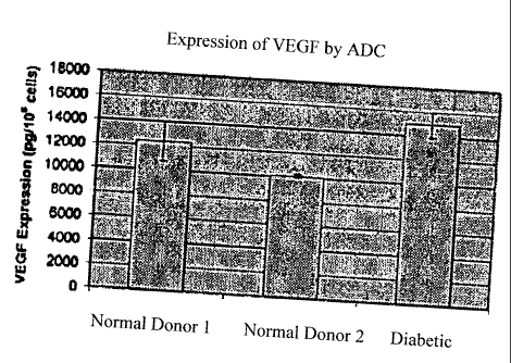
Figures 5A and 5B depict the expression of VEGF (5A) and PIGF (5B) protein by
cultured adipose derived stem cells.
Figure 6 depicts detection of endothelial progenitor cells within adipose
derived stem
cell populations.
Figures 7A and 7B depict the in vitro development of vascular structures in
both
normal (7A) and streptozotocin-treated (7B) mice.
Figure 8 depicts the increased average restoration of blood flow in hindlimb
ischemia
mice treated with adipose derived stem cell compared to a negative control.
-5-
CA 02516510 2005-08-18
WO 2004/074457 PCT/US2004/005117
Figures 9A and 9B shows that increasing adipose derived stem cell dose
improves graft
survival and angiogenesis (9A) and depicts the retention of adipose tissue
architecture in
histologic specimen (9B).
Figure 10 depicts the histological timeline of engraftment of donor derived
adipose
derived stem cells in the area of infarcted myocardium.
Figure 11 depicts dual positive staining for both beta-galactosidase and
myosin heavy
chain. Highlighted cells exhibit both blue betagalactosidase staining,
demonstrating their origin
from donor adipose tissue cells, and brown staining indicating expression of
the cardiac muscle
protein myosin heavy chain. Cells exhibiting both brown and blue staining (as
indicated by
arrows) are adipose tissue-derived cells that have taken on the phenotype of
cardiac muscle
cells.
Figure 12 depicts clusters of donor derived adipose derived stem cells in a
region of
infarcted myocardium following occlusion/reperfusion injury in the rat.
DETAILED DESCRIPTION OF THE INVENTION
The present invention provides, for the first time, proven methods for
treating
cardiovascular conditions, diseases and disorders using adipose derived stem
and progenitor
cells. Specifically, the present invention demonstrates, for the first time,
that the adipose
derived stem and progenitor cells of the invention (1) express angiogenic and
arteriogenic
growth factors, including Placenta Growth Factor (PIGF) and Vascular
Endothelial Growth
Factor (VEGF), (2) contain endothelial progenitor cells (EPC) which have a
well-established
function in blood vessel formation, (3) develop into blood vessels in vitro,
(4) support ischemic
tissue survival in vivo, (5) induce reperfusion following
occlusion/reperfusion injury of the hind
limb, (6) when injected into animals after heart injury home to the heart, and
(7) when injected
into an animals after heart injury differentiate into cells expressing markers
consistent with
their differentiation into cardiac myocytes. Accordingly, the instant
disclosure conclusively
demonstrates that the inventive adipose derived stem and progenitor cells of
the present
invention are useful for the treatment of cardiovascular diseases and
disorders.
In order that the present invention may be more readily understood, certain
terms are
first defined. Additional definitions are set forth throughout the detailed
description.
As used herein, the term "adipose tissue" refers to a tissue containing
multiple cell
types including adipocytes and microvascular cells. Adipose tissue includes
stem cells and
endothelial precursor cells. Accordingly, adipose tissue refers to fat
including the connective
tissue that stores the fat.
-6-
CA 02516510 2005-08-18
WO 2004/074457 PCT/US2004/005117
As used herein, the term "unit of adipose tissue" refers to a discrete or
measurable
amount of adipose tissue. A unit of adipose tissue may be measured by
determining the weight
and/or volume of the unit. Based on the data identified above, a unit of
processed lipoaspirate,
as removed from a patient, has a cellular component in which at least 0.1% of
the cellular
component is stem cells. In reference to the disclosure herein, a unit of
adipose tissue may
refer to the entire amount of adipose tissue removed from a patient, or an
amount that is less
than the entire amount of adipose tissue removed from a patient. Thus, a unit
of adipose tissue
may be combined with another unit of adipose tissue to form a unit of adipose
tissue that has a
weight or volume that is the sum of the individual units.
As used herein, the term "portion" refers to an amount of a material that is
less than a
whole. A minor portion refers to an amount that is less than 50%, and a major
portion refers to
an amount greater than 50%. Thus, a unit of adipose tissue that is less than
the entire amount of
adipose tissue removed from a patient is a portion of the removed adipose
tissue.
As used herein, the term "stem cell" refers to multipotent cells with the
potential to
differentiate into a variety of other cell types, which perform one or more
specific functions and
have the ability to self-renew. Some of the stem cells disclosed herein may be
pluripotent.
As used herein, the term "progenitor cell" refers to unipotent, bipotent, or
multipotent
cells with the ability to differentiate into one or more cell types, which
perform one or more
specific functions and which have limited or no ability to self-renew. Some of
the progenitor
cells disclosed herein may be pluripotent.
As used herein "stem cell number" or "stem cell frequency" refers to the
number of
colonies observed in a clonogenic assay in which adipose derived cells (ADC)
are plated at low
cell density (<10,000 cells/well) and grown in growth medium supporting MSC
growth (for
example, DMEM/F12 medium supplemented with 10% fetal calf serum, 5% horse
serum, and
antibiotic/antimycotic agents. Cells are grown for two weeks after which
cultures are stained
with hematoxylin and colonies of more than 50 cells are counted as CFU-F. Stem
cell
frequency is calculated as the number of CFU-F observed per 100 nucleated
cells plated (for
example; 15 colonies counted in a plate initiated with 1,000 nucleated ADC
cells gives a stem
cell frequency of 1.5%). Stem cell number is calculated as stem cell frequency
multiplied by
the total number of nucleated ADC cells obtained. A high percentage (100%) of
CFU-F
grown from ADC cells express the cell surface molecule CD105 which is also
expressed by
marrow-derived stem cells (Barry et al., 1999). CD105 is also expressed by
adipose tissue-
derived stem cells (Zuk et al., 2002).
As used herein, the term "processed lipoaspirate" refers to adipose tissue
that has been
processed to separate the active cellular component (e.g., the component
containing stem and
progenitor cells) from the mature adipocytes and connective tissue. This
fraction is referred to
herein as "adipose-derived cells" or "ADC." Typically, ADC refers to the
pellet of cells
-7-
CA 02516510 2005-08-18
WO 2004/074457 PCT/US2004/005117
obtained by washing and separating the cells from the adipose tissue. The
pellet is typically
obtained by centrifuging a suspension of cells so that the cells aggregate at
the bottom of a
centrifuge container.
As used herein, the phrase "cardiovascular condition, disease or disorder" is
intended
to include all disorders characterized by insufficient, undesired or abnormal
cardiac function,
e.g., ischemic heart disease, hypertensive heart disease and pulmonary
hypertensive heart
disease, valvular disease, congenital heart disease and any condition which
leads to congestive
heart failure in a subject, particularly a human subject. Insufficient or
abnormal cardiac
function can be the result of disease, injury and/or aging. By way of
background, a response to
myocardial injury follows a well-defined path in which some cells die while
others enter a state
of hibernation where they are not yet dead but are dysfunctional. This is
followed by
infiltration of inflammatory cells, deposition of collagen as part of
scarring, all of which happen
in parallel with in-growth of new blood vessels and a degree of continued cell
death. As used
herein, the term "ischemia" refers to any localized tissue ischemia due to
reduction of the
inflow of blood. The term "myocardial ischemia" refers to circulatory
disturbances caused by
coronary atherosclerosis and/or inadequate oxygen supply to the myocardium.
For example, an
acute myocardial infarction represents an irreversible ischemic insult to
myocardial tissue. This
insult results from an occlusive (e.g., thrombotic or embolic) event in the
coronary circulation
and produces an environment in which the myocardial metabolic demands exceed
the supply of
oxygen to the myocardial tissue.
As used herein, the term "angiogenesis" refers to the process by which new
blood
vessels are generated from existing vasculature and tissue (Folkman, 1995).
The phrase "repair
or remodeling" refers to the reformation of existing vasculature. The
alleviation of tissue
ischemia is critically dependent upon angiogenesis. The spontaneous growth of
new blood
vessels provides collateral circulation in and around an ischemic area,
improves blood flow,
and alleviates the symptoms caused by the ischernia. As used herein, the term
"angiogenic
factor" or "angiogenic protein" refers to any known protein capable of
promoting growth of
new blood vessels from existing vasculature ("angiogenesis"). Suitable
angiogenic factors for
use in the invention include, but are not limited to, Placenta Growth Factor
(Luttun et al.,
2002), Macrophage Colony Stimulating Factor (Aharinejad et al., 1995),
Granulocyte
Macrophage Colony Stimulating Factor (Buschmann et al., 2003), Vascular
Endothelial
Growth Factor (VEGF)-A, VEGF-A, VEGF-B, VEGF-C, VEGF-D, VEGF-E (Mints et al.,
2002), neuropilin (Wang et al., 2003), fibroblast growth factor (FGF)-1, FGF-
2(bFGF), FGF-3,
FGF-4, FGF-5, FGF-6 (Botta et al., 2000), Angiopoietin 1, Angiopoietin 2
(Sundberg et al.,
2002), erythropoietin (Ribatti et al., 2003), BMP-2, BMP-4, BMP-7 (Carano and
Filvaroff,
2003), TGF-beta (Xiong et al., 2002), IGF-1 (Shigematsu et al., 1999),
Osteopontin (Asou et
al., 2001), Pleiotropin (Beecken et al., 2000), Activin (Lamouille et al.,
2002), Endothelin-1
-8-
CA 02516510 2006-02-20
(Bagnato and Spinella, 2003) and combinations thereof. Angiogenic factors can
act
independently, or in combination with one another. When in combination,
angiogenic factors
can also act synergistically, whereby the combined effect of the factors is
greater than the sum of
the effects of the individual factors taken separately. The term "angiogenic
factor" or
"angiogenic protein" also encompasses functional analogues of such factors.
Functional
analogues include, for example, functional portions of the factors. Functional
analogues also
include anti-idiotypic antibodies which bind to the receptors of the factors
and, thus, mimic the
activity of the factors in promoting angiogenesis and/or tissue remodeling.
Methods for
generating such anti-idiotypic antibodies are well known in the art and are
described, for
example, in WO 97/23510.
Angiogenic factors used in the present invention can be produced or obtained
from any
suitable source. For example, the factors can be purified from their native
sources, or produced
synthetically or by recombinant expression. The factors can be administered to
patients as a
protein composition. Alternatively, the factors can be administered in the
form of an expression
plasmid encoding the factors. The construction of suitable expression plasmids
is well known in
the art. Suitable vectors for constructing expression plasmids include, for
example, adenoviral
vectors, retroviral vectors, adeno-associated viral vectors, RNA vectors,
liposomes, cationic
lipids, lentiviral vectors and transposons.
As used herein, the term "arteriogenesis" refers to the process of enhancing
growth of
collateral arteries and/or other arteries from pre-existing arteriolar
connections (Carmeliet, 2000;
Scholz et al., 2001; Scholz et at., 2002). More particularly, arteriogenesis
is the in situ
recruitment and expansion of arteries by proliferation of endothelial and
smooth muscle cells
from pre-existing arteriolar connections supplying blood to ischemic tissue,
tumor or site of
inflammation. These vessels largely grow outside the affected tissue and are
important for the
delivery of nutrients to the ischemic territory, the tumor or the site of
inflammation.
Arteriogenesis is part of the normal response to myocardial ischemia (Mills et
al., 2000;
Monteiro et at., 2003). In addition, the common surgical technique of a
coronary artery bypass
graft (CABG) is, in effect, no more than creation of an artificial collateral
vessel (Sergeant et at.,
1997). Thus, processes which enhance arteriogenesis following an infarct will
improve blood
flow to ischemic tissue resulting in decreased cell death and decreased
infarct size. These
improvements will result in improved cardiac function and therapeutic benefit.
As used herein, the term "treating" includes reducing or alleviating at least
one adverse
effect or symptom of a cardiovascular condition, disease or disorder, i.e.,
any disorder
characterized by insufficient or undesired cardiac function. Adverse effects
or symptoms of
cardiac disorders are well-known in the art and include, but are not limited
to, dyspnea, chest
pain, palpitations, dizziness, syncope, edema, edema, cyanosis, pallor,
fatigue and death.
As used herein, the terms "administering," "introducing" and "transplanting"
are
used
-9-
CA 02516510 2011-09-08
interchangeably herein and refer to the placement of the ADC of the invention
into a subject by
a method or route which results in at least partial localization of the ADC at
a desired site. The
ADC can be administered by any appropriate route which results in delivery to
a desired
location in the subject where at least a portion of the cells or components of
the cells remain
viable. The period of viability of the cells after administration to a subject
can be as short as a
few hours, e.g., twenty-four hours, to a few days, to as long as several
years.
As used herein, the term "subject" includes warm-blooded animals, preferably
mammals, including humans. In a preferred embodiment, the subject is a
primate. In an even
more preferred embodiment, the subject is a human..
Reference will now be made in detail to the presently preferred embodiments of
the
invention, examples of which are illustrated in the accompanying figures.
Wherever possible,
the same or similar reference numbers are used in the drawings and the
description to refer to
the same or like parts. If should be noted that the drawings are in simplified
form and are not to
precise scale. In reference to the disclosure herein, for purposes of
convenience and clarity
only, directional terms, such as, top, bottom, left, right, up, down, over,
above, below, beneath,
rear, and front, are. used with respect to the accompanying drawings. Such
directional terms
should not be construed to limit the scope of the invention in any manner.
Although the disclosure herein refers to certain illustrated embodiments, it
is to be
understood that these embodiments are presented by way of example and not by
way of
limitation. The intent of the following detailed description, although
discussing exemplary
embodiments, is to be construed to cover all modifications, alternatives, and
equivalents of the
embodiments as may fall within the scope of the invention as defined by the
appended claims. The present invention may be practiced in conjunction with
various cell or
tissue separation techniques that are conventionally used in the art, and only
so much of the
commonly practiced process steps are included herein as are necessary to
provide an
understanding of the present invention.
Accordingly, in one embodiment, the present invention is directed to a cell
population
present in adipose tissue, and systems and methods for administering the cell
population into a
human or animal patient for the treatment of cardiovascular diseases and
disorders. The cell
population of the adipose tissue may be used as a source of cells for
therapeutic applications.
Among other things, the cells may be used for regenerative medicine, such as
diseases that can
be treated with regenerating cells, including cardiovascular diseases and
disorders. The cells of
the population may be administered to a patient suffering from a
cardiovascular disease or
disorder without other adipocytes or connective tissue.
In particular, the present invention is directed to adipose tissue-derived
cells and
methods of using same that have several properties which can contribute to
minimizing damage
and promoting myocardial repair and regeneration during this process. These
include, among
-10-
CA 02516510 2005-08-18
WO 2004/074457 PCT/US2004/005117
others: the ability to synthesize and secrete growth factors stimulating new
blood vessel
formation; the ability to synthesize and secrete growth factors stimulating
cell survival and
proliferation; the ability to proliferate and differentiate into cells
directly participating in new
blood vessel formation; the ability to engraft damaged myocardium and inhibit
scar formation
(collagen deposition and cross-linking); the ability to proliferate and
differentiate into muscle
cells capable of contributing to myocardial contractility; and the ability to
proliferate and
differentiate into myocardial cells.
I. Methods of the Invention
1. Methods of Obtaining Processed Lipoaspirate (ADC)
It has been discovered that adipose tissue is an especially rich source of
stem and
progenitor cells. This finding may be due, at least in part, to the ease of
removal of the major
non-stem cell component of adipose tissue, the adipocyte. Thus, in both human
and animal
studies, processed lipoaspirate (ADC) contains stem cells at a frequency of at
least 0.1%, and
more typically greater than 0.5%. In certain embodiments of the invention, ADC
has been
obtained which contains between about 2-12% stem cells. In even further
embodiments, the
ADC is processed to obtain a population of cells where the stem cells
constitute up to 100% of
the cells in the population. The purity/frequency of stem cells obtained in
accordance with the
invention herein disclosed is substantially greater than the published
frequency of 1 in 100,000
(0.001%) in marrow (D'Ippolito et al., 1999; Banfi et al., 2001; Falla et al.,
1993; Muschler et
al., 2001). Furthermore, collection of adipose tissue is associated with lower
morbidity than
collection of a similar volume of marrow (Nishimori et al., 2002). In
addition, adipose tissue
contains endothelial precursor cells, which are capable of providing therapy
to patients (see
(Asahara et al., 1999; Kaushal et al., 2001; Kawarnoto et al., 2003; Kawarnoto
et aL, 2001)).
In practicing the methods disclosed herein, the cells that are administered to
a patient
are obtained from adipose tissue. Adipose tissue can be obtained by any method
known to a
person of ordinary skill in the art. For example, adipose tissue may be
removed from a patient
by suction-assisted lipoplasty, ultrasound-assisted lipoplasty, and excisional
lipectomy. In
addition, the procedures may include a combination of such procedures, such as
a combination
of excisional lipectomy and suction-assisted lipoplasty. As the tissue or some
fraction thereof
is intended for re-implantation into a patient the adipose tissue should be
collected in a manner
that preserves the viability of the cellular component and that minimizes the
likelihood of
contamination of the tissue with potentially infectious organisms, such as
bacteria and/or
viruses. Thus, the tissue extraction should be performed in a sterile or
aseptic manner to
minimize contamination. Suction assisted lipoplasty may be desirable to remove
the adipose
tissue from a patient as it provides a minimally invasive method of collecting
tissue with
- 11 -
CA 02516510 2005-08-18
WO 2004/074457 PCT/US2004/005117
minimal potential for stem cell damage that may be associated with other
techniques, such as
ultrasound assisted lipoplasty.
For suction-assisted lipoplastic procedures, adipose tissue is collected by
insertion of a
cannula into or near an adipose tissue depot present in the patient followed
by aspiration of the
adipose into a suction device. In one embodiment, a small cannula may be
coupled to a
syringe, and the adipose tissue may be aspirated using manual force (Asken,
1990). Using a
syringe or other similar device may be desirable to harvest relatively
moderate amounts of
adipose tissue (e.g., from 0.1 ml to several hundred milliliters of adipose
tissue). Procedures
employing these relatively small devices have the advantage that the
procedures can be
performed with only local anesthesia, as opposed to general anesthesia. Larger
volumes of
adipose tissue above this range (e.g., greater than several hundred
milliliters) may require
general anesthesia at the discretion of the donor and the person performing
the collection
procedure. When larger volumes of adipose tissue are desired to be removed,
relatively larger
cannulas and automated suction devices may be employed in the procedure
(Commons et al.,
2001).
Excisional lipectomy procedures include, and are not limited to, procedures in
which
adipose tissue-containing tissues (e.g., skin) is removed as an incidental
part of the procedure;
that is, where the primary purpose of the surgery is the removal of tissue
(e.g., skin in bariatric
or cosmetic surgery) and in which adipose tissue is removed along with the
tissue of primary
interest.
The adipose tissue that is removed from a patient is collected into a device
for further
processing. As discussed herein, and in one embodiment, the device is designed
for and
dedicated to the purpose of collecting tissue for manufacture of a processed
adipose tissue cell
population, which includes stem cells and/or endothelial precursor cells. In
other embodiments,
the device may be any conventional device that is typically used for tissue
collection by
physicians performing the extraction procedure.
The amount of tissue collected will be dependent on a number of variables
including,
but not limited to, the body mass index of the donor, the availability of
accessible adipose
tissue harvest sites, concomitant and pre-existing medications and conditions
(such as
anticoagulant therapy), and the clinical purpose for which the tissue is being
collected.
Experience with transplant of hematopoietic stem cells (bone marrow or
umbilical cord blood-
derived stem cells used to regenerate the recipient's blood cell-forming
capacity) shows that
engraftment is cell dose-dependent with threshold effects (Smith and
Sweetenham, 1995;
Barker et al., 2001). Thus, it is likely that the general principle that "more
is better" will be
applied within the limits set by other variables and that where feasible the
harvest will collect
as much tissue as possible.
It has been discovered that the stem cell percentage of 100 ml of adipose
tissue
-12-
CA 02516510 2005-08-18
WO 2004/074457 PCT/US2004/005117
extracted from a lean individual is greater than that extracted from an obese
donor (Table 1).
This reflects a dilutive effect of the increased fat content in the obese
individual. Therefore, it
may be desirable, in accordance with one aspect of the invention, to obtain
larger amounts of
tissue from overweight donors compared to the amounts that would be withdrawn
from leaner
patients. This observation also indicates that the utility of this invention
is not limited to
individuals with large amounts of adipose tissue.
Table 1: Effect of Body Mass Index on Tissue and Cell Yield
Body Mass Index Status Amount of Tissue Total Cell Yield (xlO')
Obtained (g)
Normal 641 142 2.1 0.4
Obese 1,225 173 2.4 0.5
p value 0.03 0.6
The concentrated stem cells may be administered in a composition comprising
adipose-
derived stem cells and/or endothelial precursor cells substantially free from
mature adipocytes
and connective tissue. In certain embodiments, the composition has a cellular
component in
which at least 0.1% of the cells are stem cells. In other embodiments, the
composition has a
cellular component in which the stem cells comprise between about 2% and 12%
of the cellular
component. Higher concentrations of stem cells, such as up to 100%, are also
included in
different compositions. The composition may include additional components,
such as cell
differentiation factors, growth promoters, immunosuppressive agents, or
medical devices, as
discussed herein. To obtain certain compositions in which the composition
primarily contains
one type of cell (e.g., adipose-derived stem cells or adipose-derived
endothelial precursor
cells), any suitable method for separating the different cell types may be
employed, such as the
use of cell-specific antibodies that recognize and bind antigens present on
either stein cells or
endothelial precursor cells.
For most applications preparation of the active cell population will require
depletion of
the mature fat-laden adipocyte component of adipose tissue. This is typically
achieved by a
series of washing and disaggregation steps in which the tissue is first rinsed
to reduce the
presence of free lipids (released from ruptured adipocytes) and peripheral
blood elements
(released from blood vessels severed during tissue harvest), and then
disaggregated to free
intact adipocytes and other cell populations from the connective tissue
matrix. In certain
embodiments, the entire adipocyte component, or non-stem cell component, is
separated from
the stem cell component of the adipose tissue. In other embodiments, only a
portion or portions
of the adipocyte component is separated from the stem cells. Thus, in certain
embodiments, the
stem cells can be administered with endothelial precursor cells.
-13-
CA 02516510 2005-08-18
WO 2004/074457 PCT/US2004/005117
Rinsing is an optional, but preferred, step in which the tissue is mixed with
solutions to
wash off free lipid and single cell components, such as those components in
blood, leaving
behind intact adipose tissue fragments. In one embodiment, the adipose tissue
that is removed
from the patient is mixed with isotonic saline or other physiologic
solution(s) (e.g.,
Plasmalyte , of Baxter Inc. or Normoso of Abbott Labs). Intact adipose
tissue fragments
can be separated from the free lipid and cells by any means known to persons
or ordinary skill
in the art including, but not limited to, filtration, decantation,
sedimentation, or centrifugation.
In the illustrated embodiment of the invention, the adipose tissue is
separated from non-adipose
tissue by employing a filter disposed within a tissue collection container, as
discussed herein.
In other embodiments, the adipose tissue is separated from non-adipose tissue
using a tissue
collection container that utilizes decantation, sedimentation, and/or
centrifugation techniques to
separate the materials.
The intact tissue fragments are then disaggregated using any conventional
techniques
or methods, including mechanical force (mincing or shear forces), enzymatic
digestion with
single or combinatorial proteolytic enzymes, such as collagenase, trypsin,
lipase, liberase H1,
or members of the Blendzyme family as disclosed in U.S. Pat. No. 5,952,215,
and pepsin, or a
combination of mechanical and enzymatic methods. For example, the cellular
component of
the intact tissue fragments may be disaggregated by methods using collagenase-
mediated
dissociation of adipose tissue, similar to the methods for collecting
microvascular endothelial
cells in adipose tissue, as disclosed in U.S. Patent No. 5,372,945. Additional
methods using
collagenase that may be used in practicing the invention are disclosed in U.S.
Patent No.
5,830,714 and 5,952,215, and by Williams et al., 1995 (Williams et al., 1995).
Similarly, a
neutral protease may be used instead of collagenase, as disclosed in
(Twentyman and Yuhas,
1980) Furthermore, methods may employ a combination of enzymes, such as a
combination of
collagenase and trypsin, as disclosed in (Russell et aL, 1976); or a
combination of an enzyme,
such as trypsin, and mechanical dissociation, as disclosed in (Engelholm et
al., 1985).
The active cell population (processed lipoaspirate) may then be obtained from
the
disaggregated tissue fragments by reducing the presence of mature adipocytes.
A suspension of
the processed lipoaspirate and the liquid in which the adipose tissue was
disaggregated is then
passed to another container, such as a cell collection container. The
suspension may flow
through one or more conduits to the cell collection container by using a pump,
such as a
peristaltic pump, that withdraws the suspension from the tissue collection
container and urges it
to the cell collection container. Other embodiments may employ the use of
gravity or a vacuum
while maintaining a closed system. Separation of the cells in the suspension
may be achieved
by buoyant density sedimentation, centrifugation, elutriation, filtration,
differential adherence
to and elution from solid phase moieties, antibody-mediated selection,
differences in electrical
charge; immunomagnetic beads, fluorescence activated cell sorting (FACS), or
other means.
-14-
CA 02516510 2005-08-18
WO 2004/074457 PCT/US2004/005117
Examples of these various techniques and devices for performing the techniques
may be found
in (Hemstreet et al., 1980; Schweitzer et al., 1995; Gryn et al., 2002; Prince
et al., 2002; Watts
et al., 2002; Mainwaring and Rowley, 1985; Greenberg and Hammer, 2001) and
U.S. Pat. Nos.
6,277,060; 6,221,315; 6,043,066; 6,451,207; 5,641,622; and 6,251,295.
In the illustrated embodiment, the cells in the suspension are separated from
the
acellular component of the suspension using a spinning membrane filter. In
other
embodiments, the cells in the suspension are separated from the acellular
component using a
centrifuge. In one such exemplary embodiment, the cell collection container
may be a flexible
bag that is structured to be placed in a centrifuge (e.g., manually or by
robotics). In other
embodiments, a flexible bag is not used. After centrifugation, the cellular
component forms a
pellet, which may then be resuspended with a buffered solution so that the
cells can be passed
through one or more conduits to a mixing container, as discussed herein. The
resuspension
fluids may be provided by any suitable means. For example, a buffer may be
injected into a
port on the cell collection container, or the cell collection container may
include a reserve of
buffer that can be mixed with the pellet of cells by rupturing the reserve.
When a spinning
membrane filter is used, resuspension is optional since the cells remain in a
volume of liquid
after the separation procedure.
Although certain embodiments of the invention are directed to methods of fully
disaggregating the adipose tissue to separate the active cells from the mature
adipocytes and
connective tissue, additional embodiments of the invention are directed to
methods in which the
adipose tissue is only partially disaggregated. For example, partial
disaggregation may be
performed with one or more enzymes, which are removed from the at least a part
of the adipose
tissue early, relative to an amount of time that the enzyme would otherwise be
left thereon to
fully disaggregate the tissue. Such a process may require less processing
time.
In one particular embodiment, the tissue is washed with sterile buffered
isotonic saline
and incubated with collagenase at a collagenase concentration, temperature,
and time sufficient
to provide adequate disaggregation. In a preferred embodiment, the collagenase
enzyme used
will be approved for human use by the relevant authority (e.g., the U.S. Food
and Drug
Administration). Suitable collagenase preparations include recombinant and non-
recombinant
collagenase. Non-recombinant collagenase may be obtained from F. Hoffmann-La
Roche Ltd,
Indianapolis, IN and/or Advance Biofactures Corp., Lynbrook, NY Recombinant
collagenase
may also be obtained as disclosed in U.S. Patent No. 6,475,764.
In one embodiment, solutions contain collagenase at concentrations from about
10
g/ml to about 50 gg/ml and are incubated at from about 30 C to about 38 C for
from about 20
minutes to about 60 minutes. These parameters will vary according to the
source of the
collagenase enzyme, optimized by empirical studies, in order to validate that
the system is
effective at extracting the desired cell populations in an appropriate time
frame. A particular
-15-
CA 02516510 2005-08-18
WO 2004/074457 PCT/US2004/005117
preferred concentration, time and temperature is 20 gg/ml collagenase (mixed
with the neutral
protease dispase; Blendzyme 1, Roche) incubated for 45 minutes, at about 370
C. An
alternative preferred embodiment applies 0:5 units/mL collagenase (mixed with
the neutral
protease thermolysin; Blendzyme 3). In a particularly preferred embodiment the
collagenase
enzyme used is material approved for human use by the relevant authority
(e.g., the U.S. Food
and Drug Administration). The collagenase used should be free of micro-
organisms and
contaminants, such as endotoxin.
Following disaggregation the active cell population may be washed/rinsed to
remove
additives and/or by-products of the disaggregation process (e.g., collagenase
and newly-
released free lipid). The active cell population could then be concentrated by
centrifugation or
other methods known to persons of ordinary skill in the art, as discussed
above. These post-
processing wash/concentration steps may be applied separately or
simultaneously.
In one embodiment, the cells are concentrated and the collagenase removed by
passing
the cell population through a continuous flow spinning membrane system or the
like, such as,
for example, the system disclosed in U.S. Patent Numbers 5,034,135; and
5,234,608.
In addition to the foregoing, there are many post-wash methods that may be
applied for
further purifying the active cell population. These include both positive
selection (selecting the
target cells), negative selection (selective removal of unwanted cells), or
combinations thereof.
In one embodiment, a solid phase material with adhesive properties selected to
allow
for differential adherence and/or elution of a subpopulation of cells within
the processed
lipoaspirate is inserted into the system after the cell washing step. This
general approach has
been performed in clinical blood transfusion in which filters differentially
capturing leukocytes
are used to deplete transfused red cells of contaminating white blood cell
(Soli et al., 2001).
Filters of this type are distributed by Pall Bedical (Leulcogard RS and
Purecell RCQ) and Asahi
(RS2000). Differential adherence has also been applied to positive selection
of monocytes
(Berdel et al., 1982) and epidermal stem cells (Bickenbach and Dunnwald,
2000). In this
embodiment the processed lipoaspirate would be passed through a filter
material under flow
and buffer conditions pre-determined to promote differential adherence of
target cells and
unwanted cell populations. For positive selection the filter material and
conditions would allow
preferential adherence of target cells while unwanted material would pass
freely through the
filter and be washed away with excess buffer. Target cells would be eluted
from the filter by
changing the conditions such as flow rate, pH, ionic strength, and/or presence
of cations
necessary for adhesion. The filter material could be in the form of a three-
dimensional mesh,
packed cassette of small particles, hollow-fibers or other mechanism with high
surface area. In
a preferred embodiment, this filter device would be an integral part of the
disposable set shown
in Figure 1 and would be inserted into the device shown in Figure 4. Both the
set and device
would have to be modified slightly from those examples shown in the specified
figures; Figure
-16-
CA 02516510 2005-08-18
WO 2004/074457 PCT/US2004/005117
1 to include the filter and housing and Figure 4 to allow for insertion of the
filter housing and
tubing (including valves) necessary for maintenance of a closed, sterile fluid
pathway.
Alternatively the mixing chamber (Component 108 of Figure 4; component 30 of
Figure 1)
could be replaced by the device fittings and filter/housing respectively.
An alternate embodiment of this differential adherence approach would include
use of
antibodies and/or combinations of antibodies recognizing surface molecules
differentially
expressed on target and unwanted cells. Selection on the basis of expression
of specific cell
surface markers (or combinations thereof) is another commonly applied
technique in which
antibodies are attached (directly or indirectly) to a solid phase support
structure (Geiselhart et
al., 1996; Formanek et al., 1998; Graepler et al., 1998; Kobari et al., 2001;
Mohr et al., 2001).
This approach has obvious applications in both positive and negative selection
in which, for
example, residual white blood cells might be removed by use of the CD45
antibody).
Similarly, Reyes et al have applied a complex blend of antibodies in the
selection of a
multipotential adult progenitor cell from human bone marrow (Reyes et al.,
2001). For
example, an antibody such as AP2 (Joyner et al., 1999) which specifically
binds to adipocytic
cells could be employed to preferentially deplete residual adipocytic cells
(including immature
adipocytes and adipoblasts). Positive selection could be applied by use of
antibodies specific
for the target cell population(s). For example, Quirici et al. have used
antibodies to the Nerve
Growth Factor Receptor to enrich bone marrow-derived mesenchymal stem cells
(Quirici et al.,
2002).
In one embodiment of an antibody-based approach, an antibody (for example AP2)
or a
cocktail of antibodies (for example AP2, CD3, CD19, CD1lb) would be added to
the processed
lipoaspirate. Many other antibodies and combinations of antibodies will be
recognized by one
skilled in the art and these examples are provided by way of example only.
After incubation,
under conditions pre-determined to allow for optimal binding of these
antibodies to their
cognate antigens, the cells would be washed by passing through the spinning
membrane filter
or other embodiment of the cell washing chamber to remove unbound, excess
antibody. The
cells would then be passed over a solid phase structure similar to that
described in the
embodiment above but in which the solid phase has attached a secondary
antibody capable of
high affinity attachment to the primary antibodies now bound to the cell
surface. Target cells,
for example the adipose tissue-derived stem cell, would pass freely through
this filter by virtue
of the absence of expression of cell surface antigens recognized by the
selected antibody
(antibody cocktail) thereby creating a negative selection system. In this
embodiment the
disposable set (Figure 3) and device (Figure 4) would be subject to minor
modifications very
similar to those described in the above embodiment.
An antibody-mediated positive selection embodiment could be achieved in very
similar
fashion by including a third additive that facilitates detachment of the cells
from the solid phase
-17-
CA 02516510 2005-08-18
WO 2004/074457 PCT/US2004/005117
support. In this embodiment, the enzyme papain or chymopapain could be added
to cleave the
antibody molecules and release cells from the solid phase support (Civin et
al., 1990). Another
alternative would be the use of specific peptides that would compete with the
cell surface
antigen for binding to the antibodies, as described by Tseng-Law et al., US
Patent No.
6,017,719.
In another embodiment the cell pellet could be resuspended, layered over (or
under) a
fluid material formed into a continuous or discontinuous density gradient and
placed in a
centrifuge for separation of cell populations on the basis of cell density.
Examples of media
suitable for formation of such gradients include Percoll and Ficoll-Paque
(Qian et al., 1998)
(Smits et al., 2000) or Ficoll-Paque (Lehner and Holter, 2002; Van, V et al.,
2001). This
embodiment would be capable of separating out certain residual blood cell
populations and
immature adipocytes (pre-adipocytes) from the cell population.
In a similar embodiment continuous flow approaches such as apheresis (Smith,
1997),
and elutriation (with or without counter-current) (Lasch et al., 2000) (Ito
and Shinomiya, 2001)
may also be employed. Such mechanisms have been used to fractionate blood
cells, including
separation of red blood cells on the basis of age (Larch et al., 2000) and
application of this
general approach to further purification of cells of interest from processed
lipoaspirate will be
readily apparent to one skilled in the art. This embodiment may require
modification of the
device in Figure 4 and the disposable set (Figure 3) such that the device
would be integrated
with a second device providing the apheresis or elutriation capability.
Adherence to plastic followed by a short period of cell expansion has also
been applied
in bone marrow-derived adult stem cell populations (Jaiswal et al., 2000).
This approach uses
culture conditions to preferentially expand one population while other
populations are either
maintained (and thereby reduced by dilution with the growing selected cells)
or lost due to
absence of required growth conditions. Sekiya et al have described conditions
which might be
employed in this regard for bone marrow-derived stem cells (Sekiya et al.,
2002). This
approach (with or without differential adherence to the tissue culture
plastic) could be applied
to a further embodiment of this invention. In this embodiment the cells are
removed from the
device shown in Figure 4 and placed into a second device providing the cell
culture component.
This could be in the form of a conventional laboratory tissue culture
incubator or a Bioreactor-
style device such as that described by Tsao et al., US Patent No. 6,001,642,
or by Armstrong et
al., US Patent No. 6,238,908. In an alternative embodiment, the mixing
component
(component 108 of the device shown in Figure 4; component 30 in Figure 3)
could be replaced
by a Bioreactor component allowing for short-term adherence and/or cell
culture of the
processed lipoaspirate. This alternate embodiment would permit integration of
the Bioreactor
component to the device and remove the need for removing the cells from this
device and
placement within another.
-18-
CA 02516510 2005-08-18
WO 2004/074457 PCT/US2004/005117
i. Illustrated Method for Obtaining Processed Lipoaspirate
An example of a tissue removal system for removing adipose tissue from a
patient is
illustrated in Fig. 1. In a broad embodiment, tissue removal system 10
includes a tissue
collecting container 12 and a mixing container 30 coupled to the tissue
collecting container 12.
The coupling between mixing container 30 and tissue collecting container 12
preferably defines
a closed system in which the tissue that is directed from tissue collecting
container 12 to mixing
container 30 is not exposed to the external environment. System 10 also
includes an outlet 32
that is structured to permit concentrated stem cells to be removed from tissue
collection system
10 to be administered to a patient. The tissue collection container 12
includes a tissue
collecting inlet port 14 and a filter 16. Filter 16 is disposed within the
container, and is
structured to retain adipose tissue and to pass non-adipose tissue as, for
example, the tissues are
removed from the patient. More specifically, filter 16 allows passage of free
lipid, blood, and
saline, while retaining fragments of adipose tissue during, or in another
embodiment after, the
initial harvesting of the adipose tissue. In that regard, filter 16 includes a
plurality of pores, of
either the same or different sizes, but ranging in size from about 20 gm to 5
mm. In a preferred
embodiment, the filter is a medical grade polyester mesh of around 200 gm
thickness with a
pore size of around 265 j,m and around 47 % open area. This material holds
the tissue during
rinsing but allows cells to pass out through the mesh following tissue
disaggregation. Thus,
when the tissues are aspirated from the patient, the non-adipose tissue may be
separated from
the adipose tissue. Mixing container 30 includes an additive port 31 that is
structured to allow
a user to administer an additive to the mixing container 30 to mix with stem
cells contained in
the mixing container 30. In a preferred embodiment, the dimensions of the
tissue collection
container 12 should be such as to allow retention of approximately 1 liter of
tissue fragments
within the filter. In other embodiments, the tissue collection container 12
may be sized to hold
a greater or smaller volume of tissue fragments; for example, the tissue
collection container
may be sized to store at least 100 mL of adipose tissue fragments, and up to
about 2 L of
adipose tissue fragments.
Referring to additional features present in system 10 of Fig. 1, tissue inlet
port 14 is
coupled to cannula 24 by way of tubing 22 to define a tissue removal line. In
the illustrated
embodiment, cannula 24 is an integrated, single-use liposuction cannula, and
the tubing is a
flexible tubing. The cannula is dimensioned to be inserted into a patient to
remove adipose
tissue from the patient. The tubing 22 used in the system should be capable of
withstanding
negative pressure associated with suction assisted lipoplasty to reduce the
likelihood of
collapsing. Tissue collection container 12 also includes an aspiration port 18
disposed on the
opposite side of filter 16 from tissue inlet port 14. Aspiration port 18 is
structured to be
coupled to a suction device 20, which may be manually or automatically
operated. Suction
-19-
CA 02516510 2005-08-18
WO 2004/074457 PCT/US2004/005117
device 20 may be a syringe or may be an electric vacuum, among other things.
Suction device
20 should be capable of providing a sufficient negative pressure to container
12 and cannula 24
to aspirate tissue from a patient. As illustrated, suction device 20 is
coupled to aspiration port
18 by way of tubing 22.
Tissue removal system 10 is illustrated as also including a cell collection
container 26
positioned between tissue collection container 12 and mixing container 30.
Cell collection
container 26 is positioned within system 10 so that cells, such as stem cells,
pass from tissue
collection container 12 to the cell collection container 26 before being
passed to mixing
container 30. In the illustrated embodiment, cell collection container 26 is
coupled to tissue
collection container 12 by way of cell collecting port 48. In one embodiment
of system 10, cell
collection container 26 includes a cell concentrator (not shown) that
facilitates separation of the
cells in a suspension. An example of a cell concentrator is a centrifuge
device that may
separate cells from other materials based on, for example, the size or density
of the cells.
Another example is a spinning membrane filter, as discussed above. System 10
is also
illustrated as including a filter 28 structured to pass the cells from cell
collection container 26 to
mixing container 30, and to prevent passage of material that is, for example,
larger than, the
cells. Cell collection container 26 also includes an outlet to waste container
36. The direction
of flow of the material contained in cell collection container 26 is
determined by the positioning
of one or more valves which can control whether the material flows to waste
container 36 or
mixing container 30.
In the illustrated embodiment, cell filter 28 comprises a plurality of pores
having a
diameter, or length less than 200 gm. In certain embodiments, the pores may
have diameters
that are smaller than 200 gm. In other embodiments, the pores have diameters
between 20 and
200 gm. Cell filter 28 may be spaced apart from cell collection container 26
or may be
contained within cell collection container 26. Cell filter 28 may also be
integrally formed in
cell collection container 26. Additional embodiments of system 10 do not
include filter 28.
Cell collection container may be fabricated from any suitable material. For
example, cell
collection container 26 may be a plastic bag, such as those conventionally
used in processing
blood in blood banks; or in other embodiments, it may be structurally rigid.
In certain
embodiments, cell collection container 26 may include a component preparation
chamber and a
cell washing/separation chamber.
In certain embodiments, the component preparation chamber includes one or more
ports for addition of agents that can enhance the process of separating stem
cells for
administering to a patient, such as growth factors or buffers for resuspending
the cells, as
discussed above. In these embodiments, component preparation chamber
preferably includes a
mixing device to mix or agitate the cells and additives in the container.
Component preparation
chamber also includes one or more ports for removing the cells collected
therein. One port may
-20-
CA 02516510 2005-08-18
WO 2004/074457 PCT/US2004/005117
be provided to pass the cells toward mixing container 30. Other ports may be
provided to
direct cells, or a portion of the cells, to other targets, such as implant
materials, including bone
fragments, or to cell culturing or purification devices. In one embodiment,
the cell
washing/separation chamber includes a spinning membrane filter component,
which may be
used as the cell concentrator in addition to or, preferably, as an alternative
to a centrifuge
device.
System 10 is also illustrated as including a tissue retrieval line 34 which is
positioned
to provide a conduit from tissue collection container 12 to mixing container
30. Thus, tissue
retrieval line 34 passes or directs tissue contained within tissue collection
container 12 to
mixing container 30 where the tissue can be mixed with cells obtained from
cell collection
container 26. In the illustrated embodiment, tissue retrieval line 34 extends
into tissue
container 12 to remove adipose tissue that is contained in filter 16. Tissue
is passed or directed
through tissue retrieval line 34 using one or more pumps or suction devices to
pass adipose
tissue that has been rinsed, but not necessarily disaggregated.
In one embodiment, system 10 includes a temperature control device that is
positioned
with respect to system 10 to adjust the temperature of the material contained
in the tissue
collection container 12. In certain embodiments, the temperature control
device is a heater, and
in other embodiments, temperature control device is a cooler. In additional
embodiments, the
temperature control device may be able to switch between a heater and a
cooler. The
temperature control device may be a device that adjusts the temperature of the
adipose tissue
contained in tissue collecting container 12, or may be a device that is
positioned to change the
temperature of fluid being delivered to tissue collecting container 12. It has
been found that
heating the adipose tissue facilitates disaggregation of the tissue to enhance
the separation of
the active cell component. In addition, it is desirable in certain embodiments
to cool a portion
of the tissue, preferably the active cell component to provide protection to
the cells. Even mild
cooling of the cells may provide suitable protection to enhance cell survival
during the
processing.
Outlet 32 of tissue removal system 10 is illustrated as being a component of
mixing
container 30. In additional embodiments, outlet 32 is spaced apart from mixing
container 30.
Outlet 32 preferably comprises a closure that maintains the sealed
configuration of tissue
removal system 10, and in certain embodiments, outlet 32 comprises a fluid
impermeable
membrane (e.g., a membrane that is impermeable to liquid and air). Outlet 32
should be
structured to pass the composition in mixing container 30 to a patient under
the appropriate
conditions. For example, if a syringe is used to withdraw the composition,
outlet 32 should be
able to accommodate a needle of the syringe without compromising the sterility
of the system
or composition. In additional embodiments, if the outlet is coupled to a
device that is
configured to administer the composition, but not to withdraw the composition,
such as a
-21-
CA 02516510 2005-08-18
WO 2004/074457 PCT/US2004/005117
cannula that administers the composition by applying positive pressure to
displace the
composition through the cannula, outlet 32 should be configured to allow the
composition
contained in mixing container 30 to be passed into the cannula. Iri other
embodiments, outlet
32 may comprise, or be coupled in a closed-system fashion to, the device for
administering the
composition, such as a needle of a syringe or a cannula for administering the
composition by
applying positive pressure.
Tissue removal system 10 is also illustrated as including a waste container 36
positioned to collect waste from tissue collection container 12. In the
illustrated embodiment,
waste container 36 is also coupled and positioned to receive waste from cell
collection
container 26. A wash container 38 is provided in fluid communication with wash
line 39 to
deliver a washing fluid, such as saline or any other suitable buffer, via wash
port 46 to tissue
collection container 12. Tissue collection container 12 also includes an air
inlet 40 for
controlling the amount of pressure within tissue collection container 12. An
additive line 42 is
provided on tissue collection container 12 to permit an additive to be added
to tissue collection
container 12. In reference to the methods disclosed herein, additive line 42
is provided to
deliver one or more enzymes to tissue collection container 12 to facilitate
the separation of the
active cell component from the rest of the adipose tissue contained in filter
16. As illustrated,
additive line 42 comprises a needle 44 which can be used to receive the enzyme
from a suitable
container.
A particular embodiment of the components of tissue removal system 10 are
illustrated
in Figs. 2 and 3 where like numbers represent like parts. In the particular
embodiment of Figs.
2 and 3, tissue collection container 12 includes a body that retains its form
when suction is
applied to the container. More specifically, tissue collection container 12
includes a rigid body,
for example, a, body constructed of a medical grade polycarbonate containing a
roughly conical
filter pocket of medical grade polyester with a mesh size of 275 pm. The rigid
tissue collection
container may have a size of approximately eight inches high and approximately
five inches in
diameter; the wall thickness may be about 0.125 inches. The interior of the
cylinder is accessed
through two ports for suction tubing, two ports with tubing for connection
through sterile
docking technology, and two ports for needle puncture access through a rubber
septum. The
same functionality could be achieved with different materials, mesh size, and
the number and
type of ports. For example, mesh pore sizes smaller than 100 m or as large as
several
thousand microns would achieve the same purpose of allowing passage of saline
and blood
cells while retaining adipose tissue aggregates and fragments. Similarly, the
device purpose
could be achieved by use of an alternative rigid plastic material, by
substitution of the
disposable cannula with a non-disposable, multi-use sterile cannula, or by
many other
modifications that would be known to those skilled in the art However, in
other embodiments
of tissue removal system 10, tissue collection container 12 may include a
collapsible body, such
-22-
CA 02516510 2005-08-18
WO 2004/074457 PCT/US2004/005117
as a tissue collection bag. In such systems, the bag is preferably provided
with a support, such
as an internal or external frame, that helps reduce the likelihood that the
bag will collapse upon
the application of suction to the bag.
In order to reduce contamination within tissue removal system 10, one or more
clamps
23 may be provided on the various lines or conduits to control the flow of
material through the
lines to the various components of the system. Clamps 23 permit a user to
effectively seal
various regions of tissue removal system 10. In a preferred embodiment, one or
more of the
components of system 10 are disposable. Avoiding reusing the components in
this embodiment
helps to reduce contamination that may be associated with repeated use of
various components.
In addition, providing the components in a disposable set provides an
advantage of being able
to sterilize all of the components at a single time, which may substantially
reduce the time
required for practicing the methods disclosed herein. In fully or partially
automated
embodiments, computer-controlled valves may be implemented in addition to or
as an
alternative to clamps 23.
In addition, tissue removal system 10 may include additional devices or
components
that permit, among other things, determination of the volume of material
retained in the filter
16, to allow recording of written information regarding the extraction or
processing procedure,
or perform other supplementary functions such as attaching the device to a
stand or bedding
during operation.
The components of the tissue removal system 10 should be made of materials
that are
non-reactive with biological fluids or tissues, and non-reactive with agents
used in processing
biological fluids and tissues. In addition, the materials from which the
various components are
made should be capable of withstanding sterilization, such as by autoclaving,
and irradiation,
including but not limited to beta- or gamma-irradiation. The tubing and the
cannula handle
may be made of any suitable material, such as polyethylene. The cannula may be
made of any
suitable material, including stainless steel.
In accordance with the invention herein disclosed, the tissue removal system
10
provides a closed system that is convenient for removal, processing, and
administration of adult
stem cells found in adipose tissue. The system can be placed near the patient
for removal of
adipose tissue, and the tissue can be processed without requiring the tissue
to be removed from
the system. Thus, a system is provided that can provide fresh stem cell
enhanced compositions
to a patient, and reduces potential risks associated with culturing and or
preserving stem cells.
Accordingly, based on the instant disclosure, the present invention provides a
method
for extracting tissue from a patient using the following steps: (i) preparing
the patient as for
traditional lipoplasty; (ii) removing the cannula and the tissue removal
system from the
packaging materials to the sterile field; (iii) connecting a liposuction pump
(with conventional
trap and in-line microbial filters) to the hose adaptor leading from the
tissue collection
-23-
CA 02516510 2005-08-18
WO 2004/074457 PCT/US2004/005117
container; (iv) ensuring that the tubing screw clamps are not engaged on the
suction ports of the
tissue collection container; (v) using the cannula as a regular liposuction
cannula to remove
unwanted adipose tissue; (vi) applying in a manual operation embodiment two
tubing screw
clamps to seal the tissue collection container after the desired amount of
adipose tissue have
been collected with the tissue collection container; (vii) ensuring that the
tissue collection
container is properly labeled with a patient identification label, and
recording other information
on the label (date and time of procedure, etc.) in accordance with
institutional practice, and
(viii) extracting adipose tissue from the patient.
Referring to the illustrated tissue removal system 10, tissue is collected
directly into the
processing components by attaching the tubing 22 to the suction source 20 with
an in-line fluid
trap and inserting the cannula 24 into the harvest site. Adipose tissue is
then aspirated into the
tissue collecting container 12 where it is retained by the filter 16 held
within the tissue
collection container 12. Following tissue collection the collected adipose
tissue can be rinsed
with a washing fluid, such as sterile isotonic saline, contained in wash
container 38 added to
tissue collection container 12 via wash line 39. When the tissue collecting
container 12 is made
of a rigid material in the illustrated embodiment to support collection under
suction, the air
displaced from the housing during addition of saline can be vented through the
air-inlet port 40.
Alternatively the air may be displaced into the waste container 36 or similar
holding place.
Once the tissue is rinsed the waste material can be allowed to flow into the
waste container 36.
After the tissue has been collected, needle 44 can be inserted into a sterile
vial of
collagenase-containing enzyme solution which is then passed into tissue
collection container 12
where it is mixed with the adipose tissue at or around 37 C for 15-60
minutes. Washing steps
may be repeated as needed and the disaggregated tissue may be washed following
elution of the
active cell population in order to maximize yield. At the end of tissue
disaggregation the tissue
collection container 12 is placed upright to allow flotation of the
adipocytes. The active cell
population is then allowed to flow into cell collection container 26 where the
cells are separated
from collagenase and residual free lipid. Cells may be washed and/or
concentrated by any
method known to persons of ordinary skill in the art including but not limited
to sequential
centrifugation/re-suspension washes or continuous flow mechanisms. The
concentrated,
washed cells are then allowed to flow into mixing container 30 where they can
be mixed with
intact tissue from tissue retrieval line 34 and/or any intended additives
before being removed
through the outlet 32 for administration to a patient. The material contained
in cell collecting
container 26 may be filtered using cell filter 28 following washing to enhance
removal of
unwanted residual cell and tissue aggregates that could lead to embolism upon
application.
During the processing, one or more additives may be added to the various
containers as
needed to enhance the results. Some examples of additives include agents that
optimize
washing and disaggregation, additives that enhance the viability of the active
cell population
-24-
CA 02516510 2006-02-20
during processing, anti-microbial agents (e.g., antibiotics), additives that
lyse adipocytes and/or
red blood cells, or additives that enrich for cell populations of interest (by
differential adherence
to solid phase moieties or to otherwise promote the substantial reduction or
enrichment of cell
populations).
In the above embodiment, the tissue collecting container 12 is intrinsic to
the processing
components of the tissue removal system 10. Alternatively a separate tissue
collecting
container, such as that described in Patent Application No. 10/242,094,
entitled
PRESERVATION OF NON EMBRYONIC CELLS FROM NON HEMATOPOIETIC
TISSUES, filed September 12, 2002 and published as U.S. 2003/0054331, could be
employed in
whole or in part with subsequent transference of the disaggregated material to
the processing
components. Additional potential tissue collecting containers are disclosed in
U.S. Patent Nos.
6,316,247 and 5,372,945.
As indicated above, in certain embodiments of the invention, the methods may
be
automated by providing one or more additional devices that can automatically
perform the steps
of the methods. In such embodiments, a processing device (e.g., microprocessor
or personal
computer) is a device to partially or completely automate the steps described
above. Examples
of steps amenable to such automation include, but are not limited to,
controlling the ingress and
egress of fluids and tissues along particular tubing paths by controlling
pumps and valves of the
system or processing device; detecting blockages with pressure sensors; mixing
mechanisms,
measuring the amount of tissue and/or fluid to be moved along a particular
pathway using
volumetric mechanisms; maintaining temperatures of the various components
using heat control
devices; washing and concentrating the cell, and integrating the process with
timing and
software mechanisms. In one embodiment, software can control the parameters of
the process to
allow production of a cell population prepared to specific operator-defined
parameters. Thus,
the automation device or devices improve the performance of the procedures,
and provide
automatic harvesting of adipose tissue and processing of the adipose tissue
for administration to
a patient.
One particular automation device is illustrated in Fig. 4. A tissue removal
container
(not shown) is placed into a device 100 using color-coded guide marks 112-118
to properly align
and insert the tubing into appropriate paths. Device 100 includes a plurality
of valves 105 and
110, and a plurality of pumps 104 and 109. Tubing is placed into a series of
valves 105, 110 and
pumps 104, 109 which are controlled by an integrated microprocessor system to
coordinate fluid
and tissue flow in accordance with the user defined program. Program selection
is mediated
through a user interface panel 106. A saline container is placed onto a
holding structure 101 and
attached to the tissue collection container. A vial or tube of
-25-
CA 02516510 2005-08-18
WO 2004/074457 PCT/US2004/005117
collagenase or other tissue dissociation medium or mixture (not shown) is
inserted into the
tissue collection container at point 103. A waste bag (not shown) is inserted
into a holding
structure 111, the cell separation chamber/cell collection container is placed
into a holding
structure 107, and the tissue/cell mixing container is placed into the holding
structure 108. The
tissue collection container is placed into the agitation/incubation chamber
102.
Adipose tissue may be collected into the tissue collecting container while the
container
is in position within the device or prior to placement within the device. The
device may
contain an optional transparent insert 119 or other device allowing
determination of the volume
of tissue within the tissue collecting container. Alternatively volume may be
determined by
measurement of the weight of material contained in the agitation/incubation
chamber 102
(corresponding to tissue collecting container 12). This volume may be
displayed on the user
interface screen 106.
The microprocessor then opens the valves 105 on lines 114 and 115 and
activates the
pumps 104 on line 114 for introduction of saline into the collection chamber
102 and removal
of waste material 115 to the waste bag 111. During this process the collection
chamber is
agitated by rocking, and is maintained at a programmed temperature by warming
devices
integrated into the chamber 102. In certain embodiments, tissue processing may
use pre-
warmed saline in which case the role of the warming device of the
agitation/incubation
chamber is to maintain temperature at the determined preprogrammed point
rather than to
increase the temperature.
Once the tissue is washed some fraction from 0% to 100% of the intact, washed
adipose tissue maybe removed from the incubation chamber 102 by activation of
the pump 109
and valve 110 on line 116. Material withdrawn at this time is held in the
mixing chamber 108.
Dissociation medium 103 is added to material remaining in the chamber 102 by
opening the
valve 105 on line 113, closing other valves and activating pump 104 on line
113. After
addition of dissociation medium the chamber 102 is agitated and maintained at
temperature as
described above. At the conclusion of the programmed incubation period
agitation is halted to
allow flotation of adipocytes. Additional saline may be added to facilitate
this process.
Following flotation of adipocytes, the valves on lines 112 and 115 are opened
to allow removal
of the target cell population from the chamber 102 into the cell washing
chamber 107. Washed
cells are removed through line 117 into the mixing chamber 108, supernatant
and washing
solution are removed into the waste chamber 111 through line 118. Additional
saline is passed
into the system through line 114 to complete the washing process. Cells are
mixed in the
chamber 108 with any intact tissue removed through line 116 earlier in
processing. Mixing
may be achieved by any means known to those skilled in the art including but
not limited to
agitation rocking/inversion of chamber, or by compression pulsed or by moving
rollers. Mixed
material may then be removed through the port in the mixing chamber of the
disposable set.
-26-
CA 02516510 2005-08-18
WO 2004/074457 PCT/US2004/005117
The device includes a microprocessor-controlled mechanism for automating the
process according to pre-programmed parameters 106. This system would also
include use of
pressure sensors for detection of blockages and similar safety and quality
control mechanisms.
In a preferred embodiment the software component of the system would include
automated
collection of "run data" including, for example, the lot numbers of disposable
components,
temperature and volume measurements, tissue volume and cell number parameters,
dose of
enzyme applied, incubation time, operator identity, date and time, patient
identity, etc.. In a
preferred embodiment of the device a bar code reading system would be
integrated to permit
data entry of these variables (for example disposable set lot number and
expiration date, lot
number and expiration date of the Collagenase, patient/sample identifiers,
etc.) into the device
controller as part of documentation of processing. This would reduce the
opportunity for data
entry errors. This device could be easily incorporated into the controller
system using a USB or
other interface port and system known to the art. In this way the device would
provide
integrated control of the data entry and documentation of the process. A print-
out report of
these parameters would be part of the user-defined parameters of a programmed
operation of
the device. Naturally this would require integration of a printer component
(hardware and
driver) or printer driver in software plus an interface output connector for a
printer (e.g., a USB
port) in the hardware of the device.
In a further embodiment, software incorporated into the controller would
prompt users
through the steps necessary for proper insertion of tubing and other elements
into the device.
Software would also initiate automated testing to confirm correct insertion of
tubing, absence
of blockages, etc.
The general approach to processing in this device would use the same
parameters as
those described elsewhere in this disclosure for manual cell processing.
Many other conformations of the staged mechanisms used for cell processing
will be
apparent to one skilled in the art and the present description is included as
one example only.
For example, mixing of tissue and saline during washing and disaggregation may
occur by
agitation as in the present example or by fluid recirculation. Cell washing
may be mediated by
a continuous flow mechanism such as the spinning membrane approach,
differential adherence,
differential centrifugation (including, but not limited to differential
sedimentation, velocity, or
gradient separation), or by a combination of means. Similarly, additional
components to allow
further manipulation of cells including addition of growth factors or other
biological response
modifiers (Lind, 1998) (Hanada et al., 1997) (Lieberman et al., 1998), mixing
of cells with
natural or synthetic components intended for implant with the cells into the
recipient (Fukuda,
2001; Sodian et al., 2002; Ye et al., 2000).
Post-processing manipulation may also include cell culture (Caplan and Bruder,
2001;
De Ugarte et al., 2003; Zuk et al., 2001), gene transfer (Luskey et al., 1990;
Morizono et al.,
-27-
CA 02516510 2005-08-18
WO 2004/074457 PCT/US2004/005117
2003), or further cell purification (Greenberg and Hammer, 2001; Mainwaring
and Rowley,
1985; Schweitzer et al., 1995). Mechanisms for performance of such functions
may be
integrated within the device shown in Figure 4 or may be incorporated in
separate devices.
In additional embodiments of the invention, tissue collected into a
conventional
adipose tissue trap could be transferred into a processing set designed for
processing other
tissues. For example, Baxter Inc. manufacture and sell a series of plastic
bags and filters
intended for use in the setting of a bone marrow transplant harvest ("Bone
Marrow Collection
Kit with Flexible Pre-Filters and Inline Filters", Product Code, 4R2107, U.S.
Pat. Nos.
4,346,703 and 5,724,988). This bag set contains a large conical bag with an
integrated 800 m
filter which could be used for washing the collected adipose tissue. In this
example adipose
tissue fragments larger than 800 m would be retained in the bag. These
fragments could then
be washed by repeated addition of saline (or other washing solution) followed
by removal of
waste material through ports below the filter. Mixing could be achieved
manually or by use of
a bench top rocking device and warming could be applied by use of a heating
pad.
Disaggregation could occur within the lumen of this bag. Following
disaggregation cells would
pass through the integrated 800 m filter (and optionally through one or more
filters of smaller
mesh size provided with the kit) and collected into a collection bag (also
provided). This bag
could then be placed into a centrifuge (e.g., a Sorval RC-3C) where cells
could be serially
washed and concentrated. Cells could also be washed using existing cell
washing devices
(largely developed for washing human blood products) such as those sold by
Baxter Inc
(Cytomate or Baxter CS3000) or by Cobe Inc. (Cube Spectra). The disposable
elements may
be integrated using the fittings provided by the manufacturer or they may be
linked by use of a
sterile connecting device such as those manufactured by Terumo Inc. Similarly
the mechanisms
described in this less integrated approach could be linked to a central
controller and assembled
as components of a more integrated device. A peristaltic pump or battery of
pumps could be
used to automate fluid flow with use of manual or automated clamping to open
and close fluid
pathways.
In a preferred embodiment of the invention, the tissue removal system and
processing
set would be present in the vicinity of the patient receiving the treatment,
such as the operating
room or out-patient procedure room (effectively at the patient's bedside).
This allows rapid,
efficient tissue harvest and processing, remove the opportunity for specimen
handling/labeling
error and thereby allow for performance of the entire process in the course of
a single surgical
procedure.
The following examples are provided to demonstrate particular situations and
settings
in which this technology may be applied and are not intended to restrict the
scope of the
invention and the claims included in this disclosure.
-28-
CA 02516510 2005-08-18
WO 2004/074457 PCT/US2004/005117
2. Methods of Treating Cardiovascular Diseases and Disorders Using Processed
Lipoaspirate (ADC)
As demonstrated in the instant disclosure, in a particularly preferred
embodiment, the
ADC of the invention can be used to treat cardiovascular diseases and
disorders. Adipose
tissue-derived stem and progenitor cells as obtained by practicing the methods
of the present
invention have several properties that can contribute to reducing and/or
minimizing damage
and promoting myocardial or cardiovascular repair and regeneration following
damage. These
include, among other things, the ability to synthesize and secrete growth
factors stimulating
new blood vessel formation, the ability to synthesize and secrete growth
factors stimulating cell
survival and proliferation, the ability to proliferate and differentiate into
cells directly
participating in new blood vessel formation, the ability to engraft damaged
myocardium and
inhibit scar formation (collagen deposition and cross-linking), the ability to
proliferate and
differentiate into muscle cells capable of contributing to myocardial
contractility, and the
ability to proliferate and differentiate into myocardial cells.
The foregoing means for reducing and/or minimizing damage and promoting
myocardial or cardiovascular repair and regeneration following damage using
the adipose
derived adult stem cells of the invention are described in detail below in the
Examples portion
of the instant disclosure. Specifically, the present invention demonstrates,
for the first time,
that the adipose derived stem cells (or ADC) of the invention express numerous
angiogenic
growth factors, including Placenta Growth Factor (PIGF) and Vascular
Endothelial Growth
Factor (VEGF), contain endothelial progenitor cells (EPC) which have a well-
established
function in blood vessel formation, develop into blood vessels in vitro,
support ischemic tissue
survival in vivo, induce reperfusion following occlusion/reperfusion injury of
the hind limb,
home to the heart when injected into animals after heart injury, and
differentiate into cells
expressing markers consistent with their differentiation into cardiac myocytes
when injected
into an animals after heart injury.
Accordingly, in one aspect of the present invention, adipose tissue-derived
cells are
extracted from a donor's adipose tissue and are used to elicit a therapeutic
benefit to damaged
or degenerated myocardium or other cardiovascular tissue through one or more
of the
mechanisms demonstrated herein. In a preferred embodiment the cells are
extracted from the
adipose tissue of the person into whom they are to be implanted, thereby
reducing potential
complications associated with antigenic and/or immunogenic responses to the
transplant.
Patients are typically evaluated to assess myocardial damage or disease by one
or more of the
following procedures performed by a physician or other clinical provider:
patient's health
history, physical examination, and objective data including but not limited to
EKG, serum
cardiac enzyme profile, and echocardiography.
In one embodiment, the harvesting procedure is performed prior to the patient
receiving
-29-
CA 02516510 2005-08-18
WO 2004/074457 PCT/US2004/005117
any products designed to reduce blood clotting in connection with treatment of
the myocardial
infarction. However, in certain embodiments, the patient may have received
aspirin prior to the
harvesting procedure. In addition, one preferred method includes collection of
adipose tissue
prior to any attempted revascularization procedure. However, as understood by
persons skilled
in the art, the timing of collection is expected to vary and will depend on
several factors
including, among other things, patient stability, patient coagulation profile,
provider
availability, and quality care standards. Ultimately, the timing of collection
will be determined
by the practitioner responsible for administering care to the affected
patient.
The volume of adipose tissue collected from the patient can vary from about 0
cc to
about 2000 cc and in some embodiments up to about 3000 cc. The volume of fat
removed will
vary from patient to patient and will depend on a number of factors including
but not limited to:
age, body habitus, coagulation profile, hemodynamic stability, severity of
infarct, co-
morbidities, and physician preference.
Cells may be administered to a patient in any setting in which myocardial
function is
compromised. Examples of such settings include, but are not limited to, acute
myocardial
infarction (heart attack), congestive heart failure (either as therapy or as a
bridge to transplant),
and supplementation of coronary artery bypass graft surgery, among other
things. The cells
may be extracted in advance and stored in a cryopreserved fashion or they may
be extracted at
or around the time of defined need. As disclosed herein, the cells may be
administered to the
patient, or applied directly to the damaged tissue, or in proximity of the
damaged tissue,
without further processing or following additional procedures to further
purify, modify,
stimulate, or otherwise change the cells. For example, the cells obtained from
a patient may be
administered to a patient in need thereof without culturing the cells before
administering them
to the patient. In one embodiment, the collection of adipose tissue will be
performed at a
patient's bedside. Hemodynamic monitoring may be used to monitor the patients
clinical
status.
In accordance with the invention herein disclosed, the adipo-derived cells can
be
delivered to the patient soon after harvesting the adipose tissue from the
patient. For example,
the cells may be administered immediately after the processing of the adipose
tissue to obtain a
composition of adipo-derived stem cells. In one embodiment, the preferred
timing of delivery
should take place on the order of hours to days after the infarction to take
advantage of the
neurohormonal environment which exists after cardiac injury. Ultimately, the
timing of
delivery will depend upon patient availability and the processing time
required to process the
adipose tissue. In another embodiment, the timing for delivery may be
relatively longer if the
cells to be re-infused to the patient are subject to additional modification,
purification,
stimulation, or other manipulation, as discussed herein. Furthermore, adipo-
derived cells may
be administered multiple times after the infarction. For example, the cells
may be administered
-30-
CA 02516510 2005-08-18
WO 2004/074457 PCT/US2004/005117
continuously over an extended period of time (e.g., hours), or may be
administered in multiple
bolus injections extended over a period of time. In certain embodiments, an
initial
administration of cells will be administered within about 12 hours after an
infarction, such as at
6 hours, and one or more doses of cells will be administered at 12 hour
intervals.
The number of cells administered to a patient may be related to, for example,
the cell
yield after adipose tissue processing. A portion of the total number of cells
may be retained for
later use or cyropreserved. In addition, the dose delivered will depend on the
route of delivery
of the cells to the patient. Fewer cells may be needed when epicardial or
endocardial delivery
systems are employed, as these systems and methods can provide the most direct
pathway for
treating cardiovascular conditions. In one embodiment of the invention, a
number of cells, e.g.,
unpurified cells, to be delivered to the patient is expected to be about 5.5 x
104 cells. However,
this number can be adjusted by orders of magnitude to achieve the desired
therapeutic effect.
The cells may also be applied with additives to enhance, control, or otherwise
direct the
intended therapeutic effect. For example, in one embodiment, and as described
herein, the cells
may be further purified by use of antibody-mediated positive and/or negative
cell selection to
enrich the cell population to increase efficacy, reduce morbidity, or to
facilitate ease of the
procedure. Similarly, cells may be applied with a biocompatible matrix which
facilitates in
vivo tissue engineering by supporting and/or directing the fate of the
implanted cells. In the
same way, cells may be administered following genetic manipulation such that
they express
gene products that are believed to or are intended to promote the therapeutic
response(s)
provided by the cells. Examples of manipulations include manipulations to
control (increase or
decrease) expression of factors promoting angiogenesis or vasculogenesis (for
example VEGF),
expression of developmental genes promoting differentiation into specific cell
lineages (for
example MyoD) or that stimulate cell growth and proliferation (for example
bFGF-1).
The cells may also be subjected to cell culture on a scaffold material prior
to being
implanted. Thus, tissue engineered valves, ventricular patches, pericardium,
blood vessels, and
other structures could be synthesized on natural or synthetic matrices or
scaffolds using ADC
prior to insertion or implantation into the recipient (Eschenhagen et al.,
2002; Zimmermann et
al., 2004; Zimmermann et al., 2002; Nerem and Ensley, 2004). Indeed, in vitro
differentiation
of adipose derived stem and progenitor cells into cells expressing markers of
cardiac myocytes
has been demonstrated (Gaustad et al., 2004; Rangappa et al., 2003).
3. Routes of Administration of the ADC for the Treatment of Cardiovascular
Disease and Disorders
In one embodiment, direct administration of cells to the site of intended
benefit is
preferred. This may be achieved by direct injection into the external surface
of the heart
(epicardial), direct injection into the myocardium through the internal
surface (endocardial)
-31-
CA 02516510 2005-08-18
WO 2004/074457 PCT/US2004/005117
through insertion of a suitable cannula, by arterial or venous infusion
(including retrograde
flow mechanisms) or by other means disclosed herein or known in the art.
Routes of
administration known to one of ordinary skill in the art, include but are not
limited to,
intravenous, intracoronary, endomyocardial, epimyocardial, intraventicular,
retrosinus or
intravenous.
As mentioned above, cells may be applied by several routes including systemic
administration by venous or arterial infusion (including retrograde flow
infusion) or by direct
injection into the heart. Systemic administration, particularly by peripheral
venous access, has
the advantage of being minimally invasive relying on the natural perfusion of
the heart and the
ability of adipose tissue-derived cells to target the site of damage. Cells
may be injected in a
single bolus, through a slow infusion, or through a staggered series of
applications separated by
several hours or, provided cells are appropriately stored, several days or
weeks. Cells may also
be applied by use of catheterization such that the first pass of cells through
the heart is
enhanced by using balloons to manage myocardial blood flow. As with peripheral
venous
access, cells may be injected through the catheters in a single bolus or in
multiple smaller
aliquots. Cells may also be applied directly to the myocardium by epicardial
injection. This
could be employed under direct visualization in the context of an open heart
procedure (such as
a Coronary Artery Bypass Graft Surgery) or placement of a ventricular assist
device. Catheters
equipped with needles may be employed to deliver cells directly into the
myocardium in an
endocardial fashion which would allow a less invasive means of direct
application.
In one embodiment, the route of delivery will include intravenous delivery
through a
standard peripheral intravenous catheter, a central venous catheter, or a
pulmonary artery
catheter. In other embodiments, the cells may be delivered through an
intracoronary route to be
accessed via currently accepted methods. The flow of cells may be controlled
by serial
inflation/deflation of distal and proximal balloons located within the
patient's vasculature,
thereby creating temporary no-flow zones which promote cellular engraftment or
cellular
therapeutic action. In another embodiment, cells may be delivered through an
endocardial
(inner surface of heart chamber) method which may require the use of a
compatible catheter as
well as the ability to image or detect the intended target tissue.
Alternatively, cells may be
delivered through an epicardial (outer surface of the heart) method. This
delivery may be
achieved through direct visualization at the time of an open heart procedure
or through a
thoracoscopic approach requiring specialized cell delivery instruments.
Furthermore, cells
could be delivered through the following routes, alone, or in combination with
one or more of
the approaches identified above: subcutaneous, intramuscular, sublingual,
retrograde coronary
perfusion, coronary bypass machinery, extracorporeal membrane oxygenation
(ECMO)
equipment and via a pericardial window.
In one embodiment, cells are administered to the patient as an intra-vessel
bolus or
-32-
CA 02516510 2006-02-20
timed infusion. In another embodiment, cells may be resuspended in an
artificial or natural
medium or tissue scaffold prior to be administered to the patient.
The cell dose administered to the patient will be dependent on the amount of
adipose
tissue harvested and the body mass index of the donor (as a measure of the
amount of available
adipose tissue). The amount of tissue harvested will also be determined by the
extent of the
myocardial injury or degeneration. Multiple treatments using multiple tissue
harvests or using a
single harvest with appropriate storage of cells between applications are
within the scope of this
invention.
Portions of the processed lipoaspirate may be stored before being administered
to a
patient. For short term storage (less than 6 hours) cells may be stored at or
below room
temperature in a sealed container with or without supplementation with a
nutrient solution.
Medium term storage (less than 48 hours) is preferably performed at 2-8 C in
an isosmotic,
buffered solution (for example Plasmalyte ) in a container composed of or
coated with a
material that prevents cell adhesion. Longer term storage is preferably
performed by appropriate
cryopreservation and storage of cells under conditions that promote retention
of cellular
function, such as disclosed in commonly owned and assigned PCT application
number
PCT/IJS02/29207, filed September 13, 2002.
In accordance with one aspect of the invention, the adipose-tissue derived
cells that are
administered to a patient can act as growth factor delivery vehicles. For
example, by
engineering the cells to express one or more growth factors suitable for
alleviating symptoms
associated with a cardiovascular disorder or disease, the cells can be
administered to a patient,
and engineered to release one or more of the growth factors. The release can
be provided in a
controlled fashion for extended periods of time. For example, the release can
be controlled so
that the growth factor(s) are released in a pulsed or periodic manner such
that there are local
elevations in growth factor, and/or local recessions in the amount of growth
factor in proximity
to an injured area of tissue.
The cells that are administered to the patient not only help restore function
to damaged
or otherwise unhealthy tissues, but also facilitate remodeling of the damaged
tissues.
Cell delivery may take place but is not limited to the following locations:
clinic, clinical
office, emergency department, hospital ward, intensive care unit, operating
room, catheterization
suites, and radiologic suites.
In one embodiment, the effects of cell delivery therapy would be demonstrated
by, but
not limited to, one of the following clinical measures: increased heart
ejection fraction,
decreased rate of heart failure, decreased infarct size, decreased associated
morbidity
(pulmonary edema, renal failure, arrhythmias) improved exercise tolerance or
other quality of
-33-
CA 02516510 2006-02-20
life measures, and decreased mortality. The effects of cellular therapy can be
evident over the
course of days to weeks after the procedure. However, beneficial effects may
be observed as
early as several hours after the procedure, and may persist for several years.
Patients are typically monitored prior to and during the deliver of the cells.
Monitoring
procedures include, and are not limited to: coagulation studies, oxygen
saturation,
hemodynamic monitoring, and cardiac rhythm monitoring. After delivery of
cells, patients may
require an approximate 24 hour period of monitoring for adverse events. Follow-
up studies to
assess functional improvements from the procedures may include and are not
limited to: patient
functional capacity (e.g., dyspnea on exertion, paroxysmal nocturnal dysnpea,
angina),
echocardiography, nuclear perfusion studies, magnetic resonance imaging,
postiron emission
topography, and coronary angiography.
As previously set forth above, in a preferred embodiment, the ADC, i.e., the
active
adipose derived stem cell population, is administered directly into the
patient. In other words,
the active cell population (e.g., the stem cells and/or endothelial precursor
cells) are administered
to the patient without being removed from the system or exposed to the
external environment of
the system before being administered to the patient. Providing a closed system
reduces the
possibility of contamination of the material being administered to the
patient. Thus, processing
the adipose tissue in a closed system provides advantages over existing
methods because the
active cell population is more likely to be sterile. In such an embodiment,
the only time the stem
cells and/or endothelial precursor cells are exposed to the external
environment, or removed
from the system, is when the cells are being withdrawn into an application
device and being
administered to the patient. In one embodiment, the application device can
also be part of the
closed system. Thus, the cells used in these embodiments are not processed for
culturing or
cryopreservation and may be administered to a patient without further
processing, or may be
administered to a patient after being mixed with other tissues or cells.
In other embodiments, at least a portion of the active cell population is
stored for later
implantation/infusion. The population may be divided into more than one
aliquot or unit such
that part of the population of stem cells and/or endothelial precursor cells
is retained for later
application while part is applied immediately to the patient. Moderate to long-
term storage of all
or part of the cells in a cell bank is also within the scope of this
invention, as disclosed in U.S.
Patent Application No. 10/242,094, entitled PRESERVATION OF NON EMBRYONIC
CELLS FROM NON HEMATOPOIETIC TISSUES, filed September 12, 2002. At the end of
processing, the concentrated cells may be loaded into a delivery device, such
as a syringe, for
placement into the recipient by any means known to one of ordinary skill
-34-
CA 02516510 2005-08-18
WO 2004/074457 PCT/US2004/005117
in the art.
II. Pharmaceutical Compositions
The active cell population may be applied alone or in combination with other
cells,
tissue, tissue fragments, growth factors such as VEGF and other known
angiogenic or
arteriogenic growth factors, biologically active or inert compounds,
resorbable plastic scaffolds,
or other additive intended to enhance the delivery, efficacy, tolerability, or
function of the
population. The cell population may also be modified by insertion of DNA or by
placement in
cell culture in such a way as to change, enhance, or supplement the function
of the cells for
derivation of a structural or therapeutic purpose. For example, gene transfer
techniques for
stem cells are known by persons of ordinary skill in the art, as disclosed in
(Morizono et al.,
2003; Mosca et al., 2000), and may include viral transfection techniques, and
more specifically,
adeno-associated virus gene transfer techniques, as disclosed in (Walther and
Stein, 2000) and
(Athanasopoulos et al., 2000). Non-viral based techniques may also be
performed as disclosed
in (Muramatsu et al., 1998).
In another aspect, the cells could be combined with a gene encoding pro-
angiogenic
and/or cardiomyogenic growth factor(s) which would allow cells to act as their
own source of
growth factor during cardiac repair or regeneration. Genes encoding anti-
apoptotic factors or
agents could also be applied. Addition of the gene (or combination of genes)
could be by any
technology known in the art including but not limited to adenoviral
transduction, "gene guns,"
liposome-mediated transduction, and retrovirus or lentivirus-mediated
transduction, plasmid,
adeno-associated virus. Cells could be implanted along with a carrier material
bearing gene
delivery vehicle capable of releasing and/or presenting genes to the cells
over time such that
transduction can continue or be initiated in situ, Particularly ~trrhen the
cells and/or tissue
containing the cells are administered to a patient other than the patient from
whom the cells
and/or tissue were obtained, one or more immunosuppressive agents may be
administered to
the patient receiving the cells and/or tissue to reduce, and preferably
prevent, rejection of the
transplant. As used herein, the term "immumosuppressive drug or agent" is
intended to include
pharmaceutical agents which inhibit or interfere with normal immune function.
Examples of
immunosuppressive agents suitable with the methods disclosed herein include
agents that
inhibit T-cell/B-cell costimulation pathways, such as agents that interfere
with the coupling of
T-cells and B-cells via the CTLA4 and B7 pathways, as disclosed in U.S. Patent
Pub. No.
20020182211. A preferred immunosuppressive agent is cyclosporine A. Other
examples
include myophenylate mofetil, rapamicin, and anti-thymocyte globulin. In one
embodiment,
the immunosuppressive drug is administered with at least one other therapeutic
agent. The
immunosuppressive drug is administered in a formulation which is compatible
with the route of
administration and is administered to a subject at a dosage sufficient to
achieve the desired
-35-
CA 02516510 2006-02-20
therapeutic effect. In another embodiment, the immunosuppressive drug is
administered
transiently for a sufficient time to induce tolerance to the ADC of the
invention.
In certain embodiments of the invention, the cells are administered to a
patient with one
or more cellular differentiation agents, such as cytokines and growth factors.
Examples of
various cell differentiation agents are disclosed in (Gimble et at., 1995;
Lennon et at., 1995;
Majumdar et al., 1998; Caplan and Goldberg, 1999; Ohgushi and Caplan, 1999;
Pittenger et al.,
1999; Caplan and Bruder, 2001; Fukuda, 2001; Worster et al., 2001; Zuk et al.,
2001).
The present invention is further illustrated by the following examples which
in no way
should be construed as being further limiting.
EXAMPLES
The ADC or active population of adipose derived stem cells used throughout the
examples set forth below were obtained by the method(s) described in the
instant disclosure
and/or the method(s) described in U.S. Application Serial No. 10/316,127,
entitled SYSTEMS
AND METHODS FOR TREATING PATIENTS WITH PROCESSED LIPOASPIRATE
CELLS, filed December 9, 2002.
EXAMPLE 1: Expression of Angiogenic Growth Factor, VEGF, by ADC
Vascular Endothelial Growth Factor (VEGF) is one of the key regulators of
angiogenesis
(Nagy et al., 2003; Folkman, 1995). Placenta Growth Factor, another member of
the VEGF
family, plays a similar role in both angiogenesis as well as in
arteriogenesis, the process by
which collateral vessels are recruited and expanded in response to increased
perfusion and shear
force (Nagy et al., 2003; Pipp et al., 2003; Scholz et al., 2003).
Specifically, transplant of wild-
type (PIGF +/+) cells into a PIGF knockout mouse restores ability to induce
rapid recovery from
hind limb ischemia (Scholz et al., 2003).
Given the importance of both angiogenesis and arteriogenesis to the
revascularization process, PIGF and VEGF expression by ADC cells was examined
using an
ELISA assay (R&D Systems, Minneapolis, MN) using ADC cells from three donors.
One donor
had a history of hyperglycemia and Type 2 diabetes (a condition highly
associated with
microvascular and macrovascular disease, including patients with coronary
artery disease).
ADC cells from each donor were plated at 1,000 cells/cm2 in DMEM/F-12 medium
supplemented with 10% FCS and 5% HS and grown until confluent. Supernatant
samples were
taken and assayed for expression
-36-
CA 02516510 2005-08-18
WO 2004/074457 PCT/US2004/005117
of PIGF and VEGF protein. As shown in Figures 5A and 5B, the results
demonstrate robust
expression of both VEGF (Figure 5A) and PIGF (Figure 5B) by the adipose
derived stem cells
of the invention.
These data demonstrate that adipose tissue derived stem and progenitor cells
from both
normal and diabetic patients express angiogenic and arteriogenic growth
factors. This is
important as patients with diabetes are at increased risk of cardiovascular
disease and these data
indicate that ADC cells retain their angiogenic ability in the diabetic
setting. Thus, diabetic
patients can derive angiogenic benefit from their own ADC cells.
EXAMPLE 2: ADC Contains Cell Populations That Participate in Angiogenesis
Endothelial Progenitor Cells (EPCs) are known to participate in angiogenesis.
Circulating endothelial precursor cells have been detected in peripheral
blood, cord blood,
marrow, and fetal liver (Takahashi, 1999; Asahara, 1999; Asahara, 1997;
Loomans, 2004;
Shintani, 2001; Vasa, 2001). To determine the frequency of EPCs in adipose
derived stem
cells, an EPC assay was performed. ADC cells were plated onto fibronectin-
coated plates and
cultured in endothelial cell medium for three days to remove mature
endothelial cells.
Nonadherent cells were removed and re-plated. After 14 days colonies were
identified by
staining with FITC-conjugated Ulex europacus Agglutinin-1 (Vector Labs,
Burlingame, CA)
and Dil-labeled acetylated LDL (Molecular Probes, Eugene, OR). As shown in
Figure 6, the
results indicate an EPC frequency of approximately 500 EPC/106 ADC cells.
The presence of EPCs within the adipose tissue derived stem and progenitor
cell
population indicates that this population can participate directly in
development of new blood
vessels and enhance angiogenesis and reperfusion, thereby reducing the
duration of ischemia
following myocardial infarction or in congestive heart failure.
EXAMPLE 3: In Vitro Development of Vascular Structures in ADC
An art-recognized assay for angiogenesis is one in which endothelial cells
grown on a
feeder layer of fibroblasts develop a complex network of CD3 1 -positive tubes
reminiscent of a
nascent capillary network (Donovan et al., 2001). ADC form similar networks in
the absence of
a feeder layer (Figure 7A). Notably, ADC cells obtained from hyperglycemic
mice with
streptozotocin (STZ)-induced Type 1 diabetes eight weeks following
administration of STZ
form similar structures at a similar frequency to those of untreated mice
(Figure 7B).
This is important as patients with diabetes are at increased risk of
cardiovascular
disease and these data indicate that ADC cells retain their angiogenic ability
in the diabetic
setting. Thus, diabetic patients can derive angiogenic benefit from their own
ADC cells.
-37-
CA 02516510 2005-08-18
WO 2004/074457 PCT/US2004/005117
In summary, the results of Examples 1 through 3, above, indicate that adipose
derived
stem cells contain populations of cells that can promote angiogenesis and
arteriogenesis. The
results also indicate that adipose derived stem cells from a diabetic human
donor or from STZ-
treated mice exhibiting sustained hyperglycemia are likely not deficient in
cells that promote
angiogenesis. Thus, adipose may represent a reservoir of regenerative cells
that is not
compromised by the diabetic setting in which risk of cardiovascular disease is
substantially
increased.
EXAMPLE 4: In Vivo Development of Vascular Structures in ADC
to In vitro angiogenic potential, while promising, is of little value if the
cells do not exert
in vivo angiogenic activity. Surgical induction of critical limb ischemia in
rodents is a well-
recognized model in which concurrent processes of arteriogenesis (recruitment
and expansion
of collateral vessels largely in response to increased shear force) and
angiogenesis
(development of new vessels in response to ischemia) can be observed
{Schatteman, 2000;
Scholz, 2002; Takahashi, 1999). This model was developed in immunodeficient
(NOD-SCID)
mice in which the ability of human cells to drive reperfusion could be
observed. Specifically,
animals were anesthetized with ketamine and xylazine (80mg.kg; 7.5mg/kg) and
placed on the
operating surface in the supine position. Pre-operative blood flow values were
determined for
both hind limbs as described below. Animals were prepped with Betadine and
draped in the
usual sterile fashion and placed on a circulating waterbath. A unilateral
1.5cm incision was
made extending from the origin of the hind-limb to just proximal of the knee
to expose the iliac
artery, proximal to its bifurcation into the deep and superficial femoral
arteries. The
vasculature was tied off with a 3-0 silk ligature at the following sites: 1)
iliac artery proximal to
its bifurcation, 2) just distal to the origin of deep femoral artery, 3) just
proximal to branching
of the superficial femoral artery. After ligation, the vasculature was removed
en bloc. An effort
was then made to identify any obvious collaterals which were ligated and
subsequently
removed. The wound and the muscle layer were closed with 4-0 vicryl and the
skin closed with
5-0 vicryl. Animals were treated post-operatively with buprenorphine
(0.5mg/kg) and
recovered on the circulating water bath until spontaneously recumbent. Twenty
four hours
after surgery animals were injected with 5x10⁶ ADC cells through the tail
vein. NOD-SCID
mice were injected with human donor cells, including in one study, cells from
a patient with
diabetes. Flow was imaged 14 days following treatment.
In these studies, ADC-treated animals showed statistically significant
improvement in
retention of limb structures (limb salvage; 2/3 untreated mice lost all lower
hind limb structures
compared with 0/5 ADC-treated animals) and restoration of flow (Figure 8).
Most notably, in
NOD SCID mice receiving diabetic human donor cells, day 14 flow was restored
to 50 11% in
-38-
CA 02516510 2005-08-18
WO 2004/074457 PCT/US2004/005117
treated animals compared to 10 10% in untreated animals (p<0.05). By day 19
rebound had
occurred such that perfusion in the experimental limb was greater than that of
the control
(136 37%). This response is within the range observed with cells obtained from
two normal
(non-diabetic) donors (50-90%).
In a similar experiment in immunocompetent mice (129S mice) in which the
effects of
autologous cell transfer could be determined ADC cell treated mice exhibited
80 12%
restoration of flow at day 14 compared to 56 4% in untreated mice.
In this model restoration of blood flow comes from the recruitment and
expansion of
collateral vessels and by angiogenesis in the lower limb. These processes also
are key to
restoration of flow in the heart following infarct. Thus, the ability of ADC
to stimulate these
processes in vivo strongly supports application of ADC cells in the setting of
a myocardial
infarction. It is also important to note that ADC cells obtained from a
diabetic donor (a member
of a patient population at higher risk of cardiovascular disease) also
demonstrated this activity.
EXAMPLE 5: Increasing ADC Dose Is Associated with Improved Graft Survival and
Angiogenesis
Transplant of autologous adipose tissue is a relatively common procedure in
plastic and
reconstructive surgery {Fulton, 1998; Shiff nan, 2001 }. However, this
procedure is limited by
the fact that the adipose tissue fragments are transferred without a vascular
supply and, as a
result, graft survival is dependent upon neovascularization (Coleman, 1995;
Eppley et al.,
1990). Thus, in a limited way, the transplanted tissue represents an ischemic
tissue.
A study in Fisher rats was performed in which adipose tissue fragments were
transplanted into the subcutaneous space over the muscles of the outer thigh.
The right leg was
transplanted with 0.2g of adipose tissue fragments alone, the left leg with
0.2g of adipose tissue
fragments supplemented by addition of adipose derived stem cells at three
different doses
(1.7x10⁵-1.3x10⁶ cells/graft; three animals per dose); in this way the
contralateral leg acted as a
control. Animals were then maintained for one month after which the animals
were euthanized
and the grafts recovered, weighed, fixed in formalin and embedded in paraffin
for histologic
analysis.
As shown in Figure 9A, the results show minimal retention of grafted tissue in
the
control leg and a dose-dependent increase in retention of graft weight in the
treated leg.
Further, immunohistochemical analysis of the grafts showed considerable
neoangiogenesis and
perfusion in the adipose derived stem cell treated grafts (Figure 9B, arrows).
This was also
associated with retention of adipose tissue morphology (Figure 9B).
As above, demonstration that ADC cells promote survival of inadequately
perfused,
ischemic tissue is an important indicator of clinical potential in
cardiovascular disease.
-39-
CA 02516510 2005-08-18
WO 2004/074457 PCT/US2004/005117
EXAMPLE 6: MyocardialEngraftment by ADC
Cryoinjury to the myocardium is a well established surgical model to
investigate the
role of cellular therapy in myocardial regeneration (Marchlinski et al.,
1987). To demonstrate
the ability of ADC cells to engraft damaged myocardium and thereby inhibit
scar formation
(collagen deposition and cross-linking), myocardial cryoinjury in B6129SF1/J
mice was
performed. Immediately after injury, 1 million (1.0 x 10⁶) ADC cells harvested
from ROSA26
mice which are transgenic for the lacZ gene were injected via an intra-
ventricular route.
Recipient heart tissue stained with B-galactosidase will detect the presence
of donor adipose
derived stem cells by staining blue. Mice hearts were harvested and processed
at the following
5 time-points after injection: day 1, day 7, day 14, day 28, day 84. As shown
in Figure 10, the
results demonstrate engraftment of donor derived adipose derived stem cells in
the area of
infracted myocardium at all timepoints referenced above. Figure 10
demonstrates a histological
timeline of engraftment.
Importantly, immunohistochemical analysis of donor-derived (beta galactosidase-
positive) cells at day 14 indicated that many donor-derived cells expressed
the cardiac myocyte
marker myosin heavy chain (Figure 11). This indicates that ADC cells are
capable of homing to
the site of injury in a damaged heart and of differentiating into cardiac
myocytes. Thus, ADC
cells may be capable of replenishing cardiac myocytes that are lost following
a heart attack
(myocardial infarction).
To extend these findings across species, engraftment of donor derived
processed
lipoaspirate in a rat occlusion/reperfusion model was studied. In this
experimental set-up, the
main coronary artery (left anterior descending) of an immunocompetent Wistar
rat was
temporarily occluded using 7-0 prolene and a small piece of silastic tubing
acting as a snare
over the artery. After one hour, the occlusion is released and blood is
allowed to reperfuse the
ischemic myocardium. This model more closely represents the mechanisms of
injury and repair
present in the human clinical paradigm. Immediately after reperfusion,
approximately 1
million (1 x 10⁶) ADC cells obtained from Rosa 26 mice were injected via an
intra-ventricular
route. Hearts were harvested one week following injection. As shown in Figure
12, the results
demonstrate engraftment of donor derived ADC cells.
EXAMPLE 7: Treatment of Acute Heart Damage
Acute myocardial infarct (heart attack) results in ischemic injury to the
myocardium.
Tissue damage can be minimized by reperfusion of the damaged tissue and by
regeneration of
myocardial tissue (Murry et al., 1996; Orlic et al., 2001; Orlic et al., 2003;
Rajnoch et al., 2001;
Strauer et al., 2002; Assmus et al., 2002). Adipo-derived cellular therapy, as
disclosed herein,
seeks to provide a superior source of regenerative cells relative to non-adipo-
derived cellular
-40-
CA 02516510 2005-08-18
WO 2004/074457 PCT/US2004/005117
therapies, due to for example at least one of the use of a greater number of
non-cultured cells
and more pure cells with attenuated morbidity associated with non-adipo-
derived therapies,
such as bone marrow harvesting.
A patient is suspected of having suffered from a myocardial infarction. The
patient is
admitted within an hour of experiencing the infarction. The patient is
prescribed an adipo-
derived cellular therapy. The patient's habitus is examined for a site
suitable for adipose tissue
collection. Harvest sites are characterized by at least one of the following:
potential space(s)
limited by normal anatomical structures, no major vascular or visceral
structures at risk for
damage, and ease of access. Virgin harvest sites are preferred, but a previous
harvest site does
not preclude additional adipose tissue harvest. Potential harvest sites
include, but are not
limited to, the following: lateral and medial thigh regions of bilateral lower
extremities,
anterior abdominal wall pannus, and bilateral flank regions.
The patient receives a subcutaneous injection of a tumescent fluid solution
containing a
combination of lidocaine, saline, and epinephrine in for example different
standardized dosing
regimens. Using a scalpel (e.g., an 11-blade scalpel), a small puncture wound
is made in the
patient's medial thigh region of his right and/or left legs in order to
transverse the dermis. The
blade is turned 360 degrees to complete the wound. A blunt tip cannula (e.g.,
14-guage
cannula) is inserted into the subcutaneous adipose tissue plane below the
incision. The cannula
is connected to a power assisted suction device. The cannula is moved
throughout the adipose
tissue plane to disrupt the connective tissue architecture. Approximately 500
cc of aspirate is
obtained. After removal of the adipose tissue, hemostasis is achieved with
standard surgical
techniques and the wound is closed.
The lipoaspirate is processed in accordance with the methods disclosed
hereinabove to
obtain a unit of concentrated adipo-derived stem cells. Approximately six.
hours after the
infarction, the patient is administered the stem cells. Based on the
processing of the
lipoaspirate, it is estimated that the patient receives an initial dose of
stem cells in a range of
between approximately 5.5 x 10⁴ stem cells and 5.5 x 10⁵ stem cells. The
patient receives two
supplemental dosages at 12 hour intervals after the initial administration.
The stem cells are
administered to the patient through a central venous catheter. To promote
cellular engraftment
in the target region, the flow of stem cells is controlled by a balloon
located downstream of the
target site and by a balloon upstream of the target site to create regions of
low or minimal blood
flow.
Improvements in the patient are noted within approximately six hours after the
cell
administration procedure. Several days after the cell administration procedure
further
improvement of the patient is noted evidenced by increased blood volume
ejection fraction,
decreased rate of heart failure, decreased infarct size, improved exercise
tolerance and other
quality of life measures.
-41-
CA 02516510 2005-08-18
WO 2004/074457 PCT/US2004/005117
REFERENCES
Abbate,A., Biondi-Zoccai,G.G., and Baldi,A. (2002). "Pathophysiologic role of
myocardial
apoptosis in post-infarction left ventricular remodeling." J Cell Physiol 193,
145-53.
Aharinejad,S., Marks,S.C., Jr., Bock,P., Mason-Savas,A., MacKay,C.A.,
Larson,E.K.,
Jackson,M.E., Luftensteiner,M., and Wiesbauer,E. (1995). "CSF-1 treatment
promotes
angiogenesis in the metaphysics of osteopetrotic (toothless, tl) rats." Bone
16, 315-324.
Alison,M. (1998). "Liver stem cells: a two compartment system." Curr Opin Cell
Biol 10, 710-
5.
American-Heart-Association (2002). Heart Disease and Stroke Statistics-2003
Update. (Dallas,
Texas: American Heart Association).
Arvidsson,A., Collin,T., Kirik,D., Kokaia,Z., and Lindvall,O. (2002).
"Neuronal replacement
from endogenous precursors in the adult brain after stroke." Nat Med 8, 963-
70.
Asahara,T., Takahashi,T., Masuda,H., Kalka,C., Chen,D., Iwaguro,H., Inai,Y.,
Silver,M., and
Isner,J.M. (1999). "VEGF contributes to postnatal neovascularization by
mobilizing bone
marrow-derived endothelial progenitor cells." Embo J 18, 3964-72.
Ashjian,P.H., Elbarbary,A.S., Edmonds,B., DeUgarte,D., Zhu,M., Zuk,P.A.,
Lorenz,H.P.,
Benhaim,P., and Hedrick,M.H. (2003). "In vitro differentiation of human
processed lipoaspirate
cells into early neural progenitors." Plast Reconstr Surg 111, 1922-31.
Asken,S. (1990). "Microliposuction and autologous fat transplantation for
aesthetic
enhancement of the aging face." JDermatol Surg Oncol 16, 965-72.
Asou,Y., Rittling, S.R., Yoshitake, H., Tsuji, K., Shinomiya,K., Nifuji,A.,
Denhardt,D.T., and
Noda,M. (2001). "Osteopontin facilitates angiogenesis, accumulation of
osteoclasts, and
resorption in ectopic bone." Endocrinology 142, 1325-1332.
Assady,S., Maor,G., Amit,M., Itskovitz-E1dor,J., Skorecki,K.L., and
Tzukerman,M. (2001).
"Insulin production by human embryonic stem cells." .Diabetes 50, 1691-7.
Assmus,B., Schachinger,V., Teupe,C., Britten,M., Lehmami,R., Dobert,N.,
Grunwald,F.,
Aicher,A., Urbich,C., Martin,H., Hoelzer,D., Dinumeler,S., and Zeiher,A.M.
(2002).
"Transplantation of Progenitor Cells and Regeneration Enhancement in Acute
Myocardial
Infarction (TOPCARE-AMI)." Circulation 106, 3009-17.
Athanasopoulos,T., Fabb,S., and Dickson,G. (2000). "Gene therapy vectors based
on adeno-
associated virus: characteristics and applications to acquired and inherited
diseases (review)."
bat JMol Med 6, 363-75.
Bagnato,A. and Spinella,F. (2003). "Emerging role of endothelin-1 in tumor
angiogenesis."
Trends Endocrinol. Metab 14, 44-50.
Banfi,A., Bianchi,G., Galotto,M., Cancedda,R., and Quarto,R. (2001). "Bone
marrow stromal
damage after chemo/radiotherapy: occurrence, consequences and possibilities of
treatment."
LeukLyrnphoma 42, 863-70.
Barker,J.N., Davies,S.M., DeFor,T., Ramsay,N.K., Weisdorf,D.J., and
Wagner,J.E. (2001).
"Survival after transplantation of unrelated donor umbilical cord blood is
comparable to that of
-42-
CA 02516510 2005-08-18
WO 2004/074457 PCT/US2004/005117
human leukocyte antigen-matched unrelated donor bone marrow: results of a
matched-pair
analysis." Blood 97, 2957-61.
Barry,F.P., Boynton,R.E., Haynesworth,S., Murphy,J.M., and Zaia,J. (1999).
"The monoclonal
antibody SH-2, raised against human mesenchymal stem cells, recognizes an
epitope on
endoglin (CD105)." Biochem Biophys Res Commun 265, 134-9.
Beecken,W.D., Kramer,W., and Jonas,D. (2000). "New molecular mediators in
tumor
angiogenesis." J Cell Mol Med 4, 262-269.
Berdel,W.E., Fink,U., Thiel,E., Stunkel,K., Greiner,E., Schwarzkopf,G.,
Reichert,A., and
Rastetter,J. (1982). "Purification of human monocytes by adherence to
polymeric fluorocarbon.
Characterization of the monocyte-enriched cell fraction." bnmunobiology 163,
511-520.
Bickenbach,J.R. and Dunnwald,M. (2000). "Epidermal stem cells: characteristics
and use in
tissue engineering and gene therapy." Adv Dermatol 16, 159-83.
Bonner-Weir,S. and Sharma,A. (2002). "Pancreatic stem cells." JPathol. 197,
519-526.
Botta,M., Manetti,F., and Corelli,F. (2000). "Fibroblast growth factors and
their inhibitors."
Curr. Pharm. Des 6, 1897-1924.
Buschmann,I.R., Busch,H.J., Mies,G., and Hossmann,K.A. (2003). "Therapeutic
induction of
arteriogenesis in hypoperfused rat brain via granulocyte-macrophage colony-
stimulating
factor." Circulation 108, 610-615.
Caplan,A.I. and Bruder,S.P. (2001). "Mesenchymal stem cells: building blocks
for molecular
medicine in the 21st century." Trends Mol Med 7, 259-64.
Caplan,A.I. and Goldberg,V.M. (1999). "Principles of tissue engineered
regeneration of
skeletal tissues." Clin Orthop 12-6.
Carano,R.A. and Filvaroff,E.H. (2003). "Angiogenesis and bone repair." Drug
Discov. Today 8,
980-989.
Carmeliet,P. (2000). "Mechanisms of angiogenesis and arteriogenesis." Nat Med
6, 389-395.
Civin,C.I., Strauss,L.C., Fackler,M.J., Trischmann,T.M., Wiley,J.M., and
Loken,M.R. (1990).
"Positive stem cell selection--basic science." Prog Clin Biol Res 333, 387-
401.
Clarke,D. and Frisen,J. (2001). "Differentiation potential of adult stem
cells." Curr Opin Genet
Dev 11, 575-80.
Coleman,S.R. (1995). "Long-term survival of fat transplants: controlled
demonstrations."
Aesthetic Plast Surg 19, 421-5.
Commons,G.W., Halperin,B., and Chang,C.C. (2001). "Large-volume liposuction: a
review of
631 consecutive cases over 12 years." Plast Reconstr Surg 108, 1753-63.
Crosby,H.A. and Strain,A.J. (2001). "Adult liver stem cells: bone marrow,
blood, or liver
derived?" Gut 48, 153-4.
D'Ippolito,G., Schiller,P.C., Ricordi,C., Roos,B.A., and Howard,G.A. (1999).
"Age-related
osteogenic potential of mesenchymal stromal stem cells from human vertebral
bone marrow." J
Bone Miner Res 14, 1115-22.
-43-
CA 02516510 2005-08-18
WO 2004/074457 PCT/US2004/005117
De Ugarte,D.A., Ashjian,P.H., Elbarbary,A., and Hedrick,M.H. (2003). "Future
of fat as raw
material for tissue regeneration." Ann Plast Surg 50, 215-9.
Donovan,D., Brown,N.J., Bishop,E.T., and Lewis,C.E. (2001). "Comparison of
three in vitro
human'angiogenesis' assays with capillaries formed in vivo." Angiogenesis 4,
113-121.
Engelholm,S.A., Spang-Thomsen,M., Brunner,N., Nohr,I., and Vindelov,L.L.
(1985).
"Disaggregation of human solid tumors by combined mechanical and enzymatic
methods." Br J
Cancer 51, 93-8.
Eppley,B.L., Smith,P.G., Sadove,A.M., and Delfino,J.J. (1990). "Experimental
effects of graft
revascularization and consistency on cervicofacial fat transplant survival." J
Oral Maxillofac
Surg 48, 54-62.
Eschenhagen,T., Didie,M., Munzel,F., Schubert,P., Schneiderbanger,K., and
Zimmermann,W.H. (2002). "3D engineered heart tissue for replacement therapy."
Basic Res
Cardiol 97 Suppl 1, I146-I152.
Falla,N., Van,V., Bierkens,J., Borremans,B., Schoeters,G., and Van Gorp,U.
(1993).
"Characterization of a 5-fluorouracil-enriched osteoprogenitor population of
the murine bone
marrow." Blood 82, 3580-91.
Folkman,J. (1995). "Angiogenesis in cancer, vascular, rheumatoid and other
disease." Nat Med
1,27-31.
Formanek,M., Temmel,A., Knerer,B., Willheim,M., Millesi,W., and Komfehl,J.
(1998).
"Magnetic cell separation for purification of human oral keratinocytes: an
effective method for
functional studies without prior cell subcultivation." Eur Arch.
Otorhinolar3wgol. 255, 211-
215.
Fukuda,K. (2001). "Development of regenerative cardiomyocytes from mesenchymal
stem
cells for cardiovascular tissue engineering." Artif Organs 25, 187-93.
Gaustad,K.G., Boquest,A.C., Anderson,B.E., Gerdes,A.M., and Collas,P. (2004).
"Differentiation of human adipose tissue stem cells using extracts of rat
cardiomyocytes."
Biochem. Biophys. Res Commun. 314, 420-427.
Geiselhart,A., Neu,S., Buchholz,F., Lang,P., Niethammer,D., and
Handgretinger,R. (1996).
"Positive selection of CD56+ lymphocytes by magnetic cell sorting." Nat Immun.
15, 227-233.
Gimble,J.M., Morgan,C., Kelly,K., Wu,X., Dandapani,V., Wang,C.S., and Rosen,V.
(1995).
"Bone morphogenetic proteins inhibit adipocyte differentiation by bone marrow
stromal cells."
J Cell Biochem 58, 393-402.
Graepler,F., Lauer,U., and Gregor,M. (1998). "Magnetic cell sorting for
parietal cell
purification using a new monoclonal antibody without influence on cell
function." JBiochem.
Biophys. Methods 36,143-155.
Greenberg,A.W. and Hammer,D.A. (2001). "Cell separation mediated by
differential rolling
adhesion." Biotechnol Bioeng 73, 111-24.
Gryn,J., Shadduck,R.K., Lister,J., Zeigler,Z.R., and Raymond,J.M. (2002).
"Factors affecting
purification of CD34(+) peripheral blood stem cells using the Baxter Isolex
300i." J
Hematother Stem Cell Res 11, 719-30.
-44-
CA 02516510 2005-08-18
WO 2004/074457 PCT/US2004/005117
Hagege,A.A., Carrion,C., Menasche,P., Vilquin,J.T., Duboc,D., Marolleau,J.P.,
Desnos,M., and
Bruneval,P. (2003). "Viability and differentiation of autologous skeletal
myoblast grafts in
ischaemic cardiomyopathy." Lancet 361, 491-2.
Hanada,K., Dennis,J.E., and Caplan,A.I. (1997). "Stimulatory effects of basic
fibroblast growth
factor and bone morphogenetic protein-2 on osteogenic differentiation of rat
bone marrow-
derived mesenchymal stem cells." JBone Miner Res 12, 1606-14.
Hemstreet,G.P.3., Enoch,P.G., and Pretlow,T.G.2. (1980). "Tissue
disaggregation of human
renal cell carcinoma with further isopyknic and isokinetic gradient
purification.". Cancer Res
40, 1043-9.
Horwitz,E.M., Prockop,D.J., Gordon,P.L., Koo,W.W., Fitzpatrick,L.A.,
Neel,M.D.,
McCarville,M.E., Orchard,P.J., Pyeritz,R.E., and Brenner,M.K. (2001).
"Clinical responses to
bone marrow transplantation in children with severe osteogenesis imperfecta."
Blood 97, 1227-
31.
Ito,Y. and Shinomiya,K. (2001). "A new continuous-flow cell separation method
based on cell
density: principle, apparatus, and preliminary application to separation of
human buffy coat." J
ClinApheresis. 16, 186-191.
Jacobson,L., Kahan,B., Djamali,A., Thomson,J., and Odorico,J.S. (2001).
"Differentiation of
endoderm derivatives, pancreas and intestine, from rhesus embryonic stem
cells." Transplant
Proc 33, 674.
Jaiswal,R.K., Jaiswal,N., Bruder,S.P., Mbalaviele,G., Marshak,D.R., and
Pittenger,M.F. (2000).
"Adult human mesenchymal stem cell differentiation to the osteogenic or
adipogenic lineage is
regulated by mitogen-activated protein kinase." JBiol Cliem 275, 9645-52.
Jiang,Y., Jahagirdar,B.N., Reinhardt,R.L., Schwartz,R.E., Keene,C.D., Ortiz-
Gonzalez,X.R.,
Reyes,M., Lenvik,T., Lund,T., Blackstad,M., Du,J., Aldrich,S., Lisberg,A.,
Low,W,C.,
"Largaespada,D.A., and Verfaillie,C.M. (2002a). Pluripotency of mesenchymal
stem cells
derived from adult marrow." Nature 418, 41-9.
Jiang,Y., Vaessen,B., Lenvik,T., Blackstad,M., Reyes,M., and Verfaillie,C.M.
(2002b).
"Multipotent progenitor cells can be isolated from postnatal murine bone
marrow, muscle, and
brain." Exp Hematol 30, 896-904.
Joyner,C.J., Triffitt,J., Puddle,B., and Athanasou,N.A. (1999). "Development
of a monoclonal
antibody to the aP2 protein to identify adipocyte precursors in tumours of
adipose
differentiation." Patliol. Res Pract. 195, 461-466.
Katz,A.J., Hedrick,M.H., Llull,R., and Futrell,J.W. (2001a). "A novel device
for the simple and
efficient refinement of liposuctioned tissue." Plast Reconstr Surg 107, 595-7.
Katz,B.E., Bruck,M.C., and Coleman,W.P.3. (2001b). "The benefits of powered
liposuction
versus traditional liposuction: a paired comparison analysis." Dermatol Surg
27, 863-7.
Kaushal,S., Amiel,G.E., Guleserian,K.J., Shapira,O.M., Perry,T.,
Sutherland,F.W., Rabkin,E.,
Moran,A.M., Schoen,F.J., Atala,A., Soker,S., Bischoff,J., and Mayer,J.E., Jr.
(2001).
"Functional small-diameter neovessels created using endothelial progenitor
cells expanded ex
vivo." Nat Med 7, 1035-40.
-45-
CA 02516510 2005-08-18
WO 2004/074457 PCT/US2004/005117
Kawamoto,A., Gwon,H.C., Iwaguro,H., Yamaguchi,J.I., Uchida,S., Masuda,H.,
Silver,M.,
Ma,H., Kearney,M., Isner,J.M., and Asahara,T. (2001). "Therapeutic potential
of ex vivo
expanded endothelial progenitor cells for myocardial ischemia." Circulation
103, 634-7.
Kawamoto,A., Tkebuchava,T., Yamaguchi,J., Nishimura,H., Yoon,Y.S.,
Milliken,C.,
Uchida,S., Masuo,O., Iwaguro,H., Ma,H., Hanley,A., Silver,M., Kearney,M.,
Losordo,D.W.,
Isner,J.M., and Asahara,T. (2003). "Intramyocardial transplantation of
autologous endothelial
progenitor cells for therapeutic neovascularization of myocardial ischemia."
Circulation 107,
461-8.
Kobari,L., Giarratana,M.C., Pflumio,F., Izac,B., Coulombel,L., and Douay,L.
(2001). "CD133+
cell selection is an alternative to CD34+ cell selection for ex vivo expansion
of hematopoietic
stem cells." JHematother Stem Cell Res 10, 273-281.
Lamouille,S., Mallet,C., Feige,J.J., and Bailly,S. (2002). "Activin receptor-
like kinase I is
implicated in the maturation phase of angiogenesis." Blood 100, 4495-4501.
Lasch,J., Kullertz,G., and Opalka,J.R. (2000). "Separation of erythrocytes
into age-related
fractions by density or size? Counterflow centrifugation." Clin Chem Lab Med
38, 629-632.
Lehner,M. and Holter,W. (2002). "Endotoxin-free purification of monocytes for
dendritic cell
generation via discontinuous density gradient centrifugation based on diluted
Ficoll-Paque
Plus." Int Arch. Allergy Immunol. 128, 73-76.
Lennon,D.P., Haynesworth,S.E., Young,R.G., Dennis,J.E., and Caplan,A.I.
(1995). "A
chemically defined medium supports in vitro proliferation and maintains the
osteochondral
potential of rat marrow-derived mesenchymal stem cells." Exp. Cell Res 219,
211-22.
Lieberman,J.R., Le,L.Q., Wu,L., Finerman,G.A., Berk,A., Witte,O.N., and
Stevenson,S.
(1998). "Regional gene therapy with a BMP-2-producing murine stromal cell line
induces
heterotopic and orthotopic bone formation in rodents." J Orthop Res 16, 330-9.
Lind,M. (1998). "Growth factor stimulation of bone healing. Effects on
osteoblasts, osteomies,
and implants fixation." Acta Orthop Scand Suppl 283, 2-37.
Luskey,B.D., Lim,B., Apperley,J.F., Orkin,S.H., and Williams,D.A. (1990).
"Gene transfer into
murine hematopoictic stem cells and bone marrow stromal cells." Ann N YAcad
Sci 612, 398-
406.
Luttun,A., Tjwa,M., Moons,L., Wu,Y., Angelillo-Scherrer,A., Liao,F.,
Nagy,J.A., Hooper,A.,
Priller,J., De Klerck,B., Compernolle,V., Daci,E., Bohlen,P., Dewerchin,M.,
Herbert,J.M.,
Fava,R., Matthys,P., Carmeliet,G., Collen,D., Dvorak,H.F., Hicklin,D.J., and
Carmeliet,P.
(2002). "Revascularization of ischemic tissues by P1GF treatment, and
inhibition of tumor
angiogenesis, arthritis and atherosclerosis by anti-Flti." Nat Med 8, 831-40.
Mainwaring,G. and Rowley,A.F. (1985). "Separation of leucocytes in the dogfish
(Scyliorhinus
canicula) using density gradient centrifugation and differential adhesion to
glass coverslips."
Cell Tissue Res 241, 283-90.
Majumdar,M.K., Thiede,M.A., Mosca,J.D., Moorman,M., and Gerson,S.L. (1998).
"Phenotypic
and functional comparison of cultures of marrow-derived mesenchymal stem cells
(MSCs) and
stromal cells." J Cell Physiol 176, 57-66.
-46-
CA 02516510 2005-08-18
WO 2004/074457 PCT/US2004/005117
Marchlinski,F.E., Falcone,R., Iozzo,R.V., Reichek,N., Vassallo,J.A., and
Eysmann,S.B. (1987).
"Experimental myocardial cryoinjury: local electromechanical changes,
arrhythmogenicity, and
methods for determining depth of injury." Pacing Clin Electrophysiol 10, 886-
901.
Mills,J.D., Fischer,D., and Villanueva,F.S. (2000). "Coronary collateral
development during
chronic ischemia: serial assessment using harmonic myocardial contrast
echocardiography." J
Am Coll Cardiol 36, 618-624.
Mints,M., Blomgren,B., Falconer,C., and Palmblad,J. (2002). "Expression of the
vascular
endothelial growth factor (VEGF) family in human endometrial blood vessels."
Scand. J Clin
Lab Invest 62, 167-175.
Mizuno,H., Zuk,P.A., Zhu,M., Lorenz,H.P., Benhaim,P., and Hedrick,M.H. (2002).
"Myogenic
differentiation by human processed lipoaspirate cells." Plast Reconstr Surg
109, 199-209.
Mohr,M., Dalmis,F., Hilgenfeld,E., Oelmann,E., Zuhlsdorf,M., Kratz-Albers,K.,
Nolte,A.,
Schmitmann,C., Onaldi-Mohr,D., Cassens,U., Serve,H., Sibrowski,W., Kienast,J.,
and
Berdel,W.E. (2001). "Simultaneous immunomagnetic CD34+ cell selection and B-
cell
depletion in peripheral blood progenitor cell samples of patients suffering
from B-cell non-
Hodgkin's lymphoma." Clin Cancer Res 7, 51-57.
Monteiro,P., Antunes,A., Goncalves,L.M., and Providencia,L.A. (2003). "Long-
term clinical
impact of coronary-collateral vessels after acute myocardial infarction." Rev.
Port. Cardiol 22,
1051-1061.
Morizono,K., De Ugarte,D.A., Zhu,M., Zuk,P., Elbarbary,A., Ashjian,P.,
Benhaim,P.,
Chen,I.S., and Hedrick,M.H. (2003). "Multilineage cells from adipose tissue as
gene delivery
vehicles." Hum. Gene Ther. 14, 59-66.
Mosca,J.D., Hendricks,J.K., Buyaner,D., Davis-Sproul,J., Chuang,L.C.,
Majumdar,M.K.,
Chopra,R., Barry,F., Murphy,M., Thiede,M.A., Junker,U., Rigg,R.J.,
Forestell,S.P.,
Bohnlein,E., Storb,R., and Sandmaier,B.M. (2000). "Mesenchymal stem cells as
vehicles for
gene delivery." Clin Orthop 71-90.
Muramatsu,T., Nakamura,A., and Park,H.M. (1998). "In vivo electroporation: a
powerful and
convenient means of nonviral gene transfer to tissues of living animals
(Review)." Jut JMol
Mcd 1, 55-62.
Muny,C.E., Wiseman,R.W., Schwartz,S.M., and Hauschka,S.D. (1996). "Skeletal
myoblast
transplantation for repair of myocardial necrosis." J Clin Invest 98, 2512-23.
Muschler,G.F., Nitto,H., Boehm,C.A., and Easley,K.A. (2001). "Age- and gender-
related
changes in the cellularity of human bone marrow and the prevalence of
osteoblastic
progenitors." J Orthop Res 19, 117-25.
Nagy,J.A., Dvorak,A.M., and Dvorak,.. (2003). "VEGF-A(164/165) and P1GF: roles
in
angiogenesis and arteriogenesis." Trends Cardiovasc Med 13, 169-175.
Nerem,R.M. and Ensley,A.E. (2004). "The tissue engineering of blood vessels
and the heart."
Am J Transplant. 4 Suppl 6, 36-42.
Nishimori,M., Yamada,Y., Hoshi,K., Akiyama,Y., Hoshi,Y., Morishima,Y.,
Tsuchida,M.,
Fukuhara,S., and Kodera,Y. (2002). "Health-related quality of life of
unrelated bone marrow
donors in Japan." Blood 99, 1995-2001.
-47-
CA 02516510 2005-08-18
WO 2004/074457 PCT/US2004/005117
Odorico,J.S., Kaufman,D.S., and Thomson,J.A. (2001). "Multilineage
differentiation from
human embryonic stem cell lines." Stem Cells 19, 193-204.
Ohgushi,H. and Caplan,A.I. (1999). "Stem cell technology and bioceramics: from
cell to gene
engineering." JBiomed Mater Res 48, 913-27.
Orlic,D., Kajstura,J., Chimenti,S., Bodine,D.M., Leri,A., and Anversa,P.
(2001). "Transplanted
adult bone marrow cells repair myocardial infarcts in mice." Ann N YAcad Sci
938, 221-9.
Orlic,D., Kajstura,J., Chimenti,S., Bodine,D.M., Leri,A., and Anversa,P.
(2003). "Bone marrow
stem cells regenerate infarcted myocardium." Pediatr. Transplant. 7 Suppl 3,
86-88.
Pera,M.F., Reubinoff,B., and Trounson,A. (2000). "Human embryonic stem cells."
J Cell Sci
113 (Pt l), 5-10.
Pipp,F., Heil,M., Issbrucker,K., Ziegelhoeffer,T., Martin,S., van den,H.J.,
Weich,H.,
Fernandez,B., Golomb,G., Carmeliet,P., Schaper,W., and Clauss,M. (2003).
"VEGFR-1-
selective VEGF homologue PIGF is arteriogenic: evidence for a monocyte-
mediated
mechanism." Circ. Res 92, 378-385.
Pittenger,M.F., Mackay,A.M., Beck,S.C., Jaiswal,R.K., Douglas,R., Mosca,J.D.,
Moorman,M.A., Simonetti,D.W., Craig,S., and Marshak,D.R. (1999). "Multilineage
potential
of adult human mesenchymal stem cells." Science 284, 143-7.
Prince,H.M., Bashford,J., Wall,D., Rischin,D., Parker,N., Toner,G.C.,
Seymour,J.F., Blakey,D.,
Haylock,D., Simmons,P., Francis,P., Wolf,M., Januszewicz,E.H., Richardson,G.,
Scarlett,J.,
and Briggs,P. (2002). "Isolex 300i CD34-selected cells to support multiple
cycles of high-dose
therapy." Cytotherapy 4, 137-45.
Qian,X., Jin,L., and Lloyd,R.V. (1998). "Percoll Density Gradient-Enriched
Populations of Rat
Pituitary Cells: Interleukin 6 Secretion, Proliferative Activity, and Nitric
Oxide Synthase
Expression." Endocr. Pathol. 9, 339-346.
Quirici,N., Soligo,D., Bossolasco,P., Servida,F., Lumini,C., and
Deliliers,G.L. (2002).
"Isolation of bone marrow mesenchymal stem cells by anti-nerve growth factor
receptor
antibodies." E rp Henaatol 30, 783-91.
Rajnoch,C., Chachques,J.C., Berrebi,A., Bruneval,P., Benoit,M.O,, and
Carpentier,A. (2001).
"Cellular therapy reverses myocardial dysfunction." J Thorac Cardiovasc Surg
121, 871-8.
Rangappa,S., Fen,C., Lee,E.H., Bongso,A., and Wei,E.S. (2003). "Transformation
of adult
mesenchymal stem cells isolated from the fatty tissue into cardiomyocytes."
Ann Thorac. Surg
75, 775-779.
Remme,W.J. (2000). "Overview of the relationship between ischemia and
congestive heart
failure." Clin Cardiol 23, 4-8.
Reyes,M., Lund,T., Lenvik,T., Aguiar,D., Koodie,L., and Verfaillie,C.M.
(2001). "Purification
and ex vivo expansion of postnatal human marrow mesodermal progenitor cells."
Blood 98,
2615-2625.
Ribatti,D., Vacca,A., Roccaro,A.M., Crivellato,E., and Presta,M. (2003).
"Erythropoietin as an
angiogenic factor." Eur J Clin Invest 33, 891-896.
-48-
CA 02516510 2005-08-18
WO 2004/074457 PCT/US2004/005117
Russell,S.W., Doe,W.F., Hoskins,R.G., and Cochrane,C.G. (1976). "Inflammatory
cells in solid
murine neoplasms. I. Tumor disaggregation and identification of constituent
inflammatory
cells." Int J Cancer 18, 322-30.
Scholz,D., Cai,W.J., and Schaper,W. (2001). "Arteriogenesis, a new concept of
vascular
adaptation in occlusive disease." Angiogenesis. 4, 247-257.
Scholz,D., Elsaesser,H., Sauer,A., Friedrich,C., Luttun,A., Carmeliet,P., and
Schaper,W.
(2003). "Bone marrow transplantation abolishes inhibition of arteriogenesis in
placenta growth
factor (P1GF) -/- mice." JMo1 Cell Cardiol 35, 177-184.
Scholz,D., Ziegelhoeffer,T., Helisch,A., Wagner,S., Friedrich,C.,
Podzuweit,T., and
Schaper,W. (2002). "Contribution of arteriogenesis and angiogenesis to
postocclusive hindlimb
perfusion in mice." JMo1 Cell Cardiol 34, 775-787.
Schwartz,R.E., Reyes,M., Koodie,L., Jiang,Y., Blackstad,M., Lund,T.,
Lenvik,T., Johnson,S.,
Hu,W.S., and Verfaillie,C.M. (2002). "Multipotent adult progenitor cells from
bone marrow
differentiate into functional hepatocyte-like cells." J Clin Invest 109, 1291-
302.
Schweitzer,C.M., van, der, Schoot, Ce, Drager,A.M., van, der, Valk, P,
Zevenbergen,A.,
Hooibrink,B., Westra,A.H., and Langenhuijsen,M.M. (1995). "Isolation and
culture of human
bone marrow endothelial cells." Exp Heinatol 23, 41-8.
Sekiya,I., Larson,B.L., Smith,RR., Pochampally,R., Cui,J.G., and Prockop,D.J.
(2002).
"Expansion of human adult stem cells from bone marrow stroma: conditions that
maximize the
yields of early progenitors and evaluate their quality." Stein Cells 20, 530-
541.
Sergeant,P., Blackstone,E., and Meyns,B. (1997). "Early and late outcome after
CABG in
patients with evolving myocardial infarction." Eur J Cardiothorac. Surg 11,
848-856.
Shigematsu,S., Yamauchi,K.,Nakajima,K., Iijima,S., Aizawa,T., and Hashizume,K.
(1999).
"IGF-1 regulates migration and angiogenesis of human endothelial cells."
Endoer. J 46 Suppl,
S59-S62.
Smith,J.W. (1997). "Apheresis techniques and cellular immunomodulation." Ther.
Apher. 1,
203-206.
Smith,R.J. and Sweetenham,J.W. (1995). "A mononuclear cell dose of 3 x
10(8)/kg predicts
early multilineage recovery in patients with malignant lymphoma treated with
camiustine,
etoposide, Ara-C and melphalan (BEAM) and peripheral blood progenitor cell
transplantation."
Exp Heinatol 23, 1581-8.
Smits,G., Holzgreve,W., and Hahn,S. (2000). "An examination of different
Percoll density
gradients and magnetic activated cell sorting (MACS) for the enrichment of
fetal erythroblasts
from maternal blood." Arch. Gynecol. Obstet. 263, 160-163.
Sodian,R., Lemke,T., Fritsche,C., Hoerstrup,S.P., Fu,P., Potapov,E.V.,
Hausmann,H., and
Hetzer,R. (2002). "Tissue-engineering bioreactors: a new combined cell-seeding
and perfusion
system for vascular tissue engineering." Tissue Eng 8, 863-870.
Soli,M., Blanco,L., Riggert,J., Martinez-Clavel,A., Lucas,C., Lunghi,M.,
Belloni,M., Wolf,C.,
van Waeg,G., and Antoon,M. (2001). "A multicentre evaluation of a new
filtration protocol for
leucocyte depletion of high-haematocrit red blood cells collected by an
automated blood
collection system." Vox Sang. 81, 108-112.
-49-
CA 02516510 2005-08-18
WO 2004/074457 PCT/US2004/005117
Stamm,C., Westphal,B., Kleine,H.D., Petzsch,M., Kittner,C., Klinge,H.,
Schumichen,C.,
Nienaber,C.A., Freund,M., and Steinhoff,G. (2003). "Autologous bone-marrow
stem-cell
transplantation for myocardial regeneration." Lancet 4, 45-46.
Strauer,B.E., Brehm,M., Zeus,T., Kostering,M., Hernandez,A., Sorg,R.V.,
Kogler,G., and
'5 Wernet,P. (2002). "Repair of infarcted myocardium by autologous
intracoronary mononuclear
bone marrow cell transplantation in humans." Circulation 106, 1913-8.
Sundberg,C., Kowanetz,M., Brown,L.F., Detmar,M., and Dvorak,H.F. (2002).
"Stable
expression of angiopoietin- 1 and other markers by cultured pericytes:
phenotypic similarities to
a subpopulation of cells in maturing vessels during later stages of
angiogenesis in vivo." Lab
Invest 82, 387-401.
Toma,J.G., Akhavan,M., Fernandes,K.J., Bamabe-Heider,F., Sadikot,A.,
Kaplan,D.R., and
Miller,F.D. (2001). "Isolation of multipotent adult stem cells from the dermis
of mammalian
skin." Nat Cell Biol 3, 778-84.
Twentyman,P.R. and Yuhas,J.M. (1980). "Use of bacterial neutral protease for
disaggregation
of mouse tumours and multicellular tumor spheroids." Cancer Lett 9, 225-8.
Van,M., V, Meyer,E., Dosogne,H., and Burvenich,C. (2001). "Separation of
bovine bone
marrow into maturation-related myeloid cell fractions." Vet. Immunol.
linmunopathol. 83, 11-
17.
Walther,W. and Stein,U. (2000). "Viral vectors for gene transfer: a review of
their use in the
treatment of human diseases." Drugs 60, 249-71.
Wang,L., Zeng,H., Wang,P., Soker,S., and Mukhopadhyay,D. (2003). "Neuropilin-l-
mediated
vascular permeability factor/vascular endothelial growth factor-dependent
endothelial cell
migration." JBiol Chem 278, 48848-48860.
Watts,M.J., Somervaille,T.C., Ings,S.J., Ahmed,F., Khwaja,A., Yong,K., and
Linch,D.C.
(2002). "Variable product purity and functional capacity after CD34 selection:
a direct
comparison of the C1iniMACS (v2. 1) and Isolex 300i (v2.5) clinical scale
devices." Br J
Haematol 118, 117-23.
Williams,S.K., McKerniey,S., and Jarrell,B.E. (1995). "Collagenase lot
selection and
purification for adipose tissue digestion." Cell Transplant 4, 281-9.
Worster,A.A., Brower-Toland,B.D., Fortier,L.A., Bent,S.J., Williains,J., and
Nixon,A.J. (2001).
"Chondrocytic differentiation of mesenchymal stem cells sequentially exposed
to transforming
growth factor-betal in monolayer and insulin- like growth factor-I in a three-
dimensional
matrix." J Orthop Res 19, 738-49.
Xiong,B., Gong,L.L., Zhang,F., Hu,M.B., and Yuan,H.Y. (2002). "TGF betal
expression and
angiogenesis in colorectal cancer tissue." World J Gastroenterol. 8, 496-498.
Ye,Q., Zund,G., Benedikt,P., Jockenhoevel,S., Hoerstrup,S.P., Sakyama,S.,
Hubbell,J.A., and
Turina,M. (2000). "Fibrin gel as a three dimensional matrix in cardiovascular
tissue
engineering." Eur J Cardiothorac Surg 17, 587-91.
Young,H.E., Steele,T.A., Bray,R.A., Hudson,J., Floyd,J.A., Hawkins,K.,
Thomas,K., Austin,T.,
Edwards,C., Cuzzourt,J., Duenzl,M., Lucas,P.A., and Black,A.C., Jr. (2001).
"Human reserve
pluripotent mesenchymal stem cells are present in the connective tissues of
skeletal muscle and
dermis derived from fetal, adult, and geriatric donors." Anat Rec 264, 51-62.
-50-
CA 02516510 2006-02-20
Zimmermann,W.H., Didie,M., Wasmeier,G.H., NixdorffU., Hess,A., Melnychenko,l.,
Boy,O.,
Neuhuber,W.L., Weyand,M., and Eschenhagen,T. (2002). "Cardiac grafting of
engineered heart
tissue in syngenic rats." Circulation 106,1151-1157.
Zimmermann,W.H., Melnychenko,l., and Eschenhagen,T. (2004). "Engineered heart
tissue for
regeneration of diseased hearts." Biomaterials 25, 1639-1647.
Zuk,P.A., Zhu,M., Ashjian,P., De Ugarte,D.A., Huang,J.l., Mizuno,H.,
Alfonso,Z.C.,
Fraser,J.K., Benhaim,P., and Hedrick,M.H. (2002). "Human adipose tissue is a
source of
multipotent stem cells.' Mol Biol Cell 13, 4279-95.
Zuk,P.A., Zhu,M., Mizuno,H., Huang,J., Futrell,J.W., Katz,A.J., Benhaim,P.,
Lorenz,H.P., and
Hedrick,M.H. (2001). "Multilineage cells from human adipose tissue:
implications for cell-
based therapies." Tissue Eng 7, 211-28.
Any feature or combination of features described herein are included within
the scope
of the present invention provided that the features included in any such
combination are not
mutually inconsistent as will be apparent from the context, this
specification, and the knowledge
of one of ordinary skill in the art. For purposes of summarizing the present
invention, certain
aspects, advantages and novel features of the present invention have been
described herein. Of
course, it is to be understood that not necessarily all such aspects,
advantages or features will be
embodied in any particular embodiment of the present invention. Additional
advantages and
aspects of the present invention are apparent in the following detailed
description and claims.
The above-described embodiments have been provided by way of example, and the
present invention is not limited to these examples. Multiple variations and
modification to the
disclosed embodiments will occur, to the extent not mutually exclusive, to
those skilled in the art
upon consideration of the foregoing description. Additionally, other
combinations, omissions,
substitutions and modifications will be apparent to the skilled artisan in
view of the disclosure
herein. Accordingly, the present invention is not intended to be limited by
the disclosed
embodiments, but is to be defined by reference to the appended claims.
EQUIVALENTS
Those skilled in the art will recognize , or be able to ascertain using no
more
than routine experimentation, many equivalents to the specific embodiments of
the invention
described herein. Such equivalents are intended to be encompassed by the
following claims.
-51-