Note: Descriptions are shown in the official language in which they were submitted.
CA 02516533 2005-08-18
WO 2005/021049 PCT/US2004/005924
DISINFECTION, DESTRUCTION OF
NEOPLASTIC GROWTH, AND STERILIZATION
BY DIFFERENTIAL ABSORPTION OF
ELECTROMAGNETIC ENERGY
CROSS-REFERENCE TO RELATED APPLICATION
[0001] This application is related to United States Provisional Patent
Application No.
60/450,736, filed February 28, 2003, and claims all benefits legally capable
of being offered
r by the provisional patent application. The entire contents of the
provisional patent
application are incorporated herein by reference.
BACKGROUND OF THE INVENTION
[0002] The prolific use of synthetic chemicals, antibiotics, pesticides, and
herbicides is of
increasing concern to environmentalists, health care professionals, and the
public at large as
the scientific community gains a better understanding of the implications that
these
substances have for medicine, agriculture, and global society in general.
Antibiotics and
medicines are widely used for therapeutic purposes in treatments of a vast
array of afflictions
and infestations; chemicals and radiation are widely used on both humans and
crops;
insecticides are even used on children to kill head lice. While these
treatments are useful and
effective, there is increasing concern over the threats, both potential and
actual, that the
chemicals pose to the environment and to long-term human health.
[0003] A further concern in the use of chemical agents is the ability of the
microbes,
pathogens, bacteria and pests that are the targets of these agents to develop
resistance to
them. Agriculturists and physicians now believe that the typical chemical
agent will have a
life span of only five to ten years from its introduction before the target
organism develops
enough resistance to render the agent ineffective. Many of the most effective
pesticides and
herbicides are expected to lose their approval rating under the Food
Protection Act and the
Clean Air Act. These losses of effectiveness and approval rating have
generated a sense of
CA 02516533 2005-08-18
WO 2005/021049 PCT/US2004/005924
urgency among agriculturists around the world in their search for ways to
remain competitive
and to maintain their market share on an international basis.
SUMMARY OF THE INVENTION
[0004] It has now been discovered that electromagnetic radiation of a
wavelength and flux
density chosen to meet particular parameters is an effective treatment for a
variety of
afflictions and infestations, including the presence and growth of neoplastic
tissue, of viral
infections, and of conditions in general whose development and proliferation
are mediated by
enzyme activity or by other bio-reactive substances. The present invention
thus resides in the
identification of substances that occur in pathogenic material and are
critical to its survival or
proliferation, and the selective exposure of matter or organisms that are
adversely affected by
such pathogenic material to achieve beneficial results, including clinical
therapy, sterilization,
and in general the remediation of environmentally or physiologically
unfavorable conditions.
The matter or organism to be treated is thus subjected to electromagnetic
radiation whose
spectral properties reflect the spectral differences between the host material
or organism and
the offending substance or component of the host. The selective irradiation
produces
differential absorption that induces wavelength-dependent photochemical and/or
photomechanical reactions that cause changes or quantum transitions to occur
in the
vibrational, rotational, magnetic, and/or electronic states of the molecules,
producing a
beneficial effect by means such as selective disinfection, denaturation,
disruption, and/or
dehydration. Photochemical reactions are those that are initiated or
influenced by light, such
as ultraviolet light, for example. Photomechanical reactions are light-induced
reactions that
arise from mechanical actions on a molecular scale such as bending,
stretching, rocking,
rotation, and vibration.
(0005] Among the many advantages that the present invention offers are the
fact that both
photochemical and photomechanical reactions are pollution-free, and that pests
or pathogens
camlot develop resistance to heat or to the absorption of electromagnetic
radiation.
Additionally, the invention does not require the investments of time and
expense that are
needed in discovering, screening, and registering new chemicals or drugs, and
the invention
is readily amenable to scale-up for commercial implementation. Since the
invention can be
performed without causing chemical changes to the host, the invention is well
suited for
organic applications as well as commercial applications. In many embodiments
of the
invention, the irradiation lacks sufficient energy to break a chemical bond
and is insufficient
to cause ionization.
2
CA 02516533 2005-08-18
WO 2005/021049 PCT/US2004/005924
[0006] The present invention thus resides in methods for killing neoplastic
tissue in a living
organism by the irradiation of the organism with electromagnetic radiation at
a wavelength
that is absorbed preferentially by the neoplastic tissue, and doing so at a
sufficient intensity
and for a sufficient period of time that the heat generated by the radiation
destroys the
neoplastic tissue without substantial injury to the surrounding tissues. The
present invention
also resides in methods for deactivating enzymes in living tissue, and thereby
disrupting
certain undesirable biological or physiological processes that rely on the
activity of these
enzymes, by irradiating the tissue with electromagnetic radiation at a
wavelength that is
absorbed preferentially by the enzymes, and doing so at a sufficient intensity
and for a
sufficient period of time to denature the enzymes. These undesirable
biological or
physiological processes include, but are not limited to, viral infestation or
growth or any of
the cellular processes that are mediated, disrupted, or blocked by viruses,
including the
immune response.
[0007] The invention further resides in methods for sterilizing objects or
articles such as the
medical devices and other equipment used in laboratory, clinical, or surgical
procedures
where sterile conditions are required. Such devices and equipment are
frequently constructed
of synthetic polymers and must be sterilized for re-use after having come in
contact with
biological material. In accordance with this invention, an article is
sterilized to remove any
glucose present in the article or on its surface by irradiating the article
with electromagnetic
radiation at a wavelength that is selectively absorbed by covalent O-H bonds
without causing
significant or substantial changes to the molecular structure of the synthetic
polymer from
which the article is constructed. Alternatively or in conjunction with glucose
dehydration,
proteinaceous matter is removed or decomposed by irradiation of the article
with
electromagnetic radiation at a wavelength that is selectively absorbed by
covalent N-H bonds,
again without causing significant or substantial changes to the molecular
structure of the
synthetic polymer. Alternatively or in conjunction with glucose dehydration
and protein
decomposition, further bio-reactive substances such as RNases, DNases,
pyrogens, and
nucleic acids in or on the article are decomposed or deactivated by
irradiation with
electromagnetic radiation at a wavelength that is selectively absorbed by
these substances,
again without causing significant or substantial changes to the molecular
structure of the
synthetic polymer. Still further targets, embodiments, objects, and
applications of the
invention will become apparent from the description that follows. All
literature citations in
this description are hereby incorporated herein by reference.
3
CA 02516533 2005-08-18
WO 2005/021049 PCT/US2004/005924
BRIEF DESCRIPTION OF THE FIGURES
[0008] FIG. 1 is a combined chart showing three absorption spectra,
representing the flesh,
stem, and pit, respectively, of a plum.
[0009] FIG. 2 is a chart showing an absorption spectrum of glucose
superimposed over an
absorption spectrum of low-density polyethylene.
[0010] FIG. 3 is a drawing of a medical device in the form of a glove, shown
worn by the
hand of a physician, for use in the practice of certain embodiments of the
invention.
[0011] FIG. 4 is a cross section showing use of the device of FIG. 3 on a
female patient.
[0012] FIG. 5 is a cross section showing use of the device of FIG. 3 on a male
patient.
[0013] FIG. 6 is a cross section showing a further use of the device of FIG. 3
on a male
patient.
[0014] FIG. 7 is a cross section showing a further medical device useful in
the practice of
this invention, in use on a male patient.
[0015] FIG. 8A is a top view of a device useful in testing a sample as part of
the practice of
this invention in certain embodiments. FIG. 8B is a side cross section of the
device of FIG.
8A.
[0016] FIG. 9 is a perspective view showing the performance of a test for
breast cancer
according to certain embodiments of the invention.
[0017] FIG. 10A is a side view of an optical wand that is one of the
components of the
device of FIG. 7, and FIG. l OB is an enlargement of a cross section of the
optical head of the
wand showing the optical components.
DETAILED DESCRIPTION OF THE INVENTION
AND PREFERRED EMBODIMENTS
[0018] One method for determining the parameters of the treatment of a given
host, article
or product (collectively referred to herein as the "host") in accordance with
this invention
involves analyzing the host as well as the associated target or infestation
(collectively
referred to herein as the "target") are analyzed to determine their spectral
properties. The
spectra are then compared, and the frequencies that exhibit the greatest
difference in
absorption between the host and the target, or at least those that exhibit a
sufficient difference
in absorption to permit selective absorption, are then identified. Once
identified, these
4
CA 02516533 2005-08-18
WO 2005/021049 PCT/US2004/005924
frequencies are evaluated for their availability, power conversion efficiency,
available flux
density, emission bandwidth, and efficiency after filtering or frequency
modulation, as well
as the transparency of the host to the frequencies. Flux density tests are
then conducted to
determine the maximum intensity that the host can withstand without suffering
undesirable
side effects or conversions. When the host is living or biological tissue,
these tests are
preferably performed ih viv~, particularly in those cases where damage of the
host is not
objectionable, such as food items including grains, raw meat or fish, or non-
food items such
as paint. Otherwise, these tests can be performed in vitro. Flux density tests
are also
performed on the target. The difference in absorption and the parameters for
processing are
then established. Process time, for example, is determined by the magnitude of
differential
absorption, those with a large differential permitting very short process
times provided that
sources with high intensity and narrow band emission at the desired
wavelengths) are
available. When the differential is low in magnitude, it is preferable to
identify and
determine appropriate parameters for several different wavelengths that can
then be used
simultaneously. Such multi-mode processing, i.e., mufti-wavelength treatment,
can include
irradiation at any or all wavelengths that do not produce undesirable side
effects in the host.
The location of the target relative to the host, i.e., whether the target is
embedded in the bulk
of the host or concentrated on the surface of the host, is a further factor in
the wavelength
selection. If the target is embedded in the host, the wavelength must be one
to which the host
is at least partially transparent to permit the radiation to reach the target.
For targets on the
host surface, preferred wavelengths are those to which the host is reflective
or at least
nonabsorptive. The physical state of the host and the means by which the host
is conveyed or
exposed to the radiation source are further contributing factors in some
cases.
[0019] Different types of spectra can be used in the identification of the
appropriate
wavelengths. Infrared spectroscopy, for example, can detect pathogens in grain
on conveyer
lines and IR monitoring systems are in commercial use for detecting insect
infestations in
grain bins.
[0020] Effects that can be achieved by differential absorption include
destruction of
microorganisms, disinfection or disinfestation, denaturation of proteinaceous
matter,
disruption of biological processes, and dehydration of carbohydrates and other
substances.
Disinfection or disinfestation is the ridding of the host of an infestation by
killing or
dislodging the pest or making the environment undesirable or intolerable for
the infestation.
Denaturation is the conversion of a protein by heat which renders the protein
inactive.
5
CA 02516533 2005-08-18
WO 2005/021049 PCT/US2004/005924
Dehydration is the selective reduction of the amount of water or solvent in
the host or in a
portion of the host, thereby depriving offending substances from their ability
to survive.
Sterilization is the removal or destruction of the viability of pathogens in
the host that could
cause disease or infection. Further effects that can be achieved are marking,
tagging, or
illuminating one or more of the offending substances to reference these
substances for further
targeting for action of a chemical agent, a catalyst, or a nanobot(i.e., a
robot that performs
functions on a nanometer scale).
[0021] Application of the present invention to the destruction of neoplastic
tissue and
cancer therapy in general operates through both general and specific
modalities. For
example, studies of structural changes in both malignant and normal DNA upon
irradiation
have revealed a high degree of differential at 265 nm, the malignant DNA
absorbing the
radiation to a degree about eighty times greater than the normal DNA. DNA is
known to
denature at temperatures in the range of about 75°C to about
90°C. Cancers in general as
well as specific types of cancer will require a delivery system andlor device
that supplies the
radiation to the cancer site in an amount sufficient to provide effective
treatment. Treatment
of a lesion on the skin surface, for example, can be achieved with a less
sophisticated delivery
system than a lesion or neoplastic tissue located deep inside the host will
require.
Nevertheless, delivery systems for all cancers with have certain common
features, including a
source, a means for energy or wavelength selection or optimization, means for
conveying the
radiation to the treatment site, and a system for controlling and monitoring
the energy
delivered to the site. Again, delivery systems for lesions on or near a body
surface can be
relatively simple since the primary and perhaps only requirements are that the
radiation be
focused and modulated to the appropriate flux density for effective treatment.
Systems for
delivery to deep tissue or other internal regions will require more elaborate
means.
[0022] When irradiation is performed at wavelengths that are absorbed or
scattered and
thus do not penetrate the body, the treatment site can be reached by
facilitating means such as
fiber optics, lightpipes, or wave guides. Solid-core transmitting fiber optic
devices are
available that are formed from a variety of substrates including glass and
crystalline materials
such as heavy-metal fluorides, low-molecular-weight chalcogenides (such as for
example
AsaS3), silver halides, sapphire, zinc silanide, synthetic diamond, and other
liner and non-
liner materials. Dielectric-coated metallic hollow waveguides can also be
used. Highly
flexible small-bore hollow wave guides axe also available, with bore sizes
ranging from 250
to 1,000 microns and consisting of metal and a dielectric coating deposited
inside silica
6
CA 02516533 2005-08-18
WO 2005/021049 PCT/US2004/005924
tubing. These are particularly useful for the delivery of infrared radiation.
All of these
systems are useful .in the practice of this invention.
[0023] Energy and frequency optimization, and the use of both active and
passive optical
systems, are likewise useful in the practice of this invention, particularly
in scanning and
pulsing to expose a treatment site to the proper flux density for the optimum
duration without
allowing energy to dissipate to the surrounding tissue. Delivery systems that
both monitor
and control the frequency and flux density with mode lock, through feed-back
and beam
sampling and monitoring will further assure that collateral damage due to
frequency shift or
power fluctuation are minimized or avoided. Such systems have been developed
by the
telecommunications industry and are readily available for use in the practice
of this invention.
Optically Induced Thermodynamic Reactions in Biological Matter
[0024] Organic compounds, particularly proteins, nucleic acids,
polysaccharides, and
lipids, contribute to many metabolic processes that are interdependent and
complex and that
are essential to the viability of cells and organisms. Interrupting or
diminishing one or more
of these functions will often result in the destruction of the cell or
organism. Each of these
compounds and the associated bonds are potential targets for the differential
photochemical
or photomechanical processing or treatment that occurs in the practice of this
invention.
[0025] Proteins are of particular importance among organic compounds and are
thus the
focus of many treatments and processes of this invention. The amino acid
sequence and the
three-dimensional conformation of a protein are critical to the biochemical
function of a
protein and its interactions with biological systems. Alterations in the three-
dimensional
conformation can result in deactivation of the protein and prevention of its
ability to take part
in biochemical processes.
[0026] Some of the most important proteins are enzymes. Enzymes serve as
catalysts to
reactions occurnng in biological systems and axe therefore vital to
metabolism. Enzymes are
some of the most efficient catalysts known, increasing the rate of a reaction
by a factor of up
to 1,020. Enzymes are globular proteins.
[0027] The denaturation of a protein is any non-covalent change in the
structure of the
protein. Denaturation typically alters the secondary, tertiary or quaternary
structure of the
protein, causing the protein to lose its biological activity. Denaturation of
an enzyme results
in the loss of enzymatic activity. One cause of denaturation is heat, and
depending on the
7
CA 02516533 2005-08-18
WO 2005/021049 PCT/US2004/005924
protein and on the severity of the heating, the denaturation and loss of
activity can be
reversible or irreversible. As the temperature is raised, changes to the
protein occur
progressively. The first changes are to the long-range interactions that are
needed to maintain
the tertiary structure. The interactions are weakened and then broken,
resulting in a more
flexible structure and in greater exposure of the protein to solvent. With
increased heating,
the cooperative bonds or interactions that stabilize the structure are
affected, allowing water
to interact with the amide nitrogen atoms and carbonyl oxygen atoms and to
form new
hydrogen bonds. The increased access of water also weakens nearby hydrogen
bonds by
increasing the effective dielectric constant near those bonds. This results in
the exposure of
hydrophobic groups to the solvent.
[0028] The exposure of hydrophobic groups and new hydrogen bonding groups to
the
water results in an increase in the amount of water bound by the protein
molecule, which
causes the protein to unfold. This unfolding increases the hydrodynamic radius
of the
molecule which in turn increases the viscosity of the solution. The protein
will then attempt
to minimize its free energy by burying hydrophobic groups while exposing polar
groups to
the solvent. While this is analogous to the original folding that occurred
when the protein
was first formed, this new rearrangement occurs at a much higher temperature,
which greatly
weakens the short-range interactions that initially direct protein folding.
The resulting
structure is often vastly different from that of the native protein and
therefore prevents the
protein from performing its function.
[0029) As heat-denatured proteins are cooled, the molecules are frequently not
in a
conformation having the lowest free energy and tend to aggregate through
hydrophobic
bonds, which create kinetic barriers that prevent the molecules from returning
to their native
conformation. Before the protein can re-fold and return to its native
conformation, these
hydrophobic bonds would first have to be dissociated, an event that is
energetically
unfavorable because of the exposure of large number of hydrophobic groups on
the protein to
the solvent. This transformation of the protein to a form in which it cannot
re-fold and
therefore cannot perform its biological function is a desired effect in the
disruption of the
biochemical process that is integral to the development and proliferation of
pathogens.
[0030] Polysaccharides, lipids, and lipopolysaccharides are further biological
species
whose function and activity can be eliminated in the practice of this
invention. Certain
microorganisms contain substances such as peptidoglycans and a glycocalyx that
are not
8
CA 02516533 2005-08-18
WO 2005/021049 PCT/US2004/005924
present in humans and other mammalian cell structures. These substances are
thus useful as
targets in the treatment of humans or mammalian tissue, skin or internal
organs.
[0031] Peptidoglycans, for example, are common to most all cell wall
structures of
microorganisms. Glycan backbones consists of ~-1,4 glycosidic bonds between
N-acetylmuramic acid and N-acetylglucosamine. These acetyl linkages serve as
effective
target sites for infrared radiation since the array of amino acid residues in
peptidoglycans is
stabilized largely by bridging between glycan layers. In gram-positive
organisms, these
bridges link D-alanine residues to glycine residues, and in gram negative
bacteria, these
bridges link diaminopimelic acid to D-alanine. Gram-negative organisms contain
an exterior
layer that covers the thin peptidoglycan layer. This exterior layer contains
lipopolysaccharides, intennembrane proteins, and porins. Porins are a network
of transport
proteins that are critical to vital cell function. Porins can be deactivated
by heat denaturation
using infrared radiation. Lipopolysaccharides contain a tetra- or
pentasaccharide end, keto-
deoxyoctonate as a core polysaccharide, and the toxic lipid A head. The keto-
deoxyoctonate
is also an effective target for destruction by irradiation, particularly with
infrared light, in
accordance with this invention.
[0032] A glycocalyx is a viscous gelatinous polymer surrounding bacterial
cells, and
consists of polysaccharides, polypeptides, or both. When organized and firmly
attached to
the cell wall, the glycocalyx forms a capsule which contributes~to bacterial
virulence and
protects pathogenic bacteria from phagocytosis by the cells of the host. A
capsule can also
help a bacterium become attached to any surface and survive in its natural
environment.
Bacteria that are not encapsulated are readily phagocytized and cannot cause
disease.
Destruction or weakening of a glycocalyx by irradiation that the glycocalyx
selectively
absorbs is therefore another effective means of treating bacterial infections
in accordance
with the present invention.
Teichoic Acids and Autolysins
[0033] The cell walls of many gram-positive bacteria contain teichoic acids.
These acids
are bonded to the peptidoglycan layers or to the plasma membrane and also
assume a role in
cell growth. Autolysins are enzymes that are essential to the growth of the
cell wall.
Teichoic acids regulate the activity of autolysins, preventing wall breakdown
and lysis. The
cell walls of acid-fast bacteria also contain peptidoglycan, and as much as
60% of the wall is
lipids. Autolysins are therefore effective targets for radiative destruction
by selective
9
CA 02516533 2005-08-18
WO 2005/021049 PCT/US2004/005924
absorption in accordance with this invention as a means of sterilization of
articles or the
killing of bacteria in tissue.
[0034] A cell wall can also be damaged by exposing a lysozyme. Lysozymes are
enzymes
that catalyze the hydrolysis of the bonds between sugars in the polysaccharide
chain and the
peptidoglycan. Certain antibiotics, such as peiucillin for example, destroy
bacteria by
interfering with the formation of the peptide cross bridges of peptidoglycan,
thereby
preventing the formation of a functional cell wall. The bond between the
sugars is therefore
another target site in the practice of the present invention, the destruction
of the bond causing
inhibition and breakdown of the peptide cross bridge, and thereby killing of
the bacteria.
Endospores
[0035] When essential nutrients are depleted or when water is unavailable,
certain gram-
positive bacteria, such as Clostridium and Bacillius, for example, form
specialized "resting"
cells known as endospores. Endospores are dehydrated yet highly durable
bodies. During
sporulation or sporogenesis, a structure called a forespore is created,
covered by a thick layer
of peptidoglycan. A thick spore coat of proteins is then formed over the
peptidoglycan as the
outside membrane of the endospore. This protein coat is responsible for the
resistance of the
endospore to many harsh chemicals. Both the peptidoglycan layer and the outer
protein coat
are. vulnerable to selective infrared absorption and therefore targets in the
practice of this
invention.
[0036] Once sporogenesis is completed, the endospore is depleted of most of
its water and
therefore cannot perform a metabolic function. The highly dehydrated endospore
core
contains DNA, small amounts of RNA, ribosomes, enzymes and small molecules
that include
a large amount of dipicolinic acid which is accompanied by a large calcium
ion. These
cellular components are essential for the resumption of metabolism in the
bacterium.
Germination begins with damage to the outer protein coat, and enzymes break
down the
remaining layers to let water in and allow metabolism to resume. The protein
coat, the
peptidoglycan, and the enzymes are therefore additional target sites in
preventing the
bacterium from germinating.
[0037] Arthropods and pathogens are related through their chitin-based
structure, and the
peptidoglycan layer is 50% chitin or its hydrolysis product N-acetyl-D-
glucosamine.
Preferential absorption of chitin in accordance with this invention is thus
another means of
killing the bacteria or rendering it inactive.
CA 02516533 2005-08-18
WO 2005/021049 PCT/US2004/005924
Infrared Targeting of Insects
[0038] The cuticle is of supreme importance in the survival of insects, as
reported by
Cohen, E., "Inhibition of chitin synthesis in insect systems," Chitin in
Nature arad
Technology, Muzzarelli, R., et al., eds. (New York: Plenum Press, 1985). Since
chitin is a
major structural component of the cuticle, chitin is a target for preferential
absorption in the
practice of this invention in the control of insect pests. An insect can be
targeted at several
regions of its body that relate to the cuticle, chitin, or other material that
exhibits differential
absorption of electromagnetic energy such as infrared or microwave energy. The
sensory
structures of insets, for example, such as the compound eyes, the tympanic
membranes, and
antennae can be targeted to cause blindness or deafiiess, or to render the
insect unable to
navigate or to locate a mate.
[0039] It is known that insects exposed to infrared sources experience sensory
difficulties
without behavioral recognition of the light source. Upon exposure to a
standard light source,
insects respond and flee accordingly. Some insects are virtually blind to red
wavelengths of
light but are able to see far into the ultraviolet range, as reported by
Wigglesworth, V.B.,
Insect Physiology, 8th Ed. (London: Chapman and Hall, 1984). It has been
inferred from
these experimentally recorded phenomena that no red (visible light) receptor
exists in such
insects (for example, Diptera). See Menzel, R., "Colour receptors in insects"
in Horridge,
G.A., The Compound Eye and Vision of Insects (Oxford: Clarendon Press, 1975),
and
Kirschfeld, K., "The visual system of Musca: Studies on optics, structure and
function," in
Wehner, R., ed., Infornaatiora Processing in the Visual Systems ofArthropods
(Berlin:
Springer-Verlag, 1972). According to Burkhardt, D., et al.,
"Electrophysiological studies on
the eyes Diptera, Mecoptera and Hymenoptera," in Irafornaation Processing in
the Visual
Systems of Arthropods, Wehner, R., ed. (Berlin: Springer-Verlag, 1972), this
"red blindness"
is a result of the absence of pigments which screen for longer wavelength
radiation. Insects
do however possess a strong visual correlation between ultraviolet sensitive
pigments and the
spectral sensitivity maxima at 500, 450, or 350 nanometers; these pigments
allow the insects
to respond to the stray light spectral distribution of the sky. This is
reported by Goldsmith,
T.H. et al., "The visual system of insects," in Rockstein, M., The Physiology
of Insecta, 2nd
ed. Vol. 2. (New York: Academic Press, 1972), and Hamdorf, K., et al.,
"Photoreconversion
of Invertebrate Visual Pigments," in Inforrraation Processing in the Visual
Systems of
Arthropods, Wehner, R., ed. (Berlin: Springer-Verlag, 1972). Insects have a
greater visual
response to natural, stray light rather than narrow bandwidths of radiation.
In particular,
11
CA 02516533 2005-08-18
WO 2005/021049 PCT/US2004/005924
when insects are exposed to stray light, they run, hop, jump or fly away.
Accordingly,
infrared wavelengths remain transparent (non-visible) to arthropods. The
arthropod cornea is
constructed of transparent cuticle, as reported by Dethier, V.G., The
Physiology of Insect
Senses (London: Methuen, 1963), and Land, M.F., "Mechanism of orientation and
pattern
recognition by jumping spiders (Salticidae)," in Inforznation
Pz°ocessizzg in the Visual Systems
ofArtlzropods, Wehner, R., ed. (Berlin: Springer-Verlag., 1972). The eyes of
spiders and
insects can thus be targeted by the process of the present invention. Infrared
penetration of
the cornea (or tyrnpanic membrane) can disrupt visual (or auditory) function
by the
dehydration of the tissues, causing tissue damage before rehydration of the
tissues occurs, the
damage producing subsequent blindness (or desensitization), thereby making it
difficult for
treated insect to survive.
[0040] Additionally, antenna function and leg motility are related to the
cuticle. Normally,
the cuticle is sclerotized, making it drier, stiffer, and resistant to
degradation via cross-linking
in the protein-chitin, as reported by Busca, R.C., et al., Invez~tebYates
(Sunderland: Sinauer
Associates, 1990). In the joints, however, the cuticle is unsclerotized to
allow for flexibility.
This "weakness" means that IR exposure can change the ability of the internal
chitin to retain
water in tissues necessary for mobility, such as appendage muscle, connective
tissue, and
condyles (joint tissue); such changes can cause damage to insect joints,
thereby disabling the
insect.
Chitin as a Target
[0041] Microorganisms are classified into five kingdoms based upon three
principal modes
of nutrition. The kingdoms are the Monera (bacteria), Protista (principally
algae and
protozoa), Plantae (plants), Fungi (yeast and molds), and Animalia (nematodes-
roundworms,
platyhelminthes-tapeworms/flukes, and other phyla). The first two kingdoms are
the
foundation, out of which the remaining three have evolved. The nutritional
modes upon
which this system is based are Plantae (photosynthesis), Fungi (nutrient
uptake by
adsorption), and Animalia (nutrient uptake by ingestion). Additionally, non-
cellular
infectious agents, such as viruses (animal hosts), viroids (plant hosts),
prions (infectious
proteins), and virino (nucleic acid enclosed in host protein) constitute a
microbial population
which should also be included in the taxonomy. See Pelczar, Jr., M.J.; et al.,
Micf obiology:
Concepts and Applications (New York: McGraw-Hill, 1993).
12
CA 02516533 2005-08-18
WO 2005/021049 PCT/US2004/005924
[0042] Fungal chitin is chemically identical to the chitin of arthropods and
is confined
exclusively to the cell wall in all but one class where it can also be found
as cytoplasmic
inclusion granules. This is reported by Cohen, E., "Inhibition of chitin
synthesis in insect
systems," in Chitifa ih. Nature and Technology, Muzzarelli, R., et al., eds.
(New York:
Plenum Press, 1985). In fungi, the role of chitin is to maintain cell wall
shape and rigidity.
The cell walls of fungi are composed principally of polysaccharides (sugars)
and small
amounts of lipids, proteins, and other inorganic ions. The polysaccharides are
found in two
major structures: threadlike microfibrils, and a less organized matrix. The
structure of the
microfibrils, the principle structural component of the cell wall, is that of
separate
polysaccharide chains wound about the others forming coarse, strong threads.
These threads
are embedded in the matrix, an aggregation of smaller polysaccharides that
appears
unstructured and granular. The matrix is also composed of proteins and lipids;
these make up
generally less than 10% and 8% of the matrix by weight, respectively. The
fungal wall is
analogous to reinforced concrete with the microfibrils acting as the steel
rods and the matrix
as the concrete. This is reported by Garraway, M.O. et al., Fungal
Nutf°itiof2 and Physiology.
(New York: Wiley-Interscience, 1984).
[0043] The microfibrils themselves are composed of chitin, cellulose, or other
noncellulose-based glucan. Structurally, chitin is an unbranched polymer of
(3-1,4-linked N-acetyl D-glucosamine units. The presence of chitin in the
fungal cell walls of
several of the major fungal groups is a distinguishing feature that sets fungi
apart from higher
plants. One basis of classification of fungi is the occurrence of matrix
sugars and microfibrils
since the carbohydrate distribution in the matrix differs from one taxonomic
category of
fungal groups to another. See Griffin, D.H., Fungal Physiology (New York:
Wiley-
Interscience, 1981).
[0044] The amount of chitin present (dry weight) in the fungal cell wall
differs among the
particular life cycle structures. The amount of chitin found in the
sporangiophores (the spore
forming fruiting body) in one species, Mucor ~ouxii, is 18% by dry weight. The
cell wall of
other fungi can contain as much as 39% to 58% chitin, also by dry weight.
Phospholipids
and sphingolipids are the major lipids found in fungal membranes; these lipids
are polar
molecules, which contain a hydrophilic "head" and a long hydrophobic "tail."
The plasma
membrane, which is the regulator of material passage from inside and outside
of the cell, is
composed of equal parts lipids and proteins, small amounts of carbohydrates,
and in some
cases nucleic acids.
13
CA 02516533 2005-08-18
WO 2005/021049 PCT/US2004/005924
[0045] In an Aspergillus sp. the amount of chitin increases within the cell
wall just prior to
germ tube emergence. Alterations in the concentration of cellular components,
such as chitin,
have been utilized as a way to determine fungal growth especially in assessing
the growth of
fungal plant pathogens. According to Griffin et al. cited above, controlling
pathogenic fungi
"through inhibition of chitin synthesis would seem to be an ideal mechanism
for selective
fungicides without deleterious side effects on the host. However, very few
fungicides have
been discovered with this kind of activity." Since chitin is IR active, the
chitin (and therefore
the cell walls) of fungi can be selectively disrupted in the practice of this
invention by
differential absorption of electromagnetic radiation of narrow bandwidths.
(0046] Another means of killing bacteria is by depriving them of mobility.
This is
achieved by denaturing proteins of the filament, flagella, and pili. Filament
contains the
globular protein flagellin which in most bacteria is not covered by a
membrane. Flagellae are
mechano-chemical biological motors that provide bacterial cells with the
ability to alter speed
and direction. Pili function similarly to glycocalyx by allowing bacterial
cells to adhere to
surfaces, including other cells, and thereby help bacteria to colonize.
Disrupting the proteins
in any of these structures will prevent bacteria from proliferating, and hence
all of these
structures are targets for differential absorption of electromechanical
radiation in accordance
with this invention.
Entry of the AIDS Virus into the Human Body
[0047] The preferred target of the AmS virus is the human T-cell, which fornls
an essential
part of the defense of the human body. When the virus attacks this cell, its
SU protein
attaches to CD4 receptor on the cell surface. The TM protein of the virus then
penetrates the
cell membrane and initiates a process of membrane fusion, which allows the
core of the virus
to enter the cell. Once the genetic material of the virus is in the cytoplasm
of the cell, the
process of producing DNA complementary to the viral RNA begins. The SU and TM
proteins are therefore targets for the practice of the invention in treating
an AmS infection.
[0048] Other microorganisms that can be similarly controlled or eliminated by
the present
invention are yeast, mold (Mucor, Aspergillus niger), colifonn, E. coli,
bacillus ceneus,
Enterobacter~iaceae, Clostridium pet fringeras, mesophilic anaerobes,
salmonella,
lactobacillus, and lysteria.
14
CA 02516533 2005-08-18
WO 2005/021049 PCT/US2004/005924
Farin as a Target
[0049] Furin is a cellular endoprotease that is a ubiquitously expressed 794-
amino-acid
type-1 transmembrane protein found in all vertebrates and many invertebrates.
Its large
lumenal/extracellular region has an overall homology with the same regions of
other
members of the proprotein convertase (PC) family, which belongs to the
subtilisin super-
family of serine endoproteases. In addition to the signal peptide, which
directs translocation
of the pro-enzyme into the endoplasmic reticulum (ER), furin and the other PCs
contain
prodomains that are flanked by the signal peptidase cleavage site on the amino-
terminal side
and by a conserved set of basic amino acids that comprise the autoproteolytic
cleavage site on
the carboxyl-terminal side. This essential prodomain has a crucial role in the
folding,
activation and transport of PCs, and in the regulation of PC activity. Furin
and the other PCs
also share a conserved P domain, which is essential for enzyme activity and
the modulation
of pH and calcium requirements; this P domain is absent from the related
bacterial enzymes.
The furin cytoplasmic domain controls the localization and sorting of furin in
the trans-Golgi
network (TGI~/endosomal system, and furin is an important model for
understanding the
regulation of protein trafficking in mammalian cells.
[0050] Furin proteolytically activates large numbers of proprotein substrates
in secretory
pathway compartments, including pathogenic agents. Furin also has an essential
role in
ernbryogenesis, and catalyzes the maturation of a strikingly diverse
collection of proprotein
substrates. These range from growth factors and receptors to extracellular-
matrix proteins
and even other protease systems that control disease. As a result, furin plays
a crucial role in
many different cellular events and in diseases such as Alzheimer's, anthrax,
bird flu, HIV,
cancer, dementia and Ebola fever. In view of the structural and enzymatic
properties of furin,
its autoactivation, and its intracellular localization and trafficking, furin
is a target for
differential absorption of electromagnetic energy in accordance with this
invention for the
control of viruses and bacteria through furin inhibition. Furin is also a
target for therapy of
various cancers, since the increased levels of furin that are found in tumors
correlate with an
increase in the membrane-type matrix metalloproteinases, which are critical to
rapid tumor
growth and neovascularization.
Food Processing
[OOSI] The flavor and texture of foods are affected by the presence, amount
and physical
state of numerous chemical substances. Some of these substances are proteins,
others are
CA 02516533 2005-08-18
WO 2005/021049 PCT/US2004/005924
lipids and polysaccharides. When food is heated in processing all the
components are heated
at once, proteins unfold and denature, lipids oxidize and polysaccharides
(sugars) break down
or are caramelized.
[0052] An example of the change in flavors and textures in proteins is best
referenced by
the change in flavor of meat or eggs, which change during cooking as a result
of protein
denaturation. Lipids (fats and oils) also greatly affect the taste of food,
and the oxidation rate
of lipids doubles for every 20°C rise in temperature. Upon oxidation,
lipids become
vulnerable to rancidity and develop an undesirable taste. Many of the more
subtle tastes in
food, which are attributed to essential oils, are lost as well.
[0053] Enzymes affect food in many ways, including the acceleration of
undesirable
processes such as decay, browning, and the development of off flavors. By the
selective
disabling of enzymes, these processes can be halted and food and its desirable
flavors and
shelf life can be preserved.
[0054] FIG. 1 shows three spectra, of which the uppermost is of plum flesh,
the middle is
of plum stem, and the bottom is of plum pit. The differences in these spectra
in the mid IR
range indicate wavelengths by which pits and stems can be detected, using
either "If," "And,"
or "Not" logic, and either treated or excluded in several modalities. Distinct
peaks can be
detected by their higher absorption. Identification and rejection can be
accomplished directly
. or enhanced with IR to tag for removal, or the pits and stems can be
selectively heated for
thermal scanning and removal. The four vertical lines that span each of the
three spectra
delineate three zones that are useful in differentiating the three materials.
In Zone 1, the
absorption in the stem and pits is considerably higher than the absorption in
the plum flesh.
Zone 2 likewise contains a distinct spike in the stem spectrum compared to a
relatively low
absorption in the flesh spectnun. Zone 3 likewise contains absorption in the
stem and pit that
are considerably higher than that in the flesh. Comparative spectra in any of
these three
zones can therefore be used in a process to remove both stems and pits.
[0055] Processing food with a differential photomechanical process allows for
selective
heating or the selective avoidance of heating a particular substance. This
allows for the
selective change in one substance and not another and can be used to change a
flavor or to
avoid a change in a flavor during processing.
16
CA 02516533 2005-08-18
WO 2005/021049 PCT/US2004/005924
Cancer Treatment
[0056] Infrared radiation at wavelengths that are absorbed to different
degrees for proteins
and lipids of neoplastic tissue as compared to proteins and lipids of normal
tissue can be used
in the practice of this invention to treat many types of cancer.
[0057) When molecular compositions of normal, benign and malignant skin
lesions
determined by infrared Fourier transform spectroscopy are compared, the
following skin
lesions show differences compared to normal and benign tissue: dermatofibroma,
seborrhoeic
keratosis, actinic keratosis, keratoacan thoma, basal cell carcinoma, squamous
cell
carcinoma, nevus intradermalis, nevus compositus, dysplastic nevus and lentigo
maligna.
The primary and secondary structure of the proteins is reflected by the amide
vibrations of
peptide bonds, frst overtone at 1510-1610 rim, second overtone at 1040-1070 nm
and the
fundamental at 3006-3400 nm. The principal Iipid vibrations were twisting and
wagging
(CHa) and CH stretching vibrations of the second overtone at 2460-2540 nm and
the
fundamental at 3300-3600 nm.
[0058] Histologically distinguishable lesions show specific combinations of
band changes
indicating alterations in the protein configuration and in the molecular
structure of the lipids.
Histogenetically related lesions (actinic keratosis and sqamous cell
carcinoma) produce
similar but not identical patterns of spectral changes. Because the skin
lesions produced
reproducible and unique spectra, infrared radiation at selected wavelengths is
useful in the
practice of this invention for the treatment of skin lesions as well as many
other types of
cancer. Brain, prostate, breast, and other cancers have the same general
metabolic
requirements and composition as skin cancer, since the spectra show
differences between
malignant tissue and healthy tissue. This accounts for recent advancements in
optical
detection methods. Whenever there is a sufficient amount of difference to
detect caalcer there
is sufficient difference to treat cancer.
Sterilization of Articles Constructed of Synthetic Polymers
[0059] Synthetic polymers have a multitude of applications and uses, some of
which,
primarily those in the medical and food industries, require sterility.
Products that are made
from or packaged in synthetic polymers are typically sterilized by e-beam,
gamma radiation,
ethylene tri-oxide (ETO) or heat and then tested for pathogens, endotoxins,
endonucleases
and nucleic acids on a per lot basis. Current treatments of heat, gamma
radiation and e-beam
17
CA 02516533 2005-08-18
WO 2005/021049 PCT/US2004/005924
result in a temperature rise that leads to undesirable cross-linking and to
structural changes
that can embrittle and discolor material, reducing the tensile strength by <_
20%.
[0060] For many synthetic polymers, including some polypropylenes, Teflon,
some
silicones and other copolymers, these treatments are undesirable for further
reasons. Gamma
radiation, for example, creates hazardous waste, since the source must be
"refueled" with
cobalt-60 and requires extensive shielding which increases cost and
manufacturing
complexity. Treatments involving ETO exposure take 2-4 hours, produce noxious
gases and
require special packaging. Procedures involving heat treatment are limited by
a low
penetration depth and create a risk of deformation due to improper heating.
[0061] Different types and uses of polymers require different degrees of
sterilization.
Polymers primarily used for surgical equipment, implantable devices, and other
sterile
procedures in human or animal must be void of any viable pathogen, i.e.,
microbes, virus, or
bacteria that could cause a disease or infection. The food industry likewise
requires
containers and equipment that are free from any viable pathogen that would
cause illness or
1 S disease. Polymers used in biological testing require a higher degree of
sterility. They must
not only be void and free of any pathogen they must also be free of any
reactive substance,
i.e., they must be non-bio-reactive (NBR). The standard for NBR sterilization
is to be free of
RNase, DNase, pyrogens and nucleic acids. Endotoxins and pyrogens axe
particularly
problematic. Endotoxins are lipopolysaccharides contained in gram positive
microbial cell
walls and, when liberated, cause aggressive immune responses in cells, which
interfere with
laboratory testing and research.
[0062] Photomechanical sterilization technology is very specific to chemical
bonds and
utilizes specific wavelengths to modify the target. The main structural
component of
polysaccharides and lipopolysaccharides is glucose which accounts for much of
the cell
walls. The decomposition of glucose by preferential absorption of
electromagnetic energy in
the practice of this invention is thus one means of achieving sterilization.
[0063] Biological test equipment is primarily made from one of three polymers -
-
polyethylene, polystyrene and polypropylene. These polymers do not have an
oxygen-
hydrogen (O-H) bond, such as the one that is found in glucose, which will
actively absorb
energy in the infrared. The O-H bond thus present a target for selective
absorption of
electromagnetic energy. Irradiation directed to the absorption band of the O-H
bond excites
and drives off the OH group from the molecule, causing dehydration of the
glucose. This
1g
CA 02516533 2005-08-18
WO 2005/021049 PCT/US2004/005924
will in effect kill any living organism, alleviating endotoxin or pyrogen
contamination. Since
nucleic acids have a constant chain of saccharide-phosphate bonds that
constitute a backbone
for the molecule, nucleic acids would also be affected by this irradiation. O-
H bonds absorb
radiation in the visible region and are responsible for the blue color of
water and ice, O-H
S bonds absorb in the near infrared are 1450, 193Snm. The mid infrared
absorption for O-H
bonds is 3500, 1600, 900, SOOcm-1. N-H bonds absorb in the near infrared at
1200, 1450,
1750, 2100, 2200nm. The mid infrared absorption are 3700-3000, 1650-1500, 900-
700cm-1.
[0064] Glucose, the target for the sterilization of many synthetic polymers,
exhibits
absorption differentials are wavelengths in many regions, with preferred
wavelengths in the
W-VIS, NIR, fundamental and fingerprint regions. FIG. 2 shows the spectra of
low-density
polyethylene and glucose. The polyethylene exhibits strong absorption from
3.3~ to 3.5~,,
and also from 6.8~ to 7~ and from 13.7p to 14~,. The polyethylene is
relatively transparent
(70 to 80%) to all other wavelengths in the mid-IR range. Glucose has strong
absorption
from 2.8 ~ to 3.3 ~, due to the OH bond and also from 3.3 ~ to 3. S ~, due to
the CH bond. In the
1S fingerprint region, glucose absorption occurs within the range of from 6.5~
to 10.5, at 90 to
60% (6.5~, to 10.5 transmission with only three small peaks at 11p,, 12~ and
13~.) This
indicates that selective glucose destruction can be achieved at wavelengths of
from 2.8~ to
3.5~, and from 6.5~, through 13~, with only a low degree of heating of the
polymer. Preferred
treatment ranges for selectively heating glucose are 2.5~. to 3.3~ and 8.5~.
to 10.5. A
suitable radiation source for selective heating is a gray body emitter that
emits in the vicinity
of 3~ and a CO laser that emits in the 9-11 ~, range.
[0065) There are many useful targets (bonds that preferentially absorb
particular
wavelengths) for polymer sterilization, and appropriate targets can be
selected for particular
polymers. Silicone, for example, has OH bonds which are otherwise a prime
target for
2S sterilization. To avoid destruction of the silicone, the bond between
nitrogen and hydrogen
(NH) can serve as the target when seeking to decompose proteins acid peptides.
[0066] Synthetic polymers are typically tested on a per-lot basis for
pathogens or reactive
substances. This testing most often requires two weeks, and shipments must be
held until
verifications are completed. With the practice of the present invention,
validation can be
performed on-line by monitoring the host polymer temperature during treatment
and at the
exit from treatment zone. The differential absorption process can be arranged
for example to
cause the target bio-load to reach a critical temperature of 1S0°C
while the host polymer
19
CA 02516533 2005-08-18
WO 2005/021049 PCT/US2004/005924
temperature is maintained at 70°C. The absorbed energy can be
determined by the following
relation:
~xtx(A~)=Ea =mlxCx(T~-Ta)
where
P = power
A = area
t = time
A~, = absorption factor, i.e., the absorption derived from spectra (wavelength-
dependent)
Ea = energy absorbed
ml = mass of substance
C = heat capacity
T~ = critical temperature (the desired effect)
Ta = ambient temperature
[0067] The difference in absorption and the resulting differential heating
allow one to
determine the temperature of the target by monitoring the temperature of the
host. If the host
temperature is 70°C the target pathogen temperature has reached
critical temperature of
150°C and the desired effect is achieved.
[0068] The food and beverage industry uses the same three polymers for
packaging that are
used in the medical device industry, and the same sterilization methods. The
processes most
commonly used for sterilizing food containers, bottles, lids and caps are
thermal processes.
In these processes, the bottles are hot-filled with a liquid in a hot-fill
bottling line at a
temperature high enough to kill any viable pathogens on the cap or bottle
surface. The bottle
is either over-filled with the liquid, or filled, capped, and inverted for a
period of time long
enough to expose the surfaces to the hot liquid to achieve thermal
sterilization. This process
and the required equipment are expensive and difficult to validate and involve
a high usage of
consumables. E-beam ETO and gamma radiation are also used but are often not
cost-
effective and raise toxic waste concerns.
CA 02516533 2005-08-18
WO 2005/021049 PCT/US2004/005924
[0069] Utilization of differential electromagnetic absorption in accordance
with the present
invention provides selective heating of the target pathogen and shorter
exposure times. The
short exposure times permit treatment at conveyer speeds of approximately 900
units per
minute or more. Containers that are constructed entirely from polymers can be
treated after
filling, eliminating the opportunity for reintroduction of pathogens.
Active Re-Emission Spectroscopy
[0070] Heated bodies emit electromagnetic waves over a wide spectral range.
Some of the
radiation falls in the infrared region and is often called thermal or heat
radiation. IR energy is
emitted from all bodies at every temperature above absolute zero. Thermal
radiation exists
everywhere and permeates the space between bodies and within bodies.
[0071] In the practice of this invention, electromagnetic energy of a specific
wavelength or
focused wavelength band, and at a high enough flux density, is directed to
matter or tissue,
whether human, animal, plant, bacterial, viral or chemical, to cause the
matter or tissue to re-
emit energy of a wavelength different from that of the source. Exposing a
substance to
electromagnetic energy of a specific wavelength will cause some of the energy
to be
reflected, some absorbed, and some transmitted. The relative amounts of each
are dependent
on the chemical composition of the exposed matter. The wavelength of the
reflected energy
will be the same as that of the source. The energy that is emitted as a
consequence of thermal
agitation of the molecules and atoms of the target is thermal radiation and
its emission
wavelength varies with temperature.
[0072] While reflected energy can be detected, it is often scattered and
difficult to analyze,
especially when the reflecting body is moving, as in the case of processing
lines. Emitted
thermal energy can also be detected and can aid in the identification of a
specific substance or
foreign material. Signals from both reflected and thermal energy can be
combined and
logically analyzed to significantly increase the accuracy of detection.
Combining reflectance-
generated signals with thermal imaging therefore permits data comparison and
can reduce the
occurrence of false positive readings.
(0073] Tn the practice of this invention, organic tissue, such as dried fruit
or meat for
example, is irradiated with electromagnetic energy at a selected wavelength
and the resulting
emission is used as a means of detecting and identifying foreign matter, pits,
stems, shell
fragments, bone, pathogens, or any toxic substance present in the tissue. Each
of these
undesirable components will have a characteristic absorption and reflectance.
The absorbed
21
CA 02516533 2005-08-18
WO 2005/021049 PCT/US2004/005924
energy will cause heating which will result in thermal emission. The presence
and identity of
any of these components can be determined by a combination of reflectance
spectroscopy and
thermal emission analyses.
[0074] It is known that matter or an organism can be irradiated with short-
wave ultraviolet
radiation to cause the matter or organism to fluoresce or to emit visible
light. Fluorescence
can be detected and is used as a means of identification in accordance with
procedures well
known in the art.
Full Spectrum Analysis and Treatment
[0075] A method of full spectrum analysis and treatment of matter using
electromagnetic
energy in all non-ionizing frequencies or wavelengths illustrates how the
principles of this
invention can be implemented in a wide range of applications. In analysis and
treatment,
water is particularly illustrative in regard to wavelengths longer than about
10 electron volts
(approximately 180nm) due to absorption bands that appear in the UV-visible,
infrared, and
radio bands.
I S [0076] With even the shortest wavelengths of non-ionizing, ultraviolet
spectroscopy,
analysis and comparison leads to effective therapeutic and sterilization uses.
Differential
absorptions and emissions occur in both visible light and near-infrared. Near
infrared
emissions are particularly useful when analyzing or treating the human body,
and regions of
the near infrared are frequently referred to as the therapeutic window. This
window starts at
about 800nm and extends to about 2500nm. This range of frequencies includes
first, second
and third overtone absorptions. The fundamental and strongest absorptions
takes place in the
middle infrared (mid-IR) frequencies. Absorption spectra in the mid-IR range
are used
primarily in investigative, spectroscopic identification processes containing
the fingerprint
region. Water also has its fundamental absorption in the mid-IR and will
therefore interfere
with some spectroscopic methods and processes.
[0077] Water can be removed from samples and scans run to reveal details
hidden in the
overlap of O-H absorption bands. These bands may not be useful for all
treatments but will
reveal data useful for evaluation of overtones and undertones in the near
infrared. Many
samples are separated into their components and scanned separately to reveal a
true
differential for each component. The far infrared range is for the most part
obscured by large
bands of water absorption. Several small windows are present around 20~,m
however, and
other windows are present in the range of 1000~,m. Certain gigahertz (GHz) and
terahertz
22
CA 02516533 2005-08-18
WO 2005/021049 PCT/US2004/005924
(THz) frequencies, including those in the 200-500 GHz (0.2-.OS THz) range, are
also useful
windows. At 230 GHz, for example, the transmission from malignant tissue is
approximately
18%, while the transmission from normal tissue at the same frequency is
approximately 60%,
presenting a difference of 42%. The megahertz (MHz), kilohertz (KHz) and hertz
(Hz)
S regions offer bands that are free from water absorption, although less
defined. Absorption in
the 3Mhz range is particularly useful. Use of the full range of non-ionizing
frequencies is
frequently more effective and therefore preferable to the use of but a single
point in the
spectrum. Reaction patterns appear in a full-range analysis and the manner in
which a
substance reacts in one band of frequencies often leads to information as to
how the
substance will react in a different band.
[0078] The state of the art regarding spectral analyses of agricultural
products, insects and
microbes offers enormous amounts of valuable information including details
regarding the
processing of tissues and materials. Insects are an effective model for human
tissue in view
of the multiple layers and substances in insect bodies, and the presence in
the regions of all or
most of the same chemical bonds that are present in humans. Investigations
into the effect of
electromagnetic energy on the complex respiratory, optical, reproductive,
nervous and
cardiovascular systems of insects provide insights and avenues for achieving
effective
treatments in humans and allow testing to be performed on an expendable host.
An O-H or a
C-H bond responds to electromagnetic energy in mostly the same mamler for
human,
vegetable or pathogen.
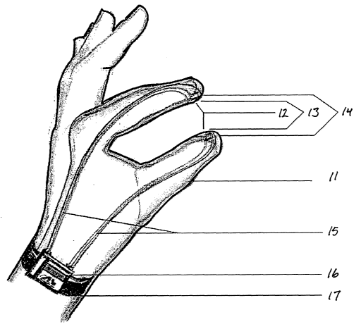
[0079] FIG. 3 illustrates a spectroscopic glove-like device that can be worn
on the hand of
a physician. This glove-like device can be formed from medical grade silicone
or other
material suitable for surgical gloves and similar devices. This device can be
equipped to
function as both a detecting device, an analytic device, a therapeutic (e.g.,
radiation delivery)
device, or a device serving a combination of these functions. The device can
thus be useful
in testing for and treating cancer. Portions of the source, means for
monitoring energy and
wavelength energy optimization, means for controlling the frequency, means for
conveying
energy to the treatment site, a thermal scan monitor, a beam sampler and a
system for
controlling and monitoring energy output can be carried by the glove-like
device and
connected to a central processing and control unit and associated hardware by
cables or fiber
optics.
23
CA 02516533 2005-08-18
WO 2005/021049 PCT/US2004/005924
[0080] The glove-like device 11 contaiizs a pair of interlace couplings 12
located on the
fingertips of the glove. These couplings form a lens system that detects
bidirectional data
coaxially from one point. A pair of miniature receiver/detectors 13 are also
located near the
tips of the forgers to function as a transition spectrophotometer. When the
glove is used for
S cancer detection, the miniature receivers and detectors 13 are connected to
the output
couplings to generate a combined signal that allows for reflection and
transmission of the
light energy through a pair of fiber optical couplings 14. Electrooptical
leads 15 extend from
the components in the fingertips to a transmitter/processor/controller unit 16
that passes
through the wristband 17 of the glove and from there to a power source (not
shown). The
transmitter/processor/controller unit 16 optically analyzes the signals from
the
receiver/detectors 13 while allowing the physician to manipulate the detection
and delivery
components to direct them to the precise area of the body where detection and
treatment are
needed.
(0081] FIG. 4 is a shows the device of FIG. 3 in use to test for cancer in the
vaginal area of
1S a female patient. One finger of the glove 11 is inserted in the vagina 2I
toward the uterus 22
and a second finger is inserted in the rectum 23, such that the tips of the
two forgers reside on
opposite sides of the pelvic tissue 24. The receiver/detectors I3 in the tips
of the two forgers
receive and send signals to the transmitterlprocessor/controller unit 16
through the
electrooptical leads 15. Malignant tissue is detected on the monitor due to
the presence of a
characteristic wavelength of the malignant tissue. Once malignant tissue has
been detected,
emitters can be attached to the glove-like device, or the device can be
replaced by a second
glove-like device to which emitters are already secured. In either case, the
emitters will be
those that emit radiation of a wavelength that will selectively destroy the
malignant tissue
without harm to the patient's normal tissue. By using the glove-like device,
the radiation can
2S be directed to the immediate area where the malignancy has been detected
and the cancer can
be treated without surgery.
[0082] The glove-like device of FIG. 3 is again shown in FIG. 5 where it is
being used to
test and/or to treat the prostrate gland of a male patient. In this case, one
finger of the glove
11 is inserted in the rectum 31 of the patient while another finger is placed
behind the
scrotum 32. Here again, the receiver/detectors 13 in the tips of the two
fingers receive and
send signals to the transmitter/processor/controller unit 16 through the
electrooptical leads 15.
Cancer in the prostate gland 33 can thus be detected and treated in the manner
set forth above
in the description of FIG. 4.
24
CA 02516533 2005-08-18
WO 2005/021049 PCT/US2004/005924
[0083] Tn FIG. 6 the same device is shown in use for testing or treatment of
cancer in a
testicle 41 of a male patient. In this procedure, the thumb and forefinger of
the glove-like
device are placed on opposite sides of the patient's testicle 41, and the
receiver/detectors 13
at the tips of the forgers send signals to the
transmitter/processor/controller unit 16 through
the electrooptical leads 15. Cancer in the testicle 41 can thus be detected
and treated in the
manner set forth above in the descriptions of the preceding Figures.
[0084] FIG. 7 depicts a device that operates at a higher power level than that
attainable
with the glove-like device of FIG. 3. The device in FIG. 7 is shown in use for
the treatment
of neoplastic tissues present in the prostate of a male patient. A scattered
light detector 51 is
placed in the bladder 52 of the patient through a catheter 53 that is inserted
in the urethra 54.
An analytical and therapeutic optical wand 55 with an optical head 56 and a
detector array 57
on the optical head is placed in the rectum 58. The bladder is filled with a
reflective fluid that
enhances the detection sensitivity of the optical wand 55 by reflecting the
signal from the
optical head 56 to the detector array 57.
[0085] FIGS. 8A and 8B are top and side views, respectively, of a sample
holder 61
designed to hold a sectioned sample for high-power Iaser spectroscopy and
destructive
radiation and to place the sample in controlled atmospheric conditions. The
sample holder 61
can be mounted to a camera or a detector array enabling the direct
transmission of radiation
by laser for spectroscopic or thermographic analysis and imaging. The holder
consists of a
cylindrical housing 62 closed at its two ends by optical filter windows 63, 64
held in place by
clamping disks 65, 66 and fitted with a vacuum line 67. Spacers 68, 69 and a
series of
transparent sample support disks 70 support the sample in the center of the
housing. The
optical filter windows 63, 64 transmit light of the selected wavelength.
[0086] FIG. 9 is a perspective view of the breasts of a female patient, one of
which has
been fitted with a detection and treatment device in accordance with the
present invention.
The device includes a cup 81 of transparent polymer that is secured to the
surface of the
breast by a low vacuum applied through a series of vacuum lines 82. Attached
to the
underside of the cup are a series of pairs of input/output couplings 83, each
of which includes
a receiver/detector that serves as a transmission spectrophotometer.
Electrooptical Ieads 84
connect each pair of input/output couplings 83 with a
transmitter/processor/controller unit
(not shown). The components operate in the same manner as those of the
structures shown in
the preceding figures. By use of this device, an image of the breast can be
formed and
CA 02516533 2005-08-18
WO 2005/021049 PCT/US2004/005924
neoplastic tissue detected, without squeezing the breast or displacing the
neoplastic tissue.
The type, location, and extent of any neoplastic tissue in the breast can
thereby be precisely
defined and mapped. Once the neoplastic tissue is precisely mapped,
electromagnetic
radiation of a selected wavelength or wavelength band is directed to the
neoplastic tissue to
S destroy the tissue without surgery and without damaging the adj acent
healthy tissue.
[0087] FIGS. 10A is a side view of the optical head 56 and optical wand 55 of
FIG. 7. The
optical head 56 is mounted at the end of a swivel handle 91, and the layers on
the optical head
axe a lens 92, a series of interlaced input/output couplings 93, an array of
optical components
94, and positive and negative electric leads 95. FIG. lOB is an enlargement of
the optical
component array of FIG. 10A. The optical components include focal plane arrays
96,
microbolometers 97, low power emitting and receiving fiber optics 98, high
power emitting
fiber optics 99, and CCD detectors 100.
26