Note: Descriptions are shown in the official language in which they were submitted.
CA 02517367 2005-08-26
WO 2004/080318 PCT/US2004/006121
POSTERIOR PEDICLE SCREW AND PLATE SYSTEM AND METHODS
FIELD OF THE INVENTION
The present invention relates to an orthopedic nnplant assembly for use in
stabilizing bone members in a desired spatial relationship in correcting bone
misalignment
disorders or for spinal or other bone fusion. In particular, the invention
concerns a multi-
axial spinal fixation system incorporating an elongated member such as a
plate.
>3ACI~GROUND OF THE INVENTION
In the art of orthopedic surgery, and particularly in spinal surgery, it has
long been
known to afftx an elongated member, such as a plate or rod, to bones in order
to hold them
and support them in a given position. For example, in a procedure to fuse
damaged,
diseased, malformed, or otherwise abnormal vertebrae, the vertebrae are
positioned in a
corrected position by a surgeon. An elongated plate is placed adjacent to the
vertebral
bone, and bone anchors, such as specially-configured screws or bolts, are
employed to
secure the plate to the bones. With such anchors, placement is accomplished by
drilling
one or more holes in the bones) and threading the anchors into the holes. As
examples,
see U.S. Pat. Nos. 5,676,666 to Oxland et al., 5,613,967 to Engelhardt et al.,
and 5,603,713
to Aust et al. An anchor can be connected to the bone, as by threading into a
vertebral
hole, through a plate, or alternatively the plate can be placed in position
over or around the
anchor after the anchor is connected to the bone. The anchor and plate are
then secured to
each other to minimize or prevent relative movement. In this way, bones may be
spinal
held and/or supported in proper alignment for healing.
It has been found desirable for implant systems to have the capability for
angular
orientation of a bolt or other anchor in multiple planes relative to the
elongated member or
other fixation mechanisms of the implant system. Such features enable bone
anchors to be
placed at angles which are optimal for anchoring, thus reducing the chance of
loosening,
pull-out, or other movement of the anchors while not compromising the optimal
positioning of the fixation plate.
Additionally, such systems allemate awkwardness frequently found in spinal
surgery due to uneven bone surfaces and the abnormality to be corrected and
generally
CA 02517367 2005-08-26
WO 2004/080318 PCT/US2004/006121
2
require less adjustment to the implant, rendering corrective surgery easier
for the surgeon
and less traumatic for the patient.
Various approaches have been used to achieve such mufti-axial capability. For
example, U.S. Pat. No. 5,735,853 to ~lerud discloses an implant device in
which a bone
bolt can occupy different angular positions in relation to a plate by
providing a
compressible spherical collar which snap-fits around the bolt, which collar is
rotatable and
tiltable in a spherical opening in a plate insert. The compression fit of the
bolt and collar
within the plate can present difficulty in assembling the apparatus,
particularly in a fluid-
prevalent environment.
Another approach is shown in U.S. Pat. No. 5,304,179 to Wagner, which shows a
bone screw fixed inside a bushing at an angle with respect to the longitudinal
axis of the
bushing. The bushing is rotatable within a portion of a connector angled with
respect to
the axis of the adjoining rod-based instrumentation. The connector is
rotatable around the
instrumentation axis. The Wagner system permits only discrete positions of a
bone screw
in three-dimensional space to be achieved, and the bushings add extra length
and profile to
the construct, as well as extra parts for the surgeon to handle and arrange.
A third approach is shown in U.S. Pat. No. 5,984,924 to Asher et. al., which
shows
a bone alignment system having an elongated bone alignment member sandwiched
between two pairs of washers. Each such pair of washers have corresponding
surfaces that
mate together in a "ball and socket" configuration to potentially occupy a
plurality of
positions. When the shaft of a bone anchor extends through each washer pair,
and also
through an aperture of the elongated member, the washer pairs enable the shaft
to be
oriented at various angles relative to the elongated member. This approach
also requires a
plurality of small parts for handling and assembly during surgery. Further,
since the
washers in that system lie outside of the elongated member, they increase the
thickness of
the overall construct, with the attendant increase in the difficulty of use in
a small surgical
space and in the potential for patient discomfort.
As noted above, in placing such implants a surgeon is commonly required to
reposition vertebrae so that a normal spinal curvature results from the
surgery. In open
surgical procedures, the surgeon may reposition vertebrae) manually or may
have tools to
assist in the repositioning. ~nce the vertebrae are repositioned, implants can
then be
CA 02517367 2005-08-26
WO 2004/080318 PCT/US2004/006121
attached in order to hold the vertebrae in the desired position.
Alternatively, it is also
known to provide a rod that is pre-bent to approximate a normal spinal
curvature and to
provide hooks or screws that can hold the rod which attach to several
vertebrae. With
such apparatus, vertebrae can be repositioned by forcing the pre-bent rod into
engagement
with the hoolcs or screws that arc already anchored in the vertebrae. Even
with that
method, however, additional tools such as a rod reducer are required. For
example, to
compress (i.e., push together) or distract (i.e., push apart) two vertebrae,
it is known to use,
among other relatively large tools, a scissors- or tongs-like device by
squeezing or pulling
apart on handles of such a tool; distal parts of the tool that contact
vertebrae or devices
attached to vertebrae will cause the distraction or compression.
Performing these tasks using traditional techniques and devices of open
surgery
has several undesirable features and consequences. Initially, such open
surgery requires a
long incision which leaves a relatively long and unappealing scar. Further,
such surgery
entails incision, retraction, and adjustment of numerous tissues in addition
the spinal
tissues. As a result, trauma to these tissues and resulting pain and
possibility of infection
are relatively high. Still further, a standard thoracotomy or other incision
may expose only
one apex of the spinal curve to be corrected, thus requiring additional long
incisions or a
longer initial incision in order to be able to fully treat the spine. Even
where the apex of
the spinal curve is adequately exposed and in good position relative to the
thoracotomy for
surgery, commonly adjacent vertebrae and intervertebral discs are not parallel
to the
exposure view provided by the incision, decreasing the effectiveness of
instrumentation
used to correct the abnormal curvature. For these reasons, an endoscopic,
thoracoscopic,
or other minimally-invasive approach is preferable.
Accordingly, there remains a need for a device that simplifies adjustment or
repositioning of vertebrae, particularly when a minimally-invasive approach is
used.
SUMMARY OF THE 1NVENTI~N
In one embodiment, an apparatus is disclosed comprising a plate member having
a
curvature, a first slot and a second slot, and sized to be inserted into the
body through a
minimally-invasive, open or other surgical opening. The slots each have a side
wall sized
t0 aCCOmlllOdate at least a portion of a bone anchor. A sloped surface is
provided within
said second slot, with the surface sloping in a longitudinal direction. A
first bone fixation
CA 02517367 2005-08-26
WO 2004/080318 PCT/US2004/006121
4
element that is adapted to engage the plate member is also provided, wherein
at least a part
of the first bone fixation element is capable of being within the first slot.
A second bone
fixation element is adapted to engage the plate member within the second slot
along the
sloped surface, whereby tightening the second boric fixation clement after
engagement
with the sloped surface causes the plate member to move with respect to the
second bone
fixation clement.
In specific embodiments, the curvature of the plate member may approximate a
natural curvature of one or more spinal segments, such as a lordotic or
kyphotic curvature.
The sloped surface may slope approximately uniformly, may be integrally formed
as a part
of the wall of the slot, and may have a downward or upward slope (viewed from
an end of
the second slot near an end of the plate member toward the end of the second
slot in the
middle of the plate member). A part of the surface may have no slope, i.e.,
may be
substantially parallel to the bottom of the plate member. A bone fixation
element retainer
(e.g. a set screw) may be provided connected to the plate member (e.g. via a
threaded
hole) adjacent the second slot. The bone fixation elements may be part of a
mufti-axial
bone fixation apparatus, standard bone screws, or screws having a head portion
with a
rounded underside and a diameter larger than a distance between adjacent
portions of the
sloped surface. The apparatus can further include additional slots sized to
accommodate at
least a portion of a bone anchor, and bone anchors for such slots.
In another embodiment, a kit is provided comprising at least one plate member,
each having a curvature, a first slot, a second slot and a sloped surface
within said second
slot. The said surface slopes in a longitudinal direction, and the plate
members) are sized
to be inserted into the body through a minimally-invasive, open or other
surgical opening.
Also provided is at least one first bone fixation element, each adapted to
engage the plate
members) and each having at least a part capable of being within the first
slot, and at least
one second bone fixation element, each adapted to engage the plate members)
along the
sloped surface. The kit rnay include a plurality of plate members not all of
the same size
and/or curvature, and some may have an upward slope while others have a
downward
slope. The plate members can be configured for attachment to the spine in one
or more of
the cervical, thoracic, lumbar, and sacral regions. The kit may also have a
plurality of the
CA 02517367 2005-08-26
WO 2004/080318 PCT/US2004/006121
first and/or second bone fixation elements adapted for attachment to the spine
in one or
more of the cervical, thoracic, lumbar, and sacral regions.
In yet another embodiment, a method is provided comprising providing a plate
member having a curvature approximating a natural spinal lordosis curvature, a
first slot
and a second slot with a sloped surface formed within said second slot, and
sized to be
inserted into the body through a minimally-invasive, open or other surgical
~pening;
inserting the plate member through the opening into a patient proximate to a
vertebra to
which the plate member is intended to be attached; placing a first anch~ring
member
through the first slot and into a first vertebra; placing a second anch~ring
member thr~ugh
the second slot and into a second vertebra; and tightening the second
anchoring member
against the sloped surface so that the plate member moves with respect to the
second
anchoring member. The first anchoring member can be tightened with respect to
said
plate member prior to tightening the second anchoring member. The method can
also
include loosening the first and second anchoring members, adjusting the plate
member
along the vertebrae, and retightening the anchoring members. Tightening the
second
anchoring member can cause one of compression and distraction of vertebrae,
and
tightening can be ceased when a predetermined amount of one of compression and
distraction of vertebrae has occurred.
In still another embodiment, a method of implanting a spinal implant having a
lordosis curvature and a plurality of longitudinal slots, at least one of the
slots having a
surface slanted along the longitudinal direction, comprises making a minimally-
invasive,
open or other surgical opening proximate to first and second vertebrae in a
patient;
preparing a first hole in the first vertebra and a second hole in the second
vertebra through
the opening; inserting a first fixation member through the opening and into
the first hole;
inserting the implant through the opening and into a position adjacent the
vertebrae such
that the first fixation member is within one of the implant's slots and
another of the slots in
the implant is adjacent the second hole, that second slot including the
slanted surface;
inserting a second fixation member through the opening and through the second
slot and
into the second hole; and tightening the second fixation member such that it
and the
second vertebra move with respect to the implant.
CA 02517367 2005-08-26
WO 2004/080318 PCT/US2004/006121
6
BRIEF DESCRIPTION OF THE DRAWINGS
FIG. 1 is a perspective view of one embodiment of a device having features
according to the present invention.
FIG. 2 is a side view of the embodiment illustrated in FIG. 1.
FIG. 3 is an exploded view of the embodiment illustrated in FIG. 1.
FIG. 4 is a cross section taken along the lines IV--IV in FIG. 1 and viewed in
the
direction of the arrows.
FIG. 5 is a magnified view of a portion of the embodiment illustrated in FIG.
1.
FIG. 6 is a perspective view of another embodiment of a device having features
according to the present invention.
DESCRIPTION OF THE PREFERRED EMBODIMENTS
For the purposes of promoting an understanding of the principles of the
invention,
reference will now be made to the embodiments illustrated herein and specific
language
will be used to describe the same. It will nevertheless be understood that no
limitation of
the scope of the invention is thereby intended. Any alterations and further
modifications
in the described processes, systems or devices, and any further applications
of the
principles of the invention as described herein, are contemplated as would
normally occur
to one skilled in the art to which the invention relates.
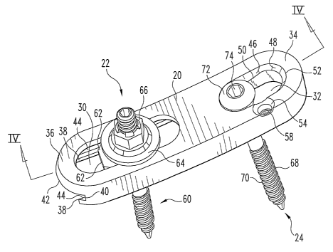
Referring now generally to FIGS. 1-5, there is shown a plate member 20 with a
mufti-axial bone fixation member 22 and a bone fixation member 24. Plate
member 20 is
elongated and includes a plurality of slots. In the illustrated embodiment,
plate member
20 includes two slots 30 and 32, although it will be appreciated additional
slots could be
placed in plate member 20. In a particular embodiment, plate member 20 and
slots 30 and
32 are sized so that plate member 20 can contact neighboring vertebrae, and
slots 30 and
32 will each be adjacent one of those vertebrae so that fixation members can
be placed
through slots 30 and 32 and into the vertebrae. Plate member 20'may also be
sized and
configured to extend across more than two adjacent vertebrae.
In the illustrated embodiment, slot 32 is near a first end 34 of plate member
20, and
slot 30 is near a second end 36 of plate member 20. Plate member 20, in the
illustrated
embodiment, is pre-bent or fomled to include a curvature, for example a
natural spinal
lordosis curvature. As shown particularly in FIGS. 2 and 3, plate member 20
has a
CA 02517367 2005-08-26
WO 2004/080318 PCT/US2004/006121
7
concave curvature, as viewed from the top. It will be understood that any
curvature
appropriate for one or more segments of the spine (whether cervical, thoracic,
lumbar or
sacral) could be incorporated into plate member 20. Such curvatures can
include entirely
convex, entirely concave, entirely straight (i.e. essentially planar), and
combinations
thereof. As noted previously, in the illustrated embodiment a lordosis
curvattbre is
depicted and is particularly a curvature characteristic of the lumbar spine.
Plate member
20 could alternatively be part-lordotic with an uncurved portion, part-
kyphotic with an
uncurved portion, wholly kyphotic, or have another curvature or combination of
curvatures.
In the illustrated embodiment, slot 30 is a longitudinal, oval-shaped slot.
Slot 30
extends through plate member 20 from top to bottom. Proximate to the bottom of
plate
member 20, a ledge 38 extends into slot 30. Ledge 38, along with surfaces 40
and 42 of
plate member 20, defines grooves 44 that run longitudinally along plate member
20 within
slot 30. Grooves 44 accommodate a part of multi-axial bone fixation element
22, as will
be described hereafter.
Slot 32 is also depicted in the illustrated embodiment as a longitudinal, oval-
shaped slot. Slot 32 extends through plate member 20, from top to bottom. A
sloped wall
portion 46 extends into slot 32 and is preferably integral with plate member
20. In the
illustrated embodiment, wall portion 46 has a concavely rounded portion 48
that ends in a
sloping edge or surface 50. By "sloping," it is meant that points along edge
or surface 50
are at different distances from the top surface of plate member 20 at
different places along
edge or surface 50, and may be approximately linear or have another suitable
configuration. In the illustrated embodiment, a point along edge or surface 50
that is
closer to first end 34 of plate member 20 than another point along edge or
surface 50 will
be closer to the top surface of plate member 20. It will be understood that
edge or surface
50 may have a flat or non-sloped portion, that is, a portion that is
substantially horizontal
or parallel to a bottom surface of plate member 20. Such a portion in the
present
embodiment may preferably be adjacent the end of slot 32 that is toward a
middle portion
of plate member 20. In a specific embodiment, an upper edge 52 of slot 30 is
sized so that
the head of a bone screw or other fixation member (e.g., fixation member 24~)
will fit
CA 02517367 2005-08-26
WO 2004/080318 PCT/US2004/006121
within edge 52, but edge or surface 50 will not allow the head of such a
fixation member
to pass.
The illustrated embodiment of plate member 20 further includes a hole ~4
adjacent
to slot 32. Hole 54 is for accommodating a retainer designed to hold fixation
member 24~
within slot 32, or to prevent fixation member 24 from backing out, such as set
screw 56
(FIG. 5), a cam or sliding wedge member, a spring-loaded member or a similar
device. In
the embodiment in which set screw 56 is used, hole 54 will include a threaded
portion 58.
Hole 54 is preferably sized to accommodate at least a portion of the retainer
(e.g., set
screw 56), so that its top extends minimally or not at all over the top
surface of plate
member 20, thus reducing the profile of the overall construct.
Multi-axial fixation member 22 includes a bone bolt 60, a stabilizing member
62, a
washer 64, and a nut 66. The respective elements of multi-axial fixation
member 22 are
described in detail in U.S. Pat. Nos. 6,280,445 and 6,315,779 to Morrison et
al., and the
entirety of those patents are incorporated herein by reference. In the
illustrated
embodiment, washer 64 and nut 66 are pre-attached, as is shown and described
in U.S.
Pat. No. 6,315,779. It will be appreciated that embodiments of the present
invention are
contemplated in which washer 64 and nut 66 are separate and are not associated
with each
other or in contact with each other until the attachment of mufti-axial
fixation member 22
to plate member 20.
Fixation member 24 is shown in one embodiment as a standard bone screw having
an attachment portion 68 with cancellous threads 70, and a head portion 72.
Head portion
72 includes a tool-engaging aperture 74, and preferably includes a rounded
underside 75.
As noted previously, the diameter of head portion 72 is preferably larger than
the distance
between sections of edge or surface 50 on opposite sides of slot 32, so that
head portion 72
cannot pass through slot 32 in plate member 20.
For ease of use, a kit containing one or more of the parts of the implant may
be
provided. For example, a kit may include several embodiments of plate member
20, or
one or more embodiments of plate member 20 in several different lengths, sizes
and/or
curvatures. Lengths or sizes appropriate for cervical, thoracic, lumbar and/or
sacral
implantation may be included. ~ne or more sets of screws, bolts, stabilizers,
washers,
and/or nuts, preferably in a variety of sizes or adapted for attachment to one
or more of the
CA 02517367 2005-08-26
WO 2004/080318 PCT/US2004/006121
9
cervical, thoracic, lumbar and sacral regions of the spine, may also be
provided in such a
kit. For example, one or more fixation members 22 and/or 24 for engaging one
or more
slots in the plate members may be included. Further, retainers for holding
fixation
member 24 within slot 32 (e.g., set screw ~6) may also be provided. In a
specific
embodiment of such a kit, each plate anember 20 is provided with stabilizing
member 62
preloaded into grooves 44 under slot 30. A catch, boss, or stop anay be
provided within
grooves 44 or on stabilizing member 62 so that stabilizing member 62 cannot
exit grooves
44 and fall out of plate member 20. Similarly, if washer 64~ and nut 66 form
an initial unit,
as shown in U.S. Pat. IVo. 6,315,779, a variety of such units may be provided
in the lcit.
Alternatively, separate quantities of nuts and washers can be provided.
A method of using the implant will now be described. As noted above, the
implant
can be used in minimally-invasive surgical procedures, and the methods
described below
reflect such procedures. It will be appreciated by those of skill in the art
that the features
that enable a minimally-invasive approach to be used with the implant will
also make the
implant easier to insert via a standard open or other surgical procedure.
Using a minimally-invasive technique, a surgeon will make an incision into the
patient at a place relatively proximate to the vertebrae or other bones) to
which the
implant is to be attached. As is known in the art, a tube of sufficient length
to extend to
the implantation site from a point above the incision in the skin is inserted
into the
incision, and visual access to the site is obtained. After the appropriate
access to the
surgical site is obtained, a portion of the inferior vertebra to be
instrumented (e.g. the
pedicle) is prepared in a standard manner. For example, an awl may be used to
prepare a
hole, which is then probed for depth and tapped as appropriate for a bolt or
screw element.
Bolt 60 of the multi-axial bone fixation member 22 is then inserted into the
hole in the
inferior vertebra. Access to a portion of the superior vertebra (e.g. the
pedicle) to be
instrumented is then obtained, either via the previous incision or via a
similar minimally-
invasive incision. The point on the superior vertebra at which the implant is
to be attached
is identified, and the vertebra is prepared as described above. However, in
the preferred
embodiment, fixation member 24~ is not yet inserted in the superior vertebra.
Plate
member 20 is then inserted (e.g., through the tube in the minimally-invasive
incision).
Plate member 20 is positioned over bone fixation member 22 and slot 30 is
bottom-loaded
CA 02517367 2005-08-26
WO 2004/080318 PCT/US2004/006121
(i.e. bone fixation member 22 is inserted into slot 30 through the bottom of
plate member
20) so that bone fixation member 22 fits within slot 30. Plate member 20 is
then translated
or otherwise moved until the hole in the superior vertebra is adjacent slot
32. In one
particular embodiment, the hole in the superior vertebra should be underneath
the
uppermost region of slot 32, i.e., the region closest to end 36 of plate
member 20. Washer
64 and nut 66 are then placed on bolt 60, and nut 66 is tightened down onto
washer 64 and
plate member 20 to hold plate member 20 in position relative to the inferior
vertebra. It
will be noted that locking plate member in position can be performed solely by
a nut 66,
without an intermediary washer 64.
10 With plate member 20 locked with respect to the inferior vertebra, fixation
member
24 can be top-loaded in plate member 20 (i.e. inserted through the top of
plate member 20
and through slot 32) into the hole prepared in the superior vertebra. Fixation
member 24 is
inserted until its head portion 72 contacts sloped edge or surface 50 within
slot 32.
Fixation member 24 is then tightened further causing the plate member 20 to
move with
respect to fixation member 24. In effect, as fixation member 24 is tightened,
plate
member 20 slides in a direction parallel to slot 32 with respect to fixation
member 24 and
the superior vertebra, so that fixation members 22 and 24 approach each other.
Since plate
member 20 is locked with respect to fixation member 22 and the inferior
vertebra, this
sliding action causes the inferior and superior pedicles to be brought closer
together,
providing compression. Further, the curvature of the plate itself can provide
correction to
the curvature of the spine if it contacts the vertebrae as fixation member 24
is tightened
with respect to plate member 20. Tightening of fixation member 24 can continue
until the
maximum amount of adjustment (e.g. compression) is obtained, or it can be
discontinued
when a predetermined amount of adjustment has occurred.
In many cases, the amount of compression or other correction of the vertebrae
can
be determined prior to surgery, and the positions of the holes in the
vertebrae can be
predetermined so that the vertebrae are in their proper, corrected positions
when fixation
member 24 is tightened to the extent that the head of fixation member 24 rests
approximately at a point at which edge or surface 50 no longer slopes, or to
the end of slot
32 that is toward the middle of plate member 20. In other words, commonly
fixation
member 24 can be tightened to a point where no further sliding action of plate
member 20
CA 02517367 2005-08-26
WO 2004/080318 PCT/US2004/006121
11
with respect to fixation member 24 is possible. It will be appreciated,
however, that in
some cases proper correction will dictate that the tightening of fixation
member 24 should
stop even though head portion 72 of fixation member 24 has not yet reached the
end of
slot 32. When the tightening of fi;~ation member 24~ is complete, a retainer
or anti-
migTation or holding element (such as set screw 56) can be inserted into
aperture 54~ or
otherwise attached to plate member 20 adjacent fixation member 24 to cover
and/or
contact a portion (e.g. head portion 72) of fixation member 24.
If it is noted that the steps above do not provide adequate compression,
fixation
member 22 can be released, i.e., nut 66 can be loosened. Plate member 20 can
then be
adjusted by moving plate member 20 with respect to bolt 60 along slot 30, and
nut 66 can
then be retightened. In this way, end 34 of plate member 20 can be moved
closer to the
hole in the superior pedicle, resulting in the opportunity for more
compression as fixation
member 24 is tightened and plate member 20 slides a further distance with
respect to
fixation member 24. Similarly, if less compression is desired, plate member 20
should be
positioned (or repositioned) such that the hole in the superior pedicle is
closer to the end of
slot 32 that is toward the middle of plate member 20. In a case in which no
compression
of the vertebrae is desired, plate member 20 should be positioned so that the
hole in the
superior pedicle is directly under the end of slot 32 closer to the middle of
plate member
20, or so that the hole in the superior pedicle is over a relatively flat or
non-sloped portion
of edge 50.
As previously noted, the above-described technique is one preferred embodiment
of a method of using the described implant. It should be noted that the
technique can be
reversed, that is, slot 32 can be adjacent a superior vertebra and slot 30 can
be adjacent an
inferior vertebra. Those with skill in the art will recognize that compression
and/or
distraction can also be obtained by using an additional external compression
or distraction
instrument.
In another embodiment of the invention, shown in FIG. 6, plate member 120 is
shown. Plate member 120 is similar in many respects to plate member 20,
described
above. However, plate member 120 is particularly useful in mufti-level
implantations (i.e.,
implantation over several respective vertebrae). Plate member 120 would
include slots
130a and 130b, similar to slot 30 of plate member 20, and a slot 132, similar
to slot 32 in
CA 02517367 2005-08-26
WO 2004/080318 PCT/US2004/006121
12
plate member 20. Slots 130a and 130b are shown with fixation members 122,
which are
similar to fixation members 22 with plate member 20. A fixation member 124
that is
similar to fixation member 24 described above is shown in slot 132. In use,
pedicles for
three vertebrae would be prepared as described above, each with a hole that
may be
tapped. The bolt portions of fixation members 124 would be inserted into the
tv~o most
inferior vertebrae and plate member 120 placed over them so that they extend
through
slots 130a and 130b. Plate member 120 would then be positioned so that the
hole in the
superior-most pediclc is under slot 132. Washers 164 and nuts 166 are placed
on bolts
160. The lower- or inferior-most fixation member 122 is then locked with
respect to plate
member 120 by tightening its respective nut 166. Fixation member 124 is then
inserted
through slot 132 and into the hole in the superior-most pedicle, and tightened
as described
above, to provide compression or distraction. The middle vertebra is allowed
to float
during the compression or distraction. Following the tightening of fixation
member 124,
the remaining fixation member 122 (attached to the middle vertebra) is locked
with respect
to plate member 120 by tightening its respective nut 166.
Plate member 120 also shows a transverse hole 200 and slot 202 at one end X134
of
plate member 120. If desired, a pin (not shown) can be placed in hole 200 and
through
slot 202 to provide a way of holding or gripping plate member 120. The surgeon
can use a
gripping tool having teeth or a rounded opening, so that the teeth grip or the
rounded
opening surrounds such pin within hole 200 and slot 202. Plate member 120 (or
plate
member 20 if provided with a similar hole and/or slot and pin) can then be
lowered into
the surgical site while the surgeon holds onto the tool that grips plate
member 120. If no
pin is placed in hole 200, a proper gripping instrument could grip plate 120
at slot 202 (or
plate member 20 if provided with a similar slot).
It will be appreciated that a plate member providing distraction can be formed
in a
similar fashion to that of plate member 20. As described above, plate member
20 includes
a slot 32 with a sloping edge or surface 50. The downward slope of edge or
surface 50 is
generally from a point near the end 36 of plate member 20 toward the middle of
plate
member 20. A distraction plate, conversely, could include a sloped edge or
surface that
slopes generally upward from a point near the end 36 of plate member 20 toward
the
middle of plate member 20. Thus, as a fixation member 24 is tightened in such
a
CA 02517367 2005-08-26
WO 2004/080318 PCT/US2004/006121
13
distraction plate, the distraction plate would slide with respect to fixation
member 24 such
that the instrumented vertebrae would be pushed apart from each other,
providing
distraction.
It will further be appreciated that the embodiments described above should be
made of materials suitable for implantation within the human or other body,
and may
consist of inert metals like titanium or stainless steel. ~ther sturdy
materials such as
certain ceramics or plastics may also be considered. Rio-resorbable materials,
such as
polylactic acid compounds, may be used along with or as a part of the parts
described
above.
As noted above, the bone fixation element 22 and 122 is described herein as
the
structures shown in U.S. Patent No. 6,20,445 and 6,315,779, the entire
disclosures of
which are incorporated herein by reference. As indicated in those patents,
such bone
fixation elements allow multi-axial positioning of the plate member 20 with
respect to the
fixation element 22. This allows for further freedom in plate positioning and
spinal
correction via a minimally-invasive incision. It will be appreciated, however,
that other
known fixation elements may be used in place of the described fixation
elements 22, 122,
24, and 124. Further, while slot 30 is described above as bottom-loading and
slot 32 as
top-loading, it will be seen that with variations in plate member 20 and/or
differing
fixation elements the slots may be loaded each from the same direction, or
slot 30 may be
loaded from the top and slot 32 from the bottom.
Additionally, the methods and structures described above have been generally
noted as effective in minimally-invasive surgical procedures, i.e. those in
which one or
more relatively small holes are opened through the skin and to the surgical
site. It will be
appreciated that the methods and structures described above can also be used
in other
types of surgical procedures, such as open procedures.
It is also contemplated that processes embodied in the present invention can
be
altered, rearranged, substituted, deleted, duplicated, combined, or added to
other processes
as would occur to those skilled in the art without departing from the spirit
of the present
invention. In addition, the various stages, steps, procedures, techniques,
phases, and
operations within these processes may be altered, rearranged, substituted,
deleted,
duplicated, or combined as would occur to those skilled in the art. All
publications,
CA 02517367 2005-08-26
WO 2004/080318 PCT/US2004/006121
14
patents, and patent applications cited in this specification are herein
incorporated by
reference as if each individual publication, patent, or patent application was
specifically
and individually indicated to be incorporated by reference and set forth in
its entirety
herein.
Further, any theory of operation, proof, or finding stated herein is meant to
further
enhance understanding of the present invention and is not intended to make the
scope of
the present invention dependent upon such theory, proof, or funding.