Note: Descriptions are shown in the official language in which they were submitted.
CA 02517452 2011-01-24
METHODS OF DETECTING NEUROLOGICAL DISORDERS
[0001]
STATEMENT REGARDING FEDEFULLY SPONSORED RESEARCH
[0002] The U.S. government may have certain rights in this invention, pursuant
to grant
nos. AG11385, NS41787, and NS43945 awarded by the National Institutes of
Health.
FIELD OF THE INVENTION
[0003] This application is in the field of cognitive impairment, and in
particular
Alzheimer's disease, cerebral amyloidosis, proteopathies of the aging central
nervous
system, and neurodegenerative disorders.
BACKGROUND OF THE INVENTION
[0004] The bright prospects of increasing life expectancy in many populations
around
the world are tempered by an alarming increase in amyloid peptide-related
neurodegenerative disorders. Alzheimer's disease, the most frequent among
these
conditions, causes an inexorable loss of memory and other cognitive functions.
Although
the etiology of most AD cases remains elusive, several key features of this
disease can be
simulated in transgenic mice, making them amenable to experimental analysis
and
manipulation. Indeed, hAPP mice are used increasingly to assess novel AD
treatments.
Amyloid plaques have remained the primary pathological outcome measure in
these
studies, although their contribution to AD-related cognitive deficits is
controversial. In
fact, it remains to be determined which of the many pathological and
biochemical
alterations identified in AD and related transgenic models contribute most
critically to
the decline in neuronal functions.
[0005] Currently, mouse models of amyloid peptide-related neurological
disorders such
as Alzheimer's Disease are used for identifying agents that are useful for
treating such
disorders. The efficacy of a given test agent is typically determined by
assessing and
scoring behavioral traits such as learning and memory. An example of such a
test is the
1
CA 02517452 2005-08-26
WO 2004/084711 PCT/US2004/009216
water maze test. Such tests are time-consuming, may be somewhat subjective,
and are
subject to a high degree of variability and imprecision.
[0006] There is a need in the art for improved methods of detecting
neurological
disorders associated with neurotoxic levels of amyloid peptide. The present
invention
addresses this need, and provides an alternative read-out for efficacy of a
test agent on
treating cognitive impairment.
Literature
[0007] Palop et al. (2003) Proc. Nail. Acad. Sci. USA 100:9572-9577; Barski et
al.
(2003) J. Neuroscience 23:3469-3477; Greene et al. (2001) Neuropathol. Applied
Neurobiol.27:339-342; Thorns et al. (2001) Neuropathol. 21:203-211; Iritani et
al.
(2001) Neuropathol. 21:162-167; Palop et al. "Immunochemical indicators of
neuronal
and behavioral deficits in transgenic models of Alzheimer's Disease," Abstract
919 (July
20-25, 2002) The 8`" International Conference on Alzheimer's Disease and
Related
Disorders, Stockholm, Sweden; Heyser et al. (1997) Proc. Nat'l. Acad. Sci. USA
94:1500-1505; Moechars et al. (1999) J. Biol. Chem. 274(10):6483-6492;
Mikkonen et
al. (1999) Neuroscience 92(2):515-32; Ichimiya, et al. (1998) Brain Res. 475,
156-159;
Hof, P. R. & Morrison, J. H. (1991) Exp. Neurol. 111, 293-301; West, M. J.,
Coleman,
P. D., Flood, D. G. & Troncoso, J. C. (1994) Lancet 344, 769-772; Crabbe et
al. (1999)
Science 284:1670-1672; Wahlsten et al. (2003) J. Neurobiol. 54:283-311.
Baimbridge
KG, et al. (1992) Trends Neurosci 15:303-308; Celio MR (1990) Neuroscience
35:375-
475; Chard PS, et al. (1995) Proc Natl Acad Sci USA 92:5144-5148; German DC,
et al.
(1997) Neuroscience 81:735-743; Geula C, et al. (2003) JCompNeurol 455:249-
259;
Greene JRT, et al. (2001) Neuropathol Appl Neurobiol 27:339-342; Guo Q, et al.
(1998)
Proc Natl Acad Sci USA 95:3227-3232; Heizmann CW, and Hunziker W (1991) Trends
Biochem Sci 16:98-103; Heyser CJ, et al. (1997) Proc Natl Acad Sci USA 94:1500-
1505; Hof PR, and Morrison JH (1991). Exp Neurol 111:293-301; Hsia A, et al.
(1999)
Proc Natl Acad Sci USA 96:3228-3233; lacopino AM, and Christakos S (1990) Proc
Natl Acad Sci USA 87:4078-4082; Iritani S, et al, Neuropathology. 2001
Sep;21(3):162-
7; Klapstein GJ, et al. (1998) Neuroscience 85:361-373; Lledo P-M, et al.
(1992) Neuron
9:943-954; Magloczky Z, et al. (1997) Neuroscience 76:377-385; Mikkonen M, et
al.
(1999) Neuroscience 92:515-532; Molinari S, et al. (1996) Proc Natl Acad Sci
USA
93:8028-8033; Mucke L, et al. (2000) J Neurosci 20:4050-4058; Nagerl UV, and
Mody
I (1998) J Physiol 509:39-45; Nagerl UV, et al. (2000) J Neurosci 20:1831-
1836; Pasti
L, et al. (1999) Neuroreport 10:2367-2372; Potier B, et al. (1994) Brain Res
661:181-
2
CA 02517452 2011-01-24
188; Thorns V, et al, Neuropathology. 2001 Sep;21(3):203-11; Vig PJS, et al.
(2001)
Brain Res. Bull. 56:221-225.
SUMMARY OF THE INVENTION
[0008] The present invention provides methods of detecting an amyloid peptide-
related
neurological disorder in an individual; and methods for staging an amyloid
peptide-related
neurological disorder in an individual. The methods involve detecting a level
of a calcium-
responsive gene product, such as calbindin, in a hippocampal neuron,
especially
a granule cell of the dentate gyros. The invention further provides
identifying an agent
that treats an amyloid peptide-related neurological disorder, as well as
agents identified
by the methods.
[0008A] Various embodiments of this invention provide a method for detecting
an
amyloid peptide-related neurological disorder in a non-human animal model, the
method
comprising: detecting a level of a calcium-responsive gene product in brain
tissue of the
animal model, wherein the calcium responsive gene product is selected from a
calbindin
polypeptide, a Fos polypeptide, an Arc polypeptide, a calbindin mRNA, and a
Fos mRNA,
and an Arc mRNA, and wherein the brain tissue is a granule cell of the dentate
gyrus;
wherein detection of a level of calcium-responsive gene product in the brain
tissue that is
lower than a level of the calcium-responsive gene product associated with a
normal control
animal is indicative of an amyloid peptide-related neurological disorder in
the animal.
[0008B] Various embodiments of this invention provide a method for identifying
a
candidate agent for treating an amyloid peptide-related neurological disorder,
the method
comprising: detecting a level of a calcium-responsive gene product in vitro in
brain tissue
of a non-human animal model of an amyloid peptide-related neurological
disorder in
response to a test agent, wherein the calcium responsive gene product is
selected from a
calbindin polypeptide, a Fos polypeptide, an Arc polypeptide, a calbindin
mRNA, and a Fos
mRNA, and an Arc mRNA, and wherein the brain tissue is a granule cell of the
dentate
gyros; wherein detection of a level of calcium-responsive gene product in the
brain tissue
that is significantly higher than a level of the calcium-responsive gene
product in the
absence of the agent indicates that the test agent is a candidate agent for
treating an amyloid
peptide-related neurological disorder.
3
CA 02517452 2011-01-24
BRIEF DESCRIPTION OF THE DRAWINGS
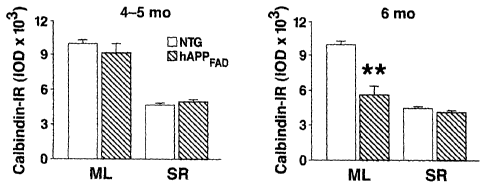
[0009] Figures 1 A-F depict results showing that calbindin and c-Fos
reductions in the
dentate gyrus depend on age and type of hAPP expressed.
[0010] Figures 2A-C depict the relationship between calbindin, c-Fos, plaque
load, and
AP levels.
[0011] Figures 3A-I depict results showing that reductions in calbindin and c-
Fos
correlate tightly with behavioral deficits.
[0012] Figure 4 depicts ectopic expression of neuropeptide Y (NPY) in mossy
fibers,
and aberrant NPY/GABAergic sprouting in the molecular layer in hAPPFAD mice.
[0013] Figure 5 depicts the observation that aberrant NPY/GABAergic sprouting
in the
molecular layer correlates with calbindin reduction (NTG; non-transgenic).
[0014] Figure 6 depicts ectopic expression of NPY in mossy fibers in hAPPFAD
mice.
[0015] Figure 7 depicts the observation that ectopic NPY expression in mossy
fibers
correlates with calbindin reductions.
[0016] Figure 8 depicts the observation that a-actinin-II is markedly reduced
in the
molecular layer of hAPPFAD mice.
[0017] Figure 9 depicts the observation that loss of a-actinin-II correlates
with calbindin
reductions in hAPPFAD mice.
[0018] Figure 10 depicts the correlation of reductions in calbindin
immunoreactivity
(IR) with reductions in calbindin protein and mRNA in hAPPFAD mice.
3a
CA 02517452 2005-08-26
WO 2004/084711 PCT/US2004/009216
DEFINITIONS
[0019] The term "amyloid peptide-related neurological disorder," as used
herein, refers to
any disorder that results from, or is associated with, accumulation of
neurotoxic levels of
amyloid peptides in the central nervous system, and/or formation of neurotoxic
amyloid
protein assemblies in the central nervous system. Such disorders include, but
are not
limited to, AD, Parkinson's disease, and Lewy body disease. The term includes
cognitive impairments associated with AD, including impairment of learning
ability, and
memory impairment.
[0020] The term "Alzheimer's disease" (abbreviated herein as "AD") as used
herein refers to
a condition associated with formation of neuritic plaques comprising amyloid
(3 protein,
as well as impairment in both learning and memory. "AD" as used herein is
meant to
encompass both AD as well as AD-type pathologies and clinical manifestations.
[0021] The terms "calcium-responsive gene product," and "calcium-dependent
gene
product," as used interchangeably herein, refer to a protein and/or an mRNA
whose level
varies with the intracellular calcium ion concentration ([Ca2+;]). Calcium-
responsive
gene products include products of genes that include a calcium-responsive
transcriptional
regulatory element; calcium-binding proteins (e.g., calbindin); neuropeptide Y
(NPY); an
a-actinin II gene product; a phospho-extracellular signal-regulated kinase
(phospho-ERK
or p-ERK) gene product; immediate early response genes (e.g., c-Fos); and the
like.
[0022] As used herein, the terms "determining," "measuring," and "assessing,"
and
"assaying" are used interchangeably and include both quantitative and
qualitative
determinations.
[0023] A "biological sample" encompasses a variety of sample types obtained
from an
individual and can be used in a diagnostic or monitoring assay. The definition
encompasses blood and other liquid samples of biological origin, solid tissue
samples
such as a biopsy specimen or tissue cultures or cells derived therefrom and
the progeny
thereof. The definition also includes samples that have been manipulated in
any way
after their procurement, such as by treatment with reagents, solubilization,
or enrichment
for certain components, such as polynucleotides. The term "biological sample"
encompasses a clinical sample, and also includes cells in culture, cell
supernatants, cell
lysates, serum, plasma, biological fluid, and tissue samples.
[0024] The term "binds specifically," in the context of antibody binding,
refers to high
avidity and/or high affinity binding of an antibody to a specific polypeptide
i.e., epitope
of a polypeptide, e.g., a calcium-responsive polypeptide, such as a calbindin
polypeptide
4
CA 02517452 2005-08-26
WO 2004/084711 PCT/US2004/009216
or a c-Fos polypeptide. For example, antibody binding to an epitope on a
specific
calbindin polypeptide or fragment thereof is stronger than binding of the same
antibody
to any other epitope, particularly those which may be present in molecules in
association
with, or in the same sample, as the specific polypeptide of interest, e.g.,
binds more
strongly to a specific calbindin epitope than to a different calbindin epitope
so that by
adjusting binding conditions the antibody binds almost exclusively to the
specific
calbindin epitope and not to any other calbindin epitope, and not to any other
calbindin
polypeptide (or fragment) or any other polypeptide which does not comprise the
epitope.
Antibodies which bind specifically to a polypeptide may be capable of binding
other
polypeptides at a weak, yet detectable, level (e.g., 10% or less of the
binding shown to
the polypeptide of interest). Such weak binding, or background binding, is
readily
discernible from the specific antibody binding to a subject polypeptide, e.g.
by use of
appropriate controls. In general, specific antibodies bind to a given
polypeptide with a
binding affinity of 10-7 M or more, e.g., 10-8 M or more (e.g., 10'9 M, 10"10
M, 10-1' M,
etc.). In general, an antibody with a binding affinity of 10-6 M or less is
not useful in that
it will not bind an antigen at a detectable level using conventional
methodology currently
used.
[0025] The terms "subject," "host," "patient," and "individual" are used
interchangeably
herein to refer to any mammalian subject for whom diagnosis or therapy is
desired,
particularly humans. Other subjects may include cattle, dogs, cats, guinea
pigs, rabbits,
rats, mice, horses, and so on.
[00261 The terms "treatment" "treating" and the like are used herein to refer
to any
treatment of any disease or condition in a mammal, particularly a human, and
includes:
a) preventing a disease, condition, or symptom of a disease or condition from
occurring
in a subject which may be predisposed to the disease but has not yet been
diagnosed as
having it; b) inhibiting a disease, condition, or symptom of a disease or
condition, e.g.,
arresting its development and/or delaying its onset or manifestation in the
patient; and/or
c) relieving a disease, condition, or symptom of a disease or condition, e.g.,
causing
regression of the condition or disease and/or its symptoms.
[0027] Before the present invention is further described, it is to be
understood that this
invention is not limited to particular embodiments described, as such may, of
course,
vary. It is also to be understood that the terminology used herein is for the
purpose of
CA 02517452 2005-08-26
WO 2004/084711 PCT/US2004/009216
describing particular embodiments only, and is not intended to be limiting,
since the
scope of the present invention will be limited only by the appended claims.
[0028] Where a range of values is provided, it is understood that each
intervening value,
to the tenth of the unit of the lower limit unless the context clearly
dictates otherwise,
between the upper and lower limit of that range and any other stated or
intervening value
in that stated range, is encompassed within the invention. The upper and lower
limits of
these smaller ranges may independently be included in the smaller ranges, and
are also
encompassed within the invention, subject to any specifically excluded limit
in the stated
range. Where the stated range includes one or both of the limits, ranges
excluding either
or both of those included limits are also included in the invention.
[0029] Unless defined otherwise, all technical and scientific terms used
herein have the
same meaning as commonly understood by one of ordinary skill in the art to
which this
invention belongs. Although any methods and materials similar or equivalent to
those
described herein can also be used in the practice or testing of the present
invention, the
preferred methods and materials are now described. All publications mentioned
herein
are incorporated herein by reference to disclose and describe the methods
and/or
materials in connection with which the publications are cited.
[0030] It must be noted that as used herein and in the appended claims, the
singular
forms "a", "and", and "the" include plural referents unless the context
clearly dictates
otherwise. Thus, for example, reference to "a granule cell" includes a
plurality of such
cells and reference to "the agent" includes reference to one or more agents
and
equivalents thereof known to those skilled in the art, and so forth.
[0031] The publications discussed herein are provided solely for their
disclosure prior to
the filing date of the present application. Nothing herein is to be construed
as an
admission that the present invention is not entitled to antedate such
publication by virtue
of prior invention. Further, the dates of publication provided may be
different from the
actual publication dates which may need to be independently confirmed.
DETAILED DESCRIPTION OF THE INVENTION
100321 The present invention provides methods of detecting an amyloid peptide-
related
neurological disorder in an individual; and methods for staging an amyloid
peptide-
related neurological disorder in an individual. The methods involve detecting
the level
of a calcium-responsive gene product (e.g., calbindin) in a granule cell of
the dentate
gyros in an individual. The invention further provides identifying an agent
that treats an
6
CA 02517452 2005-08-26
WO 2004/084711 PCT/US2004/009216
amyloid peptide-related neurological disorder. The invention further provides
methods
of modulating levels of a calcium-responsive gene product, such as calbindin,
in a
granule cell of the dentate gyrus in an individual.
[0033] The present invention is based on the observation that reduced
calbindin levels
and reduced c-Fos levels in hippocampal neurons, particularly cells of the
dentate gyrus,
are correlated with cognitive impairment, and with the relative abundance of
hippocampal AP1_42 among A(3 peptides. Transgenic non-human animals, such as
A[3
transgenic mice, are used to identify agents that are useful for treating
amyloid peptide-
related disorders such as Alzheimer's Disease (AD). The efficacy of a given
test agent is
typically determined by assessing and scoring behavioral traits. Such tests
are time-
consuming and imprecise. Furthermore, many drugs are assessed using plaque
formation
as an outcome measure instead of behavioral testing. Because there is
increasing
evidence that plaque-independent neuronal deficits also appear to play a
critical role in
AD, measuring plaque formation may fail to identify drugs that might prevent
or
ameliorate plaque-independent neuronal deficits.
[0034] Amyloid plaques do not correlate well with cognitive impairment and
other
behavioral deficits associated with neurological disorders such as AD. Indeed,
some
amyloid protein does not form plaques, but instead forms small, neurotoxic
assemblies
that are not deposited as plaques. However, calbindin levels do correlate with
the level
and neurotoxic activity of amyloid protein, as well as with plaque-independent
cognitive
decline, and therefore provide a reliable marker for amyloid protein related
neurological
disorders.
[0035] The present invention provides an alternative read-out for the efficacy
of a test
agent on treating cognitive impairment. Because reduced calbindin levels in
hippocampal neurons, such as the granule cells of the dentate gyros, are
correlated with
cognitive impairment, the level of calbindin, as well as other calcium-
dependent
proteins, in these cells serves as a surrogate marker for behavioral
characteristics.
Calbindin levels can be quantitated. Quantitation allows a more precise
analysis of the
extent of the disease, and provides a measure of the degree of efficacy of a
given test
agent. The present invention provides for a readout of clinically relevant
impairments,
and thus presents a major advantage over the assessment of drugs by behavioral
testing
or by plaque quantitation.
7
CA 02517452 2005-08-26
WO 2004/084711 PCT/US2004/009216
DETECTION METHODS
[0036] The present invention provides methods of detecting an amyloid peptide-
related
neurological disorder in an individual, or in a biological sample derived from
an
individual. The methods generally involve detecting a level of a calcium-
responsive
gene product in a hippocampal neuron in an individual, or in a biological
sample derived
from an individual. In many embodiments, calcium-responsive gene product
levels are
detected in the dentate gyros, and in particular, a granule cell of the
dentate gyros.
[0037] Any of a number of calcium-responsive gene products can be detected in
a
granule cell of the dentate gyros in a method of the present invention.
Illustrative
examples include calbindin, a-actinin II, phospho-ERK, c-Fos, and neuropeptide
Y.
Those skilled in the art can readily apply such methods to other calcium-
responsive gene
products.
[0038] In some embodiments, a level of a calcium-dependent gene product in the
dentate
gyros of an individual that is lower than the normal level of the gene product
in the
dentate gyros indicates the presence of a neurological disorder in the
individual
(particularly, an amyloid peptide-related neurological disorder). Examples of
calcium-
dependent gene products that are lower in the dentate gyros of an individual
having an
amyloid peptide-related neurological disorder include calbindin, p-ERK, a-
actinin II, and
c-Fos. Thus, e.g., a level of a calcium-dependent gene product (e.g.,
calbindin, p-ERK,
a-actinin II, and c-Fos) that is at least about 10%, at least about 20%, at
least about 30%,
at least about 40%, at least about 50%, at least about 60%, at least about
70%, or at least
about 80%, or more, lower than the normal level of that gene product in the
dentate
gyros indicates that the individual has an amyloid peptide-related
neurological disorder.
[0039] In other embodiments, a level of a calcium-dependent gene product in
the dentate
gyros of an individual that is higher than the normal level of the gene
product in the
dentate gyros indicates the presence of a neurological disorder in the
individual
(particularly, an amyloid peptide-related neurological disorder). Examples of
calcium-
dependent gene products that are higher in the dentate gyros of an individual
having an
amyloid peptide-related neurological disorder include neuropeptide Y (NPY).
Thus, e.g.,
a level of a calcium-dependent gene product (e.g., NPY) that is at least about
10%, at
least about 20%, at least about 30%, at least about 40%, at least about 50%,
at least about
60%, at least about 70%, at least about 80%, at least about 90%, at least
about 100% (or
two-fold), at least about 2.5-fold, at least about 3-fold, at least about 4-
fold, or at least
8
CA 02517452 2005-08-26
WO 2004/084711 PCT/US2004/009216
about 5-fold, (or greater) higher than the normal level of that gene product
in the dentate
gyrus indicates that the individual has an amyloid peptide-related
neurological disorder.
Detecting polypeptide levels
[0040] In some embodiments, the methods involve detecting a calcium-responsive
protein and/or mRNA level (e.g., calbindin protein and/or rnRNA level) in a
hippocampal neuron (e.g., a granule cell of the dentate gyros) in vitro. Any
method of
detecting a calcium-responsive protein level (e.g., a calbindin protein level)
in an in vitro
biological sample can be used in conjunction with a subject method. Suitable
methods of
detecting a protein include, but are not limited to, protein blotting methods,
enzyme
linked immunosorbent assays, radioimmunoassays, and immunohistochemical
methods.
For example, a hippocampal brain section is contacted with an antibody that
binds
calbindin specifically, where the antibody is detectably labeled, either
directly or
indirectly.
[00411 Direct and indirect antibody labels are known in the art. An antibody
may be
labeled with a radioisotope, an enzyme, a fluorescer (e.g., a fluorescent
protein or a
fluorescent dye), a chemiluminescer, or other label for direct detection.
Alternatively, a
second stage antibody or reagent is used to amplify the signal. Such reagents
are well
known in the art. For example, the primary antibody may be conjugated to
biotin, with
horseradish peroxidase-conjugated avidin added as a second stage reagent.
Final
detection uses a substrate that undergoes a color change in the presence of
the
peroxidase. Alternatively, the secondary antibody conjugated to a fluorescent
compound, e.g. fluorescein, rhodamine, Texas red, etc. The absence or presence
of
antibody binding may be determined by various methods, including flow
cytometry of
dissociated cells, microscopy, radiography, scintillation counting, etc.
[00421 Fluorescent proteins include, but are not limited to, a green
fluorescent protein
(GFP), e.g., a GFP derived from Aequoria victoria or a derivative thereof, a
GFP from
another species such as Renilla reniformis, Renilla mulleri, or Ptilosarcus
guernyi, as
described in, e.g., WO 99/49019 and Peelle et al. (2001) J. Protein Chem.
20:507-519;
any of a variety of fluorescent and colored proteins from Anthozoan species,
as described
in, e.g., Matz et al. (1999) Nature Biotechnol. 17:969-973; and the like.
[00431 Enzyme labels include, but are not limited to, luciferase, P-
galactosidase, horse
radish peroxidase, luciferase, alkaline phosphatase, and the like. Where the
label is an
enzyme that yields a detectable product, the product can be detected using an
appropriate
9
CA 02517452 2005-08-26
WO 2004/084711 PCT/US2004/009216
means, e.g., R-galactosidase can, depending on the substrate, yield colored
product,
which is detected spectrophotometrically, or a fluorescent product; luciferase
can yield a
luminescent product detectable with a luminometer; etc.
[0044] In some embodiments, the level of a calcium-responsive protein, such as
calbindin, is quantitated. Methods of quantitating protein levels are known in
the art.
For example, enzyme-linked immunosorbent assay (ELISA) can provide for
quantitation
of calbindin levels. Calbindin levels in immunostained brain tissue sections
can be
quantitated by determining the integrated optical density of the
immunoreactivity of an
immunostained brain section, as described in the Example.
[0045] In other embodiments, a level of a calcium-dependent protein selected
from
calbindin, neuropeptide Y (NPY), a-actinin II, phospho-ERK (p-ERK), and c-Fos,
is
detected. Levels of proteins such as calbindin, neuropeptide Y (NPY), a-
actinin II,
phospho-ERK (p-ERK), and c-Fos are readily detected using well-known methods,
including immunological assays. For example, p-ERK polypeptide levels are
readily
detected using a chemiluminescence enzyme immunometric assay (TiterZyme CLIA;
Assay Designs, Inc., Ann Arbor, MI), involving use of a p-ERK-specific IgG
antibody,
and an alkaline phosphatase-labeled IgG-specific antibody. NPY, and methods
for
detecting NPY, are well described in the literature. See, e.g., Erickson et
al. (1996)
Nature 381:415; and Minth et al. (1984) Proc. Nail. Acad. Sci. USA 81:4577.
Alpha-
actinin-II is well described in the literature. See, e.g., Wyszynski et al.
(1998) J.
Neurosci. 18:1383. Phospho-ERK is well described in the literature. See, e.g.,
Li et al.
(2001) Neurobiol. Dis. 8:127; and Fahiman et al. (2002) Brain Res. 958:43-51.
[0046] In many embodiments, the detection method is an in vitro detection
method
involving detecting a calcium-responsive gene product, e.g., a calbindin
level, in a
granule cell of the dentate gyros in a brain sample from a transgenic non-
human animal
model of a neurodegenerative disorder. Transgenic non-human animal models of
Alzheimer's disease are well known in the art. For example, various non-human
animal
models of neurodegenerative disorders such as AD are described in, e.g., U.S.
Patent
Nos. 5,767,337; 6,046,381; 6,175,057; and 6,455,757; and Mucke et al. (2000)
J.
Neurosci. 20:4050-4058; Masliah et al. (2001) Proc. Natl. Acad. Sci. USA
98:12245-
12250; and Rockenstein et al. (1995) J. Biol. Chem. 270:28257-25267. Non-
limiting
examples of suitable animal models include hAPP transgenic mice that express
high
levels of hAPP; and hAPP transgenic mice that express low levels of hAPP and
that are
also transgenic for fyn kinase. In these embodiments, a hippocampal brain
sample is
CA 02517452 2005-08-26
WO 2004/084711 PCT/US2004/009216
taken from a transgenic non-human animal model of a neurodegenerative
disorder, and
the level of a calcium-dependent protein and/or a calcium-dependent protein-
encoding
mRNA is detected in the dentate gyrus.
Detecting mRNA levels
[0047] Where the calcium-responsive gene product is an mRNA (e.g., a calbindin
mRNA, an NPY mRNA, a c-Fos mRNA, an a-actinin II mRNA, a p-ERK mRNA, and
the like), any of a variety of known methods for detecting an mRNA can be
used. In
general, nucleic acids that hybridize specifically to a calcium-responsive
mRNA, e.g., a
calbindin mRNA, are used. A number of methods are available for analyzing
nucleic
acids for the presence and/or level of a specific mRNA in a cell or in a
sample. The
mRNA may be assayed directly or reverse transcribed into cDNA for analysis.
Suitable
methods include, but are not limited to, in situ nucleic acid hybridization
methods,
quantitative reverse transcription-polymerase chain reaction (RT-PCR), nucleic
acid
blotting methods, and the like.
[0048] The nucleic acid may be amplified by conventional techniques, such as
the
polymerase chain reaction (PCR), to provide sufficient amounts for analysis.
The
mRNA may be reverse transcribed, then subjected to PCR (rtPCR). The use of the
polymerase chain reaction is described in Saiki, et al. (1985), Science
239:487, and a
review of techniques may be found in Sambrook, et al. Molecular Cloning: A
Laboratory
Manual, CSH Press 1989, pp.14.2-14.33.
[0049] A detectable label may be included in an amplification reaction.
Suitable labels
include fluorochromes, e.g. fluorescein isothiocyanate (FITC), rhodamine,
Texas Red,
phycoerythrin, allophycocyanin, 6-carboxyfluorescein (6-FAM), 2', 7'-dimethoxy-
4',5'-
dichloro-6-carboxyfluorescein (JOE), 6-carboxy-X-rhodamine (ROX), 6-carboxy-
2',4',7',4,7-hexachlorofluorescein (HEX), 5-carboxyfluorescein (5-FAM) or
N,N,N',N'-
tetramethyl-6-carboxyrhodamine (TAMRA), radioactive labels, e.g. 32P, 35S, 3H;
etc.
The label may be a two stage system, where the amplified DNA is conjugated to
biotin,
haptens, etc. having a high affinity binding partner, e.g. avidin, specific
antibodies, etc.,
where the binding partner is conjugated to a detectable label. The label may
be
conjugated to one or both of the primers. Alternatively, the pool of
nucleotides used in
the amplification is labeled, so as to incorporate the label into the
amplification product.
[0050] A variety of different methods for determining the nucleic acid
abundance in a
sample are known to those of skill in the art, where particular methods of
interest include
those described in: Pietu et al., Genome Res. (June 1996) 6: 492-503; Zhao et
al., Gene
11
CA 02517452 2011-01-24
(April 24, 1995) 156: 207-213; Soares, Curr. Opin. Biotechnol. (October 1997)
8: 542-
546; Raval, J. Pharmacol Toxicol Methods (November 1994) 32: 125-127;
Chalifour et
al., Anal. Biochem (February 1, 1994) 216:=299-304; Stolz & Tuan, Mol.
Biotechnol.
(December 19960 6: 225-230; Hong et al., Bioscience Reports (1982) 2: 907; and
McGraw, Anal. Biochem. (1984) 143: 298. Also of interest are the methods
disclosed in
WO 97/27317.
[0051] In some embodiments, calbindin mRNA levels (and/or a NPY mRNA level, a
c,
Fos mRNA level, an a-actinin II mRNA level, a p-ERK mRNA level) are
quantitated
using quantitative rtPCR. Methods of quantitating a given message using rtPCR
are
known in the art. In some of these embodiments, dye-labeled primers are used.
In other
embodiments, a double-stranded DNA binding dye, such as SYBR ,. is used, as
described in the Examples. Quantitative fluorogenic RT-PCR assays are well
known in
the art, and can be used in the present methods to detect a level of calbindin
mRNA.
See, e.g., Pinzani et al. (2001) Regul. Pept. 99:79-86; and Yin et al. (2001)
Immunol.
Cell Biol. 79:213-221.
[0052] Other examples of calcium-responsive. gene products that are suitable
for
detection using a method of the present invention is immediate early gene
products such
as c-Fos and Arc. Thus, in some embodiments, the method involves detecting a
level of
c-Fos protein in the hippocampus, e.g., in a granule cell of the dentate
gyrus. As
discussed in the Example, a reduced level of c-Fos in hippocampal neurons
correlates
with behavioral deficits, such as cognitive impairment, associated with AD.
Thus, in
some embodiments, the methods involve detecting a level of c-Fos protein
and/or mRNA
in a sample. In many embodiments, c-Fos levels are detected in a granule cell
of the
dentate gyrus. A level of c-Fos polypeptide is detected by employing
immunological
methods as described above, using antibody specific for c-Fos.
[0053] Alternatively, c-Fos mRNA is detected, using nucleic acids that
hybridize
specifically to c-Fos nucleic acids. A number of methods are available for
analyzing
nucleic acids for the presence and/or level of a specific mRNA in a cell, as
described
above. The mRNA may be assayed directly or reverse transcribed into cDNA for
analysis.
[0054] The level of a calcium-responsive gene product in an animal model of an
amyloid
peptide-related neurodegenerative disorder is compared to the level of the
calcium-
responsive gene product in control animals, e.g., animals of the same species
that are not
models for the disorder. For example, where the animal model is a non-human
animal
12
CA 02517452 2005-08-26
WO 2004/084711 PCT/US2004/009216
transgenic for a neurodegeneration-promoting protein such as A131-42, suitable
controls
are wild-type, e.g., not transgenic for the neurodegeneration-promoting
protein. For
example, where the animal is a transgenic hAPP animal, a non-transgenic animal
of the
same species serves as a control. Typically, the test animal and the control
animal are
the same sex.
[0055] While not required, validation of the method may be carried out by
detecting a
level of AP31-42 protein in the hippocampus (e.g., in a granule cell of the
dentate gyros),
and correlating a level of calbindin in the hippocampus (e.g., granule cell of
the dentate
gyros) with the level of API-42 protein in the hippocampus. A(31-42 levels are
measured
using standard immunological methods, as described above, using antibody
specific for
API-42.
[0056] In some embodiments, detection of calcium-responsive gene product
levels in
granule cells of the dentate gyros is carried out in vivo in a living subject,
including a
living mouse model of an amyloid peptide-related neurological disorder, and a
living
human subject. Thus, the present invention provides methods for antemortem
detection
of calcium-responsive gene product levels, e.g., calbindin levels, in granule
cells of the
dentate gyros. In some embodiments, the methods involve administering to a
living
subject a detectably labeled compound that specifically binds the calcium-
responsive
gene product, and detecting binding between the detectably labeled compound
and the
calcium-responsive gene product in a granule cell of the dentate gyros. In
other
embodiments, the methods involve administering to a living subject a
detectably labeled
agent that binds a factor that decreases along with a decrease in calcium-
responsive gene
product levels; and detecting binding between the agent and the factor.
Suitable
detection methods include, but are not limited to, magnetic resonance imaging
(MRI),
positron emission tomography, single photon emission computed tomography,
functional
MRI, and the like.
[0057] In other embodiments, detection of calcium-responsive gene product
level in
granule cells of the dentate gyros is carried out postmortem on a biological
sample of a
human subject. Detection of calcium-responsive gene product levels is carried
out as
described above, e.g., by detecting calbindin and/or mRNA levels in a dentate
gyros
sample taken from a deceased human subject.
Detecting multiple calcium-dependent gene products
[0058] In some embodiments, two or more calcium-responsive gene products are
detected. As one non-limiting example, in some embodiments, both calbindin
protein
13
CA 02517452 2005-08-26
WO 2004/084711 PCT/US2004/009216
levels and c-Fos protein levels are detected. Thus, in some embodiments, a
subject
method involves detecting a level of calbindin protein in a brain sample, and
detecting a
level of c-Fos protein in the brain sample, e.g., in granule cells of the
dentate gyros. In
other embodiments, two or more polypeptides selected from a calbindin
polypeptide, an
NPY polypeptide, a c-Fos polypeptide, a p-ERK polypeptide, and an a-actinin II
polypeptide are detected in the brain sample, e.g., in granule cells of the
dentate gyros.
Thus, in some embodiments, a subject method involves detecting a level of two
or more
proteins selected from a calbindin polypeptide, an NPY polypeptide, a c-Fos
polypeptide, a p-ERK polypeptide, and an a-actinin II polypeptide in a brain
sample,
e.g., in granule cells of the dentate gyros.
[0059] In other embodiments, both calbindin mRNA and c-Fos mRNA levels are
detected. Thus, in some embodiments, a subject method involves detecting a
level of
calbindin mRNA in a brain sample, and detecting a level of c-Fos mRNA in the
brain
sample, e.g., in granule cells of the dentate gyros. In other embodiments, two
or more
mRNAs selected from a calbindin mRNA, an NPY mRNA, a c-Fos mRNA, a p-ERK
mRNA, and an a-actinin II mRNA are detected. Thus, in some embodiments, a
subject
method involves detecting a level of two or more mRNAs selected from a
calbindin
mRNA, an NPY mRNA, a c-Fos mRNA, a p-ERK mRNA, and an a-actinin II mRNA.
SCREENING METHODS
[0060] The invention provides methods of identifying agents that improve
cognition;
methods of identifying agents that increase a level of a calcium-responsive
gene product
(e.g., calbindin, a-actinin II, p-ERK, c-Fos) in a hippocampal neuron in an
individual;
methods of identifying agents that block the effect of amyloid peptide on
neurons;
methods of identifying agents that reduce the level of abnormal amyloid
assemblies;
methods of identifying agents that increase clearance of amyloid peptides, and
methods
of identifying agents that decrease neurotoxic levels of amyloid peptides. The
methods
generally involve administering a test agent to a transgenic non-human animal
model of
a neurodegenerative disorder, where the test agent contacts a hippocampal
neuron (e.g., a
granule cell of the dentate gyros); and detecting a level of calcium-
responsive gene
product (e.g., calbindin, a-actinin II, p-ERK, c-Fos, NPY, etc.) in vitro in
brain tissue of
the animal. Detection of a level of a calcium-dependent gene product in the
brain tissue
that is significantly different from a level of the calcium-responsive gene
product in the
14
CA 02517452 2005-08-26
WO 2004/084711 PCT/US2004/009216
absence of the agent indicates that the test agent modulates a level of the
calcium-
responsive gene product in the brain tissue of the animal.
[0061] In general, a test agent that effects an increase or a decrease in a
hippocampal
neuron calcium-responsive gene product level (e.g., a calbindin level in a
granule cell of
the dentate gyros), such that the increase or decrease is toward a normal
level of the gene
product, is a candidate agent for treating a cognitive impairment. For
example, a test
agent that effects an increase in a calbinding, an a-actinin II, or a p-ERK
gene product in
the dentate gyros is a candidate agent for treating a cognitive impairment
(e.g., an
amyloid peptide-related cognitive impairment). As another example, a test
agent that
increases a level of an NPY gene product in the dentate gyros is a candidate
agent for
treating a cognitive impairment (e.g., an amyloid peptide-related cognitive
impairment).
In many embodiments, the method provides for identification of agents that
modulate a
cognitive impairment in the animal. In particular embodiments, the level of a
calcium-
dependent gene product, e.g., a calcium-dependent gene product selected from a
calbindin gene product, an a-actinin II gene product, an NPY gene product, and
a p-ERK
gene product, in a granule cell of the dentate gyros is detected.
Test agents
100621 The terms "candidate agent," "test agent," "agent", "substance" and
"compound"
are used interchangeably herein. Candidate agents encompass numerous chemical
classes, typically synthetic, semi-synthetic, or naturally-occurring inorganic
or organic
molecules. Candidate agents include those found in large libraries of
synthetic or natural
compounds. For example, synthetic compound libraries are commercially
available from
Maybridge Chemical Co. (Trevillet, Cornwall, UK), ComGenex (South San
Francisco,
CA), and MicroSource (New Milford, CT). A rare chemical library is available
from
Aldrich (Milwaukee, Wis.). Alternatively, libraries of natural compounds in
the form of
bacterial, fungal, plant and animal extracts are available from Pan Labs
(Bothell, WA) or
are readily producible. Additionally, natural or synthetically produced
libraries and
compounds are readily modified through conventional chemical, physical and
biochemical means, and may be used to produce combinatorial libraries. Known
pharmacological agents may be subjected to directed or random chemical
modifications,
such as acylation, alkylation, esterification, amidification, etc, to produce
structural
analogs. New potential therapeutic agents may also be created using methods
such as
rational drug design or computer modeling.
CA 02517452 2005-08-26
WO 2004/084711 PCT/US2004/009216
[0063] Candidate agents are generally small organic or inorganic compounds
having a
molecular weight of more than 50 and less than about 2,500 daltons. Candidate
agents
may comprise functional groups necessary for structural interaction with
proteins,
particularly hydrogen bonding, and may include at least an amine, carbonyl,
hydroxyl or
carboxyl group, and may contain at least two of the functional chemical
groups. The
candidate agents may comprise cyclical carbon or heterocyclic structures
and/or aromatic
or polyaromatic structures substituted with one or more of the above
functional groups.
Candidate agents are also found among biomolecules including peptides,
saccharides,
fatty acids, steroids, purines, pyrimidines, derivatives, structural analogs
or combinations
thereof.
[0064] Screening may be directed to known pharmacologically active compounds
and
chemical analogs thereof, or to new agents with unknown properties such as
those
created through rational drug design.
[0065] Assays of the invention include controls, where suitable controls
include a
control animal (e.g., an animal of the same genotype) not administered with
the test
agent. In some embodiments, a plurality of assays is run in parallel with
different agent
concentrations to obtain a differential response to the various
concentrations. Typically,
one of these concentrations serves as a negative control, i.e. at zero
concentration or
below the level of detection.
[0066] Agents that have an effect in an assay method of the invention may be
further
tested for cytotoxicity, bioavailability, and the like, using well known
assays. Agents
that have an effect in an assay method of the invention may be subjected to
directed or
random and/or directed chemical modifications, such as acylation, alkylation,
esterification, amidification, etc. to produce structural analogs. Such
structural analogs
include those that increase bioavailability, and/or reduced cytotoxicity.
Those skilled in
the art can readily envision and generate a wide variety of structural
analogs, and test
them for desired properties such as increased bioavailability and/or reduced
cytotoxicity
and/or ability to cross the blood-brain barrier.
[0067] In some embodiments, e.g., where the level of the calcium-dependent
gene
product is reduced in the dentate gyros of an individual having an amyloid
peptide-
related neurological disorder (including cognitive impairment), a test agent
that effects
an increase in a hippocampal neuron level of a calcium-responsive gene product
(e.g., an
agent that effects an increase in a calbindin level), particularly an increase
in a level of a
calcium-responsive gene product in a granule cell of the dentate gyros, is a
candidate
16
CA 02517452 2005-08-26
WO 2004/084711 PCT/US2004/009216
agent for treating a cognitive impairment. A test agent that effects an
increase in a
hippocampal neuron calcium-responsive gene product level (e.g., a calbindin
level in a
granule cell of the dentate gyrus) by at least about 5%, at least about 10%,
at least about
15%, at least about 20%, at least about 25%, at least about 30%, at least
about 35%, at
least about 40%, at least about 45%, at least about 50%, or more, compared to
the level
in a hippocampal neuron (e.g., granule cell of the dentate gyrus) not
contacted with the
test agent, indicates that the test agent is a candidate agent for treating an
amyloid
peptide-related cognitive impairment.
[0068] In other embodiments, e.g., where the level of the calcium-dependent
gene
product is increased in the dentate gyrus of an individual having an amyloid
peptide-
related neurological disorder (including cognitive impairment), a test agent
that effects a
decrease in a hippocampal neuron level of a calcium-responsive gene product
(e.g., an
agent that effects a reduction in an NPY gene product level), particularly a
reduction in a
level of a calcium-responsive gene product in a granule cell of the dentate
gyrus, is a
candidate agent for treating a cognitive impairment. A test agent that effects
a reduction
in a hippocampal neuron calcium-responsive gene product level (e.g., an NPY
level in a
granule cell of the dentate gyros) by at least about 5%, at least about 10%,
at least about
15%, at least about 20%, at least about 25%, at least about 30%, at least
about 35%, at
least about 40%, at least about 45%, or at least about 50%, or more, compared
to the
level in a hippocampal neuron (e.g., granule cell of the dentate gyros) not
contacted with
the test agent, indicates that the test agent is a candidate agent for
treating an amyloid
peptide-related cognitive impairment.
Administration
[00691 A test agent is administered in vivo to a transgenic non-human animal
model of a
neurodegenerative disorder, where the test agent contacts a hippocampal
neuron.
Transgenic non-human animal models of amyloid peptide-related
neurodegenerative
disorders are well known in the art. For example, various non-human animal
models of
neurodegenerative disorders are described in, e.g., U.S. Patent Nos.
5,767,337,-
6,046,381; 6,175,057; and 6,455,757; and Mucke et al. (2000) J. Neurosci.
20:4050-
4058; Masliah et al. (2001) Proc. Natl. Acad. Sci. USA 98:12245-12250; and
Rockenstein et al. (1995) J Biol. Chem. 270:28257-25267.
17
CA 02517452 2005-08-26
WO 2004/084711 PCT/US2004/009216
[0070] The test agent is administered by any convenient route of
administration,
including, but not limited to, intragastric, intracranial, intramuscular,
intravenous,
topical, subcutaneous, intratracheal, and the like.
[0071] In some embodiments, a subject screening method provides for
determining
whether a test agent crosses the blood brain barrier. For example, a test
agent that is
effective in increasing hippocampal neuron calcium-responsive gene product
levels when
administered intracranially is modified in vitro and the derivatives thus
formed are
administered to the animal intravenously. If the agent is effective when
administered
intravenously, then it likely crosses the blood brain barrier. Various
modifications to a
test agent, e.g., acetylations, acylations, phophorylations, and the like, can
be tested in
this manner.
In vitro screening
[0072] After the animal is administered with the test agent, the level of
calcium-
responsive gene product (e.g., a protein and/or mRNA selected from calbindin,
a-actinin
IT, c-Fos, NPY, and p-ERK) in brain tissue of the animal is detected in an in
vitro assay.
In vitro screening is conducted following administration of the test agent,
e.g., usually
after a period of about 10 minutes, about 15 minutes, about 30 minutes, about
60
minutes, about 2 hours, about 4 hours, or more, following administration of
the test
agent.
[0073] Typically, a hippocampal brain sample is taken from the animal, and
calcium-
responsive protein (e.g., calbindin) is detected. In many embodiments, the
brain sample
is a dentate gyros sample. Samples derived from an animal model of amyloid
peptide-
related cognitive impairment are used in assays. Typically, samples are
hippocampal
samples, e.g., dissociated hippocampal neurons, hippocampal brain sections,
and the like.
The number of cells in a sample will generally be at least about 103, usually
at least 104
more usually at least about 105. The cells may be dissociated, in the case of
solid tissues,
or tissue sections may be analyzed. Alternatively a lysate of the cells may be
prepared.
[0074] As described below, in some embodiments, a level of calcium-responsive
protein
in a hippocampal brain sample is detected. In some embodiments, a level of
calbindin
protein is detected. In other embodiments, a level of c-Fos protein in a
hippocampal
neuron is detected. Where a level of c-Fos protein is detected, the methods
discussed for
detecting calbindin protein are used, except that an antibody specific for c-
Fos is
employed.
18
CA 02517452 2005-08-26
WO 2004/084711 PCT/US2004/009216
[0075] As described below, in some embodiments, a level of calcium-responsive
mRNA
in a hippocampal brain sample is detected. In some of these embodiments, a
level of
calbindin mRNA is detected in the hippocampal brain sample. Alternatively, a
level of
c-Fos mRNA is detected.
Detecting calcium-responsive proteins
[0076] In many embodiments, an antibody specific for a given calcium-
responsive
polypeptide is used. As one non-limiting example, a calbindin level is
detected, and an
antibody specific for calbindin is used. The antibody is detectably labeled,
either directly
or indirectly. Various labels include radioisotopes, fluorescers,
chemiluminescers,
enzymes, specific binding molecules (e.g., members of specific binding pairs),
particles,
e.g. magnetic particles, and the like. Specific binding molecules include
pairs, such as
biotin and streptavidin, digoxin and antidigoxin, lectin and carbohydrate,
antibody and
antigen, antibody and hapten, etc. For the specific binding members, the
complementary
member would normally be labeled with a molecule that provides for detection,
in
accordance with known procedures. Direct and indirect antibody labels are
known in the
art. An antibody may be labeled with a radioisotope, an enzyme, a fluorescer
(e.g., a
fluorescent protein or a fluorescent dye), a chemiluminescer, or other label
for direct
detection. Alternatively, a second stage antibody or reagent is used to
amplify the signal.
Such reagents are well known in the art, and are discussed above.
[00771 A variety of other reagents may be included in the screening assay.
These
include reagents like salts, neutral proteins, e.g. albumin, detergents, etc
that are used to
facilitate optimal protein-protein binding and/or reduce non-specific or
background
interactions. Reagents that improve the efficiency of the assay, such as
protease
inhibitors, anti-microbial agents, etc. may be used. The mixture of components
is added
in any order that provides for the requisite binding (e.g., of antibody to
calbindin in the
sample). Incubations are performed at any suitable temperature, typically
between 4 and
40 C. Incubation periods are selected for optimum activity, but may also be
optimized to
facilitate rapid high-throughput screening. Typically between 0.1 and 1 hour
will be
sufficient.
[00781 Detection of calcium-responsive protein (e.g., calbindin) may utilize
staining of
cells or histological sections, performed in accordance with conventional
methods. The
antibodies of interest are added to the cell sample or brain section sample,
and incubated
for a period of time sufficient to allow binding to the epitope, usually at
least about 10
19
CA 02517452 2005-08-26
WO 2004/084711 PCT/US2004/009216
minutes. The antibody may be labeled with radioisotopes, enzymes, fluorescers,
chemiluminescers, or other labels for direct detection. Alternatively, a
second stage
antibody or reagent is used to amplify the signal. Such reagents are well
known in the
art. For example, the primary antibody may be conjugated to biotin, with
horseradish
peroxidase-conjugated avidin added as a second stage reagent. Final detection
uses a
substrate that undergoes a color change in the presence of the peroxidase. The
absence
or presence of antibody binding may be determined by various methods,
including flow
cytometry of dissociated cells, microscopy, radiography, scintillation
counting, etc.
[0079] An alternative method depends on the in vitro detection of binding
between
antibodies and calcium-responsive protein (e.g., calbindin) in a cell lysate.
Measuring
binding between an antibody and a protein in a sample or fraction thereof may
be
accomplished by a variety of specific assays. A conventional sandwich type
assay may
be used. For example, a sandwich assay may first attach specific antibodies to
an
insoluble surface or support. The particular manner of binding is not crucial
so long as it
is compatible with the reagents and overall methods of the invention. They may
be
bound to the plates covalently or non-covalently.
[0080] The insoluble supports may be any compositions to which polypeptides
can be
bound, which is readily separated from soluble material, and which is
otherwise
compatible with the overall method. The surface of such supports may be solid
or
porous and of any convenient shape. Examples of suitable insoluble supports to
which
the receptor is bound include beads, e.g. magnetic beads, membranes and
microtiter
plates. These are typically made of glass, plastic (e.g. polystyrene),
polysaccharides,
nylon or nitrocellulose. Microtiter plates are especially convenient because a
large
number of assays can be carried out simultaneously, using small amounts of
reagents and
samples.
[0081] Detection may utilize staining of cells or histological sections,
performed in
accordance with conventional methods. The antibodies of interest are added to
the cell
sample, and incubated for a period of time sufficient to allow binding to the
epitope,
usually at least about 10 minutes. The antibody may be labeled with
radioisotopes,
enzymes, fluorescers, chemiluminescers, or other labels for direct detection.
Alternatively, a second stage antibody or reagent is used to amplify the
signal. Such
reagents are well known in the art. For example, the primary antibody may be
conjugated to biotin, with horseradish peroxidase-conjugated avidin added as a
second
stage reagent. Final detection uses a substrate that undergoes a color change
in the
CA 02517452 2005-08-26
WO 2004/084711 PCT/US2004/009216
presence of the peroxidase. The absence or presence of antibody binding may be
determined by various methods, including flow cytometry of dissociated cells,
microscopy, radiography, scintillation counting, etc.
Detecting c-Fos
[0082] In some embodiments, a level of c-Fos in a brain tissue is also
detected in an in
vitro assay. Methods for detecting c-Fos are described above. In some of these
embodiments, a test agent that effects an increase in both a hippocampal
neuron
calbindin level and a hippocampal c-Fos level is a candidate agent for
treating a
cognitive impairment. A test agent that effects an increase in a hippocampal
neuron
calbindin level by at least about 5%, at least about 10%, at least about 15%,
at least about
20%, at least about 25%, at least about 30%, at least about 35%, at least
about 40%, at
least about 45%, at least about 50%, or more, and that effects an effects an
increase in a
hippocampal neuron c-Fos level by at least about 5%, at least about 10%, at
least about
15%, at least about 20%, at least about 25%, at least about 30%, at least
about 35%, at
least about 40%, at least about 45%, at least about 50%, or more compared to
the
calbindin and c-Fos levels in a hippocampal neuron not contacted with the test
agent,
indicates that the test agent is a candidate agent for treating an amyloid
peptide-related
neurodegenerative disorder, such as cognitive impairment. In particular
embodiments,
the level of calbindin in a granule cell of the dentate gyrus is detected.
THERAPEUTIC AGENTS
[0083] The invention provides agents identified using the methods described
herein.
Agents that increase a level of a calcium-responsive gene product (e.g.,
calbindin, a-
actinin-II, p-ERK), where the level of the gene product is decreased in the
dentate gyrus
in a neuropathology, are used to treat amyloid peptide-related neurological
disorders,
particularly cognitive impairment. Agents that reduce a level of a calcium-
dependent
gene product (e.g., NPY), where the level of the gene product is increased in
the dentate
gyrus under neuropathological conditions, are use to treat amyloid peptide-
related
neurological disorders, particularly cognitive impairment. An effective amount
of the
active agent is administered to the host, where "effective amount" means a
dosage
sufficient to produce a desired result. Generally, the desired result is at
least an
amelioration of at least one neuropathological symptom (e.g., at least a
reduction in a
learning deficit) as compared to a control.
21
CA 02517452 2005-08-26
WO 2004/084711 PCT/US2004/009216
Formulations
[0084] In the subject methods, the active agent(s) may be administered to the
host using
any convenient means capable of resulting in the desired reduction in any
amyloid
peptide-related neurological disorder, particularly amyloid peptide-related
cognitive
impairment.
[0085] Thus, the agent can be incorporated into a variety of formulations for
therapeutic
administration. More particularly, the agents of the present invention can be
formulated
into pharmaceutical compositions by combination with appropriate,
pharmaceutically
acceptable carriers or diluents, and may be formulated into preparations in
solid,
semi-solid, liquid or gaseous forms, such as tablets, capsules, powders,
granules,
ointments, solutions, suppositories, injections, inhalants and aerosols.
[0086] In pharmaceutical dosage forms, the agents may be administered in the
form of
their pharmaceutically acceptable salts, or they may also be used alone or in
appropriate
association, as well as in combination, with other pharmaceutically active
compounds.
The following methods and excipients are merely exemplary and are in no way
limiting.
[0087] For oral preparations, the agents can be used alone or in combination
with
appropriate additives to make tablets, powders, granules or capsules, for
example, with
conventional additives, such as lactose, mannitol, corn starch or potato
starch; with
binders, such as crystalline cellulose, cellulose derivatives, acacia, corn
starch or
gelatins; with disintegrators, such as corn starch, potato starch or sodium
carboxymethylcellulose; with lubricants, such as talc or magnesium stearate;
and if
desired, with diluents, buffering agents, moistening agents, preservatives and
flavoring
agents.
[0088] The agents can be formulated into preparations for injection by
dissolving,
suspending or emulsifying them in an aqueous or nonaqueous solvent, such as
vegetable
or other similar oils, synthetic aliphatic acid glycerides, esters of higher
aliphatic acids or
propylene glycol; and if desired, with conventional additives such as
solubilizers,
isotonic agents, suspending agents, emulsifying agents, stabilizers and
preservatives.
[0089] The agents can be utilized in aerosol formulation to be administered
via
inhalation. The compounds of the present invention can be formulated into
pressurized
acceptable propellants such as dichlorodifluoromethane, propane, nitrogen and
the like.
[0090] Furthermore, the agents can be made into suppositories by mixing with a
variety
of bases such as emulsifying bases or water-soluble bases. The compounds of
the present
22
CA 02517452 2005-08-26
WO 2004/084711 PCT/US2004/009216
invention can be administered rectally via a suppository. The suppository can
include
vehicles such as cocoa butter, carbowaxes and polyethylene glycols, which melt
at body
temperature, yet are solidified at room temperature.
[0091] Unit dosage forms for oral or rectal administration such as syrups,
elixirs, and
suspensions may be provided wherein each dosage unit, for example,
teaspoonful,
tablespoonful, tablet or suppository, contains a predetermined amount of the
composition
containing one or more inhibitors. Similarly, unit dosage forms for injection
or
intravenous administration may comprise the inhibitor(s) in a composition as a
solution
in sterile water, normal saline or another pharmaceutically acceptable
carrier.
[0092] The term "unit dosage form," as used herein, refers to physically
discrete units
suitable as unitary dosages for human and animal subjects, each unit
containing a
predetermined quantity of compounds of the present invention calculated in an
amount
sufficient to produce the desired effect in association with a
pharmaceutically acceptable
diluent, carrier or vehicle. The specifications for the novel unit dosage
forms of the
present invention depend on the particular compound employed and the effect to
be
achieved, and the pharmacodynamics associated with each compound in the host.
[0093] Other modes of administration will also find use with the subject
invention. For
instance, an agent of the invention can be formulated in suppositories and, in
some cases,
aerosol and intranasal compositions. For suppositories, the vehicle
composition will
include traditional binders and carriers such as, polyalkylene glycols, or
triglycerides.
Such suppositories may be formed from mixtures containing the active
ingredient in the
range of about 0.5% to about 10% (w/w), preferably about 1% to about 2%.
[0094] Intranasal formulations will usually include vehicles that neither
cause irritation
to the nasal mucosa nor significantly disturb ciliary function. Diluents such
as water,
aqueous saline or other known substances can be employed with the subject
invention.
The nasal formulations may also contain preservatives such as, but not limited
to,
chlorobutanol and benzalkonium chloride. A surfactant may be present to
enhance
absorption of the subject proteins by the nasal mucosa.
[0095] An agent of the invention can be administered as injectables.
Typically,
injectable compositions are prepared as liquid solutions or suspensions; solid
forms
suitable for solution in, or suspension in, liquid vehicles prior to injection
may also be
prepared. The preparation may also be emulsified or the active ingredient
encapsulated
in liposome vehicles.
23
CA 02517452 2005-08-26
WO 2004/084711 PCT/US2004/009216
[0096] Suitable excipient vehicles are, for example, water, saline, dextrose,
glycerol,
ethanol, or the like, and combinations thereof. In addition, if desired, the
vehicle may
contain minor amounts of auxiliary substances such as wetting or emulsifying
agents or
pH buffering agents. Actual methods of preparing such dosage forms are known,
or will
be apparent, to those skilled in the art. See,, Remington's Pharmaceutical
Sciences,
Mack Publishing Company, Easton, Pennsylvania, 17th edition, 1985. The
composition
or formulation to be administered will, in any event, contain a quantity of
the agent
adequate to achieve the desired state in the subject being treated.
[0097] The pharmaceutically acceptable excipients, such as vehicles,
adjuvants, carriers
or diluents, are readily available to the public. Moreover, pharmaceutically
acceptable
auxiliary substances, such as pH adjusting and buffering agents, tonicity
adjusting
agents, stabilizers, wetting agents and the like, are readily available to the
public.
Dosages
[0098] Although the dosage used will vary depending on the clinical goals to
be
achieved, a suitable dosage range is one which provides up to about 1 g to
about 1,000
g or about 10,000 pg of an agent that increases a level of calcium-responsive
gene
product in a hippocampal neuron (e.g., a granule cell of the dentate gyros)
and can be
administered in a single dose. Alternatively, a target dosage of an agent that
increases a
level of calbindin in a hippocampal neuron can be considered to be about in
the range of
about 0.1-1000 M, about 0.5-500 M, about 1-100 M, or about 5-50 M in a
sample of
host blood drawn within the first 24-48 hours after administration of the
agent.
[0099] Those of skill will readily appreciate that dose levels can vary as a
function of the
specific compound, the severity of the symptoms and the susceptibility of the
subject to
side effects. Preferred dosages for a given compound are readily determinable
by those
of skill in the art by a variety of means.
Routes of administration
[00100] An agent that increases a level of calcium-responsive gene product in
a granule
cell of the dentate gyros is administered to an individual using any available
method and
route suitable for drug delivery, including in vivo and ex vivo methods, as
well as
systemic and localized routes of administration.
[00101] Conventional and pharmaceutically acceptable routes of administration
include
intranasal, intramuscular, intracranial, intratracheal, intratumoral,
subcutaneous,
24
CA 02517452 2005-08-26
WO 2004/084711 PCT/US2004/009216
intradermal, topical application, intravenous, rectal, nasal, oral and other
parenteral
routes of administration. Routes of administration may be combined, if
desired, or
adjusted depending upon the agent and/or the desired effect. The composition
can be
administered in a single dose or in multiple doses.
[00102] The agent can be administered to a host using any available
conventional
methods and routes suitable for delivery of conventional drugs, including
systemic or
localized routes. In general, routes of administration contemplated by the
invention
include, but are not necessarily limited to, enteral, parenteral, or
inhalational routes.
[00103] Parenteral routes of administration other than inhalation
administration include,
but are not necessarily limited to, topical, transdermal, subcutaneous,
intramuscular,
intraorbital, intracapsular, intraspinal, intrasternal, intracranial, and
intravenous routes,
i.e., any route of administration other than through the alimentary canal.
Parenteral
administration can be carried to effect systemic or local delivery of the
agent. Where
systemic delivery is desired, administration typically involves invasive or
systemically
absorbed topical or mucosal administration of pharmaceutical preparations.
[00104] The agent can also be delivered to the subject by enteral
administration. Enteral
routes of administration include, but are not necessarily limited to, oral and
rectal (e.g.,
using a suppository) delivery.
[00105] Methods of administration of the agent through the skin or mucosa
include, but
are not necessarily limited to, topical application of a suitable
pharmaceutical
preparation, transdermal transmission, injection and epidermal administration.
For
transdermal transmission, absorption promoters or iontophoresis are suitable
methods.
Iontophoretic transmission may be accomplished using commercially available
"patches"
which deliver their product continuously via electric pulses through unbroken
skin for
periods of several days or more.
[00106] By treatment is meant at least an amelioration of the symptoms
associated with
the pathological condition afflicting the host, where amelioration is used in
a broad sense
to refer to at least a reduction in the magnitude of a parameter, e.g.
symptom, associated
with the pathological condition being treated, such as a behavioral deficit
associated with
AD. As such, treatment also includes situations where the pathological
condition, or at
least symptoms associated therewith, are completely inhibited, e.g. prevented
from
happening, or stopped, e.g. terminated, such that the host no longer suffers
from the
pathological condition, or at least the symptoms that characterize the
pathological
condition.
CA 02517452 2005-08-26
WO 2004/084711 PCT/US2004/009216
[00107] A variety of hosts (wherein the term "host" is used interchangeably
herein with
the terms "subject" and "patient") are treatable according to the subject
methods.
Generally such hosts are "mammals" or "mammalian," where these terms are used
broadly to describe organisms which are within the class mammalia, including
the orders
carnivore (e.g., dogs and cats), rodentia (e.g., mice, guinea pigs, and rats),
and primates
(e.g., humans, chimpanzees, and monkeys). In many embodiments, the hosts will
be
humans.
[00108] Kits with unit doses of the active agent, e.g. in oral or injectable
doses, are
provided. In such kits, in addition to the containers containing the unit
doses will be an
informational package insert describing the use and attendant benefits of the
drugs in
treating pathological condition of interest. Preferred compounds and unit
doses are those
described herein above.
Crossing the blood-brain barrier
[00109] The blood-brain barrier limits the uptake of many therapeutic agents
into the
brain and spinal cord from the general circulation. Molecules which cross the
blood-
brain barrier use two main mechanisms: free diffusion; and facilitated
transport.
Because of the presence of the blood-brain barrier, attaining beneficial
concentrations of
a given therapeutic agent in the central nervous system (CNS) may require the
use of
drug delivery strategies. Delivery of therapeutic agents to the CNS can be
achieved by
several methods.
[00110] One method relies on neurosurgical techniques. In the case of gravely
ill patients
such as accident victims or those suffering from various forms of dementia,
surgical
intervention is warranted despite its attendant risks. For instance,
therapeutic agents can
be delivered by direct physical introduction into the CNS, such as
intraventricular or
intrathecal injection of drugs. Intraventricular injection may be facilitated
by an
intraventricular catheter, for example, attached to a reservoir, such as an
Ommaya
reservoir. Methods of introduction may also be provided by rechargeable or
biodegradable devices. Another approach is the disruption of the blood-brain
barrier by
substances which increase the permeability of the blood-brain barrier.
Examples include
intra-arterial infusion of poorly diffusible agents such as mannitol,
pharmaceuticals
which increase cerebrovascular permeability such as etoposide, or vasoactive
agents such
as leukotrienes. Neuwelt and Rappoport (1984) Fed. Proc. 43:214-219; Baba et
al.
26
CA 02517452 2005-08-26
WO 2004/084711 PCT/US2004/009216
(1991) J. Cereb. Blood Flow Metab. 11:638-643; and Gennuso et al. (1993)
Cancer
Invest. 11:638-643.
[00111] Further, it may be desirable to administer the pharmaceutical agents
locally to the
area in need of treatment; this may be achieved by, for example, local
infusion during
surgery, by injection, by means of a catheter, or by means of an implant, said
implant
being of a porous, non-porous, or gelatinous material, including membranes,
such as
silastic membranes, or fibers.
[00112] Therapeutic compounds can also be delivered by using pharmacological
techniques including chemical modification or screening for an analog which
will cross
the blood-brain barrier. The compound may be modified to increase the
hydrophobicity
of the molecule, decrease net charge or molecular weight of the molecule, or
modify the
molecule, so that it will resemble one normally transported across the blood-
brain
barrier. Levin (1980) J. Med. Chem. 23:682-684; Pardridge (1991) in: Peptide
Drug
Delivery to the Brain; and Kostis et al. (1994) J. Clin. Pharmacol. 34:989-
996.
[00113] Encapsulation of the drug in a hydrophobic environment such as
liposomes is
also effective in delivering drugs to the CNS. For example WO 91/04014
describes a
liposomal delivery system in which the drug is encapsulated within liposomes
to which
molecules have been added that are normally transported across the blood-brain
barrier.
[00114] Another method of formulating the drug to pass through the blood-brain
barrier is
to encapsulate the drug in a cyclodextrin. Any suitable cyclodextrin which
passes
through the blood-brain barrier may be employed, including, but not limited
to, a-
cyclodextrin, (3-cyclodextrin and derivatives thereof. See generally, U.S.
Patent Nos.
5,017,566, 5,002,935 and 4,983,586. Such compositions may also include a
glycerol
derivative as described by U. S. Patent No. 5,153,179.
[00115] Delivery may also be obtained by conjugation of a therapeutic agent to
a
transportable agent to yield a new chimeric transportable therapeutic agent.
For
example, vasoactive intestinal peptide analog (VIPa) exerted its vasoactive
effects only
after conjugation to a monoclonal antibody (mAb) to the specific carrier
molecule
transferrin receptor, which facilitated the uptake of the VIPa-mAb conjugate
through the
blood-brain barrier. Pardridge (1991); and Bickel et al. (1993) Proc. Natl.
AcadSci.
USA 90:2618-2622. Several other specific transport systems have been
identified, these
include, but are not limited to, those for transferring insulin, or insulin-
like growth
factors I and II. Other suitable, non-specific carriers include, but are not
limited to,
pyridinium, fatty acids, inositol, cholesterol, and glucose derivatives.
Certain prodrugs
27
CA 02517452 2005-08-26
WO 2004/084711 PCT/US2004/009216
have been described whereby, upon entering the central nervous system, the
drug is
cleaved from the carrier to release the active drug. U.S. Patent No.
5,017,566.
Subjects suitable for treatment
[00116] Subjects suitable for treatment with a subject agent include those
having any
amyloid peptide-related neurodegenerative disorders. Subjects suitable for
treatment
with a subject agent include individuals diagnosed as having AD or probable
AD.
Subjects suitable for treatment includes those exhibiting AD-related cognitive
impairments and/or genetic, imaging, or biochemical evidence indicating that
they are at
increased risk of developing such deficits. Also suitable for treatment are
individuals
who are at increased risk of developing AD (e.g., individuals with two apoE4
alleles).
UTILITY
[00117] The instant methods are useful for detecting an amyloid peptide-
related
neurological disorder (e.g., AD); for determining the severity of an amyloid
peptide-
related neurological disorder; for monitoring the progression of an amyloid
peptide-
related disorder; for monitoring the response of an individual to a drug for
treating an
amyloid peptide-related disorder; and for identifying pathways and molecular
manipulations that affect AD-type pathology in animal models of AD.
[00118] The instant methods are useful for diagnosing an amyloid peptide-
related
neurological disorder in a non-human animal model of amyloid peptide-related
neurological disorders. Thus, e.g., a reduction in hippocampal (especially
dentate gyros)
calbindin levels (and/or a-actinin-II levels, and/or p-ERK levels) of at least
about 5%, at
least about 10%, at least about 15%, at least about 20%, at least about 25%,
at least about
30%, at least about 35%, at least about 40%, at least about 45%, at least
about 50%, or
more, compared with a control, is diagnostic for an amyloid peptide-related
neurological
disorder.
[00119] The instant methods are useful to assess the severity of an amyloid
peptide-
related neurological disorder in a non-human animal model of an amyloid
peptide-related
neurological disorder. The severity of an amyloid peptide-related neurological
disorder
can be assessed by comparing the detected levels of calbindin with levels of
calbindin in
samples, and associating the level with the severity of the amyloid peptide-
related
neurological disorder. In this embodiment, a relatively very low level of
calbindin is
usually associated with severe (i.e. highly progressed) amyloid peptide-
related
neurological disorder, a relatively low level of calbindin is usually
associated with
28
CA 02517452 2005-08-26
WO 2004/084711 PCT/US2004/009216
moderate amyloid peptide-related neurological disorder, and somewhat lower
than
normal level is usually associated with mild amyloid peptide-related
neurological
disorder. The severity of the disease may allow the selection of more
efficacious
therapies, for example a mild case of Alzheimer's disease may be more
susceptible to
certain drugs than a severe case.
[00120] The instant methods are also useful for monitoring the response to
treatment with
a drug in a non-human animal model of an amyloid peptide-related neurological
disorder. For example, levels of dentate calbindin are detected in a non-human
animal
model of an amyloid peptide-related neurological disorder that is administered
with a
drug being tested for its efficacy in treating behavioral deficits associated
with an
amyloid peptide-related neurological disorder, such as AD. Increase in the
levels of
calbindin following treatment with a drug, relative to a control animal not
treated with
the drug, indicates that the drug treatment is effective in treating
behavioral deficits.
[00121] The instant methods are also useful for in vivo detection methods in
humans. In
vivo detection of calbindin levels in dentate gyrus granule cells in humans
are useful for
diagnosis of an amyloid peptide-related neurodegenerative disorder, for
staging of an
amyloid peptide-related neurodegenerative disorder, and for assessing an
individual's
response to drug treatment for an amyloid peptide-related neurodegenerative
disorder.
[00122] The instant methods are also useful for postmortem analysis of human
biological
samples, e.g., postmortem analysis of granule cells of the dentate gyrus. Such
analyses
are useful to stage a neurodegenerative disease, such as AD, and/or to assess
the
effectiveness of a given treatment for a neurodegenerative disease, such as
AD, in a
human subject.
[00123] The instant methods are also useful for identifying pathways and
molecular
manipulations that affect AD-type pathologies in animal models of AD.
EXAMPLES
[00124] The following examples are put forth so as to provide those of
ordinary skill in
the art with a complete disclosure and description of how to make and use the
present
invention, and are not intended to limit the scope of what the inventors
regard as their
invention nor are they intended to represent that the experiments below are
all or the only
experiments performed. Efforts have been made to ensure accuracy with respect
to
numbers used (e.g. amounts, temperature, etc.) but some experimental errors
and
deviations should be accounted for. Unless indicated otherwise, parts are
parts by
29
CA 02517452 2005-08-26
WO 2004/084711 PCT/US2004/009216
weight, molecular weight is weight average molecular weight, temperature is in
degrees
Celsius, and pressure is at or near atmospheric. Standard abbreviations may be
used,
e.g., bp, base pair(s); kb, kilobase(s); pl, picoliter(s); s, second(s); min,
minute(s); h or hr,
hour(s); and the like.
Example 1: Correlation of hippocampal calbindin levels with relative levels of
AR1_42
and with cognitive impairment
MATERIALS AND METHODS
Animals and behavioral testing
[001251 This study included 148 mice from line J20 (80 nontransgenic, 68
transgenic) and
57 mice from line 15 (25 nontransgenic, 33 transgenic), representing F6-F10
offspring
from crosses of heterozygous transgenic mice with C57BL/6 nontransgenic
breeders.
Mucke et al. (2000) J. Neurosci. 20:4050-4058. Comparisons between transgenic
and
nontransgenic mice were performed on littermates. Mice had free access to food
(Rodent
Diet 20, PicoLab) and water. They were singly housed for 24 h before the
behavioral
testing and group housed otherwise. The light/dark cycle was 12 h with lights
on at 6:00
a.m. Behavioral testing was carried out during the light cycle. Male mice were
used for
behavioral testing and female mice for AD measurements by ELISA. Other
measurements were carried out on gender-balanced groups. No significant
differences in
calbindin, c-Fos-IR granule cells, and plaque load were identified between age-
and
genotype-matched male and female mice.
[001261 The water maze consisted of a pool (diameter, 122 cm) of opaque water
(24 C)
with a platform (diameter, 10 cm) submerged 2.0 cm below the surface. For the
cued
training sessions, a white pen (length, 14.5 cm) with a red cap was mounted on
the
platform to indicate its location. The pen was removed for the hidden platform
session.
Mice were trained to locate first the visible platform (sessions 1-4) and then
the hidden
platform (sessions 5-10) in two daily sessions (3.5 h apart), each consisting
of three 60-s
trials (15-min intertrial interval). In the cued training, the location of the
platform was
changed with each session. In the hidden platform training, the platform
location
remained constant for each mouse. Mice were placed into the periphery of the
pool and
entry points were changed semirandomly between trials. Mice that failed to
locate the
platform were assigned an escape latency value of 60 s for that trial.
Decreases in the
average time (latency) and path length required to navigate to the platform in
each
CA 02517452 2005-08-26
WO 2004/084711 PCT/US2004/009216
session were used as putative measures of learning. One hour after completion
of the
hidden platform training, a 60-s probe trial (platform removed) was performed
to
determine whether mice spent more time in the quadrant where the platform was
previously hidden (target quadrant) than in the other quadrants. Entry points
for the
probe trial were in the quadrant opposite to the target quadrant. The
performance of the
mice was monitored with an EthoVision video-tracking system (Noldus
Instruments) set
to analyze two samples per second.
[00127] For histological and biochemical analyses, mice were anesthetized with
chloral
hydrate and flush perfused transcardially with phosphate-buffered saline
(PBS). Brains
were removed and divided sagittally. For A[3 ELISAs, the hippocampus of one
hemibrain
was dissected on ice, immediately frozen on dry ice, and stored at -70 C until
analysis.
The other hemibrain was postfixed in phosphate-buffered 4% paraformaldehyde,
pH 7.4,
at 4 C for 48 h.
Human brain tissues
[00128] Human brain tissues were obtained postmortem from people examined
neurologically and psychometrically (including Blessed score, Mini Mental
State
Examination, and dementia-rating scale) at the Alzheimer Disease Research
Center of
the University of California at San Diego within 12 months before death.
Fifteen AD
cases (9 females, ages 75-90 years (84.6 4.6, mean SD); 6 males, ages 71-
92 years
(82.2 9.2)) and two nondemented controls (1 female, age 74; 1 male, age 71)
were
included in this study. Neocortical, limbic, and subcortical tissues from each
case were
fixed in 10% buffered formalin, embedded and sectioned in paraffin, stained
with
haematoxylin/eosin or with thioflavine-S, and analyzed by light microscopy to
determine
the extent of plaques and tangles and the Braak stage. Cases were divided into
AD and
nondemented controls following the diagnostic criteria of the Consortium to
Establish a
Registry for Alzheimer's disease (CERAD) and the National Institute on Aging
(NIA).
For immunohistochemical analyses, hippocampal tissues were postfixed for 72 h
in 4%
phosphate-buffered paraformaldehyde and serially sectioned with a vibratome.
Immunhistochemistry
[00129] Free-floating vibratome sections (50 m) of mouse tissues were used
for
fluorescence double immunolabeling (calbindin and Neu-N) and free-floating
freeze
31
CA 02517452 2011-01-24
sliding microtome sections (30 m) for single immunolabeling with the standard
avidin-
biotin/peroxidase method (calbindin, c-Fos, or Aa). Free-floating vibratome
sections (40
pm) of human tissues were used for calbindin immunoperoxidase staining. After
quenching of endogenous peroxidase activity and blocking of nonspecific
binding sites,
sections were incubated overnight with primary antibodies in 3% preimmune
serum from
the species in which the secondary antibody was raised, 0.2% gelatin, and 0.5%
TritonTM
X-100 in PBS. The following primary antibodies were used: anti-calbindin
(rabbit
polyclonal, Swant, 1:15,000), anti-c-Fos (rabbit polyclonal Ab-5, Oncogene,
1:10,000),
anti-Neu-N (mouse monoclonal, Chemicon International, 1:5,000), and anti-AP
(mouse
monoclonal 3D6, Elan Pharmaceuticals, 1:500). Secondary antibodies consisted
of
fluorescein conjugated donkey anti-rabbit (Jackson ImmunoResearch, 1:3 00),
Texas
Red-conjugated donkey anti-mouse (Jackson ImmunoResearch, 1:300), biotinylated
goat
anti-rabbit (Vector Laboratories, 1:300), biotinylated goat anti-mouse (Vector
Laboratories, 1:600), and biotinylated goat anti-rabbit (Vector Laboratories,
1:200).
Diaminobenzidine was used as a chromagen in immunoperoxidase reactions.
Immunofluorescence was visualized by confocal microscopy (Radiance 2000,
BioRad)
and immunoperoxidase staining by light microscopy.
Quantitation of immunoreactive structures
[00130] Digitized images of immunostained sections were obtained with a DEI-
470
digital camera (Optronics) mounted on a BX-60 microscope (Olympus) at final
magnifications of x300 (calbindin), x100 (c-Fos), and x60 (AR). Calbiadin
levels were
quantitated as follows. For each mouse, two coronal sections (300 p.m apart)
were
selected between -2.54 and -2.80 mm from the bregma. The integrated optical
density
(IOD) of immunoreactivities was determined with the BioQuant Image Analysis
package
(R&M Biometrics) in two areas (0.04 mm2 each) of the molecular layer of the
dentate
gyrus and two areas (0.04 mm2 each) of the stratum radiatum of the CAl region.
These
measurements were used to calculate average IODs for each brain region.
Relative
calbindin levels were expressed as the ratio of IOD readings obtained in the
molecular
layer and the stratum radiatum of the same section. The mean ratio obtained in
nontransgenic controls was arbitrarily defined as 1Ø
[00131] Relative levels of c-Fos-IR granule cells were determined by counting
all c-Fos-
IR cells in the granular layer of the dentate gyrus in every tenth coronal
section (serial
32
CA 02517452 2011-01-24
sections, 30- pm thick) throughout the rostrocaudal extent of the granule cell
layer. The
average percent area of the hippocampus occupied by A[i-immunoreactive
deposits was
determined in four coronal sections (300 .tm apart) per mouse with the
BioQuant Image
Analysis package.
Western blot and quantitative fluorogenic reverse transcriptase polymerase
chain
reaction (qfRT-PCR) analysis of microdissected dentate gyrus
[001321 Mice were anesthetized and flush perfused transcardially with RNase-
free PBS.
Hemibrains were dissected, immediately frozen on dry ice and stored at -70 C.
To
obtain tissue samples of the dentate gyrus, hemibrains were thawed on ice and
sliced
with a vibratome into 450-pm thick sagittal sections. From each section, the
dentate
gyrus was isolated on ice under a binocular microscope.
[001331 For protein quantifications, samples were immediately stored in 50%
glycerol in
PBS at -70 C. Samples of the dentate gyrus isolated from sagittal sections at
comparable medial-lateral levels were individually sonicated three times (5
sec each) at
4 C in lysis buffer containing 1 mM HEPES pH 7.4, 150 mM NaCl, 50 mM NaF, 1
tpM
EDTA, 1 mM DTT, I mM PMSF, 1 mM Na3VO4, and 10 .g/ml leupeptin, 10 .tg/ml
aprotinin, and 1% SDS. After incubation at 4 C for 15 min, samples were
centrifuged
for 10 min at 5,000 x g. Protein concentration was determined by Bradford
assay and
equal amounts of protein were loaded per lane, resolved by SDS-PAGE, and
transferred
to nitrocellulose membranes. After blocking in 5% nonfat dry milk in Tris-
buffered
saline/0.05%TweenTM 20, membranes were labeled with anti-calbindin (rabbit
polyclonal,
Swant, 1:20,000), anti-hAPP (mouse monoclonal 8E5, Elan Pharmaceuticals,
1:1,000),
or anti-a-tubulin (mouse monoclonal B512, Sigma, 1:100,000), followed by
incubation
with HRP-conjugated goat anti-rabbit IgG (Chemicon, 1:5,000) or goat anti-
mouse IgG
(Chemicon, 1:10,000) secondary antibodies. Bands were visualized by ECL and
quantitated densitometrically with Quant One 4.0 software (BioRad). Calbindin
and
hAPP levels were normalized to a-tubulin levels.
[001341 For mRNA quantification, total RNA was isolated and DNase treated with
an
RNeasy kit (Qiagen). RT reactions (Applied Biosystems) contained 120 ng total
RNA
and 2.5 p.M each of random hexamer and oligo d(T) primers. After the RT
reaction,
samples were diluted 1:60 and analyzed by PCR with SYBR Green reagents
(Molecular
Probes) and an ABI Prism 7700 sequence detector (Applied Biosystems). Levels
of
calbindin, hAPP, and GAPDH cDNAs were determined relative to standard curves
33
CA 02517452 2005-08-26
WO 2004/084711 PCT/US2004/009216
established with serial dilutions (1:3) of pooled cDNAs from all samples. The
slopes of
the standard curves were: calbindin -3.48, hAPP -3.51, and GAPDH -3.40. The
purity
of PCR products was confirmed with dissociation curves. No significant signal
was
detected when RT was omitted from reactions. Since GAPDH cDNA levels were
comparable among genotypes, they were used to control for nonspecific
variations in
cDNA content among samples.
1001351 Primer sequences: calbindin (calbindin forward
GGAAAGGAGCTGCAGAACTTGAT (SEQ ID NO:01); calbindin reverse
TTCCGGTGATAGCTCCAATCC (SEQ ID NO:02)), c-Fos (c-Fos forward
AACCTGGTGCTGGATTGTATCTAGT (SEQ ID NO:03); c-Fos reverse
TTCTTAGTTTAATATTGGTCGTTTCTAATTG (SEQ ID NO:04)); GAPDH (GAPDH
forward GGGAAGCCCATCACCATCTT (SEQ ID NO:05); GAPDH reverse
GCCTTCTCCATGGTGGTGAA (SEQ ID NO:06)); and hAPP (hAPP forward
GAGGAGGATGACTCGGATGTCT (SEQ ID NO:07); hAPP reverse
AGCCACTTCTTCCTCCTCTGCTA (SEQ ID NO:08)).
Statistics
[001361 Statistical analyses were carried out with SPSS 10.0 program (SPSS,
Chicago,
IL). Unless indicated otherwise, quantitative data were expressed as mean
SEM, and
differences between means were assessed by unpaired two-tailed Student's t-
test.
Differences among means were evaluated by analysis of variance (ANOVA) and
Tukey-
Kramer posthoc test. Multiple stepwise linear regression was carried out for
multivariate
analysis. Age was included as an independent variable, and levels of soluble
A131-42,
A[31-x, and plaque load were expressed in Log natural scale. a = 0.05 for all
analyses.
RESULTS
[001371 The relationship between morphological, biochemical, and behavioral
alterations
in transgenic mice, in which neuronal expression of hAPP is directed by the
platelet-
derived growth factor (PDGF) 0 chain promoter, was investigated. Mucke et at.
(2000)
J. Neurosci. 20:4050-4058.
[001381 Mice from line J20 express familial AD-mutant (K670N, M671L, V717F;
hAPP770 numbering) hAPP (hAPPF,,D) and have high levels of human A[3 in the
hippocampal formation, which includes the dentate gyrus and is critically
involved in
34
CA 02517452 2005-08-26
WO 2004/084711 PCT/US2004/009216
learning and memory. Mucke et al. (2000), supra. The expression of calcium-
dependent
proteins in these brain regions was analyzed. First, calbindin, a 28-kD
calcium-binding
protein that is particularly abundant in neurons of the dentate gyrus and
highly
responsive to alterations in calcium influx, was analyzed.
1001391 Most hAPPFAD mice had significantly lower neuronal calbindin levels in
the
dentate gyrus than nontransgenic controls (Fig. la). The calbindin reduction
was most
prominent in the granular layer, as well as in the molecular layer into which
the granule
cells extend their dendrites, whereas it did not notably affect the pyramidal
cells in the
CAI region or their dendrites in the stratum radiatum of the hippocampus (Fig.
1 a).
Double-labeling of brain sections from hAPPFAD mice for calbindin and the
neuronal
marker Neu-N indicated that the calbindin reduction in the dentate gyrus
primarily
reflects a decrease in neuronal calbindin levels rather than a loss of
calbindin-producing
neurons.
[001401 Although loss of calbindin-positive neurons in cortical areas of AD
cases has
been observed previously, to the best of our knowledge, no studies have
reported
calbindin reductions in granule cells of the dentate gyrus in AD. In fact,
granule cells are
particularly resistant to AD-associated cell death. Yet, we found strong
reductions in
neuronal calbindin levels in the dentate gyrus of AD cases, with the most
striking
depletions seen in the most severely demented individuals. These results
support the
clinical relevance of the calbindin reductions we observed in hAPPFAD mice.
They also
demonstrate that neuronal populations resisting cell death in AD can still be
drastically
altered at the molecular level. Although many more cases will need to be
analyzed to
establish the extent to which calbindin reductions correlate with cognitive
deficits in AD,
it is tempting to speculate that such molecular alterations may have
functional
implications.
[001411 The calbindin reduction in hAPPFAD mice was age dependent in that it
was highly
significant at 6 months but barely notable at 4-5 months (Fig. 1 a). PDGF-hAPP
transgenic mice from line 15 (Mucke et al. (2000) supra) with neuronal
expression of
wildtype hAPP (hAPPWT) showed no significant reductions in calbindin at 6-9
months
(Fig. lb, lc) or at 11-13 and 13-15 months (n = 8-12 hAPPWT mice and n = 5-11
nontransgenic controls per age group), suggesting that the prominent calbindin
reductions in hAPPFAD mice are causally related to the FAD mutation and its
pathophysiological consequences.
CA 02517452 2005-08-26
WO 2004/084711 PCT/US2004/009216
[00142] Reductions in calbindin immunoreactivity in 6-7-month-old hAPPFAD mice
correlated tightly with calbindin protein and mRNA levels in the dentate gyrus
of the
opposite hemibrain (Fig. 1 c and 1 d), indicating a mechanism affecting gene
expression.
Since calbindin expression is strongly influenced by calcium, we next examined
the
expression of the immediate early gene, c-fos, which is also critically
dependent on
calcium. The number of c-Fos immunoreactive (IR) neurons in the granule cell
layer of
the dentate gyros was significantly reduced in hAPPFAD mice even at 4-5 months
of age,
and further decreases were observed by 6-7 months (Fig. le, If). At the latter
age, c-Fos
reductions in hAPPFAD mice were significant at all rostrocaudal levels of the
dentate
gyros analyzed (Fig. If).
[00143] Figures la-f. Calbindin and c-Fos reductions in the dentate gyros
depend on age
and on the type of hAPP expressed. Figures 1 a, lb, Coronal brain sections
were obtained
from hAPPFA D mice of line J20 (a), hAPPWT mice of line 15 (b), and
nontransgenic
(NTG) littermate controls. Ages in months (mo) are indicated above each panel
(n = 10-
13 mice per age and genotype). IOD, integrated optical density. ML, molecular
layer.
SR, stratum radiatum. Significant calbindin reductions in hAPPFA D mice were
also
detected at 6-7, 9-11, and 14-15 months (n = 4-8 mice per age and genotype)
but were
not worse than those at 6 months of age (data not shown). Figures 1 c, 1 d,
Total protein
and RNA were extracted from dentate gyros samples from hAPPFAD mice of line
J20,
hAPPWT mice of line 15, and nontransgenic controls. Levels of calbindin
protein (c; d,
left) and mRNA (d, right) were determined by western blot analysis and gfRT-
PCR,
respectively, and expressed as calbindin/a-tubulin (d, left) and
calbindin/GAPDH (d,
right) ratios. Figures 1 e, If, Coronal brain sections from hAPPFAD mice of
line J20 and
nontransgenic controls (n = 13-18 mice per age and genotype) were
immunolabeled for
c-Fos and the relative number of c-Fos-1R neurons in the granular layer was
determined.
Data represent group means for all sections analyzed (e) or for different
rostrocaudal
levels of the dentate gyros 9. For all panels *P < 0.05, **P < 0.001.
[00144] In contrast to nontransgenic controls, hAPPFAD mice showed substantial
interindividual variations in calbindin and c-Fos (Fig. 2a). However,
calbindin and c-Fos
reductions in hAPPFAD mice were tightly correlated (Fig. 2a), suggesting that
the
mechanisms underlying these variations are nonrandom and overlapping.
[00145] In the dentate gyros of 6-7-month-old hAPPFAD mice (n = 9), levels of
calbindin
(mRNA, protein, or IR) did not correlate with levels of hAPPFAD (mRNA or
protein) (P
> 0.7 for all six calbindin-hAPPFAD correlations), suggesting that the
calbindin
36
CA 02517452 2005-08-26
WO 2004/084711 PCT/US2004/009216
reductions in hAPPFAD mice are not caused by the expression of hAPPFA D per
se. To
assess whether reductions in calbindin and c-Fos may be caused by AD, we
analyzed
their relationship with Al) deposits (plaques), levels of soluble A[31-42 and
Al) 1-x, and
A[31-42/A(31-x ratios. Calbindin and c-Fos reductions in hAPPFAD mice did not
correlate
with the extent of Al) deposition (Fig. 2b) but correlated strongly with the
A131-42/A(31-
x ratio (Fig. 2c), which reflects the abundance of Al) ending at residue 42
relative to
other, mostly shorter, AD peptides.
[001461 These results are consistent with mounting evidence that AD-related
neuronal
deficits may be caused by nondeposited Al) assemblies rather than by plaques.
They are
also consistent with studies suggesting that, above an absolute threshold
concentration,
the formation of neurotoxic AD assemblies depends more on relative than
absolute levels
of A[31-42. Although Al) production is dependent on hAPP levels, the formation
of
neurotoxic Al) assemblies may be strongly affected by proteins that bind or
degrade Al).
This may explain why reductions in calbindin and c-Fos correlated with the
relative
abundance of A[31-42 but not with hAPPFAD levels. The exact mechanisms by
which Al)
assemblies may reduce calbindin and c-Fos levels remain to be determined. They
could
involve destabilization of the neuronal calcium homeostasis by chronic
inflammation,
formation of pores in cell membranes, and alterations in the function of
calcium channels
and other membrane proteins.
[00147) Figures 2a-c. Relationship between calbindin, c-Fos, plaque load, and
Al) levels.
Brain sections and snap-frozen hippocampi were obtained from hAPPFAD mice from
line
J20 (Figures 2a-c) and nontransgenic controls (Figure 2a). R2 and P values
refer to
hAPPFAD mice only. Figure 2a, Relative levels of calbindin and c-Fos-IR
granule cells in
the dentate gyros were strongly correlated in hAPPFAD mice but not in
nontransgenic
controls (n = 48-60 per genotype, age: 4-7 months). Figure 2b, Neither
calbindin nor c-
Fos-IR granule cells correlated with hippocampal plaque load in hAPPFAD mice
with
early plaque formation (n = 39, age: 4-7 months). Figure 2c, Hippocampal
levels of
AD 1-42 and A(31-x (approximates total Al)) were determined in hAPPFAD mice (n
= 18)
over a wider range of ages (4-22 months, mean SD: 10.7 6.7 months). The
levels of
calbindin and c-Fos-IR granule cells correlated inversely with A(31-42/A(31-x
ratios, but
not with plaque load (P > 0.6).
[001481 To further assess the pathophysiological significance of calbindin and
c-Fos
reductions in hAPPFAD mice, we analyzed hAPPFAD mice and nontransgenic
controls in a
37
CA 02517452 2005-08-26
WO 2004/084711 PCT/US2004/009216
Morris water maze test, which provides putative measures of learning and
memory.
Behavioral deficits in hAPPFAD mice showed a striking relationship to neuronal
reductions in calbindin and c-Fos (Fig. 3A-I).
[00149] The majority of hAPPFAD mice learned to navigate to a visible
platform,
demonstrating efficient cued learning (sessions 1-4), but showed significant
deficits in
the spatial component of the test, during which they had to use extramaze cues
to locate a
hidden platform (sessions 5-10) (Fig. 3a). These hAPPFAD mice were also
impaired in
the probe trial (Fig. 3b), which provides a putative measure of memory
retention.
However, they did not differ from nontransgenic controls in swim speed (Fig.
3c),
suggesting that their longer escape latencies during the hidden platform
sessions were
not due to motor deficits.
[00150] In nontransgenic controls and in hAPPFAD mice with deficits in
spatial, but not
cued, learning, calbindin levels in the dentate gyrus did not correlate with
cued learning
(Fig. 3d). Calbindin levels were also unrelated to the performance of hAPPFAD
mice and
nontransgenic controls in the first two sessions of the hidden platform
training (Fig. 3e,
sessions 5-6), before significant spatial learning was evident in the controls
(Fig. 3a).
However, in contrast to nontransgenic mice, hAPPFAD mice showed a tight
correlation
between calbindin levels and spatial learning deficits in the last four
sessions of hidden
platform training (Fig. 3e, sessions 7-10), when spatial learning was clearly
occurring in
control mice (Fig. 3a). This correlation remained strong when escape latencies
and path
lengths were averaged over all hidden platform trials (Fig. 3f). The relative
level of c-
Fos-IR granule cells also correlated strongly with spatial learning in hAPPFAD
mice but
not in nontransgenic controls (Fig. 3g).
[00151] Some hAPPFAD mice were excluded from the above analysis of spatial
learning
because they had significant deficits even in cued learning (Fig. 3h).
Interestingly, these
mice also had the most prominent reductions in both calbindin and c-Fos-IR
granule cells
(Fig. 3i). The mechanisms underlying these severe behavioral impairments might
differ
from those causing more selective spatial learning deficits (Fig. 3a-g)
quantitatively,
qualitatively, or both. The visible and hidden platform components of the
water maze test
appear to involve overlapping cognitive functions, raising possibilities for
extensions of
deficits from one component to the other. While selective lesions of the
hippocampal
formation typically impair learning in the spatial, but not cued, component of
the test,
AD affects many brain regions besides the hippocampus combining spatial
learning
38
CA 02517452 2005-08-26
WO 2004/084711 PCT/US2004/009216
deficits with other cognitive impairments. Widespread neuronal expression of
hAPPFAD/A[3 may have similar effects in severely impaired transgenic mice.
[001521 Figures 3a-i. Reductions in calbindin and c-Fos correlate tightly with
behavioral
deficits. hAPPFAD mice from line J20 (black dots or columns) and nontransgenic
littermate controls (empty dots or columns) (n = 12 males per genotype, age: 6-
7
months) were trained in a Morris water maze. After the behavioral testing,
relative levels
of calbindin and c-Fos-IR neurons in the dentate gyrus were measured. Figures
3a-c,
Learning curves (Figure 3a), probe trial performance (Figure 3b), and average
swim
speeds (Figure 3c) of nontransgenic controls (n = 12) and of hAPPFAD mice (n =
8)
showing learning deficits when the platform was hidden but not when it was
visible.
Assessment of session effects in (Figure 3a) by repeated measures ANOVA
revealed that
hAPPFAD mice learned the cued task (P < 0.001) but not the spatial task (P >
0.95),
whereas nontransgenic controls learned both tasks (P < 0.001). Average swim
speeds in
(Figure 3c) were calculated for sessions 5 and 10 from all trials performed in
the
respective session. Figures 3d-e, Relationship of relative calbindin-IR levels
and escape
latencies during sessions in which the platform was visible (Figure 3d) or
hidden (Figure
3e). See (Figure 3a) for sequence of sessions. Dots represent mean latency
values of
individual mice calculated from the sessions indicated above each panel. Bars
represent
the slope coefficient "b" in the linear regression equation (y = a + bx) for
hAPPF1 mice
in the training sessions indicated. Figure 3f, Correlation of relative
calbindin levels with
average escape latencies (left) and path lengths (right) calculated from all
sessions of
hidden platform training. Calbindin levels did not correlate with average swim
speeds in
the visible (P = 0.86) or hidden (P = 0.47) platform component of the test.
Figure 3g,
Correlation of relative levels of c-Fos-IR granule cells with average escape
latencies
(left) and path lengths (right) calculated as in (Figure 3f). The mean number
of c-Fos-IR
granule cells identified in nontransgenic controls was arbitrarily defined as
1Ø Figure
3h, Four hAPPFAD mice (grey columns) were excluded from the above analysis
because
they showed a significant deficit in the visible platform training, defined
here as an
average latency (mean of all trials in sessions 3 and 4) exceeding the average
latency
plus two SD in nontransgenic controls. In contrast to the other mice, this
group of
hAPPFAD mice did not consistently find the visible platform, although they
oriented
normally to the investigator when allowed to exit the water maze (not shown).
Figure 3i,
Relative levels of calbindin and c-Fos-IR granule cells in nontransgenic
contols and
hAPPFAD mice that did or did not show deficits in the visible platform
training. *P <
39
CA 02517452 2005-08-26
WO 2004/084711 PCT/US2004/009216
0.05. R2 and P values in (Figures 3e-g) refer to hAPPFAD mice only; no
significant
correlations were identified in nontransgenic controls.
[00153] Figures 4-10 present evidence of altered levels of other calcium-
dependent
proteins in animal models of amyloid-related pathologies. Figure 4 depicts
ectopic
expression of NPY in mossy fibers, and aberrant NPY/GABAergic sprouting in the
molecular layer in hAPPF1 mice. Figure 5 depicts the observation that aberrant
NPY/GABAergic sprouting in the molecular layer correlates with calbindin
reduction
(NTG; non-transgenic). Figure 6 depicts ectopic expression of NPY in mossy
fibers in
hAPPFAD mice. Figure 7 depicts the observation that ectopic NPY expression in
mossy
fibers correlates with calbindin reductions. Figure 8 depicts the observation
that a-
actinin-II is markedly reduced in the molecular layer of hAPPFAD mice. Figure
9 depicts
the observation that loss of a-actinin-II correlates with calbindin reductions
in hAPPFAD
mice. Figure 10 depicts the correlation of reductions in calbindin
immunoreactivity (IR)
with reductions in calbindin protein and mRNA in hAPPFAD mice.
[00154] Our findings that hAPPFAD/A(3 is sufficient to reduce neuronal
calbindin and c-
Fos levels in vivo and that this effect is tightly associated with behavioral
deficits has
practical implications, particularly in light of increasing efforts to assess
novel therapies
for AD in transgenic mouse models. The behavioral testing of mice is time
consuming,
and test results obtained in different laboratories can vary widely. Crabbe,
J. C.,
Wahlsten, D. & Dudek, B. C. Genetics of mouse behavior: Interactions with
laboratory
environment. Science 284, 1670-1672 (1999). Reliable molecular indicators of
behavioral deficits circumvent these obstacles and facilitate the preclinical
assessment of
AD treatments.
[00155] While the present invention has been described with reference to the
specific
embodiments thereof, it should be understood by those skilled in the art that
various
changes may be made and equivalents may be substituted without departing from
the
true spirit and scope of the invention. In addition, many modifications may be
made to
adapt a particular situation, material, composition of matter, process,
process step or
steps, to the objective, spirit and scope of the present invention. All such
modifications
are intended to be within the scope of the claims appended hereto.
CA 02517452 2011-01-24
SEQUENCE LISTING
<110> MUCKS, LENNART
PALOP, JORGE J.
<120> METHODS OF DETECTING NEUROLOGICAL
DISORDERS
<130> UCAL-280WO
<150> 60/457,200
<151> 2003-03-24
<160> 8
<170> FastSEQ for Windows Version 4.0
<210> 1
<211> 23
<212> DNA
<213> Artificial Sequence
<220>
<223> primer
<400> 1
ggaaaggagc tgcagaactt gat 23
<210> 2
<211> 21
<212> DNA
<213> Artificial Sequence
<220>
<223> primer
<400> 2
ttccggtgat agctccaatc c 21
<210> 3
<211> 25
<212> DNA
<213> Artificial Sequence
<220>
<223> primer
<400> 3
aacctggtgc tggattgtat ctagt 25
<210> 4
<211> 31
<212> DNA
<213> Artificial Sequence
<220>
<223> primer
41
CA 02517452 2011-01-24
<400> 4
ttcttagttt aatattggtc gtttctaatt g 31
<210> 5
<211> 20
<212> DNA
<213> Artificial Sequence
<220>
<223> primer
<400> 5
gggaagccca tcaccatctt 20
<210> 6
<211> 20
<212> DNA
<213> Artificial Sequence
<220>
<223> primer
<400> 6
gccttctcca tggtggtgaa 20
<210> 7
<211> 22
<212> DNA
<213> Artificial Sequence
<220>
<223> primer
<400> 7
gaggaggatg actcggatgt ct 22
<210> 8
<211> 23
<212> DNA
<213> Artificial Sequence
<220>
<223> primer
<400> 8
agccacttct tcctcctctg cta 23
42