Note: Descriptions are shown in the official language in which they were submitted.
CA 02517580 2005-08-30
DESCRIPTION
Method for Examining Cancer Cells and Reagent Therefor
Technical Field
The present invention relates to a method for examining cancer cells and
reagenttherefor.
Background Art
Collection of cells expressing a characteristic antigen on the cell surface is
hitherto carried out by utilizing the characteristic antigen as a marker. The
most
commonly employed method today is the method in which a fluorescence-labeled
antibody is reacted with the antigen on the cells, and the cells are separated
by flow
cytometry utilizing the fluorescent label of the bound antibody (for example,
section
of "cell sorter" in Nikkei Bio Latest Dictionary, 5th Edition). This apparatus
is
called cell sorter. However, the cell sorter is an expensive apparatus which
costs as
high as several tens million Yen. Although the apparatus is convenient, the
purity
of the collected cell population is not necessarily high satisfactorily.
A method for separating and collecting cells expressing a marker antigen on
the cell surfaces using magnetic beads on which an antibody to the marker
antigen
expressed on the cell surfaces is also known (e.g., Japanese Laid-open PCT
Application Nos. 8-510390 and 2001-522806). However, it is not known to search
2 0 the nucleic acid of the cell population collected by using the magnetic
beads and to
use the cells for examination of cancer.
Disclosure of the Invention
An object of the present invention is to provide a method for examining
cancer cells by which examination of cancer cells may be carried out simply
and
2 5 efficiently without using an expensive apparatus, and a reagent therefor.
The present inventors intensively studied to discover that diagnosis of cancer
may be attained by collecting cancer cells expressing SF-25 antigen by binding
the
CA 02517580 2005-08-30
2
cancer cells to magnetic beads using an anti-SF-25 antibody, and examining the
cells
bound to the magnetic beads, thereby completing the present invention.
That is, the present invention provides a method for examining cancer cells,
comprising binding cancer cells separated from the body, which cells express
SF-25
antigen on their surfaces, to magnetic beads utilizing antigen-antibody
reaction
between said cancer cells and an anti-SF-25 antibody or antigen-binding
fragment
thereof, then collecting said magnetic beads by magnetic force, and examining
said
cancer cells bound to said magnetic beads. The present invention also provides
a
reagent for examination of cancer cells for carrying out the method according
to the
present invention, comprising magnetic beads on which an anti-SF-25 antibody
or
antigen-binding fragment thereof is immobilized.
By the method of the present invention, examination of cancer cells may be
attained simply and efficiently without using an expensive apparatus such as
cell
sorter. Collection of magnetic beads may be carried out by an apparatus using
magnet or manually using a magnet, therefore it can be carried out much more
inexpensively than the method using a cell sorter. Further, by the method
according
to the present invention using the magnetic beads, the purity of the collected
cells is
higher than that attained by the method using a cell sorter, that is, SF-25
antigen-
expressing cells alone may be accurately collected, so that the subsequent
2 0 examination may be carried out efficiently and accurately.
Brief Description of the Drawings
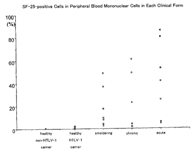
Fig. 1 shows the ratio of SF-25 antigen-positive mononuclear cells in the
total
mononuclear cells, which was measured by flow cytometry using an anti-SF-25
monoclonal antibody, using the mononuclear cells in peripheral blood from
acute
2 5 ATL patients, chronic ATL patients, smoldering ATL patients, healthy ATL
carriers
and healthy individuals (not infected with HTLV-1) as samples, in an Example
of the
present invention.
CA 02517580 2005-08-30
3
Fig. 2 shows the relationship between the number of cycles and the
fluorescence intensity when pX gene at various concentrations was amplified in
an
Example of the present invention.
Best Mode for Carrying Out the Invention
As mentioned above, according to the method for examining cancer cells of
the present invention, the cancer cells separated from the body, which express
SF-25
antigen on their surfaces, are bound to magnetic beads utilizing antigen-
antibody
reaction between the cancer cells and the anti-SF-25 antibody or antigen-
binding
fragment thereof.
SF-25 is a glycoprotein antigen having a molecular weight of about 125 kDa
found in 1987 (W089/05307; European Patent No. 0 397 700; U.S. Patent No.
5,212,085; Takahashi H, Wilson B, Ozturk M, Motte P, Strauss W, Isselbacher KJ
and Wands JR. In vivo localization of colon adenocarcinoma by monoclonal
antibody binding to a highly expressed cell surface antigen. Cancer Research
1988;
48: 6573-6579., Wilson B, Ozturk M, Takahashi H, Motte P, Kew M, Isselbacher
KJ
and Wands JR. Cell surface changes associated with transformation of human
hepatocytes to the malignant phenotype. Proc. Natl. Acad. Sci. USA 1988; 85:
3140-
3144., Takahashi H, Carlson R, Ozturk M, Sun S, Motte P, Strauss W,
Isselbacher
KJ, Wands JR and Shouval D. Radioimmunolocalization of hepatic and pulmonary
2 0 metastasis of human colon adenocarcinoma. Gastroenterology 1989; 96: 1317-
1329.,
Hurwitz E, Stancovski I, Wilcheck M, Shouval D, Takahashi H, Wands JR, Sela M.
A conjugate of 5-Fluorourodine-poly(L-lysine) and an antibody reactive with
human
colon carcinoma. Bioconjugate Chemistry 1990; 1: 285-290., Wands JR, Takahashi
H. Studies on cell surface changes associated with transformation of human
2 5 hepatocytes to the malignant phenotype and their role as potential
immunotargeting
sites. In Frontiers ofMucosal Immunology. Volume 2. Eds by Tsuchiya M. 1991
pp.
295-298. Hurwitz E, Adler R, Shouval D, Takahashi H, Wands JR, Sela M.
CA 02517580 2005-08-30
4
Immunotargeting of daunomycin to localized and metastatic human colon
adenocarcinoma in athymic mice. Cancer Immunology Immunotherapy 1992; 35:
186-192., Takahashi H, Nakada T, Puisieux I. Inhibition of human colon cancer
growth by antibody-directed human LAK cells in SCID mice. Science 1993; 259:
1460-1463., Takahashi H, Nakada T, Nakaki M, Wands JR. Inhibition of hepatic
metastases of human colon cancer in nude mice by a chimeric SF-25 monoclonal
antibody. Gastroenterology 1995; 108: 172-182). SF-25 antigen is known to
express on human colon cancer cell lines (e.g., LS 180 (ATCC No. CL0187), COLD
320 (ATCC No. CCL-220.1), WiDr (ATCC No. CCL-218), Caco-2 (HTB-37) and on
human liver cancer cell lines (e.g., FOCUS (Lun H. et al., in Vitro 20; 493-
504
(1984)). Anti-SF-25 monoclonal antibodies are also known (W089/05307,
European Patent No. 0 397 700 and U.S. Patent No. 5,212,085), and a hybridoma
producing an anti-SF-25 monoclonal antibody is deposited with ATCC (ATCC No.
HB9599).
The anti-SF-25 antibody may preferably be a monoclonal antibody. As
mentioned above, anti-SF-25 monoclonal antibodies are known and one of them
has
been deposited. The monoclonal antibody produced by the deposited hybridoma
ATCC No. HB9599 was prepared by immunizing mice with the above-described
human liver cancer cell line FOCUS, preparing hybridomas producing monoclonal
2 0 antibodies, and selecting a monoclonal antibody which undergoes antigen-
antibody
reaction with the above-described various human colon cancer cell lines. In
the
method of the present invention, the deposited anti-SF-25 monoclonal antibody
may
be employed, and other monoclonal antibodies prepared by the similar method
may
also be employed. As concretely described in W089/05307, European Patent No. 0
2 5 397 700 and U.S. Patent No. 5,212,085, describing ATCC No. HB9599, since a
number of hybridomas other than ATCC No. HB9599 were obtained in one
preparation process, an anti-SF-25 monoclonal antibody may be easily prepared
by
CA 02517580 2005-08-30
the known method. Therefore, the monoclonal antibody used in the method of the
present invention is not restricted to that produced by the deposited
hybridoma.
Further, not only the entire antibody, but also fragments thereof having an
ability to
bind with the antigen, such as Fab fragment and F(ab')2 fragment, may also be
5 employed.
Magnetic beads are particles prepared by giving magnetism to latex particles
or polystyrene particles by, for example, blending ferrite. Magnetic beads
carrying
an antigen or antibody are well-known in the field of immunoassay and are
commercially available. As the magnetic beads used in the present invention,
those
which do not adsorb the cells, by non-specific interaction, other than the
target cells
bound through SF-25 antibody are desired, and DYNABEADS (trademark, Dynal
Biotech) and the like may preferably be employed.
The method for binding the cancer cells separated from the body, which cells
express on their surfaces the SF-25 antigen to the magnetic beads utilizing
the
antigen-antibody reaction between the cancer cells and the anti-SF-25 antibody
or
antigen-binding fragment thereof include direct method and indirect method,
and
either of them may be employed. The direct method is the method in which an
anti-
SF-25 antibody or the antigen-binding fragment thereof is immobilized on the
magnetic beads, and the cancer cells are directly bound to the magnetic beads
by the
2 0 antigen-antibody reaction between the anti-SF-25 antibody or antigen-
binding
fragment thereof and the cancer cells. The direct method has an advantage that
it
can be carried out quickly and simply because the antigen-antibody reaction is
carried
out only once.
The method per se for immobilizing an antibody or the antigen-binding
2 5 fragment thereof to the magnetic beads is well-known. For example,
magnetic
beads may be applied to a solution of the antibody or its antigen-binding
fragment so
as to physically adsorb the antigen or the antigen-binding fragment on the
magnetic
CA 02517580 2005-08-30
6
beads. In this case, the concentration of the antibody or the antigen-binding
fragment thereof in the mixture may be, although not restricted, usually about
0.001
to 1 % by weight, and the concentration of the magnetic beads may be, although
not
restricted, usually about 0.1 to 50% by weight. Although the conditions for
the
physical adsorption are not restricted, it may be usually carried out by
incubating the
mixture at 4°C to 45°C for about 0.5 to 48 hours. The method for
immobilization of
the antibody or the antigen-binding fragment thereof onto the magnetic beads
is not
restricted to physical adsorption, and other known methods, for example,
covalently
bonding the antibody or the antigen-binding fragment thereof using functional
groups
such as amino groups or carboxyl groups bound to the magnetic beads may also
be
employed.
On the other hand, the indirect method is a method in which the cancer cells
and a labeled or non-labeled anti-SF-25 antibody or antigen-binding fragment
thereof
are subjected to antigen-antibody reaction to bind the antibody or antigen-
binding
fragment thereof to the cells, and subsequently or simultaneously, the cells
are bound
to the magnetic beads through the antibody or antigen-binding fragment thereof
by
specific reaction between the antibody or antigen-binding fragment thereof or
the
label attached thereto and the magnetic beads. In cases where a non-labeled
antibody or antigen-binding fragment thereof is used, an antibody or antigen-
binding
2 0 fragment thereof which undergoes antigen-antibody reaction with the non-
labeled
antibody or antigen-binding fragment thereof is immobilized on the magnetic
beads.
For example, in cases where the anti-SF-25 antibody which is to be reacted
with the
cancer cells is a mouse IgG, an anti-mouse IgG antibody may be immobilized on
the
magnetic beads. In cases where a labeled antibody or antigen-binding fragment
2 5 thereof is bound to the cancer cells, an antibody or antigen-binding
fragment thereof
or a substance which specifically binds to the label is immobilized on the
magnetic
beads. Therefore, as the label, any substance which has antigenecity and which
CA 02517580 2005-08-30
7
does not hinder the antigen-antibody reaction between the cancer cells and the
anti-
SF-25 antibody or antigen-binding fragment thereof may be employed. For
example, in Examples below, fluorescein isothiocyanate (FITC) as a label,
which is
well-known as a fluorescent label, is bound to an anti-SF-25 monoclonal
antibody,
and the FITC is bound to anti-FITC antibody-immobilized magnetic beads.
Alternatively, biotin may be used as the label, and the biotin may be bound to
the
magnetic beads on which avidin or an avidin derivative such as streptoavidin
is
immobilized. Thus, as the label, any substance may be employed, which can
specifically bind to another substance, and which does not hinder the antigen-
antibody reaction between the cancer cells and the anti-SF-25 antibody or
antigen-
binding fragment thereof. Anti-mouse IgG antibody-immobilized magnetic beads,
anti-FITC antibody-immobilized magnetic beads, streptoavidin-immobilized
magnetic beads and the like are widely used in the field of immunoassay, and
are
commercially available because their versatility is high. In the method of the
present invention, these commercially available magnetic beads may preferably
be
employed.
The cancer cells which may be examined by the method of the present
invention are any cancer cells expressing the SF-25 antigen on their surfaces,
and
examples of the cancer cells include leukemia cells, colon cancer cells, small
2 0 intestinal cancer cells, gastric cancer cells, esophagus cancer cells,
bile duct cancer
cells, gallbladder cancer cells, thyroid cancer cells, parathyroid cancer
cells, prostate
cancer cells, uterine cancer cells, ovarian cancer cells, choriocarcinoma
cells,
orchioncus cells, bladder cancer cells, renal cancer cells, adrenal cancer
cells, brain
tumor cells, melanoma cells, skin cancer cells, lung cancer cells, breast
cancer cells,
2 5 pancreatic cancer cells and liver cancer cells. Examples of the leukemia
cells
include transformed lymphocytes and mononuclear cells. In Example 1 below,
adult T cell leukemia (ATL) accompanying transformed lymphocytes is examined,
CA 02517580 2005-08-30
but it was hitherto not known that SF-25 antigen is expressed on transformed
lymphocytes.
In a preferred mode of the present invention, blood or cells separated from
the
blood are used as the test sample. Among the various cancer cells described
above,
leukemia cells are contained in the blood, so that it is natural that the
blood or the
cells separated from the blood may be tested by the method of the present
invention.
Further, since solid cancer cells, peeled off from the tissues, such as colon
cancer
cells, gastric cancer cells, lung cancer cells, breast cancer cells,
pancreatic cancer
cells and liver cancer cells, are also contained in the blood, cerebrospinal
fluid, bone
marrow, pleural effusion, ascites, pancreatic juice, duodenal juice, bile,
feces or urine,
examination of these solid cancer cells may be attained by the method of the
present
invention using blood or the like. Although biopsy is necessary for obtaining
solid
cancer cells from an organ tissue, by the method of the present invention,
blood that
can be much more easily and safely obtained than organ tissues may be used as
the
test sample, which is advantageous.
In the direct method, although the conditions for the antigen-antibody
reaction
between the magnetic beads on which the antibody or antigen-binding fragment
thereof is immobilized and the cancer cells are not restricted, in cases where
the test
cells (a mixture of normal cells and cancer cells) have already been
separated, the
2 0 antigen-antibody reaction may be carried out by bringing the magnetic
beads into
contact with the cancer cells, for example, at about 4°C to 45°C
for about 0.5 to 24
hours. The population density in the mixture used herein is not restricted,
and
usually about 1 cell/ml to 106 cells/ml, and the concentration of the magnetic
beads
may be, although not restricted, usually about 0.1 to 10% by weight. In cases
where
2 5 blood is made to contact with magnetic beads, it may be carried out at a
temperature
between about 4 and 45°C for about 0.5 to 24 hours. In this case, the
concentration
of the magnetic beads in the mixture may be as described above. In the case of
CA 02517580 2005-08-30
9
indirect method, the concentration of the anti-SF-25 antibody or antigen-
binding
fragment thereof which is subjected to the antigen-antibody reaction with the
cancer
cells may be, although not restricted, usually about 0.001 to 1 % by weight.
As for
the conditions for the antigen-antibody reaction, either the reaction between
the
cancer cells and the anti-SF-25 antibody or antigen-binding fragment thereof
or the
reaction between the produced antigen-antibody complex and the antibody or
antigen-binding fragment thereof on the magnetic beads may be carried out
under the
conditions similar to those employed in the above-described direct method. In
the
indirect method, the antigen-antibody reaction between the cancer cells and
the anti-
SF-25 antibody or antigen-binding fragment thereof may be carried out firstly,
and
then the produced antigen-antibody complex may be reacted with the magnetic
beads.
Alternatively, the cancer cells, the anti-SF-25 antibody or antigen-binding
fragment
thereof and the magnetic beads may be made to co-exist, and the above-
described
two reactions may be carried out in parallel. In cases where a specifically
binding
substance such as biotin is used as the label, the reaction may be carried out
under the
conditions well-known for the respective label.
Thereafter, magnetic beads are collected by magnetic force. As mentioned
above, since immunoassay using magnetic beads is well-known, and since the
apparatuses for collecting magnetic beads by magnetic force are commercially
2 0 available, collection of the magnetic beads may be carried out easily
using a
commercially available apparatus. Alternatively, the collection may be
attained
manually by simply using a magnet.
By the above-described steps, the cells expressing SF-25 antigen on their
surfaces are bound to the magnetic beads. Examination of the cancer cells
bound to
2 5 the magnetic beads is then carried out. It is preferred to conduct a
washing step in
which the magnetic beads are washed with a buffer solution and the magnetic
beads
are collected again by magnetic force, before the examination. The examination
per
CA 02517580 2005-08-30
I
se of the cells may be carried out by a method known for the respective cancer
cells.
For example, an examination by which the cells can be identified as cancer
cells,
such as examination of nucleic acid, pathological examination or biochemical
examination is carried out. In Examples 1 and 3 below, DNAs are collected from
mononuclear cells from adult T cell leukemia (ATL) patients, and the proviral
HTLV-1 gene causative of ATL is detected by inverse PCR and subsequent
Southern
blot, thereby diagnosing ATL. A preferred example of the examination is the
examination of nucleic acid by which such a pathogenic virus or a marker gene
for
various cancer cells is detected. Examples of such genes include HTLV-1 (ATL),
Rb (retinoblastoma, lung cancer, breast cancer), p53 (colon cancer, breast
cancer,
lung cancer and the like), WTI (Wilms tumor), APC (colon cancer, gastric
cancer),
p 1 b (melanoma, esophagus cancer), NFI (melanoma, neuroblastoma), NF2
(meningioma, esophagus cancer), VHL (renal cancer), DPC-4 (pancreatic cancer),
SMAD2 (colon cancer), PTEN (glioblastoma), PTC (dermal basal cell carcinoma),
int-2/hst-1/cycDl (head and neck cancer, esophagus cancer, bladder cancer),
MDM-2
(sarcoma, brain tumor), erbB 1 (polymorphic glioma, breast cancer), erbB2(neu)
(breast cancer, gastric cancer, ovarian cancer), c-myc (uterine cancer, small
cell
carcinoma of the lung, breast cancer), N-myc (neuroblastoma, small cell
carcinoma
of the lung, sarcoma), H-ras (uterine cancer), K-ras (gastric cancer), c-met
(gastric
2 0 cancer), K-sam (gastric cancer), AKT-1, AKT-2(S/T-PK) (both of them are
markers
of gastric cancer and ovarian cancer), and Aurora-2(S/T-PK) (colon cancer).
Other
than the examination of nucleic acids, examinations of EGF receptor (breast
cancer
and the like), p53 protein (colon cancer, liver cancer), vascular epidermal
growth
factor (VEGF) (liver cancer, colon cancer and the like), TGF-(3, annexin'-I
and the
2 5 like are exemplified. It is also preferred to search the expression of a
gene such as
4F2 gene or PCD1 gene by RT-PCR, of which expression is ubiquitously increased
in cancer cells (see Example 4). The nucleotide sequences of these pathogenic
virus
CA 02517580 2005-08-30
11
genes and cancer marker genes or cDNAs thereof are known and are included in
databases such as GenBank (freely available at
http://www.ncbi.nlm.nih.gov/Genbank/index.html). Therefore, by carrying out a
search using the name of the pathogenic virus or the cancer marker as the
keyword,
the nucleotide sequence thereof may easily be found. If the nucleotide
sequence of
the pathogenic virus gene or the cancer marker gene or the cDNA thereof is
known,
whether the cells contain the pathogenic virus gene or the cancer marker gene
or not,
or the gene is expressed in the cells or not may be determined by, for
example,
subjecting the gene or cDNA to a well-known nucleic acid-amplification method
such as PCR for amplifying an optional region in the gene or cDNA, and
determining
whether amplification occurs or not.
Since the nucleic acid-amplification methods such as PCR and the methods
for the subsequent detection of the amplification product are well-known in
the art
and since reagent kits and apparatuses therefor are commercially available,
those
skilled in the art can easily practice the nucleic amplification methods. The
size of
the primers used in PCR is not less than 15 bases, preferably about 18 to 50
bases.
By subjecting the target pathogenic virus gene or cancer marker gene or its
cDNA to
a nucleic acid-amplification method using a pair of primers which are a part
of the
target pathogenic virus gene or cancer marker gene or its cDNA, and a part of
the
2 0 complementary chain thereof, respectively, and using the test nucleic acid
as the
template, the test nucleic acid is amplified. In contrast, if the test nucleic
acid is not
contained in the test sample, amplification of the nucleic acid does not
occur.
Therefore, by detecting the amplification product, whether the test nucleic
acid exists
in the sample or not can be determined. Detection of the amplification product
may
2 5 be carried out by a method in which the reaction solution after
amplification is
electrophoresed, and staining the bands with ethidium bromide or the like, or
by a
method comprising immobilizing the amplification product after electrophoresis
on a
CA 02517580 2005-08-30
12
solid phase such as a nylon membrane, hybridizing a labeled probe which
specifically
hybridizes with the test nucleic acid with the immobilized amplification
product, and
detecting the label after washing. The test nucleic acid in the sample may
also be
quantified by conducting the so called real-time detection PCR (see Examples
below)
using a quencher fluorescent pigment and a reporter fluorescent pigment. Since
kits
for real-time detection PCR are also commercially available, it can be easily
carried
out. Further, the test nucleic acid may also be semi-quantified based on the
intensity of the electrophoretic band. The test nucleic acid may be a mRNA or
a
cDNA reverse-transcribed from the mRNA. In cases where a mRNA as a test
nucleic acid is amplified, NASBA method (3SR method or TMA method) using the
above-mentioned pair of primers may also be employed. Since NASBA method per
se is well-known, and since the kits therefor are commercially available,
NASBA
may easily be carried out using the above-mentioned pair of primers.
The pathogenic virus gene or the cancer marker gene or the cDNA thereof
may also be detected by a method using a nucleic acid probe. The method using
a
nucleic acid probe is also well-known, and if the nucleotide sequence of the
test
nucleic acid is known, the method may be carried out by hybridizing a labeled
nucleic acid probe with the test nucleic acid, which probe has a nucleotide
sequence
complementary to a region of the test nucleic acid, and then detecting the
label. As
2 0 the probe, a nucleic acid having a nucleotide sequence complementary to a
part of the
nucleotide sequence of the pathogenic virus gene or cancer marker gene or cDNA
thereof, which nucleic acid is labeled with a fluorescent label, radioactive
label,
biotin label or the like may be employed. Whether the test nucleic acid exists
in the
sample or not may be determined by immobilizing the test nucleic acid or an
2 5 amplification product thereof on a solid phase, hybridizing it with the
labeled probe,
and measuring the label bound to the solid phase after washing. Alternatively,
the
detection of the test nucleic acid may also be carried out by immobilizing a
nucleic
CA 02517580 2005-08-30
13
acid for measurement on a solid phase, hybridizing the test nucleic acid
therewith and
detecting the test nucleic acid bound to the solid phase by a labeled probe or
the like.
In such a case, the nucleic acid for measurement immobilized on the solid
phase is
also called a probe.
As the pathological tests and biochemical tests of the cancer cells trapped on
the magnetic beads by the above-described method, the following methods are
exemplified. That is, examples of the pathological tests include detection of
Philadelphia chromosome in chronic myelocytic leukemia, detection of signet-
ring
cell carcinoma observed in gastric cancer cells or the like, detection of
coffee bean-
like morphology observed in the nuclei of cancer cells, and detections of
tumor
markers such as CEA (carcinoembryonic antigen), AFP(alfa-feto protein), CA19-
9,
DUPAN-2, TPA (tissue polypeptide antigen) and the like. Examples of the
biochemical tests include measurements of the above-mentioned tumor markers
expressed by cancer cells by ELISA and measurements of isozymes (e.g., LDH
isozyme and 5'-NPD-V isozyme) expressed by cancer.
The present invention will now be described more concretely by way of
examples thereof. It should be noted that the present invention is not
restricted to
the examples below.
Reference Example 1 Detection of ATL
2 0 1. Preparation of Samples
Mononuclear cells were separated from peripheral blood from 7 acute ATL
patients, 5 chronic ATL patients, 9 smoldering ATL patients, 42 healthy ATL
carriers
and 8 healthy individuals (not infected with HTLV-1), respectively. The
separation
of the mononuclear cells was carried out as follows. In a centrifugal tube, 5
ml of
2 5 Lymphaprep (trademark) (Axis-Sheld PoC AS, Oslo, Norway) was placed and 5
ml
of venous blood supplemented with heparin was gently overlaid thereon. Then
the
sample was centrifuged at 400 x g for 30 minutes, and the mononuclear cells
CA 02517580 2005-08-30
14
suspended in the form of a band between the blood plasma and the separation
solution were recovered with a capillary pipette. The recovered mononuclear
cells
was subjected to centrifugal washing three times with phosphate-buffered
saline
(PBS), and the separated mononuclear cells were used in the subsequent
experiments.
2. Flow Cytometry
Mouse anti-SF-25 monoclonal antibody (produced by ATCC No. HB9599)
was labeled with FITC by a conventional method. Using the fluorescence-labeled
anti-SF-25 monoclonal antibody, flow cytometry was carried out for the
mononuclear
cells separated in 1 described above. The flow cytometry was carried out as
follows.
From a mononuclear cell suspension in PBS of which population density was
adjusted to 5 x 10⁶ cells/ml, 0.1 ml aliquots were sampled into two small test
tubes.
To one of the test tubes, 10 ~1 of diluted fluorescence-labeled SF-25
monoclonal
antibody was added. To the other test tube, 10 ~l of FITC-labeled mouse IgGI
was
added. Each of the mixtures was allowed to react at 4°C for 30 minutes
while
gently agitating the mixture at 10 minutes' intervals. After the reaction, PBS
was
added, and centrifugal washing was carried out twice. Thereafter, the mixture
was
applied to a flow cytometer EPICS XL (trademark), Coulter, Miami, Florida,
U.S.A.),
and about 10,000 cells were counted using FITC-labeled mouse IgGI as a
control, to
determine the ratio of the positive cells.
2 0 By the above-described flow cytometry, the ratio of the mononuclear cells
expressing SF-25 antibody was determined. The results are shown in Table 1
below
and in Fig. 1.
CA 02517580 2005-08-30
Table 1
Examinee Acute Chronic SmolderingHealthy Healthy
ATL ATL ATL HTLV-I Individuals
PatientsPatients Patients Carriers
Ratio (%) 42.7 27.9 15.2 0.6 0.4
of
Mononuclear
Cells Expressing
SF-25 Antigen
(average)
As is apparent from Table 1 and Fig. 1, by using SF-25 antigen on
mononuclear cells as a marker for ATL, the group of healthy individuals and
healthy
carriers, and the group of acute, chronic, and smoldering ATL may be clearly
5 distinguished.
Example 1
Collection of SF-25-expressing Mononuclear Cells Using Magnetic Beads and Test
of Nucleic Acids Thereof
Whether or not HTLV-1 gene was inserted in the form of provirus in the
10 chromosomal DNAs in the mononuclear cells expressing SF-25 antigen was
examined by inverse PCR and Southern blot. These were carried out as follows.
( 1 ) Collection of Mononuclear Cells Expressing SF-25 Antigen
In 60 ~l of phosphate-buffered saline (pH 7.2, containing 0.5% bovine serum
albumin and 2 mM EDTA, hereinafter referred to as "buffer"), 10~ mononuclear
cells
15 separated from peripheral blood from each smoldering ATL patient were
suspended.
Then 10 ~l of FITC-labeled mouse anti-SF-25 monoclonal antibody (produced by
ATCC No. HB9599) solution (concentration: 0.1 %) was added thereto and the
mixture was allowed to react at 4°C for 5 minutes, followed by washing
twice to
remove the non-adsorbed antibody. To the resulting cells, 90 ~l of buffer was
2 0 added to suspend the cells again, and 10 pl of anti-FITC micromagnetic
beads
(Miltenyi Biotec GmbH, Bergish Gladbach, Germany, particle diameter: 50 nm)
was
added to the suspension, followed by allowing the labeling at 6°C for
15 minutes,
CA 02517580 2005-08-30
16
thereby binding the mononuclear cells having SF-25 antigen on their surfaces
to the
magnetic beads. The cells were washed twice to remove the non-adsorbed beads,
and the resulting cells were re-suspended in 500 pl of buffer. Using MACS Midi
Set (Miltenyi Biotec GmbH, Bergish Gladbach, Germany), the cells were applied
to a
MACS Separation Positive Selection MS Column (Miltenyi Biotec GmbH, Bergish
Gladbach, Germany) (washed once with 500 ~.l of buffer), and the cells labeled
with
the magnetic beads (SF-25 antigen-positive cells) and the cells not labeled
with
magnetic beads (SF-25 antigen-negative cells) were fractioned by magnetic
force.
The fractioned cells were used in the subsequent experiments as samples.
(2) Inverse PCR
Inverse PCR was performed as follows based on the report by Takemoto S et
al (Blood Vol 84 No9 3080-3085, 1994). Chromosomal DNAs were extracted from
each sample using DNAzoI (Molecular Research Center, Inc., Montgomery Rd.,
Cincinnati, Ohio), and the DNAs were cleaved by Sau 3AI, followed by
subjecting
the resultant to self ligation using T4 DNA ligase. By this method, DNA
consisting
of HTLV-1 5'LTR and the gag sequence, as well as DNA consisting of HTLV-1 3'-
LTR and chromosomal DNA was constructed. To remove the DNA consisting of
the 5' proviral DNA of HTLV-1, the mixture was treated with Sac II under heat.
Using the resulting DNA as a template, two-step nested PCR was performed. The
2 0 first step PCR was performed using primer 1: 5'-aagccggcagtcagtcgtga-3'
(8946-
8927nt in nucleotide sequence of HTLV-1) and primer 2: 5'-aagtaccggcaactctgctg-
3'
(8958-8977nt in nucleotide sequence of HTLV-1). Then the second step PCR was
performed using primer 3: 5'-gaaagggaaaggggtggaac-3' (8924-8905nt in
nucleotide
sequence of HTLV-1) and primer 4: 5'-ccagcgacagcccattctat-3' (8986-9005nt in
2 5 nucleotide sequence of HTLV-1 ). Each PCR was performed using Thermal
Cycler
by repeating 50 times in the first step and 35 times in the second step the
cycle of
94°C for 20 seconds, 55°C for 20 seconds and 72°C for 30
seconds. A 5 pl aliquot
CA 02517580 2005-08-30
17
of the PCR product was sampled and subjected to electrophoresis on 2% agarose
gel.
The gel was stained with ethidium bromide and the band was examined for the
existence of the incorporation of clonal HTLV-1.
(3) Southern Blot
The above-described electrophoresed product was transferred to a nylon
membrane filter, and incorporation of HTLV-I was examined using an
oligonucleotide (5'-ctccaggagagaaatttagtacac-3', 9012-9035nt in nucleotide
sequence
of HTLV-1) as a probe. According to the report by Takemoto et al., the US
region
of 3'LTR of HTLV-1 containing the chromosomal gene is thought to be amplified
by
this method. Although the incorporation of HTLV-1 gene in ATL patients is
random between different cases, the incorporation of HTLV-1 gene in ATL cells
in
one patient is monoclonal, so that whether the amplification of the gene is
monoclonal or not can be determined by amplifying the US region in the 3'LTR
of
HTLV-1 containing the chromosomal DNA. In fact, they confirmed it by
sequencing the DNA in the US region of the 3'LTR of HTLV-1 containing the
chromosomal DNA.
As a result, in the mononuclear cells expressing SF-25 antigen, the HTLV-I
proviral DNA was monoclonally incorporated, and in the SF-25-negative cells,
proviral HTLV-I was not detected. By this, it was confirmed that acute,
chronic and
2 0 smoldering ATL could be detected by the method of the present invention.
Example 2
Synthesis of Magnetic Beads Sensitized with Anti-SF-25 Antibody
Magnetic beads for separation of cells (DYNAL, M-450 Goat anti-Mouse
IgG) in the form of a suspension in an amount of 10 mL (solid content: 300 mg)
were
2 5 rinsed 4 times with PBS, and then suspended in 6 mL of PBS. To this
magnetic
beads, 0.60 mL of an anti-SF-25 antibody (1.0 mg/mL) was added, and the
mixture
was allowed to react at 20°C for 4 hours, followed by washing the
resultant with
CA 02517580 2005-08-30
18
physiological saline to obtain magnetic beads sensitized with anti-SF-25
antibody.
The obtained beads were dispersed in phosphate buffer solution (PBS)
supplemented
with 0.1 % bovine serum albumin to a solid content of 2%.
Example 3
Synthesis of Magnetic Beads Sensitized with Anti-SF-25 Antibody (Chemical
Antibody-Sensitization Method)
In MES buffer solution (5 mM, pH 6.0), 100 mg of magnetic beads for
separation of cells (DYNAL, M-270 Carboxylic Acid) were suspended to a solid
content of 5%. To bind an antibody to the carboxyl groups on the magnetic
beads,
20 mg of EDC hydrochloric acid salt (1-ethyl-3(3-
dimethylaminopropyl)carbodiimide hydrochloride) which was a water-soluble
carbodiimide reagent was added, and the mixture was allowed to react at
20°C for 1
hour to activate the beads. Then the beads were washed once with HEPES buffer
solution (0.1 M, pH 7.4) and then suspended in 2 mL of the same buffer
solution.
To the resulting magnetic beads suspension, 0.4 mL of anti-SF-25 antibody (1.0
mL/ml) was added, and the resulting mixture was allowed to react at
20°C for 4
hours, followed by washing the resultant with physiological saline to obtain
magnetic
beads to which anti-SF-25 antibody was chemically bound. The obtained beads
were dispersed in PBS supplemented with 0.1% BSA to a solid content of 2%.
2 0 Example 4
Separation of KUT-2 Cells
A 5 mL of citrated blood sampled from a healthy donor was centrifuged and
the buffy coat was collected. To the buffy coat, 10 mL of 0.17 M aqueous
ammonium chloride solution was added and the mixture was left to stand at room
2 5 temperature for 10 minutes, followed by centrifuging the mixture. The
obtained
precipitate was washed twice with PBS containing 2 mM EDTA, and suspended in
4.5 mL of PBS containing 0.5% BSA and 0.6% citric acid to obtain normal human
CA 02517580 2005-08-30
19
nucleated cell suspension.
A cell culture of KUT-2 cells (Hanada S, Tsubai F and Namba Y. The
Characteristics of T-cell lines derived from peripheral blood of patients with
adult T-
cell leukemia-lymphoma. Recent Advances in RES Research 1985; 25: 124-133) in
RPMI1640 medium supplemented with 10% FCS, cultured at 37°C in an
incubator
under 5% C02 was centrifuged. The obtained KUT-2 cells were washed twice with
PBS containing 2 mM EDTA, and suspended in 4.0 mL of PBS containing 0.5%
BSA and 0.6% citric acid to obtain KUT-2 cell suspension having a population
density of 6.0 x 10⁵ cells/mL. The normal human cell suspension and the KUT-2
cell suspension obtained above were mixed so as to yield the number of fed
cells
shown in Table 5, and the mixture was diluted with PBS containing 0.5% BSA and
0.6% citric acid to a total volume of 1.0 mL. Selective separation of KUT-2
cells
from the cell mixture was performed using the magnetic beads sensitized with
the
anti-SF-25 antibody, prepared in Examples 2 and 3. To 1.0 mL of the cell
mixture
solution, 50 ~L (beads content: 1 mg) of the magnetic beads sensitized with
the anti-
SF-25 antibody prepared in Example 2 or 3 was added, and the resulting mixture
was
mixed by inversion for 30 minutes at 4°C. The magnetic beads were
separated by
magnetic separation, and the beads were washed three times with PBS containing
0.5% BSA and 0.6% citric acid, thereby removing the cells adsorbed to the
beads by
2 0 weak interaction. The beads were then washed once with PBS containing 2 mM
EDTA, and separated by magnetic force, followed by removing the washing
solution
to eliminate citric acid which adversely affects the nucleic acid-
amplification reaction.
To elute nucleic acids from the cells trapped on the magnetic beads, 50 ~L of
Proteinase K solution (0.8 mg/mL) diluted with 10 mM Tris-HCl (pH8.3) was
added
2 5 and the mixture was allowed to react at 55°C for 15 minutes to lyse
the cells. Then
the reaction was allowed to occur at 95°C for 20 minutes to inactivate
Proteinase K
used for the cell lysis and to inactivate the substances originated from the
cells,
CA 02517580 2005-08-30
which inhibit the nucleic acid-amplification reaction.
Using 20 ~L aliquot of the nucleic acid solution, quantitative PCR was
carried out to assess the number of the KUT-2 cells and the number of the
normal
human cells trapped on the magnetic beads. The quantification of the KUT-2
cells
5 was carried out by amplifying and quantifying a 102mer region in the pX gene
region
originated from HTLV-1 virus. The nucleotide sequences of the primers and of
the
probe are shown in Table 2.
The amount of normal human cells were determined by amplifying and
quantifying a 1 l Omer region in (3-globin gene, and the total number of the
captured
10 KUT-2 cells and normal human cells, and then subtracting the number of the
KUT-2
cells. The nucleotide sequences of the primers and of the probe are shown in
Table
3.
The quantitative PCR was performed using ABI PRISM 7700 Sequence
Detection System (Applied Biosystems). The number of the cells was determined
15 based on the number of cycles (Th cycle) at which the fluorescence
intensity
exceeded a threshold value (Th), by extrapolating a calibration curve prepared
at the
same time.
For the preparation of calibration curves of the pX gene and the (3-globin
gene,
a serial dilution of nucleic acid solution was used, which was obtained by
adding
2 0 Proteinase K solution (0.8 mg/mL) to a prescribed amount of KUT-2 cells or
normal
human cells, allowing the mixture to react at 55°C for 15 minutes to
lyse the cells and
then reacting the mixture at 95°C for 20 minutes. The relationship
between the
number of cycles and the fluorescence intensity when the pX gene was amplified
is
shown in Fig. 2. Quantitative amplification was confirmed between 0.5 cell to
5 x
2 5 105 cells. At this time, (Number of Cells) = 10~(30.77/3.46-Th
cycle/3.46),
correlation coefficient r = 1.00.
CA 02517580 2005-08-30
21
Table 2
Quantification 5'-sequence-3'
of pX
Gene
Forward Primer TTC CCA GGG TTT GGA CAG AG
Reverse Primer CGA AGA TAG TCC CCC AGA GA
TaqMan Probe FAM-ATA CCC AGT CTA CGT GTT TGG AGA C-
TAMRA
Table 3
Quantification of 5'-sequence-3'
(3-globin
Gene
Forward Primer ACA CAA CTG TGT TCA CTA GC
Reverse Primer CAA CTT CAT CCA CGT TCA CC
TaqMan Probe FAM-AAC AGA CAC CAT GGT GCA TCT GAC
T-TAMRA
The PCR solution contained reagents for hot-start (Roche Diagnostics) and
had the composition shown in Table 4. To 30 ~,L of the solution, 20 ~L of the
nucleic acid extract obtained above was added, and the resulting mixture was
subjected to amplification.
CA 02517580 2005-08-30
22
Table 4 Composition of PCR Solution
PCR grade distilled water 17.95 ~1
x 10 reaction buffer 3 pl
MgCl2 (25mM) 5 pl
dATP, dGTP, dCTP (IOmM) each 0.5 ~1
dUTP (20mM) 0.5 pl
forward primer (50 ~M) 0.6 pl
reverse primer (50 ~M) 0.6 ~l
TaqMan probe (25 ~M) 0.2 ~l
Fast Start DNA polymerase (SU/p,L) 0.4 ~l
ROX reference dye 0.25 pl
(Invitrogen)
Total 30 ~1
The PCR was performed as follows:
Activation of Polymerase 95°C for 10 minutes
PCR Cycle 60°C for 60 seconds
95°C for 10 seconds
This PCR cycle was repeated 50 times.
2 0 From the determined number of KUT-2 cells and of normal human cells, the
ratio of capturing KUT-2 cells and purity thereof were calculated according to
the
following equation, and the measured values of 10 runs were averaged, which
are
shown in Table 5.
Ratio of Capturing (%) = Number of Captured KUT-2 Cells/Number of Added KUT-2
Cells
2 5 x 100
Purity (%) = Number of Captured KUT-2 Cells/(Number of Captured KUT-2 Cells +
Number of Captured Normal Human Cells) x 100
CA 02517580 2005-08-30
23
Table 5
Beads Number Number Number Number Ratio Purity
Used of Addedof Addedof of of of
KUT-2 Human CapturedCaptured CapturedKUT-2
CELLS Normal KUT-2 Human KUT-2 Cells
Cells Cells Normal Cells (%)
Cells (%)
Exam let 3.Ox 6.Ox 2.8x 2.7x 10 92 99
10 10 10
Exam le 3.0 x 6.0 x 2.9 x 1.2 x 10 95 96
2 10 10 10
Example 3.0 x 6.0 x 2.7 x 2.6 x 10 90 91
2 10 10 10
Example 3.0 x 6.0 x 2.8 x 1.2 x 10 94 96
2 10 10 10
Example 3.0 x 6.0 x 2.9 x 2.7 x 10 98 99
3 10 10 10
Exam lea 3.0x10 6.0x10 2.9x10 9.0x10 97 97
Exam le 3.0 x 6.0 x 3.0 x 6.3 x 10 99 98
3 10 10 10
Exam le 3.0 x 6.0 x 3.0 x 1.3 x 10 99 96
3 10 10 10
As shown in Table 5, by using the magnetic beads sensitized with the anti-SF-
25 antibody, the target KUT-2 cells were able to be recovered efficiently with
a high
purity by a simple procedure. Also, the quantity and purity of DNA eluted from
the
recovered cells was sufficient to achieve quantitative PCR. By the method of
the
present invention, the accuracy of gene diagnosis may be increased, and
diseases
such as cancers can be detected at an earlier stage.
Example 4
Extraction of Nucleic Acid from Human Cancer Cells and Amplification
Heparinated blood collected from a healthy donor was centrifuged and buffy
coat was recovered. The buffy coat was overlaid on Ficoll-Paque Plus (Amersham
Pharmacia) and the resultant was centrifuged. The resultant was suspended in
PBS
and the human normal nucleated cell fraction was recovered.
Human gastric cancer cells OCUM-2M LN (Fujihara T, Sawada T, Chung K
H-YS, Yashiro M, moue T and Sowa M. Establishment of lymph node metastatic
model for human gastric cancer in nude mice and analysis of factors associated
with
metastasis), lung cancer cells Calu-6 (ATCC Number: HTB-56, J Fogh (editor).
Human tumor cells in vitro. New York: Plenum Press; 1975. pp.l 15-159.),
pancreatic
cancer cells Capan-2(ATCC Number: HTB-80, Dahiya R, Kwak KS, Byrd JC, Ho S,
CA 02517580 2005-08-30
24
Yoon W, and Kim YS. Mucin synthesis and secretion in various human epithelial
cancer cell lines that express the MUC-1 mucin gene. Cancer Research 1993; 53:
1437-1443.), colon cancer cells HT-29(ATCC Number: HTB-38, J Fogh (editor).
Human tumor cells in vitro. pp.l 15-159. New York: Plenum Press; 1975.),
uterine
cancer cells HeLa (ATCC Number: CCL-2 Gey GO, Coffman WD and Kubicek MT.
Tissue culture studies of the proliferative capacity of cervical carcinoma and
normal
epithelium. Cancer Research 1952; 12: 264-265.) were suspended in PBS
respectively, to prepare cell suspensions having a prescribed concentration.
To extract nucleic acids from human normal cells and various human cancer
cells, SMITEST EX-R&D (Genome Science Laboratories) was used. More
specifically, after centrifuging the cell suspension, supernatant was removed,
and 15
~1 of enzyme solution, 480 ~l of sample diluent and 5 g.l of coprecipitation
agent
were added thereto, followed by allowing the resulting mixture at 55°C
for 30
minutes after mixing. To the resultant, 400 ~1 of protein solution was added
and the
resulting mixture was allowed to react at 55°C for 15 minutes after
mixing. Then
800 ~l of isopropanol was added thereto, and the mixture was centrifuged after
mixing, followed by removal of the supernatant. To the resultant, 500 ~l of
70%
ethanol was added, and the resultant was centrifuged after washing, followed
by
removal of the supernatant similarly. The resultant was dissolved in
DNase/RNase
2 0 free water to obtain a nucleic acid extract solution.
From this nucleic acid extract solution, genes were amplified and quantified
by quantitative RT-PCR.
For the reverse transcription reaction, Omniscript reverse transcriptase
(QIAGEN) was used, and the composition shown in Table 6 was employed. To 15
2 5 pl aliquot thereof, 5 ~l of the nucleic acid extract obtained above was
added and the
reaction was allowed to occur.
CA 02517580 2005-08-30
Table 6 Composition of Reverse Transcription Reaction Solution
x 10 Reaction buffer 2 ~,1
dNTP mix (SmM) 2 ~l
Random hexamer primer (CLONTECH) (20 ~M) 1 ~,1
5 RNasin ribonuclease inhibitor (Promega) (40 Units/gl) 0.25 ~1
Omniscript reverse transcriptase 1 ~1
RNase-free water 8 75 ul
Total 15 ~,l
The reverse transcription reaction was carried out under the following
conditions:
10 Activation of reverse transcriptase 37°C for 60 minutes
Inactivation of reverse transcriptase 93°C for 5 minutes
Then the obtained single-stranded DNA was amplified and quantified by PCR.
As the target genes to be amplified and quantified, a 101 mer region in PCD 1
gene
and a 1 O1 mer region in 4F2 gene were employed, of which expressions are
increased
15 in various cancer cells. The nucleotide sequences of the primers and the
probes are
shown in Tables 7 and 8. In addition, human endogenous housekeeping genes,
that
is, genes of human (3-actin, human GAPDH and 18S ribosomal RNA were amplified
and quantified similarly.
Table 7
Quantification 5'-sequence-3'
of
PCD 1 Gene
Forward primer GAC AAG GCT GCC CTC TCC TA
Reverse primer TTA AAT CAA GAC CAG ATG TGG AAG AC
TaqMan probe FAM-CTT TCC CAA GAC CAG GCT GCC ACT TCT-TAMRA
CA 02517580 2005-08-30
26
Table 8
Quantification 5'-sequence-3'
of 4F2
Gene
Forward primer TCC TTC TTG CCG GCT CAA C
Reverse rimer GCA TCC AGG CCA ATC TCA TC
TaqMan probe FAM-CGA CTC TAC CAG CTG ATG CTC TTC ACC
C-
TAMRA
The quantitative PCR was performed using ABI PRISM 7700 Sequence
Detection System (Applied Biosystems). The PCR solution contained QuantiTect
Probe PCR (QIAGEN), and had the composition shown in Table 9. To 47 gl
aliquot thereof, 6 ~l of the reaction solution obtained in the reverse
transcription
reaction was added, and the mixture was subjected to amplification.
Table 9 Example of Composition of Quantitative PCR Solution
2 x QuantiTect Probe PCR Master Mix 25 gl
Forward primer (100 ~M) 0.2 ~1
Reverse primer (100 gM) 0.2 ~l
TaqMan probe (13 gM) 0.36 gl
Distilled water 18.24 gl
Total 44 ~l
The PCR was carried out under the following conditions:
Activation of polymerase 95°C for 15 minutes
PCR cycle 94°C for 15 seconds
60°C for 60 seconds
2 0 This PCR cycle was repeated 50 times. The quantification of the expression
of each gene was determined based on the threshold value (Th), by
extrapolating a
calibration curve prepared at the same time. For the preparation of
calibration
curves, a serial dilution of a prescribed amount of cells was used.
CA 02517580 2005-08-30
27
For the human normal cells and for the human cancer cells, the expression
ratios of the PCD 1 gene and 4F2 gene, respectively, to that of the human
endogenous
housekeeping genes were determined. It was proved that expressions of 4F2 gene
and PCD I gene were higher in tumor cells than in human normal cells from the
level
of several cells.
Amplifications of the genes which are strongly expressed in cancer cells were
observed in a small amount of cells as well. By using the magnetic beads
sensitized
with anti-cancer-specific SF-25 antibody, cancer cells in the blood may be
recovered
at a high efficiency to a high purity, and the cancer cells may be detected by
the
quantitative RT-PCR of a specific gene. By the method of the present
invention,
early detection and diagnosis of cancers using a sample separated from the
body,
such as blood, may be attained.
Industrial Availability
By the method of the present invention, cancer cells can be examined simply
and efficiently without using an expensive apparatus such as a cell sorter.
CA 02517580 2005-08-30
1/6
SEQUENCE LISTING
<110> TAKAHASHI Hiroshi and HANADA Shuichi
<120> Method for Examining Cancer Cells and Reagent Therefor
<130> 03PF273-PCT
160> 17
<210> 1
<211> 20
<212> DNA
<213> Artificial Sequence
<220>
<223> Oligonucleotide forward primer used in inverse PCR for amplifying
a region of HTLU-1 gene
<400> 1
aagccggcag tcagtcgtga 20
<210> 2
<211> 20
<212> DNA
<213> Artificial Sequence
<220>
<223> Oligonucleotide reverse primer used in inverse PCR for amplifying
a region of HTLU-1 gene
<400> 2
aagtaccggc aactctgctg 20
<210> 3
<211> 20
CA 02517580 2005-08-30
2/6
<212> DNA
<213> Artificial Sequence
<220>
<223> Oligonucleotide forward primer used in inverse PCR for amplifying
a region of HTLV-1 gene
<400> 3
gaaagggaaa ggggtggaac 20
<210> 4
<211> 20
<212> DNA
<213> Artificial Sequence
<220>
<223> Oligonucleotide reverse primer used in inverse PCR for amplifying
a region of HTLV-1 gene
<400> 4
ccagcgacag cccattctat 20
<210>5
<211>24
<212>DNA
<213>Artificial Sequence
<220>
<223> Oligonucleotide probe used for detecting a region of HTLV-1 gene
<400> 5
ctccaggaga gaaatttagt acac 24
<210> 6
CA 02517580 2005-08-30
3/6
<211> 20
<212> DNA
<213> Artificial Sequence
<220>
<223> Oligonucleotide forward primer used for measuring pX gene in HTL
V-1 gene
<400> 6
ttcccagggt ttggacagag 20
<210> 7
<211> 20
<212> DNA
<213> Artificial Sequence
<220>
<223> Oligonucleotide reverse primer used for measuring pX gene in HTL
V-1 gene
<400> 7
cgaagatagt cccccagaga 20
<210> 8
<211> 25
<212> DNA
<213> Artificial Sequence
<220>
<223> Oligonucleotide TaqMan probe used for measuring pX gene in HTLV-1
gene
<400> 8
atacccagtc tacgtgtttg gagac 25
CA 02517580 2005-08-30
4/6
<210> 9
<211> 20
<212> DNA
<213> Artificial Sequence
<220>
<223> Oligonucleotide forward primer used for measuring beta-globin gen
a
<400> 9
acacaactgt gttcactagc 20
<210> 10
<211> 20
<212> DNA
<213> Artificial Sequence
<220>
<223> Oligonucleotide reverse primer used for measuring beta-globin gen
a
<400> 10
caacttcatc cacgttcacc 20
<210> 11
<211> 25
<212> DNA
<213> Artificial Sequence
<220>
<223> Oligonucleotide probe used for measuring beta-globin gene
<400> 11
CA 02517580 2005-08-30
5/6
aacagacacc atggtgcatc tgact 25
<210> 12
<211> 20
<212> DNA
<213> Artificial Sequence
<220>
<223> Oligonucleotide forward primer used for measuring PCD1 gene
<400> 12
gacaaggctg ccctctccta 20
<210> 13
<211> 26
<212> DNA
<213> Artificial Sequence
<220>
<223> Oligonucleotide reverse primer used for measuring PCD1 gene
<400> 13
ttaaatcaag accagatgtg gaagac 26
<210> 14
<211> 27
<212> DNA
<213> Artificial Sequence
<220>
<223> Oligonucleotide TaqMan probe used for measuring PCD1 gene
<400> 14
ctttcccaag accaggctgc cacttct 27
CA 02517580 2005-08-30
6/6
<210> 15
<211> 19
<212> DNA
<213> Artificial Sequence
<220>
<223> Oligonucleotide forward primer used for measuring 4F2 gene
<400> 15
tccttcttgc cggctcaac 19
<210> 16
<211> 20
<212> DNA
<213> Artificial Sequence
<220>
<223> Oligonucleotide reverse primer used for measuring 4F2 gene
<400> 16
gcatccaggc caatctcatc 20
<210> 17
<211> 28
<212> DNA
<213> Artificial Sequence
<220>
<223> Oligonucleotide TaqMan probe used for measuring 4F2 gene
<400> 17
cgactctacc agctgatgct cttcaccc 28