Note: Descriptions are shown in the official language in which they were submitted.
CA 02517734 2005-08-29
WO 2004/082467 PCT/US2004/008324
EXTRAC0RP0REAL BLOOD HANDLING SySTEl WITH
AUTOMATIC FLOW CONTROL AND METHODS OF USE
Field of the Invention
[0007.] The present invention relates to an
extracorporeal blood handling system with automatic flow
control and methods for use for monitoring and detecting
error conditions, and modulating flow through the
extracorporeal blood handling system in response to the
detected error conditions.
Background of the Invention
[0002] For more than thirty years, vascular diseases
have been treated using open surgical procedures. In 1999
alone, 753,000 open-heart procedures, including coronary
artery bypass grafting (CABG), valve replacements, and
heart transplants, were performed. During a typical CABG
procedure, a sternotomy is performed to gain access to
the pericardial sac, the patient is put on
cardiopulmonary bypass (CPB), and the heart is stopped
using a cardioplegia solution.
[0003] Generally, previously-known CPB is accomplished
by constructing an extracorporeal blood handling system
including, inter alia, a venous line, a venous reservoir,
a centrifugal or roller pump that perfuses blood through
the extracorporeal circuit and the patient, an oxygenator
for oxygenating the blood, an arterial line for returning
CA 02517734 2005-08-29
WO 2004/082467 PCT/US2004/008324
oxygenated blood to the patient, and an arterial filter
located in the arterial line.
[0004] Previously-known methods of CPB are susceptible
to several error or trigger Conditions. For instance,
one trigger Condition is the inadvertent introduction of
air into the extraCOrporeal circuit. This may occur in a
number of ways, including inadvertent opening of a vent
line, improper priming of the circuit, or by turning the
heart during surgery. If returned to the patient, air
can cause significant patient injury such as brain
damage, cardiac dysfunction, and myocardial damage.
Further, an air-blood mixture may cause turbulence and
high shear stresses within the circuit, resulting in
hemolysis and platelet activation.
[0005] Previously known CPB systems, such as the S3
System sold by Stockert GmbH, Munich, Germany, the HL 20
Heart Lung Machine sold by Jostra Corp., The Woodlands,
Texas, USA and the Sarns Modular Perfusion System 8000,
sold by Terumo Cardiovascular Systems, Ann Arbor, MI,
USA, each include a level detector in the venous
reservoir that slows and then stops delivery of blood to
a patient if the volume of blood in the venous reservoir
falls below a minimum volume. Each of these systems also
includes a bubble detector that abruptly stops the pump
if a predetermined number of bubbles larger than a
predetermined size are detected.
(0006] The system shutdown strategy used in previously
known CPB systems is designed to prevent de-priming of
the venous reservoir and other components of the CPB
circuit until the perfusionist can correct the problem.
Due to the extended periods of time required to prime
previously-l~nown CPB systems, such a strategy is critical
to avoid de-priming. Unfortunately, this strategy leads
2
CA 02517734 2005-08-29
WO 2004/082467 PCT/US2004/008324
to no forward flow to the patient, with potentially
serious consequences if flow is not restored promptly.
[0007] Another previously-known method for handling
air entrained in the blood is described in U.S. Patent
No. 5,188,604 to Orth. The system described in that
patent includes an air sensor disposed in the arterial
line, a controller, and a series of solenoid-controlled
valves, and a shunt circuit. If air is detected in blood
passing to the arterial line, the controller actuates the
solenoid-controlled valves to stop flow in the arterial
line and simultaneously opens the shunt circuit to
redirect the air-laden blood back into the blood
treatment system. Like the previously-described CPB
systems described above, the system described in the Orth
patent results in no forward flow to the patient until
the error condition is corrected.
[0008] Another trigger condition is low venous
pressure, which may be caused by occlusions within the
circuit or the introduction of a large bolus of air. Low
venous pressure is a known risk factor for air
entrainment and may result in depletion of the venous
reservoir, thus requiring blood delivery to the patient
to be suspended while the condition is corrected or the
CPB system is re-primed.
[0009] This problem is manifested with previously-
known CPB systems in that there is minimal reaction time
available to the perfusionist to correct trigger
conditions. For instance, should the venous return flow
stop due to a trigger condition, such as detection of a
large bubble, the perfusionist has only a few seconds to
stop the heart-lung machine before the bubble is pumped
into the patient.
[0010] bet another problem with previously-known
extracorporeal blood handling systems is the substantial
3
CA 02517734 2005-08-29
WO 2004/082467 PCT/US2004/008324
suction force required for proper air evacuation due to
an open air source. An open air source enables the pump
to pull in large amounts of air, overwhelming the ability
of an air evacuation line, if present, t~ remove the air.
[0011] In view of the aforementioned limitations, it
would be desirable to provide an extrasorporeal blood
handling system that monitors and automatically modulates
blood flow in response to trigger conditions thereby
increasing the time available to the perfusionist to
correct trigger conditions.
[0012] It also would be desirable to provide an
extracorporeal blood handling system that monitors and
automatically system operation in response to the
detection of gas in the system, to enhance the ability of
an air evacuation line to remove the air and avoid de-
priming the pump.
[0013] It further would be desirable to provide an
extracorporeal blood handling system that automatically
modulates pump speed in response to the detection of a
massive air bolus in the extracorporeal blood circuit.
[0014] It still further would be desirable to provide
an extracorporeal blood handling system that
automatically modulates system operation in response to
the detection of discrete trigger conditions, monitors
such conditions, and resumes normal operation when the
triggering conditions resolve.
[0015] It even further would be desirable to provide
an extracorporeal blood handling system that
automatically modulates pump speed in response to the
detection of low venous pressures in the extracorporeal
blood circuit.
4
CA 02517734 2005-08-29
WO 2004/082467 PCT/US2004/008324
Summary of the Invention
[0016] In view of the foregoing, it is an object of
the present invention to provide an extracorporeal blood
handling system that monitors and automatically modulates
system operation in response to trigger conditions,
thereby increasing the time available for a perfusionist
to correct trigger conditions.
[0017] It is another object of the present invention
to provide an extracorporeal blood handling system that
monitors and automatically modulates pump speed in
response to the detection of gas in the system, to
enhance the ability of an air evacuation line to remove
the air and avoid depriming of the pump.
[0018] It is an additional object of the present
invention to provide an extracorporeal blood handling
system that automatically system operation in response to
the detection of a massive bolus of air in the circuit.
[0019] It is a further object of the present invention
to provide an extracorporeal blood handling system that
automatically modulates system operation in response to
the detection of discrete triggering conditions, such a
small amounts of air, monitors such conditions, and
resume normal operation once the triggering conditions
subside.
[0020] It is an even further object of the present
invention to provide an extracorporeal blood handling
system that automatically modulates pump speed in
response to the detection of low venous pressures.
[0021] These and other objects of the present
invention are accomplished by providing an extracorporeal
blood handling system with automatic flow control, such
as a microprocessor controlled system whereby pump speed
is regulated in response to detected trigger conditions.
5
CA 02517734 2005-08-29
WO 2004/082467 PCT/US2004/008324
(0022] In a preferred embodiment, the automatic flow
control feature of the apparatus is a microprocessor-
controlled system that monitors the extracorporeal
circuit and automatically modulates pump speed or the
system configuration in response to detected trigger
conditions. The apparatus comprises an extracorporeal
circuit, a controller coupled to an air evacuation system
and sensors positioned to sense air and venous pressures
within the extracorporeal circuit. The controller is
electrically coupled to a pump, such as a centrifugal
pump, to modulate the pump speed in response to detected
trigger conditions in the extracorporeal circuit.
(0023] In a first mode, the automatic flowrcontrol
system of the present invention comprises a controller
coupled to at least one sensor disposed to sense air.
Upon sensing a bolus of air, the microprocessor reduces
the pump speed to a predetermined lower limit. The
predetermined lower limit preferably is determined such
that forward blood flow is maintained through the
extracorporeal circuit to the sterile field.
[0024] In a second mode, the speed of the centrifugal
pump is reduced in response to the detection of discrete
amounts of air. In this case, the pump speed is reduced
by a predetermined percentage until either the trigger
condition is resolved or the pump speed reaches a
predetermined lower limit.
(0025] In a third mode, the controller is coupled to a
second sensor disposed to sense bubbles in the venous
line. The speed of the centrifugal pump is reduced in
response to the detection of bubbles greater than a
predetermined concentration. In this case, the pump
speed is reduced by a predetermined percentage until
either the trigger condition is resolved or the pump
speed reaches a predetermined lower limit.
6
CA 02517734 2005-08-29
WO 2004/082467 PCT/US2004/008324
[0026] In a fourth mode, the controller is coupled to
a third sensor disposed to sense venous pressure. The
speed of the centrifugal pump is reduced in response to
the sensing of venous pressure below a predetermined
level. In this case, the pump speed is reduced by a
fired step until the venous pressure is no longer below
the predetermined level or until a predetermined lower
limit is attained.
[002'7] In alternative embodiments, the system
configuration may be altered in response to the detection
of trigger conditions, by constricting outflow from the
system or by rerouting flowpaths within the system. In
accordance with the principles of the present invention,
some degree of forward flow to the patient is maintained
in these alternative embodiments.
[0028] Methods of operating the automatic flow control
features of the present invention also are provided.
Brief Description of the Drawings
(0029] The above and other objects and advantages of
the present invention will be apparent upon consideration
of the following detailed description, taken in
conjunction with the accompanying drawings, in which like
reference characters refer to like parts throughout, and
in which:
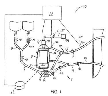
[0030] FIG. 1 is a schematic view of a preferred
extracorporeal circuit incorporating the automatic flow
control system of the present invention;
[0031] FIGS. 2A and 2B are, respectively, perspective
and exploded views of a preferred blood processing
component suitable for implementing the automatic flow
control features of the present invention;
[0032] FIG. 3 is a side-sectional view of the blood
processing component of FIGS. 2 and 3;
7
CA 02517734 2005-08-29
WO 2004/082467 PCT/US2004/008324
[0033] FIGS. 4A and 4B are, respectively, perspective
and cross-sectional views of a filter element of the
blood processing component of FIG. 3;
[0034] FIGS. 5A and. 5B are, respectively, front and
rear perspective views of a preferred blood handling
system incorporating the automatic flow control system of
the present invention;
[0035] FIGS. 6A and 6B are, respectively,
representative screens depicting the display of
parameters monitored and/or controlled by the blood
processing system of FIGS. 6;
[0036] FIG. 7 is a flowchart depicting a first
operational mode of the automatic flow control feature of
the present invention for handling introduction of a
massive bolus of air;
[0037] FIG. 8 is a flowchart depicting a first
operational mode of the automatic flow control feature of
the present invention for handling introduction of
discrete, relatively small boluses of air;
[0038] FIG. 9 is a flowchart depicting a third
operational mode of the automatic flow control feature of
the present invention for handling the occurrence of
bubbles in the venous line;
[0039] FIG. 10 is a flowchart depicting a fourth
operational mode of the automatic flow control feature of
the present invention for handling low venous pressure;
[0040] FIG. 11A is a representative screen depicting
the display of parameters monitored and/or controlled by
the automatic flow control feature of the present
invention;
[004.] FIG. 11B is a representation depicting the
various states of the automatic flow control button of
the present invention;
8
CA 02517734 2005-08-29
WO 2004/082467 PCT/US2004/008324
[0042] FIG. 12A is a graph showing how the automatic
flow control system responds to the detection of low
venous pressure trigger over time; and
[0~431 FIG. 12B is a graph showing how the automatic
flow control system responds to the detection of gas over
time.
Detailed DesCri tion of the Invention
~v~~~i ~~~ ~.f' ~ P.~~.f~~r~~ ,~1 ~~~ Ff~ra~l a.~g~ S,y~ t ~r~
[0044] Referring to FIG. 1, a preferred extracorporeal
blood handling system 10 suitable for use with the
automatic flow Control system of the present invention is
described. ExtraCOrporeal blood handling system 10 is
designed to maintain a patient on full or partial bypass
support, for example, during a coronary artery bypass
graft procedure or mitral valve repair procedure, in
either a full-bypass or beating heart mode of operation.
[0045] Extracorporeal blood handling system 10
includes an extracorporeal blood circuit 11 having a
perfusion circuit comprising venous line 12, perfusion
line segments 13a, 13b and arterial line 14, and a
priming circuit comprising line 16, priming line 17, and
segments 18a and 18b. The ends of perfusion line
segments 13a, 13b are shown extending into the sterile
field as they would appear during use, where they are
coupled to venous and arterial Cannulae respectively.
[0046] Extracorporeal blood circuit 11 illustratively
includes pinch clamps 19 and sampling manifolds 20
disposed on various of the lines. wick-disconnect
couplings 21 are provided at the junctions of venous line
12 and venous segment of perfusion line 13a and arterial
line 14 and arterial segment of perfusion line 13b.
These couplings 21 permit venous line 12 to be directly
coupled to arterial line 14 during priming. In addition,
9
CA 02517734 2005-08-29
WO 2004/082467 PCT/US2004/008324
another quick-disconnect coupling 22 is provided in line
16 to permit, for example, the inclusion of a heat
exchanger when the priming circuit is used for
recirculation.
[0047] ExtraCOrporeal blood handling system 10
further includes an integrated blood processing Component
31 Coupled to a drive unit 32 and controller 33. In
addition, the blood handling system 10 includes a gas
removal system including sensors 25-27, and valve 36
coupled to suction source 34 via line 35. The sensors
25-27, valve 36 and drive unit 32 preferably are
electrically coupled to Controller 33 so that Controller
33 regulates operation of valve 36 and drive unit 3'2 in
response to output of the sensors 25-27. As explained in
greater detail hereinafter, the gas removal system of the
present invention removes air and other gases from
extracorporeal blood circuit 11 and blood processing
component 31 during priming and operation of the bypass
system.
[0048] Referring now to FIGS. 2A, 2B and 3, integrated
blood processing component 31 provides in a single
disposable unit a blood oxygenator, blood pump, and blood
filter, and optionally, a heat exchanger. Blood
processing component 31 includes housing 40 having blood
inlet 41, blood outlet 42, recirculation outlet 43, gas
inlet port 44, gas outlet port 45 and gas removal port
46. Blood outlet 42 and recirculation outlet 43 are
disposed from blood outlet manifold 47, which is located
diametrically opposite blood inlet manifold 48 on housing
40. Blood processing Component 31 preferably includes
tabs 49 or other means for Coupling blood processing
component 31 to reusable drive unit 32.
[004~~] Referring to FIG. 3, housing 40 Comprises a
series of compartments, including: gas collection plenum
CA 02517734 2005-08-29
WO 2004/082467 PCT/US2004/008324
50, central void 51, upper gas plenum 52, annular fiber
bundle compartment 53, lower gas plenum 54 and pump space
55. In a preferred embodiment, central void 51 includes
a larger diameter upper portion and a smaller diameter
lower portion that couples to pump space 55.
[0050] Gas Collection plenum 50 encloses filter 56
that disposed within upper portion of Central void 51.
Filter 56 comprises generally conical, fluid impermeable
upper wall 57 having outlet 80, baffled support structure
58 and filter material 59. Filter 56 Causes gas
entrained in blood introduced into the gas collection
plenum to separate and Collect in the upper portions of
gas collection plenum 50. Blood inlet 41 is displaced
tangentially relative to the centerline of housing 40, so
that blood passing through blood inlet 41 into gas
collection plenum 50 swirls around upper wall 57.
[0051] Upper wall 57 also preferably has a portion
defining an interior chamber that Communicates with the
upper portion of gas Collection plenum 50 through outlet
80. This Configuration allows any gas that passes
through filter material 59 to escape through outlet 80 in
upper wall 57 and be evacuated from gas collection plenum
50. Advantageously, this feature facilitates rapid and
easy priming of the blood processing component 31.
[0052] Filter material 59 comprises one or multiple
layers of a screen-like material, and is mounted to
baffled support structure 58. Filter material 59 serves
to exclude bubbles from the blood flow by maintaining the
swirling action of the blood in the central void for a
sufficient time to allow the bubbles to rise to the gas
Collection plenum. Because the blood Circulates around
the outside of gas removal/blood filter 56 in central
void 51, bubbles impinge against filter material 59
tangentially, and thus "bounce off." Filter material 59
11
CA 02517734 2005-08-29
WO 2004/082467 PCT/US2004/008324
preferably also forms a first stage of a progressive
blood filter that is distributed throughout the blood
processing component, and filters out relatively large
particulate matter.
[0053] t~s illustrated in FIGS. 4t=~ and 4B, support
structure 58 forms a fluid impermeable Cruciform
structure 63 having longitudinal struts f1 and support
rings 62. Struts 61 serve as baffles to reduce swirling
of blood once the blood has passed through filter
material 59.
[0054] Referring again to FIG. 3, blood oxygenation
element 70 is disposed within annular fiber bundle
compartment 53, and comprises a multiplicity of gas
permeable fibers arranged in an annular bundle. As is
well known in the art, the gas permeable fibers are
potted near the upper and lower ends of the bundle so gas
may pass through the interior of the fibers, while
allowing blood to pass along the exterior of the fibers.
The bundle of fibers has an upper potting region 71 that
separates the blood flow region within the annular bundle
from upper gas plenum 52, and lower potting region 72
that separates blood flow region from the lower gas
plenum 54.
[0055] Blood passing into annular fiber bundle
compartment 53 from blood inlet manifold 48 therefore
flows through blood oxygenation element 70 and to blood
outlet manifold 47. The annular fiber bundle also
provides some filtration of blood passing through blood
processing component 31, by filtering out particulate
matter that has passed through filter material 59
employed in gas removal/blood filter 56.
[0056] The lower portion of Central void 51
Communicates with pump space 55, in which pump 55a is
disposed. In a preferred embodiment, pump 55a is a
12
CA 02517734 2005-08-29
WO 2004/082467 PCT/US2004/008324
centrifugal pump including an impeller 75 having a
plurality of vanes 76 and is mounted on shaft 77 via
bearings 78. Impeller 75 preferably comprises an
injection-molded part that encloses a ferromagnetic disk,
so that the disk may be magnetically coupled to drive
unit 32 (see FIG. 1). Blood accelerated by impeller 75
is ejected from pump space 55 via a passageway that
includes Curved ramp 79. Ramp 79 serves to redirect
radially outward blood flow from impeller to a
longitudinal flow within blood inlet manifold 48.
[005'7] In a preferred embodiment, oxygen is introduced
into upper gas plenum 52 through gas inlet port 44 and
passes through the interiors of the multiplicity of
hollow fibers in blood oxygenation element 70. Carbon
dioxide, any residual oxygen, and any other gases
exchanged through blood oxygenation element 70 exits into
lower gas plenum 54 and are exhausted through gas outlet
port 45.
[0058] Referring again to FIG. 1, and in accordance
with the present invention, the extracorporeal blood
handling system 10 also includes sensors 25, 26 and 27
that monitor system parameters. Sensor 25 monitors the
level of gas or blood in gas collection plenum 50.
Sensor 26 detects the presence of gas in venous line 12,
while sensor 27 monitors the pressure in the venous line.
[0059] Sensor 25 is configured to sense a parameter
indicative of a level or volume of air or other gas, or
detect the absence of blood, and preferably operates by a
non-contact method. Suitable sensor methods include
electrical-charge based, optical and acoustic methods. A
resistive contact method also Could be employed, in which
a low electrical Current is passed between adjacent
electrodes only in the presence of blood.
[0060] Sensor 25 preferably is of a known capacitance
13
CA 02517734 2005-08-29
WO 2004/082467 PCT/US2004/008324
type that detects a change in electrical capacitance
between the bulk of a liquid (in this case, blood or
saline) and gas. Alternatively, sensor 25 may be optical
in nature, and uses a light source that has a wavelength
that is minimally attenuated by blood. In this case, the
light source is directed, at an oblique angle, through.
the blood towards a photodetector, and sensor 25 is
positioned to detect the change in the refractive index
of the blood (or saline prime) caused by the presence of
air or other gases. In another alternative embodiment,
sensors 25 may use an ultrasonic energy source and
receiver to detect the presence of gas or absence of
blood by the change in acoustic transmission
characteristics.
[0061] The output of sensor 25 is supplied to
controller 33 (see FIG. 1), which in turn regulates valve
36. When sensor 25 outputs a signal indicating that gas
is present in the extracorporeal blood handling system
10, controller 33 opens valve 36, thereby coupling gas
collection plenum 50 to suction source 34. Suction
source 34 may be any suitable suction source such as a
vacuum bottle, pump or standard operating room suction
port. Once the gas is evacuated, arid sensor 25 detects
blood at an appropriate level, and changes its output so
that controller 33 closes valve 36. In this manner, gas
is continuously monitored and then automatically removed
from the blood by the blood handling system 10.
[0062] Sensor 26 monitors for entrained air in the
venous blood and comprises a sensor of the type described
with respect to sensor 25. Preferably, sensor 26 uses
ultrasound to detect the presence of air entrained in
venous blood, and is coupled to controller 33 so that an
output of the sensor is used to evaluate one or more
trigger conditions, as described hereinafter. Sensor 26
14
CA 02517734 2005-08-29
WO 2004/082467 PCT/US2004/008324
also may be used as a back-up to sensor 25 in the event
sensor 25 fails. Sensor 27 may be any suitable pressure
sensor such as a piezoelectric transducer or an
electrostatic capacitance sensor, and is also Coupled to
Controller 33 and provides an output Corresponding to the
pressure in venous line 13a.
[0063) In operation, deoxygenated blood from the
sterile field is routed through venous line 12 to blood
inlet 41 of integrated blood processing component 31.
Blood entering gas collection plenum 50 is induced to
circulate around the exterior of filter 56 until air or
other gases entrapped in the blood separate out of the
blood and collect in the upper portion of the gas
collection plenum 50. Responsive to the detection of the
presence of a predetermined level or volume of gas by
sensor 25, controller 33 controls operation of valve 36
to evacuate the gas.
[0064 The gas removal system incorporated in the
system of FIGS. 1-3 is capable of removing large amounts
of air from extracorporeal blood circuit 11 during
initial startup, thereby greatly reducing the amount of
saline or donor blood required to prime the system.
Advantageously, this feature facilitates rapid and easy
set-up of blood handling system 10, as well as reduces
the amount of saline or donor blood delivered to the
patient.
[0065] As blood. circulates around filter 56 in central
void 51, it is drawn by the negative pressure head
created by impeller 75 through filter material 59 and
down through central void 51 into pump space 55.
Rotation of impeller 75 caused by drive unit 32, under
the control of controller 33, propels blood up Curved
ramp 79 into blood inlet manifold 43.
CA 02517734 2005-08-29
WO 2004/082467 PCT/US2004/008324
[0066] From blood inlet manifold 48, the blood
traverses blood oxygenation element 70 where it exchanges
carbon dioxide and other gases for oxygen. Qxygenated
blood then passes into blood outlet manifold 47.
~xygenated blood then is directed back to the sterile
field through arterial line 14.
[0067] FIGS. 5A and 5B depict a preferred embodiment
of a blood handling system suitable for implementing the
automatic flow control features of the present invention.
All blood, gas and electrical lines have been omitted for
clarity fro FIGS. 5, and microprocessor-driven controller
33 (see FIG. 1) and a back-up battery are enclosed in
wheeled base 90. Pole 91 is mounted in base 90, and
includes support arm 92 that supports blood processing
component 31 on drive unit 32. Support arm 92 also
carries solenoid 93 that controls valve 36, which is in
turn coupled to suction source 34. Pole 91 also carries
support arm 93, which carries display screen 95. Screen
95 preferably is a touch-sensitive screen coupled to the
controller, and serves as both an input device for the
extracorporeal blood handling system 10 and a display of
system function.
[0068] FIGS. 6A and 6B provide representative samples
of the information displayed on the main windows of the
blood handling system 10. As will of course be
understood by one of ordinary skill in the art of
computer-controlled equipment, the software used to
program operation of the controller may include a number
of set-up screens to adjust particular system parameters.
FIGS. 6A and 6B are therefore the windows that will most
commonly be displayed by the clinician during a
procedure.
16
CA 02517734 2005-08-29
WO 2004/082467 PCT/US2004/008324
[0069] The display of FIG. 6A includes an indicator
of battery status, a series of timers for pump operation,
duration of cross-clamping, and an auxiliary timer,
arterial and venous temperatures and pressures, the speed
of centrifugal pump 55a and the corresponding blood flow
rate. Preferably, controller 33 is programmed with a
number of algorithms for determining an appropriate blood
flow rate during the procedure, as determined based on
body surface area (BSA). The window also may display the
value of BSA determined by the selected algorithm based
on the patient's dimensions, and the suggested blood flow
rate.
[0070] The display of FIG. 6B includes much of the
same information provided in the window of FIG. 6A, but
further shows temperatures and pressures graphically as
well as numerically, so that the clinician can quickly
identify trends in the data and take appropriate
corrective measures. In addition, a lower portion of the
windows displayed in FIGS. 6A and 6B may present system
status or help messages, and include touch sensitive
buttons that permit to access the other available
functions.
Description of the Automatic Flow Control System
of the Present Invention
[0071] In accordance with the principles of the
present invention, microprocessor-based controller 33 of
the extracorporeal blood handling system 10 of FIG. 1 is
programmed to provide at least one automatic flow control
feature. More particularly, controller 33 is programmed
to evaluate the outputs of sensor 25, 26 and 27 to
evaluate the onset or existence of certain trigger
conditions and to modulate system operation to avoid
17
CA 02517734 2005-08-29
WO 2004/082467 PCT/US2004/008324
adverse impacts to system operation. In a preferred
embodiment, modulation of system operation comprises
regulating the speed of pump 55a.
[0072, For example, the outputs of sensors 25-27 may
detect non-negligible levels of gas in the blood and/or
low venous pressure, and reduce the speed of pump 55a and
the blood flow rate. These reductions are expected to
increase the time available for a perfusionist to Correct
the trigger Conditions. In addition, reducing pump speed
lengthens the residence time of blood in filter 56,
thereby permitting air to be evacuated through valve 36
instead of being drawn through blood processing component
31 by pump 55a.
[0073] In a first alternative embodiment, controller
33 may modulate a solenoid-driven clamp on the arterial
line to selectively reduce flow rate through the system.
The pressure increase in the arterial line created by
partially occluding that line is transmitted back to the
pump, thereby reducing blood flow through blood
processing component 31, and again lengthening the period
of time available for the perfusionist to correct the
trigger condition or for the trigger condition to
resolve.
[0074] In yet another embodiment, controller 33 may
modulate a solenoid-controlled valve on the priming
circuit so that blood is shunted from arterial line 14
back to the inlet of blood processing component 31. ~nce
recirculation is established by opening the valve in the
priming circuit, the flow rate through the arterial line
will decrease. This decrease in flow will again provide
needed time for the perfusionist to correct the trigger
condition.
[0075] Referring again to FIG. 1, extracorporeal blood
handling system 10 with automatic flow Control includes
18
CA 02517734 2005-08-29
WO 2004/082467 PCT/US2004/008324
extracorporeal blood circuit 11, blood processing
component 31, and controller 33. Preferably, the
controller 33 includes a microprocessor having software
including machine-readable instructions for interpreting
sensor input and regulating pump speed and gas removal
during automatic flow control.
[007] According to one aspect of the present
invention, controller 33 is electrically coupled to drive
unit 32 of pump 55a and to sensors 25-27. As disclosed
above, the sensors are positioned within extracorporeal
blood circuit 11 to detect the presence of air and/or
measure venous pressure. Preferably, sensor 25 monitors
the level of gas or blood in gas collection plenum 50,
sensor 26 detects the presence of gas or blood in venous
line 12 and sensor 27 monitors the pressure in venous
line 12. When a trigger condition is detected,
controller 33 modulates drive unit 32 to lower the speed
of pump 55a, thus lowering the blood flow rate through
arterial line 14.
[0077] Automatic flow control software is programmed
to provide a reduction phase, a hold phase and a resume
phase in response to a trigger condition. During the
reduction phase, pump speed is reduced to lower the rate
of blood flow through ex.tracorporeal blood circuit 11.
Depending on the type and magnitude of the error
condition, pump speed may be reduced by a fixed step, by
a percentage of the initial pump speed or by rapidly
dropping the pump speed to a predetermined lower limit.
In addition, pump speed may be manually regulated.
[00°70 After reducing the pump speed, the automatic
flow control algorithm enters a hold phase, wherein pump
speed is maintained. at the lower level. In the hold
phase, the perfusionist is prompted to enable the resume
phase as soon as the trigger condition has been resolved.
19
CA 02517734 2005-08-29
WO 2004/082467 PCT/US2004/008324
During the resume phase, pump speed is gradually
increased back to the initial level.
[0079] The automatic flow control system includes
algorithms to implement a number of different control
modes of operation. The system preferably will not lower
the pump speed below a predetermined lower limit, which
is chosen so that forward blood flow is maintained
through extracorporeal blood circuit 11 and to the
sterile field.
[0080] In a preferred embodiment, the automatic flow
control system includes a plurality of operational modes
that respond to different trigger conditions, including a
massive air detection mode, a discrete air detection
mode, a bubble detection mode and a low venous pressure
detection mode. These reduction modes are now be
described with respect to FIGS. 7-10.
[0081] A first mode of operation is designed to handle
the introduction of a large bolus of air into
extracorporeal system 10 - the ~~massive air detection
mode." This is a high priority mode that is triggered
when valve 36 (see FIG. 1) is opened to remove a large
amount of air from gas collection plenum 50. Referring
to FIG. 7, method 100 of automatic flow control is now
described. At step 101, controller 33 detects the
opening of valve 36 in response to gas within gas
collection plenum 50 (see FIG. 1). At step 102,
controller 33 checks whether valve 36 remains open for a
predetermined duration. If so, the first operational
mode is triggered (step 104) and controller 33 reacts by
rapidly dropping the pump speed to the predetermined
lower limit (step 105). According to a preferred
embodiment, the predetermined lower limit is 1800 RPM.
[0082] At step 106, the automatic flow control enters
the hold phase, wherein pump speed is maintained at the
CA 02517734 2005-08-29
WO 2004/082467 PCT/US2004/008324
predetermined lower limit until the trigger condition is
resolved (step 107) and the perfusionist enables the
resume phase (step 108). During the resume phase, pump
speed is gradually increased back to the initial level
set by the perfusionist.
[0083] If valve 36 closes before the e~.piration of the
predetermined duration, a second mode of operation - °'the
discrete air detection mode" - is triggered at step 103.
The discrete air detection mode is designed to handle the
presence of discrete boluses of air in the venous blood.
This is a medium priority mode that is triggered when
valve 36 is briefly opened to remove discrete amounts of
gas from the extracorporeal blood handling system 10.
[0084] Referring to FIG. 8, method 110 of automatic
flow control following detection of discrete quantities
of air is described. At step 111, valve 36 is opened to
remove a discrete amount of gas from gas collection
plenum 50 (see FIG. 1). At step 112, controller 33
checks whether valve 36 remains open for a predetermined
minimum amount of time. If valve 36 remains open for
more than the predetermined minimum amount of time, the
massive air detection mode of FIG. 7 is triggered at step
113. The discrete air detection mode is triggered at
step 114 if valve 36 closes before the predetermined
amount of time has elapsed.
[0085] At step 115, controller 33 reacts by rapidly
dropping the pump speed by a predetermined percentage;
the algorithm then enters a hold phase. In the hold
phase, pump speed is maintained at the current level
until either the trigger condition is resolved (step 117)
or sensor 25 detects further discrete amounts of air, in
which case the method proceeds to step 111. After the
trigger condition is resolved (step 117), the
perfusionist enables the resume phase (step 118). In a
21
CA 02517734 2005-08-29
WO 2004/082467 PCT/US2004/008324
preferred embodiment, the predetermined lower limit for
the pump speed in the discrete air detection mode is 2500
RPM. If pump speed reaches this level, automatic flow
control will remain in the hold phase (step 116) until
the trigger condition is resolved (step 117) and the
perfusionist enables the resume phase (step 118).
[008~a~ A third operational mode - °°the bubble
detection mode" - is designed to handle the presence of
bubbles in the venous line. This is a medium priority
mode, and is triggered when sensor 26 detects gas bubbles
in venous line 12. Referring t~ FIG. 9, method 120 of
automatic flow control following bubble detection in
venous line 12 is described. At step 121, sensor 26
detects the presence of gas bubbles in venous line 12
(see FIG. 1). At step 122, controller 33 reacts by
rapidly lowering the pump speed by a predetermined
percentage. Next, controller 33 waits for a
predetermined duration (step 123) before checking the
status of sensor 26. At step 124, controller 33
determines whether sensor 26 continues to detect the
presence of gas bubbles in venous line 12. If gas
bubbles remain in venous line 12, the method proceeds to
step 122, wherein controller 33 further reduces pump
speed by a predetermined percentage.
[0087] However, if the gas bubbles have dissipated,
the automatic flow control system enters a hold phase at
step 125. In the hold phase, pump speed is maintained at
the then-current level until the trigger condition has
been resolved (step 126) and the perfusionist enables the
resume phase (step 127). In a preferred embodiment, the
predetermined lower limit for pump speed in the bubble
detection mode is 2500 RPM. If pump speed reaches this
level, automatic flow control will remain in the hold
phase (step 125) until the trigger condition is resolved
22
CA 02517734 2005-08-29
WO 2004/082467 PCT/US2004/008324
(step 126) and the perfusionist enables the resume phase
(step 127). Alternatively, controller 33 may be
programmed to enter the resume phase automatically if no
further bubbles are detected within a predetermined time
period.
[0088] A further operational mode - "the low venous
pressure detection mode°' - is designed to handle low
venous pressure in venous line 12. This is a low
priority mode, and is triggered when sensor 27 detects
low venous pressure in venous line 12. Referring to FIG.
10, method 130 of automatic flow control following low
venous pressure detection is described. At step 131,
sensor 27 detects that the venous line pressure has
fallen below a predetermined threshold for a
predetermined minimum duration. At step 132, controller
33 reacts by lowering the pump speed by a predetermined
increment. Next, controller 33 waits for a predetermined
duration (step 133) to allow conditions to stabilize.
Then, at step 134, controller 33 determines whether
sensor 27 continues to detect low venous pressure in
venous line 12. If venous pressure remains below the
predetermined threshold, then the method proceeds to step
132 and controller 33 further reduces the pump speed by
the predetermined increment.
[0089] If venous pressure is no longer below the
predetermined value, automatic flow control algorithm
enters a hold phase (step 135). In the hold phase, pump
speed is maintained at then-current level until the
trigger condition is resolved (step 136) and the
perfusionist enables the resume phase (step 137). In a
preferred embodiment, the predetermined lower limit for
pump speed in the low venous pressure detection mode is
1800 RPi~i. If pump speed reaches this level, automatic
flow control will remain in the hold phase (step 135)
23
CA 02517734 2005-08-29
WO 2004/082467 PCT/US2004/008324
until the trigger condition is resolved (step 136) and
the perfusionist enables the resume phase (step 137).
Controller 33 also may be programmed to enter the resume
phase automatically when the pressure in venous line 12
is detected to exceed a preset level for a predetermined
time period.
LO090] In the event that multiple reduction modes are
triggered at the same time, the highest priority mode
will take precedence. According to a preferred
embodiment, the massive air detection mode is the highest
priority mode followed by the discrete air detection
mode, the bubble detection mode and the low venous
pressure detection mode. In cases where a lower priority
mode is interrupted by a higher priority mode, control
returns to the lower priority mode only after the trigger
condition causing the higher priority mode has been
resolved. By way of example, if discrete air is sensed
by sensor 25 during low venous pressure detection mode,
then the automatic flow control system automatically
switches to the discrete air detection mode. After the
discrete air detection mode trigger condition (i.e., the
presence of discrete amounts of air in the gas collection
plenum) has been resolved, automatic flow control
automatically returns to the low venous pressure
detection mode.
[0091] FIG. 11A is an illustrative display of main
screen 140, similar to FIGS. 6, that incorporates the
automatic flow control system of the present invention.
As will be understood by one of ordinary skill in the art
of computer-controlled equipment, the software used to
program operation of controller 33 may include a number
of set-up screens to adjust particular system parameters.
FIG. 11A depicts screen 140 that will most commonly be
displayed by the perfusionist during automatic flow
24
CA 02517734 2005-08-29
WO 2004/082467 PCT/US2004/008324
control.
[0092] As shown in FIG. 11A, main screen 140 includes
a series of timers for pump operation, duration of cross-
clamping, and an cardioplegia timer, arterial and venous
temperatures and pressures, as measured, for example, at
the blood inlet and blood outlet of the blood processing
component 31, the speed of the centrifugal pump (RPM) and
the corresponding blood flow rate.
[0093] Controller 33 preferably is programmed with a
number of algorithms for determining appropriate blood
flow rates and pump speeds during the procedure and for
evaluating the outputs of sensors 25-27 in accordance
with methods 100, 110, 120 and 130 described hereinabove.
Controller 33 also preferably includes storage that is
programmed with default values for the pump speed limit
values, sensor threshold values, and time periods for
invoking and exiting the various operational modes.
Alternatively, these values may be computed based on
target flow rate values computed, for example, based on
the patient's BSA value, or these values may be input
directly via an alpha-numeric display mode of screen 140
(not shown) .
[0094] In addition, a portion of main screen 140
includes touch sensitive buttons that permit access to
the other available functions. More particularly, main
screen 140 includes button 141 for manually overriding
automatic flow control. With this feature a
perfusionist may at any time disable or partially disable
the automatic flow control system by pressing button 141.
Button 141 functions both as a system control and a
prominent indicator of the automatic flow control status.
To increase its visibility to a perfusionist, button 141
preferably is optionally located in the region of screen
140 that includes pump speed and flow values.
CA 02517734 2005-08-29
WO 2004/082467 PCT/US2004/008324
[0095] As illustrated in FIG. 11B, button 141
preferably has three states including an enabled state
(''AFR ENABLED"), a partially disabled state ("AFR NO
Pven") and a disabled state (°°AFR DISABLED'°).
Optionally, button 141 includes different shades or
colors as a further visual indication of automatic flow
control system status. According to a preferred
embodiment, button 141 has a green tint to indicate that
aut~matic flow control is enabled, a yellow tint to
indicate automatic flow control is partially disabled and
a red tint to indicate automatic flow control is
disabled.
[0096] Referring again to FIG. 11A, main screen 140
also includes button 142 to be used after resolving the
trigger condition(s). Pressing button 142 gradually
increases pump speed back to the initial level (i.e., the
pump speed at which the first trigger condition
occurred). Optionally, button 142 is darkened when pump
speed returns to the initial level and/or when automatic
flow control is re-triggered.
[0097] The system also optionally includes knob 143
for manually controlling the flow rate within the
extracorporeal blood circuit 11. Activating (i.e.,
turning) knob 143 immediately returns blood handling
system 10 to normal operation and darkens button 142.
Using knob 143, a perfusionist may manually control how
quickly the pump speed and flow rate are returned to
their initial levels following automatic flow control.
[0098] Main screen 140 further may include status bar
144 to present system status and/or help messages. These
messages are optionally displayed on the display unit
during reduction and hold phases to indicate the present
mode or modes of operation. As shown in FIG. 11A, status
bar 144 indicates that automatic flow control was enabled
26
CA 02517734 2005-08-29
WO 2004/082467 PCT/US2004/008324
due to a combination of the presence of discrete air in
the system arid low venous pressure. However, since these
trigger conditions have cleared, button 142 is available.
The status messages are removed if button 142 is pressed
or knob 143 is activated. When button 142 is pressed,
the message °°AFR Resuming RPM°° is displayed.
[~0~°] FIGS. 12A and 12H are illustrative graphs
showing how the automatic flow control system responds to
various triggers over time. For exemplary purposes,
actual values for variables such as pressure, pump speed
and time, are described below in parentheses.
[0100] FIG. 12A is a graph depicting how the automatic
flow control system responds over time to a low venous
pressure trigger. Initially, venous pressure drops
briefly below a predetermined threshold (-100mmHg), but
not for a predetermined minimum duration (2 sec). Thus,
the low venous pressure detection mode is not trige~ered.
Thereafter, venous pressure drops below the predetermined
threshold (-100mmHg) for a duration exceeding a
predetermined minimum duration (2 sec) and the low venous
pressure detection. mode is triggered. The automatic flow
control algorithms cause the pump speed to be reduced by
a predetermined increment (e.g., 300 RPM) from 4000 to
3700 RPM. After waiting for a predetermined period of
time (1 sec), the venous pressure is evaluated by
controller 33 using the output of sensor 27, and is
determined still to be below the predetermined threshold
(-100mmHg). Thus, pump speed is again reduced by the
predetermined increment (300 RPM) from 3700 to 3400 RPM.
[0101] After waiting for an additional period of
predetermined duration (e. g., 1 sec), the ven~us pressure
is again evaluated, and the pump speed is reduced for a
third time by the predetermined increment (300 RPM) from
3400 to 3100 RPM. This time, when venous pressure is
27
CA 02517734 2005-08-29
WO 2004/082467 PCT/US2004/008324
evaluated, it is above the predetermined threshold (-
100mmHg). The automatic flow control system now enters
the hold phase, in which the pump speed (3100 RPM) is
maintained until the perfusionist can resolve the trigger
condition. After resolving the trigger condition, the
venous pressure increases (to -GOmmHg) and the
perfusionist presses button 142 to signal automatic flow
control to begin the resume phase. During the resume
phase, pump speed is increased non-linearly over a period
of time (5-6 sec) t~ the initial level (4000 RPM) .
[0102] FIG. 12B is a graph depicting how the automatic
flow control system responds over time to the detection
of various amounts of air in the extracorporeal blood
handling system 10. As shown in FIG. 12B, the automatic
flow control system initially responds to a discrete air
trigger and then responds to a massive air trigger. At
the outset, valve 36 opens momentarily. Since valve 36
remains open for less than a predetermined duration (1/4
sec), the discrete air detection mode is triggered
instead of the massive air detection mode. The automatic
flow control system rapidly decreases pump speed by a
predetermined percentage (by 30% of initial RPM = ~~000 x
.3 - 1200 RPM) to a new level (4000 RPM - 1200 RPM = 2800
RPM). Then, automatic flow control begins a hold phase
at the new pump speed (2800 RPM). Next, valve 36 again
opens momentarily for less than the predetermined
duration (1/4 sec) triggering another rapid reduction in
pump speed by the predetermined percentage (by 30% of
2800 RPM = 840 RPM) toward a new level (2800 RPM - 840
RPM = 1960 RPM).
[0103 However, before reaching the new level (1960
RPM), the pump speed hits the predetermined lower limit
speed (2500 RPM) and automatic flow control begins a hold
phase at the predetermined lower limit pump speed (2500
28
CA 02517734 2005-08-29
WO 2004/082467 PCT/US2004/008324
RPM). When valve 36 again opens momentarily for less
than the predetermined duration (1/4 sec), pump speed
stays at the lower limit value (2500 RPM). Once the
perfusionist Corrects the problem and presses button 142,
pump speed is increased non-linearly over a period of
time (5-6 sec) to the initial level (4000 RPN).
[010~~] Subsequently, valve 36 again opens, but this
time for greater than the predetermined duration (1/4
sec) and the massive air detection mode is triggered. In
this Case, the automatic flow Control system rapidly
reduces the pump speed to the predetermined lower limit
for this mode of operation (1800 RPM).
[0105] Referring now to FIG. 13, an alternative
embodiment of an automatic flow control system in
accordance with the principles of the present invention
is described. Blood processing system 150 includes all
of the Components blood processing system 10 described in
FIG. 1, including microprocessor-based controller 33.
Unlike the embodiment of the automatic flow control
system described above with respect to FTGS. 7-12, the
automatic flow control system of FIG. 13 uses solenoid-
Controlled pinch valve 151, or other suitable valve, to
restrict flow to arterial line 14, rather than relying on
modulation of the pump speed.
[0106] Valve 151 is coupled to Controller 33, and is
activated by controller 33 responsive to outputs
generated by sensors 25, 26 and 27. Controller 33 may be
programmed with multiple operational modes, as described
hereinabove, and selectively restricts the flow through
arterial line 14, either with or without pump speed
modulation. For e~.ample, in a discrete air detection
mode, controller 33 activates valve 151 to Constrict the
flow diameter by a predetermined percentage (e.g., 50%).
This Constriction reduces flow through the arterial line,
29
CA 02517734 2005-08-29
WO 2004/082467 PCT/US2004/008324
and creates a backpressure that reduces the output flow
rate of the centrifugal pump. This reduction in flow
rate through the pump consequently extends the residence
time of blood flowing through filter 56 and gas
collection plenum 50 (see FIG. 3), and thereby enhances
the ability of the gas removal system to evacuate gas
from the blood processing system.
[0107] As another example, controller 33 may be
programmed with algorithms that provide a massive air
detection operational mode, in which. valve 151 is
actuated to reduce the flow rate through arterial line by
800. In addition, the controller also may reduce the
pump speed, thereby extending the time during which the
perfusionist can correct the trigger condition and avoid
de-priming of the blood processing component 31.
Controller 33 may be programmed to modulate valve 151,
either alone or in conjunction with pump speed, to
implement strategies for handling the presence of bubbles
or low pressure in venous line 12.
[0108] Referring to FIG. 14, a further alternative
embodiment of an automatic flow control system in
accordance with the principles of the present invention
is described. Blood processing system 160 includes all
of the components blood processing system 10 described in
FIG. 1, including microprocessor-based controller 33.
Unlike the previously-described embodiments of the
automatic flow control system, the automatic flow control
system of FIG. 14 uses solenoid-controlled pinch valve
161, or other suitable valve, to selectively open a
recirculation loop using the priming circuit.
[010°] Valve 161 is coupled to controller 33, and is
activated by controller 33 responsive to outputs
generated by sensors 25, 26 and 27. Controller 33 may be
programmed with multiple operational modes, as described
CA 02517734 2005-08-29
WO 2004/082467 PCT/US2004/008324
hereinabove, and selectively opens a bypass or
recirculation loop between the outlet and inlet of blood
processing component 31. Valve 161 may be used either
alone, or in conjunction with modulation of pump speed,
arterial line constriction, or both.
[~110 For example, in a discrete air detection mode,
controller 33 activates valve 161 to fully open valve 161
from either a partially or completely closed
configuration. The creation of a bypass flow path
reduces flow through the arterial line, and
preferentially shunts the output of the centrifugal pump
to the inlet of blood processing component 31. The
reduction in flow rate to the arterial line reduces the
risk of perfusing air-laden air to the patient.
Moreover, recirculating the blood to the inlet of blodd
processing component 31 consequently extends the
residence time of blood flowir~g through filter 56 and gas
collection plenum 50 (see FIG. 3), and enhances the
ability of the gas removal system to evacuate gas from
the blood processing system.
[0111 Controller 33 also may be programmed with
algorithms that provide a massive air detection
operational mode, in which valve 161 is actuated to
bypass a substantial portion of the blood flow at the
outlet of blood processing component 31 to the inlet of
component 31. In addition, the controller also may
reduce the pump speed, and actuate valve 151 (if present)
to extend the time during which the perfusionist can
correct the trigger condition and avoid de-priming of the
blood processing component 31. Controller 33 may be
programmed to modulate valve 161, either alone or in
conjunction with pump speed, to implement strategies for
ha11d1111g the presence of bubbles or low pressure in
venous line 12.
31
CA 02517734 2005-08-29
WO 2004/082467 PCT/US2004/008324
[0112] Although preferred illustrative embodiments of
the present invention are described above, it will be
evident to one skilled in the art that various changes
and modifications may be made without departing from the
invention. It is intended in the appended claims to Cover
all such Changes and modifications that fall within the
true spirit and scope of the invention.
32