Note: Descriptions are shown in the official language in which they were submitted.
CA 02517841 2005-08-31
WO 2004/084702 PCT/US2004/006939
METHOD AND APPARATUS FOR TRACKING INSERTION DEPTH
FIELD OF THE INVENTION
[0001] The present invention relates generally to endoscopes and
endoscopic medical procedures. More particularly, it relates to methods and
apparatus for tracking the insertion and/or withdrawal of a flexible endoscope
along a tortuous path, such as for oolonoscopic examination and treatment.
BACKGROUND OF THE INVENTION
[0002] An endoscope is a medical instrument for visualizing the interior of
a patient's body. Endoscopes can be used for a variety of different diagnostic
and
interventional procedures, including colonoscopy, bronchoscopy, thoracoscopy,
laparoscopy and video endoscopy. '
[0003] Colonoscopy is a medical procedure in which a flexible endoscope,
or colonoscope, is inserted into a patient's colon for diagnostic examination
and/or surgical treatment of the colon. A standard colonoscope is typically
135-
185 crn in length and 12-19. mm in diameter, and includes a fiberoptic imaging
bundle or a miniature camera located at the instrument's tip, illumination
fibers,
one or two instrument channels that may also be used for insufflation or
irrigation,
air and water channels, and vacuum channels. The colonoscope is usually
inserted via the patient's anus and advanced through the colon, allowing
direct
visual examination of the colon, the ileocecal valve and portions of the
terminal
ileum. ~ Insertion of the colonoscope is complicated by the fact that the
colon
represents a tortuous and convoluted path. Considerable manipulation of the
colonoscope is often necessary to advance the colonoscope through the colon,
making the procedure more difficult and time consuming and adding to the
potential for complications, such as intestinal perforation. Steerable
colonoscopes
have been devised to facilitate selection of the correct path though the
curves of
the colon. However, as the colonoscope is inserted farther and farther into
the
colon, it becomes more difficult to advance the colonoscope along the selected
path. At each turn, the wall of the colon must maintain the curve in the
CA 02517841 2005-08-31
WO 2004/084702 PCT/US2004/006939
-2_
colonoscope. The colonoscope rubs against the mucosal surface of the colon
along the outside of each turn. Friction and slack in the colonoscope build up
at
each turn, making it more and more difficult to advance and withdraw the
colonoscope. In addition, the force against the wall of the colon increases
with
the buildup of friction. In cases of extreme tortuosity, it may become
impossible
to advance the colonoscope all of the way through the colon.
[0004] Another problem which arises, for example, in colonoscope
procedures, is the formation of loops in the long and narrow tube of the
colonoscope. Such loops may arise when the scope encounters an obstacle, or
gets stuck in a narrow passage. Instead of progressing, the scope forms loops
within the patient. In an attempt to proceed in insertion of the. colonoscope,
excess force may be exerted, damaging delicate tissue in the patient's body.
The
physician may proceed with the attempted insertion of the endoscope without
realizing there is a problem.
[0005] Through a visual imaging device the user can observe images
transmitted from the distal end of the endoscope. From these images and from:
knowledge of the path the endoscope has followed, the user can ordinarily
determine the position of the endoscope. However, it is difficult to determine
the
endoscope position within a patient's body with any great degree of.accuracy.
This becomes even more difficult when attempting to determine endoscopic .
positioning using, e.g., automatically controlled endoscopic devices, as
described
in U.S. Pat. No. 6,468,203; U.S. Pat. App. No. 09/969,927 filed October 2,
2001;
U.S. Pat. App. No. 101229,577 filed August 27, 2002; U.S. Pat. App. No.
10/087,100 filed March 1, 2002; and U.S. Pat. App. No. 10/139,289 filed May 2,
2002, each of which is incorporated herein by reference in its entirety.
(0006] Another method used to determine the configuration of the
endoscope is x-ray imaging. Yet another method used is magnetic field
positioning, which avoids the x-ray exposure to the patient and the operator.
Such
a method typically uses magnetic position determination via low frequency
magnetic fields to determine the position of a miniature sensor embedded
within
CA 02517841 2005-08-31
WO 2004/084702 PCT/US2004/006939
-3-
the endoscope tube. Based on the position of the sensor at sequential time
periods, an image of the configuration of the endoscope tube is produced.
[0007] Another method involves the placement of a series of markings on
the endoscope that can aid the physician in proper placement of the device in
the
patient's body during a procedure. These markings can include bands, dots,
lettering, numbering, colors, or other types of indicia to indicate position
or
movement of the device within the body. Visually distinguishable marks are
often located at regular predetermined intervals. Such a system of indicia can
be
made to be visible under fluoroscopy by the use of certain radiopaque metals,
or
compounds incorporated into or printed on the device.
[0008] However, each of these methods are limited in their flexibility and
applicability when the position of the endoscope within a patient's body is
desired
with any accuracy. Furthermore, such conventional position determinations
methods in many cases may also fail to account for the real-time position of
the
endoscope during advancement and/or withdrawal into the patient.
BRIEF SUMMARY OF THE INVENTION
[0009] The information on the length of an endoscope or colonoscope
inserted into a body organ within a patient may be used to aid in mapping the
body organ, anatomical landmarks, anomalies, etc., and/or to maintain real-
time
knowledge along the entire length of the endoscope position within the body:
This is particularly useful when used in conjunction with various endoscopes
and/or colonoscopes having a distal steerable portion and an automatically
controlled proximal portion which may be automatically controlled by, e.g., a
controller. Examples of such devices are described in detail in the following
granted patents and co-pending applications: U.S. Pat. No. 6,468,203; U.S.
Pat.
App. No. 09/969,927 filed October 2, 2001; U.S. Pat. App. No. 10/229,577 filed
August 27, 2002; U.S. Pat. App. No. 10/087,100 filed March 1, 2002; and U.S.
Pat. App. No. 10/139,289 filed May 2, 2002, each of which has been
incorporated
by reference above.
CA 02517841 2005-08-31
WO 2004/084702 PCT/US2004/006939
-4-
[0010] One method for determining endoscopic insertion depth and/or
position is to utilize a fully instrumented endoscopic device which
incorporates
features or elements configured to determine the endoscope's depth of
insertion
without the need for a separate or external sensing device and to relay this
information to the operator, surgeon, nurse, or technician involved in
carrying out
a procedure. Another method is to utilize a sensing device separate from and
external to the endoscope that may or may not be connected to the endoscope
and
which interacts with the endoscope to determine which portion of the endoscope
has passed through or by a reference boundary. The external sensing device may
also be referred to herein interchangeably as a datum or datum device as it
rnay
function, in part, as a point of reference relative to a position of the
endoscope
and/or patient. This datum may be located externally of the endoscope and
either
internally or externally to the body of the patient; thus, the interaction
between the
endoscope and the datum may be through direct contact or through non-contact
interactions.
[0011] An instrumented endoscope may accomplish measurementby
polling the status of the entire scope (or at least a portion of the scope
length), and
then determining the endoscope position in relation to an anatomical boundary
or
landmark such as, e.g., the anus in the case of a colonoscope. The polled
information may be obtained by a number of sensors located along the length of
the device. Because the sensed information may be obtained from the entire
endoscope length (or at least a portion of its length), the direction of
endoscope
insertion or withdrawal from the body may be omitted because the instantaneous
status of the endoscope may be provided by the sensors.
[0012] Aside from endoscopes being instrumented to measure insertion
depth, other endoscope variations may be used in conjunction with a separate
and
external device that may or may not be attached to the body and which is
configured to measure and/or record endoscope insertion depth. This device may
be referred to as an external sensing device or as a datum or datum device.
These
terms are used interchangeably herein as the external sensing device may
function, in part, as a point of reference relative to a position of the
endoscope
CA 02517841 2005-08-31
WO 2004/084702 PCT/US2004/006939
-5-
and/or patient. This datum may be located externally of the endoscope and
either
internally or externally of the body of the patient; thus, the interaction
between the
endoscope and the datum may be through direct contact or through non-contact
interactions. Moreover, the datum may be configured to sense or read
positional
information by polling the status of sensors, which may be located along the
body
of the endoscope, as the endoscope passes into the body through, e.g., the
anus.
The datum may be positioned external to the patient and located, e.g., on the
bed
or platform that the patient is positioned upon, attached to a separate cart,
or
removably attached to the patient body, etc.
[0013] If the patient is positioned so that they are unable to move with any
significant movement during a procedure, the datum may function as a fixed
point
of reference by securing it to another fixed point in the room. Alternatively;
the
datum may be attached directly to the patient in a fixed location relative
to,,the
point~of entry of the endoscope into the patient's body. For instance, for
colonoscopic procedures the datum may be positioned on the patient's body near
the anus. The location where the datum is positioned is ideally a place that
moves
minimally relative to the anus because during such a procedure, the
patientrmay
shift position, twitch, flex, etc., and disturb the measurement of the
endoscope.
Therefore, the datum may be positioned in one of several places on the.body.
[0014] One location may be along the natal cleft, i.e., the crease defined
between the gluteal muscles typically extending from the anus towards the
lower
back. The natal cleft generally has little or no fat layers or musculature and
does
not move appreciably relative to the anus. Another location may be directly on
the gluteal muscle adjacent to the anus.
BRIEF DESCRIPTION OF THE DRAWINGS
[0015] Fig. 1 A shows an example of an endoscope having an electrical
circuit throughout the length of the instrument.
(0016] Fig. 1B shows an example of the device of Fig. 1A prior to being
inserted into a patient.
CA 02517841 2005-08-31
WO 2004/084702 PCT/US2004/006939
-6-
[0017] Fig. 1C shows a device sensing its position as it is advanced
through the anus of the patient.
[0018] Fig. 1D shows a cross-sectional view of one variation of the
endoscope of Fig. 1 A.
(0019] Figs. 2A and 2B show an endoscopic device having a series of
individual sensors or switches for sensing its insertion depth or position.
[0020] Fig. 3A shows another example of an endoscope which may have.a
number of sensors positioned along the length at discrete locations.
[0021] Fig. 3B shows the device of Fig. 3A with individual sensor wires
leading to each of the sensors along the length.
[0022] Fig. 4 shows another example in which pairs of sensor wires may
be placed along the length of the endoscope terminating at discrete locations.
[0023] Figs. SA to SD show another example of an endoscope in which
the endoscope position may be determined in part by the resistance measured
between adjacent~sensor rings.
[0024] Fig. 6 shows an example of an algorithm which may be utilized for
determining and recording insertion depth of an endoscope.
[0025] Figs. 7A and 7B show an example of an endoscope which may
utilize an external device for determining endoscope position.
[0026] Fig. 7C shows another example of an endoscope having a non- .
uniform diameter utilizing an external device for deterinining endoscope
position.
[0027] Fig. 8 shows another example of an external device which may be
used to determine endoscope position.
[0028] Fig. 9 shows another example of an external device which may be
used to detect sensors positioned on the endoscope.
[0029] Fig. 10 shows one example of determining endoscope insertion
andlor withdrawal using at least two sensors.
[0030] Figs. 11 A and 11B show examples of plots indicating sensor
readings from the two sensors of Fig. 10 which may be used to determine
whether
the endoscope is being advanced or withdrawn.
CA 02517841 2005-08-31
WO 2004/084702 PCT/US2004/006939
_7_
[0031] Figs. 12A to 12D show at least four situations, respectively, on
how the direction of travel for the endoscope may be determined using the two
sensors of Fig. 10.
[0032] Fig. 13 shows an example of an algorithm which may be utilized
for determining the endoscope direction of travel. -
[0033] Fig. 14 shows a simplified example for determining endoscope
position with an external device.
[0034] Fig. 15 shows an example illustrating the positioning which may
be utilized for an external device with an endoscope.
[0035] Fig. 16 shows a schematic variation utilizing a single magnetic
device and multiple sensors. ~ .
[0036] Figs. 17A and 17B illustrate one example for sensing individual
segments of an endoscopic device as it passes the sensor.
[0037] Fig. 18 shows another example for sensing individual segments of
an endoscopic device having discrete permanent magnets or electromagnets
positioned along the endoscope.
(0038] Figs. 19A and 19B illustrate another example for sensing
individual segments of an endoscopic device using multiple permanent magnets
or
electromagnets.
[0039] Fig. 20 shows only the vertebrae of an endoscopic device, for
clarity, with discrete permanent magnets or electromagnets positioned along
the
endoscope.
[0040] Figs. 21A and 21B show side and cross-sectional views,
respectively, of another example for magnet positioning along the endoscope.
[0041] Figs. 22A and 22B show another example for applying ferrous
material, other materials that may alter or affect a magnetic field, permanent
magnets, or electromagnets along the endoscope.
[0042] Fig. 23 shows another example in which magnets or ferrous
material, or other materials that may alter or affect a magnetic field, may be
positioned along an elongate support or tool which may then be positioned
within
the working lumen of a conventional endoscope.
CA 02517841 2005-08-31
WO 2004/084702 PCT/US2004/006939
_$_
[0043] Figs. 24A to 24C show various examples for attaching ferrous
materials or other materials that may alter or affect a magnetic field to
individual
vertebrae of an endoscope.
[0044] Figs. 25A and ZSB show examples of alternative sensing
mechanisms using, e.g., force measurement. -
[0045] Figs. 26A and 26B show another example of alternative sensing
mechanisms using, e.g., a rotatable wheel having discrete permanent magnets or
electromagnets integrated within or upon the wheel.
[0046] Fig. 27 shows one example of a datum which may be positioned
along or within the natal cleft.
[0047] Fig. 28 shows another example of a datum which may also be
aligned along or within the natal cleft using a flexible and elongate member.
[0048] Figs. 29A and 29B show one possible configuration for the datum
sensor.
[0049] Figs. 30 A and 30B show another example of datum positioning. for
securing the sensor to the patient.
[0050] Fig. 31 shows another example of a datum for use with a sensor
within a disposable substrate.
[0051] Figs. 32A and 32B show another example of a datum which may
be positioned on a single cheek adjacent to the anus.
[0052] Figs. 33A to 33C show another example of a datum which may
also be positioned on a single cheek adjacent to the anus.
(0053] Fig., 34 shows yet another example of a datum which may also be
positioned on a single cheek adjacent to the anus.
[0054] Fig. 35 shows yet another example of a datum having multiple
sensors which may also be positioned on a single cheek adjacent to the anus.
[0055] Fig. 36 shows an example of an encased datum.
[0056] Fig. 37 shows an example of a datum which may be placed upon
both cheeks while spanning the natal cleft.
[0057] Figs. 38A and 38B show an example of a datum which may be
used to encircle the endoscope when in use.
CA 02517841 2005-08-31
WO 2004/084702 PCT/US2004/006939
=9-
[0058] Fig. 39 shows an example of a datum which may be incorporated
into the fabric of an undergarment in the region surrounding the anus.
DETAILED DESCRIPTION OF THE INVENTION
[0059] A determination of the length of an endoscope or colonoscope
inserted into a body organ within a patient, or generally into any enclosed
space,
is useful information which may be used to aid in mapping the body organ,
anatomical landmarks, anomalies, etc., and/or to maintain real-time knowledge
of
the endoscope position within the body. The term endoscope and colonoscope
may be used herein interchangeably but shall refer to the same type of device.
This is particularly useful when used in conjunction with various endoscopes
and/or colonoscopes having a distal steerable portion and an automatically
controlled proximal portion which may be automatically controlled by, e.g,, a
controller. Examples of such devices are described in detail in the following
granted patents and co-pending applications: U.S. Pat. No. 6,468,203; U.S.
Pat.
App. No. 09/969,927 filed October 2, 2001; U.S. Pat. App. No. 10/229,577'
filed
August 27, 2002; U.S. Pat: App. No. 10/087,100 filed March 1, 2002; and U.S.
Pat. App. No. 101139,289 filed May 2, 2002, each of which has been
incorporated
by reference above.
[0060] There are at least two different approaches which may be utilized
in determining endoscopic insertion depth and/or position when an endoscope
has
been inserted within the body. One method is to utilize a fully instrumented
endoscopic device which incozporates features or elements which are configured
to determine the endoscope's depth of insertion and to relay this information
to
the operator, surgeon, nurse, or technician involved in carrying out a
procedure.
[0061] Another method is to utilize a sensing device separate from and
external to the endoscope and which interacts with the endoscope to determine
which portion of the endoscope has passed through or by a reference boundary.
The external sensing device may also be referred to herein interchangeably as
a
datum or datum device as it may function, in part, as a point of reference
relative
to a position of the endoscope and/or patient. This datum may be located
CA 02517841 2005-08-31
WO 2004/084702 PCT/US2004/006939
-10-
externally of the endoscope and either internally or externally to the body of
the
patient; thus, the interaction between the endoscope and the datum may be
through direct contact or through non-contact interactions.
INSTRI1MENTED ENDOSCOPES -
[0062] ~ One method of determination for endoscopic insertion depth andlor
position is through an endoscopic device which may be configured to determine
its depth of insertion. That is, an endoscopic device rnay be configured to
indicate
the portion of the endoscope that has been inserted into a body organ without
the
need for a separate or external sensing device. This type of determination
rnay
reflect an endoscope configured such that its depth measurement is independent
of
its progress during insertion or withdrawal into the body organ and instead
reflects
its depth instantaneously without regards to its insertion history.
[0063] Such an endoscopic device may accomplish this, in part, by polling
the status of the entire scope (or at least a portion of the scope length),
and then
determining the endoscope position in relation to an anatomical boundary or
landmark such as, e.g., the anus in the case of a colonoscope. The polled
information may be obtained by a number of sensors located along the length of
the device, as described in further detail below. Because the sensed
information
may be obtained from the entire endoscope length (or at least a portion of its
length), the direction of endoscope insertion or withdrawal from the body may
be '
omitted because the instantaneous status of the endoscope may be provided by
the
sensors. Directional information or history of the endoscope position during
an
exploratory or diagnostic procedure may optionally be recorded and/or stored
by
reviewing the endoscope time history of insertion depth.
[0064] One variation is seen in Fig. 1A which shows endoscope assembly
10. Endoscope 12 may be configured to have at least a single circuit 14 wired
through the length of the shaft of endoscope 12. Circuit 14 may also be wired
through only a portion of the shaft length or through a majority of the shaft
length
depending upon the desired proportion of the shaft that the operator, surgeon,
or
technician desires to act as a sensor. The single circuit 14 may thus
configure the
CA 02517841 2005-08-31
WO 2004/084702 PCT/US2004/006939
-11-
endoscope 12 to function as a single continuous sensor. Depending upon the
type
of sensors implemented, as described in further detail below, changes in an
output
variable received by the sensors may be measured and recorded. The degree of
change in the output variable may then be correlated to the length of the
endoscope 12 inserted into the body. The change in the output variable may
also
be based upon varying environmental factors experienced by the endoscope 12.
For instance, one example of an environmental factor which may instigate
changes in the output variable sensed by the circuit 14 may include pressure
sensed from the surrounding tissue, e.g., from the anus, where endoscope 12 is
initially inserted into the body. Another factor may include changes in
electrical
conductivity, e.g., from the tissue, when the endoscope 12 is inserted into
the
body.
[0065] Endoscope 12 may alternatively be configured to detect and
correlate the length of the endoscope 12 remaining outside the body rather
than
inside the body to indirectly calculate the insertion depth. Moreover, the
endoscope 12 may additionally detect and correlate both the length of the
endoscope 12 remaining outside the body as well as the length of endoscope 12
inserted within the body. Alternatively, endoscope 12 may sense the location
of
the orifice or anus 20 along the length of the device and then calculate
either the
length remaining outside the body or the insertion length relative to the
position of
anus 20.
[0066] Another example of changing environmental factors leading to a
change in an output variable is shown in Figs. 1B and 1C, which show an
example
of endoscope assembly 10 configured as a capacitive sensing endoscopic device.
As seen in Fig. 1B, patient 18 may be positioned upon table andlor grounding
pad
16 which may be connected to electrical ground.22. Fig. 1C shows endoscope 12
inserted within anus 20 of patient 18. Prior to or while endoscope 12 is
inserted in
patient 18, a constant input current may be provided to endoscope 12 and the
voltage may be measured in accordance. Endoscope 12 may thus act as a plate
within a capacitor while grounding pad 16 placed under patient 18 may function
as a second opposing plate to endoscope 12, as represented in the schematic
24.
CA 02517841 2005-08-31
WO 2004/084702 PCT/US2004/006939
-12-
The resulting capacitance between endoscope 12 and grounding pad 16 may be
calculated based upon the value of the current, i, over a time period, t,
andlor upon
the measured difference in phase shift between the input frequency and the
resulting frequency. As endoscope x2 is inserted or withdrawn from anus 20,
the
calculated capacitance will vary according to differences in the dielectric
constants between the tissue of patient 18 and that of air. This capacitance
change
may be constantly monitored and mapped against the length of endoscope 12 to
indicate the length of insertion within patient 18.
[0067] Another variation on endoscopic sensing may utilize resistivity
rather than capacitance. For instance, continuous circuit 14 may be configured
into a single printed circuit with an overlay of conductive printed carbon. .
Fig. 1D
shows one variation on a cross-section of endoscope 12 which may be configured
as such. As seen, conductive printed carbon layer 25 may be positioned
circumferentially within printed flex circuit 26 while surrounding endoscope:
interior 28. The endoscope 12 may be optionally covered by an outer jacket or
sheath 27 to cover the endoscope and its electronics. In use, when the
endoscope
12 is inserted into the patient 18 through, e.g., the anus 20, pressure from
the .
surrounding tissue at the point of insertion into the body may force contact
between carbon layer 25 and flex circuit 26 within endoscope 12 and thereby
close the circuit 14 at the point of insertion. As endoscope 12 is inserted
and
withdrawn from anus 20, the contact point between carbon layer 25 arid flex
circuit 26 will vary according to where the pressure is applied at the point
of
insertion and the resistance of the circuit 14 at any one time may be measured
and
mapped against the length of endoscope 12 to indicate the length of insertion
within anus 20.
[0068] Another variation is shown in Figs. 2A and 2B, which show an
endoscopic device having a series of individual sensors or switches fox
sensing its
insertion depth or position. Endoscope 30 is shown as having a continuous
circuit
with a plurality of open, individual switches or conductive sections 32
positioned
along the length of the device 30. Switches, S~ to SN, may be positioned at
regular
intervals along endoscope 12. The spacing between the switches may vary and
CA 02517841 2005-08-31
WO 2004/084702 PCT/US2004/006939
-13-
may depend upon the desired degree of accuracy in endoscope position
determination. Switches may be positioned closely to one another to provide
for a
more accurate reading, while switches spaced farther apart from one another
may
provide for a less accurate determination. Moreover, the switches may be
positioned at uniform distances from one another, or alternatively they may be
spaced apart at irregular intervals, depending upon the desired results. The
switches may also take a variety of electrically conductive forms, e.g.,
membrane
switches, force sensitive resistors (FSR), etc.
[0069] Another variation on the type of switch which may be used is light-
detecting transducers. The switches S~ to SN, may be configured as one of a
variety of different types of photo-sensitive switches, e.g., photoernissive .
detectors, photoconductive cells, photovoltaic cells, photodiodes,
phototransistors,
etc. The switches S1 to SN, may be located at predetermined positions along
the
length of the endoscope 30. As the endoscope 30 is inserted into the patient
18,
the change in ambient light from outside the patient 18 to inside the patient
18
may result in a voltage change in the switches inserted within the body 18.,
This
transition may thereby indicate the insertion depth of the endoscope 30 within
the
body 18 or the length of the endoscope 30 still located outside the body 18.
The
types of photo-sensitive switches aforementioned may have a current running
through them during a procedure, with the exception of photovoltaic switches,
which may be powered entirely by the ambient light outside the body 18.
[0070] Fig. 2B shows a schematic representation 34 of the device of Fig.
2A. As shown, switches, S1 to SN, may be configured such that they are in
parallel to one another. Insertion or withdrawal of the endoscope 12 within
patient 18 may activate or close a switch through, e.g., interaction with
electrically
conductive tissue, pressure from the anus closing the switch, changes in
moisture
or pH, temperature changes, light intensity changes, etc. The closing of a
particular switch will vary according to how deep the endoscope 12 is inserted
within the anus 20. When a particular switch is electrically activated, a
corresponding resistance value, ranging from Rl to RN, may be measured and
then
mapped against the endoscope 12 to indicate the length of insertion.
CA 02517841 2005-08-31
WO 2004/084702 PCT/US2004/006939
-14-
(0071] Another variation is shown in Figs. 3A and 3B which show an
endoscope 40 having a number of sensors positioned along the, length of the
endoscope 40 at discrete locations. In this variation, a number of sensor
wires
may be placed along the length of the endoscope 12 such that each wire
terminates at subsequent locations along the endoscope 12, as shown in Fig.
3B.
Although only three wires are shown, this is merely intended to be
illustrative and
any number of fewer or additional wires may be utilized depending upon the
desired length of the endoscope 12 to be instrumented. The placement of the
distal ends of sensor wires 46', 48', 50' may coincide with the number of
vertebrae or links of the endoscope 12 structure. The sensor wires 46', 48',
50'
may be simply routed through within the endoscope 12 length or they may be
placed along the exterior of the device. The distal ends of the wires may be ~
exposed to allow for communication with the tissue or they may
alternatively,be
each connected to corresponding conductors 42 which divide the endoscope 12 up
into a number of segments 44. These optional conductors 42 may be formed in
the shape of rings to allow for circumferential contact with the tissue. Each
sensor wire 46', 48', 50' may thus be in electrical communication with a
corresponding conductor 46, 48, 50, respectively, and so on, depending upon
the
number of wires and corresponding conductors~utilized. The individual sensors
may also be networked together on a single bus and more complex networking
and placement of sensors may also be implemented to yield additional '
information, e.g., rotational position of the endoscope 12. The proximal ends
of
the sensor wires 46', 48', 50' may each be connected to a corresponding
processor
52, 54, 56, respectively, such that the length of the endoscope 12 inserted
within
the anus 20 may be determined by polling the status of each individual sensor
wire 46', 48', 50'.
[0072] Fig. 4 shows another endoscopic assembly variation 60 in which
corresponding pairs of wire sensors may be positioned along an endoscope 62
body. A first pair 64 of wire sensors may extend along the endoscope 62 and
terminate at a first distal location; a second pair 66 of wire sensors may
also
extend along the endoscope 62 and terminate at a second distal location which
is
CA 02517841 2005-08-31
WO 2004/084702 PCT/US2004/006939
-15-
proximal of the first distal location; and a third pair 68 of wire sensors may
also
extend along the endoscope 62 and terminate at a third distal location which
is
proximal of the second distal location, and so on. Any number of wire pairs
may
be used and the distances between each of the first, second, third, etc.,
distal
locations may be uniform or irregular, depending upon the desired measurement
results. This variation 60 may operate in the same manner as above by
measuring
which pair of wire sensors is disrupted when inserted or withdrawn from a
patient.
[0073] Yet another example is shown in Figs. SA to SD which shows
endoscope assembly 70 which may comprise an endoscope 72 having at least one
or more, preferably at least two or more, conductive sensors 74 positioned
along
the length of endoscope 72. Sensors 74 may be in the shape of rings and may be
further configured to measure resistance between each adjacent ring. Fig. SB
is a
detailed view of a portion of endoscope 72 which shows first sensor 76 and
adjacent second sensor 78. Each sensor 76, 78 may be connected to a separate
sensor wire 76', 78' such that the electrical resistance, e.g., Rl, between
adjacent
sensors, e.g., sensors 76, 78, may be measured when contacting a region
ofaissue.
Fig. SC shows sensors 76, 78 contacting tissue 79. As the endoscope 72 is
advanced or withdrawn from the tissue, resistance values between adjacent
sensors may be measured to determine the position of the' endoscope 72 within
the.
patient 18. As seen in Fig. SD, resistance values may be subsequently.measured
between each adjacent sensor, shown as sensors 1, 2, 3, etc., as the device is
advanced into patient 18. This may be accomplished, in part, by correlating
measured resistance values between sensors where R .~:oowhen sensors are
measured outside of the body, and R « when sensors are measured inside the
body when surrounded by tissue.
[0074] As mentioned above, other output variables aside from pressure or
force, capacitance, and resistance measurements may also be employed to
determine endoscopic insertion depth. For instance, moisture or pH sensors may
be utilized since moisture or pH values change dramatically with insertion
into the
body. Temperature or heat flux sensing may also be utilized by placing
temperature sensors, e.g., thermistors, thermocouples, etc., at varying
locations
CA 02517841 2005-08-31
WO 2004/084702 PCT/US2004/006939
- 16-
along the endoscope body. Temperature sensing may take advantage of the
temperature differences between air and the body. Another alternative may
include heating or cooling the interior of the endoscope at ranges above or
below
body temperature. Thus, the resultant heat flux into or out of the endoscope,
depending upon the interior endoscope temperature, may be monitored to
determine which portion of the endoscope are in contact with the body tissue.
Another alternative may include light sensing by positioning light sensors at
locations along the endoscope body. Thus, light intensity differences may be
determined between outside and inside the body to map endoscope insertion
depth. Alternatively, sound waves or other pressure waves, ultrasound,
inductive
proximity sensors, etc., may also be utilized.
[0075] In utilizing sensors positioned upon the endoscope body, an
algorithm may be utilized for determining and recording the insertion depth of
the
endoscope within a patient, as shown in Fig. 6. This variation on an algorithm
operates on the general principle that each of the sensors are triggered
sequentially
as the endoscope is inserted or withdrawn from the patient. A register maybe
used to record and keep track of the latest insertion depth, i.e., the most
recent and
valid triggered sensor. The endoscope and algorithm may be configured such
that
sensor readings that are considered valid are those readings which are
triggered by
the same sensor or adjacent sensors such that insertion, withdrawal, or no
motion
may be indicated. Other sensor triggers can be ignored or rejected while valid
sensor triggers may cause the register to update.
[0076] Such an algorithm may be implemented with any of the devices
described above to eliminate false measurements and to maintain accurate
insertion depth measurements: Step 80 indicates the start of the algorithm as
the
endoscope waits for a sensor to be triggered 82. If a sensor has not been
triggered
84, the algorithm would indicate a "No" and the device would continue to wait
for
a trigger signal. Upon an indication that a sensor has been triggered 84, a
comparison of the triggered signal takes place to compare whether the sensed
signal is from an adjacent sensor 85 by comparing the triggered sensor
information to stored register information in sensor register 88. If the
triggered
CA 02517841 2005-08-31
WO 2004/084702 PCT/US2004/006939
-17-
signal is not from an adjacent sensor, the signal is rejected as a false
signal 87 and
the endoscope goes back to waiting for a sensor to be triggered 82. However,
if
the triggered signal is from an adjacent sensor when compared to the value
stored
in register 88, register 88 is updated 86 with the new sensor information and
the
endoscope then continues to wait for another sensor to be triggered 82.
ENDOSCOPES USING EXTERNAL SENSING DEVICES
[0077] Aside from endoscopes being instrumented to measure insertion
depth, other endoscopes may be used in conjunction with a separate device
configured to measure and/or record endoscope insertion depth. This separate
device may be referred to as an external sensing device or as a datum or datum
device. These terms are used interchangeably herein as the external sensing
device may function, in part, as a point of reference relative to a position
of the
endoscope andlor patient. This datum may be located externally of the
endoscope
and either internally or externally to the body of the patient; thus, the
interaction
between the endoscope and the datum may be through direct contact or through
non-contact interactions. Moreover, the datum may be configured to sense or
read
positional information by polling the status of sensors or transponders, which
may
be located along the body of the endoscope, as the endoscope passes into the
body
through, e.g., the anus. Alternatively, the datum may be configured to detect
sensors or transponders only within a limited region or area. The datum may be
positioned external to the patient and located, e.g., on the bed or platform
that the
patient is positioned upon, attached to a separate cart, or removably attached
either internally or externally to the patient body, etc.
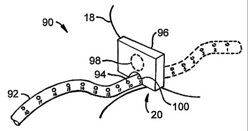
[0078] Figs. 7A and 7B show one variation in using an endoscope
assembly 90 in conjunction with external sensing device or datum 96. Datum 96
may be positioned externally of patient 18 adjacent to an opening into a body
cavity, e.g., anus 20 for colonoscopic procedures. Datum 96 may accordingly
have a sensor or reader 98 located next to opening 100, which may be used as a
guide fox passage of endoscope 92 therethrough into anus 20. Endoscope 92 may
be configured to have a number of tags 94, e.g., sensors, transponders, etc.,
CA 02517841 2005-08-31
WO 2004/084702 PCT/US2004/006939
-18-
located along the body of endoscope 92. These tags 94 may be positioned at
regular intervals along endoscope 92. The spacing between the tags 94 may vary
and may also depend upon the desired degree of accuracy in endoscope position
determination. Tags 94 may be positioned closely to one another to provide for
a
more accurate reading, while tags 94 spaced farther apart from one another may
provide for a less accurate determination. Moreover, tags 94 may be positioned
at
uniform distances from one another, or alternatively they may be spaced apart
are
irregular intervals, depending upon the desired results. Moreover, tags 94 may
be
positioned along the entire length of endoscope 92 or only along a portion of
it,
depending upon the desired results. As shown in Fig. 7B, as endoscope 92 is
passed through datum 96 via opening 100 and into anus 20, reader 98 located
within datum 96 may sense each of the tags 94 as they pass through opening
100.
Accordingly, the direction and insertion depth of endoscope 92 may be recorded
and/or maintained for real-time positional information of the endoscope 92.
(0079] Any number of technologies may be utilized with tags 94. For
instance, one variation may have tags 94 configured as RF identification tags
or
antennas. Reader 98 may accordingly be configured as a RF receiving device.
Each tag 94 may be encoded with, e.g., position information such as the
distance
of a particular tag 94 from the distal end of endoscope 92. The reader 98 may
be
configured to thus read in only certain regions or zones, e.g., reader 98 may
read
only those RF tags passing through opening 100 or only those tags adjacent to
anus 20. Alternatively, the RF tags may be configured to transmit the status
of,
e.g., pressure switches as described above, to datum 96 to determine the
length of
insertion.
[0080] Another variation on tags 94 may be to configure the tags for
ultrasonic sensing. For example, each tag 94 may be configured as
piezoelectric
transducers or speakers positioned along the endoscope 92. The reader 98 may
thus be configured as an ultrasonic receiver for receiving positional
information
from tuned transducers or tags 94 each of which relay its positional
information.
Alternatively, optical sensors may be used as tags 94. In this variation, each
tag
94 may be configured as a passive encoded marker located on an outer surface
of
CA 02517841 2005-08-31
WO 2004/084702 PCT/US2004/006939
- 19-
endoscope 92. These markers may be in the form of a conventional bar code,
custom bar code, color patterns, etc., and each may be further configured to
indicate directional motion, i.e., insertion or withdrawal. Furthermore, each
tag
94 may be configured as active encoded markers, e.g., LEDs which may be
blinking in coded patterns. Reader 98 may thus be configured as an-optical
sensor.
[0081] Another alternative may be to configure tags 94 and reader 98 for
infrared (IR) sensing in which case IR emitters may be positioned along the
length
of endoscope 92 such that each IR emitter or tag 94 is configured to emit
light at a
specific frequency according to its position along the endoscope 92. Reader 98
may thus be configured as an IR receiver for receiving the different
frequencies of
light and mapping the specific frequency detected against the length of
endoscope
92. Yet another alternative may be to have tags 94 configured magnetically.
such
that a magnetic reader in datum 96 can read the position of the device, as
described in further detail below.
[0082] Yet another alternative may be to configure the datum and
endoscope assembly as a linear cable transducer assembly. In this variation,
reader 98 may be configured as a transducer having a cable, wire, or some
other
flexible member extending from reader 98 and attached to the distal end of
endoscope 92. While the datum 96 remains external to the patient and further
remains in a fixed position relative to the patient, the endoscope 92 may be
advanced within the patient while pulling the cable or wire from reader 98.
The
proximal end of the cable or wire may be attached to a spool of cable or wire
in
electrical communication with a mufti-turn potentiometer. To retract the cable
or
wire when the endoscope 92 is withdrawn, the spool may be biased to urge the
retraction of the cable or wire back onto the spool. Thus, the change of wixe
length may be correlated to an output of the reader 98 or of the potentiometer
to a
length of the extended cable and thus the length of the endoscope 92 inserted
within the patient.
[0083] Yet another alternative may be to mount rollers connected to, e.g.,
mufti-turn potentiometers, encoders, etc., on datum 96. These rollers may be
CA 02517841 2005-08-31
WO 2004/084702 PCT/US2004/006939
configured to be in direct contact with the endoscope 92 such that the rollers
rotate in a first direction when endoscope 92 is advanced and the rollers
rotate in
the opposite direction when endoscope 92 is withdrawn. The turning and number
of revolutions turned by the rollers may be correlated into a length of the
insertion
depth of endoscope 92. -
[0084] Yet another alternative may be to use the endoscopes, or any of the
endo.scopes described herein, in conjunction with conventional imaging
technologies which are able to produce images within the body of a patient.
For
instance, any one of the imaging technologies such as x-ray, fluoroscopy, .
computed tomography (CT), magnetic resonance imaging (MRI), magnetic field
location systems, etc., may be used in conjunction with the endoscopes
described .
herein for determining the insertion depth.
[0085] In yet another alternative, the datum may be used to sense the
positional information from the endoscope through the~use of one or several
pressure sensors located on the datum, e.g., datum 96. The pressure sensor may
be positioned upon datum 96 such that it may press up against the endoscope 92
as it is advanced or withdrawn. This pressure sensor may be configured, e:g.,
as a
switch, or it alternatively be configured to sense certain features on the
endoscope
92, e.g., patterned textures, depressions, detents, etc., which are located at
predetermined lengths or length intervals to indicate to the pressure switch
the
insertion depth of endoscope 92.
[0086] , Yet another alternative is to sense changes in the diameter of the
endoscope body inserted into the patient, as seen in Fig. 7C. The insertion
length
of the endoscope may have multiple sections each having a unique diameter,
e.g.,
a distal most section 102 may have the smallest diameter and each successive
proximal section 104,106 may have incrementally larger diameters.
Alternatively, successive sections may have alternating diameter sizes where a
first section may have a first diameter, a second section may have a second
larger
diameter, and the third section may have a diameter equal to the first
diameter or
larger than the second diameter, and so on. The differences in endoscopic
diameter may be used to detect the endoscopic insertion depth by using a datum
CA 02517841 2005-08-31
WO 2004/084702 PCT/US2004/006939
-21 -
108 which may be configured to maintain contact with the endoscope and move
according to the diameter changes of the endoscope, as shown by the arrows.
This diameter referencing device and method may be used independently or in
conjunction with any of the other methods described herein as a check to
ensure
that the position of the endoscope concurs with the results using other
methods of
sensing.
[0087] Fig. 8 shows another example in endoscope assembly 110 in which
endoscope 112 may have a number of sensors or tags 114 located along the body
of the endoscope 112. As endoscope 112 is advanced or withdrawn from anus Z0,
datum 116, which may be mounted externally of the patient and at a distance
from
endoscope 112, may have a receiver or reader 118 configured in any of the
variations described above. For instance, receiver or reader 118 may be
adapted
to function as a RF receiver, ultrasonic receiver, optical sensor, or as any
of the
other variations described above, to read only those tags 114 adjacent to anus
20
and to map their position on the endoscope 112 and thus, the length of
insertion.
[0088] If reader 118 were configured as an optical sensor, it may further
utilize a light source, e.g., LED, laser, carbon, etc., within datum 116. This
light
source may be utilized along with a CCD or CMOS imaging system connected to
a digital signal processor (DSP) within reader 118. The light may be used to
illuminate markings located at predetermined intervals along endoscope 112.
Alternatively, the markings may be omitted entirely and the CCD or CMOS
imaging system may be used to simply detect irregularities normally present
along
the surface of an endoscope. While the endoscope is moved past the light
source'
and reader 118, the movement of the endoscope may be detected and correlated
accordingly to indicate insertion depth.
[0089] Fig. 9 shows another variation with endoscope assembly 120 in
which endoscope 122 may have a number of sensors 124 located along the length
of endoscope 122. These sensors 124 may be configured as Hall-effect type
sensors, as will be described in greater detail below. The datum 126 may be
configured as a ring magnet defining an endoscope guide 128 therethrough such
that the magnetic field is perpendicularly defined relative to the sensors
124.
CA 02517841 2005-08-31
WO 2004/084702 PCT/US2004/006939
-22-
Thus, sensors 124 may interact with magnet 126 as they each pass through guide
128. As a Hall sensor 124 passes through datum 126, the sensor 124 may
experience a voltage difference indicating the passage of a certain sensor
through
datum 126. These types of sensors will be described in greater detail below.
[0090] ' In order to determine the direction of the endoscope when it is
either advanced or withdrawn from the patient, directional information may be
obtained using any of the examples described above. Another example is to
utilize at least two or more sensors positioned at a predetermined distance
from
one another. Fig. 10 shows one variation illustrating sensor detection
assembly
130 with first sensor 132 and second sensor 134. First and second sensors 132,
134 may be positioned at a predetermined distance, d, from one another. As
endoscope 136 is advanced or withdrawn past sensor assemb1y.130, the direction
of travel 138 of endoscope 136 may be determined by examining and comparing
the signals received from each sensor 132,134. By determining which sensor has
a rising edge or input signal first received relative to the other sensor, the
direction
of travel 138 may be determined. As shown in Fig. 1 lA, plot 140 generally
illustrates signals received from first sensor 132. From position x =1 to
position
x = 2, a rise in the signal is measured thus sensing a peak in advance of the
signal
measured from position x =1 to position x = 2 in plot 142, which is the signal
received from second sensor 134, as seen in Fig. 11B. Thus, a first direction
of
travel, e.g., insertion, may be indicated by the relative comparisons between
signals in plots 140 and 142. If endoscope 136 were traveling in the opposite
direction, e.g., withdrawal, second sensor 134 would sense a peak in advance
of
first sensor 132.
[0091] A more detailed description fox determining the endoscope's
direction of travel follows below. Figs. 12A to 12D illustrate various cases
for
determining endoscopic direction of travel using first sensor 150 and second
sensor 152. First and second sensors 150,152 are preferably at a predetermined
distance from one another while an endoscope is passed adjacent to the
sensors.
For the purposes of this illustration, a direction to the right shall indicate
a first
direction of travel for an endoscope device, e.g., insertion into a body,
while a
CA 02517841 2005-08-31
WO 2004/084702 PCT/US2004/006939
- 23 -
direction to the left shall indicate a second direction of travel opposite to
the first
direction, e.g., withdrawal from the body.
[0092] Fig. 12A shows a situation in which first sensor 150 measures a
voltage less than the voltage measured by second sensor 152, as indicated by
plot
154. If first and second sensors 150,152 both measure a decrease in voltage,
this
may indicate a motion of the endoscope to the right while an increase voltage
in
both first and second sensors 150,152 rnay indicate a motion of the endoscope
to
the left. Fig. 12B shows another situation in which first sensor 150 measures
a
voltage greater than the voltage measured by second sensor 152, as indicated
by
plot 156. If first and second sensors 150,152 both measure an increase in
voltage,
this may indicate a motion of the endoscope to the right. However, if both
first
and second sensors 150,152 measure a decrease in voltage, this may indicate a
motion of the endoscope to the left.
[0093] Fig. 12C shows another situation where first sensor 150 measures a
voltage equal to a voltage measured by second sensor 152, as shown by plot
158.
In this case, if first sensor 150 measures an increase in voltage prior to
second
sensor 152 also measuring an increase in voltage, this may be an indication of
the
endoscope moving to the right. On the other hand, if second sensor 152
measures
an increase prior to first sensor 150 measuring an increase in voltage, this
may
indicate movement of the endoscope to the left. Fig. 12D shows a final
situation
in plot 160 where first sensor 150 again measures a voltage equal to a voltage
measured by second sensor 152. In this case, the opposite to that shown in
Fig.
12C occurs. For instance, if the voltage measured by first sensor 150
decreases
prior to the voltage measured by second sensor 152, this indicates a movement
of
the endoscope to the right. However, if second sensor 152 measures a voltage
which decreases prior to a decrease in voltage measured by first sensor 150,
this
may indicate a movement of the endoscope to the left.
[0094] Fig. 13 shows one variation of an algorithm which may be
implemented as one method for determining whether an endoscope is being
advanced or withdrawn from the body. Fig. 13 illustrates how the various
determinations described above may be combined into one variation fox an
CA 02517841 2005-08-31
WO 2004/084702 PCT/US2004/006939
-24-
algorithm. As seen, the algorithm begins with step 170. In step 172 an initial
step
of determining whether first sensor 150 measures a voltage greater than second
sensor 152 is performed. If first sensor 150 does measure a voltage greater
than
second sensor 152, then a second determination may be performed in step 174
where a determination may be made as to whether the voltages measured by both
sensors 150,152 are increasing or not. If both voltages are increasing, step
178
may indicate that the endoscope is being inserted. At this point, the position
of
the endoscope and its fractional position, i.e., the distance traveled by the
endoscope since its last measurement, may be determined and the algorithm may
then return to step 172 to await the next measurement.
(0095] If, however,. first sensor 150 does not measure a voltage greater
than second sensor 152 in step 172, another determination may be performed in
step 176 to determine whether the voltages measured by sensors 150,152 are
equal. If the voltages are not equivalent, the algorithm proceeds to step 180'
where
yet another determination may be performed in step 180 to determine if both
voltages are increasing. If they are not, then step 178 is performed, as
described
above. If both voltages are increasing, then step 184 may indicate that the
endoscope is being withdrawn. At this point, the position of the endoscope and
its
fractional position, i.e., the distance traveled by the endoscope since its
last
measurement, may again be determined and the algorithm may then return to step
172 to await the next measurement.
(0096] In step 176, if the voltages measured by first sensor 150 and
second sensor 152 are equivalent, then the algorithm may await to determine
whether a peak voltage is detected in step 182. If a peak voltage is detected,
step
186 increments the insertion count. However, if a peak is not detected, then
step
188 decrements the insertion count. Regardless of whether the insertion count
is
incremented or decremented, the algorithm may return to step 172 to await the
next measurement.
ENDOSCOPES USING MAGNETIC SENSING DEVICES
CA 02517841 2005-08-31
WO 2004/084702 PCT/US2004/006939
-25-
[0097] One particular variation on measuring endoscopic insertion depth
may utilize magnetic sensing, in particular, taking advantage of the Hall
effect.
Generally, the Hall effect is the appearance of a transverse voltage
difference in a
sensor, e.g., a conductor, carrying a current perpendicular to a magnetic
field.
This voltage difference is directly proportional to the flux density through
the
sensing element. .A permanent magnet, electromagnet, or other magnetic field
source may be incorporated into a Hall effect sensor to provide the magnetic
field.
If a passing object, such as another permanent magnet, ferrous material, or
other
magnetic field-altering material, alters the magnetic field, the change in the
Hall-
effect voltage may be measured by the transducer.
(0098] Fig. 14 illustrates generally Hall effect sensor assembly 190 which
shows conductor or sensor 192 maintained at a distance, d, as it is passed
over
magnets 194, 196,198 at distances xl, xz, x3, respectively. Each magnet may be
positioned such that the polarity o'f adjacent magnets is opposite to one
another or
such that the polarity of adjacent magnets is the same. As sensor 192 is
passed,
voltage differences may be measured to indicate which magnet sensor 192 is
adjacent to.
[0099] Fig. 15 shows one variation illustrating the general application for
implementing Hall effect sensors for endoscopic position measurement. As
shown, sensor assembly 200 illustrates one variation having magnet 202 with
first
sensor 204 and second sensor 206 adjacent to magnet 202. Magnet 202 may be a
permanent magnet or it may also be an electromagnet. First and second sensors
204, 206 are connected to a power supply (not shown) and are positioned from
one another at a predetermined distance. Both sensors 204, 206 may also be
located at a predetermined distance from magnet 202. A general representation
of
endoscope 208 is shown to reveal the individual links or vertebrae 210 that
may
comprise part of the structure of the endoscope, as described in further
detail in
any of the references incozporated above. Each vertebrae 210 is shown as being
schematically connected to adjacent vertebrae via joints 212 which may allow
for
endoscope articulation through tortuous paths. Endoscope 208 may be passed by
sensor assembly 200 at a predetermined distance as it is inserted or withdrawn
CA 02517841 2005-08-31
WO 2004/084702 PCT/US2004/006939
-26-
from an opening in a patient. Each or a selected number of vertebrae 210 may
be
made of a ferrous material or other material that may alter or affect a
magnetic
field or have ferrous materials incorporated in the vertebrae 210. Thus, as
endoscope 208 passes first and second sensors 204, 206, the ferrous vertebrae
210
may pass through and disrupt a magnetic field generated by magnet 202 and
cause
a corresponding voltage measurement to be sensed by sensors 204, 206.
Direction
of travel for endoscope 208, i.e., insertion or withdrawal, as well as depth
of
endoscope insertion may be determined by applying any of the methods described
above.
(0100] Another variation is shown in Fig. 16 which illustrates a schematic
representation 220 of Hall effect sensing in which the sensors may be located
on
the endoscope 226 itself. Magnet 222 may be positioned adjacent to, e.g., the
anus of a patient, such that endoscope 226 passes adjacent to magnet 222 when.
inserted or withdrawn from the patient. Endoscope 226 may have a number of
discrete Hall switches 228 positioned along the body of endoscope 226. As
endoscope 226 passes magnet 222, the magnetic field lines 224 may disrupt a
switch 228 passing adjacently. Hall switches 228 may be bipolar, unipolar,
latched, analog, etc. and may be used to determine the total resistance Rl_2
in
order to determine insertion length of the endoscope 226. .
[0101] Figs. 17A and 17B show another variation for Hall sensor .
positioning. Fig. 17A shows a sensor assembly 230 adjacent to an individual
vertebrae 232 of an endoscope. A single vertebrae 232 is shown only for the
sake
of clarity. As seen, when vertebrae 232 is directly adjacent to magnet 234,
magnetic flux lines 238 are disrupted and are forced to pass through sensor
236.
Flux lines 238 passing through sensor 236 may cause a disruption in the
current
flowing therethrough and may thus indicate the passage of the endoscope. Fig.
17B shows the assembly of Fig. 17A when endoscope 230 has been advanced or
withdrawn fractionally such that magnet 234 is positioned inbetween adjacent
vertebrae 232 and 232'. When a vertebra is not immediately adjacent to magnet
234, flux lines 238' may return to their normal undisturbed state such that
sensor
236 is also undisturbed by magnetic flux. The resumption of current within
CA 02517841 2005-08-31
WO 2004/084702 PCT/US2004/006939
-27-
sensor 236 may indicate that endoscope 230 has been moved relative to sensor
assembly 230.
[0102] Fig. 18 shows another variation in assembly 240 where a discrete
magnet 248 may be positioned on individual vertebrae 242 to produce a more
pronounced effect in sensor measurement. Magnets 248 may be positioned along
the longitudinal axis of the endoscope for creating a uniform magnetic field
radially about the endoscope. Discrete magnets 248 may be permanent magnets
or they may alternatively be electromagnets. In either case, they may be
placed
on as many or as few vertebrae or at various selected positions along the
endoscope body depending upon the desired measurement results. As shown,
when vertebrae 242 having discrete magnet 248 mounted thereon is brought into
the vicinity of magnet 244, the interaction between the magnets produces an
enhanced flux interaction 250 such that Hall sensor 246 is able to sense a
more
pronounced measurement. The polarity of each individual magnet 248 located
along the endoscope body may be varied from location to location but the
polarity
of adjacent magnets on the endoscope body are preferably opposite to one
another.
[0103] Alternatively, a number of magnets each having a unique magnetic
signature may be placed at predetermined positions along the length of the
endoscope. Each magnet 248 may be mapped to its location along the endoscope .
so when a magnet having a specific magnetic signature is detected, the
insertion
depth of the endoscope may be correlated. The magnets 248 may have unique
magnetic signatures, e.g., measurable variations in magnetic field strength,
alternating magnetic fields (if electromagnets are utilized), reversed
polarity, etc.
[0104] Figs. 19A and 19B show yet another variation in assembly 260 in
which more than one magnet may be used in alternative configurations. A first
magnet 262 may be positioned at an angle relative to a second magnet 264 such
that the combined flux lines 268 interact in accordance with each magnet.
Thus,
the polarity of each magnet 262, 264 may be opposite to one another as shown
in
the figures. Sensor 266 may be positioned such that the undisturbed field
lines
268 pass through sensor 266. As vertebrae 270 is passed adjacent to sensor
266,
CA 02517841 2005-08-31
WO 2004/084702 PCT/US2004/006939
-28-
the disturbed flux lines 268', as shown in assembly 260' in Fig. 19B, may be
altered such that they no longer pass through sensor 266 due to the
interaction
with vertebrae 270. Alternatively, the field lines 268 passing through sensor
266
may be altered in strength as vertebrae 270 passes.
[0105] Fig. 20 shows yet another variation in which discrete magnets may
be placed on each individual vertebrae of an endoscope assembly. As shown,
sensor assembly 280 shows only the vertebrae 282 of an endoscope for clarity.
Discrete magnets 284 having a first orientation may be placed on alternating
vertebrae 282 while magnets 286 having a second orientation may be placed on
alternating vertebrae 282 inbetween magnets 284. Thus, when the endoscope is
moved, e.g., along the direction of travel 292, flux lines 288 having
alternating
directions on each vertebrae 282 can be sensed by sensor 290. The measured
alternating flux lines may be used as an indication of endoscope movement in a
first or second direction. Each of the magnets may be positioned along the
periphery of the vertebrae on a single side; however, they may.also be
positioned
circumferentially, as described below in fizrther detail.
[0106] Figs. 21A and 21B show side and cross-sectional views,
respectively, of another alternative in magnet positioning. Fig. 21A shows a
side
view of endoscope assembly 300 in which a number of magnets 304 having a first
orientation may be positioned circumferentially about endoscope 302. A number
of magnets 306 having a second orientation opposite to the fiirst orientation
may
also be positioned circumferentially about endoscope 302 separated a distance,
d,
longitudinally away from magnets 304. With discrete magnets positioned
circumferentially about endoscope 302, the rotational orientation of endoscope
302 becomes less important as it passes sensor 308 in determining the
insertion
depth of the device. Fig. 21B shows a cross-sectional view of the device of
Fig.
21 A and shows one example of how magnets 304 may be positioned about the
circumference. Although this variation illustrates magnets 304 having a "N"
orientation radially outward and a "S" orientation radially inward of
endoscope
302, this orientation may be reversed so long as the adjacent set of
circumferential
magnets is preferably likewise reversed. Moreover, although seven magnets are
CA 02517841 2005-08-31
WO 2004/084702 PCT/US2004/006939
-29-
shown in each circumferential set in the figure, any number of fewer or more
magnets may be used as practicable.
[0107] Fig. 22A shows yet another variation in which endoscope 310 may
have discrete circumferentially positioned magnets 312 placed at each
vertebrae
312 on an outer surface of the endoscope 310. As endoscope 310 is-passed into
anus 20, Hall sensor 314 may be positioned adjacent to anus 20 such that
sensor
314 is able to read or measure the discrete magnets 312. as they pass into
anus 20.
Fig. 22B shows yet another variation in which endoscope assembly 320 may have
endoscope 322 in which individual vertebrae 326 may have some ferromagnetic
material 328 integrated or mounted onto or within the vertebrae 326. The
ferromagnetic material 328 may be in the form of a band, coating, or other non-
obstructive shape for integration onto vertebrae 326 or for coating over
portions of
vertebrae 326. A sheath or skin 324 may be placed over the vertebrae 326 to;
provide for a lubricious surface. Inbetween vertebrae 326, non-magnetic
regions
330 may be maintained to provide for the separation between vertebrae 326 and
between ferromagnetic material 328. Moreover, ferromagnetic material 328 may
be applied retroactively not only to endoscopes having vertebrae, but also
other
conventional endoscopes for which a determination of insertion depth is
desired.
As endoscope 322 passes magnet 332, sensor 334 may detect disturbances in flux
lines 336 as the regions having the ferromagnetic material 328 passes.
Additionally, endoscope 322 may be passed at a distance, h, from sensor 334'
which is sufficiently close to enable an accurate measurement but far enough
away so as not to interfere with endoscope 322 movement.
[0108] Fig. 23 shows yet another variation in which conventional
endoscopes may be used with any of the Hall sensor datum devices described
herein. As shown, elongate support or tool 337 may have a number of magnets
338, or ferrous material or other materials that may alter or affect a
magnetic field,
positioned along the tool at predetermined intervals. Magnets 338 may be
positioned along the length of tool 337 such that the adjacent magnets are
either
alternating in polarity or uniform in polarity. Furthermore, magnets 338 may
be
made integrally within the tool 337 or they may be made as wireforms or
CA 02517841 2005-08-31
WO 2004/084702 PCT/US2004/006939
-30-
members which may be crimped about tool 337. Tool 337 may be positioned
within the working lumen 339 of any conventional endoscope for use with a
datum device as described herein. The inclusion of the tool 337 may then
enable
the determination of insertion depth of a conventional or instrumented
endoscope.
If a conventional endoscope is used, tool 337 may be securely held within the
working lumen 339 during an exploratory procedure. Tool 337 may optionally be
removed during a procedure to allow for the insertion of another tool and then
reinserted within lumen 339 at a later time to proceed with the insertion
andlor
withdrawal of the endoscope.
[0109] Figs. 24A to 24C show perspective views of alternative variations
for attaching permanent magnets, ferrous materials, or other materials that
may
alter or affect a magnetic field, onto individual vertebrae. Fig. 24A shows
one
variation in which vertebrae 340 may be manufactured with a notch or channel
342 circumferentially defined along its outer surface 344. A ring made of a '
ferrous material or other material 'that may alter or affect a magnetic field,
such as
permanent magnets, may be placed within notch 342. Fig. 24B shows another
variation in which a formed ring 348 made of a permanent magnet or other: such
materials may be separately formed and attached onto vertebrae 346. Fig. 24C
shows yet another variation in which a wire form 354 made from a ferrous
material or other material that may alter or affect a magnetic field, such as
a .
permanent magnet, may be placed within notch 352 of vertebrae 350.
Alternatively, ferrous powder may be molded into a circumferential shape and
placed within notch 352. Another alternative may be to simply manufacture the
entire vertebrae from a ferrous metal'or simply cover a vertebrae or a portion
of
the vertebrae with a ferrous coating.
(0110] Another alternative for utilizing Hall sensors is seen in Figs. 25A '
and 25B. The variation in Fig. 25A may have a fixed platform 360 upon which a
magnet 364 may be mounted. A pressure sensor or microforce sensor 362 may be
placed inbetween magnet 364 and platform 360. As an endoscope is passed
adjacent to magnet 364, the magnet 364 may be attracted to vertebrae 366 as it
passes adjacently. Vertebrae 366 may optionally include ferrous materials or
CA 02517841 2005-08-31
WO 2004/084702 PCT/US2004/006939
-31-
other materials that may alter or affect a magnetic field as described above
to
enhance the attraction andlor repulsion. As magnet 364 is pulled or repulsed
by
the magnetic force, pressure sensor 362 may record the corresponding positive
or
negative force values for correlating to endoscope insertion depth. Fig. 25B
shows another example in which magnets 368 may be attached to a pressure
gauge 370, e.g., a Chatillon~ gauge made by Ametek, Inc. As the endoscope
passes magnets 368 at some distance, h, the attraction andlor repulsion
between
magnets 368 and vertebrae 366 may be accordingly measured by gauge 370 and
similarly correlated to endoscope insertion depth.
[0111] Yet another variation is shown in Figs. 26A and 26B in assembly
380. Rather than utilizing the linear motion of an endoscope past a static
datum, a
rotatable datum 382 may be used to record insertion length. Datum wheel 382
may be configured to rotate about pivot 384 while sensing the movement of
endoscope 386, which shows only schematic representations of the vertebrae for
clarity. The datum wheel 382 may have a number of magnets 398 incorporated
around the circumference of wheel 382. Each magnet may be arranged in
alternating pole configurations or alternatively in the same pole arrangement.
Each of the magnets 398 are also preferably spaced apart from one another at
intervals equal to the linear distances between the magnets 388, 390 or
permanent
magnet located along the body of endoscope 386. Ferrous materials, or
materials
that may otherwise alter a magnetic field, may be used in place of the
permanent
magnets. As endoscope 386 is moved past datum wheel 382, wheel 382 rotates in
corresponding fashion with the linear movement of endoscope 386 past the datum
382.
(0112] The rotation of datum wheel 382 that results when endoscope 386
is moved past can be sensed by a variety of methods. One example includes
rotary optical encoders, another example includes sensing the movement of
magnets 398 on datum wheel 382 as they rotate relative to a fixed point as
measured by, e.g., Hall effect sensors or magnetoresistive sensors. As datum
wheel 382 rotates with the linear movement of endoscope 386, datum wheel 382
may directly touch endoscope 386 or a thin material may separate the wheel 382
CA 02517841 2005-08-31
WO 2004/084702 PCT/US2004/006939
-32-
from the body of endoscope 386. Fig. 26B shows one variation of an assembly
view of datum wheel 382 which may be rotatably attached to housing 392.
Housing 392 may be connected to stem or support 394, which may extend from
housing 392 and provide a support member for affixing datum wheel 382 to the
patient, an examination table, a stand, or any other platform. Support 394 may
also be used to route any cables, wires, connectors, etc., to housing 392
and/or
datum wheel 382. The associated sensors and various support electronics, e.g.,
rotary encoders, magnetic field sensors, etc., may also be located within
housing
392. Support 394 may further include an optional flexible joint 396 to allow
datum wheel 382 to track the movement of endoscope 386 as it passes into or
out
of a patient.
EXAMPLES OF EXTERNAL SENSING DEVICES
[0113] The external sensing devices, or datum, may function in part as a
point of reference relative to a position of the endoscope and/or patient, as
described above. The datum may accordingly be located externally of the
endoscope and either internally or externally to the body of the patient. If
the:
patient is positioned so that they are unable to move with any significant
movement during a procedure, the datum may function as a fixed point of
reference by securing it to another fixed point in the room, e.g., examination
table,
procedure cart, etc. Alternatively, the datum may be attached directly to the
patient in a fixed location relative to the point of entry of the endoscope
into the
patient's body. The datum variations described herein may utilize any of the
sensing and measurement methods described above.
[0114] For instance, for colonoscopic procedures the datum may be
positioned on the patient's body near the anus. The location where the datum
is
positioned is ideally a.place that moves minimally relative to the anus
because
during such a procedure, the patient may shift position, twitch, flex, etc.,
and
disturb the measurement of the endoscope. Therefore, the datum may be
positioned in one of several places on the body.
CA 02517841 2005-08-31
WO 2004/084702 PCT/US2004/006939
-33-
[0115] One location may be along the natal cleft, i.e., the crease defined
between the gluteal muscles typically extending from the anus towards the
lower
back. The natal cleft generally has little or no fat layers or musculature and
does
not move appreciably relative to the anus. Another location may be directly on
the gluteal muscle adjacent to the anus. -
[0116] One variation for the datum for positioning along the natal cleft
408 is shown in Fig. 27. Datum 400 may have sensor 402 positioned in the
distal
tip of the sensing device, which may be placed adj scent to anus 20. The.
datum
itself may be positioned within the natal cleft 408 and temporarily held in
place on
the patient with adhesive 406. The datum may have a connector 404 extending
via a wire or cable for connection to a processor (not shown).
[0117] Another variation is shown in Fig. 28 in which the datum 410 may
have a base comprising a substrate. The substrate may have an adhesive side
that
may be placed against the small of the patient's back. An elongate flexible
member or arm 412 may extend from the substrate and lie within or against the
natal cleft such that the distal end 414 of member 412 is adjacent to anus 20.
Distal end 414 may have a sensor mounted within for sensing the movement of an
endoscope as it is passed through anus 20. The flexible member 412 rnay be
secured along the natal cleft using, e.g., adhesive tape, to prevent excessive
.
movement of the device.
[0118] Figs. 29A and 29B show a detailed vievcl of a variation of the '
datum device 410 of Fig. 28. Fig. 29A shows another view for possible
positioning of datum 410 on patient 18. The substrate may be positioned
proximal of anus 20 while member 412 extends along the natal cleft for
positioning sensor tip 414 proximally adjacent to anus 20. Fig. 29B shows
datum
410 laid out and having a substrate 420 upon which sensors and electronics may
be positioned. Substrate 420, as mentioned above, may have an adhesive backing
for temporary placement against the patient 18. Moreover, datum 410, or any of
the other datum examples described herein, may be optionally configured to be
disposable for one-time use on a patient. Support electronics 422 may
optionally
be placed upon substrate 420 and sensor 426 may be positioned within the
distal
CA 02517841 2005-08-31
WO 2004/084702 PCT/US2004/006939
-34-
end 414 at or near the end of the flexible member or arm 412. An optional
magnet 428 may be positioned along member 412 proximally of sensor 426.
Connector 424 may extend via a wire or cable from datum 410 for connection to
a
processor.
[0119] Another variation is shown in Figs. 30A and 30B which shows
datum substrate 430 having sensor 436 positioned within the distal end of
elongate flexible assembly 434 for placement adjacent to anus 20. Connector
432
may be provided for connection to a processor. Here, elongate assembly 434 may
be secured against or within the natal cleft by use of, e.g., an adhesive
strip 438.
Fig. 30B shows a cross-sectional top-down view of elongate assembly 434
positioned against the natal cleft. A sponge, silicone wedge, or some other
wedging device 440 may be positioned inbetween elongate assembly 434 and
adhesive strip 438 to ensure secure positioning of the datum device
relative.to
anus 20.
[0120] Fig. 31 shows another variation on the datum device which may
utilize a disposable substrate. Datum assembly 450 may have substrate 452 for
placement against the patient. A retaining pocket 454 may be defined within or
upon substrate 452 and it may be configured to allow for a reusable electronic
sensor assembly 458 to be placed within pocket 454. Sensor assembly 458 may
have a wire or cable 462 extending therefrom and it may further have a sensor
460
positioned or potted upon sensor assembly 458. The sensor assembly 458 may be
positioned within pocket 454 by slipping sensor assembly 458 through an
opening
456 defined within substrate 452 and sensor assembly 458 is preferably
positioned
within pocket 454 such that sensor 460 is positioned at the distal end of
substrate
452 to allow for positioning adjacent the anus.
[0121] Another variation for positioning a datum is directly on the gluteal
muscle adjacent to the anus. Generally, the sensor and associated circuitry
rnay
be incozporated into a patch or small chassis that may then be attached to the
muscle adjacent to the anus. The entire datum assembly may optionally be
mounted onto a bandage-like package with an adhesive backing. Figs. 32A and
32B show a variation in datum 470 which is formed into a small chassis having
CA 02517841 2005-08-31
WO 2004/084702 PCT/US2004/006939
-35-
connector 472 extending therefrom. Datum 470 may be attached temporarily to
patient 18 via adhesive 474 adjacent to anus 20. A guide, ramp, or other
similar
structure 476 for situating, orienting, or guiding endoscope relative to datum
470
may be optionally incorporated into the device.
[0122] Fig. 33A shows another variation of the device in datum 480. In
this example, datum 480 may be in the form of a patch with sensor 482
positioned
thereon. The device may be placed upon one of the gluteal muscles such that
sensor 482 is adjacent to anus 20. Fig. 33B shows a detailed view of how datum
480 may be positioned upon the gluteal muscle adjacent to anus 20. Adhesive
484 may be placed over datum 480 to temporarily hold it onto the gluteal
muscle
as shown. Fig. 33C shows an example of how datum 480 may interact with
endoscope 486 as it is advanced or withdrawn from anus 20. Because datum 480
may have a relatively small diameter, D, discomfort may be reduced for the
patient and close proximity to anus 20 may be assured. As endoscope 486 moves
past datum 480, the sensors within datum 480 may measure the insertion depth.
Zone 488 shows generally the zone of operation, i.e., the region within which
the
operator's or surgeon's hands generally operate during a colonoscopy
procedure.
Because of the small diameter of datum 480 and its position adjacent anus 20,
it is
generally out of the way of the operator or surgeon during a procedure and
thereby allows for unhindered operation of the endoscope 486 while maintaining
accurate measurement or sensing with datum 480.
[0123] Fig. 34 shows yet another variation in datum 490 which may have
a substrate with sensor 494 mounted at one end. Support electronics 492 may be
optionally mounted on datum 490 and wire or cable 496 may be used to transmit
the measured signals from sensor 494. Datum 490 may be in a triangular shape
for placement upon a single gluteal muscle, as shown, such that a vertex of
the
substrate is positioned adjacent to anus 20 to allow sensor 494 to sense or
measure
signals as endoscope 498 is advanced or withdrawn into anus 20. Although
shown in this variation in a triangular pattern, this is not intended to be
limiting
and is intended merely to illustrate one possible shape fox the datum.
CA 02517841 2005-08-31
WO 2004/084702 PCT/US2004/006939
-36-
[0124] Another variation is shown in Fig. 35 in which datum 500 may
incorporate multiple sensors. Datum 500 may be placed on a single gluteal
muscle and it may define an insertion region 508 at which the anus of the
patient
may be positioned. Each of the sensors 502, 504, 506 may thereby be configured
to sense or read the endoscope as it passes through or past the insertion
region
508. Although three sensors are shown in this configuration, fewer or more
sensors may be utilized depending upon the configuration of the datum 500 and
the desired signal processing results.
[0125] Fig. 36 shows yet another variation in which datum 510 may be
encased in a rigid housing. Datum 510 may thus encapsulate support electronics
512 within with sensor 514 directed towards one end of the housing. The
housing
may incorporate a connector 516 attached via a wire or cable extending from
the
datum 510. The rigid housing may be temporarily adhered to the patient on a
gluteal' muscle in the same fashion as described above.
[0126] Fig. 37 shows yet another variation in which datum 520 may be
configured to extend across the natal cleft to position an opening defined in
the
datum over the anus of the patient. As shown, an adhesive substrate 522 may be
configured, e.g., into a "butterfly" configuration. Substrate 522 may have at
least
iwo wings or flaps 524 for adhering to each gluteal muscle across the natal
cleft
while sensor 526 and support electronics 528 may be contained ad3acent an
opening 534 defined at or near the center of substrate 522. Sensor 526 and
support electronics 528 may be potted or contained within a housing 530 on
substrate 522. Connector 532 may be attached via a wire or cable for
connection
to a processor.
[0127] A datum device may also be configured to encircle an endoscope
as it passes into the body. Such a datum configuration may be useful when
using
sensing technology such as RF. In the case of RF, the datum may be in a looped
configuration to facilitate the exchange of RF signals with components or
sensors
mounted along the endoscope, as described above. One variation of a looped
datum configuration is shown in Figs. 38A and 38B. As shown, datum 540 may
have a loop 542 defined at a distal end to function as a signal receiver,
e.g., RF
CA 02517841 2005-08-31
WO 2004/084702 PCT/US2004/006939
-37-
signals, and/or as a guide loop. The datum 540 may be aligned along the natal
cleft 408 and secured in place with adhesive tape 544. A connector 546 may be
attached to datum 540 via a wire or cable at a first end of datum 540 while
sensor
548 may be positioned at the opposing end of datum 540. Sensor 548 may be
positioned adjacent to anus 20, while loop 542 encircles the openingof anus
20.
The loop 542 may define an insertion region 550 through which an endoscope
may be passed. The loop 542 may be made of a thin, flexible material such as
mylar and it optionally have an adhesive backing for placement upon the tissue
surrounding anus 20. Although shown in a circle configuration, loop 542 may be
in a variety of looped configuration and is not limited by its shape.
[0128] Yet another variation is shown in Fig. 39 where a supporting
garment 560, e.g., a pair of underpants, may define an opening 562 in the
region
surrounding the anus 20. A loop 564 may be incorporated into the fabric such
that
the loop surrounds the opening 562. The fabric in the middle of loop 564 may
either be removable at the time of the procedure or omitted altogether.
Connection to the loop 564 may be made through connector 566, which can be
connected via a wire or cable extending from, e.g., the waistband, front, or
side of
garment 560.
[0129] Aside from colonoscopy, other applications rnay include uses in .
minimally invasive surgery (MIS). MIS typically depends upon the use of long,
thin tools for insertion into the body via small incisions, e.g., often
through a
cannula. Instruments typically employed during MIS may include rigid
endoscopes, laparoscopes, thoracoscopes, needle drivers, clamps, etc. Because
each of these tools must pass through an opening in the body, a datum device
may
be used adjacent to that body opening for tracking instrument insertion depth.
In
situations where cannulas are used, the cannula itself may be instrumented
through one of the methods described above.
[0130] For other types of endoscopy procedures, various types of flexible
endoscopes may be used, e.g., upper endoscopes, duodenoscopes, sigmoidscopes,
bronchoscopes, neuroscopes, ENT scopes, etc. Any of the devices and methods
described above may be utilized and configured to maintain insertion depth fox
CA 02517841 2005-08-31
WO 2004/084702 PCT/US2004/006939
- 38 -
any of these types of endoscopes. For instance, for flexible endoscopes that
enter
the body transorally, a mouthpiece configured as a datum may be utilized.
[0131] The applications of the devices and methods discussed above are
not limited to regions of the body but may include any number of further
treatment applications. Other treatment sites may include other areas or
regions
of the body. Additionally, the present invention may be used in other
environments such as exploratory procedures on piping systems, ducts, etc.
Modification of the above-described assemblies and methods for carrying out
the
invention, and variations of aspects of the invention that are obvious to
those of
skill in the art are intended to be within the scope of the claims.