Note: Descriptions are shown in the official language in which they were submitted.
CA 02518124 2005-09-06
-1-
FIXATION IMPLANT FOR A BONE GRAFT WITHIN A JOINT FOR THE
PURPOSE OF ENSURING ARTHRODESIS OF THE JOINT
This invention relates to the general technical field of surgical fixation
implants,
and in particular to the sector of fixation implants designed to be used to
achieve
arthrodesis.
This invention relates more particularly to a fixation implant for a bone
graft
arranged between the bones located on both sides of an articular space, for
the purpose of
ensuring arthrodesis of a joint.
to The fixation implant according to the invention is specifically designed to
achieve
arthrodesis of a joint, in particular, but not exclusively, the ankle joint.
This invention also relates to a surgical method for emplacement of a fixation
implant according to the invention.
Arthrodesis is a surgical intervention intended to almost completely suppress
the
is mobility of a joint by causing "bone fusion." Such a surgical intervention
may prove to
be necessary in the case in which the patient is suffering from severe and
final arthrosis,
or when the cartilage of the bones forming the joint is very damaged.
In order to carry out such interventions, we know to resect the damaged
cartilaginous surfaces of the joint in such a way as to bring in contact, by
compression,
2o the facing bone surfaces of the bones forming the joint, thus making
possible
osteosynthesis.
However, such a method can lead to a shortening of the limb involved, which is
quite obviously undesirable, not only from the aesthetic point of view, but
also on
account of the complications (excessive claudication, for example) which this
may lead
25 to.
In order to mitigate these disadvantages, we know to contrive a housing on
both
sides of the articular space by cutting out fragments of bone at the opposite
ends of the
bones forming the joint. This housing, preferably cylindrical, is then filled
up with a bone
graft, such as an approximately cylindrical core sample of bone taken from the
same
3o patient, for example from the iliac crest.
21449265.1
CA 02518124 2005-09-06
-2-
This manipulation thus makes it possible to dispose of the degraded
cartilaginous
surfaces and to replace the damaged bone ends with a healthy bone graft
without
shortening the limb in question. The bone graft is then fixed relative to the
bones forming
the joint with the aim of enabling osteosynthesis between the bone graft on
the one hand
and the bones on the other hand, thus ensuring arthrodesis of the joint.
Various fixation implants can be used for the purpose of ensuring fixation of
the
bone graft and arthrodesis of the joint. Thus, in the case of a joint between
a first and a
second bone, a Steinman pin is routinely used. The Steinman pin presents
itself in the
form of an elongated pin of length sufficient to successively cross the first
bone of the
to joint, the bone graft and the second bone of the joint. Such fixation
implants, while they
make it possible to obtain significant results as far as joint immobilisation
is concerned,
nevertheless suffer from non-negligible disadvantages.
In the first place, these fixation implants require, for their emplacement, an
additional incision added to the incision already made for the purpose of
putting in the
1 s housing designed for receiving the bone graft. This additional incision
has the effect of
significantly increasing the risk of infection and operatory or post-operatory
complications.
In addition, Steinman pin fixation implants must generally be arranged in such
a
way as to extend obliquely or perpendicularly with respect to the articular
space for the
2o purpose of ensuring an effective maintenance of the joint. For certain
joints, such as the
ankle joint, such an orientation of the implant is undesirable because there
is a risk that
during patient management, and in particular during walking, the implant might
cross the
plantar cortex of the calcaneum and project outside the plantar facies.
Moreover, if the fixation implant is poorly positioned, it can also lead to
damage
2s of the soft tissues of the plantar fades. Now, fixation implants such as
Steinman pins can
prove to be difficult to position, in particular when the joints involve bones
of small
dimensions. Thus, in the case of poor orientation of the implant, it can
happen that the
bone graft is not maintained, the implant thus set up being then virtually
ineffective.
The objects assigned to the invention consequently aim at remedying the
various
3o disadvantages previously enumerated and at proposing a novel fixation
implant for a
bone graft arranged between the bones located on both sides of an articular
space, for the
21449265.1
CA 02518124 2005-09-06
-3-
purpose of ensuring arthrodesis of a joint, which makes it possible to ensure,
in a simple
way, particularly effective and stable maintenance of the joint without
risking damage to
the cortex on the one hand, and the soft tissues in the neighbourhood of the
bones
forming the joint on the other hand.
Another object of the invention aims at proposing a novel fixation implant
particularly suitable for immobilising a fragmented and/or fissured graft.
Another object of the invention aims at proposing a novel fixation implant
adapted for effectively withstanding the mechanical stresses exerted on the
joint, in
particular during walking in the case of the ankle j oint.
to Another object of the invention aims at proposing a novel fixation implant
that
does not require an additional incision for its emplacement.
Another object of the invention aims at proposing a novel fixation implant
that is
less intrusive than known implants.
Another object of the invention aims at proposing a novel fixation implant
is presenting a structure and a shape adapted to the anatomy of the joint.
Another object of the invention aims at proposing a novel fixation implant
that
makes possible a solid and comfortable maintenance of the joint.
Another object of the invention aims at proposing a novel fixation implant
whose
manipulation is made easy and which makes possible reduction of operatory
errors.
2o The objects assigned to the invention are achieved by means of a fixation
implant
for a bone graft arranged between the bones located on both sides of an
articular space,
with the purpose of ensuring arthrodesis of a joint, said fixation implant
comprising:
-at least two anchoring elements designed to be introduced into the bones,
and equipped with a proximal end and a distal end, said distal end being
adapted to be
2s introduced into the bones, and said anchoring elements being connected to
each
other by at least one connection element extending outside of the joint,
-a means for immobilisation of the bone graft, arranged between the anchoring
elements and connected to the connection element in such a way as to ensure,
in
cooperation with the anchoring elements, blocking of the bone graft with
respect to
3o the bones of the joint and vice-versa, the immobilisation means being
formed by a
plate.
21449265.1
CA 02518124 2005-09-06
-4-
Other special features and advantages of the invention will appear in greater
detail
upon reading of the description which follows, and by means of the appended
drawings
provided in a purely illustrative and non-restrictive way, in which:
-Figure 1 depicts, in a perspective view, an ankle joint with a bone graft
arranged
between the bones located on both sides of an articular space.
-Figure 2 depicts, in a perspective view, a fixation implant according to the
invention in its functional position for maintenance of the joint.
-Figure 3 depicts, in a perspective view, a first embodiment of a fixation
implant
according to the invention.
to -Figure 4 depicts, in a side view, another embodiment of a fixation implant
according to the invention.
-Figure 5 depicts, in a frontal view, a fixation implant according to the
invention
in its functional position for compression of the bone graft and maintenance
of the joint.
-Figure 6 depicts, in a cross-sectional view along line A-A depicted in figure
5,
I s the fixation implant depicted in figure 5.
-Figure 7 depicts, in a perspective view, an ankle joint and a bone graft
arranged
between the bones located on both sides of the articular space, the bone graft
being
formed by two fragments separated by the articular space.
-Figure 8 depicts, in a perspective view, a fixation implant according to the
2o invention put in place within the joint depicted in figure 7.
-Figure 9 depicts, in a side view in perspective, an embodiment of the
fixation
implant according to the invention, equipped with a compression unit.
-Figure 10 depicts, in a side view in perspective, an improved variant of
realisation of the fixation implant equipped with a compression unit according
to the
25 invention.
-Figures 11 to 13 depict, in side views in perspective, various embodiments of
a
compression unit according to the invention.
-Figure 14 depicts, in a schematic view, an implant according to another
embodiment of the invention.
3o Figures 1 and 7 depict two surgical methods that make it possible to
achieve
arthrodesis of the ankle joint.
21449265. I
CA 02518124 2005-09-06
-5-
Arthrodesis of a joint becomes necessary when the joint is in such a degraded
condition that other less severe surgical interventions, such as for example
those
consisting in the placement of prostheses, prove to be ineffective. It becomes
necessary,
in this case, to completely immobilise the joint. This invention is depicted
in the case of
s an ankle joint but could be applied to all types of joints in the human or
animal body.
A damaged joint is characterized in particular by the condition of the
cartilage of
the bones delimiting the articular space. In the case of severe arthrosis,
this cartilage is
particularly worn out and can lead to pain or inflammation of the joint.
Figures 1 and 7 depict a joint 1 formed from at least two bones, namely a
first
to bone 2, and a second bone 3 located on both sides of an articular space 4.
However, joint
1 could quite obviously comprise a third bone, for example located between the
first bone
2 and the second bone 3, and this without leaving the framework of the
invention.
Several arthrodesis techniques are conceivable but the fixation implant
according
to the invention is more specifically designed to be used for the purpose of
achieving
Is arthrodesis by means of a bone graft 5 arranged between bones 2 and 3
located on both
sides of the articular space 4. A first method that is known thus consists in
putting in a
housing 6, for example cylindrical, in the ends 2A and 3A of bones 2 and 3
delimiting the
articular space 4.
According to this first known method, the bone fragments contained in the
2o housing 6 are extracted and a bone core sample is taken from another part
of the patient's
body, for example from the iliac crest, with the purpose of introducing it
within the
housing 6 once the bone fragments are withdrawn. This core sample constitutes,
after its
emplacement within the housing 6, a bone graft 5, approximately solid, i.e.
neither
hollow nor split, suitable to be fixed relative to the bones 2 and 3 for the
purpose of
2s ensuring osteosynthesis between the bone graft 5 and bones 2 and 3.
Specifically,
osteosynthesis is achieved between the bleeding outer bone surface SA of the
bone graft 5
and the surface of section 6A, also bony, of bones 2 and 3. For the purpose of
the
invention, the bleeding outer bone surface SA of the bone graft 5 corresponds
to the
surface according to which the bone graft 5 was cut out. In the case of a
cylindrical core
3o sample, the bleeding outer bone surface SA therefore corresponds to the
lateral surface of
21449265.1
CA 02518124 2005-09-06
-6-
the core sample. The surface of section 6A corresponds approximately, for the
purpose of
the invention, to the inner wall 6I of the housing 6.
A variant of this method was invented by the applicant. Like the preceding
method, the method according to the applicant's invention consists in putting
in a housing
s 6, preferably cylindrical, on both sides of the articular space 4. On the
other hand, the
contents of housing 6 are not hollowed out, i.e. there is no withdrawal of
bone fragments
2' and 3' cut out, respectively, from the ends of bones 2 and 3 located on
both sides of
the articular space 4. Bone fragments 2'and 3', juxtaposed within the housing
6 and
separated by an interstice I derived for example from the articular space 4
then constitute
to the bone graft 5. The worn-out cartilaginous surfaces of the joint are thus
located on both
sides of the interstice I separating the bone fragments 2' and 3' and
therefore at the centre
of the bone graft 5.
The applicant's method consists then in displacing the bone graft 5, formed
from
bone fragments 2' and 3', within its housing 6, by making it turn on itself
for example by
is a quarter turn in the direction of rotation R indicated in figure 7. In
this way, the bleeding
outer bone surface 2'A of the first bone fragment 2'is put facing the surface
of the section
3 S, also bony, of the second bone 3. In the same way, the bleeding outer bone
surface
3'A of the second bone fragment 3' is put facing the surface of section 2S of
the first
bone 2. This thus makes possible osteosynthesis of bone fragments 2' and 3'
with,
2o respectively, bones 3 and 2 and more generally osteosynthesis of the bone
graft 5 with
bones 2 and 3.
For the purpose of the invention, the expression "bone graft" therefore refers
either to a bone transplant, i.e. a piece of bone, preferably of one piece,
taken from a part
of the body located at a distance from the joint 1 and introduced within the
housing 6, or
2s to an assembly of bone fragments 2' and 3'cut out within the joint 1 during
construction
of the housing 6, and which has been caused to undergo a displacement, such as
a
rotation R within the housing 6. The bone graft 5 therefore constitutes a
unitary totality,
formed either from a one-piece bone fragment, or by a plurality of bone
fragments
juxtaposed within the housing 6.
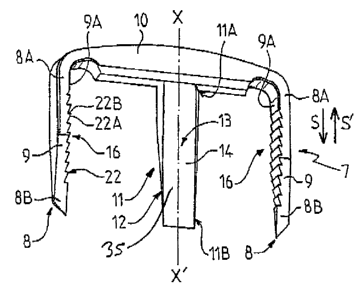
3o Figures 3 to 6 depict several variants of realisation of a fixation implant
7,
designed in particular to be used to fix the bone graft 5, in one piece,
depicted in figure 1.
21449265.1
CA 02518124 2005-09-06
_7_
Figures 9 to 14 depict other variants of realisation of the fixation implant
7,
designed in particular to be used to ensure fixation of the bone graft 5
formed by the bone
fragments 2' and 3' depicted in figure 7.
According to the invention, the fixation implant 7 comprises at least 2
anchoring
s elements 8 designed to be introduced in bones 2 and 3. Preferentially, the
fixation implant
7 comprises as many anchoring elements 8 as the joint comprises bones 2 and 3.
Thus, if
the joint 1 is formed from two bones 2 and 3, the fixation implant will
preferably
comprise two anchoring elements 8, as is shown in figures 3, 4 and 9. It is
however quite
obviously conceivable to equip the fixation implant with several anchoring
elements for
to the same bone, and this without leaving the framework of the invention.
Each anchoring element 8 extends preferentially between a proximal end 8A and
a
distal end 8B. The distal end 8B is in addition adapted to be introduced into
bones 2 and
3 and is to that end preferentially tapered, or pointed. By means of this
technical measure,
each anchoring element 8 presents a self perforating character, making
possible the
1 s penetration of the anchoring element 8 into the bone.
The anchoring elements 8 are to best advantage connected to each other through
at least one connection element 10. As depicted in figures 2 and 8, the
connection
element 10 extends outside of the joint l and overlaps the articular space 4,
thus forming
an essentially rigid connection bridge between the anchoring elements 8 and
therefore
2o between bones 2 and 3. This connection bridge 10 thus confers on the
fixation implant 7
its structural rigidity, which makes it possible for it to better withstand
the various
mechanical stresses to which the joint 1 is subjected. The connection element
10 being
arranged, in its functional position, between the joint l and the soft tissues
(not shown), it
preferably presents rounded edges which confer upon it an essentially non-
invasive
2s character with respect to the surrounding soft tissue.
Preferentially, the anchoring elements 8 are formed from anchoring arms 9
whose
distal ends 8B are substantially tapered in such a way as to facilitate their
penetration into
the bone tissues.
To best advantage, the anchoring elements 8, in particular the anchoring arms
9,
3o extend essentially parallel to the articular space 4, and are essentially
perpendicular to the
connection element 10. The fixation implant 7 then presents to best advantage
a U shape,
21449265.1
CA 02518124 2005-09-06
-g-
the arms of the U being formed from the anchoring arms 9, and the base of the
U being
formed from the connection element 10.
It is, however, quite obviously conceivable to construct a fixation implant
whose
anchoring arms 9 extend obliquely and not perpendicularly with respect to the
connection
s element 10 and in such a way as to come close to each other. The anchoring
arms 9 can
thus to best advantage be constructed from a material with shape memory so as
to shrink,
and to come close to each other once introduced into bones 2 and 3, in order
to ensure
effective compression of bones 2 and 3 against each other.
According to the invention, the fixation implant 7 also comprises a means for
to immobilisation 11 of the bone graft 5, arranged between the anchoring
elements 8 and
connected to the connection element 10 in such a way as to ensure, in
cooperation with
the anchoring elements 8, blocking of the bone graft 5 with respect to bones 2
and 3 of
the joint 1 and conversely.
The expression "in cooperation" refers to the fact that the immobilisation
means
is 11 acts together with the anchoring elements 8 to immobilise the bone graft
5 with
respect, on the one hand, to the anchoring elements 8 and, on the other hand,
to bones 2
and 3 of the joint 1. To best advantage, the immobilisation means 11 extends
longitudinally, in a longitudinal direction X-X' approximately parallel to the
articular
space 4, and this contrary to the devices of prior art, such as Steinman pins,
which extend
2o in an essentially perpendicular or oblique fashion with respect to the
articular space.
The immobilisation means 11 presents, of course, a shape and dimensions
adapted
for ensuring stable and reliable immobilisation of the graft. In that, the
immobilisation
means is not simply a means for indexing the implant in position relative to
the graft, but
rather a means for mechanical embedding of the implant in the graft.
2s The immobilisation means 11 is also distinct from and exogenous to the
graft and
adapted for cooperation with the latter. The immobilisation means 11 therefore
does not
directly, of itself, form, for the purpose of the invention, a substrate for
growth or bone or
tissue regeneration, but rather a unit interacting mechanically with the graft
to block the
latter.
so The immobilisation means I 1 thus to best advantage extends, in the
longitudinal
direction X-X', between a proximal part I 1A connected to the connection
element 10 and
21449265.1
CA 02518124 2005-09-06
-9-
a distal part 11B arranged on the opposite side from the proximal part 1 1A.
The
immobilisation means 11 is thus arranged perpendicularly to the connection
element 10.
Preferentially, the anchoring arms 9 extend longitudinally in an essentially
parallel fashion in the longitudinal extension direction X-X' of the
immobilisation means
11. In addition, the anchoring arms 9 present to best advantage, along their
length, a
variable thickness which decreases between their proximal end 8A and their
distal end 8B
in such a way as to facilitate their penetration into bones 2 and 3.
As depicted in figure 3, the anchoring arms 9 preferentially have the same
length
as the immobilisation means 11. It is, however, quite obviously conceivable to
construct
to anchoring arms 9 of different lengths. Thus, it is conceivable to construct
a fixation
implant 7 equipped with anchoring arms 9 appreciably longer than the
immobilisation
means 11 (figure 4).
To best advantage, the immobilisation means 11 comprises a unit for
introduction
12 within the bone graft 5. The introduction unit 12 is thus adapted to
penetrate either
1 s inside a one-piece bone graft 5, formed from a single bone fragment
(figure 2), or into the
interstice I separating bone fragments 2' and 3' within the bone graft 5
(figures 7 and 8).
To this end, the distal part 11B of the immobilisation means 11, which forms
the
introduction unit 12, is preferably shaped to a point or tapered.
In order to ensure effective maintenance of the bone graft 5 and to make
possible
20 osteosynthesis with bones 2 and 3, the immobilisation means 11 comprises to
best
advantage means for rotation blocking 13 adapted to prevent rotation of the
bone graft S
around the fixation implant 7, and vice-versa.
As depicted in figure 3, the rotation blocking means 13 are to best advantage
formed from at least one flat part 14, arranged along the immobilisation means
11. The
2s immobilisation means 11 can thus be presented in the shape of a point
comprising at least
one essentially flat outer surface forming flat part 14.
In accordance with the invention, as depicted in figures 3, 6 and 9 to 14, the
immobilisation means 11 is formed from at least one plate 35, i.e. from a two-
dimensional element. By "two-dimensional element," an element of flattened
shape is
3o designated here, whose thickness is slight compared to its length and
width. In other
words, such an immobilisation means 11 presents a blade shape and extends
principally
21449265.1
CA 02518124 2005-09-06
-10-
in two directions of space, and not essentially in a single direction of
space, like the screw
15 described in more detail below.
Plate 35 can, for the purpose of the invention, present a relatively spread-
out
shape, as in the variant of figures 9 to 14, or slender, as in the variant of
figure 3.
Implementation of an immobilisation means 11 formed from a plate 35 makes
possible excellent maintenance of the bone graft 5, and in particular allows
use of a graft
that is fissured, split or even broken into several fragments. In this case,
implementation
of an immobilisation means 11 in plate shape makes it possible, in fact, to
ensure a
relative blocking of the fragments that is sufficiently robust and stable to
withstand the
to loads to which the joint may be subjected (in particular when the latter is
an ankle joint,
on which the weight of the patient's body is exerted).
The plate 35 can have a thickness that is essentially uniformly constant. In
the
case in which the graft is formed from several fragments, the dimensions of
the plate, and
in particular its thickness, will preferably be chosen as a function of the
free space
is between the fragments in such a way that the plate occupies a volume
sufficient to block
the fragments.
Plate 35 can, however, possibly be made up of several sections, each
presenting a
thickness that is essentially constant and different from the thickness of the
other
sections. In this case, coupling between each section can be sharp, and be
presented for
2o example in the shape of a shoulder or a "step."
It is also conceivable, in a preferential variant of realisation depicted in
figures 3
and 9 to 13, and described in more detail below, that the plate 35 present a
"wedge-like,"
tapered shape, i.e. whose thickness increases progressively, in the
longitudinal direction,
over at least a part of the plate, from its distal end 11 B towards its
proximal end 11 A.
2s In this case, described in more detail in what follows, the immobilisation
means
forms (or is formed) from a compression unit 30.
According to a variant of realisation of the invention depicted in figure 4,
the
immobilisation means 11 may, however, be formed from a screw 15, preferably
self
drilling and self threading, and equipped to this end with preparation means
17 formed
3o from at least one tooth 17A extending essentially axially in the
longitudinal direction X-
X'.
21449265.1
CA 02518124 2005-09-06
-11-
To best advantage, the screw 15 also comprises grooves 18 arranged along its
length in such a way as to make possible progressive evacuation of excess bone
matter
during its screwing inside the bone graft 5.
According to a first embodiment of the invention, depicted in figure 4, the
s immobilisation means 11, for example the screw 15, is mounted in a removable
way on
the connection element 10. The latter is to this end to best advantage
equipped with a
through-hole 19, preferentially fitted approximately in the centre of the
connection
element 10 and adapted to receive the immobilisation means 11. Thus, the screw
15 can
to best advantage comprise, towards its proximal end 15B, a head 20 designed
to support
to a shoulder 21, forming an abutment, fitted within through-hole 19.
According to another variant of realisation of the invention depicted in
figures 3, 9
and 10, the immobilisation means 11 is to best advantage permanently united
with the
connection element 10 and for example made in one piece with the latter, thus
forming a
one-piece totality.
1 s Preferentially, and as depicted in figures 3 and 4, the anchoring means 8
are
formed from two lateral and spaced-out anchoring arms 9, arranged oppositely
on both
sides of the connection element 10, parallel with respect to each other. The
anchoring
arms 9 are preferably made in one piece with the connection element 10 but can
quite
obviously be formed from distinct parts of the connection element 10, and
united with the
20 latter, for example by means of fixation screws (variant not shown). In a
particularly
advantageous way, the anchoring arms 9 are preferentially identical and
symmetrically
arranged on both sides of the immobilisation means 11.
In addition, the immobilisation means 11 is to best advantage formed from a
central arm approximately parallel to the lateral anchoring arms 9, in such a
way as to
2s extend perpendicularly with respect to the connection element 10.
In an even more preferential way, the anchoring arms 9 and the immobilisation
means 11 are made of one piece, thus forming a one-piece fixation implant 7.
Such a
fixation implant withstands particularly well the mechanical stresses to which
the joint is
subj ected.
3o To best advantage, and as shown in figures 3, 4, 9 and 10, the anchoring
arms 9
are equipped with reverse-lock means 16 specifically designed to prevent
displacement of
21449265.1
CA 02518124 2005-09-06
-12-
the fixation implant 7 in a direction S' opposite to its direction of
introduction S into the
bone graft 5. To best advantage, the reverse-lock means 16 are preferentially
formed
from at least one protuberance 22 protruding from the outer surface of the
anchoring arms
9. In an even more preferential way, the reverse-lock means 16 are formed from
a
s plurality of protuberances 22 arranged along the anchoring arms 9, in the
longitudinal
direction X-X'.
The anchoring arms 9 comprise to best advantage an inner surface 9A, located
essentially facing the immobilisation means 11, on which the reverse-lock
means 16 are
arranged. The inner surface 9A thus presents a notched appearance, each
protuberance 22
Io forming a notch and presenting an inclined surface 22A designed to
facilitate the
introduction of the anchoring arms 9 into the bone tissue, and a horizontal
surface 22B,
approximately perpendicular to longitudinal direction X-X' and in the
direction S of
introduction of the fixation implant 7 so as to prevent disengagement of said
fixation
implant 7 once the latter is put in place within the joint 1.
1 s According to a particularly advantageous characteristic of the invention,
the
immobilisation means 11 is formed from a compression unit 30 adapted to
support the
bone graft 5 and to exert on the latter a pressure sufficient so that the bone
graft 5
supports, at least partially, bones 2 and 3 of the joint 1 in such a way as to
promote
osteosynthesis between the bone graft 5 and bones 2 and 3.
2o In the case of the configuration depicted in figure 5, in which the bone
graft is
formed from a single fragment of bone 5', preferably of one piece, arranged
within the
housing 6, the compression unit 30 is adapted to support at least part of the
outer surface
5'A of bone fragment 5' in such a way as to compress the latter in a direction
of
compression F against the inner wall 6I of the housing 6.
2s To this end, as was set forth in the preceding, the compression unit 30 is
preferentially formed from a plate 35, one of whose surfaces comes in contact
with the
outer surface 5'A of bone fragment 5'. Bone fragment 5' can to best advantage
be
presented in the shape of a hemicylindrical block not occupying all of the
housing 69
presenting an approximately flat portion of the outer surface 5'A which the
plate 35 is
capable of supporting.
21449265.1
CA 02518124 2005-09-06
-13-
According to the method depicted in figures 7 and 8, the bone graft S
comprises at
least two bone fragments 2' and 3', separated by the interstice I. The
compression unit 30
is then adapted to be introduced within the interstice I, with a low
clearance, and to exert
external centrifugal or radial compression, following arrows F', on bone
fragments 2' and
s 3', for the purpose of pushing them back against the inner wall 6I of the
housing 6 and
thus ensuring the expansion of the bone graft 5 and its blocking within the
housing 6, and
more generally within the joint 1.
According to a particularly advantageous characteristic of the invention, the
compression unit 30 is equipped with progressive spreading means 32, adapted
to ensure,
to as the penetration of the compression unit 30 into interstice I progresses,
progressive
compression of bone fragments 2' and 3'. As depicted in figure 10, the
spreading means
32 are to best advantage formed from a section 33 of the compression unit 30
whose
thickness is variable. Thus section 33 extends, in the direction of
introduction S of the
compression unit 30, between a proximal limit 33A, located on the side of the
connection
is element 10, and a distal limit 33B opposite. The thickness of section 33
increases to best
advantage, for example continuously, between distal limit 33B and proximal
limit 33A in
such a way as to ensure progressive spreading of bone fragments 2' and 3'.
It is, however, conceivable that the thickness of section 33 could be
approximately
constant, or could vary by sharp levels, without on this account leaving the
framework of
2o the invention.
In a particularly advantageous way, the compression unit 30 comprises a
tapered
distal part, designed to facilitate its introduction into the bone graft 5,
and to best
advantage formed from the spreading means 32. The compression unit 30 in
addition
comprises a proximal part 34 essentially thicker than its distal part which
corresponds to
2s section 33. In an even more preferential way, proximal part 34 presents a
thickness
approximately equal to the width of the interstice I, itself approximately
identical to the
width of the articular space 4, so as to avoid the phenomenon of the
shortening of the
limb of the patient comprising the joint 1.
To best advantage, the compression unit 30 is formed from a plate 35,
3o approximately prismatic and flattened, forming a wedge. Plate 35 can to
best advantage
be formed from a material with shape memory, and be designed to expand after
its
21449265.1
CA 02518124 2005-09-06
-14-
introduction within the interstice I for the purpose of ensuring compression
and
progressive spreading of bone fragments 2' and 3'. The plate 35 preferably
comprises
two surfaces 35A and 35B, at least one of said surfaces 35A comprising grooves
36.
According to a first variant of realisation depicted in figure 10, grooves 36
can to
best advantage extend in a direction approximately parallel to longitudinal
extension
direction X-X' of the compression unit 30. Such a configuration in particular
makes it
possible to appreciably improve the effectiveness of compression.
According to another variant depicted in figure 9, the grooves 36 preferably
extend in a direction approximately perpendicular to longitudinal extension
direction X-
to X' of the compression unit 30. According to this configuration, the grooves
36 to best
advantage form reverse-lock means opposing extraction of the implant once the
latter is
introduced within the bone graft 5.
According to a preferential variant depicted in particular in figure 10, the
immobilisation means 11 or the compression unit 30, formed from the plate 35,
extends
1 s in a principal extension plane P and the anchoring arms 9 are to best
advantage located in
this same principal extension plane P.
According to an advantageous characteristic of the invention, the fixation
implant
7 comprises gripping means 40 to best advantage formed from at least one
groove and
preferably two grooves 41 arranged on both sides of the fixation implant 7,
preferentially
2o between the connection element 10 and the immobilisation means 11.
Alternatively, as depicted in figure 14, gripping means 40 are formed from an
orifice 41 A fitted through the thickness of the implant, preferably in the
neighbourhood
of the junction between the immobilisation means and the connection element.
This
orifice 41A makes possible the introduction of an extraction instrument in the
shape of a
2s rod, in particular making it possible to exert a lever arm on the implant
in order to remove
it from the bones if necessary.
In a particularly advantageous embodiment, and which moreover constitutes an
invention in its own right, the compression unit 30 is independent of the
anchoring
elements 8, i.e. it is not connected to the latter by means of the connection
element l00
o The fixation implant 7 is then formed exclusively from the compression unit
30,
and does not comprise the anchoring element 8.
21449265.1
CA 02518124 2005-09-06
-15-
As depicted in figures 11 to 13, the compression unit 30 comprises all
characteristics previously described but is to best advantage designed to be
introduced
within the bone graft 5 independently of the anchoring elements 8. It is then
possible to
use other fixation means, for example fixation screws, in order to unite bone
fragment 5'
s or each of fragments 2' and 3' to bones 2 and 3, independently of the
compression unit
30.
The compression unit 30 is then, according to the case, specifically adapted
to
support bone fragments 5' or 2' and 3' and to exert a sufficient pressure on
the latter to
push them back against the inner wall 6I of the housing 6 and to ensure
blocking of the
io bone graft 5 within joint 1.
Whatever their embodiment, the fixation implant 7, the anchoring elements 8,
the
immobilisation means 11 or the compression unit 30 are to best advantage made
of a bio-
resorbable material, which makes it possible to avoid a new surgical
intervention for the
purpose of withdrawing them.
t s The surgical method for emplacement of the fixation implant 7 will now be
described with reference to figures 1 to 13.
The surgical method according to the invention includes, subsequently to the
step
of emplacement or positioning of the bone graft 5 within its housing 6, a step
for fixation
of the bone graft 5 by means of the fixation implant 7 previously described.
2o This fixation step comprises at first a step a) of immobilisation relative
to bones 2
and 3 forming the joint. This step a) is carried out by means of one or more
impacts,
performed for example by means of an appropriate impactor, on the implant, and
for
example either on the connection element 10, or on the anchoring elements 8,
in such a
way as to cause the latter to penetrate into each of bones 2 and 3 of the
joint 1. Bones 2
2s and 3 are then united with each other through the connection element 10.
The surgical method also includes a step b) for blocking of the bone graft 5
with
respect to bones 2 and 3 forming the joint. This step b) can be carried out
simultaneously
with step a) or subsequently to step a), in particular when the immobilisation
means 11 is
not of one piece with the anchoring elements 8. In the case in which the
immobilisation
3o means 11 is formed from an independent part, it is possible to introduce
the latter within
the bone graft 5 in particular by means of one or more impacts carried out on
the
21449265.1
CA 02518124 2005-09-06
-16-
proximal part 11 A of the immobilisation means 11. This way of proceeding can
prove to
be judicious for example in the case in which the immobilisation means 11 is
formed
from a prismatic plate 35 forming a wedge. On the other hand, if the
immobilisation
means 11 is formed from a screw 15, step b) will preferentially be carried out
by
s introducing the screw 15 within through-hole 19 and then screwing the latter
inside the
bone graft 5, for example within one-piece bone fragment 5'.
In the case in which the immobilisation means 11 is of one piece with the
anchoring elements 8, the immobilisation means 11 is introduced within bone
graft 5 at
approximately the same time as the anchoring elements 8 penetrate inside bones
2 and 3
1 o under the action of the impactor.
The surgical method according to the invention additionally comprises to best
advantage a step c) for compression of the bone graft 5 within the housing 6
arranged on
both sides of the articular space 4.
To best advantage, steps a), b) and c) are carried out simultaneously.
15 In particular, the bone graft 5 being formed from at least two bone
fragments 2'
and 3', step c) involves the exertion of an external radial or centrifugal
compression,
following arrows F', on bone fragments 2'and 3' for the purpose of pushing
them back
towards the inner wall 6I of the housing 6, thus ensuring their blocking as
well as that of
the bone graft 5, on the one hand within the housing 6 and on the other hand
within the
2o joint 1.
Such a step can be conducted independently of steps a) and b) described
previously and then constitutes an invention in its own right. This step is to
best
advantage carried out by introducing the compression unit 30 within the
interstice I
between bone fragments 2' and 3', said compression unit 30 being or not being
25 associated with the anchoring elements 8 in order to form the fixation
implant 7.
The fixation implant 7 according to the invention therefore makes it possible
to
ensure, temporarily or permanently, effective maintenance of the joint 1 and
the bone
graft 5 within said joint l, and therefore to facilitate osteosynthesis
between the bone
graft 5 and each of bones 2 and 3.
21449265.1
CA 02518124 2005-09-06
17-
Another advantage of the fixation implant 7 according to the invention derives
from its ease of emplacement, by means of a simple impactor, and without an
additional
incision being necessary for its implantation.