Note: Descriptions are shown in the official language in which they were submitted.
CA 02518532 2005-09-08
33XZ 155828
SYSTEM AND METHOD FOR AN ADAPTIVE MORPHOLOGY X-RAY BEAM
IN AN X-RAY SYSTEM
RELATED APPLICATIONS
Not applicable.
FEDERALLY SPONSORED RESEARCH OR DEVELOPMENT
Not applicable.
BACKGROUND OF THE INVENTION
The present invention generally relates to an x-ray imaging system. In
particular, the
present invention relates to a system and method for x-ray imaging with
spatial
modulation of the x-ray beam.
Conventional x-ray imaging systems consist of an x-ray source exposing an
object to
an essentially uniform x-ray beam. As the beam passes through the object,
varying
radiographic densities throughout the object cause varying portions of x-ray
flux to be
attenuated (for example, absorbed or scattered) in the object. After passing
through
the object, the remaining beam strikes a detector. As the detector receives
the beam
with varying intensities, the detector measures and communicates the beam
intensities
to a data acquisition system. The data acquisition system may then use the
beam
intensities to create a shadow image.
Several fundamental problems exist in this conventional approach. For example,
the
entirety of the imaged object receives a relatively high x-ray dose
independently of
varying radiographic thicknesses throughout the object, regardless of the
presence of
motion in imaged objects and/or the degree to which various object volumes are
of
interest to the viewer.
A large dose is commonly used to ensure that the object volumes that attenuate
the
largest amount of the beam receive sufficient photon flux to provide an image
of those
volumes. If a beam striking an object volume with a large radiographic
thickness has
1
CA 02518532 2005-09-08
33XZ 155828
insufficient intensity to allow a sufficient number of x-ray photons to reach
the
detector, then the resultant shadow image may not produce sufficient contrast
for
features in the object volume. A sufficient number of photons must reach the
detector
to allow differentiating objects' radiographic thickness variations from
fluctuations in
the detected numbers of photons. These fluctuations are known as quantum noise
or
mottle.
However, the high x-ray doses also strike object volumes with smaller
radiographic
thicknesses, which require much less dose to be imaged adequately. Excessive
exposures of the thin object volumes may be harmful. In addition they may
cause
additional imaging problems, such as, for example, (a) increased x-ray
scatter, (b)
increased veiling glare, and (c) detector saturation. Current high-performance
x-ray
detectors may allow imaging object volumes with both large and small
radiographic
thicknesses without saturation. However, such systems may still expose object
volumes with smaller radiographic densities to unnecessarily large x-ray
doses. In
addition, such high-performance detectors add considerable expense to an x-ray
system.
Another problem with conventional x-ray imaging are high doses to object
volumes
imaged for reference only without the need for high spatial and grayscale
resolution.
These volumes may be imaged with a decreased dose rate and still provide
adequate
information while object volumes that require high grayscale and spatial
resolutions
may still need to be exposed to usual doses.
Another problem with conventional fluoroscopy is excessive exposure rates to
object
volumes where little change occurs from frame to frame and, therefore, little
new
information is present. If an image region is known to contain little object
motion, it
may be possible to reduce dose and increase information reuse from previous
frames
to render an accurate representation of the object. Moving or changing object
volumes may still need to be exposed to regular dose rates to provide adequate
image
quality.
Several beam modulation techniques have already been proposed. These
techniques
may be classified into two general categories based on the goals they pursue:
(a)
2
CA 02518532 2005-09-08
33XZ 155828
Beam Equalization methods attempt to equalize or homogenize the detector
exposure
spatially; and (b) Region-of Interest Radiography and Fluoroscopy methods
attempt
to reduce exposure to anatomical volumes of lesser clinical interest. Some
examples
of each will be given below.
Another categorization of beam modulation methods is based on whether or not
the
displayed image is compensated for the introduced brightness modulation. In
many
applications this compensation is unnecessary as the uncompensated images are
of
equal or greater value to the user as the uncompensated images. In other
applications,
it may be necessary to present image intensities that accurately represent
true
radiographic thicknesses in the imaged objects and, before presenting the
output
image, the system may need to reverse the intensity variation introduced into
the x-ray
beam.
Beam modulation methods may also be categorized based on whether the beam
modulation is configured and invoked automatically or manually. Thus,
automatic and
manual beam modulation methods are distinguished.
Several techniques have been proposed to equalize or make uniform the exposure
to
the x-ray detector for the purpose of dose reduction, x-ray scatter reduction,
or to
prevent detector saturation. These techniques typically consist of placing an
equalizing beam filter between the x-ray source and imaged objects. For
example, in
Sirvin, U.S. Patent No. 5,185,775, entitled "X-ray Apparatus Including a
Homogenizing Filter", a filter matching the morphology of the imaged object is
placed between the x-ray source and the imaged object to homogenize detector
exposure and to improve the quality of angiographic images.
Several technologies have been proposed to quickly produce filters matching
the
morphology of arbitrary objects. One such technology is disclosed in Boone,
U.S.
Patent No. 5,107,529, entitled "Radiographic Equalization Apparatus and
Method."
Boone describes the utilization of a plurality of juxtaposed discs used in the
filtration
of an x-ray beam. Each disc includes a complex attenuation pattern and is
individually rotatable in order to obtain numerous attenuation patterns. Based
on a
single scout image, discs are rotated so as to create an optimal attenuation
pattern.
3
CA 02518532 2005-09-08
33XZ 155828
The attenuation pattern provides for increased beam attenuation in areas of
the imaged
object corresponding to overexposed areas of the preliminary image. In this
way,
Boone describes an x-ray filtering apparatus and method for equalizing x-ray
beam
intensity received at a detector.
Another proposed solution is disclosed in Edholm et al., U.S. Patent No.
3,755,672,
entitled "Exposure Compensating Device for Radiographic Apparatus." Edholm
describes an x-ray filter that may alter an amount of x-ray absorption. The
filter has a
variable shape such that the amount of x-ray absorption within different
portions of
the filter can be independently altered. In addition, the amounts of x-ray
absorption in
portions of the filter are automatically adjusted in response to signals based
on a
preliminary or scout image detected by radiation detecting means located below
the
imaging plane. Edholm therefore describes an x-ray filter that can
automatically alter
an amount of x-ray attenuation based on x-ray intensities detected during a
preliminary image.
Another proposed solution is disclosed in bobbins, III, U.S. Patent Nos.
4,868,857
and 5,081,659, entitled "Variable Compensation Method and Apparatus for
Radiological Images." bobbins describes the modulation of an x-ray beam based
on a
preliminary or scout low-dose x-ray image. As above with regards to Boone and
Edholm, bobbins therefore describes a static x-ray filtration method and
apparatus.
The modulation is based on a digital beam attenuator mask that provides for an
x-ray
beam that is equalized when received at the detector. The digital beam
attenuated
mask of bobbins is combined digitally with detected x-ray intensities to form
a final
x-ray image.
Region-of Interest Fluoroscopy ("ROIF") has been proposed to address the
problem
of excessive exposures to less important object volumes (e.g. Rudin et al,
"Region of
Interest Fluoroscopy", J. of Med. Phys., 1992 Sep-Oct; 19(5):pp. 1183-9). In
ROIF, a
procedure-specific filter is placed between the x-ray source and the imaged
object to
selectively attenuate the x-ray beam in regions of lesser clinical interest.
Prior to the
procedure, compensating mask images are acquired by taking an image of the
attenuating filter alone. During the procedure, the mask image is subtracted
digitally,
4
CA 02518532 2005-09-08
33XZ 155828
similarly to digital subtraction angiography techniques, to recover true
attenuations of
the imaged object.
Many of the proposed systems require human intervention to produce or select
beam
filters, to position them in the beam, and to perform image compensation.
Several
solutions have been proposed to automate portions or the entirety of the beam
equalization process. These solutions collectively are known as Computed
Equalization Radiography. Some categories of such solutions are: (a) scanning
or
raster systems (e.g. Vlasbloem et al, "AMBER: A Scanning Multiple-Beam
Equalization System for Chest Radiography", Radiology, vol. 169, No. 1, pp. 29-
34),
(b) solutions using x-ray absorbing liquids or deformable substances whose
volumetric shapes are controlled mechanically or electronically (e.g. Tang,
Mather
and Zhou, "Area x-ray beam equalization for digital angiography", J. of Med.
Phys.,
1999, 26(12):pp.2684-92), (c), printing desired attenuation patterns with x-
ray
absorbing ink, (Hasegawa et al., "Geometrical properties of a digital beam
attenuator
system", Med. Phys. 14: 3, 314-21, May-Jun, 1987) (d) solutions that use multi-
leaf
or mufti-layer semitransparent filters of varying thickness whose positions
are
adjusted independently to produce desired attenuation patterns (e.g. Boone,
U.S.
Patent No. 5,107,529, entitled "Radiographic Equalization Apparatus and
Method").
The above references describe beam modulation techniques, in which the
required x-
ray intensity field is computed from a preliminary scout image or is
programmed
manually. However, as many x-ray procedures may require hundreds or thousands
of
continuous frames from multiple views, these solutions do not provide a
mechanism
for uninterruptible point-and-shoot imaging with optimized beam modulation.
Some of the proposed solutions such as raster-beam or slit-beam scanning
systems
(such as AMBER) significantly increase x-ray tube loading requirements because
only a small portion of the x-ray beam is used at any time.
Solutions that use semitransparent substances to selectively attenuate the
beam are
sensitive to the photon energies in the x-ray beam. Filters designed to
attenuate the x-
ray beam with effective x-ray photon energies around 35 keV would be too
opaque
for meaningful beam modulation when the effective photon energy is dropped to,
for
CA 02518532 2005-09-08
33XZ 155828
example, 20 keV, or too transparent when the effective photon energy is
increased to,
for example, 70 keV. Addressing the problem with specialized filters that work
with
low- and high-energy beams would require a substantial increase in the
complexity of
such systems. The amounts or thicknesses of these x-ray absorbing substances
would
need to vary by significant factors when the x-ray technique undergoes a
significant
change. For such systems to provide meaningful beam modulating factors in a
wide
range of x-ray techniques, their designs may be prohibitively complex.
In addition, automated beam modulation systems proposed in above references
may
be too bulky, slow, and expensive to provide high speed, resolution, and
dynamic
range that would make them useful in a wide spectrum of imaging applications.
To make a beam modulation system useful in dynamic imaging environments such
as
medical interventional imaging, a need exists for an improved system and
method
allowing for modulation of an x-ray beam continuously without user
intervention and
without the need for a scout shot. Such a system and method can control the x-
ray
beam intensities across the field of view prior to the x-ray beam striking the
imaged
object. The degree of variation may need to be sufficiently high, for example,
up to
one or two orders of magnitude while resolving a sufficient number of
intermediate
intensity values in a wide range of x-ray techniques. The system and method
may
also automatically reduce the x-ray exposure to regions of an imaged object
where a
lower dose is sufficient to adequately render features of interest, such as in
radiographically thin, static, or less interesting regions, for example. The
system may
also render the displayed image without compromising various aspects of image
quality, distracting the viewer, or distorting displayed images. In short,
such system
can deliver the benefits of beam equalization and region-of interest
fluoroscopy (for
example, reduced dose, reduced x-ray scatter, reduced optical glare, and
reduced
saturation) while making the displayed images appear as if produced with a
uniform
high-exposure beam. In addition, such a system and method can provide for
improved image quality by irradiating with higher doses object volumes of
interest,
object volumes with high radiographic thickness, and object volumes with
anticipated
motion.
6
CA 02518532 2005-09-08
33X2 155828
BRIEF SUMMARY OF THE INVENTION
The present invention provides for an x-ray system using spatial modulation of
an x-
ray beam and subsequent digital removal of brightness or noise distortions
introduced
by beam modulation from the output image. The system includes an x-ray source,
an
x-ray detector, a beam processor and an image processor. The source transmits
an x-
ray beam towards an object to be imaged. The beam includes a beam intensity
field
based on at least a beam intensity signal. The detector receives the beam and
measures a plurality of intensities of the beam. The detector also produces a
residual
image signal based on at least the measured intensities. The beam processor
updates
the beam intensity signal continually or periodically to maintain an optimal
beam
intensity field. The image processor produces an output image signal based on
one or
more of the residual image signal and the beam intensity signal.
The present invention also provides for a method of x-ray imaging with spatial
modulation of an x-ray beam. The method includes transmitting a spatially
modulated x-ray beam towards an object to be imaged, receiving the beam at an
x-ray
detector, measuring a plurality of beam intensities at the detector, creating
a residual
image signal based on at least the measured intensities, and producing an
output
image signal. The x-ray intensities across the initial beam are caused to vary
spatially
based on at least a beam intensity signal. The beam intensity signal is based
on, at
least, some of the following: (a) measured or predicted radiographic
thicknesses in
imaged objects, which, in turn, may be determined from the current residual
image
and the beam intensity field, (b) measured or predicted radiographic
thicknesses in
imaged objects, and (c) detected or predicted object motion. The output image
signal
is based on one or more of the residual image signal and the beam intensity
signal.
The present invention also provides for a system and method for "x-ray
dodging," a
technique for automatic and dynamic spatial modulation of an x-ray beam based
on a
beam intensity signal. X-ray dodging consists of placing arrangements of x-ray-
blocking elements in the beam. Some of the elements may overlap to various
degrees
thus varying the areas of the blocked portions of the beam. The entire
arrangement is
then caused to undergo a high-frequency periodic motion while the beam
intensity is
caused to vary in time in synchronization with the periodic motion. The
combined
7
CA 02518532 2005-09-08
33XZ 155828
effect of this process smoothens the blocked portions of the beam to result in
a
continuously varying smooth semitransparent attenuations pattern with a high
number
and range of gradation levels.
BRIEF DESCRIPTION OF FIGURES
FIG. 1 illustrates a schematic diagram on an x-ray system using x-ray beam
modulation in accordance with an embodiment of the present invention.
FIG. 2 illustrates a flowchart according to a method of generating an output
image
signal based on the above described feedback loop according to an embodiment
of the
present invention.
FIG. 3 illustrates examples of beam intensity field according to an embodiment
of the
present invention
FIG. 4 illustrates a schematic diagram of an x-ray system using spatial
modulation of
x-ray beam used in accordance with an embodiment of the present invention.
FIG. 5 illustrates a flowchart according to a method of generating an output
image
signal based on the above described feedback loop using a beam-modulating
filter in
accordance with an embodiment of the present invention.
FIGS. 6 and 7 illustrate an embodiment of the beam-modulating filter in
accordance
with an embodiment of the present invention.
FIG. 8 illustrates the effect of x-ray dodging according to an embodiment of
the
present invention.
FIG. 9 illustrates an x-ray tube current waveform such as 920 may be used to
smoothen motion blur produced by the harmonic oscillation such as 910 in
accordance with an embodiment of the present invention.
FIG. 10 illustrates a more flexible way to adjust the local attenuation level
with the
use of inter-element occlusions according to an embodiment of the present
invention.
8
CA 02518532 2005-09-08
33XZ 155828
FIG. 11 illustrates an add-on beam modulation system 1100 that works in
combination with a conventional fluoroscopic imaging system 1105 used in
accordance with an embodiment of the present invention.
The foregoing summary, as well as the following detailed description of
certain
embodiments of the present invention, will be better understood when read in
conjunction with the appended drawings. For the purpose of illustrating the
invention, certain embodiments are shown in the drawings. It should be
understood,
however, that the present invention is not limited to the arrangements and
instrumentality shown in the attached drawings.
DETAILED DESCRIPTION OF THE INVENTION
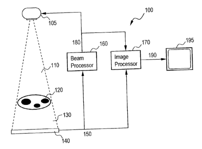
FIG. 1 illustrates a schematic diagram on an x-ray system 100 using x-ray beam
modulation in accordance with an embodiment of the present invention. System
100
includes an x-ray source 105 producing a spatially modulated beam 110, an
imaged
object 120, an x-ray detector 140, an x-ray beam processor 160, an image
processor
170, and a display device 195. Modulated beam 110 passes through imaged object
120, is attenuated to various degrees by its features, and forms residual beam
130.
Detector 140 measures the beam intensities in the residual beam 130 and
communicates a residual image 150 to the beam processor 160 and the image
processor 170. A beam intensity signal 180 can be communicated from the beam
processor 160 to the x-ray source 105 and to the image processor 170. The
image
processor produces a displayed image signal 190 and communicates to display
device
195.
As mentioned above, source 105 is capable of transmitting a spatially
modulated
beam 110 towards imaged object 120. More specifically, source 105 may be
capable
of altering x-ray intensities across beam 110 non-uniformly according to a
beam
intensity signal. A beam intensity signal is a digital representation of the
intensity
field of a spatially modulated x-ray beam 110.
Source 105 may be capable of altering the x-ray intensity field in beam 110 by
any
one of several embodiments. For example, system 100 may use a raster beam 110
by
moving a narrow beam 110 back and forth in a raster pattern over particular
areas of
9
CA 02518532 2005-09-08
33XZ 155828
object 120 while varying the beam's intensity temporally and integrating the
image in
the detector 140. In another embodiment, source 105 may include multiple beam
sources, each exposing different portions of imaged object 120. Source 105 may
then
modulate beam 110 spatially by controlling the outputs of individual x-ray
sources.
A spatially modulated x-ray beam 110 may be constructed to match a
distribution of
radiographic thicknesses of object 120. For example, object 120 may have a
known,
measured, or anticipated distribution of thickness (for example, based on
previous
frames in a fluoroscopic sequence). Based at least on this distribution, a
beam
intensity signal may be created to increase the exposure to radiographically
thick
regions and/or decrease exposure to radiographically thin regions of imaged
object
120, thereby possibly resulting in the approximate equalization of intensities
in
residual beam 130, for example. Residual beam 130 can include an x-ray beam
after
it has been attenuated by at least imaged object 120, for example.
A spatially modulated x-ray beam 110 may be constructed to match a
distribution of
region on interest in object 120. Regions of interest may be areas or volumes
in
object 120 that a user of system 100 desires to image. Regions of interest in
object
120 may be known a priori from previous scans or general atlases, programmed,
inferred, or anticipated. Based on at least the distribution of these regions
of interest,
a beam intensity signal may be created that results in increased x-ray
exposures to
regions of great interest and/or decrease x-ray exposures to regions of lesser
interest,
for example.
A spatially modulated x-ray beam 110 may also be established to match a
distribution
of regions of sustained motion in object 120. Object 120 may have regions or
volumes that are likely to move relative to imaging system 100. Other regions
are
more likely to remain static. For example, if object 120 is a chest cavity of
a human
patient, it may include the patient's heart moving relative to the rest of the
chest
cavity. Regions of motion in object 120 may be programmed by users, known a
priori, measured, or anticipated. Less exposure is necessary in regions with
little
motion where image processing techniques may be employed to reuse information
from earlier frames to produce a high-quality representation of these static
regions.
Based on at least the anticipated distribution of motion, a beam intensity
signal may
33XZ 155828
CA 02518532 2005-09-08
be created that results in increased x-ray exposures to regions with motion
and/or
decreased x-ray exposures to regions with little or no motion.
Finally, a spatially modulated x-ray beam 110 may be established to match a
combination of the three distributions described above, for example, (a)
radiographic
thicknesses, (b) regions of interests, and (c) regions of object motion may be
combined to produce an improved beam intensity signal.
Once beam 110 passes through object 120, detector 140 receives residual beam
130.
Detector 140 is a device capable of measuring or recording the intensity
pattern
projected by residual image 130. For example, detector 140 may be a solid-
state x-
ray detector, or an image intensifier coupled with a charged-coupled device
digital
video camera.
Based at least on measured intensities in residual beam 130, detector 140 may
create
residual image 150. For example, residual image 150 may comprise electronic
data
representing various residual beam 130 intensities received by detector 140.
Detector
140 communicates residual image 150 to at least one of beam processor 160 and
image processor 170.
Beam processor 160 is an image-processing component of system 100. Beam
processor 160 may be any processor capable of receiving residual image 150
from
detector 140, creating beam intensity signal 180, and communicating beam
intensity
signal 180 to at least one of source 105 and image processor 170. Beam
processor
160 may be embodied in a computer general-purpose microprocessor, a software
component, or a specialized digital signal processing ("DSP") circuit, for
example.
Beam processor 160 may be embedded in a system supplying processing for system
100, which may also perform additional tasks for system 100, such as those
performed by image processor 170.
After beam processor 160 receives residual image 150, beam processor 150
examines
residual image 150 to determine how the beam intensity signal 180 needs to be
modified. Thus beam processor 160 completes a feedback loop that may
periodically
or continuously update the beam 110 intensity field based at least on changes
in
imaged object 120. Because beam processor 160 may "know" what beam 110
CA 02518532 2005-09-08
33XZ 155828
intensity field was applied to produce the received residual image 130, beam
processor 160 may not require a uniform-beam scout shot to estimate
radiographic
thicknesses in imaged object 120 and may further be capable of periodically
and/or
continually updating beam intensity signal 180 as imaged object 120 moves or
changes throughout an imaging session.
When beam intensity signal 180 is based primarily on radiographic thicknesses
in
imaged object 120, the feedback loop may result in residual image 130 being
essentially uniform, within the beam-modulating performance limitations of x-
ray
source 105. This is to say that in some cases, the spatial resolution
limitations, the
dynamic range limitations, or grayscale resolution limitations of the beam
modulation
in x-ray source 105 will not allow complete equalization of the beam, even
though a
significant improvement may be produced thanks to partial equalization. These
limits
include spatial resolution, intensity resolution, and dynamic range. The
residual
image can include information of object movement or other changes as well as
detail
that is not resolved by the beam modulator in x-ray source 105. If the beam
modulation capabilities of x-ray source 105 approach corresponding image
acquisition capabilities of x-ray detector 140, then residual image 140 may
only
include noise and motion, if any. Thus, considerable useful information about
imaged
object may be included in beam intensity signal 180.
When beam intensity signal 180 is also based on anticipated regions of motion
and
regions of interest in object 120, then beam processor 160 may create a beam
intensity
signal 180 to cause increased beam intensity in these regions. The residual
image 140
may therefore be non-uniform and may not accurately represent radiographic
thicknesses in imaged object 120.
As described above, beam processor 160 may also communicate beam intensity
signal
180 to image processor 170. Image processor 170 may be any processor capable
of
combining two or more image signals into a third image signal using image
algebra
operators. For example, image processor 170 may be a specialized hardware
component, a programmable device, or an embedded software component running on
a general-purpose microprocessor, for example.
12
CA 02518532 2005-09-08
33XZ 155828
Image processor 170 subtracts beam intensity signal 180 from residual image
150 to
create output image 190. This subtraction may occur, for example, on a pixel-
for-
pixel basis. The specific meaning of the subtraction operation depends on the
grayscale transforms applied to constituent images. For example, if a
logarithmic
grayscale transform has been applied to the residual image and to the beam
intensity
signal, then a simple arithmetic subtraction may be used. Combined image 190
may
then accurately represent true radiographic thickness in object 120, as if
acquired with
a uniform x-ray beam, for example. Signal delays may need to be built into the
system to ensure that beam intensity signals 180 are combined with matching
residual
images 150.
Image processor 170 may also adapt its processing in accordance to the same
region-
of interest information and region-of motion information used to produce the
beam
intensity signal 180 in beam processor 160. These adaptations may include
spatial
filtration, temporal filtration, feature enhancements, noise suppression, and
others. For
example, when beam processor 160 causes a dose reduction to a region of lesser
interest, image processor 170 may increase noise reduction in corresponding
image
regions. As another example, when beam processor 160 causes a dose reduction
to a
region where little object motion is anticipated, then increased temporal
filtration may
be used to increase the reuse of previous frames to present a high-quality
image.
Multiscale image processing schemes may facilitate these solutions.
In another embodiment of the present invention, the present invention may be
embodied as an external add-on device to an existing imaging system. In FIG. 1
l,
system 1100 includes an existing conventional system 1105, demarcated by a
dash-
lined box, which, in turn, includes an x-ray source 405 and an x-ray detector
140. An
external beam-modulating device comprises an external add-on processor 1130, a
beam modulator 1115, and a display device 1140. The conventional system's
video
output 1110 is connected to the add-on processor 1130. The beam modulator 1115
is
attached to the conventional system's 1115 x-ray source 405. The add-on
processor
1130 plays the roles of the beam processor 160 and image processor 170 as in
FIG. 4.
The beam configuration signal 420 is conveyed to the beam modulation 1115
along
the modulator connection 1120. The video signal 1110 conveys residual image
signal
to add-on processor 1130.
13
CA 02518532 2005-09-08
33XZ 155828
A conventional digital fluoroscopy x-ray imaging system typically includes x-
ray
source 105, detector 140, and is capable of producing a video output signal
1110. In
operation, source 105 transmits an x-ray beam 110 towards object 120. After
beam
110 passes through object 120 and becomes residual beam 130 (as described
above),
detector 140 measures the x-ray intensities of residual beam 130. The system
1105
then converts this residual beam into a video signal 1110 which may be fed
into other
systems.
However, in this embodiment, external beam modulation device 1115 may be added
to such system to add the functionality of the present invention to an
existing imaging
system. Device 1115 is controlled by an add-on processor 1130.
In operation, processing block received video output 1110 from the
conventional
system 1105. Add-on processor 1130 then acts to achieve the same functionality
of
the beam processor 160 and image processor 170, as described above. For
example,
once add-on processor 1130 receives residual image 1110, a beam processor
similar
to beam processor 160 examines residual image video signal 1120 to determine
how a
beam intensities in beam 110 need to be modified. The beam processor of add-on
processor 1130 completes a feedback loop that may periodically or continuously
update the beam 110 intensity field based at least on changes in imaged object
120, as
described above. Device 1120 may then communicate the beam intensity signal
180
to beam modulator via the beam modulator connection 1120.
In addition, once the beam processor of add-on processor 1130 determines a
beam
intensity signal, add-on processor 1130 may also communicate the beam
intensity
signal to an internal image processor similar to image processor 170 of system
100.
The image processor of add-on 1130 then subtracts beam intensity signal 180
from
residual image 150 to create output image 190. This subtraction may occur, for
example, on a pixel-for-pixel basis. Device 1120 can then communicate the
image
190 to an external display device 1140 for display to a user of system 1100.
Therefore, the presently described embodiment provides for the simple addition
of a
beam modulation device 1120 to an existing x-ray imaging system in order to
achieve
the functionality of the present invention.
14
CA 02518532 2005-09-08
33XZ 155828
FIG. 2 illustrates a flowchart according to a method 200 of generating an
output
image signal 190 based on the above described feedback loop according to an
embodiment of the present invention. First, at step 210, an x-ray source 105
transmits
an x-ray beam 110 towards an object 120. Next, at step 220, the spatially
modulated
beam 110 passes through and is attenuated by the object 120. The resultant
beam that
exists the other side of the object 120 is a residual beam 130. At step 230, a
detector
140 measures x-ray intensities in the residual beam 130 in order to create a
residual
image 150. Next, at step 240, the detector 140 communicates the residual image
150
to a beam processor 160 and an image processor 170. Next, at step 250, the
beam
processor 160 generates a beam intensity signal 180 and communicates signal
180 to
the source 105 and image processor 170. Next, at step 260, the image processor
170
integrates the residual image 150 with the beam intensity signal 180 in order
to
produce an image output signal 190. This output signal 190 may then be
displayed on
a display device 195, for example. Next, method 200 may proceed back to step
210.
In this way, method 200 may proceed in a feedback loop manner.
The beam processor 160 may create and communicate beam intensity signal 180 on
a
regularly repeated or continuous basis such as fluoroscopic frame rates of 30,
15, or
7.5 frames per second.
In addition to combing the two constituents into the output image, beam
processor
170 may also perform other image processing tasks such as feature enhancement,
dynamic range suppression, noise reduction, digital subtraction angiography
("DSA"),
and grayscale transformations, for example. These processing tasks in image
processor 170 may be correlated with beam modulating tasks in beam processor
160.
For example, regions that are not anticipated to contain motion may receive
reduced
x-ray exposures, as controlled by beam processor 160, but they may also be
more
heavily temporally averaged to reduce image noise in image processor 170. As
another example, regions of lesser interest may receive reduced x-ray
exposures but
may also be more spatially averaged to reduce noise in image processor 170,
for
example.
Display device 195 receives output image 190 from image processor 170 and
presents
it to a viewer.
CA 02518532 2005-09-08
33XZ 155828
FIG. 3 illustrates examples of spatially modulated beam 110 according to an
embodiment of the present invention, residual image 150, and displayed image
signal
190 after the feedback loop has produced a near-optimal beam intensity field.
In FIG.
3, the beam processor is programmed to equalize the residual image without
consideration for regions of interest or anticipated object motion. In
addition, the
spatial resolution of the beam modulator is limited in FIG. 3, so the beam
intensity
signal comprises only the low-frequency image information and the residual
image
contains the remaining high-frequency image information. The combined output
image 190 appears as if acquired with a uniform-beam system at a high dose and
high
resolution, when, in fact, the averaged dose to the imaged object is
significantly
reduced.
FIG. 4 illustrates a schematic diagram of an x-ray system 400 using spatial
modulation of x-ray beam 110 used in accordance with an embodiment of the
present
invention. System 400 includes an x-ray source 405 emitting an essentially
uniform
x-ray beam 410, a beam-modulating filter 415, an imaged object 120, an x-ray
detector 140, a beam processor 160, an image processor 170, and a display
device
195. The initial beam 410 may not be completely uniform due to the Heel
effect, for
example. Beam modulating filter 415 is placed between x-ray source 405 and
imaged
object 120. X-ray source 405 transmits an essentially uniform x-ray beam 410
toward
modulating filter 415, imaged object 120, and detector 140. At least some
portion of
uniform beam 410 passes through modulating filter 415 to form modulated beam l
10.
Modulated beam 110 passes through imaged object 120, is attenuated to various
degrees by its features, and forms residual beam 130. X-ray detector 140
measures
intensities in residual beam 130, fortes the residual image 150 and
communicates it to
beam processor 160 and image processor 170. The beam processor 160 forms the
beam intensity signal 180 and communicates the signal 180 to the image
processor
170. The beam processor 160 then translates the beam intensity signal 180 into
a
modulator configuration signal 420 and communicates it to the beam-modulating
filter 415. In this way, both the beam intensity signal 180 and the modulator
configuration signal 420 act to determine the spatial modulation of an x-ray
beam.
The image processor 170 creates output image 190 and communicates it to
display
device 195. Image processor 170 may create output image 190 by integrating
16
CA 02518532 2005-09-08
33XZ 155828
intensity signal 180 and modulator configuration signal 420, similar to as
described
above in regards to FIG. 1.
Beam-modulating filter 415 may attenuate initial beam 410 according to
modulator
configuration signal 420 to various degrees across the beam field. Beam-
modulating
filter 415 may be any device capable of selectively altering an amount
attenuation of
initial beam 410 to various degrees across the beam field, thereby creating
spatially
modulated beam 110. Similar to spatially modulated beam 110 in FIG. 1, beam-
modulating filter 415 may attenuate initial beam 410 as to create a desired
beam 110
intensity field, as described above.
In an example, beam-modulating filter 415's ability to selectively alter beam
attenuations across the beam field may be compared to a liquid crystal display
("LCD") device. For example, an LCD device may control the passage of light
through pixels by applying an electric current to a matrix of liquid crystals.
By
application of the proper current, individual pixels of the LCD may change to
allow
variable amounts of light through an LCD. Similarly, beam-modulating filter
415
may employ a matrix of pixels that, based on a modulator configuration signal
420
may change to allow various amounts of x-ray beam 410 to pass, for example.
The functions of the remaining components of system 400 are similar to those
of
system 100 depicted in FIG. 1 and are described above. The functionality,
applications, and benefits of system 400 are similar to the functionality of
system 100
in FIG. 1. For example, sources 105 and 405, object 120, detector 140, beam
processor 160, image processor 170, and display device 195 may behave
similarly in
both FIGS. 1 and 4.
FIG. 5 illustrates a flowchart according to a method 500 of generating an
output
image signal 190 based on the above described feedback loop using a beam-
modulating filter in accordance with an embodiment of the present invention.
First, at
step 505, an x-ray source 405 transmits an x-ray beam 410 towards a filter or
beam
attenuator 415, as described above. Next, at step 510, a beam attenuator (or
filter)
415 attenuates the beam 410, as described above. For example, attenuator 415
may
attenuate the beam 410 non-uniformly according to a modulator configuration
signal
17
CA 02518532 2005-09-08
33XZ 1SS828
420. Once beam 410 has exited the attenuator 41 S, beam 410 becomes modulated
beam 110, as described above. Modulated beam 110 then passes through an imaged
object 120 and becomes a residual beam 130, as shown in step 520. The residual
beam 130 then strikes a detector 140. At step S30 the detector 140 measures
the x-ray
intensities of the residual beam 130 in order to create a residual image 1 S0.
Next, at
step 540, the detector 140 communicates the residual image 1 SO to a beam
processor
160 and an image processor 170, as described above. At step SSO, the beam
processor
160 generates a beam intensity signal 180 and communicates the intensity
signal 180
to the image processor 170. Next, at step 560, the beam processor 160
translates the
beam intensity signal I80 into a configuration signal 420, as described above,
and
communicates the signal 420 to the beam attenuator 415. Next, at step 570, the
image
processor 170 integrates the residual image 1 SO with the beam intensity
signal 180 in
order to produce an output image signal 190, as described above. This image
signal
190 may then be communicated to a display device 19S for display. Next, method
S00 may proceed to step SOS. In this way, method S00 may proceed in a feedback
loop manner.
The basis for a practical embodiment of a beam-modulating filter in accordance
with
this invention is referred to as "x-ray dodging". The term originates from the
dodging
and burning techniques in darkroom light photography. To control the exposure
to a
portion of a photograph, photographers may introduce an opaque mask into the
light
beam for a calculated portion of the exposure time. To feather sharp mask
edges in
the photograph, photographers may wave the mask horizontally or vertically.
The
photographic paper integrates the exposure over time, so that the variations
of total
exposure to the photographic paper may be controlled across the image by the
duration of time for which the region remains blocked by the mask.
Beam-modulating filters previously disclosed (for example, as described above)
modulate the beam by varying the thicknesses of the semi-transparent
substances
placed in the x-ray beam. In contrast, x-ray dodging uses radiographically
opaque
elements to block the beam completely but only for a controlled portion of a
frame
integration period. This strategy endows the beam modulator with flexibility,
a high
number of gradation levels, high spatial resolution, and a high dynamic range.
In
addition, unlike the previous attempted solutions (as described above), beam
18
CA 02518532 2005-09-08
33XZ 155828
modulation using x-ray dodging is not as sensitive to x-ray photon energies as
long as
the x-ray blocking elements remain radiographically opaque. In the range of x-
ray
techniques used for interventional medical fluoroscopy and diagnostic
radiography,
elements made of 0.8-1.5 mm of tungsten may be sufficient to effectively block
the x-
ray beam.
To control the exposure times, the x-ray-blocking elements may be moved,
rotated,
and/or oscillated at high speeds or frequencies with high precision. To help
reduce
the complexity of the motion, the intensity of the uniform beam rnay be varied
synchronized with the motion of the x-ray blocking elements. In practice, it
may be
easier to make these motions and beam intensity variations periodic in time.
Therefore, the x-ray dodging technique may be defined as the use of controlled
arrangements of x-ray blocking elements in the x-ray beam undergoing a high-
frequency periodic motion synchronized with periodic temporal x-ray beam
modulations and detector frame integration periods to produce desired spatial
modulation of the x-ray beam.
FIG. 8 illustrates the effect of x-ray dodging according to an embodiment of
the
present invention. In this embodiment exposed area 615 is divided into image
cells
720. A radiographically opaque element 710 may be introduced into any image
cell.
When the element 710 undergoes an oscillatory motion 810 at a high frequency
in a
plane perpendicular to the x-ray beam with the amplitude of about one cell
width, a
semitransparent blurred attenuation pattern 800 may be produced. Here the
oscillation
810 is assumed to be harmonic or sinusoidal. The oscillatory motion 810 may
not
completely remove sharp features from the attenuation pattern 800. These sharp
features may introduce artifacts in an output image 190. To remove these sharp
features, the system may vary the intensity of the initial uniform beam 410
synchronized with the phase of the oscillatory motion 810.
For example, let ~k ~9, p~ E E0,1~ be the attenuation function of the klh
basis disc
defined in polar coordinates B, p such that the center of disc rotation 630 is
at p = 0 .
The system will then shift the phases 640 of each disc k by appropriate
angular offsets
qrk to produce a desired combined attenuation function of the entire stack
19
CA 02518532 2005-09-08
33XZ 155828
~ ~8, p) _ ~ øk (B + irk , p) . Indicators 640 representing one or more phase
shifts
k
are included in FIG. 6 for demonstration purposes only. The entire disc stack
610 is
caused to undergo rotational oscillation so that its angular offset s varies
as
~ _ ~, cos ~ 2~ t ~ , where t is time, T is the oscillation period, and ~, is
the angular
oscillation amplitude (e.g. ~, = 64 ). Now, a portion of the disc stack is
exposed to a
uniform beam with time-varying intensity Io (t) . Then at any point in time t,
the
intensity of the modulated beam will be I (B, p,t~ = to (t)-~(9+s(t), p) . The
mean
intensit durin each half eriod will be: I B, p = Z Io t - ~ B + ~ t , - dt .
y g P
0
Substituting the integration variable to s , this expression becomes
I (e, p~ _ ~ f Io ~ 2 cos-' ~ ~ ~ ~ ~B - s, p) - 2 ~ - 1 2 dt . This may be
written
-a. 1 _ (E~)
as a convolution integral I (B, p~ = h (B~ * ~ (8, p~ , where
h (B~ = to ~ T cos-' a ~ . T - 1 . Now it may be shown that, by modifying
2~c ~, 2~~, 1 _ ( ~ )z
the time intensity waveform to (t) of the uniform x-ray beam 410, one can
effectively
convolve the attenuation pattern ~(B, p) with an arbitrary function g(B~ along
the
B axis. For example, one may choose g(9) to be a smoothing band-limiting
kernel
such as a Gaussian or Harming kernels. Then h(9~=g(0~ and beam intensity
waveform may be computed as to ~t ~ = g ~~, cos 2~ t ~ . 2~ . sin 2~ t for t =
~0, Z ~ (a
single pulse). Pulses may be spread out or follow each other in sequence, as
required
by the imaging application.
FIG. 9 illustrates an example of an oscillation offset function 910 that may
correspond
to the harmonic oscillation function ~(t~ described above and the x-ray tube
current
waveform 920 (mA) that produces proportional uniform beam 410 intensity to (t)
as
CA 02518532 2005-09-08
33XZ 1SS828
described above. The x-ray tube waveform 920 causes the smoothing kernel h~B~
to
become a Gaussian kernel, resulting in a smoothing effect such as illustrated
in FIG.
8.
Notice that the motion blurring in FIG. 8 smoothens the attenuation pattern
along the
direction of oscillation 810 only. The smoothness along the radial axis is
achieved due
to smooth variations of the widths of the x-ray blocking elements 710. When a
column of x-ray blocking elements 830 is smoothened by oscillations 810
synchronized with beam intensity modulation 920 of FIG. 9, the resulting beam
modulation pattern 840 may be made completely uniform along the direction of
oscillation 810 due at least in part to the band-limited convolution kernel
h~x~ .
Rows of beam blocking elements 8S0 may result in uniform attenuation
orthogonal to
oscillation 810 due at least in part to the band-limited width variations of
the x-ray
blocking elements 710. In this way, the x-ray block elements 710 combined with
periodic motion 810 and temporal beam intensity modulation may be used to
produce
smoothly varying attenuation patterns. These patterns may be critical to avoid
image
artifacts or the necessity for perfect beam alignment.
Motion blur and temporal beam modulation remove sharp features from the
attenuation pattern. The area blocked by a beam-blocking element 710 will
contribute
to the attenuation produced in the image cell in which it is placed. The
system may
regulate the local attenuation by selecting from a set of possible beam-
blocking
elements of various widths. FIG. 10 illustrates a more flexible way to adjust
the local
attenuation level with the use of inter-element occlusions according to an
embodiment
of the present invention. Two beam-blocking elements 1001 and 1002 may be
placed
in the beam. Elements 1001, 1002 may differ in size and/or shape. If these
elements
1001, 1002 are positioned in different planes, they may occlude one another.
As a
result, the total beam-blocking areas may be varied gradually, with the number
of
attenuation levels limited only by the mechanical precision.
For example, when the two elements 1001, 1002 are not occluding each other, as
in
element arrangement 1010, the resulting attenuation cell 1020 may be darker
than
when the elements 1001, 1002 occlude each other to various degrees, as in
element
21
CA 02518532 2005-09-08
33XZ 155828
arrangements 1030 and 1050 and corresponding attenuation cells 1040 and 1060).
In
addition, other ways of changing the projected area of a beam-blocking element
may
be used such as rotation of the element or moving the element closer to or
away from
the focal spot.
The design of x-ray-blocking elements may also take into consideration how
adjacent
cells interact. For example, it may be desirable to have the capability to
block a
portion of the x-ray beam completely. In order to do so, rows and/or columns
of
elements may mesh tightly so that the x-ray beam is blocked completely. Beam-
blocking elements are designed to interlock with elements from adjacent rows
as to be
configurable to block an entire area without gaps. For example, two adjacent
columns
of cells 1070 and 1075 of elements set for maximum attenuation, when combined,
may lock tightly as in arrangement 1080. After they are blurred by motion, the
smoothened attenuation pattern 1085 contains areas where the beam is
completely
blocked.
Inter-element occlusions are just one of several possible approaches of
blocking
varying portions of the x-ray beam with one or several beam-blocking elements.
For
example, rotating or rolling the elements or moving them toward or away from
the x-
ray source may be employed. Neither do inter-element occlusions need to be
limited
to two elements. Multiple elements occluding one another in various
arrangements
may provide even greater flexibility in creating desired attenuation patterns.
FIG. 6 and FIG. 7 each illustrate an embodiment of the beam-modulating filter
41 S in
accordance with an embodiment of the present invention. The x-ray source 105
produces an essentially uniform x-ray beam 410, as described above. The
uniform x-
ray beam 410 traverses a stack of disc-shaped basis filters 620. The basis
filters 620
are made of a radiographically translucent material and contain arrangements
of x-
ray-blocking elements 710.
The relative angular offsets 640 of the basis discs place different portions
of each disc
in the exposed area 615. For example, angular offset 640 of a given disc may
be
determined by an angular offset 640 of a particular x-ray blocking element 710
or
other known position marker indicated by reference 635. The discs' angular
offsets
22
CA 02518532 2005-09-08
33XZ 155828
may be controlled independently. By varying the number of discs 620, the
various
arrangements of elements 710, and the various angular displacements of the
various
discs 620, a large number of possible arrangements of x-ray blocking elements
in the
exposed area 615 are possible.
The entire disc stack may be caused to undergo a high-frequency rotational
oscillation
around the axis 630 synchronized with the periodic temporal modulation of the
uniform beam 410.
The rotational offsets of the basis discs may be controlled by the modulator
configuration signal 420 originating from the beam processor 160, as described
above. The motors and mechanics driving these offsets are not shown in FIG. 6,
but
may be embodied, for example, in a stepper motor configuration known to those
of
ordinary skill in the art.
In an example of an embodiment of the present invention, the circular exposed
area
615 of a basis filter 620 may be divided into columns and rows of cells 720,
as shown
in FIG. 7. For example, in FIG. 7, the exposed circular area 615 is divided
into five
7-cell central columns and two 3-cell boundary columns. The beam attenuation
level
of each of the 41 cells can be controlled independently with smooth
transitions
between them.
Two basis discs are assigned to each of the five central columns of image
cells (ten
basis discs total). Each disc 620 may be rotated to such a position that each
cell 720
in the exposed column will either include an x-ray-blocking element or not
contain
one. For a seven-cell column, 2'=128 such septuplets are possible. If 1
represents the
presence of an x-ray-blocking element and 0 represents the absence of an x-ray-
blocking element, then arranging the elements circularly around a basis disc
according
to the 128-element pattern 0 0 0 0 1 1 1 0 0 0 0 1 1 0 1 1 1 1 1 0 1 1 0 0 1 0
0 1 1 1 1 1
1100110001010101111000110011101011011010100110
1000100100011110100101110111001010000101100000
1 0 0 can allow rotating the disc 620 to a position producing any possible
such
septuplet. Since two discs may be assigned to each column, for each of the 35
central
cells, four possible configurations are possible: (1) no x-ray block elements
present,
23
CA 02518532 2005-09-08
33X2 155828
(2) one x-ray blocking element from first basis disc present, (3) one x-ray-
blocking
element from second basis disc present, and (4) two x-ray-blocking elements
present,
one from the first and one from the second basis disc. For example, if the
resulting
cell attenuation from an element from the first disc is 0.33 and from the
second disc -
0.67,, then when both elements are present in the cell, the attenuation may be
varied in
continuous gradation from 0.67 to 1.0 by adjusting the degree of the inter-
element
occlusions.
Many other pattern designs are possible, not necessarily based on cell
matrices. For
example, in a circular exposed area such as 615 in FIG. 7, the vertical
boundary
columns contain only three cells. Instead of using two discs with a binary
pattern of
beam-blocking elements such 715 as described above to provide four independent
attenuation levels, a quinternary attenuation pattern may be used to provide
any of
five attenuation patterns in each cell, independently. An example of such 125-
cell
circular pattern is 001112223334441133002244220033114433
2211004401234023013412403434232312120242413130
3020101414203140410213243042143103204321040.In
this pattern any contiguous triplet of digits 0 through 4 may be found. If
five types of
beam blocking elements corresponding to these digits are arranged in a circle
on a
disc, then any combination of such elements may be selected into the three
exposed
cells.
While particular elements, embodiments and applications of the present
invention
have been shown and described, it is understood that the invention is not
limited
thereto since modifications may be made by those skilled in the art,
particularly in
light of the foregoing teaching. It is therefore contemplated by the appended
claims
to cover such modifications and incorporate those features that come within
the spirit
and scope of the invention.
24