Note: Descriptions are shown in the official language in which they were submitted.
CA 02519113 2005-09-13
WO 2004/087756 PCT/EP2004/003442
1
Antibodies against insulin-like growth factor I receptor and uses thereof
The present invention relates to antibodies against human insulin-like growth
factor I receptor (IGF-IR), methods for their production, pharmaceutical
compositions containing said antibodies, and uses thereof.
Human insulin-like growth factor I receptor (IGF-IR, EC 2.7.112, CD 221
antigen)
belongs to the family of transmembrane protein tyrosine kinases (LeRoith, D.,
et
al., Endocrin. Rev. 16 (1995) 143-163; and Adams, T.E., et al., Cell. Mol.
Life Sci. 57
(2000) 1050-1063). IGF-IR binds IGF-I with high affinity and initiates the
physiological response to this ligand in vivo. IGF-IR also binds to IGF-II,
however
with slightly lower affinity. IGF-IR overexpression promotes the neoplastic
transformation of cells and there exists evidence that IGF-IR is involved in
malignant transformation of cells and is therefore a useful target for the
development of therapeutic agents for the treatment of cancer (Adams, T.E., et
al.,
Cell. Mol. Life Sci. 57 (2000) 1050-1063).
Antibodies against IGF-IR are well-known in.the state of the art and
investigated
for their antitumor effects in vitro and in vivo (Benini, S., et al., Clin.
Cancer Res. 7
(2001) 1790-1797; Scotlandi, K., et al., Cancer Gene Ther. 9 (2002) 296-307;
Scotlandi, K., et al., Int. J. Cancer 101 (2002) 11-16; Brunetti, A., et al.,
Biochem.
Biophys. Res. Commun. 165 (1989) 212-218; Prigent, S.A., et al., J. Biol.
Chem. 265
(1990) 9970-9977; Li, S.L., et al., Cancer Immunol. Immunother. 49 (2000) 243-
252; Pessino, A., et al., Biochem. Biophys. Res. Commun. 162 (1989) 1236-1243;
Surinya, K.H., et al., J. Biol. Chem. 277 (2002) 16718-16725; Soos, M.A., et
al., J.
Biol. Chem., 267 (1992) 12955-12963; Soos, M.A., et al., Proc. Natl. Acad.
Sci. USA
86 (1989) 5217-5221; O'Brien, R.M., et al., EMBO J. 6 (1987) 4003-4010;
Taylor, R.,
et al., Biochem. J. 242 (1987) 123-129; Soos, M.A., et al., Biochem. J. 235
(1986)
199-208; Li, S.L., et al., Biochem. Biophys. Res. Commun. 196 (1993) 92-98;
Delafontaine, P., et al., J. Mol. Cell. Cardiol. 26 (1994) 1659-1673; Kull,
F.C. Jr., et
al. J. Biol. Chem. 258 (1983) 6561-6566; Morgan, D.O., and Roth, R.A.,
Biochemistry 25 (1986) 1364-1371; Forsayeth, J.R., et al., Proc. Natl. Acad.
Sci. USA
84 (1987) 3448-3451; Schaefer, E.M., et al., J. Biol. Chem. 265 (1990) 13248-
13253;
Gustafson, T.A., and Rutter, W.J., J. Biol. Chem. 265 (1990) 18663-18667;
Hoyne,
P.A., et al., FEBS Lett. 469 (2000) 57-60; Tulloch, P.A., et al., J. Struct.
Biol. 125
(1999) 11-18; Rohlik, Q.T., et al., Biochem. Biophys. Res. Comm. 149 (1987)
276-
CA 02519113 2005-09-13
WO 2004/087756 PCT/EP2004/003442
-2-
281; and Kalebic, T., et al., Cancer Res. 54 (1994) 5531-5534; Adams, T. E.,
et al.,
Cell. Mol. Life Sci. 57 (2000) 1050-1063; Dricu, A., et al., Glycobiology 9
(1999)
571-579; Kanter-Lewensohn, L., et al., Melanoma Res. 8 (1998) 389-397; Li,
S.L., et
al., Cancer Immunol. Immunother. 49 (2000) 243-252). Antibodies against IgF-IR
are also described in a lot of further publications, e.g., Arteaga, C.L., et
al., Breast
Cancer Res. Treatment 22 (1992) 101-106; and Hailey, J., et al., Mol. Cancer
Ther. 1
(2002) 1349-1353.
In particular, the monoclonal antibody against IGF-IR called aIR3 is widely
used in
the investigation of studying IGF-IR mediated processes and IGF-I mediated
diseases such as cancer. Alpha-IR-3 was described by Kull, F.C., J. Biol.
Chem. 258
(1983) 6561-6566. In the meantime, about a hundred publications have been
published dealing with the investigation and therapeutic use of aIR3 in regard
to its
antitumor effect, alone and together with cytostatic agents such as
doxorubicin and
vincristine. aIR3 is a murine monoclonal antibody which is known to inhibit
IGF-I
binding to IGF receptor but not IGF-II binding to IGF-IR. However, there exist
other antibodies (e.g., 1H7, Li, S.L., et al., Cancer Immunol. Immunother. 49
(2000) 243-252) which inhibit IGF-II binding to IGF-IR more potently than IGF-
I
binding. A summary of the state of the art of antibodies and their properties
and
characteristics is described by Adams, T.E., et al., Cell. Mol. Life Sci. 57
(2000)
1050-1063.
Most of the antibodies described in the state of the art are of mouse origin.
Such
antibodies are, as is well known in the state of the art, not useful for the
therapy of
human patients without further alterations like chimerization or humanization.
Based on these drawbacks, human antibodies are clearly preferred as
therapeutic
agents in the treatment of human patients. Human antibodies are well-known in
the state of the art (van Dijk, M.A., and van de Winkel, J.G., Curr. Opin.
Pharmacol. 5 (2001) 368-374). Based on such technology, human antibodies
against a great variety of targets can be produced. Examples of human
antibodies
against IGF-IR are described in WO 02/053596.
However, there is still a need for antibodies against IGF-IR with convincing
benefits
for patients in need of antitumor therapy. The relevant benefit for the
patient is, in
simple terms, reduction in tumor growth and a significant prolongation of time
to
progression caused by the treatment with the antitumorigenic agent.
CA 02519113 2005-09-13
WO 2004/087756 PCT/EP2004/003442
-3-
Summary of the Invention
The invention comprises an antibody binding to IGF-IR and inhibiting the
binding
of IGF-I and IGF-II to IGF-IR, characterized in that said antibody is of IgG1
isotype
and shows a ratio of inhibition of the binding of IGF-I to IGF-IR to the
inhibition
of binding of IGF-II to IGF-IR of 1:3 to 3:1 and induces cell death of 20% or
more
cells of a preparation of IGF-IR expressing cells after 24 hours at a
concentration of
said antibody of 100 nM by ADCC.
Antibodies according to the invention show benefits for patients in need of
antitumor therapy and provide reduction of tumor growth and a significant
prolongation of the time to progression. The antibodies according to the
invention
have new and inventive properties causing a benefit for a patient suffering
from a
disease associated with an IGF deregulation, especially a tumor disease. The
antibodies according to the invention are characterized by the abovementioned
properties. The properties are therefore especially specific binding to IGF-
IR,
inhibiting the binding of IGF-I and IGF-II to IGF-IR at the abovementioned
ratio,
being of IgGi isotype, and having effector function in ADCC.
Preferably, in addition, the antibodies according to the invention induce cell
death
of 20% or more cells of a preparation of IGF-IR expressing cells after 4 h at
an
antibody concentration of 100 nM by CDC.
Preferably, at a concentration of 50 nM the antibodies according to the
invention
completely inhibit IGF-I mediated signal transduction of IGF-IR in tumor
cells.
The invention also comprises antibody encoding nucleic acids. The encoded
polypeptides are capable of assembling together with the respective other
antibody
chain defined below:
- an antibody heavy chain comprising as CDRs CDR1 (aa 31-35), CDR2 (aa 50-
66) and CDR3 (aa 98-108) of SEQ ID NO:1, wherein amino acid 31 can be
asparagine or serine, amino acid 66 can be glycine or deleted, and amino acid
104 can be glutamic acid or aspartic acid;
CA 02519113 2005-09-13
WO 2004/087756 PCT/EP2004/003442
-4-
- an antibody light chain comprising as CDRs CDR1 (aa 18-34 or as 24-34),
CDR2 (aa 50-56) and CDR3 (aa 89-98) of SEQ ID NO:2, wherein amino acid 96
can be proline or isoleucine, and amino acid 98 can be phenylalanine or
deleted.
The preferred CDRs are (a) CDR1 (aa 31-35), CDR2 (aa 50-65) and CDR3 (aa 98-
108) of SEQ ID NO:1, wherein amino acid 31 can be asparagine or serine and
amino acid 104 can be glutamic acid or asparatic acid, and (b) CDR1 (aa 24-
34), CDR2 (aa 50-56) and CDR3 (aa 89-97) of SEQ ID NO:2.
CDR numbering and definition is preferred according to Rabat, E. (see e.g.
Johnson, G., et al., Nucl. Acids Res. 28 (2000) 214-218).
Preferably, the nucleic acid encodes a polypeptide which is either a heavy
chain
consisting of a variable region (VH) of SEQ ID NO:1, wherein amino acid (aa)
30
denotes serine or arginine, as 31 denotes asparagine or serine, as 94 denotes
histidine or tyrosine and as 104 denotes aspartic acid or glutamic acid, and
of a
human heavy chain constant region (CH);
and a light chain consisting of a variable region (VL) of SEQ ID NO:2, wherein
as
96 denotes proline or isoleucine, as 100 denotes proline or glutamine, as 103
denotes arginine or lysine, as 104 denotes valine or leucine and as 105
denotes
aspartic acid or glutamic acid, and of a human light chain constant region
(CL).
The antibody is preferably a monoclonal antibody and, in addition, a chimeric
antibody (human constant chain), a humanized antibody and especially
preferably
a human antibody.
The antibody binds to IGF-IR human (EC 2.7.1.112, SwissProt P08069) in
competition to the antibodies characterized by the variable chains of
SEQ ID NOS:1-6.
The antibody is further characterized by an affinity of 10-8 M (IKD) or less,
preferably of about 10-8 to 10-11 M
Preferably, the invention provides antibodies comprising as complementarity
determining regions (CDRs) having the following sequences:
CA 02519113 2005-09-13
WO 2004/087756 PCT/EP2004/003442
-5-
- an antibody heavy chain comprising as CDRs CDR1 (aa 31-35), CDR2 (aa 50-
66) and CDR3 (aa 98-108) of SEQ ID NO:1, wherein amino acid 31 can be
asparagine or serine, amino acid 66 can be glycine or deleted, and amino acid
104 can be glutamic acid or aspartic acid;
- an antibody light chain comprising as CDRs CDR1 (aa 18-34 or as 24-34),
CDR2 (aa 50-56) and CDR3 (aa 89-98) of SEQ ID NO:2, wherein amino acid 96
can be proline or isoleucine, and amino acid 98 can be phenylalanine or
deleted.
The invention therefore comprises also a polypeptide and an encoding nucleic
acid
selected from the above-mentioned group consisting of CDR1, CDR2, CDR3 of
heavy chain and CDR1, CDR2, CDR3 of light chain of an IGF-IR antibody
according to the invention.
Preferably, the invention comprises an antibody characterized by a heavy chain
consisting of a variable region (VH) of SEQ ID NO:1, wherein amino acid (aa)
30
denotes serine or arginine, as 31 denotes asparagine or serine, as 94 denotes
histidine or tyrosine and as 104 denotes aspartic acid or glutamic acid, and
of a
human heavy chain constant region (CH);
and a light chain consisting of a variable region (VL) of SEQ ID NO:2, wherein
as
96 denotes proline or isoleucine, as 100 denotes proline or glutamine, as 103
denotes arginine or lysine, as 104 denotes valine or leucine and as 105
denotes
aspartic acid or glutamic acid, and of a human light chain constant region
(CL).
The constant regions provide Clq complement binding and are therefore
preferably of human IgGl type.
The combinations
as 30 Arg, as 31 Asn, as 94 Tyr and as 104 Asp (antibody 1A) or
as 30 Arg, as 31 Ser, as 94 Tyr and as 104 Asp (antibody 8) or
as 30 Ser, as 31 Asn, as 94 His and as 104 Glu (antibody 23)
in the heavy chain are preferred.
The combinations
as 96 Pro, as 100 Pro, as 103 Lys, as 104 Val and as 105 Asp (antibody 1A and
8),
as 96 Ile, as 100 Gln, as 103 Arg, as 104 Leu and as 105 Glu (antibody 23)
in the light chain are especially preferred.
CA 02519113 2005-09-13
WO 2004/087756 PCT/EP2004/003442
-6-
The combination as 30 Arg, as 31 Asn, as 94 Tyr, and as 104 Asp in the heavy
chain
and as 96 Pro, as 100 Pro, as 103 Lys, as 104 Val and as 105 Asp in the light
chain is
especially preferred.
The antibody according to the invention considerably prolongates the time to
progression in relevant xenograft tumor models in comparison with vehicle
treated
animals and reduces tumor growth. The antibody inhibits the binding of IGF-I
and
IGF-II to IGF-IR in vitro and in vivo, preferably in about an equal manner for
IGF-
I and IGF-II.
The antibody is further characterized by the ability to bind IgGFc receptor
and to
induce ADCC and preferably to bind complement component Clq and to induce
CDC.
The invention further provides hybridoma cell lines which produce such
antagonistic monoclonal antibodies according to the invention.
The preferred hybridoma cell lines according to the invention, <IGF-1R> HuMab
Clone la (antibody 1A, Ab 1A or Ak 1A), <IGF-1R> HuMab Clone 23 (antibody
23), and <IGF-1R> HuMab-Clone 8 (antibody 8) were deposited with Deutsche
Sammlung von Mikroorganismen and Zellkulturen GmbH (DSMZ), Germany:
Cell line Deposition No. Date of Deposit
<IGF-1R> HUMAB Clone la DSM ACC 2586 10.04.2003
<IGF-1R> HUMAB Clone 23 DSM ACC 2588 10.04.2003
<IGF-1R> HUMAB-Clone 8 DSM ACC 2589 24.04.2003
The antibodies obtainable from said cell lines are preferred embodiments of
the
invention.
The invention further provides nucleic acids encoding such antibodies,
expression
vectors containing said nucleic acids, and host cells for the recombinant
production
of such antibodies.
The invention further provides methods for the recombinant production of such
antibodies.
CA 02519113 2005-09-13
WO 2004/087756 PCT/EP2004/003442
-7-
The invention further provides methods for treating cancer, comprising
administering to a patient diagnosed as having cancer (and therefore being in
need
of an antitumor therapy) an effective amount of an antagonistic antibody
against
IGF-IR according to the invention. The antibody may be administered alone, in
a
pharmaceutical composition, or alternatively in combination with a cytotoxic
treatment such as radiotherapy or a cytotoxdc agent or a prodrug thereof.
The invention further comprises the use of an antibody according to the
invention
for cancer treatment and for the manufacture of a pharmaceutical composition
according to the invention. In addition, the invention comprises a method for
the
manufacture of a pharmaceutical composition according to the invention.
The invention further comprises a pharmaceutical composition containing an
antibody according to the invention with a pharmaceutically effective amount,
optionally together with a buffer and/or an adjuvant useful for the
formulation of
antibodies for pharmaceutical purposes.
The invention further provides a pharmaceutical composition comprising such an
antibody in a pharmaceutically acceptable carrier. In one embodiment, the
pharmaceutical composition may be included in an article of manufacture or
kit.
The invention further comprises a vector containing a nucleic acid according
to the
invention, capable of expressing said nucleic acid in a prokaryotic or
eukaryotic
host cell.
The invention further comprises a prokaryotic or eukaryotic host cell
comprising a
vector according to the invention.
The invention further comprises a method for the production of a recombinant
human antibody according to the invention, characterized by expressing a
nucleic
acid according to the invention in a prokaryotic or eukaryotic host cell and
recovering said antibody from said cell. The invention further comprises the
antibody obtainable by such a recombinant method.
CA 02519113 2005-09-13
WO 2004/087756 PCT/EP2004/003442
-8-
Detailed Description of the Invention
The term "antibody" encompasses the various forms of antibodies including but
not
being limited to whole antibodies, antibody fragments, human antibodies,
humanized antibodies and genetically engineered antibodies as long as the
characteristic properties according to the invention are retained.
"Antibody fragments" comprise a portion of a full length antibody, generally
at least
the antigen binding portion or the variable region thereof. Examples of
antibody
fragments include diabodies, single-chain antibody molecules, immunotoxins,
and
multispecific antibodies formed from antibody fragments. In addition, antibody
fragments comprise single chain polypeptides having the characteristics of a
VH
chain, namely being able to assemble together with a VL chain or of a VL chain
binding to IGF-1R, namely being able to assemble together with a VH chain to a
functional antigen binding pocket and thereby providing the property of
inhibiting
the binding of IGF-I and IGF-II to IGF-IR.
"Antibody fragments" also comprises such fragments which per se are not able
to
provide effector functions (ADCC/CDC) but provide this function in a manner
according to the invention after being combined with appropriate antibody
constant domain(s).
The terms "monoclonal antibody" or "monoclonal antibody composition" as used
herein refer to a preparation of antibody molecules of a single amino acid
composition. Accordingly, the term "human monoclonal antibody" refers to
antibodies displaying a single binding specificity which have variable and
constant
regions derived from human germline immunoglobulin sequences. In one
embodiment, the human monoclonal antibodies are produced by a hybridoma
which includes a B cell obtained from a transgenic non-human animal, e.g. a
transgenic mouse, having a genome comprising a human heavy chain transgene
and a light human chain transgene fused to an immortalized cell.
The term "chimeric antibody" refers to a monoclonal antibody comprising a
variable region, i.e., binding region, from one source or species and at least
a
portion of a constant region derived from a different source or species,
usually
prepared by recombinant DNA techniques. Chimeric antibodies comprising a
CA 02519113 2005-09-13
WO 2004/087756 PCT/EP2004/003442
-9-
murine variable region and a human constant region are especially preferred.
Such
murine/human chimeric antibodies are the product of expressed immunoglobulin
genes comprising DNA segments encoding murine immunoglobulin variable
regions and DNA segments encoding human immunoglobulin constant regions.
Other forms of "chimeric antibodies" encompassed by the present invention are
those in which the class or subclass has been modified or changed from that of
the
original antibody. Such "chimeric" antibodies are also referred to as "class-
switched
antibodies." Methods for producing chimeric antibodies involve conventional
recombinant DNA and gene transfection techniques now well known in the art.
See, e.g., Morrison, S.L., et al., Proc. Natl. Acad Sci. USA 81 (1984) 6851-
6855; US
Patent Nos. 5,202,238 and 5,204,244.
The term "humanized antibody" refers to antibodies in which the framework or
"complementarity determining regions" (CDR) have been modified to comprise the
CDR of an immunoglobulin of different specificity as compared to that of the
parent immunoglobulin. In a preferred embodiment, a murine CDR is grafted into
the framework region of a human antibody to prepare the "humanized antibody."
See, e.g., Riechmann, L., et al., Nature 332 (1988) 323-327; and Neuberger,
M.S., et
al., Nature 314 (1985) 268-270. Particularly preferred CDRs correspond to
those
representing sequences recognizing the antigens noted above for chimeric and
bifunctional antibodies.
The term "human antibody", as used herein, is intended to include antibodies
having variable and constant regions derived from human germline
immunoglobulin sequences. The variable heavy chain is preferably derived from
germline sequence DP-61 (GenBank M99682) and the variable light chain is
preferably derived from germline sequence L15 (GenBank K01323). The constant
regions of the antibody are constant regions of human IgG1 type. Such regions
can
be allotypic and are described by, e.g., Johnson, G., and Wu, T.T., Nucleic
Acids
Res. 28 (2000) 214-218 and the databases referenced therein and are useful as
long
as the properties of induction of ADCC and preferably CDC according to the
invention are retained.
The term "recombinant human antibody", as used herein, is intended to include
all
human antibodies that are prepared, expressed, created or isolated by
recombinant
means, such as antibodies isolated from a host cell such as a NSO or CHO cell
or
CA 02519113 2005-09-13
WO 2004/087756 PCT/EP2004/003442
_10-
from an animal (e.g. a mouse) that is transgenic for human immunoglobulin
genes
or antibodies expressed using a recombinant expression vector transfected into
a
host cell. Such recombinant human antibodies have variable and constant
regions
derived from human germline immunoglobulin sequences in a rearranged form.
The recombinant human antibodies according to the invention have been
subjected
to in vivo somatic hypermutation. Thus, the amino acid sequences of the VH and
VL regions of the recombinant antibodies are sequences that, while derived
from
and related to human germline VH and VL sequences, may not naturally exist
within the human antibody germline repertoire in vivo.
As used herein, " binding" refers to antibody binding to IGF-IR with an
affinity of
about 1011 to 10-8 M (KD), preferably of about 10-11 to 10"9 M.
The term "nucleic acid molecule", as used herein, is intended to include DNA
molecules and RNA molecules. A nucleic acid molecule may be single-stranded or
double-stranded, but preferably is double-stranded DNA.
The "constant domains" are not involved directly in binding the antibody to an
antigen but are involved in the effector functions (ADCC, complement binding,
and CDC). The constant domain of an antibody according to the invention is
therefore preferably of the human IgG1 type. Human constant domains having
these characteristics are described in detail by Rabat et al., Sequences of
Proteins of
Immunological Interest, 5th Ed. Public Health Service, National Institutes of
Health, Bethesda, MD. (1991), and by Bruggemann, M., et al., J. Exp. Med. 166
(1987) 1351-1361; Love, T.W., et al., Methods Enzymol. 178 (1989) 515-527.
Examples are shown in SEQ ID NOS:7 to 10. Other useful constant domains are
the
constant domains of the antibodies obtainable from the hybridoma cell lines
deposited with DSMZ for this invention. The constant domains useful in the
invention provide complement binding. ADCC and optionally CDC are provided
by the combination of variable and constant domains.
The "variable region" (variable region of a light chain (VL), variable region
of a
heavy chain (VH)) as used herein denotes each of the pair of light and heavy
chains
which is involved directly in binding the antibody to the antigen. The domains
of
variable human light and heavy chains have the same general structure and each
domain comprises four framework (FR) regions whose sequences are widely
CA 02519113 2005-09-13
WO 2004/087756 PCT/EP2004/003442
-11-
conserved, connected by three "hypervariable regions" (or complementarity
determining regions, CDRs). The framework regions adopt a (3-sheet
conformation
and the CDRs may form loops connecting the (3-sheet structure. The CDRs in
each
chain are held in their three-dimensional structure by the framework regions
and
form together with the CDRs from the other chain the antigen binding site. The
antibody heavy and light chain CDR3 regions play a particularly important role
in
the binding specificity/affinity of the antibodies according to the invention
and
therefore provide a further object of the invention.
The terms "hypervariable region" or "antigen-binding portion of an antibody"
when
used herein refer to the amino acid residues of an antibody which are
responsible
for antigen-binding. The hypervariable region comprises amino acid residues
from
the "complementarity determining regions" or "CDRs". "Framework" or "FR"
regions are those variable domain regions other than the hypervariable region
residues as herein defined. Therefore, the light and heavy chains of an
antibody
comprise from N- to C-terminus the domains FR1, CDR1, FR2, CDR2, FR3, CDR3,
and FR4. Especially, CDR3 of the heavy chain is the region which contributes
most
to antigen binding. CDR and FR regions are determined according to the
standard
definition of Kabat et al., Sequences of Proteins of Immunological Interest,
5th Ed.
Public Health Service, National Institutes of Health, Bethesda, MD. (1991))
and/or
those residues from a "hypervariable loop".
The term "binding to IGF-IR" as used herein means the binding of the antibody
to
IGF-IR in an in vitro assay, preferably in a binding assay in which the
antibody is
bound to a surface and binding of IGF-IR is measured by Surface Plasmon
Resonance (SPR). Binding means a binding affinity (KD) of 10-8 M or less,
preferably 10-11 to 10-8 M.
Binding to IGF-IR can be investigated by a BlAcore assay (Pharmacia Biosensor
AB,
Uppsala, Sweden). The affinity of the binding is defined by the terms ka (rate
constant for the association of the antibody from the antibody/antigen
complex, kd
(dissociation constant), and KD (kd/ka). The antibodies according to the
invention
preferably show a KD of 10-9 M or less.
The binding of IGF-I and IGF-II to IGF-IR is also inhibited by the antibodies
according to the invention. The inhibition is measured as IC50 in an assay for
CA 02519113 2005-09-13
WO 2004/087756 PCT/EP2004/003442
-12-
binding of IGF-I/IGF-II to IGF-IR on tumor cells. Such an assay is described
in
Example 7. In such an assay, the amount of radiolabeled IGF-I or IGF-II or IGF-
IR
binding fragments thereof bound to the IGF-IR provided at the surface of said
tumor cells (e.g. HT29) is measured without and with increasing concentrations
of
the antibody. The IC50 values of the antibodies according to the invention for
the
binding of IGF-I and IGF-II to IGF-IR are no more than 10 nM and the ratio of
the
IC50 values for binding of IGF-I/IGF-II to IGF-IR is about 1:3 to 3:1.
The term "inhibiting the binding of IGF-I and IGF-II to IGF-IR" as used herein
refers to inhibiting the binding of I125-labeled IGF-I or IGF-II to IGF-IR
presented
on the surface of HT29 (ATCC HTB-38) tumor cells in an in vitro assay.
Inhibiting
means an IC50 value of 10 nM or lower.
The term "IGF-IR expressing cells" refers to such cells which are
overexpressing
IGF-I receptor to about at least 20,000 receptors/cell. Such cells are, for
example,
tumor cell lines such as NCI H322M, or a cell line (e.g. 3T3) overexpressing
IGF-IR
after transfection with an expression vector for IGF-IR.
The term "antibody-dependent cellular cytotoxicity (ADCC)" refers to lysis of
human tumor target cells by an antibody according to the invention in the
presence
of effector cells. ADCC is measured preferably by the treatment of a
preparation of
IGF-IR expressing cells with an antibody according to the invention in the
presence
of effector cells such as freshly isolated PBMC or purified effector cells
from buffy
coats, like monocytes or NIA cells. ADCC is found if the antibody induces at a
concentration of 100 nM the lysis (cell death) of 20% or more of the tumor
cells
after 24 hours. The assay is performed preferably with 51Cr labeled tumor
cells and
measurement of specifically released "Cr. Controls include the incubation of
the
tumor target cells with effector cells but without the antibody.
The term "complement-dependent cytotoxicity (CDC)" refers to lysis of human
tumor target cells by the antibody according to the invention in the presence
of
complement. CDC is measured preferably by the treatment of a preparation of
IGF-
IR expressing cells with an antibody according to the invention in the
presence of
complement. CDC is found if the antibody induces at a concentration of 100 nM
the lysis (cell death) of 20% or more of the tumor cells after 4 hours. The
assay is
performed preferably with 51Cr labeled tumor cells and measurement of released
CA 02519113 2005-09-13
WO 2004/087756 PCT/EP2004/003442
-13-
51 Cr. Controls include the incubation of the tumor target cells with
complement
but without the antibody.
The term "complete inhibition of IGF-I mediated signal transduction" refers to
the
inhibition of IGF-I-mediated phosphorylation of IGF-IR. For such an assay, IGF-
IR
expressing cells, preferably H322M cells, are stimulated with IGF-I and
treated with
an antibody according to the invention (an antibody concentration of 10 nM or
lower (IC50) is useful). Subsequently, an SDS PAGE is performed and
phosphorylation of IGF-I is measured by Western blotting analysis with an
antibody specific for phosphorylated tyrosine. Complete inhibition of the
signal
transduction is found if on the Western blot visibly no band can be detected
which
refers to phosphorylated IGF-IR.
The antibodies according to the invention show a binding to the same epitope
of
IGF-IR as antibody IA or are inhibited in binding to IGF-IR due to steric
hindrance
of binding by antibody IA. Binding inhibition can be detected by an SPR assay
using immobilized antibody IA and IGF-IR at a concentration of 20-50 nM and
the
antibody to be detected at a concentration of 100 nM. A signal reduction of
50% or
more shows that the antibody competes with antibody IA. Such an assay can be
performed in the same manner by using antibody 8 or 23 as immobilized
antibodies.
The term "epitope" means a protein determinant capable of specific binding to
an
antibody. Epitopes usually consist of chemically active surface groupings of
molecules such as amino acids or sugar side chains and usually have specific
three
dimensional structural characteristics, as well as specific charge
characteristics.
Conformational and nonconformational epitopes are distinguished in that the
binding to the former but not the latter is lost in the presence of denaturing
solvents.
The antibodies according to the invention include, in addition, such
antibodies
having "conservative sequence modifications", nucleotide and amino acid
sequence
modifications which do not affect or alter the above-mentioned characteristics
of
the antibody according to the invention. Modifications can be introduced by
standard techniques known in the art, such as site-directed mutagenesis and
PCR-
mediated mutagenesis. Conservative amino acid substitutions include ones in
CA 02519113 2005-09-13
WO 2004/087756 PCT/EP2004/003442
-14-
which the amino acid residue is replaced with an amino acid residue having a
similar side chain. Families of amino acid residues having similar side chains
have
been defined in the art. These families include amino acids with basic side
chains
(e.g., lysine, arginine, histidine), acidic side chains (e.g., aspartic acid,
glutamic
acid), uncharged polar side chains (e.g. glycine, asparagine, glutamine,
serine,
threonine, tyrosine, cysteine, tryptophan), nonpolar side chains (e.g.,
alanine,
valine, leucine, isoleucine, proline, phenylalanine, methionine), beta-
branched side
chains (e.g., threonine, valine, isoleucine) and aromatic side chains (e.g.,
tyrosine,
phenylalanine, tryptophan, histidine). Thus, a predicted nonessential amino
acid
residue in a human anti-IGF-IR antibody can be preferably replaced with
another
amino acid residue from the same side chain family.
Amino acid substitutions can be performed by mutagenesis based upon molecular
modeling as described by Riechmann, L., et al., Nature 332 (1988) 323-327 and
Queen, C., et al., Proc. Natl. Acad. Sci. USA 86 (1989)10029-10033.
In a preferred embodiment of the invention, the antibodies according to the
invention are further characterized by one or more of the characteristics
selected
from the group selected from the binding parameters ka, kd and KD, binding to
the
same epitope to which antibodies 1A, 8 and 23 bind, the IC50 values for
inhibition
of binding of IGF-I and IGF-II to IGF-IR on tumor cells, and the IC50 values
for
inhibition of phosphorylation of IGF-IR upon stimulation of IGF-I in tumor
cells.
Inhibition of phosphorylation of IGF-IR leads to the inhibition of
phosphorylation
of downstream elements such as PkB, the down-regulation of IGF-IR in tumor
cells, and the influence on the three-dimensional growth of tumor cells in
vitro.
The antibodies are further preferably characterized by their pharmacokinetic
and
pharmacodynamic values, and the cross-reactivity for other species.
The antibodies according to the invention preferably inhibit IGF-IR tyrosine
phosphorylation.
The antibodies according to the invention preferably downregulate the IGF-IR
protein level in tumor cells.
CA 02519113 2005-09-13
WO 2004/087756 PCT/EP2004/003442
-15-
The antibodies according to the invention inhibit preferably the three-
dimensional
growth of tumor cells in a colony formation assay as well as proliferation of
IGF-IR
expressing cells (e.g. NIH 3T3 cells).
The antibodies according to the invention preferably show cross-reactivity
with
IGF-IR from Marmoset (Callithrix jacchus) and Cynomolgus (Macaca
fascicularis),
but not with IGF-IR from rat and mouse. After two weeks' treatment of healthy
Macaca fasciularis primates, no signs of side-effects could be detected (200
mg/kg/week).
The antibodies according to the invention preferably do not inhibit binding of
insulin to insulin receptor in a binding competition assay on insulin receptor
overexpressing 3T3 cells using the antibody in a concentration of 200 nmol/l
or
more.
The antibodies according to the invention preferably show serum half-lives of
about 10-18 days in vivo (in nude mice such as NMRI mice).
The antibodies according to the invention are preferably produced by
recombinant
means. Such methods are widely known in the state of the art and comprise
protein
expression in prokaryotic and eukaryotic cells with subsequent isolation of
the
antibody polypeptide and usually purification to a pharmaceutically acceptable
purity. For the protein expression, nucleic acids encoding light and heavy
chains or
fragments thereof are inserted into expression vectors by standard methods.
Expression is performed in appropriate prokaryotic or eukaryotic host cells
like
CHO cells, NSO cells, SP2/0 cells, HEK293 cells, COS cells, yeast, or E.coli
cells, and
the antibody is recovered from the cells (supernatant or cells after lysis).
Recombinant production of antibodies is well-known in the state of the art and
described, for example, in the review articles of Makrides, S.C., Protein
Expr. Purif.
17 (1999) 183-202; Geisse, S., et al., Protein Expr. Purif. 8 (1996) 271-282;
Kaufman, R.J., Mol. Biotechnol. 16 (2000) 151-161; Werner, R.G., Drug Res. 48
(1998) 870-880.
The antibodies may be present in whole cells, in a cell lysate, or in a
partially
purified or substantially pure form. Purification is performed in order to
eliminate
CA 02519113 2005-09-13
WO 2004/087756 PCT/EP2004/003442
-16-
other cellular components or other contaminants, e.g. other cellular nucleic
acids
or proteins, by standard techniques, including alkaline/SDS treatment, CsC1
banding, column chromatography, agarose gel electrophoresis, and others well
known in the art. See Ausubel, F., et al., ed. Current Protocols in Molecular
Biology,
Greene Publishing and Wiley Interscience, New York (1987).
Expression in NSO cells is described by, e.g., Barnes, L.M., et al.,
Cytotechnology 32
(2000) 109-123; and Barnes, L.M., et al., Biotech. Bioeng. 73 (2001) 261-270.
Transient expression is described by, e.g., Durocher, Y., et al., Nucl. Acids.
Res. 30
(2002) E9. Cloning of variable domains is described by Orlandi, R., et al.,
Proc.
Natl. Acad. Sci. USA 86 (1989) 3833-3837; Carter, P., et al., Proc. Natl.
Acad. Sci.
USA 89 (1992) 4285-4289; and Norderhaug, L., et al., J. Immunol. Methods 204
(1997) 77-87. A preferred transient expression system (HEK 293) is described
by
Schlaeger, E.-J., and Christensen, K., in Cytotechnology 30 (1999) 71-83 and
by
Schlaeger, E.-J., in J. Immunol. Methods 194 (1996) 191-199.
The control sequences that are suitable for prokaryotes, for example, include
a
promoter, optionally an operator sequence, and a ribosome binding site.
Eukaryotic cells are known to utilize promoters, enhancers and polyadenylation
signals.
Nucleic acid is "operably linked" when it is placed into a functional
relationship
with another nucleic acid sequence. For example, DNA for a presequence or
secretory leader is operably linked to DNA for a polypeptide if it is
expressed as a
preprotein that participates in the secretion of the polypeptide; a promoter
or
enhancer is operably linked to a coding sequence if it affects the
transcription of the
sequence; or a ribosome binding site is operably linked to a coding sequence
if it is
positioned so as to facilitate translation. Generally, "operably linked" means
that the
DNA sequences being linked are contiguous, and, in the case of a secretory
leader,
contiguous and in reading frame. However, enhancers do not have to be
contiguous. Linking is accomplished by ligation at convenient restriction
sites. If
such sites do not exist, the synthetic oligonucleotide adaptors or linkers are
used in
accordance with conventional practice.
The monoclonal antibodies are suitably separated from the culture medium by
conventional immunoglobulin purification procedures such as, for example,
CA 02519113 2005-09-13
WO 2004/087756 PCT/EP2004/003442
-17-
protein A-Sepharose, hydroxylapatite chromatography, gel electrophoresis,
dialysis,
or affinity chromatography. DNA and RNA encoding the monoclonal antibodies is
readily isolated and sequenced using conventional procedures. The hybridoma
cells
can serve as a source of such DNA and RNA. Once isolated, the DNA may be
inserted into expression vectors, which are then transfected into host cells
such as
HEIR 293 cells, CHO cells, or myeloma cells that do not otherwise produce
immunoglobulin protein, to obtain the synthesis of recombinant monoclonal
antibodies in the host cells.
Amino acid sequence variants of human IGF-IR antibody are prepared by
introducing appropriate nucleotide changes into the antibody DNA, or by
peptide
synthesis. Such modifications can be performed, however, only in a very
limited
range, e.g. as described above. For example, the modifications do not alter
the
abovementioned antibody characteristics such as the IgG isotype and epitope
binding, but may improve the yield of the recombinant production, protein
stability or facilitate the purification.
Any cysteine residue not involved in maintaining the proper conformation of
the
anti- IGF-IR antibody also maybe substituted, generally with serine, to
improve the
oxidative stability of the molecule and prevent aberrant crosslinking.
Conversely,
cysteine bond(s) may be added to the antibody to improve its stability
(particularly
where the antibody is an antibody fragment such as an Fv fragment).
Another type of amino acid variant of the antibody alters the original
glycosylation
pattern of the antibody. By altering is meant deleting one or more
carbohydrate
moieties found in the antibody, and/or adding one or more glycosylation sites
that
are not present in the antibody. Glycosylation of antibodies is typically N-
linked. N-
linked refers to the attachment of the carbohydrate moiety to the side chain
of an
asparagine residue. The tripeptide sequences asparagine-X-serine and
asparagine-
X-threonine, where X is any amino acid except proline, are the recognition
sequences for enzymatic attachment of the carbohydrate moiety to the
asparagine
side chain. Thus, the presence of either of these tripeptide sequences in a
polypeptide creates a potential glycosylation site. Addition of glycosylation
sites to
the antibody is conveniently accomplished by altering the amino acid sequence
such that it contains one or more of the above-described tripeptide sequences
(for
N-linked glycosylation sites).
CA 02519113 2005-09-13
WO 2004/087756 PCT/EP2004/003442
-18-
Nucleic acid molecules encoding amino acid sequence variants of anti-IGF-IR
antibody are prepared by a variety of methods known in the art. These methods
include, but are not limited to, isolation from a natural source (in the case
of
naturally occurring amino acid sequence variants) or preparation by
oligonucleotide-mediated (or site-directed) mutagenesis, PCR mutagenesis, and
cassette mutagenesis of an earlier prepared variant or a non-variant version
of
humanized anti-IGF-IR antibody.
The invention also pertains to immunoconjugates comprising the antibody
according to the invention conjugated to a cytotoxic agent such as a
chemotherapeutic agent, toxin (e.g., an enzymatically active toxin of
bacterial,
fungal, plant or animal origin, or fragments thereof), a radioactive isotope
(i.e., a
radioconjugate) or a prodrug of a cytotoxic agent. Enzymatically active toxins
and
fragments thereof which can be used include diphtheria A chain, nonbinding
active
fragments of diphtheria toxin, exotoxin A chain (from Pseudomonas aeruginosa),
ricin A chain, abrin A chain, modeccin A chain, alpha-sarcin, Aleuritesfordii
proteins, dianthin proteins, Phytolaca americana proteins (PAPI, PAPII, and
PAP-
S), momordica charantia inhibitor, curcin, crotin, sapaonaria officinalis
inhibitor,
gelonin, mitogellin, restrictocin, phenomycin, enomycin and the tricothecenes.
Conjugates of the antibody and cytotoxic agent are made using a variety of
bifunctional protein coupling agents such as N-succinimidyl-3-(2-
pyridyldithiol)
propionate (SPDP), iminothiolane (IT), bifunctional derivatives of
imidoesters;
(such as dimethyl adipimidate HCL), active esters (such as disuccinimidyl
suberate), aldehydes (such as glutaraldehyde), bis-azido compounds (such as
bis
(p- azidobenzoyl) hexanediamine), bis-diazonium derivatives (such as bis-(p-
diazoniumbenzoyl)-ethylenediatnine), diisocyanates (such as tolyene 2,6-
diisocyanate), and bis-active fluorine compounds (such as 1,5-difluoro-2,4-
dinitrobenzene). For example, a ricin immunotoxin can be prepared as described
in
Vitetta, E.S., et al., Science 238 (1987) 1098-1104). Carbon- 14-labeled 1-
isothiocyanatobenzyl-3-methyldiethylene triaminepentaacetic acid (MX-DTPA) is
an exemplary chelating agent for conjugation of radionucleotide to the
antibody.
See WO 94/11026.
Another type of covalent modification involves chemically or enzymatically
coupling glycosides to the antibody. These procedures are advantageous in that
they
CA 02519113 2005-09-13
WO 2004/087756 PCT/EP2004/003442
-19-
do not require production of the antibody in a host cell that has
glycosylation
capabilities for N- or O-linked glycosylation. Depending on the coupling mode
used, the sugar(s) may be attached to (a) arginine and histidine, (b) free
carboxyl
groups, (c) free sulfhydryl groups such as those of cysteine, (d) free
hydroxyl groups
such as those of serine, threonine, or hydroxyproline, (e) aromatic residues
such as
those of phenylalanine, tyrosine, or tryptophan, or (f) the amide group of
glutamine. These methods are described in WO 87/05330, and in Aplin, J.D., and
Wriston, J.C. Jr., CRC Crit. Rev. Biochem. (1981) 259-306.
Removal of any carbohydrate moieties present on the antibody may be
accomplished chemically or enzymatically. Chemical deglycosylation requires
exposure of the antibody to the compound trifluoromethanesulfonic acid, or an
equivalent compound. This treatment results in the cleavage of most or all
sugars
except the linking sugar (N-acetylglucosamine or N- acetylgalactosamine),
while
leaving the antibody intact. Chemical deglycosylation is described by Sojahr,
H.T.,
and Bahl, O.P., Arch. Biochem. Biophys. 259 (1987) 52-57 and by Edge, A.S., et
al.
Anal. Biochem. 118 (1981) 131-137. Enzymatic cleavage of carbohydrate moieties
on antibodies can be achieved by the use of a variety of endo- and exo-
glycosidases
as described by Thotakura, N.R., and Bahl, O.P., Meth. Enzymol. 138 (1987) 350-
359.
Another type of covalent modification of the antibody comprises linking the
antibody to one of a variety of nonproteinaceous polymers, eg., polyethylene
glycol,
polypropylene glycol, or polyoxyalkylenes, in the manner set forth in US
Patent
Nos. 4,640,835; 4,496,689; 4,301, 144; 4,670,417; 4,791,192 or 4,179,337.
In yet another aspect, the invention provides isolated B-cells from a
transgenic non-
human animal, e.g. a transgenic mouse, which express the human anti IGF-IR
antibodies according to the invention. Preferably, the isolated B cells are
obtained
from a transgenic non-human animal, e.g., a transgenic mouse, which has been
immunized with a purified or enriched preparation of IGF-IR antigen and/or
cells
expressing IGF-IR. Preferably, the transgenic non-human animal, e.g. a
transgenic
mouse, has a genome comprising a human heavy chain transgene and a human
light chain transgene encoding all or a portion of an antibody of the
invention. The
isolated B-cells are then immortalized to provide a source (e.g. a hybridoma)
of
human anti-IGF-IR antibodies. Accordingly, the present invention also provides
a
CA 02519113 2005-09-13
WO 2004/087756 PCT/EP2004/003442
-20-
hybridoma capable of producing human monoclonal antibodies according to the
invention. In one embodiment, the hybridoma includes a B cell obtained from a
transgenic non-human animal, e.g., a transgenic mouse having a genome
comprising a human heavy chain transgene and a human light chain transgene
encoding all or a portion of an antibody of the invention, fused to an
immortalized
cell.
In a particular embodiment, the transgenic non-human animal is a transgenic
mouse having a genome comprising a human heavy chain transgene and a human
light chain transgene encoding all or a portion of an antibody of the
invention. The
transgenic non-human animal can be immunized with a purified or enriched
preparation of IGF-IR antigen and/or cells expressing IGF-IR. Preferably, the
transgenic non-human animal, e.g. the transgenic mouse, is capable of
producing
IgG1 isotypes of human monoclonal antibodies to IGF-IR.
The human monoclonal antibodies according to the invention can be produced by
immunizing a transgenic non-human animal, e.g. a transgenic mouse, having a
genome comprising a human heavy chain transgene and a human light chain
transgene encoding all or a portion of an antibody of the invention, with a
purified
or enriched preparation of IGF-IR antigen and/or cells expressing IGF-IR. B
cells
(e.g. splenic B cells) of the animal are then obtained and fused with myeloma
cells
to form immortal, hybridoma cells that secrete human monoclonal antibodies
against IGF-IR.
In a preferred embodiment, human monoclonal antibodies directed against IGF-IR
can be generated using transgenic mice carrying parts of the human immune
system rather than the mouse system. These transgenic mice, referred to herein
as
"HuMab" mice, contain a human immunoglobulin gene miniloci that encodes
unrearranged human immunoglobulin genes which include the heavy ( and y) and
x light chain (constant region genes), together with targeted mutations that
inactivate the endogenous and x chain loci (Lonberg, N., et al., Nature 368
(1994)
856-859). Accordingly, the mice exhibit reduced expression of mouse IgM or K,
and
in response to immunization, the introduced human heavy and light chain
transgenes undergo class switching and somatic mutation to generate high
affinity
human IgG monoclonal antibodies (Lonberg, N., et al., Nature 368 (1994) 856-
859;
reviewed in Lonberg, N., Handbook of Experimental Pharmacology 113 (1994) 49-
CA 02519113 2011-08-22
WO 2004/087756 PCT/EP2004/003442
-21-
101; Lonberg, N., and Huszar, D., Intern. Rev. Immunol. 25 (1995) 65-93; and
Harding, F., and Lonberg, N., Ann. N. Acad. Sci 764 (1995) 536-546). The
preparation of HuMab mice is described in Taylor, L., et al., Nucleic Acids
Research
20 (1992) 6287-6295; Chen, J., et at, international Immunology 5 (1993) 647-
656;
Tuaillon, N., et at, Proc. Natl. Acad. Sci USA 90 (1993) 3720-3724; Choi,
T.K., et
al., Nature Genetics 4 (1993) 117-123; Chen, J., et al., EMBO J. 12 (1993) 821-
830;
Tuaillon, N., et al., Immunol. 152 (1994) 2912-2920; Lonberg, N., et at,
Nature 368
(1994) 856-859;.Lonberg, N., Handbook of Experimental Pharmacology 113 (1994)
49-101; Taylor, L., et al., Int. Immunol. 6 (1994) 579-591; Lonberg, N., and
Huszar,
D., Intern. Rev. Immunol. 25 (1995) 65-93; Harding, F., and Lonberg; N., Ann.
N.
Acad. Sci 764 (1995) 536-546; Fishwild, D.M., et al., Nat. Biotechnol. 14
(1996)
845-851.
See further, US Patent Nos. 5,545,806; 5,569,825; 5,625,126; 5,633,425;
5,789,650; 5,877, 397; 5,661,016; 5,814,318; 5,874,299; 5,545,807; 5,770,429;
WO 98/24884; WO 94/25585; WO 93/1227; WO 92/22645; and WO 92/03918.
To generate fully human monoclonal antibodies to IGF-IR, HuMab mice can be
immunized with a purified or enriched preparation of IGF-IR antigen and/or
cells
expressing IGF-IR in accordance with the general method, as described by
Lonberg,
N., et al., Nature 368 (1994) 856-859; Fishwild, D.M., et al., Nat.
Biotechnol. 14
(1996) 845-851 and WO 98/24884. Preferably, the mice will be 6-16 weeks of age
upon the first immunization. For example, a purified or enriched preparation
of
soluble IGF-IR antigen (e.g. purified from IGF-IR-expressing cells) can be
used to
immunize the HuMab mice intraperitoneally. In the event that immunizations
using a purified or enriched preparation of IGF-IR antigen do not result in
antibodies, mice can also be immunized with cells expressing IGF-IR, e.g., a
tumor
cell line, to promote immune responses. Cumulative experience with various
antigens has shown that the HuMab transgenic mice respond best when initially
immunized intraperitoneally (i.p.) with antigen in complete Freund's adjuvant,
followed by every other week i.p. immunizations (for example, up to a total of
6)
with antigen in incomplete Freund's adjuvant. The immune response can be
monitored over the course of the immunization protocol with plasma samples
being obtained by retroorbital bleeds. The plasma can be screened by ELISA ,
and
mice with sufficient titers of anti-IGF-IR human immunoglobulin can be used
for
immortalization of corresponding B cells. Mice can be boosted intravenously
with
antigen 3 to 4 days before sacrifice and removal of the spleen and lymph
nodes. It is
CA 02519113 2005-09-13
WO 2004/087756 PCT/EP2004/003442
-22-
expected that 2-3 fusions for each antigen may need to be performed. Several
mice
will be immunized for each antigen. For example, a total of twelve HuMab mice
of
the HCo7 and HCo12 strains can be immunized.
The HCo7 mice have a JKD disruption in their endogenous light chain (kappa)
genes (as described in Chen, J., et al., EMBO J. 12 (1993) 821-830), a CMD
disruption in their endogenous heavy chain genes (as described in Example 1 of
WO 01/14424), a KCo5 human kappa light chain transgene (as described in
Fishwild, D.M., et al., Nat. Biotechnol. 14 (1996) 845-851), and a HCo7 human
heavy chain transgene (as described in US Patent No. 5,770,429).
The HCo12 mice have a JKD disruption in their endogenous light chain (kappa)
genes (as described in Chen, J., et al., EMBO J. 12 (1993) 821-830), a CMD
disruption in their endogenous heavy chain genes (as described in Example 1 of
WO 01/14424), a KCo5 human kappa light chain transgene (as described in
Fishwild, D.M., et al., Nat. Biotechnol. 14 (1996) 845-851), and a HCo12 human
heavy chain transgene (as described in Example 2 of WO 01/14424).
The mouse lymphocytes can be isolated and fused with a mouse myeloma cell line
using PEG based on standard protocols to generate hybridomas. The resulting
hybridomas are then screened for the production of antigen-specific
antibodies. For
example, single cell suspensions of splenic and lymph node-derived lymphocytes
from immunized mice are fused to one-sixth the number of SP 2/0 nonsecreting
mouse myeloma cells (ATCC, CRL 1581) with 50% PEG. Cells are plated at
approximately 2 x 10⁵ in flat bottom microtiter plate, followed by about two
weeks
incubation in selective medium.
Individual wells are then screened by ELISA for human anti-IGF-IR monoclonal
IgM and IgG antibodies. Once extensive hybridoma growth occurs, medium is
analyzed, usually after 10-14 days. The antibody secreting hybridomas are
replated,
screened again, and if still positive for human IgG, anti-IGF-IR monoclonal
antibodies, can be subcloned at least twice by limiting dilution. The stable
subclones are then cultured in vitro to produce antibody in tissue culture
medium
for characterization.
CA 02519113 2005-09-13
WO 2004/087756 PCT/EP2004/003442
-23-
Because CDR sequences are responsible for antibody-antigen interactions, it is
possible to express recombinant antibodies according to the invention by
constructing expression vectors that include the CDR sequences according to
the
invention onto framework sequences from a different human antibody (see, e.g.,
Riechmann, L., et al., Nature 332 (1998) 323-327; Jones, P., et al., Nature
321
(1986) 522-525; and Queen, C., et al., Proc. Natl. Acad. See. U.S.A. 86
(1989)10029-
10033). Such framework sequences can be obtained from public DNA databases
that include germline human antibody gene sequences. These germline sequences
will differ from mature antibody gene sequences because they will not include
completely assembled variable genes, which are formed by V(D)J joining during
B
cell maturation. Germline gene sequences will also differ from the sequences
of a
high affinity secondary repertoire antibody at individual evenly across the
variable
region.
The invention preferably comprises a nucleic acid fragment encoding a
polypeptide
binding to IGF-IR, whereby said polypeptide inhibits the binding of IGF-I and
IGF-
II to IGF-IR, selected from the group consisting of
a heavy chain consisting of a variable region (VH) of SEQ ID NO: 1, wherein
amino
acid (aa) 30 denotes serine or arginine, as 31 denotes asparagine or serine,
as 94
denotes histidine or tyrosine and as 104 denotes aspartic acid or glutamic
acid, and
of a human heavy chain constant region (CH) or a fragment thereof;
and a light chain consisting of a variable region (VL) of SEQ ID NO:2, wherein
as
96 denotes proline or isoleucine, as 100 denotes proline or glutamine, as 103
denotes arginine or lysine, as 104 denotes valine or leucine and as 105
denotes
aspartic acid or glutamic acid, and of a human light chain constant region
(CL) or a
fragment thereof.
Particularly preferred nucleic acid fragments according to the invention are
nucleic
acid fragments encoding a polypeptide according to the invention comprising as
CDR regions
an antibody heavy chain comprising as CDRs CDR1 (aa 31-35), CDR2 (aa 50-66)
and CDR3 (aa 98-108) of SEQ ID NO:1, wherein amino acid 31 can be asparagine
or serine, amino acid 66 can be glycine or deleted, and amino acid 104 can be
glutamic acid or aspartic acid;
CA 02519113 2005-09-13
WO 2004/087756 PCT/EP2004/003442
-24-
an antibody light chain comprising as CDRs CDR1 (aa 18-34 or as 24-34), CDR2
(aa 50-56) and CDR3 (aa 89-98) of SEQ ID NO:2, wherein amino acid 96 can be
proline or isoleucine, and amino acid 98 can be phenylalanine or deleted.
The reconstructed heavy and light chain variable regions are combined with
sequences of promoter, translation initiation, constant region, 3'
untranslated,
polyadenylation, and transcription termination to form expression vector
constructs. The heavy and light chain expression constructs can be combined
into a
single vector, co-transfected, serially transfected, or separately transfected
into host
cells which are then fused to form a single host cell expressing both chains.
Accordingly, the invention provides a method for the production of a
recombinant
human antibody according to the invention, characterized by expressing a
nucleic
acid encoding
a heavy chain consisting of a variable region (VH) of SEQ ID NO: I, wherein
amino
acid (aa) 30 denotes serine or arginine, as 31 denotes asparagine or serine,
as 94
denotes histidine or tyrosine and as 104 denotes aspartic acid or glutamic
acid, and
of a human heavy chain constant region (CH);
and a light chain consisting of a variable region (VL) of SEQ ID NO:2, wherein
as
96 denotes proline or isoleucine, as 100 denotes proline or glutamine, as 103
denotes arginine or lysine, as 104 denotes valine or leucine and as 105
denotes
aspartic acid or glutamic acid, and of a human light chain constant region
(CL).
in a prokaryotic or eukaryotic host cell and recovering said antibody from
said cell.
The constant regions provide Clq complement binding and are therefore
preferably of human IgG1 type. Preferably, the heavy chain variable region
contains
the amino acid combination of
as 30 Arg, as 31 Asn, as 94 Tyr and as 104 Asp or
as 30 Arg, as 31 Ser, as 94 Tyr and as 104 Asp or
as 30 Ser, as 31 Asn, as 94 His and as 104 Glu.
Preferably, the light chain variable region contains the amino acid
combination of
as 96 Pro, as 100 Pro, as 103 Lys, as 104 Val and as 105 Asp (antibody 1A and
8),
as 96 Ile, as 100 Gln, as 103 Arg, as 104 Leu and as 105 Glu (antibody 23).
CA 02519113 2005-09-13
WO 2004/087756 PCT/EP2004/003442
-25-
The combination as 30 Arg, as 31 Asn, as 94 Tyr and as 104 Asp in the heavy
chain
and as 96 Pro, as 100 Pro, as 103 Lys, as 104 Val and as 105 Asp in the light
chain is
especially preferred.
The invention further comprises the use of an antibody according to the
invention
for the diagnosis of IGF-IR in vitro, preferably by an immunological assay
determining the binding between IGF-IR of a sample and the antibody according
to
the invention.
In another aspect, the present invention provides a composition, e.g. a
pharmaceutical composition, containing one or a combination of human
monoclonal antibodies, or the antigen-binding portion thereof, of the present
invention, formulated together with a pharmaceutically acceptable carrier.
Pharmaceutical compositions of the invention also can be administered in
combination therapy, i.e., combined with other agents. For example, the
combination therapy can include a composition of the present invention with at
least one anti-tumor agent or other conventional therapy.
A "chemotherapeutic agent" is a chemical compound useful in the treatment of
cancer. Examples of chemotherapeutic agents include Adriamycin, Doxorubicin, 5-
Fluorouracil, Cytosine arabinoside ("Ara-C"), Cyclophosphamide, Thiotepa,
Taxotere (docetaxel), Busulfan, Cytoxin, Taxol, Methotrexate, Cisplatin,
Melphalan, Vinblastine, Bleomycin, Etoposide, Ifosfamide, Mitomycin C,
Mitoxantrone, Vincreistine, Vinorelbine, Carboplatin, Teniposide, Daunomycin,
Carminomycin, Aminopterin, Dactinomycin, Mitomycins, Esperamicins (see US
Patent No. 4,675,187), Melphalan and other related nitrogen mustards.
The term "cytotoxic agent" as used herein refers to a substance that inhibits
or
prevents the function of cells and/or causes destruction of cells. The term is
intended to include radioactive isotopes, chemotherapeutic agents, and toxins
such
as enzymatically active toxins of bacterial fungal, plant or animal origin, or
fragments thereof.
CA 02519113 2005-09-13
WO 2004/087756 PCT/EP2004/003442
-26-
The term "prodrug" as used in this application refers to a precursor or
derivative
form of a pharmaceutically active substance that is less cytotoxic to tumor
cells
compared to the parent drug and is capable of being enzymatically activated or
converted into the more active parent form. See, e.g., Wilman, "Prodrugs in
Cancer
Chemotherapy" Biochemical Society Transactions, 14, pp. 375-382, 615th Meeting
Belfast (1986), and Stella et al., "Prodrugs: A Chemical Approach to Targeted
Drug
Delivery," Directed Drug Delivery, Borchardt et al., (ed.), pp. 247-267,
Humana
Press (1985). The prodrugs of this invention include, but are not limited to,
phosphate-containing prodrugs, thiophosphate- containing prodrugs, sulfate-
containing prodrugs, peptide-containing prodrugs, D-amino acid-modified
prodrugs, glycosylated prodrugs, f3-lactam ring prodrugs, optionally
substituted
phenoxyacetamide-containing prodrugs or optionally substituted phenylacetamide-
containing prodrugs, 5-fluorocytosine and other 5-fluorouridine prodrugs which
can be converted into the more active cytotoxic free drug. Examples of
cytotoxic
drugs that can be derivatized into a prodrug form for use in this invention
include,
but are not limited to, those chemotherapeutic agents described above.
As used herein, "pharmaceutically acceptable carrier" includes any and all
solvents,
dispersion media, coatings, antibacterial and antifungal agents, isotonic and
absorption delaying agents, and the like that are physiologically compatible.
Preferably, the carrier is suitable for intravenous, intramuscular,
subcutaneous,
parenteral, spinal or epidermal administration (e.g. by injection or
infusion).
A "pharmaceutically acceptable salt" refers to a salt that retains the desired
biological activity of the antibody and does not impart any undesired
toxicological
effects (see e.g. Berge, S.M., et al., J. Pharm. Sci. 66 (1977) 1-19). Such
salts are
included in the invention. Examples of such salts include acid addition salts
and
base addition salts. Acid addition salts include those derived from nontoxic
inorganic acids, such as hydrochloric salts.
A composition of the present invention can be administered by a variety of
methods known in the art. As will be appreciated by the skilled artisan, the
route
and/or mode of administration will vary depending upon the desired results.
To administer a compound of the invention by certain routes of administration,
it
may be necessary to coat the compound with, or co- administer the compound
CA 02519113 2005-09-13
WO 2004/087756 PCT/EP2004/003442
-27-
with, a material to prevent its inactivation. For example, the compound may be
administered to a subject in an appropriate carrier, for example, liposomes,
or a
diluent. Pharmaceutically acceptable diluents include saline and aqueous
buffer
solutions.
Pharmaceutically acceptable carriers include sterile aqueous solutions or
dispersions and sterile powders for the extemporaneous preparation of sterile
injectable solutions or dispersion. The use of such media and agents for
pharmaceutically active substances is known in the art.
The phrases "parenteral administration" and "administered parenterally" as
used
herein means modes of administration other than enteral and topical
administration, usually by injection, and includes, without limitation,
intravenous,
intramuscular, intraarterial, intrathecal, intracapsular, intraorbital,
intracardiac,
intradermal, intraperitoneal, transtracheal, subcutaneous, subcuticular,
intraarticular, subcapsular, subarachnoid, intraspinal, epidural and
intrasternal
injection and infusion.
These compositions may also contain adjuvants such as preservatives, wetting
agents, emulsifying agents and dispersing agents. Prevention of presence of
microorganisms may be ensured both by sterilization procedures, supra, and by
the
inclusion of various antibacterial and antifungal agents, for example,
paraben,
chlorobutanol, phenol, sorbic acid, and the like. It may also be desirable to
include
isotonic agents, such as sugars, sodium chloride, and the like into the
compositions.
In addition, prolonged absorption of the injectable pharmaceutical form may be
brought about by the inclusion of agents which delay absorption such as
aluminum
monostearate and gelatin.
Regardless of the route of administration selected, the compounds of the
present
invention, which may be used in a suitable hydrated form, and/or the
pharmaceutical compositions of the present invention, are formulated into
pharmaceutically acceptable dosage forms by conventional methods known to
those of skill in the art.
Actual dosage levels of the active ingredients in the pharmaceutical
compositions of
the present invention may be varied so as to obtain an amount of the active
CA 02519113 2005-09-13
WO 2004/087756 PCT/EP2004/003442
-28-
ingredient which is effective to achieve the desired therapeutic response for
a
particular patient, composition, and mode of administration, without being
toxic
to the patient. The selected dosage level will depend upon a variety of
pharmacokinetic factors including the activity of the particular compositions
of the
present invention employed, or the ester, salt or amide thereof, the route of
administration, the time of administration, the rate of excretion of the
particular
compound being employed, the duration of the treatment, other drugs, compounds
and/or materials used in combination with the particular compositions
employed,
the age, sex, weight, condition, general health and prior medical history of
the
patient being treated, and like factors well known in the medical arts.
The composition must be sterile and fluid to the extent that the composition
is
deliverable by syringe. In addition to water, the carrier can be an isotonic
buffered
saline solution, ethanol, polyol (for example, glycerol, propylene glycol, and
liquid
polyetheylene glycol, and the like), and suitable mixtures thereof.
Proper fluidity can be maintained, for example, by use of coating such as
lecithin,
by maintenance of required particle size in the case of dispersion and by use
of
surfactants. In many cases, it is preferable to include isotonic agents, for
example,
sugars, polyalcohols such as mannitol or sorbitol, and sodium chloride in the
composition. Long-term absorption of the injectable compositions can be
brought
about by including in the composition an agent which delays absorption, for
example, aluminum monostearate or gelatin.
When the active compound is suitably protected, as described above, the
compound may be orally administered, for example, with an inert diluent or an
assimilable edible carrier.
The antibodies according to the invention can be used for the treatment of a
patient
suffering from a tumor disease and in the need of an antitumor therapy.
Therefore,
the invention comprises a method for the treatment of a tumor patient,
preferably a
patient suffering from cancer, especially from colon, breast, prostate and
lung
cancer.
The invention further provides a method for the manufacture of a
pharmaceutical
composition comprising an effective amount of an antibody according to the
CA 02519113 2005-09-13
WO 2004/087756 PCT/EP2004/003442
-29-
invention together with a pharmaceutically acceptable carrier and the use of
the
antibody according to the invention for such a method.
The invention further provides the method of treatment, method of manfacture
and the pharmaceutical composition of an antibody according to the invention
together with a chemotherapeutic, preferably cytotoxic agent or a prodrug
thereof.
In addition, the antibodies can be used in combination with a cytotoxic
radiotherapy.
The following examples, references, sequence listing and figures are provided
to aid
the understanding of the present invention, the true scope of which is set
forth in
the appended claims. It is understood that modifications can be made in the
procedures set forth without departing from the spirit of the invention.
Description of the Figures
Figure 1 IGF-IR surface expression in low and high density cell culture.
Figure 2 WST assay for proliferation inhibition in 3D culture.
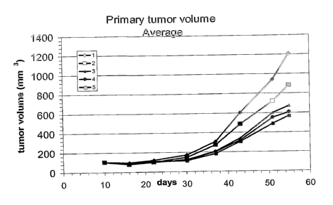
Figure 3 Primary tumor volume measured during treatment until day 55;
Vehicle: 1; antibody 1A 20 mg/kg: 2; antibody 1A 7 mg/kg: 3;
antibody 1A 2 mg/kg: 4.
Figure 4 Inhibition of I125-IGF-I binding to HT29 cells by antibodies 1A, 8
and 23.
Figure 5 Inhibition of I125-IGF-I binding to various human tumor cell lines
by antibodies against hlGF-1R.
Figure 6 Inhibition of I125_IGF-II binding to HT29 cells by antibody IA.
Figure 7 Inhibition of 1125-IGF-II bindingto HT29 cells by antibody ccIR3.
CA 02519113 2005-09-13
WO 2004/087756 PCT/EP2004/003442
-30-
Figure 8 Blockage of IGF-I induced phosphorylation of both IGF-IR and
AKT/PkB.
Figure 9 Downregulation of IGF-IR protein level on tumor cells.
Figure 10 No inhibition of 1125 -insulin binding to 3T3-IR cells by anti-hIGF-
1R antibodies. (MAX w/o Ab: maximal binding of I125-insulin;
MIN: minimal binding after competition with 1 M insulin)
Figure 11 Induction of downregulation of IGF-IR in vivo.
Figure 12 Crossreactivity of antibodies with IGF-IR from other species.
Example 1
Generation of a hybridoma cell line producing anti-IGF-IR antibodies
Culture of hybridomas
Generated HuMab hybridomas were cultured in Hybridoma Express Medium
(PAA Laboratories GmbH, Austria) supplemented with 2 mM L-glutamine
(BioWhittaker) and 4% Origen Cloning Factor (Igen, France) at 37 C and 5% C02;
or in Iscoves Modified Dulbeco's Medium (500 ml: BioWhittaker Europe, Belgium)
supplemented with Fetal Clone Serum (50 ml: Hyclone, Utah), and Origen
Hybridoma Cloning Factor (30 ml: Igen, Gaithersburg MD) at 37 C and 5% CO2.
Immunization procedure of transgenic mice
Ten HCo7 transgenic mice (4 males and 6 females), strain GG2201 (Medarex, San
Jose, CA, USA) were alternatingly immunized with 1x10⁶ NIH 3T3 cells,
transfected
with an expression vector for IGF-IR, and 20 g soluble extracellular domain
of
IGF-IR. Six immunizations were performed in total, three intraperitoneal (IP)
immunizations with the IGF-IR expressing cells and three subcutaneous (SC)
immunizations at the tail base with the recombinant protein. For the first
immunization, 100 l of 1x10⁶ NIH 3T3 IGF-IR cells was mixed with 100 l
complete Freunds' adjuvant (CFA; Difco Laboratories, Detroit, USA). For all
other
CA 02519113 2005-09-13
WO 2004/087756 PCT/EP2004/003442
-31-
immunizations, 100 l of cells in PBS were used or recombinant protein was
mixed
with 100 l incomplete Freunds' adjuvant (ICFA; Difco).
Antigen gpecific ELISA
Anti-IGF-IR titers in sera of immunized mice were determined by antigen
specific
ELISA. IGF-IR soluble extracellular domain at a concentration of 1 g/ml in
PBS
was coated overnight at 4 C, or for two hours at 37 C, to 96 wells plates.
Thereafter,
the wells were blocked with PBSTC (PBS supplemented with 0.05% Tween 0-20 and
2% chicken serum (Gibco BRL)) for 1 hour (h) at room temperature. First tap
sera
were diluted 1/50 in PBSTC, sera from all other taps were pre-diluted 1/100 in
PBSTC and serially diluted up to 1/6400. Diluted sera were added to the wells
and
incubated for 1 h at 37 C. Pre-tap serum was used as negative control. 200
ng/ml
goat anti-human IGF-IR (100 g/ml) was used as positive control. Subsequently,
plates were washed twice with PBST and incubated with horse radish peroxidase
(HRP) -conjugated rat anti-human IgG F(ab')2 (DAKO), diluted 1/2000 in PBSTC
for 1 h at 37 C. Wells were washed twice with PBST and assays were developed
with
freshly prepared ABTS solution (1 mg/ml) (ABTS: 2,2'-azino his (3-
ethylbenzthiazoline-6-sulfonic acid) for 30 minutes at room temperature (RT)
in
the dark. Absorbance was measured at 405 nm.
FAGS analysis
In addition to determination by antigen specific ELISA, anti-IGF-IR titers in
sera of
immunized mice were also determined by FACS analyses. NIH 3T3 IGF-IR cells
and the parental NIH 3T3 cells were incubated with diluted sera for 30 minutes
at
4 C. Alternating IP and SC immunizations were performed at two weeks intervals
starting with an IP immunization. Pre-tap serum (parental NIH 3T3 cells) was
used
as negative control. Initially, 200 ng/ml goat anti-human IGF-IR was used as
positive control. Cells were washed three times in PBS supplemented with 1%
bovine serum albumin and 0.01% azide. Subsequently, cells were incubated with
fluorescein isothiocyanate (FITC) -conjugated antigen binding fragments
(F(ab')2
fragments) of rat anti-human human IgG diluted 1/100 in FACS buffer, for 30
minutes at 4 C. Cells were washed twice in FACS buffer and samples were
analyzed
on a FACSCalibur (Becton Dickinson, Erembodegem-Aalst, Belgium).
CA 02519113 2005-09-13
WO 2004/087756 PCT/EP2004/003442
-32-
Boosting of mice
When serum titers of anti-IGF-IR were found to be sufficient, mice were
additionally boosted twice with 15 iag IGF-IR extracellular domain in 200 pl
PBS
intravenously (i.v.) 4 and 3 days before fusion.
Hybridoma generation
Mice were sacrificed and the spleen and lymph nodes flanking the abdominal
aorta
and vena cava were collected. Fusion of splenocytes and lymph node cells with
the
fusion partner SP 2.0 cells was performed according to standard operating
procedures.
x ELISA
To determine whether hybridomas that resulted from the fusion generate human
antibodies, a x-ELISA was performed. ELISA plates were coated with rat anti-
human IgG K-light chain antibody (DAKO) diluted 1/10000 in PBS by overnight
incubation at 4 C. After discarding the wells, plates were blocked by
incubation
with PBSTC for 1 hour at room temperature. Thereafter, wells were incubated
with
hybridoma culture supernatant, 1/2 diluted in PBSTC. Culture medium 1/2
diluted
in PBSTC was used as negative control, x-light positive mouse serum 1/100
diluted
in PBSTC served as positive control. Subsequently, wells were washed thrice
and
were incubated with HRP-conjugated rat anti-human IgG F(ab')2 (DAKO), diluted
1/2000 in PBSTC for 1 h at 37 C. Wells were washed thrice and assays were
developed with freshly prepared ABTS solution (1 mg/ml) for 30 minutes at
room
temperature (RT) in the dark. Absorbance was measured at 405 nm in an ELISA
plate reader.
Three monoclonal antibodies were prepared.
Antibody 1A: SEQ ID NOS:1 and 2
Antibody 8: SEQ ID NOS: 3 and 4
Antibody 23: SEQ ID NOS: 5 and 6.
CA 02519113 2005-09-13
WO 2004/087756 PCT/EP2004/003442
-33-
Example 2
Determination of the affinity of anti-IGF-IR antibodies to IGF-IR
Instrument: BIACORE 3000
Chip: CM5
Coupling: amine coupling
Buffer: HBS (HEPES, NaCl), pH 7.4,25'C
For affinity measurements anti human Fcy antibodies (from rabbit) have been
coupled to the chip surface for presentation of the antibody against IGF-IR.
IGF-IR
extracellular domain was added in various concentrations in solution.
Association
was measured by an IGF-IR-injection of 3 minutes; dissociation was measured by
washing the chip surface with buffer for 5 minutes. The affinity data for
antibodies
1A, 8 and 23 are shown in Table 1.
Table 1:
Affinity data measured by SPR (BIACORE 3000)
Antibody ka (1/Ms) kd (1/s) KA (1/M) KD (M)
1A 1.18 x 10⁵ 4.68 x 10⁻⁵ 2.52 x 10⁹ 3.97 x 10"10
8 8.18x10⁴ 1.61x10"4 4.98 x 10⁸ 2.01x10⁻⁹
23 8.41 x 10⁴ 1.63 x 10⁻⁵ 5.17 x 10⁹ 1.94 x 10⁻¹⁰
Example 3
WST proliferation assay
To assess the capacity of HuMab antibodies to inhibit IGF-I-induced
proliferation
of IGF-IR expressing cell lines, the WST-1 proliferation assay was performed.
The
IGF-IR expressing cell line NIH 3T3 was cultured for two days in starvation
medium (i.e., regular culture medium with 0.5% FCS instead of 10% FCS), 9x10³
cells per well in 96 wells tissue culture plates in order to bring back the
metabolic
system to a base level. Thereafter, medium was removed and wells were
replenished
with 100 l starvation medium containing the following compounds: 1) 10-9 M
IGF-I; 2) 10-9 M IGF-I plus 10 g/ml protein-A purified HuMab antibody; 3) 10-
9
M IGF-I plus 10 g/ml aIR3. As negative controls, cells were incubated with
starvation medium, as positive control cells were incubated with culture
medium
CA 02519113 2005-09-13
WO 2004/087756 PCT/EP2004/003442
-34-
only. Cells were cultured for an additional two days. Subsequently, 10 l of
WST-1
reagent (Roche Diagnostics GmbH) was added to the wells to detect live cells.
After
2-3 hours, absorbance was measured at 450 nm in an ELISA plate reader. The
percentage of inhibition of proliferation was calculated according to the
following
formula:
OD 100% growth = ODIGF-I - GDstarvation medium
Inhibition of proliferation: (1 - ODsampie / OD100% growth)x100%
Example 4
Three-dimensional growth of tumor cells and overexpression of IGF-I receptor
at
cell-cell-contact (3D culture)
Materials and Methods:
NCI H322M cells were cultured in RPMI media on optical grade glass cover
slides
either at low density or superconfluent to study the effects on IGF-IR surface
expression. In parallel, H322M xenograft tissue isolated from the control
group
(untreated mice) was shock frozen in isopentane and kryosections were cut at 5
m
thickness. Immunofluorescence labelling was performed using a mouse-anti IGF-
IR
monoclonal antibody (aIR3, 5 g/ml) or an antibody according to the invention,
followed by a goat anti-mouse-antibody or a goat anti-mouse antibody labeled
with
Cy3. Specimens were imaged on a Leica SP2 confocal microscope or analyzed by
FACS.
Results:
When H322M cells cultured at high density were imaged by confocal microscopy
it
became apparent that IGF-IR clustered specifically at the sites of cell-cell
contact.
When compared to H322M cells grown in vivo, i.e. xenograft tissue, there was a
striking similarity to densely packed in vitro cultures as far as the
organization of
surface IGF-I receptors was concerned.
The upregulation of surface IGF-I receptor in superconfluent cultures of H322M
cells was also quantified by FACS. IGF-I receptor surface expression increased
more
than 10 fold when cells were grown under high density conditions compared to
low
density without significant cell-cell contacts.
CA 02519113 2005-09-13
WO 2004/087756 PCT/EP2004/003442
-35-
Other tumor cells such as HT29, MDA231 and MCF-7 showed a similar behavior,
indicating that upregulation of IGF-I receptors on the cell surface upon
establishing
cell-cell contact sites is not an unique feature of H322M cells but appears to
be a
general property of a more tissue like organization that is also found in vivo
(Fig.
1).
Growth inhibition of H322M tumor cells expressing IGF-IR in 3D culture under
treatment with antibody 1A
Materials and Methods:
H322M cells were cultured in RPMI1640/10% NCS media on poly-HEMA (poly(2-
hydroxyethylmethacrylate)) coated dishes to prevent adherence to the plastic
surface. Under these conditions H322M cells form dense spheroids that grow
three
dimensionally (a property that is called anchorage independence). These
spheroids
represent the three dimensional histoarchitecture and organization of solid
tumors
in situ. Spheroid cultures were incubated for 9 days in the presence of
increasing
amounts of antibodies from 0-10 g/ml. Two nonspecific antibodies (antibody
against HBV and against E25) were used as a negative control. The WST
conversion
assay was used to measure growth inhibition.
Results:
When H322M spheroid cultures were treated with different concentrations of
antibody 1A (0.32-10 g/ml) a dose dependent inhibition in growth could be
observed, while the control antibodies against HBV and E25 (anti-IgE) had
little or
no effect. The reduction in WST for antibody IA is therefore primarily due to
a
reduced proliferation of the cells (Fig. 2).
Example 5
Determination of pharmacokinetic properties
Antibody 1A was administered in two different pharmacokinetic studies. For the
first study, the antibody was administered formulated as a potassium phosphate
buffer solution. For the second study, the antibody was administered
formulated as
a sodium chloride/ histidine solution.
CA 02519113 2005-09-13
WO 2004/087756 PCT/EP2004/003442
-36-
5a) First animal study
Female NMRI mice were used (18 - 23 g body weight). Antibody IA was given as a
solution (KPO4) for i.v., and i.p. administration route.
Doses:
10 mg/kg i.v. Drug concentration: lmg/mL. Administered volume: 10 mL/kg.
mg/kg i.p. Drug concentration: lmg/mL. Administered volume: 10 mL/kg.
Single dose administration.
Plasma samples were subsequently analysed for plasma levels of the compound
using a human IgG-ELISA method.
10 Reagents:
Antibodies: Capture antibody: polyclonal rabbit antibody against human kappa-
light chains IgG (Dako, Code No. A0191)
Detection antibody: polyclonal rabbit antibody against human IgG, conjugate
with
Horse raddish peroxidase (DAKO, Code No. P0214)
Assay procedure:
Coating of the microtiter plate:
Step
- dilute capture antibody 1:10000 with 100mM Na-Carbonate, pH=9,6
- add 100 l of this solution (Step 1) to each well
- incubate the plate at 4 C over night (12h)
- remove solvents of each well
- wash 3 times with 300 l/well PBST
Adding animal sample and calibration sample, respectively dilute animal sample
(range 1 to 10 up to 1 to 200000) and calibration sample, respectively
(concentration 0.625; 1.25; 2.5; 5; 10; 20 and 40 ng/ml human IgG) with 3%
BSA/PBST:
- add 100 1 animal/calibration sample to each well
- incubate for 1 h at room temperature (22 C)
CA 02519113 2005-09-13
WO 2004/087756 PCT/EP2004/003442
-37-
- wash 3 times with 300 l/well PBST
Detection:
- dilute detection antibody 1:2000 with 3% BSA/PBST
- add 100 l to each well
- incubate for 1 h at room temperature (22 C)
- wash 3 times with 300 l/well PBST
- add 100 l ABTS -solution to each well
- after ca. 10 min stop the color reaction with 50 V1/well 0.5 M oxalic acid
- measure the extinction at 405nm
CA 02519113 2005-09-13
WO 2004/087756 PCT/EP2004/003442
-38-
PK Parameters:
Table 2:
Pharmacokinetic parameters of antibody 1A fomulated
as a potassium phosphate buffer
Parameter units
Species Mouse Mouse
Strain NMRI NMRI
Gender female female
Formulation Phosphate buffer Phosphate buffer
Dose mg/kg 10.0 10.0
Administration route i.v. i.p.
CMAX ng/mL 208000 68800
CMAX_NORM ng/mL / mg/kg 20800 6880
TMAX h 0 96
AUC_0_LST h.ng/mL 36300000 32300000
AUC_0_LST_NORM h.ng/mL / mg/kg 3630000 3230000
AUC_0_INF h.ng/mL 40200000 36300000
AUC_0_INF_NORM h.ng/mL / mg/kg 4020000 3630000
PCT_AUC_EXTRA % 9.75 11.00
MRT_LST h 324 348
MRT_INF h 437 475
CL_TOTAL mL/min/kg 0.00414
VZ L/kg 0.11
VSS L/kg 0.1
HALFLIFE_Z h 308 (=12.8 days) 324 (=13.5 days)
F % 100.0 89.0
5b) Second animal study
Animals:
Female NMRI mice were used (21 - 28 g body weight). Antibody 1A was given as a
solution (histidine/sodium chloride) for i.v., and i.p. administration route.
CA 02519113 2005-09-13
WO 2004/087756 PCT/EP2004/003442
-39-
Doses:
mg/kg i.v. Drug concentration: 1 mg/mL. Administered volume: 10 mL/kg.
2 mg/kg i.p. Drug concentration: 0.2 mg/mL. Administered volume: 10 mL/kg.
mg/kg i.p. Drug concentration: 2 mg/mL. Administered volume: 10 mL/kg.
5 Single dose administration.
Plasma samples were subsequently analysed for plasma levels of the compound
using a human IgG-ELISA method. Calibration was done using the antibody IA for
preparing calibration samples.
Reagents and assay procedure: see Example 5a
CA 02519113 2005-09-13
WO 2004/087756 PCT/EP2004/003442
-40-
PK Parameters:
Table 3:
Pharmacokinetic parameters of antibody 1A fomulated as a
histidine/sodium chloride solution
Species Mouse Mouse Mouse
Strain NMRI NMRI NMRI
Gender female female female
Formulation solution solution solution
(NaCI/histidine) (NaCI/histidine) (NaCI/histidine)
Dose mg/kg 10 2 20
Administration route i.v. i.p. i.p.
CMAX ng/mL 222000 27900 186000
CMAX_NORM ng/mL / 22200 14000 9300
mg/kg
TMAX h 0 24 24
AUC_0_LST h.ng/mL 30500000 4920000 59000000
AUC_0_LST_NORM h.ng/mL / 3050000 2460000 2950000
mg/kg
AUC_0_INF h.ng/mL 36700000 6910000 72100000
AUC_0_INF_NORM h.ng/mL / 3670000 3460000 3610000
mg/kg
PCT_AUC_EXTRA % 16.90 28.80 18.20
MRT_LST h 247 217 239
MRT_INF h 381 513 389
CL_TOTAL mL/min/kg 0.00454
CL_TOTAL_CTG L,M,H
CL_ORAL mL/min/kg 0.00482 0.00462
VZ L/kg 0.102
VZ_ORAL (Vz/F) L/kg 0.166 0.108
VSS L/kg 0.1
VSS_CTG L,M,H L
HALFLIFE_Z h 260.0 399.0 270.0
IF % 100.0 80.7 96.7
CA 02519113 2005-09-13
WO 2004/087756 PCT/EP2004/003442
-41-
Abbreviations:
Abbreviation Meaning
CMAX Cmax
CMAX NORM Cmax dose-normalized
TMAX Tmax
AUC_0_INF AUC extrapolated
AUC_0_LST AUC observed
AUC_0_INF_NORM AUC extrapolated, normalized
AUC_0_LST_NORM AUC observed, normalized
PCT_AUC_EXTRA percentage AUC extrapolated
CL-TOTAL Total Clearance
VSS Steady state distribution volume
VZ Terminal distribution volume
MRT_INF Mean residence time (extrapolated)
MRT_LST Mean residence time (observed)
HALFLIFE Z Terminal half-life
F Bioavailability (i.v. 100%)
Example 6
Pharmacodynamic testing of recombinant anti-IGF-IR antibody 1A
The effects of the antibody were investigated in vivo. Tumors were induced in
athymic nude mice according to established methods. Human H322M cells were
coinjected together with Matrigel subcutaneously into 6-7 week-old athymic
nude
mice (nu/nu). For that purpose, 5 x 10⁶ H322M cells were concentrated in 100
l
culture medium and mixed with 100 l Matrigel . 200 l of this mixture were
injected into the right flanks of the mice. Treatment was initiated when
induced
tumors reached an average volume of 125 mg. Tumor volume was calculated by
measuring tumor diameters with Vernier calipers twice a week according to the
formula
tumor volume [mg] _ (length x (width)2
(Gallicchio, M.A., et al., Int. J. Cancer 94 (2001) 645-651).
CA 02519113 2005-09-13
WO 2004/087756 PCT/EP2004/003442
-42-
All antibodies were administered intraperitoneally (i.p.) at 10 ml/kg.
After tumors had grown to an average volume of 100 mg the antibody was
administered twice a week i.p. at 20 mg/kg, 7 mg/kg and 2 mg/kg twelve times
starting treatment with a doubled loading dose given once on the first day of
the
treatment period. All three doses of the antibody had an effect on primary
tumor
volume. Figure 3 shows the tumor size in relation to the various treatments
over
time. The experiment demonstrates that blocking of the IGF-IR axis by antibody
1A
results in good antitumor effects.
Example 7
Inhibition of IGF-I and IGF-II binding to tumor cells expressing IGF-IR
In order to determine the ability of the antibody of the invention to block
binding
of the ligands IGF-I and IGF-II to the IGF-I receptor (IGF-IR), competition
experiments with radioactively labeled ligand peptides were performed.
Human tumor cells (HT29, NCI H322M, 0.5 to 1 x 10⁵/ml) were plated in RPMI
1640 medium (PAA, Cat. No. E15-039) supplemented with 2 mM L-Glutamin, lx
non-essential amino acids (Gibco, Cat. No. 11140-035), 1 mM sodium pyruvate
(Gibco, Cat. No. 11360-039) and 10% heat inactivated FCS (PAA, Cat. No. A15-
771). Six bottles in the T175 format were inoculated with 20 ml cells in the
respective medium for each experiment and cultivated for two days at 37 C and
5%
CO2 to obtain confluent cell monolayers.
To collect individual cells, 2 ml of lx Trypsin/EDTA (Gibco, Cat. No. 25300-
054)
per T175 flask were added and detachment of cells monitored with a Zeiss
Axiovert25 microscope. The cells were collected and medium with 10% FCS as
described before was added to a total volume of 50 ml. Cells were reisolated
by
centrifugation for 10 minutes at 1000 rpm (Heraeus sepatech, Omnifuge 2.0 RS)
and resuspended in 50 ml of binding buffer (120 mM NaCl, 5 mM KCI, 1.2 mM
MgSO4, 1 mM EDTA, 10 mM D(+)glucose, 15 mM NaAc, 100 mM Hepes pH 7.6,
1% BSA). Cells were counted, reisolated by centrifugation and adjusted with
binding buffer to 1 x 10⁶ cells/ml.
CA 02519113 2005-09-13
WO 2004/087756 PCT/EP2004/003442
-43 -
I125-labeled IGF-I and IGF-II peptides (Amersham, -2000 Ci/mmol, Cat. No.
IM172 and IM238), solubilized in 0.1% CH3COOH, were diluted in binding buffer
to a final activity of 4 x 10⁵ counts/(minute x ml). 75 l of antibody at the
specified
concentrations together with 25 l of prediluted I125-labeled IGF-I or IGF-II
peptide
was added to 200 l of cell suspension and incubated for 3,5 h at 4 C. Cells
were
reisolated by centrifugation for 5 minutes at 2000 rpm (Eppendorf, 5415C) and
supernatant removed. After washing two times in 1 ml binding buffer, cells
were
resuspended in 1 ml binding buffer and transferred to scintillation tubes. The
amount of radioactive peptide bound to the cell surface receptors was measured
on
a scintillation counter.
The resulting IC50 curves demonstrating the ability of the antibody to inhibit
binding of IGF-I and IGF-II peptide to the IGF-I receptor are shown in Figs.
4, 5
and 6. The results for antibody aIR3 are shown in Figs. 5 and 7.
Example 8
Antibody competition assay for IGF-IR binding
For an epitope mapping of anti-IGF-IR monoclonal antibodies a similar format
as
for affinity measurement (Example 2) was selected. Antibody la was bound to
anti
human Fcy antibodies (from rabbit) which were amine-coupled to the chip
surface.
To determine whether another antibody was directed against an IGF-IR epitope
overlapping with the epitope recognized by antibody la, IGF-IR was pre-
incubated
with this antibody under saturating conditions in solution. After incubation
for at
least 30 minutes at RT the IGF-IR with pre-bound antibody was injected to the
flow
cell and binding to the reference antibody la at the chip surface was
monitored. In
case of overlapping epitope recognition of test antibody and reference
antibody 1A
binding of IGF-IR was inhibited by at least 10% compared to the binding signal
of
IGF-IR alone (100% binding signal) at a standard concentration of 50 nM. In
case
of additional antibody binding the IGF-IR binding signal was increased by at
least
10% indicating the recognition of an independent binding epitope on IGF-IR by
the test antibody.
CA 02519113 2005-09-13
WO 2004/087756 PCT/EP2004/003442
-44-
Example 9
Inhibition of IGF-I mediated phosphorylation of IGF-IR and Akt/PKB
In order to determine the ability of the antibody of the invention to inhibit
activation and phosphorylation of the IGF-I receptor (IGF-IR), competition
experiments were performed with IGF-I peptide and subsequent Western blotting
analysis with antibodies specific for phosphorylated tyrosine.
Human tumor cells (HT29, NCI H322M, 5 x 10⁴/ml) were plated in RPMI 1640
medium (PAA, Cat. No. E15-039) supplemented with 2 mM L-Glutamin, lx non-
essential aminoacids (Gibco, Cat. No. 11140-035), 1 mM sodium pyruvate (Gibco,
Cat. No. 11360-039) and 0.5% heat inactivated FCS (PAA, Cat. No. A15-771). For
determination of IC50 values, 12 well plates were inoculated with 1 ml cells
in the
respective medium for each experiment and cultivated for two days at 37 C and
5%
C02-
After 48 hours of cultivation with low serum medium, the medium was carefully
removed and replaced by different concentrations of antibody diluted in the
respective medium. After 5 minutes incubation at 37 C and 5% CO2 IGF-I peptide
was added at a final concentration of 2 nM and cells were again incubated for
10
minutes under the conditions mentioned above. The medium was carefully
removed by aspiration and 100 1 of cold lysis buffer was added per well (50mM
Hepes pH 7.2, 150 mM NaCl, 1mM EGTA, 10% glycerol, 1% Triton -X100,
100mM NaF, 10 mM Na4P2O7, Complete protease inhibitor). The cells were
detached using a cell scraper (Corning, Cat. No. 3010) and well contents
transferred to Eppendorf reaction tubes. Cell fragments were removed by
centrifugation for 10 minutes at 13000 rpm and 4 C and half of the supernatant
was
added to 2x Laemmli sample buffer in a 1:1 (v/v) ratio. For
immunoprecipitation of
IGF-IR, the remaining supernatant of cell lysates underwent a clearifying spin
(10
minutes at 13000 rpm and 4 C) right before 1 l of primary antibody was added
(C-20, Santa Cruz Biotechnologies, mAb 24-55, GroPep). After 2 hours
incubation
at 4 C in a rotating Eppendorf reaction tube, 25 l Protein G Sepharose beads
(Amersham Biosciences, Cat. No. 17-0618-01) were added followed by another
incubation step of 1 hour at 4 C. The beads with bound antibody-protein-
complexes were isolated by centrifugation (1 minute at 2000 rpm and 4 C) and
washed three times with wash buffer (lysis buffer with only 0.1% Triton -
X100).
CA 02519113 2005-09-13
WO 2004/087756 PCT/EP2004/003442
-45-
After boiling the beads in Laemmli sample buffer, cellular proteins were
separated
by SDS-PAGE and transferred to a nitrocellulose membrane (PROTRAN BA 85,
Schleicher&Schuell) by semi-dry Western blotting.
A phosphotyrosin specific antibody (<P-Tyr>, Upstate, clone 4G10, Cat. No. 05-
321) was used to determine phosphorylation status of immunopurified IGF-IR.
For
the detection of phosphorylated Akt/PKB an antibody with specificity for
phosphorylated Ser473 (<P-PkB>, Cell Signalling, Cat. No. 9271) was applied.
The observed blockage of IGF-I induced phosphorylation of both IGF-IR and
Akt/PKB is shown in Fig. 8.
Example 10
Induction of antibody mediated downregulation of IGF-IR in-vitro
In order to detect effects of the antibody of the invention on the amount of
IGF-I
receptor (IGF-IR) in tumor cells, time-course experiments and subsequent
western-
blotting analysis with IGF-IR specific antibodies were performed.
Human tumor cells (H460, QG56, MCF-7, 5 x 10⁴ cells/ml) were plated in RPMI
1640 medium (PAA, Cat. No. E15-039) supplemented with 2 mM L-Glutamin, lx
non-essential aminoacids (Gibco, Cat. No. 11140-035), 1 mM sodium pyruvate
(Gibco, Cat. No. 11360-039) and 10% heat inactivated FCS (PAA, Cat. No. A15-
771). For each incubation period one 12 well plate was inoculated with 1 ml
cells in
the respective medium for each experiment and cultivated for 24 hours at 37 C
and
5% C02-
The medium was carefully removed and replaced by different concentrations of
antibody diluted in the respective medium. In two control wells, medium was
replaced by either medium without antibody or medium with a control antibody
(AB-1, Oncogene, Cat. No. GR11). Cells were incubated at 37 C and 5% CO2 and
individual plates were taken out for further processing after 15 minutes, 24
hours
and 48 hours.
The medium was carefully removed by aspiration and 100 l of cold lysis buffer
was
added per well (50mM Hepes pH 7.2, 150 mM NaCl, 1mM EGTA, 10% glycerol,
CA 02519113 2005-09-13
WO 2004/087756 PCT/EP2004/003442
-46-
1% Triton -X100, 100mM Nall, 10 mM Na4P2O7, Complete protease inhibitor).
The cells were detached using a cell scraper (Corning, Cat. No. 3010) and well
contents transferred to Eppendorf reaction tubes. Cell fragments were removed
by
centrifugation for 10 minutes at 13000 rpm and 4 C and the supernatant was
added
to 2x Laemmli sample buffer in a 1:1 (v/v) ratio. Cellular proteins were
separated by
SDS-PAGE and transferred to a nitrocellulose membrane (PROTRAN BA 85,
Schleicher&Schuell, Cat. No. 10 401196) by semi-dry western-blotting.
An antibody specific for IGF-IR (<IGF-IR>, C-20, Santa Cruz Biotechnologies,
Cat.
No. sc-713) was used to determine protein levels of IGF-IR.
Downregulation of IGF-IR induced by the antibody of the invention after less
than
24 hours after addition of the antibody was observed. The results are shown in
Fig.
9.
Example 11
Inhibition of insulin binding to 3T3-cells expressing human insulin receptor
In order to determine whether the antibody of the invention also blocks
binding of
insulin to the insulin receptor (IR), competition experiments were performed
with
a radioactively labeled ligand peptide.
NIH-3T3 cells (1 x 10⁵/ml) expressing recombinantly high numbers (>105) human
IR were plated in MEM Dulbecco medium (DMEM) with high glucose (PAA, Cat.
No. E15-009) supplemented with 2mM L-Glutamin (Gibco, Cat. No. 25030-024)
and 10% heat inactivated FCS (PAA, Cat. No. A15-771). Six bottles in the T175
format were inoculated with 20 ml cells in the respective medium for each
experiment and cultivated for two days at 37 C and 5% CO2 to obtain confluent
cell
monolayers.
To collect individual cells, 2 nil of lx Trypsin/EDTA (Gibco, Cat. No. 25300-
054)
per T175 flask were added and detachment of cells monitored with a microscope.
The cells were collected and medium with 10% FCS as described before was added
to a total volume of 50 ml. Cells were reisolated by centrifugation for 10
minutes at
1000 rpm and resuspended in 50 ml of binding buffer (120 mM NaCl, 5 mM KC1,
1.2 mM MgSO4i 1 mM EDTA, 10 mM D(+)glucose, 15 mM NaAc, 100 mM Hepes
CA 02519113 2005-09-13
WO 2004/087756 PCT/EP2004/003442
-47-
pH 7.6, 1% BSA). Cells were counted, reisolated by centrifugation and adjusted
with binding buffer to 1 x 10⁶ cells/ml.
I125-labeled insulin peptide (Amersham, Cat. No. IM166, -2000 Ci/mmol),
solubilized in 0.1% CH3COOH, were diluted in binding buffer to a final
activity of
4x10⁵ counts/(minute x ml). 75 i1 of antibody together with 25 l of
prediluted I125-
labeled insulin peptide was added to 200 l of cell suspension (final antibody
concentration 200 nM) and incubated for 3,5 h at 4 C. Cells were reisolated by
centrifugation for 5 minutes at 2000 rpm and supernatant was removed. After
washing two times in 1 ml binding buffer, cells were resuspended in 1 ml
binding
buffer and transferred to scintillation tubes. The amount of radioactive
peptide
bound to the cell surface receptors was measured on a scintillation counter.
The results demonstrate that the antibody of the invention does not interfere
with
binding of insulin ligand to the insulin receptor (Fig. 10).
Example 12
Induction of receptor down-regulation in different tumor types
Human tumors (NCI H322M or NCI H460) were induced in nude mice and
treated with the antibody of the invention as described in Example 6. After
termination of the experiment, the tumors were extracted and homogenized under
liquid nitrogen. Cold lysis buffer was added (50mM Hepes pH 7.2, 150 mM NaCI,
1mM EGTA, 10% glycerol, 1% Triton-X100, 100mM NaF, 1 mM Na3VO4, 10 mM
Na4P2O7i Complete protease inhibitor, ImM PMSF) in a buffer-volume to tumor-
weight ratio of 3:1 and thoroughly mixed with the thawing tumor homogenate.
After solubilizing the tissue for 15 minutes on ice, insoluble fragments were
removed by centrifugation for 10 minutes at 13000 rpm and 4 C. The protein
concentration of the samples was determined with the Micro BCA Reagents
(Pierce) and lysis buffer was added to adjust equal concentrations. Part of
the
supernatant was added to 2x Laemmli sample buffer in a 1:1 (v/v) ratio.
Cellular
proteins were separated by SDS-PAGE and transferred to a nitrocellulose
membrane (PROTRAN BA 85, Schleicher&Schuell, Cat. No. 10 401196) by semi-
dry western-blotting. An IGF-IR specific antibody (C-20, Santa Cruz
Biotechnologies, Cat. No. sc-713) was used to detect IGF-IR.
CA 02519113 2005-09-13
WO 2004/087756 PCT/EP2004/003442
-48-
A dramatic decrease of IGF-IR levels in all tumors treated with the antibody
of the
invention (Fig. 11) corresponding to the results of the in vitro
downregulation
experiments was observed.
Example 13
Antibody crossreactivity with IGF-IR from rat, mouse, Marmoset and
Cynomolgus
The antibody of the invention was tested for its ability to bind to the IGF-I
receptor
of other species. Immunoprecipitation experiments were performed with the
antibody of the invention and tissue or cell lysates from different animal
species.
Frozen animal tissue was homogenized under liquid nitrogen. Cold lysis buffer
was
added (50mM Hepes pH 7.2, 150 mM NaCl, 1mM EGTA, 10% glycerol, 1%
Triton-X100, 100mM NaF, 1 mM Na3VO4, 10 mM Na4P2O7, Complete protease
inhibitor, 1mM PMSF) in a buffer-volume to tissue-weight ratio of 3:1 and
thoroughly mixed with the thawing tissue homogenate. After solubilizing the
tissue
for 15 minutes on ice, insoluble fragments were removed by centrifugation for
10
minutes at 13000 rpm and 4 C. The protein concentration of the samples was
determined with the Micro BCA Reagents (Pierce) and lysis buffer was added to
adjust equal concentrations. Animal cells were solubilized according to the
protocol
for tumor cell lines (Example 10).
For immunoprecipitation of IGF-IR, cell and tissue lysates (1 mg total
protein)
underwent a clarifying spin (10 minutes at 13000 rpm and 4 C) right before 2
g of
primary antibody 1A was added. The same experiment was also performed with an
unrelated human control antibody directed against hepatitis B virus protein
(HBV)
as a measure for unspecific binding. After 2 hours incubation at 4 C in a
rotating
eppendorf reaction tube, 25 l Protein G Sepharose beads (Amersham
Biosciences, Cat. No. 17-0618-01) were added followed by another incubation
step
of 1 hour at 4 C. The beads with bound antibody-protein-complexes were
isolated
by centrifugation (1 minute at 2000 rpm and 4 C) and washed three times with
wash buffer (lysis buffer with only 0.1% Triton -X100). After boiling the
beads in
Laemmli sample buffer, cellular proteins were separated by SDS-PAGE and
transferred to a nitrocellulose membrane (PROTRAN BA 85, Schleicher&Schuell,
Cat. No. 10 401196) by semi-dry western-blotting.
CA 02519113 2005-09-13
WO 2004/087756 PCT/EP2004/003442
-49-
An IGF-IR specific antibody with broad species cross-reactivity (C-20, Santa
Cruz
Biotechnologies, Cat. No. sc-713) was used to determine the levels of IGF-IR
immunoprecipitated with the antibody of the invention.
Cross-reactivity with IGF-IR from Cynomolgus and Marmoset monkey (Fig. 12),
but no cross-reactivity to the rat or mouse IGF-I receptor was observed.
Example 14
Clq Binding ELISA
Introduction
To determine the ability of antibodies according to the invention to fix Clq
an
ELISA approach was used. Clq is part of the adaptive immune system and, upon
binding to immune complexes, triggers the sequential activation of several
zymogens. The enzymes in turn, cause the cleavage of C3 molecules, which can
result in the onset of inflammatory reactions, opsonization of foreign or
aberrant
particles and lysis of cell membranes.
In principle, the ELISA plate is coated with concentration ranges of the
antibody, to
which human Clq is added. Clq binding is detected by an antibody directed
against human Clq followed by a peroxidase-labeled conjugate.
Materials and methods
Antibody 1A, 8 and 23 and control antibodies were tested in concentrations of
10,
5, 1 and 0.5 g/ml. Table 1 shows the specificities of the samples tested. As
a
negative control a human IgG4 (CLB, stock 0.5 1g/ml), that binds Clq very
weakly,
was used. Human IgG1 was incorporated as positive control. Human Clq stock
solution with a concentration of 2 g/ml was used. For the detection of Clq a
rabbit
antibody directed against Clq (Dako) and an anti-rabbit IgG antibody,
conjugated
with horseradish peroxidase (Sigma) were used.
CA 02519113 2005-09-13
WO 2004/087756 PCT/EP2004/003442
-50-
Calculations and curve fitting
Calculations concerning maximum binding (Bmax) of the HuMab tested were
determined using nonlinear regression curve fitting (one site binding) using
Graphpad Prism software.
Results
The antibodies according to the invention show dose dependent binding of human
Clq protein. The optical density at 405 nm (OD 405 nm) was plotted against the
HuMab concentrations and the curves were fitted using nonlinear regression.
Best
fit values for maximum binding (Bmax) are listed in Table 4, as are the
correlation
coefficient of the curve (R2) and the standard deviation for each value. The
lowest
correlation coefficient had a value of 0.950 (IgG4). With a maximum binding of
0.279, human IgG4 (negative control) shows minimum binding of C1q. Positive
controls IgG1 and IgG3 both bind C1q, as shown by a maximum binding of 1.729
and 2.223, respectively.
Table 4:
Maximum binding (Bmax) of the HuMab tested in the Clq binding ELISA (n=3)
Best fit values Bmax Standard Goodness of fit Standard
deviation R2 deviation
Bmax R2
IgGl 1.729 0.166 0.983 0.010
IgG3 2.223 0.947 0.995 0.005
IgG4 0.279 0.280 0.950 0.041
Antibody IA 1.670 0.601 0.988 0.005
Antibody 8 1.954 0.131 0.978 0.009
Antibody 23 1.872 0.558 0.990 0.004
The correlation coefficient (R2) and standard deviation and are also listed.
Compared to the Clq binding of human IgG4 (negative control, with an O.D. of
0.279), all antibodies tested are equally capable of fixing Clq.
CA 02519113 2005-09-13
WO 2004/087756 PCT/EP2004/003442
-51-
Example 15
Determination of antibody mediated effector functions by anti-IGF-IR HuMabs
In order to determine the capacity of the generated HuMab antibodies to elicit
immune effector mechanisms, complement dependent cytotoxicity (CDC) and
antibody-dependent cell cytotoxdcity (ADCC) studies were performed.
To study CDC (National Cancer Institute, lung adenocarcinoma cell line),
H322M,
H460 and NIH 3T3 cells (2-6 x 10⁶) were labeled with 100 Ci 51Cr for 45-120
minutes (Amersham Pharmacia Biotech, UK, Cat CJS11). After labeling the cells
were washed twice with 40 ml PBS and spun for 3 minutes at 1500 rpm. The cells
were then plated 5,000 per well in a round bottom plate, in a volume of 50 l.
Antibodies were added at a final concentration ranging from 25-0.1 g/ml in a
volume of 50 l cell culture medium to 50 l cell suspension and incubated for
30-
60 minutes. After incubation excess antibody was removed by washing twice with
PBS. 100 1 of active or inactive (30 minutes at 56 C) pooled human serum,
guinea
pig, rabbit or nude mice serum diluted between 1/3-1/30 was added, and the
cells
were incubated for 3 hours, after which the cells were spun down at 1500 rpm
for 3
minutes. 100 l of the supernatant was harvested, transferred to polypropylene
tubes and counted in ay-counter.
To study the effects of the antibodies in ADCC, H322M, H460 and NIH 3T3 or
other suitable IGF-IR expressing cells (2-6 x 10⁶) were labeled with 100 Ci
51Cr for
45-120 minutes (Amersham Pharmacia Biotech, UK, Cat CJS11), washed twice
with 40 ml of PBS and spun for 3 minutes at 1500 rpm. The cells were plated
5,000
per well in a round bottom plate, in a volume of 50 l. HuMab antibodies were
added at a final concentration ranging from 25-0.1 g/ml in a volume of 50 l
cell
culture medium to 50 l cell suspension and incubated for 15 minutes.
Subsequently, 50 l of effector cells, freshly isolated PBMC or purified
effector cells
from buffycoats, were added at an E:T ratio in the range of from 100:1 to 5:1.
The
plates were centrifuged for 2 minutes at 500-700 rpm, and incubated overnight
at
37 C. After incubation the cells were spun down for 3 minutes at 1500 rpm and
100
l of supernatant was harvested, transferred to polypropylene tubes and counted
in
a y-counter.
CA 02519113 2005-09-13
WO 2004/087756 PCT/EP2004/003442
-52-
The magnitude of cell lysis by CDC or ADCC is expressed as % of the maximum
release of radioactivity from the target cells lysed by detergent corrected
for
spontaneous release of radioactivity from the respective target cells.
CA 02519113 2005-09-13
WO 2004/087756 PCT/EP2004/003442
-53-
List of References
Adams, T.E., et al., Cell. Mol. Life Sci. 57 (2000) 1050-1063
Aplin, J.D., and Wriston, J.C. Jr., CRC Crit. Rev. Biochem. (1981) 259-306
Arteaga, C.L., et al., Breast Cancer Res. Treatment 22 (1992) 101-106
Ausubel, F., et al., ed. Current Protocols in Molecular Biology, Greene
Publishing
and Wiley Interscience, New York (1987)
Barnes, L.M., et al., Biotech. Bioeng. 73 (2001) 261-270
Barnes, L.M., et al., Cytotechnology 32 (2000) 109-123
Benini, S., et al., Clin. Cancer Res. 7 (2001) 1790-1797
Berge, S.M., et al., J. Pharm. Sci. 66 (1977) 1-19
Bruggemann, M., et al., J. Exp. Med. 166 (1987) 1351-1361
Brunetti, A., et al., Biochem. Biophys. Res. Commun. 165 (1989) 212-218
Carter, P., et al., Proc. Natl. Acad. Sci. USA 89 (1992) 4285-4289
Chen, J., et al., EMBO J. 12 (1993) 821-830
Chen, f., et al., International Immunology 5 (1993) 647-656
Choi, T.K., et al., Nature Genetics 4 (1993) 117-123
Delafontaine, P., et al., J. Mol. Cell. Cardiol. 26 (1994) 1659-1673
Dricu, A., et al., Glycobiology 9 (1999) 571-579
Durocher, Y., et al., Nucl. Acids. Res. 30 (2002) E9
Edge, A.S., et al. Anal. Biochem. 118 (1981) 131-137
Fishwild, D.M., et al., Nat. Biotechnol. 14 (1996) 845-851
Forsayeth, J.R., et al., Proc. Natl. Acad. Sci. USA 84 (1987) 3448-3451
Gallicchio, M.A., et al., Int. J. Cancer 94 (2001) 645-651
Geisse, S., et al., Protein Expr. Purif. 8 (1996) 271-282
Gustafson, T.A., and Rutter, W.J., J. Biol. Chem. 265 (1990) 18663-18667
Hailey, J., et al., Mol. Cancer Ther. 1 (2002) 1349-1353
Harding, F., and Lonberg, N., Ann. N. Acad. Sci 764 (1995) 536-546
Hoyne, P.A., et al., FEBS Lett. 469 (2000) 57-60
Johnson, G., and Wu, T.T., Nucleic Acids Res. 28 (2000) 214-218
Jones, P., et al., Nature 321 (1986) 522-525
Kabat et al., Sequences of Proteins of Immunological Interest, 5th Ed. Public
Health
Service, National Institutes of Health, Bethesda, MD. (1991)
Kalebic, T., et al., Cancer Res. 54 (1994) 5531-5534
Kanter-Lewensohn, L., et al., Melanoma Res. 8 (1998) 389-397
Kaufman, R.J., Mol. Biotechnol. 16 (2000) 151-161
CA 02519113 2005-09-13
WO 2004/087756 PCT/EP2004/003442
-54-
Kull, F.C. Jr., et al. J. Biol. Chem. 258 (1983) 6561-6566
LeRoith, D., et al., Endocrin. Rev. 16 (1995) 143-163
Li, S.L., et al., Biochem. Biophys. Res. Commun. 196 (1993) 92-98
Li, S.L., et al., Cancer Immunol. Immunother. 49 (2000) 243-252
Lonberg, N., and Huszar, D., Intern. Rev. Immunol. 25 (1995) 65-93
Lonberg, N., et al., Nature 368 (1994) 856-859
Lonberg, N., Handbook of Experimental Pharmacology 113 (1994) 49-101
Love, T.W., et al., Methods Enzymol. 178 (1989) 515-527
Makrides, S.C., Protein Expr. Purif. 17 (1999) 183-202
Morgan, D.O., and Roth, R.A., Biochemistry 25 (1986) 1364-1371
Morrison, S.L., et al., Proc. Natl. Acad Sci. USA 81 (1984) 6851-6855
Neuberger, M.S., et al., Nature 314 (1985) 268-270
Norderhaug, L., et al., J. Immunol. Methods 204 (1997) 77-87
O'Brien, R.M., et al., EMBO J. 6 (1987) 4003-4010
Orlandi, R., et al., Proc. Natl. Acad. Sci. USA 86 (1989) 3833-3837
Pessino, A., et al., Biochem. Biophys. Res. Commun. 162 (1989) 1236-1243
Prigent, S.A., et al., J. Biol. Chem. 265 (1990) 9970-9977
Queen, C., et al., Proc. Natl. Acad. Sci. USA 86 (1989)10029-10033
Riechmann, L., et al., Nature 332 (1988) 323-327
Rohlik, Q.T., et al., Biochem. Biophys. Res. Comm. 149 (1987) 276-281
Schaefer, E.M., et al., J. Biol. Chem. 265 (1990) 13248-13253
Schlaeger, E.-J., and Christensen, K., Cytotechnology 30 (1999) 71-83
Schlaeger, E.-J., in J. Immunol. Methods 194 (1996) 191-199,
Scotlandi, K., et al., Cancer Gene Ther. 9 (2002) 296-307
Scotlandi, K., et al., Int. J. Cancer 101 (2002) 11-16
Sojahr, H.T., and Bahl, O.P., Arch. Biochem. Biophys. 259 (1987) 52-57
Soos, M.A., et al., Biochem. J. 235 (1986) 199-208
Soos, M.A., et al., J. Biol. Chem. 267 (1992) 12955-12963
Soos, M.A., et al., Proc. Natl. Acad. Sci. USA 86 (1989) 5217-5221
Stella et al., "Prodrugs: A Chemical Approach to Targeted Drug Delivery,"
Directed
Drug Delivery, Borchardt et al., (ed.), pp. 247-267, Humana Press
(1985)
Surinya, K.H., et al., J. Biol. Chem. 277 (2002) 16718-16725
Taylor, L., et al., Int. Immunol. 6 (1994) 579-591
Taylor, L., et al., Nucleic Acids Research 20 (1992) 6287-6295
Taylor, R., et al., Biochem. J. 242 (1987) 123-129
CA 02519113 2005-09-13
WO 2004/087756 PCT/EP2004/003442
-55-
Thotakura, N.R., and Bahl, O.P., Meth. Enzymol. 138 (1987) 350-359
Tuaillon, N., et al., Immunol. 152 (1994) 2912-2920
Tuaillon, N., et al., Proc. Natl. Acad. Sci USA 90 (1993) 3720-3724
Tulloch, P.A., et al., J. Struct. Biol. 125 (1999) 11-18
US Patent No. 4,179,337
US Patent No. 4,301,144
US Patent No. 4,487,603
US Patent No. 4,496,689
US Patent No. 4,640,835
US Patent No. 4,670,417
US Patent No. 4,675,187
US Patent No. 4,791,192
US Patent No. 5,202,238
US Patent No. 5,204,244
US Patent No. 5,545,806
US Patent No. 5,545,807
US Patent No. 5,569,825
US Patent No. 5,625,126
US Patent No. 5,633,425
US Patent No. 5,661,016
US Patent No. 5,770,429
US Patent No. 5,789,650
US Patent No. 5,814,318
US Patent No. 5,874,299
US Patent No. 5,877,397
van Dijk, M.A., and van de Winkel, J.G., Curr. Opin. Pharmacol. 5 (2001) 368-
374
Vitetta, E.S., et al., Science 238 (1987) 1098-1104
Werner, R.G., Drug Res. 48 (1998) 870-880
Wilman, "Prodrugs in Cancer Chemotherapy" Biochemical Society Transactions,
14, pp. 375-382, 615th Meeting Belfast (1986)
WO 01/14424
WO 02/053596
WO 87/05330
WO 92/03918
WO 92/22645
WO 93/1227
CA 02519113 2005-09-13
WO 2004/087756 PCT/EP2004/003442
-56-
WO 94/11026
WO 94/25585
WO 98/24884
CA 02519113 2005-09-13
1
SEQUENCE LISTING
<110> F. Hoffmann-La Roche AG
<120> Antibodies against insulin-like growth factor I receptor and uses
thereof
<130> 08903862CA
<140>
<141> 2004-04-01
<150> US 60/459,837
<151> 2003-04-02
<150> US 60/463,003
<151> 2003-04-15
<160> 10
<170> Patentln version 3.2
<210> 1
<211> 119
<212> PRT
<213> Homo sapiens
<400> 1
Glu Val Gln Leu Val Gln Ser Gly Gly Gly Leu Val His Pro Gly Gly
1 5 10 15
Ser Leu Arg Leu Ser Cys Ala Gly Ser Gly Phe Thr Phe Arg Asn Tyr
20 25 30
Ala Met Tyr Trp Val Arg Gln Ala Pro Gly Lys Gly Leu Glu Trp Val
35 40 45
Ser Ala Ile Gly Ser Gly Gly Gly Thr Tyr Tyr Ala Asp Ser Val Lys
50 55 60
Gly Arg Phe Thr Ile Ser Arg Asp Asn Ala Lys Asn Ser Leu Tyr Leu
65 70 75 80
Gln Met Asn Ser Leu Arg Ala Glu Asp Met Ala Val Tyr Tyr Cys Ala
85 90 95
Arg Ala Pro Asn Trp Gly Ser Asp Ala Phe Asp Ile Trp Gly Gln Gly
100 105 110
Thr Met Val Thr Val Ser Ser
115
CA 02519113 2005-09-13
WO 2004/087756 PCT/EP2004/003442
-2-
<210> 2
<211> 107
<212> PRT
<213> Homo sapiens
<400> 2
Asp Ile Gln Met Thr Gin Ser Pro Ser Ser Leu Ser Ala Ser Val Gly
1 5 10 15
Asp Arg Val Thr Ile Thr Cys Arg Ala Ser Gln Gly Ile Ser Ser Trp
20 25 30
Leu Ala Trp Tyr Gln Gln Lys Pro Glu Lys Ala Pro Lys Ser Leu Ile
35 40 45
Tyr Ala Ala Ser Ser Leu Gln Ser Gly Val Pro Ser Arg Phe Ser Gly
50 55 60
Ser Gly Ser Gly Thr Asp Phe Thr Leu Thr Ile Ser Ser Leu Gln Pro
65 70 75 80
Glu Asp Phe Ala Thr Tyr Tyr Cys Gln Gln Tyr Asn Ser Tyr Pro Pro
85 90 95
Thr Phe Gly Pro Gly Thr Lys Val Asp Ile Lys
100 105
<210> 3
<211> 119
<212> PRT
<213> Homo sapiens
<400> 3
Glu Val Gln Leu Val Gln Ser Gly Gly Gly Leu Val His Pro Gly Gly
1 5 10 15
Ser Leu Arg Leu Ser Cys Ala Gly Ser Gly Phe Thr Phe Arg Ser Tyr
20 25 30
Ala Met Tyr Trp Val Arg Gin Ala Pro Gly Lys Gly Leu Glu Trp Val
35 40 45
Ser Ala Ile Gly Ser Gly Gly Gly Thr Tyr Tyr Ala Asp Ser Val Lys
50 55 60
CA 02519113 2005-09-13
WO 2004/087756 PCT/EP2004/003442
-3-
Gly Arg Phe Thr Ile Ser Arg Asp Asn Ala Lys Asn Ser Leu Tyr Leu
65 70 75 80
Gln Met Asn Ser Leu Arg Ala Glu Asp Met Ala Val Tyr Tyr Cys Ala
85 90 95
Arg Ala Pro Asn Trp Gly Ser Asp Ala Phe Asp Ile Trp Gly Gln Gly
100 105 110
Thr Met Val Thr Val Ser Ser
115
<210> 4
<211> 107
<212> PRT
<213> Homo sapiens
<400> 4
Asp Ile Gln Met Thr Gln Ser Pro Ser Ser Leu Ser Ala Ser Val Gly
1 5 10 15
Asp Arg Val Thr Ile Thr Cys Arg Ala Ser Gln Gly Ile Ser Ser Trp
20 25 30
Leu Ala Trp Tyr Gln Gln Lys Pro Glu Lys Ala Pro Lys Ser Leu Ile
35 40 45
Tyr Ala Ala Ser Ser Leu Gln Ser Gly Val Pro Ser Arg Phe Ser Gly
50 55 60
Ser Gly Ser Gly Thr Asp Phe Thr Leu Thr Ile Ser Ser Leu Gln Pro
65 70 75 80
Glu Asp Phe Ala Thr Tyr Tyr Cys Gln Gln Tyr Asn Ser Tyr Pro Pro
85 90 95
Thr Phe Gly Pro Gly Thr Lys Val Asp Ile Lys
100 105
<210> 5
<211> 119
<212> PRT
<213> Homo sapiens
<400> 5
CA 02519113 2005-09-13
WO 2004/087756 PCT/EP2004/003442
-4-
Glu Val Gin Leu Val Gln Ser Gly Gly Gly Leu Val His Pro Gly Gly
1 5 10 15
Ser Leu Arg Leu Ser Cys Ala Gly Ser Gly Phe Thr Phe Ser Asn Tyr
20 25 30
Ala Met Tyr Trp Val Arg Gln Ala Pro Gly Lys Gly Leu Glu Trp Val
35 40 45
Ser Ala Ile Gly Ser Gly Gly Gly Thr Tyr Tyr Ala Asp Ser Val Lys
50 55 60
Gly Arg Phe Thr Ile Ser Arg Asp Asn Ala Lys Asn Ser Leu Tyr Leu
65 70 75 80
Gln Met Asn Ser Leu Arg Ala Glu Asp Met Ala Val Tyr His Cys Ala
85 90 95
Arg Ala Pro Asn Trp Gly Ser Glu Ala Phe Asp Ile Trp Gly Gln Gly
100 105 110
Thr Met Val Thr Val Ser Ser
115
<210> 6
<211> 107
<212> PRT
<213> Homo sapiens
<400> 6
Asp Ile Gln Met Thr Gln Ser Pro Ser Ser Leu Ser Ala Ser Val Gly
1 5 10 15
Asp Arg Val Thr Ile Thr Cys Arg Ala Ser Gln Gly Ile Ser Ser Trp
20 25 30
Leu Ala Trp Tyr Gln Gln Lys Pro Glu Lys Ala Pro Lys Ser Leu Ile
35 40 45
Tyr Ala Ala Ser Ser Leu Gln Ser Gly Val Pro Ser Arg Phe Ser Gly
50 55 60
CA 02519113 2005-09-13
WO 2004/087756 PCT/EP2004/003442
-5-
Ser Gly Ser Gly Thr Asp Phe Thr Leu Thr Ile Ser Ser Leu Gln Pro
65 70 75 80
Glu Asp Phe Ala Thr Tyr Tyr Cys Gln Gln Tyr Asn Ser Tyr Pro Ile
85 90 95
Thr Phe Gly Gln Gly Thr Arg Leu Glu Ile Lys
100 105
<210> 7
<211> 990
<212> DNA
<213> Homo sapiens
<220>
<221> CDS
<222> (1)..(990)
<400> 7
gcc tcc acc aag ggc cca tcg gtc ttc ccc ctg gca ccc tcc tcc aag 48
Ala Ser Thr Lys Gly Pro Ser Val Phe Pro Leu Ala Pro Ser Ser Lys
1 5 10 15
agc acc tct ggg ggc aca gcg gcc ctg ggc tgc ctg gtc aag gac tac 96
Ser Thr Ser Gly Gly Thr Ala Ala Leu Gly Cys Leu Val Lys Asp Tyr
20 25 30
ttc ccc gaa ccg gtg acg gtg tcg tgg aac tca ggc gcc ctg acc agc 144
Phe Pro Glu Pro Val Thr Val Ser Trp Asn Ser Gly Ala Leu Thr Ser
35 40 45
ggc gtg cac acc ttc ccg get gtc cta cag tcc tca gga ctc tac tcc 192
Gly Val His Thr Phe Pro Ala Val Leu Gln Ser Ser Gly Leu Tyr Ser
50 55 60
ctc agc agc gtg gtg acc gtg ccc tcc agc agc ttg ggc acc cag acc 240
Leu Ser Ser Val Val Thr Val Pro Ser Ser Ser Leu Gly Thr Gln Thr
65 70 75 80
tac atc tgc aac gtg aat cac aag ccc agc aac acc aag gtg gac aag 288
Tyr Ile Cys Asn Val Asn His Lys Pro Ser Asn Thr Lys Val Asp Lys
85 90 95
aaa gtt gag ccc aaa tct tgt gac aaa act cac aca tgc cca ccg tgc 336
Lys Val Glu Pro Lys Ser Cys Asp Lys Thr His Thr Cys Pro Pro Cys
100 105 110
cca gca cct gaa ctc ctg ggg gga ccg tca gtc ttc ctc ttc ccc cca 384
Pro Ala Pro Glu Leu Leu Gly Gly Pro Ser Val Phe Leu Phe Pro Pro
115 120 125
aaa ccc aag gac acc ctc atg atc tcc cgg acc cct gag gtc aca tgc 432
Lys Pro Lys Asp Thr Leu Met Ile Ser Arg Thr Pro Glu Val Thr Cys
130 135 140
CA 02519113 2005-09-13
WO 2004/087756 PCT/EP2004/003442
-.6 -
gtg gtg gtg gac gtg agc cac gaa gac cct gag gtc aag ttc aac tgg 480
Val Val Val Asp Val Ser His Glu Asp Pro Glu Val Lys Phe Asn Trp
145 150 155 160
tac gtg gac ggc gtg gag gtg cat aat gcc aag aca aag ccg cgg gag 528
Tyr Val Asp Gly Val Glu Val His Asn Ala Lys Thr Lys Pro Arg Glu
165 170 175
gag cag tac aac agc acg tac cgt gtg gtc agc gtc ctc acc gtc ctg 576
Glu Gln Tyr Asn Ser Thr Tyr Arg Val Val Ser Val Leu Thr Val Leu
180 185 190
cac cag gac tgg ctg aat ggc aag gag tac aag tgc aag gtc tcc aac 624
His Gln Asp Trp Leu Asn Gly Lys Glu Tyr Lys Cys Lys Val Ser Asn
195 200 205
aaa gcc ctc cca gcc ccc atc gag aaa acc atc tcc aaa gcc aaa ggg 672
Lys Ala Leu Pro Ala Pro Ile Glu Lys Thr Ile Ser Lys Ala Lys Gly
210 215 220
cag ccc cga gaa cca cag gtg tac acc ctg ccc cca tcc cgg gat gag 720
Gln Pro Arg Glu Pro Gln Val Tyr Thr Leu Pro Pro Ser Arg Asp Glu
225 230 235 240
ctg acc aag aac cag gtc agc ctg acc tgc ctg gtc aaa ggc ttc tat 768
Leu Thr Lys Asn Gln Val Ser Leu Thr Cys Leu Val Lys Gly Phe Tyr
245 250 255
cCC agc gac atc gcc gtg gag tgg gag agc aat ggg cag ccg gag aac 816
Pro Ser Asp Ile Ala Val Glu Trp Glu Ser Asn Gly Gln Pro Glu Asn
260 265 270
aac tac aag acc acg cct ccc gtg Ctg gac tcc gac ggc tcc ttc ttc 864
Asn Tyr Lys Thr Thr Pro Pro Val Leu Asp Ser Asp Gly Ser Phe Phe
275 280 285
ctc tac agc aag ctc acc gtg gac aag agc agg tgg cag cag ggg aac 912
Leu Tyr Ser Lys Leu Thr Val Asp Lys Ser Arg Trp Gln Gln Gly Asn
290 295 300
gtc ttc tca tgc tcc gtg atg cat gag get ctg cac aac cac tac acg 960
Val Phe Ser Cys Ser Val Met His Glu Ala Leu His Asn His Tyr Thr
305 310 315 320
cag aag agc ctc tcc ctg tct ccg ggt aaa 990
Gln Lys Ser Leu Ser Leu Ser Pro Gly Lys
325 330
<210> 8
<211> 330
<212> PRT
<213> Homo sapiens
<400> 8
Ala Ser Thr Lys Gly Pro Ser Val Phe Pro Leu Ala Pro Ser Ser Lys
1 5 10 15
CA 02519113 2005-09-13
WO 2004/087756 PCT/EP2004/003442
-7-
Ser Thr Ser Gly Gly Thr Ala Ala Leu Gly Cys Leu Val Lys Asp Tyr
20 25 30
Phe Pro Glu Pro Val Thr Val Ser Trp Asn Ser Gly Ala Leu Thr Ser
35 40 45
Gly Val His Thr Phe Pro Ala Val Leu Gln Ser Ser Gly Leu Tyr Ser
50 55 60
Leu Ser Ser Val Val Thr Val Pro Ser Ser Ser Leu Gly Thr Gln Thr
65 70 75 80
Tyr Ile Cys Asn Val Asn His Lys Pro Ser Asn Thr Lys Val Asp Lys
85 90 95
Lys Val Glu Pro Lys Ser Cys Asp Lys Thr His Thr Cys Pro Pro Cys
100 105 110
Pro Ala Pro Glu Leu Leu Gly Gly Pro Ser Val Phe Leu Phe Pro Pro
115 120 125
Lys Pro Lys Asp Thr Leu Met Ile Ser Arg Thr Pro Glu Val Thr Cys
130 135 140
Val Val Val Asp Val Ser His Glu Asp Pro Glu Val Lys Phe Asn Trp
145 150 155 160
Tyr Val Asp Gly Val Glu Val His Asn Ala Lys Thr Lys Pro Arg Glu
165 170 175
Glu Gln Tyr Asn Ser Thr Tyr Arg Val Val Ser Val Leu Thr Val Leu
180 185 190
His Gln Asp Trp Leu Asn Gly Lys Glu Tyr Lys Cys Lys Val Ser Asn
195 200 205
Lys Ala Leu Pro Ala Pro Ile Glu Lys Thr Ile Ser Lys Ala Lys Gly
210 215 220
Gln Pro Arg Glu Pro Gln Val Tyr Thr Leu Pro Pro Ser Arg Asp Glu
225 230 235 240
Leu Thr Lys Asn Gln Val Ser Leu Thr Cys Leu Val Lys Gly Phe Tyr
245 250 255
CA 02519113 2005-09-13
WO 2004/087756 PCT/EP2004/003442
-8-
Pro Ser Asp Ile Ala Val Glu Trp Glu Ser Asn Gly Gln Pro Glu Asn
260 265 270
Asn Tyr Lys Thr Thr Pro Pro Val Leu Asp Ser Asp Gly Ser Phe Phe
275 280 285
Leu Tyr Ser Lys Leu Thr Val Asp Lys Ser Arg Trp Gln Gln Gly Asn
290 295 300
Val Phe Ser Cys Ser Val Met His Glu Ala Leu His Asn His Tyr Thr
305 310 315 320
Gln Lys Ser Leu Ser Leu Ser Pro Gly Lys
325 330
<210> 9
<211> 321
<212> DNA
<213> Homo sapiens
<220>
<221> CDS
<222> (1)..(321)
<400> 9
cga act gtg get gca cca tct gtc ttc atc ttc ccg cca tct gat gag 48
Arg Thr Val Ala Ala Pro Ser Val Phe Ile Phe Pro Pro Ser Asp Glu
1 5 10 15
cag ttg aaa tct gga act gcc tct gtt gtg tgc ctg ctg aat aac ttc 96
Gln Leu Lys Ser Gly Thr Ala Ser Val Val Cys Leu Leu Asn Asn Phe
20 25 30
tat ccc aga gag gcc aaa gta cag tgg aag gtg gat aac gcc ctc caa 144
Tyr Pro Arg Glu Ala Lys Val Gln Trp Lys Val Asp Asn Ala Leu Gln
35 40 45
tcg ggt aac tca cag gag agc gtc aca gag cag gac agc aag gac agc 192
Ser Gly Asn Ser Gln Glu Ser Val Thr Glu Gln Asp Ser Lys Asp Ser
50 55 60
acc tac agc ctc agc agc acc ctg acg ctg agc aaa gca gac tac gag 240
Thr Tyr Ser Leu Ser Ser Thr Leu Thr Leu Ser Lys Ala Asp Tyr Glu
65 70 75 80
aaa cac aaa gtc tac gcc tgc gaa gtc acc cat cag ggc ctg agc tcg 288
Lys His Lys Val Tyr Ala Cys Glu Val Thr His Gln Gly Leu Ser Ser
85 90 95
CA 02519113 2005-09-13
WO 2004/087756 PCT/EP2004/003442
-9-
ccc gtc aca aag agc ttc aac agg gga gag tgt 321
Pro Val Thr Lys Ser Phe Asn Arg Gly Glu Cys
100 105
<210> 10
<211> 107
<212> PRT
<213> Homo sapiens
<400> 10
Arg Thr Val Ala Ala Pro Ser Val Phe Ile Phe Pro Pro Ser Asp Glu
1 5 10 15
Gln Leu Lys Ser Gly Thr Ala Ser Val Val Cys Leu Leu Asn Asn Phe
20 25 30
Tyr Pro Arg Glu Ala Lys Val Gln Trp Lys Val Asp Asn Ala Leu Gln
35 40 45
Ser Gly Asn Ser Gln Glu Ser Val Thr Glu Gln Asp Ser Lys Asp Ser
50 55 60
Thr Tyr Ser Leu Ser Ser Thr Leu Thr Leu Ser Lys Ala Asp Tyr Glu
65 70 75 80
Lys His Lys Val Tyr Ala Cys Glu Val Thr His Gln Gly Leu Ser Ser
85 90 95
Pro Val Thr Lys Ser Phe Asn Arg Gly Glu Cys
100 105