Note: Descriptions are shown in the official language in which they were submitted.
CA 02519166 2011-01-06
77553-43
1
MEDICAL DEVICES
TECHNICAL FIELD
The invention relates to medical devices, including, for example,
those are visible by magnetic resonance imaging.
BACKGROUND
Certain medical devices are inserted and/or implanted into the body
of a patient. Examples of these devices include catheters, guidewires, medical
balloons, stents, and stent-grafts. When a device is advanced through the
body,
its progress can be monitored, e.g., tracked, so that the device can be
delivered
properly to a target site. After the device is delivered to the target site,
the device
can be monitored to determine whether it has been placed properly and/or is
functioning properly.
One method of monitoring a medical device is magnetic resonance
imaging (MRI). MRI is a non-invasive technique that uses a magnetic field and
radio waves to image the body. In some MRI procedures, the patient is exposed
to a magnetic field, which interacts with certain atoms, e.g., hydrogen atoms,
in
the patient's body. Incident radio waves are then directed at the patient. The
incident radio waves interact with atoms in the patient's body, and produce
characteristic return radio waves. The return radio waves are detected by a
scanner and processed by a computer to generate an image of the body.
In some MRI procedures, a contrast medium or agent is introduced
into the body to enhance the visibility of an image. For example, the contrast
agent can produce an area that is darker or lighter relative to other areas to
enhance visibility. The contrast agent can alter the response of atoms near
the
contrast agent to the magnetic field. As a result, the interaction between the
incident radio waves and the atoms can be altered, which consequently, can
affect
the return radio waves produced and the image generated.
CA 02519166 2011-01-06
77553-43
la
SUMMARY
The invention relates to medical devices.
According to an aspect of the present invention, there is provided a
medical device, comprising: a body; a member within the body; and a contrast
agent within the member, wherein the device is visible by magnetic resonance
imaging, and the member is a hollow fiber.
According to another aspect of the present invention, there is
provided a method of making a medical device, the method comprising: extruding
a body comprising a polymer, and a hollow fiber member having a contrast agent
within the member, wherein the device is visible by magnetic resonance
imaging.
According to another aspect of the present invention, there is
provided a method of making a medical device, the method comprising: forming a
mixture comprising a polymer, and a hollow fiber member having a contrast
agent
within the member; and forming the mixture into the medical device.
In one aspect, some embodiments of the invention feature a medical
device having a member, e.g., a sealed, hollow, elongated member, and a
CA 02519166 2011-01-06
77553-43
2
contrast agent in the member. The contrast agent enhances the visibility of
the device
during MRI, X-ray fluoroscopy, and~or ultrasound imaging.
In another aspect, some embodiments of the invention feature a medical device
including a body, a
sealed member in the body, the member being different than the body, and a
contrast
agent surrounded by or encapsulated by the member. The device is visible by
magnetic
resonance imaging.
A device is- visible by magnetic resonance imaging when the device has a
sufficient contrast,to noise ratio under MRI. For example, a sufficient
contrast to noise
ratio may allow a user to define an edge of a device. In some embodiments, the
device
9o has a contrast to noise ratio greater than about 3, such as greater than 4,
5, 6, 7, 8 or
higher.
Embodiments can include one or more of the following features. The contrast
agent includes a liquid and/or a solid. The contrast agent includes a TI
relaxation time
shortening agent. The contrast agent includes water and a chemical agent. The
contrast
agent includes a heavy metal complex, such as, for example, Gd-DTPA. The
contrast
agent includes glycerin. The contrast agent includes a T2 relaxation time
shortening
agent.
The member can have an aspect ratio greater than about one. The member can
be a hollow, fiber. The member can include a polymer material, such as, for
example,
polypropylene ;~polyethylene, Nylon, polyethyleneterephthalate (PET), or
polyacetonitrile. The member can include a metal. The device can include a
plurality
of crisscrossing members in the body.
The device can further include a solid chemical agent encapsulated by the
member. The solid chemical agent can be radiopaque. Examples of the solid
chemical
agent include materials having of gold, tantalum, barium, bismuth, or
tungsten. The
member can extend helically about the body.
The device can be a guidewire, a catheter, a vascular graft, a stent-graft, a
stent,
or a medical balloon, or a balloon catheter.
In another aspect, the invention features a method of making a medical device.
3o The method can include extruding a body having a polymer and a member
surrounding
or encapsulating a contrast agent, wherein the device is visible by magnetic
resonance
imaging. The contrast agent can include a liquid and/or a solid.
CA 02519166 2011-01-06
77553-43
3
In another aspect, some embodiments of the invention feature a method of
making a medical device
including forming a mixture having a polymer and a member encapsulating a
contrast
agent, and forming the mixture into the medical device. The method can include
injecting the mixture into a mold, and/or extruding the mixture. The contrast
agent can
include a liquid and/or a solid.
Embodiments can include one or more of the following advantages. The
medical device has enhanced visibility, for example, during MRl, X-ray
fluoroscopy,
and/or ultrasound imaging. The contrast agent can be placed in a variety of
devices.
The details of one or more embodiments are set forth in the accompanying
lo drawings and the description below. Other aspects, features, and advantages
of the
invention will be apparent from the description, drawings, and claims.
DESCRIPTION OF DRAWINGS
Fig. IA is an illustration of a portion of an embodiment of a catheter; and
Fig.
lB is a cross-sectional view of the catheter of Fig. IA, taken along line lB-
lB.
Fig. 2 is an illustration of a portion of an embodiment of a medical device.
Fig. ')A and 3B are illustrations of portions of embodiments of medical
devices.
Fig. 4 is an illustration of a portion of-an embodiment of a medical device.
Fig. 5 is an illustration of a portion of an embodiment of a balloon catheter.
Fig. 6 is an illustration of a portion of an embodiment of a stmt-graft.
Fig. 7 is an illustration of a portion of an embodiment of a stent.
Fig. 8 is an illustration of a portion of an embodiment of a vascular graft.
Fig. 9 is an illustration of a portion of an embodiment of a catheter.
DETAILED DESCRIPTION
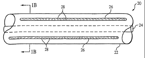
Referring to Figs. lA and 1B, a medical device 20, here, a portion of a
catheter,
includes a tubular body 22 defining a lumen 24, and a plurality of members 26
in the
body. The catheter can be, e.g., a guiding catheter or a catheter shaft of a
medical
device such as a balloon catheter. Members 26 are sealed to encapsulate a
contrast
agent 2S, and together, the members and the contrast agent can enhance the
magnetic
resonance imaging (MI) visibility of catheter 20. As described below, members
26
3o and contrast agent 28 can be incorporated into other types of medical
devices to
enhance the MRl visibility of the devices.
CA 02519166 2011-01-06
77553-43
4
Contrast agent 28 can be any material that enhances the MRI visibility of
device
20. For example, contrast agent 28 can have an MRI response that is different
than the
MRI response of the medical device in which the contrast agent is used, and/or
the MRI
response of the bodily tissue or fluid near the contrast agent during use. The
MRI
response is the response to a magnetic field or radio waves used during A.I.
In some
embodiments, contrast agent 2S can include a material having a TI relaxation
time that
is different than that of the medical device and/or tissue. For example,
contrast agent
28 can include a liquid, such as a solution having a TI (longitudinal)
relaxation time
shortening agent and a proton-containing fluid, such as water or glycerin.
Examples of
Ti relaxation time shortening agents include a paramagnetic metal salt or a
paramagnetic metal chelate compound, such as heavy metal complexes, e.g.,
gadolinium diethylenetrianiinepentaacetic acid (e.g., a 1% Gd-DTPA aqueous
solution), GdDTPA-BMA, and GdHP-DO3A (e.g., available from Schering, Nycomed
and Bracco under the trade marks MAGNEVIST . OMNISCAN , and
PROHANCE ). In some embodiments, the concentration of the Ti relaxation time
shortening agent-is about I L of Gd-DTPA as supplied (e.g., 0.5 mole) per
milliliter of
saline to about '7 gL!mL, e.g., about 3 KL/mL. Alternatively or in addition,
contrast
agent 28 can include a material having a T2 relaxation time that is different
than that of
the medical, device and/or tissue. For example, contrast agent 28 can include
carrier or
a fluid having ferromagnetic, ferrimagnetic, or superparamagnetic
nanoparticles, such
as iron oxide, dysprosium oxide, and/or gadolinium oxide. The particles can be
surface
modified, e.g., made hydrophilic,, to suspend the particles in the fluid and
reduce the
occurrence of precipitation and/or coagulation. Examples of particles and
methods of
modifying the particles are described in U.S. Patent Nos. 6,123,920 and
6,423,296,
In certain embodiments, a member 26 includes both
Ti and a T2 relaxation time shortening agents. In.some embodiments having
multiple
members 26, selected members include Ti relaxation time shortening agents,
while
different selected members include T2 relaxation time shortening agents.
Member 26 can be formed of one or more materials, such as non-magnetically
3o active materials, that do not interfere with the MRl visibility of device
20. The material
can be visibly transparent or opaque, Preferably, the material has a low
permeability,
e.g., impermeability, to fluids, such as water, in contrast agent 28. In some
cases, the
permeability is about equal to or less than the permeability of Nylon or PET.
The low
CA 02519166 2011-01-06
77553-43
permeability to fluids reduces or prevents fluids from exiting member 26 and
reducing
the visibility of the contrast agent. Examples of suitable materials include
polymers
such as polypropylene, polyethylene, polysulfonate, Nylon,
polyethyleneterephthalate
(PET), or polyacetonitrile. Other materials include glass, and non-magnetic
metals,
5 such as aluminum. The material can be radiopaque, e.g., visible by X-ray
fluoroscopy,
Examples of radiopaque materials include those having a densitygreater than
about 10
g/cc, gold, tantalum, tungsten, platinum, palladium, or their alloys.
Different members
26 can be formed of different materials.
Member 26 is generally configured to house contrast agent 28. Member 26 can
1o generally be a thin-walled receptacle defining a hollow cavity for housing
contrast
agent 26. The thin wall of member 26 allows more contrast agent 28 to be
loaded into
a given member 26, which can enhance visibility of the member, or reduce the
overall
size of the, member.
Particular dimensions and/or configuration of member 26 can be function of,
for
example, the device in which the member is used, other components (such as
guidewires) that can be used with the device, and/or the desired MRI
visibility.
Generally, member 28 can have a variety of configurations or shapes. Member 26
can
be substantially spherical, oval, or elongated and relatively flexible. Member
26 can
have a cross section that is circular or non-circular, such as oval, or
regularly or
in-egularly polygonal having 3, 4, 5, 6, 7, or 8 or more sides. The outer
surface of
member 26 can be relatively smooth, e.g., cylindrical or rod-like, or faceted.
Member
26 can have uniform or non-uniform thickness, e.g., the member can taper along
its
length. Different combinations of members 26 having two or more different
configurations or shapes can be used in a medical device.
As noted above, the dimensions of member 26 can vary. In some cases, the
width or outer diameter is between about 50 to about 500 microns. Member 26
can
have a width or outer diameter greater than or equal to about 50 microns, 100
microns,
200 microns, 300 microns, or 400 microns; and/or less than or equal to about
500
microns, 400 microns, 300 microns, 200 microns, or 100 microns. The widths or
outer
diameters of multiple members 26 in a device can be uniform or relatively
random.
Member 26 can have a wall thickness between about 10 microns and about 500
microns. The wall thickness can be greater than or equal to about 10, 50, 100,
1S0,
CA 02519166 2011-01-06
77553-43
6
200, 250, 300, 350, or 450 microns; and/or less than or equal to about 500,
450, 400,
350, 300, 250, 200, 150, 100, or 50 microns. .
Different arrangements of members 26 are possible. An elongated member 26
can extend substantially an entire length of a medical device. Alternatively
or in
addition, members having similar or different dimensions can extend at
selected
portion(s) of the device. For example, members can act as marker, e.g., by
placing the
markers near the distal end of the device, or as a measuring tool, e.g., by
placing the
members at predetermined intervals. An elongated member can extend
continuously or
with gaps between members. Any number members can be included in a medical
9o device, e.g., the device can have one or more elongated members, e.g., 2,
3, 4, 5, 6, 7, 8
or more. Member 26 can be equally and/or unequally spaced in a device. For
example,
looking at a radial cross section of a device having six members 26, the
members can
be formed at 2 o'clock, 3 o'clock, 4 o'clock, 8 o'clock, 9 o'clock, and 10
o'clock.
Member 26 at 3 o'clock is equally spaced from the member at 2 o'clock and 4
o'clock;
but, for example, the member at 4 o'clock is unequally spaced from member at 3
o'clock and 8 o'clock. Member 26 can be symmetrically or asymmetrically
positioned
in a medical device.
In some cases, members 26 are formed into relatively small fibers, e.g.,
chopped
fibers, generally having lengths greater than widths or diameters. The fibers
can have a
length of about 0.5 mm to about 5 mm. In some embodiments, the fibers can have
a.
length greater than or equal to about 0.5, 1.0, 1.5, 2.0, 2.5, 3.0, 3.5, 4.0,
or 4.5 mm;
and/or less than or equal to about 5.0, 4.5, 4.0, 3.5, 3.0, 2.5, 2.0, 1.5, or
1.0 mm. The
lengths of the fibers can be uniform or relatively random. The fibers can be a
width or
outer diameter, and/or wall thickness generally as described above. The widths
or outer
diameters, and/or wall thickness can be uniform or relatively random.
In some embodiments, the fibers can be characterized as having a length to
width aspect ratios from about 3:1 to about 20:1, although higher aspect
ratios are
possible. In some embodiments, the length to width aspect ratios can be
greater than
about 3:1, 5:1, 10:1, or 15:i; and/or less than about 20:1,15:1, 10:1, or 5:1.
The width
used to determine the aspect ratio can be the narrowest or broadest width or
outer
diameter. The length can be the largest dimension of a fiber. Mixtures of
fibers having
two or more different aspect ratios can be used.
CA 02519166 2011-01-06
77553-43
7
For a given medical device, the concentration of members 26 in fiber form is a
function of, for example, the size of the fibers, the amount of contrast agent
28, the
desired visibility, and/or the type of medical device- In some embodiments,
the
concentration is between about 10 and about 60 percent by weight. The
concentration
can be greater than or equal to about 10, 20, 30, 40, or 50 percent by weight;
and/or less
than or equal to about 60, 50, 40, 30, or 20 percent by weight.
Methods of forming member 26 can depend on the materials in the member.
Generally, member 26 can be formed using conventional techniques, e.g.,
techniques
used to form a hollow tube or filament, such as extrusion, drawing, or
casting. Member
26 can be loaded with contrast agent 2S by injecting the contrast agent into
the member
or by soaking the member in the contrast agent. Member 26 can be sealed by
heating
(e.g., melting), gluing, and/or. mechanically working (e.g., crimping) the
member.
Similarly, methods of incorporating member 26 into a medical device can
depend on the type of device. In some embodiments, a member, such as a
relatively
long polymer member, can be co-extruded with the device, such as a catheter,
according to conventional techniques. In embodiments in which members 26 are
in
fiber form, the fibers can be mixed with a polymer of a device prior to
extrusion or
forming. The fibers can be randomly oriented or preferentially oriented.
Methods of
orienting a fiber are described in US Patent No. 7,029,495.
Injection molding techniques can be used. For example, members 26 can be
placed in a
cavity of a mold, and material(s) of a device can be injected into the mold to
form the
device, such as a tube or a parison. In other embodiments, members 26 can be
mixed
with a device material and injected into the mold. Suitable device materials
are
described in US Patent Application Publication No. 2002/0 1 65 5 23.
Other methods of incorporating member 26 into a medical device are possible.
Referring to Fill 2, all elongated and flexible member 30, e.g., made of a
polymer,
containing contrast agent 28 is helically wrapped around a portion of a
medical device
32. Member 30 can be wrapped such that portions of the member contact each
other
around device 32 or wrapped such that the portions are spaced from each other
(Fig. 2).
3o Device 32 can be, for example, a guiding catheter, a guidewire, or a
balloon catheter.
Member 30 can be secured to device 32 by an adhesive and/or by selectively
heating
and melting the member to the device. In other embodiments, more than one
elongated
member 30 can be wrapped around device 32. Referring to Fig. 3A, multiple
members
CA 02519166 2011-01-06
77553-43
8
30 can be wrapped around device 32 in a woven, braided, or knitted pattern,
which can
enhance the strength of the device. In certain embodiments, multiple members
30 cafe
be wrapped around device 32 without criss-crossing each other (Fig. 3B). The
multiple
members 30 can be the same or different. For example, selected members can
have
different contrast agents and/or be formed of different materials, as
described above.
In some embodiments, an elongated and flexible member, such as member 30,
can be wound to form a medical device. Referring to Fig. 4, a medical device
110,
such as a catheter, can be formed by winding an elongated member (e.g., member
30)
around a mandrel, securing the member in place (e.g., by an adhesive), and
removing
the mandrel. The member defines a tube, which can be used, e.g., as a catheter
or a
vascular graft. The member can also be braided, knitted, or woven as.
described above
to for-ii a medical device (Figs. 3A or 3B).
After a medical device is incorporated with or formed by one or more members,
the medical device can be used by conventional methods. The device can also be
imaged by conventional imaging techniques.
As noted. above, member 26 or 30 and contrast agent 28 can be used in a
variety
of medical devices. Referring to Fig. 5, a balloon catheter 50 includes a
catheter 52 and
a medical balloon 54 carried by the catheter. - In some embodiments, catheter
52
includes member 26 and/or 30 and contrast agent 28, as described above.
Alternatively
or in addition, medical balloon 54 can include member 26 and/or 30 and
contrast agent
28 to enhance the balloon's MRI visibility. For example, fibers of member 26
can be
compounded with a. polymer and extruded to form a tube that can be :formed
into a
balloon. Alternatively or in addition, balloon catheter 50 can include MRI
visible
markers 56 at selected portions, as shown, at the proximal and distal ends of
balloon 54.
Markers 56, for example, can indicate the location of balloon 54 when it is
inflated to a
recommended inflation volume. As shown, markers 56 are elongated members wound
around selected portions of catheter 52. In other embodiments, members can be
formed
in catheter 52 at the selected portions, as described above. Examples of
balloon
catheters are described in, for example, Wang U.S. 5,195,969, and Hamlin U.S.
5,270,086, and are exemplified by the Ranger" system available from Boston
Scientific
Scimed, Maple Grove, MN.
Referring to Fig. 6, a stent-graft 60 includes a stent 62 and a graft 64 on
the
scent. Graft 64 includes member 26 and/or 30 and contrast agent 28, as
described
CA 02519166 2011-01-06
= 77553-43
9
above. As shown, stent-graft 60 is carried by a support 66, which can be a
catheter or a
medical balloon, depending on the type of stent 62. Graft 64 can be formed of
a
biocompatible, non-porous or semi-porous polymer matrix made of
polyte.trafluoroethylene (PTFE), expanded PTFE, polyethylene, urethane, or
polypropylene. Fibers of members 26 can be compounded with the polymer matrix.
An elongated member 301can extend, e_g., helically, around graft 63. Graft 64
can be
defined by one or more members, e.g. Figs. 3A, 3B, or'4. In some embodiments,
stent-
graft 60 includes a.releasable therapeutic agent or a pharmaceutically active
compound,
such as described in U.S. Patent No. 5,674,242, and U.S. Patent No. 6,676,987.
The
therapeutic agents or pharmaceutically active compounds can include, for
example, anti-
thrombogenic agents, antioxidants, anti-inflammatory agents, anesthetic
agents, anti-
coagulants, and antibiotics.
In general, stent 62 can be of any desired shape and size (e.g., coronary
stents,
aortic scents, peripheral stents, gastrointestinal scents, urology scents and
neurology
stents). In certain embodiments, a coronary stent can have an expanded
diameter of
from about 2 millimeters to about 6 millimeters. In some embodiments, a
peripheral
stent can have an expanded diameter of from about 5 millimeters to about 24,
millimeters. In certain embodiments, a gastrointestinal and/or urology scent
can have
an expanded diameter of from about 6 millimeters to about 30 millimeters. In
some
embodiments, a neurology stent can have an expanded diameter of from about 1
millimeter to about 12 millimeters. Stent 62 can be balloon-expandable, self-
expandable, or.a combination of both (e.g., as described in U.S. Patent No.
5,366,504).
Stent-graft 60 can be used, e.g., delivered and expanded, according to
conventional methods. Suitable catheter systems are described in, for example,
Wang
U.S_ 5,195,969, and Hamlin U.S. 5,270,086. Suitable delivery methods are also
exemplified by the NIR on Rangerii system, available from Boston Scientific
Sciined.
Maple Grove, MN.
Referring to Fig. 7, member 26 or 30 can be incorporated into a stent 80.
Stent
80 can be generally sized as described above for scent 62. Stent 80 includes a
polymer
matrix 82, and as shown, preferentially oriented fibers 84. Member 26 and/or
30 can be
incorporated to matrix 82, as described above. Multiple embodiments of stent
80
including a polymer and methods of making the stents are described in
CA 02519166 2011-01-06
77553-43
U.S. Patent No. 7,029,495. In some embodiments, elongated members having
a contrast agent can be used with conventional stent materials, e.g.,
stainless steel or,
Iitinol wires, to form a stent. For example, the elongated members can be co-
knitted
with the stmt materials. Methods of forming the stent are described, for
example, in
5 Heath, U.S. 5,725,570, Mayer, U.S. 5,679,470, and Andersen, U.S. 5,366,504.
Referring to Fig. 8, a vascular graft 70 includes a plurality of members 26
containing contrast agent 28. Member 26 and contrast agent 28 can be as
generally
described above, and can be arranged in the graft 70 as described above for
catheter 20.
For example, members 26 can be fibers compounded with a graft material and
extruded
19 to form graft- Alternatively or in addition, members 30 can be placed
between multiple
layers of graft material, with or without members 26 in the layers. Examples
of graft
materials, such as PTFE and expanded PTFE, and methods of making a graft are
described in U.S. Patent No. 6,428,571. Graft 70 can be formed by winding,
braiding,
weaving, or knitting elongated member(s), as described above (Figs. 2, 3A, or
3B).
Member 26 and/or 30 can be incorporated into a guidewire. Polymer
guidewires and methods of making them are described in U.S. Patent. No.
6,436,056.
Member 26 and/or 30 can be incorporated into medical tubing, such as medical
catheters. In general, the size and configuration of the tubing is not
limited. In some
embodiments, a catheter is in the form of a 10 French catheter or smaller,
e_g., a 8
French, 6 French, 4 French, or 2 French catheter. A catheter can have a length
of, for
example, about 240 cm to about 3.5 meters. Examples of catheters include
balloon
catheters, aneurysm catheters, guide catheters, urology catheters, and
microcatheters
(all available from Boston Scientific Corp., Natick, MA).
In other embodiments, member 26 or 30 can contain a radiopaque material
and/or an ultrasound contrast agent. Examples of radiopaque materials include
tantalum, tungsten, platinum, palladium, or gold. The radiopaque material can
be
placed inside an member. Alternatively or in addition, the radiopaque
material, e.g., a
band of radiopaque material, can be placed on a medical device at selected
positions,
such as, for example, on a catheter adjacent to a balloon.
The ultrasound contrast agent can be any material that enhances visibility
during ultrasound iruaging. An ultrasound contrast agent can include a
suspension
having trapped bubbles of sufficient size to deflect sound waves. The
ultrasound
contrast agent can be incorporated into any embodiments of member 26 or 30
described
CA 02519166 2011-01-06
77553-43
11
above. Any of the medical devices described above can include members 26 or
30 having an MRI contrast agent, a radiopaque material, and/or an ultrasound
contrast agent, in any combination or arrangement. For example, a device can
include some members having an MRI contrast agent, and some members having
an ultrasound contrast agent. A member can include more than one contrast
agent, or one or more contrast agent with a radiopaque material.
In some embodiments, a member is not used to contain a contrast
agent. For example, referring to Fig. 9, a catheter 100 can be formed to
define a
lumen 102, which is subsequently filled with a contrast agent, and sealed.
Catheter 100 can additionally include members 26 or 30 with any contrast agent
described above.
Other embodiments are within the scope of the following claims.