Note: Descriptions are shown in the official language in which they were submitted.
CA 02519975 2005-09-22
WO 2004/090121
PCT/1L2004/000315
Stem cells having increased sensitivity to a chemoattractant and
methods of generating and using same.
HELD OF THE INVENTION
The present invention relates to stem cells which exhibit increased
sensitivity
to a chemoattractant and, more particularly, to methods of generating and
using them
such as in clinical applications involving stem cell transplantation.
BACKGROUND OF THE INVENTION
Medical treatment of disorders caused by abnormal organ function typically
employ pharmaceutical agents designed for either compensating for such
abnormal
organ function or treating the dysfunctional organ tissue. However, in some
cases,
pharmaceutical therapy cannot be instated since organ function is oftentimes
complex
and/or not completely understood.
In such cases, the only viable alternative is surgical replacement of the non-
functional organ, which is now widely used for treatment of liver and kidney
failure,
both acute and chronic, as well as for cancer and certain inborn
abnormalities.
However, the need for donor organs far exceeds the supply. Organ shortage has
resulted in new surgical techniques, such as splitting adult organs for
transplant.
Despite fairly good results, such techniques still suffer from a lack of donor
tissue.
The lack of viable donor tissue has led to the emergence of stem cell
replacement therapy, which relies on stem cell plasticity i.e., the ability to
give rise to
cell types in a new location that are not normally present in the organ in
which the
stem cells are located.
Stem cells are generally classified according to their origin, essentially
adult,
embryonic or neonatal origin. Embryonic stem cells deriving from the inner
cell mass
of the blastocyst are pluripotential , bring capable of giving rise to cells
found in all
three germ layers. Despite long held belief adult stem cells are not as
lineage
restricted as previously thought. In particular, haematopoietic and neural
stem cells
appear to be the most versatile at cutting across lineage boundaries. For
example,
recent reports suggest that hematopoietic stem cells (HSCs) of human origin
have a
hepatic potential. Studies of liver or bone marrow transplantation from sex
mismatched donors, identified bone marrow-derived hepatocytes in recipients
[Alison
(2000) Nature 406:257; Theise (2000) Hepatology 32:11-16; Korbling (2002) N
Engl J
SUBSTITUTE SHEET (RULE 26)
CA 02519975 2005-09-22
WO 2004/090121
PCT/1L2004/000315
2
Med 346:738-746.]. Murine and rat HSCs were also found to migrate to
irradiated or
injured adult livers, and to differentiate into hepatic cells [Petersen (1999)
Science
284:1168-1170; Theise (2000) Hepatology 31:235-240; Lagasse (2000) Nat Med
6:1229-1234]. Furthermore, single murine hematopoietic stem cell
transplantation has
resulted in detection of HSC-derived cells in the liver of irradiated
recipients with a
low percentage of transplanted cells exhibiting immunohistochemical and
morphologic properties of hepatic epithelial cells [Krause (2001) Cell 105:369-
377].
The mechanisms that guide circulating hematopoietic stem cells are clinically
significant because the success of stem cell transplantation depends on
efficient
targeting (also referred to as homing) of grafted cells to the recipient
target tissue
[Mazo and von Adrian (1999) Journal of leukocyte Biology 66,25-32]. It is due
to
this homing of transplanted cells that bone marrow transplantations do not
require
invasive surgery, as in the case with the transplantation of any other organ,
but rather
can be effected by simple intravenous infusion.
Homing of HSCs can be defined as the set of molecular interactions that
allows circulating HSCs to recognize, adhere to, and migrate across bone
marrow
endothelial cells resulting in the accumulation of HSCs in the unique
hematopoiesis-
promoting microenvironment of the bone marrow. Homing of progenitor cells can
be
conceived as a multi-step phenomenon [Voermans (2001) J. Hematother. Stem Cell
Res. 10:725-738, Lapidot (2002) Leukemia 16:1992-2003]. HSCs arriving
to the
bone marrow must first interact with the luminal surface of the bone marrow
endothelium. This interaction must occur within seconds after the HSCs have
entered
the bone marrow microvasculature and provide sufficient mechanical strength to
permit the adherent cell to withstand the shear force exerted by the flowing
blood.
Adherent HSCs must then pass through the endothelial layer to enter the
hematopoietic compartment. After extravasation, HSCs encounter specialized
stromal
cells whose juxtaposition supports maintenance of the immature pool by self-
renewal
process in addition to lineage-specific HSCs differentiation, proliferation
and
maturation, a process that involves stroma-derived cytoldnes and other growth
signals.
Only a limited number of factors involved in stem cells homing are known to
date; these include, the ligand for c-kit, stem cell factor, which has been
shown to play
a central role in adherence of HSCs to the stroma [Peled (1999) Science
283:845-848];
CA 02519975 2005-09-22
WO 2004/090121 PCT/1L2004/000315
3
and inte grin interactions (e.g., 131-Intergrins ), which were shown to be
crucial to the
migration of HSCs to the foetal liver [Zanjani (1999) Blood 94:2515-2522].
One important molecular interaction which is considered central to HSC
homing is that of chemokine stromal derived factor (SDF-1) and its cognate
receptor,
CXCR4. =
SDF-1 is the only known powerful chemoattractant of hematopoietic stem cells
of both human [Aiuti (1997) J. Exp. Med. 185:111-120] and murine origin
[Wright
(2002) J. Exp. Med. 195:1145-1154] known to date. SDF-1 is widely expressed in
many tissues during development [McGrath (1999) Dev. Biol. 213:442-456] and
adulthood [Nagasawa (1994) Proc Natl Acad Sci U S A 91:2305-2309; Imai (1999)
Br
J Haematol 106:905-911; Pablos (1999) Am J Pathol 155:1577-1586], such as for
example the liver [Shirozu (1995) Genomics 28:495-500; Nagasawa (1996) Nature
382:635-638; Goddard (2001) Transplantation 72:1957-1967]. Previously, the
present
inventors were able to show that bone marrow homing and repopulation by sorted
human CD34+/CD3841' stem cells transplanted into the tail vein of irradiated
immune
deficient NOD/SCID and NOD/SCID/B2m null mice, are dependent on SDF-
1/CXCR4 interactions [Peled (1999) Science 283:845-848; Kollet (2001) Blood
97:3283-3291]. More recently, the present inventors also established a role
for these
interactions in G-CSF-induced mobilization of murine and human stem cells
[Petit
(2002) Nat Immunol 3:687-694].
In view of the ever-expanding use of stem cell therapy, it is highly desirable
to
further elucidate the mechanism behind stem cell homing and target
repopulation in
order to improve the efficiency and success rate of cell replacement therapy.
Hepatocyte growth factor (HGF), initially identified as a potent mitogen for
mature hepatocytes, is a kringle-containing polypeptide growth factor sharing
structural homology with plasminogen [Nakamure (1984) Biohcem. Biophys. Res.
Commun. 122:1450; Nakamura (1987) FEBS Lett. 224:311; Gohda (1988) J. Clin.
Invest. 81:414; Zanegar Cancer Res. 49:3314; Nakamura (1989) Nature 342:440].
HGF is a mesenchyme-derived pleiotropic factor which regulates cell growth,
cell motility and morphogenesis in various types of cells [Matsumoto (1993) .
Goldberg ID, Rosen EM (eds): Hepatocyte Growth Factor-Scatter Factor (HG-SF)
and C-met Receptor, Basel, Switzerland, Birkhauser Verlag, 1993, 225; Gherardi
(1990) Nature 346:228; Weidner (1990) J. Cell Biol. 111:2097; Higasho (1990)
CA 02519975 2005-09-22
WO 2004/090121
PCT/1L2004/000315
4
Biochem. Biophys. Res. Commun. 170:397; Rubin (1991) Proc. Natl. Acad. Sci.
USA
88:415]. C-met proto-oncogene is the natural and only receptor for HGF known
to
date. HGF is considered a humoral mediator of epithelial-mesenchymal
interactions
responsible for organogenesis of various tissues and organs, regeneration of
organs
and growth, invasion and metastasis of tumor cells [Matsumoto (1996) J.
Biochem.
119:591]. In the hematopoietic system, HGF augments the growth of
hematopoietic
progenitor cells [Krniecik (1992) Blood 80:2454; Nishino (1995) Blood 85:3093;
Mizuno (1993) Biochem Biophys Res. Commun. 194:178 Galimi (1994) J. Cell Biol.
127:1743]. Interestingly, an acute liver injury has been reported to trigger
expression
of HGF as determined by in-situ hybridization of HGF after stimulation of rat
liver
with carbon tetrachloride [CC14, Armbrust (2002) Liver 22:486-494].
Information pertaining to the interaction of HGF with hematopoietic cells is
incomplete, although a possible role in hematopoiesis has been suggested. HGF
was
found to be constitutively produced by human bone marrow stromal cells [Takai
(1997) Blood 89:1560-1565]. Recently, HGF was found to be synergistic with GM-
CSF and IL-3 in proliferation of murine myeloid progenitor cell line and
murine
hemopoietic progenitor cells (HPCs) enriched from bone marrow (BM) or fetal
liver
[Kmiecik (1992) Blood 80:2454-2457; Mizuno (1993) Supra; Nishino (1995) Blood
85:3093-3100]. In addition, a synergistic proliferative effect of HGF with
other
growth factors on human HPCs has been observed and expression of C-met on
CD34+ HPC was detected as well [Galami (1994) J. Cell Biol. 127:1743-1754;
Goff
(1996) Stem Cells 14:592-602; Weimer (1998) Exp. Hematol. 26:885-894].
While reducing the present invention to practice the present inventors have
uncovered that HGF can upregulate CXCR4 expression and promote SDF-1/CXCR4
dependent stem cell motility and migration to the target tissue. These
findings
provide a novel approach for sensitizing stem cell recruitment to a target
tissue and as
such can be used in various cell and tissue replacement protocols.
SUMMARY OF THE INVENTION
According to one aspect of the present invention there is provided a method of
increasing sensitivity of stem cells to a chemoattractant, the method
comprises
exposing the stem cells to HGF or an active portion thereof, which is capable
of
CA 02519975 2005-09-22
WO 2004/090121
PCT/1L2004/000315
increasing a level of at least one chemoattractant receptor of the stem cells
to thereby
increase the sensitivity of the stem cells to the chemoattractant.
According to another aspect of the present invention there is provided a
method of treating a disorder requiring cell or tissue replacement, the method
5 comprises
providing to a subject in need thereof a therapeutically effective amount of
stem cells pretreated with HGF or an active portion thereof, which is capable
of
increasing a level of at least one chemoattractant receptor of the stem cells,
thereby
treating the disorder requiring cell or tissue replacement in the subject.
According to yet another aspect of the present invention there is provided a
method of treating a disorder requiring cell or tissue replacement, the method
comprises providing to a subject in need thereof a therapeutic effective
amount of
HGF or an active portion thereof, which is capable of increasing a level of at
least one
chemoattractant receptor of stem cells, thereby treating the disorder
requiring cell or
tissue replacement.
According to still another aspect of the present invention there is provided a
use of HGF or an active portion thereof for the manufacture of a medicament
for
increasing homing of stem cells to a target tissue.
According to an additional aspect of the present invention there is provided a
method of generating stem cells suitable for transplantation, the method
comprises: (a)
collecting stem cells; (b) exposing the stem cells to HGF or an active portion
thereof;
and (c) isolating stem cells having CXCR4 levels above a predetermined
threshold, to
thereby generate stem cells' suitable for transplantation.
According to further features in preferred embodiments of the invention
described below, collecting the stem, cells is effected by: (i) a stem cell
mobilization
procedure; and/or (ii) a surgical procedure.
According to still further features in the described preferred embodiments the
isolating stem cells having CXCR4 levels above the predetermined threshold is
effected by FACS.
According to still further features in the described preferred embodiments the
method further comprises determining homing capabilities of the stem cells
having
CXCR4 levels above the predetermined threshold following step (c).
According to yet an additional aspect of the present invention there is
provided
a nucleic acid construct comprising a first polynucleotide sequence encoding
HGF or
CA 02519975 2005-09-22
WO 2004/090121
PCT/1L2004/000315
6
an active portion thereof and an inducible cis-acting regulatory element for
directing
expression of the polynucleotide in cells.
According to still further features in the described preferred embodiments the
inducible cis-acting regulatory element is a shear stress activation element.
According to still further features in the described preferred embodiments the
'
nucleic acid construct further comprises a second polynucleotide sequence
being
translationally fused to the first polynucleotide sequence, the second
polynucleotide
sequence encoding a signal peptide capable of directing secretion of the HGF
or the
active portion thereof out of the cells.
According to still an additional aspect of the present invention there is
provided a eukaryotic cell comprising the nucleic acid construct.
According to a further aspect of the present invention there is provided a
cell-
line comprising stem cells transformed to express an exogenous polynucleotide
encoding HGF or an active portion thereof.
According to yet a further aspect of the present invention there is provided a
cell culture comprising: (i) stem cells; and (ii) feeder cells expressing HGF
or an
active portion thereof each being capable of increasing a level of at least
one
chemoattractant receptor of the stem cells.
According to still a further aspect of the present invention there is provided
a
method of increasing sensitivity of stem cells to a chemoattractant, the
method
comprises, upregulating an expression or activity of endogenous HGF or an
active
portion thereof of the stem cells to thereby increase the sensitivity of the
stem cells to
the chemoattractant
According to still a further aspect of the present invention there is provided
a
method of increasing stem cell motility, the method comprises exposing the
stem cells
to HGF or an active portion thereof which is capable of increasing motility of
the
stem cells.
According to still an additional aspect of the present invention the at least
one
chemoattractant receptor is CXCR4.
According to still an additional aspect of the present invention the method
further comprises exposing the stem cells to a growth factor and/or a
cytokine.
According to still an additional aspect of the present invention the growth
factor and/or cytokine are selected from the group consisting of SCF and IL-6.
CA 02519975 2005-09-22
WO 2004/090121
PCT/1L2004/000315
7
According to still an additional aspect of the present invention the stem
cells
are hematopoietic stem cells.
According to still an additional aspect of the present invention the
hematopoietic stem cells are CD34+ hematopoietic stem cells.
According to still an additional aspect of the present invention the
hematopoietic stem cells are CD34+/CD3841' hematopoietic stem cells.
According to still an additional aspect of the present invention the stem
cells
are mesenchymal stem cells.
According to still an additional aspect of the present invention exposing the
1.0 stem cells
to the HGF or the active portion thereof, is effected by: (i) expressing a
polynucleotide encoding the HGF or the active portion thereof in the stem
cells; and/or
(ii) contacting the stem cells with the HGF or the active portion thereof.
According to still an additional aspect of the present invention the method
further comprises exposing the stem cells to HGF-receptor.
According to still an additional aspect of the present invention provides a
method of facilitating self repopulation and/or self engraftment of self
progenitor cells
to an injured organ, comprising administration of HGF or an active portion
thereof
alone or together with SCF to a subject suffering of organ inflammation and/or
injury.
According to still an additional aspect of the present invention provides a
pharmaceutical composition comprising HGF or an active portion thereof
which is capable of increasing motility of the stem cells and SCF, and more
specifically to a pharmaceutical composition for treating a disorder requiring
cell or
tissue replacement.
The present invention successfully addresses the shortcomings of the presently
known configurations by providing stem cells, which exhibit increased
sensitivity to a
chemoattractant and methods of generating and using the same.
Unless otherwise defined, all technical and scientific terms used herein have
the same meaning as commonly understood by one of ordinary skill in the art to
which
this invention belongs. Although methods and materials similar or equivalent
to those
described herein can be used in the practice or testing of the present
invention, suitable
methods and materials are described below. In case of conflict, the patent
CA 02519975 2005-09-22
WO 2004/090121
PCT/1L2004/000315
8
specification, including definitions, will control. In addition, the
materials, methods,
and examples are illustrative only and not intended to be limiting.
plurality of instructions.
BRIEF DESCRIPTION OF THE DRAWINGS
The invention is herein described, by way of example only, with reference to
the accompanying drawings. With specific reference now to the drawings in
detail, it
is stressed that the particulars shown are by way of example and for purposes
of
illustrative discussion of the preferred embodiments of the present invention
only, and
are presented in the cause of providing what is believed to be the most useful
and
readily understood description of the principles and conceptual aspects of the
invention. In this regard, no attempt is made to show structural details of
the invention
in more detail than is necessary for a fundamental understanding of the
invention, the
description taken with the drawings making apparent to those skilled in the
art how the
several forms of the invention may be embodied in practice.
In the drawings:
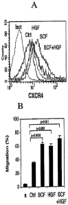
FIG. 1 a is a graph depicting cytokine induced expression of CXCR4 in CB
CD34+ cells as determined using flow cytometry.
FIG. lb is a histogram depicting SDF-1 mediated directional migration of
CD34+ cells in the presence of HGF, SCF or a combination thereof, as
determined
using a Transwell migration assay. Data represent percentage of migration. The
(a)
character denotes spontaneous migration in the absence of SDF-1.
FIG. 2 Shows increased expression of HGF mRNA in the BM and liver of
irradiated mice. NOD/SCID mice were sublethally irradiated (375 cGy). Liver
and
BM samples collected 24 and 48 hours later, together with a sample from a non-
irradiated mouse, were homogenized in Tryreagent (MRC). mRNA was extracted
using standard protocol. RNA was subjected to RT-PCR.
FIG. 3 Shows that HGF increases the potential of BM cells to migrate towards
SDF-1 and the rate of progenitor cell mobilization, following CC14 injury.
NOD/SCID mice were treated with a single injection of CC14 alone (10 ml,
CC14), or
with CC14 followed by 4 consecutive daily injections of HGF (1.5 mg/mouse,
CC14+HGF) starting 2 days later. SDF-1-induced migration by BM cells and the
level
CA 02519975 2005-09-22
WO 2004/090121
PCT/1L2004/000315
9
of progenitors in the blood circulation of these mice were compared to non
treated
mice (ctrl) or to mice treated with G-SCF (300 mg/Kg, 5 consecutive days).
DESCRIPTION OF THE PREFERRED EMBODIMENTS
The present invention relates to stem cells which exhibit increased
sensitivity
to a chemoattractant and to methods of generating and using the same.
Specifically,
the present invention allows to treat disorders requiring cell or tissue
replacement such
as for example to treat chronic or acute liver damage.
The principles and operation of the present invention may be better understood
to with reference to the drawings and accompanying descriptions.
Before explaining at least one embodiment of the invention in detail, it is to
be
understood that the invention is not limited in its application to the details
set forth in
the following description or exemplified by the Examples. The invention is
capable of
other embodiments or of being practiced or carried out in various ways. Also,
it is to
be understood that the phraseology and terminology employed herein is for the
purpose of description and should not be regarded as limiting.
The use of cellular therapy is growing rapidly, and is gradually becoming an
important therapeutic modality in treatment of various disorders.
Hematopoietic stem
cell (HSC) (e.g., bone marrow, umbilical cord blood or mobilized peripheral
blood)
transplantation is one example of a routinely practiced, insurance-reimbursed
cellular =
therapy. However, many other cellular therapies are being developed as well,
including immunotherapy for cancer and infectious diseases, chondrocyte
therapy for
cartilage defects, neuronal cell therapy for neurodegenerative diseases, and
stem cell
therapy for numerous applications [Forbes (2002) Clinical Science 103:355-
369].
One of the problems associated with stem cell therapy is the difficulty of
achieving long-term successful engraftinent of cells at the target tissue.
Currently,
patients which were successfully transplanted exhibit very low levels of stem
cells and
immature progenitors which generate cells with the desired phenotype.
Thus, the success of stem cell transplantation depends on the ability of
intravenously infused stem cells to lodge in the target tissue (e.g., bone
marrow), a
process referred to as homing. It is hypothesized that homing is a multistep
process,
consisting of adhesion of the stem cells to endothelial cells of the marrow
sinusoids,
followed by transendothelial migration directed by chemoattractants, and
finally
CA 02519975 2005-09-22
WO 2004/090121
PCT/1L2004/000315
anchoring within the extravascular bone marrow spaces where proliferation and
differentiation will occur.
Studies have shown that numerous factors are involved in the homing process
including, adhesion molecules, cytokines and growth factors. In 1997 studies
5 uncovered
that migration of CD34+ cells was goverened by the chemoattractant, SDF-
1. Subsequent studies have shown that SDF-1 activates integrins on HSCs and
induces trans-endothelial migration of HSCs in vitro. The receptor for SDF-1
is a G-
protein coupled receptor, termed CXCR-4. In SDF-1 or CXCR-4 knock-out mice
hematopoietic precursors do not shift to the bone marrow during fetal
development
10 suggesting
that SDF-1/CXCR-4 interactions play an important role in stem cell
migration [for review see Voermans (2001) J. Hematother. Stem Cell Res. 10:725-
738,
Lapidot (2002) Leukemia 16:1992-20031.
Despite preliminary understanding of the homing process, information about
regulation of migration of stem cells is still incomplete and scattered. It is
well
appreciated that improving the efficacy of stem cell transplantation may be
achieved
by modulating the ability of stem cells to home to the target tissue.
While reducing the present invention to practice the present inventors have
uncovered that HGF upregulates CXCR4 expression and promotes SDF-1/CXCR4
dependent stem cell motility and migration to a damaged target tissue.
As illustrated hereinunder and in the Examples section which follows, the
present inventors illustrate that hepatic injury upregulates HGF, which
induces
cytoskeletal rearrangements, increases the motility and potentiates the
response of
immature CD34+ cells to SDF-1 signaling by inducing CXCR4 upregulation and
synergizing with stem cell factor (S CE).
Although HGF has been previously shown to be upregulated following liver
injury [Armbrust Liver 2002 Dec;22(6):486-94], the present inventors are the
first to
show that this upregulation in HGF activity leads to upregulation in CXCR4
expression to cytoskeletal rearrangements and accelerated homing of cells
expressing
the same.
The present findings enable the generation of stem cells, which can be
efficiently recruited to a target tissue and as such can be used in numerous
clinical
applications, such as in repair of liver injury and in liver or bone marrow
transplantation.
CA 02519975 2005-09-22
WO 2004/090121
PCT/1L2004/000315
11
Thus, according to one aspect of the present invention there is provided a
method of increasing sensitivity of stem cells to a chemoattractant.
According to another aspect of the present invention there is provided a
method
comprising the administration of HGF to a subject suffering of organ
inflammation
and/or injury, to facilitate self repopulation and/or self engraftrnent to the
injured
organ, due to increased progenitor stem cell levels in the cell blood
circulation.
As used herein, the phrase "stem cells" refers to cells, which are capable of
to differentiating into other cell types having a particular, specialized
function (i.e.,
"fully differentiated" cells).
The method according to this aspect of the present invention includes exposing
the stem cells to HGF or an active portion thereof which is capable of
increasing the
level of at least one chemoattractant receptor of the stem cells to thereby
increase the
sensitivity of the stem cells to the chemoattractant.
Alternatively, increasing sensitivity of stem cells to a chemoattractant can
also
be effected by upregulating expression or activity of endogenous HGF of the
stem
cells.
As is further described herein under, exposing the stem cells to HGF or an
active portion thereof can be effected by either contacting the cells with the
protein or
an active portion thereof, or by expressing the protein or an active portion
thereof
within these cells or expressing HGF or an active portion thereof in non-stem
cells
cultured therewith (e.g., fibroblasts used as a feeder layer).
As is clearly demonstrated in the Examples section which follows, exposure of
stem cells to HGF substantially increased their motility and their ability to
migrate to a
chemoattractant thereof i.e., SDF-1 .
The invention relates to HGF and to its salts, functional derivatives,
precursors and active fractions as well as its active mutants, i.e. other
proteins or
polypeptides wherein one or more amino acids of the structure are eliminated
or
substituted by other amino acids or one or more amino acids were added to that
sequence in order to obtain polypeptides or proteins having the same activity
of the
HGF and comprises also the corresponding "fusion proteins" i.e. polypeptides
comprising the HGF or a mutation thereof fused with another protein . The HGF
CA 02519975 2005-09-22
WO 2004/090121
PCT/1L2004/000315
12
can therefore be fused with another protein such as, for example, an
immunoglobulin.
The term "salts" herein refers to both salts of carboxyl groups and to acid
addition salts of amino groups of the HGF protein of the invention or muteins
thereof
Salts of a carboxyl group may be formed by means known in the art and include
inorganic salts, for example, sodium, calcium, ammonium, ferric or zinc salts,
and the
like, and salts with organic bases as those formed, for example, with amines,
such as
triethanolamine, arginine or lysine, piperidine, procaine and the like. Acid
addition
salts include, for example, salts with mineral acids such as, for example,
hydrochloric
acid or sulphuric acid, and salts with organic acids such as, for example,
acetic acid or
oxalic acid. Of course, any such salts must have substantially similar
activity to the
HGF protein of the invention or its muteins.
The definition "functional derivatives" as herein used refers to derivatives
which can be prepared from the functional groups present on the lateral chains
of the
amino acid moieties or on the terminal N- or C- groups according to known
methods
and are comprised in the invention when they are pharmaceutically acceptable
i.e.
when they do not destroy the protein activity or do not impart toxicity to the
pharmaceutical compositions containing them. Such derivatives include for
example
esters or aliphatic amides of the carboxyl-groups and N-acyl derivatives of
free amino
groups or 0-acyl derivatives of free hydroxyl-groups and are formed with acyl-
groups
as for example alcanoyl- or aroyl-groups.
"Fragment" of the protein the present invention refers to any fragment
or precursor of the polypeptidic chain of the compound itself, alone or in
combination
with related molecules or residues bound to it, for example residues of sugars
or
phosphates, or aggregates of the polypeptide molecule when such fragments or
precursors show the same activity of the HGF as medicament.
The term "circularly permuted" as used herein refers to a linear molecule in
which the termini have been joined together, either directly or through a
linker, to
produce a circular molecule, and then the circular molecule is opened at
another
location to produce a new linear molecule with termini different from the
termini in
the original molecule. Circular permutations include those molecules whose
structure
is equivalent to a molecule that has been circularized and then opened. Thus,
a
circlularly permuted molecule may be synthesized de novo as a linear molecule
and
CA 02519975 2011-10-19
13
never go through a circularization and opening step. The particular circular
permutation of a molecule is designated by brackets containing the amino acid
residues between which the peptide bond is eliminated. Circularly permuted
molecules, which may include DNA, RNA and protein, are single-chain molecules
15 The terms "polypeptide and protein" in the present specification are
interchangeable.
The present invention also concerns muteins of the above HGF protein of the
invention, which muteins retain essentially the same biological activity of
the HGF
protein having essentially only the naturally occurring sequences of the HGF.
Such
These muteins are prepared by known synthesis and/or by site-directed
Any such mutein preferably has a sequence of amino acids sufficiently
duplicative of that of the basic the HGF such as to have substantially similar
activity
thereto. Thus, it can be determined whether any given mutein has substantially
the
same activity as the basic protein of the invention by means of routine
experimentation
Muteins of the HGF protein which can be used in accordance with the present
invention, or nucleic acid coding thereof, include a finite set of
substantially the HGF
CA 02519975 2005-09-22
WO 2004/090121
PCT/1L2004/000315
14
corresponding sequences as substitution peptides or polynucleotides which can
be
routinely obtained by one of ordinary skill in the art, without undue
experimentation,
based on the teachings and guidance presented herein. For a detailed
description of
protein chemistry and structure, see Schulz, G.E. et al., Principles of
Protein Structure,
Springer-Verlag, New York, 1978; and Creighton, T.E., Proteins: Structure and
Molecular Properties, W.H. Freeman & Co., San Francisco, 1983, which are
hereby
incorporated by reference. For a presentation of nucleotide sequence
substitutions,
such as codon preferences, see. See Ausubel et al., Current Protocols in
Molecular
Biology, Greene Publications and Wiley Interscience, New York, NY, 1987-1995;
Sambrook et al., Molecular Cloning: A Laboratory Manual, Cold Spring Harbor
Laboratory, Cold Spring Harbor, NY, 1989.
Preferred changes for muteins in accordance with the present invention are
what are known as "conservative" substitutions. Conservative amino acid
substitutions
of those in the protein having essentially the naturally-occurring HGF
sequences, may
include synonymous amino acids within a group, which have sufficiently similar
physicochemical properties that substitution between members of the group will
preserve the biological function of the molecule, see Grantham, Science, Vol.
185, pp.
862-864 (1974). It is clear that insertions and deletions of amino acids may
also be
made in the above-defined sequence without altering its function, particularly
if the
insertions or deletions only involve a few amino acids, e.g., under 50, and
preferably
under 20 HGF and do not remove or displace amino acids which are critical to a
functional conformation, e.g., cysteine residues, Anfinsen, "Principles That
Govern
The Folding of Protein Chains", Science, Vol. 181, pp. 223-230 (1973). Muteins
produced by such deletions and/or insertions come within the purview of the
present
invention.
Preferably, the synonymous amino acid groups are those defined in Table A.
More preferably, the synonymous amino acid groups are those defined in Table
B; and
most preferably the synonymous amino acid groups are those defined in Table C.
TABLE A Preferred Groups of Synonymous Amino Acids
CA 02519975 2005-09-22
WO 2004/090121 PCT/1L2004/000315
Amino Acid Synonymous Group
Ser Ser, Thr, Gly, Asn
Arg Arg, Gin, Lys, Glu, His
Leu Ile, Phe, Tyr, Met, Val, Leu
5 Pro Gly, Ala, Thr, Pro
Thr Pro, Ser. Ala, Gly, His, Gin, Thr
Ala Gly, Thr, Pro, Ala
Val Met, Tyr, Phe, Ile, Leu, Val
Gly Ala, Thr, Pro, Ser. Gly
10 = Ile Met, Tyr, Phe, Val, Leu, Ile
Phe Trp, Met, Tyr, Ile, Val, Leu, Phe
Tyr Trp, Met, Phe, Ile, Val, Leu, Tyr
Cys Ser, Thr, Cys
His Glu, Lys, Gin, Thr, Arg, His
15 Gin Glu, Lys, Asn, His, Thr, Arg, Gin
Asn Gin, Asp, Ser, Asn
Lys Glu, Gin, His, Arg, Lys
Asp Glu, Asn, Asp
Glu Asp, Lys, Asn, Gin, His, Arg, Glu
Met Phe, Ile, Val, Leu, Met
Trp Trp
TABLE B More Preferred Groups of Synonymous Amino Acids
Amino Acid Synonymous Group
Sers Sers
Arc His, Lys, Arg
Leu Ile, Phe, Met, Leu
Pro Ala, Pro
Thr Thr
Ala Pro, Ala
CA 02519975 2005-09-22
WO 2004/090121
PCT/1L2004/000315
16
Val Met, Ile, Val
Gly Gly
Ilea Ile, Met, Phe, Val, Leu
Phe Met, Tyr, Ile, Leu, Phe
Try Phi, Try
Cys Ser, Cys
His Arg, Gin, His
Gin Glu, His, Gin
Asn Asp, Asn
Lys Arg, Lys
Asp Asn, Asp
Glu FLN, Glu
Met Phe, Ile, Val, Leu, Met
Trp Tip
TABLE C Most Preferred Groups of Synonymous Amino Acids
Amino Acid Synonymous Group
Sers Sers
Arc Arc
Leu Ile, Met, Leu
Pro Pro
Thr Thar
Alan Alan
Val Val
Gly Gly
Ilea Ile, Met, Leu
Phi Phi
Try Tyr
Cys Ser, Cys
His His
Gin Gin
Asn Asn
CA 02519975 2005-09-22
WO 2004/090121
PCT/1L2004/000315
17
Lys Lys
Asp Asp
Glu Glu
Met Ile, Leu, Met
Trp Trp
Examples of production of amino acid substitutions in proteins which can be
used for obtaining muteins of the protein for use in the present invention
include any
to known method steps, such as presented in US patents RE 33,653,
4,959,314,
4,588,585 and 4,737,462, to Mark et al; 5,116,943 to Koths et al., 4,965,195
to Namen
et al; 4,879,111 to Chong et al; and 5,017,691 to Lee et al; and lysine
substituted
proteins presented in US patent No. 4,904,584 (Straw et al).
In another preferred embodiment of the present invention, any mutein of the
HGF protein for use in the present invention has an amino acid sequence
essentially
corresponding to that of the above noted HGF protein of the invention. The
term
"essentially corresponding to" is intended to comprehend muteins with minor
changes
to the sequence of the basic protein which do not affect the basic
characteristics
thereof, particularly insofar as its ability to the HGF is concerned. The type
of
changes which are generally considered to fall within the "essentially
corresponding
to" language are those which would result from conventional mutagenesis
techniques
of the DNA encoding the HGF protein of the invention, resulting in a few minor
modifications, and screening for the desired activity for example increasing
the
sensitivity of stem cells to a chemoattractant.
The present invention also encompasses HGF variants. A preferred HGF
variant are the ones having at least 80% amino acid identity, a more preferred
the HGF
variant is one having at least 90% identity and a most preferred variant is
one having
at least 95% identity to HGF amino acid sequence.
The term "sequence identity" as used herein means that the amino acid
sequences are compared by alignment according to Hanks and Quinn (1991) with a
CA 02519975 2005-09-22
WO 2004/090121
PCT/1L2004/000315
18
refinement of low homology regions using the Clustal-X program, which is the
Windows interface for the ClustalW multiple sequence alignment program
(Thompson
et al., 1994). The Clustal-X program is available over the interne at
ftp://ftp-igbmc.u-
strasbg.fr/pubiclustalx./. Of course, it should be understood that if this
link becomes
inactive, those of ordinary skill in the art could find versions of this
program at other
links using standard interne search techniques without undue experimentation.
Unless
otherwise specified, the most recent version of any program referred herein,
as of the
effective filing date of the present application, is the one, which is used in
order to
practice the present invention.
Another method for determining "sequence identity" is he following. The
sequences are aligned using Version 9 of the Genetic Computing Group's GDAP
(global alignment program), using the default (BLOSUM62) matrix (values -4 to
+11)
with a gap open penalty of -12 (for the first null of a gap) and a gap
extension penalty
of -4 (per each additional consecutive null in the gap). After alignment,
percentage
identity is calculated by expressing the number of matches as a percentage of
the
number of amino acids in the claimed sequence.
Muteins in accordance with the present invention include those encoded by a
nucleic acid, such as DNA or RNA, which hybridizes to DNA or RNA under
stringent
conditions and which encodes a the HGF protein in accordance with the present
invention, comprising essentially all of the naturally-occurring sequences
encoding the
HGF and sequences which may differ in its nucleotide sequence from the
naturally-
derived nucleotide sequence by virtue of the degeneracy of the genetic code,
i.e., a
somewhat different nucleic acid sequence may still code for the same amino
acid
sequence, due to this degeneracy.
The term "hybridization" as used herein shall include any process by which a
strand of nucleic acid joins with complementary strand through a base pairing
(Coombs J, 1994, Dictionary of Biotechnology, stokton Press, New York NY).
"Amplification" is defined as the production of additional copies of a nucleic
acid
sequence and is generally carried out using polymerase chain reaction
technologies
well known in the art (Dieffenbach and Dveksler, 1995, PCR Primer, a
Laboratory
Manual, Cold Spring Harbor Press, Plainview NY).
"Stringency" typically occurs in a range from about Tm-5 C (5 C below the
melting temperature of the probe) to about 20 C to 25 C below Tm.
CA 02519975 2005-09-22
WO 2004/090121
PCT/1L2004/000315
19
The term "stringent conditions" refers to hybridization and subsequent washing
conditions, which those of ordinary skill in the art conventionally refer to
as
"stringent". See Ausubel et al., Current Protocols in Molecular Biology,
Greene
Publications and Wiley Interscience, New York, NY, 1987-1995; Sambrook et al.,
Molecular Cloning: A Laboratory Manual, Cold Spring Harbor Laboratory, Cold
Spring Harbor, NY, 1989.
As used herein, stringency conditions are a function of the temperature used
in
the hybridization experiment, the molarity of the monovalent cations and the
percentage of formamide in the hybridization solution. To determine the degree
of
stringency involved with any given set of conditions, one first uses the
equation of
Meinkoth et al. (1984) for determining the stability of hybrids of 100%
identity
expressed as melting temperature Tm of the DNA-DNA hybrid:
Tm = 81.5 C + 16.6 (LogM) + 0.41 (%GC) - 0.61 (% form) - 500/L
where M is the molarity of monovalent cations, %GC is the percentage of G
and C nucleotides in the DNA, % form is the percentage of formamide in the
hybridization solution, and L is the length of the hybrid in base pairs. For
each 1 C
that the Tm is reduced from that calculated for a 100% identity hybrid, the
amount of
mismatch permitted is increased by about 1%. Thus, if the Tm used for any
given
hybridization experiment at the specified salt and forrnamide concentrations
is 10 C
20- below the Tm calculated for a 100% hybrid according to the equation of
Meinkoth,
hybridization will occur even if there is up to about 10% mismatch.
As used herein, "highly stringent conditions" are those which provide a Tm
which is not more than 10 C below the Tm that would exist for a perfect duplex
with
the target sequence, either as calculated by the above formula or as actually
measured.
"Moderately stringent conditions" are those, which provide a Tm, which is not
more
than 20 C below the Tm that would exist for a perfect duplex with the target
sequence,
either as calculated by the above formula or as actually measured. Without
limitation,
examples of highly stringent (5-10 C below the calculated or measured Tm of
the
hybrid) and moderately stringent (15-20 C below the calculated or measured Tm
of the
hybrid) conditions use a wash solution of 2 X SSC (standard saline citrate)
and 0.5%
SDS (sodium dodecyl sulfate) at the appropriate temperature below the
calculated Tm
of the hybrid. The ultimate stringency of the conditions is primarily due to
the
washing conditions, particularly if the hybridization conditions used are
those, which
CA 02519975 2005-09-22
WO 2004/090121
PCT/1L2004/000315
allow less stable hybrids to form along with stable hybrids. The wash
conditions at
higher stringency then remove the less stable hybrids. A common hybridization
condition that can be used with the highly stringent to moderately stringent
wash
conditions described above is hybridization in a solution of 6 X SSC (or 6 X
SSPE
5 (standard saline-phosphate-EDTA), 5 X Denhardt's reagent, 0.5% SDS, 100
µ
g/ml denatured, fragmented salmon sperm DNA at a temperature approximately 20
to
C below the Tm. If mixed probes are used, it is preferable to use tetramethyl
ammonium chloride (TMAC) instead of SSC (Ausubel, 1987, 1999).
Non-limiting examples of stem cells, which can be used according to this
10 aspect of the present invention, are hematopoietic stem cells (HSCs) and
mesenchymal stem cells (MS Cs) obtained from bone marrow tissue of an
individual
at any age or from cord blood of a newborn individual, embryonic stem (ES)
cells
obtained from the embryonic tissue formed after gestation (e.g., blastocyst),
or
embryonic germ (EG) cells obtained from the genital tissue of a fetus any time
during
15 gestation, preferably before 10 weeks of gestation. Further description
of stem cells,
which can be used according to this aspect of the present invention is
summarized
hereinbelow.
HSCs - Hematopoietic stem cells (HSCs) are the formative pluripotential blast
cells found inter alia in bone marrow, fetal liver, umbilical cord blood and
peripheral
20 blood which are capable of differentiating into any of the specific
types of
hematopoietic or blood cells, such as erythrocytes, lymphocytes, macrophages
and
megakaryocytes. Typically, within the bone marrow, HSCs reside in niches that
support all the requisite factors and adhesive properties to maintain their
ability and
produce an appropriate balanced output of mature progeny over the life time of
the
25 organism [Whetton (1999) Trends Cell Biol 9:233-238; Weissman (2000)
Cell
100:157-168; Jankowska-Wieczorek (2001) Stem Cells 19:99-107; Chan (2001) Br.
J.
Haematol. 112:541-557].
HSCs according to this aspect of the present invention are preferably CD34+
cells and more preferably CD34+/CD3841' cells, which are a more primitive stem
cell
population and are therefore less lineage-restricted and are the major long-
term bone
marrow repopulating cells.
MSCs ¨ Mesenchymal stem cells are the formative pluripotential blast cells
found inter alia in bone marrow, blood, dermis and periosteum that are capable
of
CA 02519975 2005-09-22
WO 2004/090121
PCT/1L2004/000315
21
differentiating into more than one specific type of mesenchymal or connective
tissue
(i.e. the tissues of the body that support the specialized elements; e.g.
adipose,
osseous, stoma, cartilaginous, elastic and fibrous connective tissues)
depending upon
various influences from bioactive factors, such as cytokines.
Approximately, 30 % of human marrow aspirate cells adhering to plastic are
considered as MSCs. These cells can be expanded in vitro and then induced to
differentiate. The fact that adult MSCs can be expanded in vitro and
stimulated to
form bone, cartilage, tendon, muscle or. fat cells render them attractive for
tissue
engineering and gene therapy strategies. In vivo assays have been developed to
assay
MSC function. MSCs injected into the circulation can integrate into a number.
of
tissues described hereinabove. Specifically, skeletal and cardiac muscle can
be
induced by exposure to 5-azacytidine and neuronal differentiation of rat and
human
MSCs in culture can be induced by exposure to 13-mercaptoethanol, DMSO or
butylated hydroxyanisole [Tomita (1999) 100:11247-11256; Woodbury (2000) J.
Neurosci. Res. 61:364-370]. Furthermore, MSC-derived cells are seen to
integrate
deep into brain after peripheral injection as well as after direct injection
of human
MSCs into rat brain; they migrate along pathways used during migration of
neural
stem cells developmentally, become distributed widely and start lose markers
of HSC
specialization [Azizi (1998) Proc. Natl. Acad. Sci. USA 95:3908-39131 Methods
for
promoting mesenchymal stem and lineage-specific cell proliferation are
disclosed in
U.S. Pat. No. 6,248,587.
Epitopes on the surface of the human mesenchymal stem cells (hMSCs) such
as SH2, SH3 and SH4 described in U.S. Pat. No. 5,486,359 can be used as
reagents to
screen and capture mesenchymal stem cell population from a heterogeneous cell
population, such as exists, for example, n bone marrow. Precursor mesenchymal
stem cells which are positive for CD45 are preferably used according to this
aspect of
the present invention, since these precursor mesenchymal stem cells can
differentiate
into the various mesenchymal lineages.
Preferred stem cells according to this aspect of the present invention are
human stem cells.
Table 1, below provides examples of adult stem cells, which can be used to
obtain the indicated phenotype in a target tissue of interest, according to
this aspect of
the present invention.
CA 02519975 2005-09-22
WO 2004/090121
PCT/1L2004/000315
22
Table I
Stem cell Differentiated Target tissue Reference
phenotype
Bone marrow Oval cells, Liver Petersen (1999) Science
284:1168-
Hepatocytes 1170
KTLS cells Hepatocytes Liver Lagasse (2000) Nat. Med. 6:1229-
1234
Bone marrow Hepatocytes Liver Alison (2000) Nature 406:257;
Thiese
(2000) Hepatology 32:11-16
Pacreatic exocrine Hepatocytes Liver Shen (2000) Nat. Cell Biol. 2:879-
887
cells
Pacreas Hepatocytes Liver Wang (2001) Am. J. Pathol. 158:571-
579
Bone marrow Endothelium Liver Gao (2001) Lancet 357:932-933
Bone marrow Tubular Kidney Poulsom (2001) J. Pathol. 195:229-
epithelium, 235
glomeruli
Bone marrow Endothelium Kidney Lagaaij (2001) Lancet 357:33-37
Extra renal Endothelium Kidney Williams (1969) Surg. Forum 20:293-
294
Bone marrow Myocardium Heart Orlic (2001) Nature 410:701-704
Bone marrow Cardiomyocytes Heart Jackson (2001) J. Clin Invest.
and Endothelium 107:1395-1402
Bone marrow Type 1 Lung Krause (2001) Cell 105:369-377
pneumocytes
Neuronal Multiple Marrow Bjornson (1999) Science 283:534-
537
hematopoietic
lineages
Bone marrow Neurons CNS Mezey (2000) Science 290:1779-1782
Bone marrow Microglia and CNS Eglitis (1997) Proc. Natl. Acad.
Sci.
Astrocyes USA 94:4080-4085
Abbreviations: SP- Side population cells; CNS ¨ central nervous system;
As mentioned hereinabove the stem cells according to this aspect of the
present
invention are exposed to HGF or an active portion thereof.
HGF is a heterodimeric polypeptide including a 62 lcDa a-subunit and a 34
lcDa 13-subunit. The a-subunit contains an N-terminal hairpin domain including
27
amino acids followed by four canonical kringle domains which are 80-amino acid
double looped structures stabilized by three internal disulfide bridges
[Comoglio
(1999) Exp. Cell. Res. 253:88-99; Comoglio (1993) Exs 65:131-65; Comoglio
(1996)
Genes Cells 1:347-54]. The kringle domains are important for protein-protein
interaction. The first kringle domain contains the high affinity binding
domain for the
HGF-receptor, while the second kringle domain contains a low affinity binding
site to
membrane associated heparan-sulfate proteoglycans. The low affinity
interaction
maintains a high concentration of HGF in the vicinity of target cells. The HGF
gene
encodes a single pro-HGF protein, which is cleaved to form the active
heterodimeric
molecule with high affinity for the receptor.
CA 02519975 2005-09-22
WO 2004/090121
PCT/1L2004/000315
23
As used herein an active portion of HGF, refers to the minimal HGF sequence,
which is sufficient to increase sensitivity of the stem cells of the present
invention to
the chemoattractant. As used herein an active portion of HGF, refers also to a
mutein,
fusion protein, functional derivative , fragment, circularly permutated HGF
and/or salt
thereof.
To determine the active portion of HGF according to the present invention,
stem cells can be contacted with an HGF segment and response of the cells
thereto can
be monitored molecularly, biochemically or functionally (e.g., motility,
homing,
migration assays) using methods which are well known to those of skill in the
art and
are further described hereinbelow.
As described hereinabove exposing the stem cells to HGF or an active portion
thereof increases the level of at least one type of chemoattractant receptor
of the stem
cells.
A number of chemotactic cell receptors are known to participate in
transendothelial migration of stem cells. Many of these receptors belong to
the family
of G protein-coupled seven-transmembrane receptors (7-TMR). Signaling via G
proteins, particularly Gi proteins, results in a chemotactic response of the
cells towards
a gradient of the corresponding ligand [Voermans (2001) J. Hematother. Stem
Cell
Res. 10:725-738]. Recent studies have provided evidence for expression of
several 7-
TMR on immature hematopoietic progenitor cells, which potentially mediate
chemotactic effects: chemokine receptors (e.g., CXCR4, receptor for stromal
cell-
derived factor-1), receptors for lipid mediators (e.g., the cysteinyl
leukotriene receptor
cysLT1 and the peripheral cannabinoid receptor cb2), and receptors for
neuroendocrine hormones (e.g., the somatostatin receptor sst2). From these
studies it
can be concluded that migration of hematopoietic progenitor and stem cells is
controlled by a variety of chemotactic factors rather than by a single
chemokine (e.g.,
SDF-1).
According to preferred embodiments of this aspect of the present invention the
chemotactic receptor is CXCR4.
It will be appreciated that since HGF exerts its biological activities through
binding to an HGF-receptor i.e., c-met, the present invention also
contemplates
exposing the cells to HGF receptor, i.e. c-met which may be a limiting factor
in
HGF-signaling.
CA 02519975 2005-09-22
WO 2004/090121
PCT/1L2004/000315
24
As mentioned hereinabove, exposing the stem cells to HGF or an active portion
thereof can be effected by contacting the stem cells with the protein or by
expressing
the protein within the stem cells.
Contacting stem cells with HGF or an active portion thereof is preferably
effected using harvested cells, although the present invention also
contemplates
mobilization of stem cells from tissue into circulation and exposure of
circulating stem
cells to the HGF or an active portion thereof.
Adult stem cells can be obtained using a surgical procedure such as bone
marrow aspiration or can be harvested using commercial systems such as those
available from Nexell Therapeutics Inc. Irvine, CA, USA.
Stem cells utilized by the present invention are preferably collected (i.e.,
harvested) using a stem cell mobilization procedure, which utilizes
chemotherapy or
cytokine stimulation to release of HSCs into circulation of subjects. Stem
cells are
preferably retrieved using this procedure since mobilization is known to yield
more
HSCs and progenitor cells than bone marrow surgery.
Stem cell mobilization can be induced by a number of molecules. Examples
include but are not limited to cytokines such as, granulocyte colony-
stimulating factor
(G-CSF), granulocyte-macrophage colony-stimulating factor (GM-CSF),
interleukin
(IL)-7, IL-3, IL-12, stem cell factor (SCF), and fit-3 ligand; chemokines like
IL-8,
Mip-la, GroP, or SDF-1; and the chemotherapeutic agents cyclophosphamide (Cy)
and paclitaxel. It will be appreciated :that these molecules differ in
kinetics and
efficacy, however, according to presently known embodiments G-CSF is
preferably
used alone or in combination such as with cyclophosphamide to mobilize the
stem
cells. Typically, G-CSF is administered daily at a dose of 5-10 jig/kg for 5-
10 days.
Methods of mobilizing stem cells are disclosed in U.S. Pat. Nos. 6,447,766 and
6,162,427.
Human embryonic stem cells can be isolated from human blastocysts. Human
blastocysts are typically obtained from human in vivo preimplantation embryos
or
from in vitro fertilized (IVF) embryos. Alternatively, a single cell human
embryo can
be expanded to the blastocyst stage. For the isolation of human ES cells the
zona
pellucida is removed from the blastocyst and the inner cell mass (ICM) is
isolated by
immunosurgery, in which the trophectoderm cells are lysed and removed from the
intact ICM by gentle pipetting. The ICM is then plated in a tissue culture
flask
CA 02519975 2005-09-22
WO 2004/090121
PCT/1L2004/000315
containing the appropriate medium which enables its outgrowth. Following 9 to
15
days, the ICM derived outgrowth is dissociated into clumps either by a
mechanical
dissociation or by an enzymatic degradation and the cells are then re-plated
on a fresh
tissue culture medium. Colonies demonstrating undifferentiated morphology are
5 individually selected by micropipette, mechanically dissociated into
clumps, and re-
plated. Resulting ES cells are then routinely split every 1-2 weeks. For
further details
on methods of preparation human ES cells see Thomson et al., [U.S. Pat. No.
5,843,780; Science 282: 1145, 1998; Curr. Top. Dev. Biol. 38: 133, 1998; Proc.
Natl.
Acad. Sci. USA 92: 7844, 1995]; Bongso et al., [Hum Reprod 4: 706, 1989];
Gardner
10 et al., [Fertil. Steril. 69: 84, 1998].
It will be appreciated that commercially available stem cells can be also be
used according to this aspect of the present invention. Human ES cells can be
purchased from the NIH human embryonic stem cells registry
(<http://escr.nih.gov>).
Non-limiting examples of commercially available embryonic stem cell lines are
15 BG01, BG02, BG03, BG04, CY12, CY30, CY92, CY10, TE03, TE32.
Human EG cells can be retrieved from the primordial germ cells obtained from
human fetuses of about 8-11 weeks of gestation using laboratory techniques
known to
anyone skilled in the arts. The genital ridges are dissociated and cut into
small chunks,
which are thereafter disaggregated into cells by mechanical dissociation. The
EG cells
20 are then grown in tissue culture flasks with the appropriate medium. The
cells are
cultured with daily replacement of medium until a cell morphology consistent
with EG
cells is observed, typically after 7-30 days or 1-4 passages. For additional
details on
methods of preparing EG cells see Shamblott et al., [Proc. Natl. Acad. Sci.
USA 95:
13726, 1998] and U.S. Pat. No. 6,090,622.
25 It will be appreciated that enrichment of stem cell population
exhibiting
pluripotency may be preferably effected. Thus, for example, as outlined
hereinabove,
CD34+ stem cells can be concentrated using affinity columns or FACS as further
described hereinunder.
Culturing of stem cells under proliferative conditions may also be effected in
cases where stem cell numbers are too low for use in treatment. Culturing of
stem
cells is described in U.S. Pat. Nos. 6,511,958, 6,436,704, 6,280,718,
6,258,597,
6,184,035, 6,132708 and 5,837,5739.
CA 02519975 2005-09-22
WO 2004/090121
PCT/1L2004/000315
26
Once stem cells are obtained, they are contacted with HGF or an active portion
thereof.
Soluble HGF, and in particular, active portion thereof can be biochemically
synthesized by using, for example, standard solid phase techniques. These
methods
include exclusive solid phase synthesis, partial solid phase synthesis
methods,
fragment condensation, classical solution synthesis. Solid phase peptide
synthesis
procedures are well known in the art and further described by John Morrow
Stewart
and Janis Dillaha Young, Solid Phase Peptide Syntheses (2nd Ed., Pierce
Chemical
Company, 1984).
Synthetic peptides can be purified by preparative high performance liquid
chromatography [Creighton T. (1983) Proteins, structures and molecular
principles.
WH Freeman and Co. N.Y.] and the composition of which can be confirmed via
amino acid sequencing.
It will be appreciated that HGF can also be obtained from. commercial
suppliers such as, Sigma-Aldrich, Rehovot, Israel; Product No. H1404.
In cases where large amounts of HGF or the active portion thereof are desired,
such polypeptides are preferably generated using recombinant techniques.
To recombinantly synthesize such polypeptides, an expression construct (i.e.,
expression vector), which includes a polynucleotide encoding the HGF or the
active
portion thereof positioned under the transcriptional control of a regulatory
element,
such as a promoter, is introduced into host cells.
The "transformed" cells are cultured under suitable conditions, which allow
the expression of the fusion protein encoded by the polynucleotide.
Following a predetermined time period, the expressed protein is recovered
from the cell or cell culture, and purification is effected.
A variety of prokaryotic or eukaryotic cells can be used as host-expression
systems to express the modified polypeptide coding sequence. These include,
but are
not limited to, microorganisms, such as bacteria transformed with a
recombinant
bacteriophage DNA, plasmid DNA or cosmid DNA expression vector containing the
desired coding sequence; Mammalian expression systems are preferably used to
express the HGF or the active portion thereof, since eukaryotic cells enable
the
generation of post-translational modified proteins. However, bacterial systems
are
typically used to produce recombinant proteins since they enable a high
production
CA 02519975 2005-09-22
WO 2004/090121
PCT/1L2004/000315
27
volume at low cost. Thus, the host system is selected according to the
recombinant
protein to be generated and the end use thereof.
In bacterial systems, a number of expression vectors can be advantageously
selected depending upon the use intended for the modified polypeptide
expressed.
For example, when large quantities of conjugates are desired, vectors that
direct the
expression of high levels of the protein product, possibly as a fusion with a
hydrophobic signal sequence, which directs the expressed product into the
periplasm
of the bacteria or the culture medium where the protein product is readily
purified
may be desired. Certain fusion protein engineered with a specific cleavage
site to aid
in recovery of the conjugate may also be desirable. Such vectors adaptable to
such
manipulation include, but are not limited to, the pET series of E. coli
expression
vectors [Studier et al. (1990) Methods in Enzymol. 185:60-89).
Other expression systems such as insects and mammalian host cell systems,
which are well known in the art can also be used by the present invention (see
U.S.
Pat. No. 6,541,623).
In any case, transformed cells are cultured under effective conditions, which
allow for the expression of high amounts of recombinant polypeptide. Effective
culture conditions include, but are not limited to, effective media,
bioreactor,
temperature, pH and oxygen conditions that permit protein production. An
effective
medium refers to any medium in which a cell is cultured to produce the
recombinant
modified polypeptide of the present invention. Such a medium typically
includes an
aqueous solution having assimilable carbon, nitrogen and phosphate sources,
and
appropriate salts, minerals, metals and other nutrients, such as vitamins.
Cells of the
present invention can be cultured in conventional fermentation bioreactors,
shake
flasks, test tubes, microtiter dishes, and petri plates. Culturing can be
carried out at a
temperature, pH and oxygen content appropriate for a recombinant cell. Such
culturing conditions are within the expertise of one of ordinary skill in the
art.
The resultant recombinant proteins of the present invention are preferably
secreted into the growth (e.g., fermentation) medium.
Following a predetermined time in culture, recovery of the recombinant
protein is effected. The phrase "recovering the recombinant protein" refers to
collecting the whole growth medium containing the protein and need not imply
additional steps of separation or purification (see Example 2 of the Examples
CA 02519975 2005-09-22
WO 2004/090121
PCT/1L2004/000315
28
section). Proteins of the present invention can be purified using a variety of
standard
protein purification techniques, such as, but not limited to, affinity
chromatography,
ion exchange chromatography, filtration, electrophoresis, hydrophobic
interaction
chromatography, gel filtration chromatography, reverse phase chromatography,
concanavalin A chromatography, chromatofocusing and differential
solubilization.
Proteins of the present invention are preferably retrieved in "substantially
pure" form. As used herein, "substantially pure" refers to a purity that
allows for the
effective use of the protein in the diverse applications, described
hereinbelow.
It will be appreciated that recombinant production of HGF or the active
portion thereof of the present invention can also be effected in-vitro.
HGF or the active portion thereof can be included in a culture medium utilized
for culturing or sustaining the harvested stem cells. Such a culture medium
typically
includes a buffer solution (i.e., growth medium) suitable for stem cell
culturing. The
culture medium can also include serum or serum replacement which include
growth
factors which support growth and survival of the stem cells. The culture
medium can
also include homing agents such as SDF-1, IL-6, SCF and the like. Additionally
the
growth medium of the present invention may also include differentiation-
inhibiting
agents such as leukemia inhibitor factor (LIF).
The stem cells of the present invention can also be contacted with HGF
secreting cells. This can be effected by co-culturing the stem cells of the
present
invention with cells which express HGF or an active portion thereof. For
example,
fibroblast feeder cells, which are oftentimes-co-cultured with stem cells to
support
proliferation thereof in a non-differentiated state, can express HGF, thereby
performing a dual role i.e., growth support and increase of migration and
motility
potential of stem cells.
However since the stem cells of the present invention are preferably used for
clinical applications, measures are taken to isolate the stem cells from the
second
HGF-expressing cell population following induction of sufficient level of the
at least
one chemoattractant receptor of the stem cells. Methods of sorting cell
populations are
further described hereinbelow.
Alternatively, the stem cells of the present invention can be transformed with
an expression construct such as that described above in order to express HGF
or the
active portion thereof in the stem cells.
CA 02519975 2005-09-22
WO 2004/090121
PCT/1L2004/000315
29
In such cases, the expression construct includes a cis-acting regulatory
element active in mammalin cells (examples above), preferably under inducible,
growth specific or tissue specific conditions.
Examples of cell type-specific and/or tissue-specific promoters include
promoters such as albumin that is liver specific [Pinkert et al., (1987) Genes
Dev.
1:268-277], lymphoid specific promoters [Calame et al., (1988) Adv. Immunol.
43:235-275]; in particular promoters of T-cell receptors [Winoto et al.,
(1989) EMBO
J. 8:729-733] and immunoglobulins; [Baneiji et al. (1983) Cell 33729-740],
neuron-
specific promoters such as the neurofilament promoter [Byrne et al. (1989)
Proc.
Natl. Acad. Sci. USA 86:5473-5477], pancreas-specific promoters [Edlunch et
al.
(1985) Science 230:912-916] or mammary gland-specific promoters such as the
milk
whey promoter (U.S. Pat. No. 4,873,316 and European Application Publication
No.
264,166). The nucleic acid construct of the present invention can further
include an
enhancer, which can be adjacent or distant to the promoter sequence and can
function
in up regulating the transcription therefrom.
Preferably, the inducible cis-acting regulatory element is regulatable by
changes in the environment of the stem cells during the homing-implantation
process.
During their migration, stem cells are subjected to shear forces generated by
movement of the cells within circulating blood; once implanted, stem cells are
no
longer subjected to such shear forces. Since the HGF is preferably active
during the
homing stage (migration), the use of a cis-acting regulatory element which is
active
only at the stage of migration is particularly advantageous. One such
regulatory
element is the shear stress responsive element described by Resnick et al., in
PNAS
USA 90:4591-4595, 1993.
Genetic modification of mesenchymal stem cells is discussed in U.S. Pat. No.
5,591,625. Genetic modofocation of HSCs is discussed in Zheng 2000 Nat.
Biotechnol. 18:176-180 and Lotti 2002 J. Virol. 76(8)3996-4007.
Once exposed to HGF or an active portion thereof, stem cells exhibiting
increased expression levels of the chemoattractant receptor and as a result,
increased
sensitivity to the chemoattractant are preferably identified and isolated.
Although such
a step enriches for highly chemotactic cells, use of a non-enriched HGF-
treated
population is also envisaged by the present invention.
CA 02519975 2005-09-22
WO 2004/090121
PCT/1L2004/000315
Identification and isolation of such cells according to this aspect of the
present
invention can be effected using a number of cytological, biochemical and
molecular
methods which are well known in the art.
For example, analysis of receptor level can be effected by flow cytometry.
5 This approach employs instrumentation that scans single cells flowing
past excitation
sources in a liquid medium. The technology can provide rapid, quantitative,
multiparameter analyses on single living (or dead) cells based on the
measurement of
visible and fluorescent light emission. This basic protocol focuses on:
measure
fluorescence intensity produced by fluorescent-labled antibodies and ligands
that bind
10 specific cell-associated molecules. To isolate cell populations
using fluorescence
activated cell sorter stem cells of the present invention are contacted with
anti CXCR4
commercially available from R&D, 614 McKinley Place NE Minneapolis, MN.
Other cytological or biochemical methods for quanfitavely assessing the level
of the chemotactic receptor expression include but are not limited to binding
analysis
15 using a labeled (e.g., radioactively labeled) chemokine, western blot
analysis, cell-
surface biotinylation and immunofluorescent staining.
It will be appreciated that the receptor expression levels can also be
determined
at the mRNA level. For example, CXCR4 mRNA may be detected in cells by
hybridization to a specific probe. Such probes may be cloned DNAs or fragments
20 thereof, RNA, typically made by in-vitro transcription, or
oligonucleotide probes,
usually generated by solid phase synthesis. Methods for generating and using
probes
suitable for specific hybridization are well known and used in the art.
Quantification
of mRNA levels can be also effected using an amplification reaction [e.g.,
PCR, "PCR
Protocols: A Guide To Methods And Applications", Academic Press, San Diego, CA
25 (1990)], employing primers, which hybridize specifically to the mRNA
of a
chemotactic receptor of interest.
A variety of controls may be usefully employed to improve accuracy in
mRNA detection assays. For instance, samples may be hybridized to an
irrelevant
probe and treated with RNAse A prior to hybridization, to assess false
hybridization.
30 Functional assays can also be used to determine the chemotactic
receptor
expression. For example, a chemotaxis assay which employs a gradient of the
chemotactic agent (e.g., SDF-1) and follows stem cell migration through a
membrane
towards the chemotactic agent can be utilized to identify and isolate stem
cells
CA 02519975 2005-09-22
WO 2004/090121
PCT/1L2004/000315
31
exhibiting increased chemotaxis. If the cells do not express enough levels of
the
chemotactic receptor (e.g., CXCR4), then the majority of the cells will remain
on the
membrane. However, upon increased expression of the chemoattractant receptor
of the
present invention, cells will migrate through the membrane and settle on the
bottom of
It will be appreciated that a functional homing assay can also be utilized by
the
method of the present invention. Such an assay is described in Kollet (2001)
Blood
97:3283-3291.
Immunofluorescent staining can also be employed in a functional assay for
Stem cells exhibiting an increased sensitivity to the chemoattractant can be
Thus, according to another aspect of the present invention there is provided a
method of treating a disorder requiring cell or tissue replacement. The method
is
effected by providing to a subject in need thereof a therapeutically effective
amount of
stem cells pretreated with HGF or an active portion thereof selected capable
of
Disorders requiring cell or tissue replacement include but are not limited to
various immunodeficiencies such as in T and/or B lymphocytes, or immune
disorders,
infections, HTLVI, HTLVII, HTLVIII, severe exposure to radiation, cancer
therapy
or the result of other medical treatment; Hematological deficiencies including
but not
limited to leukemias, such as acute lymphoblastic leukemia (ALL), acute
nonlymphoblastic leukemia (ANLL), acute myelocytic leukemia (AML) or chronic
not limited to, severe combined immunodeficiency (SCID) syndromes (such as,
for
example adenosine deaminase (ADA) deficiency and X-linked SC]]) (XSCID)),
osteopetrosis, aplastic anemia, Gaucher's disease, thalassemia and other
congenital or
CA 02519975 2005-09-22
WO 2004/090121
PCT/1L2004/000315
32
genetically-determined hematopoietic abnormalities; Other disorders requiring
cell or
tissue replacement include those associated with liver failure, pancretic
failure,
neurological disorders, those disorders requiring augmented bone formation
such as
osteoartbritis, osteoporosis, traumatic or pathological conditions involving
any of the
connective tissues, such as a bone defects, connective tissue defects,
skeletal defects
or cartilage defects.
Preferred individual subjects according to the present invention are mammals
such as canines, felines, ovines, porcines, equines, bovines and preferably
humans.
The stem cells according to this aspect of the present invention are
preferably
obtained from the subject to be treated. However stem cells may also be
obtained
from a syngeneic, allogeneic and less preferably from a xenogeneic donor.
It will be appreciated that when allogeneic or xenogeneic stem cells are used,
the recipient subject and/or cells are preferably treated to prevent graft
versus host and
host versus graft rejections. Immunosuppression protocols are well known in
the art
and some are disclosed in U.S. Pat. No. 6,447,765.
It will be appreciated that the stem cells of the present invention can be
genetically modified to express any therapeutic gene such as an antiviral
agent against
hepatitis further described in U.S. Pat. No. 5,928,638.
The stem cells are transplanted into the recipient subject. This is generally
effected using methods well known in the art, and usually involves injecting
or
introducing the treated stem cells into the subject using clinical tools well
known by
those skilled in the art (U.S. Pat. No. 6,447,765, 6,383,481, 6,143,292, and
6,326,198).
For example, introduction of the stem cells of the present invention can be
effected via intravascular administration, including intravenous or
intraarterial
administration, intraperitoneal administration, and the like. Cells can be
injected into a
50 mol Fenwall infusion bag using sterile syringes or other sterile transfer
mechanisms. The cells can then be immediately infused via IV administration
over a
period of time, such as 15 minutes, into a free flow IV line into the patient
In some
embodiments, additional reagents such as buffers or salts may be added as
well. The
composition for administration must be formulated, produced and stored
according to
standard methods complying with proper sterility and stability.
CA 02519975 2005-09-22
WO 2004/090121
PCT/1L2004/000315
33
Stem cell dosages can be determined according to the prescribed use. In
general, in the case of parenteral administration, it is customary to
administer from
about 0.01 to about 5 million cells per kilogram of recipient body weight. The
number
of cells used will depend on the weight and condition of the recipient, the
number of or
frequency of administrations, and other variables known to those of skill in
the art.
After administering the cells into the subject, the effect of the treatment
may be
evaluated, if desired, as known in the art. The treatment may be repeated as
needed or
required.
The invention also provides a pharmaceutical composition comprising a
therapeutically effective amount of an HGF, or an active portion thereof to be
administrated alone or co-administrated with SCF. More specifically the
invention
provides pharmaceutical compositions for treating a disorder requiring cell or
tissue
replacement.
Additional objects, advantages, and novel features of the present invention
will
become apparent to one ordinarily skilled in the art upon examination of the
following
examples, which are not intended to be limiting. Additionally, each of the
various
embodiments and aspects of the present invention as delineated hereinabove and
as
claimed in the claims section below finds experimental support in the
following
examples.
EXAMPLES
Reference is now made to the following examples, which together with the
above descriptions, illustrate the invention in a non limiting fashion.
Generally, the nomenclature used herein and the laboratory procedures utilized
in the present invention include molecular, biochemical, microbiological and
recombinant DNA techniques. Such techniques are thoroughly explained in the
literature. See, for example, "Molecular Cloning: A laboratory Manual"
Sambrook et
al., (1989); "Current Protocols in Molecular Biology" Volumes I-III Ausubel,
R. M.,
ed. (1994); Ausubel et al., "Current Protocols in Molecular Biology", John
Wiley and
Sons, Baltimore, Maryland (1989); Perbal, "A Practical Guide to Molecular
Cloning",
John Wiley & Sons, New York (1988); Watson et al., "Recombinant DNA",
Scientific
American Books, New York; Birren et al. (eds) "Genome Analysis: A Laboratory
CA 02519975 2011-10-19
34
Manual Series", Vols. 1-4, Cold Spring Harbor Laboratory Press, New York
(1998);
methodologies as set forth in U.S. Pat. Nos. 4,666,828; 4,683,202; 4,801,531;
5,192,659 and 5,272,057; "Cell Biology: A Laboratory Handbook", Volumes I-III
Cellis, J. E., ed. (1994); "Current Protocols in Immunology" Volumes I-III
Coligan J.
E., ed. (1994); Stites et al. (eds), "Basic and Clinical Immunology" (8th
Edition),
Appleton & Lange, Norwalk, CT (1994); Mishell and Shiigi (eds), "Selected
Methods
in Cellular Immunology", W. H. Freeman and Co., New York (1980); available
immunoassays are extensively described in the patent and scientific
literature, see, for
example, U.S. Pat. Nos. 3,791,932; 3,839,153; 3,850,752; 3,850,578; 3,853,987;
3,867,517; 3,879,262; 3,901,654; 3,935,074; 3,984,533; 3,996,345; 4,034,074;
4,098,876; 4,879,219; 5,011,771 and 5,281,521; "Oligonucleotide Synthesis"
Gait, M.
J., ed. (1984); "Nucleic Acid Hybridization" Hames, B. D., and Higgins S. 1,
eds.
(1985); "Transcription and Translation" Hames, B. D., and Higgins S. J., Eds.
(1984);
"Animal Cell Culture" FresImey, R. I., ed. (1986); "Immobilized Cells and
Enzymes"
IRL Press, (1986); "A Practical Guide to Molecular Cloning" Perbal, B., (1984)
and
"Methods in Enzymology" Vol. 1-317, Academic Press; "PCR Protocols: A Guide To
Methods And Applications", Academic Press, San Diego, CA (1990); Marshak et
al.,
"Strategies for Protein Purification and Characterization - A Laboratory
Course
Manual" CSHL Press (1996). Other general references are provided throughout
this
document. The procedures therein are believed to be well known in the art and
are provided
for the convenience of the reader.
EXAMPLE 1
Increased HGF mediated motility and CXCR4 mediated migration of human CD34+
progenitors to an injured liver
It is well known that HGF is upregulated in the injured liver [Armbrust (2002)
Liver 22:486-494] and that addition of human HGF increases the levels of human
albumin producing cells in CC14 injured-livers in engrafted immune deficient
murine
chimeras [Wang (2003) Blood 101:2924-2931 (epub December 12th, 2002)].
Therefore, it
was hypothesized
CA 02519975 2005-09-22
WO 2004/090121
PCT/1L2004/000315
that HGF may also participate in the regulation of human CD34+ cell migration
and
recruitment to the injured liver.
Human cells - CB and adult mobilized peripheral blood (MPB) were obtained
following informed consent and in accordance with procedures approved by the
5 human ethics committee of the Weizmann Institute. CD34+ cell enrichment
was
effected using magnetic bead separation as previously described [Kollet (2001)
Blood
97:3283-3291]. CXCR4 expression was determined by flow cytometry using
purified
anti human CXCR4 (clone 12G5, R&D, Minneapolis, MN) and secondary F(ab')2
fragment of goat anti mouse IgG FITC (Jackson, West Grove, PA).
10 Liver injury - Mice were injected intra peritoneously (IP) with 10,
15 or 30
I/mouse of CC14 and liver samples were collected within a few hours, or 1-2
days
following injection, as indicated.
Immunocytochemistly - CB CD34+ enriched cells were incubated for 40
hours in RPMI supplemented with 10% FCS in the absence of cytokines, or in the
15 presence of SCF (50 ng/ml, R&D), HGF (100 ng/ml, PeproTech) or both
cytokines.
Cells were plated on fibronectin (10 g/cm2, Calbiochem, San Diego, CA) coated
glass cover slips (2 hours, 37 C, 5% CO2), fixed for 25 minutes in 3%
paraformaldehyde, permeabilized for 5 minutes in 0.5% TritonX100, both in PBS.
Cells were indirectly immunolabeled with rabbit anti human CXCR4 polyclonal Ab
20 (Chemicon, Temecula, CA), washed extensively with PBS and incubated with
Phalloidin-TRITC (Sigma, Rehovot, IL) and goat anti rabbit Alexa 488
(Molecular
Probes, Eugene, OR). Following extensive washing with PBS, samples were
mounted in Elvanol (Mowiol 4-88; Hoechst, Frankfurt, Germany); All procedures
were carried out in a humidified atmosphere, at room temperature.
25 Immunofluorescence was viewed and analyzed using a BioRad confocal
microscope,
at 100 x magnification.
Chemotaxis - Migration of enriched CD34+ cells towards a gradient of SDF-1
was determined by transwell assay as described [Peled (1999) Science 283:845-
848].
CD34+ cells were incubated for 40 hours in RPMI supplemented with 10% FCS in
the
30 absence of human cytokines, or with SCF (50 ng/ml, 614 McKinley Place NE
Minneapolis, MN), HGF (100 ng/ml, PeproTech, Rocky Hill, NJ) or both cytokines
prior to migration towards 125 ng/ml of SDF-1.
CA 02519975 2005-09-22
WO 2004/090121 PCT/1L2004/000315
36
Results
Enriched CB CD34 cells were cultured for 40 hours in the absence of
cytokines, or in the presence of stem cell factor, which is known to induce
CXCR4
expression and SDF-1 dependent migration [Peled (1999) Science 283:845-848],
HGF
or a combination of both cytokines. Cells were then incubated with anti CXCR4
and/or anti-polymerized actin, washed extensively with PBS and incubated with
Phalloidin-TRITC and goat anti rabbit Alexa 488. While CD34+ cells cultured in
the
absence of cytokines maintained a round shape, cells cultured with SCF were
spread
and polarized. Interestingly, HGF alone induced formation of actin-based
protrusions
from the cell surface and the combination of SCF and HGF promoted lamellipodia
formation, a phenotype distinct from that observed with SCF or HGF alone (data
not
shown). Most importantly, these cytoskeletal rearrangements were associated
with
CXCR4 upregulation (Figure ra) and a functionally enhanced chernotactic
response to
SDF-1 (Figure lb). HGF did not induce chemotaxis of human progenitors alone
(data
not shown). However, HGF increased the motility of human progenitors and
synergized with SCF to potentiate both CXCR4 expression and SDF-1-induced
directional migration.
These unexpected findings suggest an important role for HGF in facilitating
motility and directional migration of human CD34+ cells in response to injured
liver
stress signals.
Increased expression of HGF mRNA in the BM and liver of irradiated mice.
EXAMPLE 2
Irradiation upregulates of HGF in the liver and in the bone marrow
It was explored whether irradiation, which is used prior to transplantation
and
is known to upregulate a variety of cytokines such as SDF-1, induces
upregulation of
HGF in the liver and in the bone marrow.
NOD/SCID mice were sublethally irradiated (375 cGy). Liver and BM sampl(
collected 24 and 48 hours later, together with a sample from a non-irradiated
mouse,
were homogenized in Tryreagent (MRC). The mRNA was extracted and subjected tc
RT-PCR with specific primers. Figure 2 shows that expression of HGF is
increasedl
irradiation in both, the BM and in the liver.
CA 02519975 2005-09-22
WO 2004/090121 PCT/1L2004/000315
37
EXAMPLE 3
HGF synergies with SCF to increase the repopulating potential of CB
CD34+ cells.
As demonstrated in example 1, HGF increases the motility of human
progenitors and synergies with SCF to potentiate both, CXCR4 expression and
SDF-1-
induced directional migration.
Further experiments were carried out to explore whether HGF synergies with
SCF to increase the repopulating potential of hematopoietic precursors. Thus,
human
CB CD34+ cells were cultured in the presence of SCF, HGF or the combination of
both. NOD/SCID mice were irradiated (375 cGy) and transplanted with the
cultured
cells. One month later, BM of transplanted mice was examined for the level of
human
engrafting cells.
CB CD34+ cells were cultured in the presence of SCF (50 ng,/m1), HGF (100
ng/ml) or both, in 10% FCS+RPMI, for 36h. NOD/SCID mice (3 per group) were
irradiated (375 cGy) and transplanted 24h later with cultured cells derived
from 2x10⁵
cells/mouse. One month later, BM of transplanted mice was examined for the
level of
human engrafting cells, by using anti human CD45-FITC Ab and FACSCalibur.
Table 2
Treatment Human cell engrafbnent (%)
Control 16.9 10.2 2.6
SCF 23.6 19.3 1.5
HGF 7.9 7.4 16.9
SCF + HGF 7.0 33.7 21.2
The results in Table 2 show that cells treated with HGF alone repopulate BM
similarly to the control cells and at a significant lesser extent than cells
pretreated with
SCF. In contrast cells co-stimulated with SCF and HGF show increased
repopulation
capability in comparison to cell treated with HGF or SCF alone.
EXAMPLE 4
HGF increases the potential of BM cells to migrate towards SDF-1 and the rate
of
progenitor cell mobilization, following CC14
CA 02519975 2005-09-22
WO 2004/090121
PCT/1L2004/000315
38
As demonstrated in example 1, ex-vivo HGF administration to progenitor stem
cells
induces CXCR4 expression on the cell surface and enhance migration of the
cells to
SDF-1 .
The following experiments were carried out to find the effect of in vivo
administration
of HGF in repopulation following organ injury.
For that purpose, NOD/SCID mice were treated with CC14 alone (to induce
injury),
with the combination of CC14 followed by HGF or remained untreated (control).
The
mice were sacrificed, the bone marrow cells extracted, and migration of such
BM
cells to SDF-1 was monitored in-vitro (Figure 3).
NOD/SCID mice were treated with a single injection of CC14 alone (10 ml CC14),
or
with CC14 followed by 4 consecutive daily injections of HGF (1.5 mg/mouse,
CC14+HGF) starting 2 days later. SDF-1-induced migration by BM cells derived
from
these treated mice were compared to SDF-1-induced migration by BM cells
derived
from non treated mice (ctrl) or from mice treated with G-SCF (300 mg/Kg, 5
consecutive days).
BM cells isolated from CC14 treated mice showed similar level of SDF-1
dependent
migration as the level of migration obtained with BM cells from control non
treated
mice. In contrast, BM cells isolated from G-SCF treated mice showed enhanced
level of SDF-1 dependent migration in comparison to the level of migration
observed
with BM from control non treated mice. Unexpectedly, BM cells isolated from
mice
receiving the combined treatment with HGF and CC14 showed enhanced level of
SDF-1 dependent migration in comparison to the level of migration observed
with
BM cells from CC14 treated or non-treated mice.
In light of the increase in the migration capability of BM cells observed in
HGF/CC14 treated mice, it was hypothesized that HGF/CC14 treated mice may
exhibit
also an increase in the level of precursor progenitor cell in the blood
circulation,
consequently to BM mobilization.
To explore this hypothesis, the number of progenitor precursors in the
circulation was
monitored in CC14 treated, in CC14 and HGF treated versus non-treated mice
(Figure
3).
CA 02519975 2011-10-19
39
The results obtained show that induction of injury or inflammation , by CC14
treatment, increased the number of progenitor precursors in the blood
circulation.
This results suggests that induction of injury or inflammation by CC14
facilitates
mobilization of progenitor precursors from the bone marrow to the blood but at
a
lesser extent than the positive control for mobilization (see G-SCF). However,
inflammation or injury induced by CC14 treatment in combination with HGF
administration, induced a further increase in the level of progenitor
precursors in the
blood circulation.
The results obtained suggest administration of HGF to a subject suffering of
organ
inflammation and/or injury, to facilitate self repopulation and/or self
engraftnient to
the injured organ, due to increased progenitor levels in the cell blood
circulation.
It is appreciated that certain features of the invention, which are, for
clarity,
described in the context of separate embodiments, may also be provided in
combination in a single embodiment. Conversely, various features of the
invention,
which are, for brevity, described in the context of a single embodiment, may
also be
provided separately or in any suitable subcombination.
Although the invention has. been described in conjunction with specific
embodiments thereof, it is evident that many alternatives, modifications and
variations
will be apparent to those skilled in the art. Accordingly, it is intended to
embrace all
such alternatives, modifications and variations that fall within the broad
scope of the
appended claims. In addition, citation or identification of any reference in
this application
CA 02519975 2005-09-22
WO 2004/090121 PCT/1L2004/000315
shall not be construed as an admission that such reference is available as
prior art to
the present invention.