Note: Descriptions are shown in the official language in which they were submitted.
CA 02520252 2005-09-23
WO 2004/091432 PCT/US2004/011079
TITLE: PERCUTANEOUSLY IMPLANTABLE MEDICAL DEVICE
CONFIGURED TO PROMOTE TISSUE INGROWTH
FIELD OF THE INVENTION
This invention relates generally to medical devices intended to be
surgically implanted in a patient's body. More particularly, the invention is
directed to an implantable, percutaneous device, and method of implantation,
especially configured to promote soft tissue ingrowth for creating an
infection
resistant barrier and for anchoring the implanted device in place.
BACKGROUND OF THE INVENTION
It is generally known that a porous outer surface can be used on an
implantable medical device to promote bone ingrowth to facilitate device
anchoring. It has also been suggested that the application of a porous
surface to a percutaneous implantable device can be helpful for promoting
tissue ingrowth. For example, see European Patent Publication 0367354B1
published 09.05.90 entitled "A percutaneous implant". It is also noted that so
called "dacron cuffs" have been used to accommodate tissue ingrowth for
anchoring percutaneous catheters.
SUMMARY OF THE INVENTION
The present invention is directed generally to medical devices and
more particularly to a structural configuration and method of implantation for
promoting tissue ingrowth around a percutaneously projecting portion, or stud,
of the medical device so as to create an infection resistant barrier, provide
effective anchoring and inhibit marsupialization (which reduces
vascularization of the local tissue).
An implantable device in accordance with the invention includes a
housing having a projecting stud defining an outer end and a peripheral
surface extending longitudinally inwardly from said outer end. A
longitudinally
extending porous peripheral layer is formed on the peripheral surface
characterized by a porosity conducive to promoting tissue ingrowth. When
the device is implanted, the stud projects through a skin incision and places
the porous peripheral layer adjacent to the skin surrounding the incision. The
CA 02520252 2005-09-23
WO 2004/091432 PCT/US2004/011079
device is intended to be implanted so that the porous layer is oriented
substantially perpendicular to the patent's skin plane adjacent to the
incision
site and extends longitudinally below the skin surface.
A porous layer in accordance with the invention is preferably formed by
mounting a layer of fibrous material on the projecting stud of the implantable
device housing. The fibrous layer is preferably fabricated from bio compatible
metallic materials, such as titanium, nitinol, silver, or stainless steel, or
from
polymeric materials, such as polyolefins, Teflon, nylon, Dacron, or silicone.
Fibers can be wound directly onto the housing stud or alternatively a separate
structure (e.g., mesh or sintered polymeric or metallic material) can be
fabricated and then attached to the stud using mechanical or adhesive
techniques. To adequately promote soft tissue ingrowth, the resulting fibrous
layer should preferably contain pore sizes on the order of 50 to 200 microns
with a porosity of 60 to 95%.
A preferred implantable device in accordance with the invention
includes a housing having a stud projecting percutaneously through an
incision in the patient's skin. The stud defines a peripheral surface
extending
longitudinally inwardly from the stud outer end. The longitudinal peripheral
surface is used as a substrate to carry the aforementioned fibrous layer. The
peripheral fibrous layer is located so that when implanted, it extends to
below
the patient's epidermal and dermal skin layers for promoting laterally
directed
soft tissue ingrowth.
In accordance with the invention, the housing also defines a lateral
shoulder surface oriented substantially perpendicular to the longitudinal
peripheral surface. The shoulder surface, when implanted, is located just
inwardly from the patient's outer skin surface. The shoulder surface also
carries a porous layer conducive to promoting tissue ingrowth. The provision
of both lateral and longitudinal porous layers on the device allows tissue
ingrowth, i.e., tissue growth into the interstices of both porous layers, to
promote vascularization and form an enhanced infection resistant barrier
while also providing improved device anchoring.
Moreover, it may sometimes be desirable to selectively incorporate
appropriate substances into, or adjacent to, the porous layers for various
medical reasons such as promoting tissue healing and infection resistance
2
CA 02520252 2011-08-02
78661-23
and inflammation. Such substances are known in the literature and include,
e.g., antibiotics, silver compounds, and steroid based agents.
According to one aspect of the present invention, there is provided a
medical device comprising: a housing body having a longitudinal peripheral
surface
defining a substantially uniform lateral dimension configured for subcutaneous
implantation by surgical tunneling; a stud projecting longitudinally from said
housing
body configured for percutaneous implantation having an inner end adjacent to
said
housing body and an outer end spaced longitudinally therefrom to define a
longitudinal peripheral surface; a shoulder surface on said housing body
extending
laterally from said housing body longitudinal peripheral surface to said stud
longitudinal peripheral surface; a laterally extending first porous layer
carried by said
shoulder surface having a lateral dimension no greater than said housing body
lateral
dimension; a second porous layer carried by said stud longitudinal peripheral
surface
extending longitudinally from said first porous layer and terminating inwardly
of said
stud outer end, said second porous layer having a lateral dimension no greater
than
said housing body lateral dimension; and wherein said first and said second
porous
layers orthogonally abut one another and wherein each of said porous layers is
characterized by a pore size within the range of 50 to 200 microns with a
porosity of
between 60 to 95% for promoting soft tissue ingrowth.
According to another aspect of the present invention, there is provided
a method of configuring a medical device for implantation by surgical
tunneling from a
proximal site to a distal site, said method comprising: providing a housing
body
having a longitudinal peripheral surface defining a substantially uniform
lateral
dimension suitable for subcutaneous implantation by surgical tunneling from
said
proximal site; providing a longitudinal stud projecting distally from said
housing body,
said stud having an inner end adjacent to said housing body and an outer end
spaced longitudinally therefrom and defining a longitudinal peripheral
surface;
providing a shoulder surface extending laterally from said housing body
peripheral
surface to said stud longitudinal peripheral surface; forming a lateral porous
layer on
3
CA 02520252 2011-08-02
78661-23
said shoulder surface having a lateral dimension no greater than said housing
body
lateral dimension and where said lateral porous layer is characterized by a
pore size
within the range of 50 to 200 microns with a porosity of between 60 to 95% for
promoting soft tissue ingrowth; forming a longitudinal porous layer on said
stud
peripheral surface having a lateral dimension no greater than said housing
body
lateral dimension and where said longitudinal porous layer extends from said
lateral
porous layer to a location longitudinally inward of said stud outer end and is
characterized by a pore size within the range of 50 to 200 microns with a
porosity of
between 60 to 95% for promoting soft tissue ingrowth; and positioning said
longitudinal porous layer to orthogonally abut said lateral porous layer
proximate to
said shoulder surface.
Embodiments of the invention can be advantageously used with a wide
variety of medical devices adapted for percutaneous implantation. By way of
example, such devices include implantable hearing aids which can
percutaneously
project into the ear canal and catheters, cables, and/or sensors which can
project
percutaneously to provide access to various internal sites, e.g., access to
the
abdominal cavity, to the inner eye, to the circulatory system, etc.
BRIEF DESCRIPTION OF THE FIGURES
Figure 1 schematically depicts an exemplary medical device (i.e., a
hearing aid, percutaneously implanted in a patient's ear canal) which can
advantageously utilize the teachings of the present invention;
Figure 2 is an enlarged schematic representation showing a
conventional medical device penetrating the epidermis and dermis skin layers
shortly
after implantation;
Figure 3 is a schematic representation similar to Figure 2 showing skin
downgrowth around the conventional medical device typically occurring after
implantation;
3a
CA 02520252 2011-08-02
78661-23
Figure 4 schematically shows part of a device housing having a
portion, or stud, adapted to project percutaneously in accordance with the
invention having longitudinally and laterally extending surfaces for
respectively supporting porous layers;
Figure 5 is a side sectional view of the device housing of Figure 4 also
showing an optional end cap;
Figure 6 is an enlarged schematic representation of a fibrous mesh
which can form a porous layer in accordance with the invention;
Figure 7 is similar to Figure 5 but" additionally represents the inclusion
of an optional supplemental agent for cooperating with the porous layer to
promote tissue healing and/or resist infection and inflammation;
Figure 8 is a schematic representation similar to Figure 3 but
representing tissue ingrowth into the interstices of longitudinal and lateral
porous layers in accordance with the present invention;
3b
CA 02520252 2005-09-23
WO 2004/091432 PCT/US2004/011079
Figures 9A-9D illustrate various configurations showing the use of a
transitional surface installed on a housing stud to beneficially modify
healing;
Figure 10 schematically depicts the use of an invention embodiment in
a vascular application where a catheter, cable, or sensor extends
percutaneously through the patient's skin layers;
Figure 11 schematically depicts the use of an invention embodiment in
an ocular application for providing access to the inner eye via a percutaneous
catheter, cable, or sensor;
Figure 12 is an isometric view of one preferred embodiment of the
invention intended for implanting adjacent to a patient's ear canal to promote
hearing (as generally depicted in Figure 1); and
Figures 13A, 13B, and 13C respectively show top, side, and end views
of the device of Figure 12.
DETAILED DESCRIPTION
Attention is initially directed to Figure 1 which schematically depicts an
exemplary application of the teachings of the invention. Figure 1 represents a
fragmentary front view of a patient's head 20 (i.e., as seen when looking at
the patient's face) showing the patient's ear 22, pinna 24 (sometimes referred
to as "auricle"), and an ear canal 26. The soft tissue space behind the pinna
24 is often referred to as the retro-auricular space or cavity 28.
Figure 1 also depicts a generic hearing aid 30 implanted within a
recess 32 behind the patient's ear canal 26. The recess 32 can be readily
formed by a relatively simple surgical procedure involving for example,
tunneling through the space 28. The recess nominally extends from a
proximal end 36 to a distal end 38 located at an incision site 40 opening into
the ear canal 26. The hearing aid 30 depicted in Figure 1 is comprised of a
generally elongate, e.g., cylindrical, tubular housing 42 having a proximal
end
44 and a distal end 46. The housing 42 is preferably formed of a
biocompatible material, e.g., titanium.
The tubular housing 42 typically contains electronic circuitry for driving
a sound generator, i.e., an electroacoustic transducer (not shown) located
within the housing proximate to the distal end 46. The housing distal end, as
shown in Figure 1, preferably projects percutaneously through the incision
site
4
CA 02520252 2005-09-23
WO 2004/091432 PCT/US2004/011079
40 into the ear canal 26 to locate the transducer in or immediately adjacent
to
the ear canal.
Figure 2 schematically depicts the housing distal end 46 in greater
detail generally showing the formation of the patient's skin layers 50 (i.e.,
dermis 54 and epidermis 56) adjacent to the incision site 40, shortly after
implantation of the housing 42. Figure 3 schematically illustrates how, in a
typical prior art implantation, the epidermis 56 and other tissue layers, over
a
period of time, can grow downwardly along the longitudinal surface 58 of the
housing 42. This tissue downgrowth, as depicted in Figure 3, tends to
produce sinus tracts 59, susceptible for infection. Continued downgrowth can
lead to marsupialization and ultimately can result in expulsion of the
implant,
e.g. hearing aid 30, from the patient's body.
The present invention is directed primarily to means for creating an
infection resistant barrier around the housing distal end 46 at the
percutaneous penetration, i.e., incision, site 40 in order to effectively
anchor
the implanted device and avoid the aforementioned problems associated with
tissue downgrowth. Briefly, the infection resistant barrier is formed by
promoting tissue ingrowth into a porous layer(s) formed on orthogonal
(longitudinal and lateral) surfaces of the housing 42.
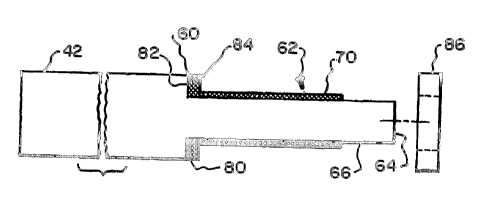
Attention is now directed to Figures 4 and 5 which illustrate a preferred
manner of configuring the housing distal end 46 in accordance with the
present invention. As shown, the housing 42 includes a lateral shoulder 60
which forms a reduced diameter stud 62 extending longitudinally therefrom
and terminating at outer end surface 64. The stud 62 defines a longitudinally
extending peripheral surface 66. The peripheral surface 66 typically has a
circular cross section but other cross sectional shapes, e.g., oval,
hexagonal,
etc. can be used. Moreover, although, the outer end surface 64 is shown as
being flat, in certain applications, it is preferable that the end surface
have a
different profile, e.g., conical or spherical.
In accordance with the invention, a first porous layer, or surface, 70 is
formed along a longitudinally extending portion of the peripheral surface 66.
The porous layer 70 is preferably formed by a mesh 72 of intersecting fibers
74 as depicted in Figure 6. The fibers can be of any suitable biocompatible
material such as a metal, e.g., titanium, nitinol, silver, or stainless steel
or a
5
CA 02520252 2005-09-23
WO 2004/091432 PCT/US2004/011079
polymeric material, e.g., polyolefins, Teflon, nylon, Dacron, or silicone. The
mesh 72 is preferably formed by cross winding the fibers 74 in multiple layers
to define a porosity conducive to promoting tissue ingrowth, e.g., with pore
sizes within a range of 50 to 200 microns and having a porosity of 60 to 95%.
The resulting porosity of the mesh 72 is a function of several factors
including
the diameter of the fibers 74 and the spacing between adjacent fibers.
The mesh 72 can be formed by directly winding the fibers 74 on the
peripheral surface 66 acting as a substrate. Alternatively, the mesh 72 can be
formed as an integral structure and then attached to the peripheral surface 66
by suitable mechanical or adhesive means. As an alternative to winding the
fiber mesh 72, a porous surface 70 can be formed by a sintered mass of
metal or polymeric material having the aforementioned porosity
characteristics.
In accordance with the invention, as seen in Figure 5, a second porous
layer, or surface, 80 is provided oriented substantially perpendicular to the
longitudinally extending porous surface 70. The porous layer 80 is formed on
the laterally oriented surface 82 formed by shoulder 60. The porous layer 80
is preferably formed by a disk 84 formed of porous material having a central
aperture for passing stud 62. The disk 84 can be adhered or mechanically
attached to the lateral shoulder surface 82. The disk 84 can be formed of a
fiber mesh (Figure 6) or a sintered mass, as previously described, to provide
porosity characteristics consistent with the previously mentioned porosity
characteristics.
Figure 8 illustrates the stud 62 percutaneously penetrating the patient's
skin layers 50 and shows how the soft tissue grows into the orthogonal porous
layers 70 and 80 to create a closed infection resistant barrier around the
stud.
The ingrowth into the porous layers 70 and 80 additionally promotes
vascularization as the dermis grows into and entwines with the mesh. It is
also pointed out that Figure 8 demonstrates the use of an optional cap 86
adapted to be mounted on the stud outer end for protection of the tissue
around the incision during the healing process.
It is pointed out that it is sometimes desirable to include one or more
substances on the stud (32 to promote tissue healing and/or resist infection
and inflammation. Suitable substances are known in the literature and
6
CA 02520252 2005-09-23
WO 2004/091432 PCT/US2004/011079
include, for example, antibiotics, silver compounds, and steroid based agents.
Such substances can be deposited on the stud 62 as shown in Figure 7, for
example, as a sublayer 90 applied to the peripheral surface 66 beneath the
porous layer 70. Alternatively, the substances can be incorporated within the
mesh or sintered material of the porous layers 70 and 80.
Figure 9A depicts the device of Figures 4 and 5 but further shows the
utilization of a transitional layer, or surface, 92 mounted on the stud
peripheral
surface 66 between the peripheral porous surface 70 and the stud outer end
64. The transitional surface 92 can have the same or a different porosity
and/or composition as the porous surface 70 and can be variously configured
as shown, for example, in Figures 9B, 9C, 9D. The transitional surface 92 is
intended to beneficially modify the healing response of the adjacent tissue
cells after implantation.
For convenience in explanation, the description thus far has mostly
merely referred to a "stud " percutaneously projecting through the patient's
skin layers. It should be understood that the term "stud" as used herein, is
not
intended to connote any particular structural configuration but rather to
generically refer to any member percutaneously projecting from an orthogonal
shoulder surface. In different applications, the stud can variously comprise a
catheter, a cable, a sensor or other member which projects percutaneously
from a lateral shoulder surface. Figure 10 depicts an exemplary application of
the invention showing a catheter, cable, or sensor 94 which projects
percutaneously for providing vascular access. Figure 11 depicts a further
exemplary application where a catheter, cable, or sensor 96 projects
percutaneously for providing access to the inner eye.
Attention is now directed to Figures 12 and 13A, 13B, 13C which depict
a preferred embodiment '100 of the invention configured for use as a hearing
aid in the manner schematically represented in Figure 1. The embodiment
100 comprises a housing 102 having a body portion 103 and a stud portion
104. The body 103 has a substantially rectangular (with rounded corners)
cross-section (Figure 13A) defined by short sides 105 and long sides 106.
The body extends longitudinally in a forward direction from a rear face 108 to
a laterally oriented shoulder surface 110. The stud 104 extends forwardly
from the shoulder surface 110 and terminates at a stud outer face 114. The
7
CA 02520252 2011-08-02
78661-23
body 103 houses electronic circuitry (not shown) for driving a sound
generator, e.g., electroacoustic transducer (not shown), mounted in the stud
proximate to the outer face 114. It is intended that when implanted, the stud
104 will project percutaneously to place the stud face 114 in the patient's
ear
canal.
As previously described, in order to promote healthy tissue ingrowth for
anchoring the housing 102 and forming a bacteria resistant barrier, a porous
layer comprising a first portion of porous material 116 is formed on the
longitudinally extending peripheral surface 118 of stud 104 and a second
portion of porous material 120 is formed on the laterally extending shoulder
surface 110.
From the foregoing, it should now be appreciated that the application
describes a method and apparatus for creating an enhanced infection
resistant barrier around a percutaneously projecting member. Embodiments
of the invention are useful in a wide variety of medical applications for
creating
an infection resistant barrier, for effective anchoring, and for avoiding the
development of adverse conditions such as marsupial izatio n,
8