Note: Descriptions are shown in the official language in which they were submitted.
CA 02520416 2005-09-21
PATENT APPLICATION
Atty. Docket: 2886 PCT CIP II (203-3427 PCT CIP II)
ELECTRODE ASSEMBLY FOR TISSUE FUSION
BACKGROUND
The present disclosure relates to forceps used for open and/or
endoscopic surgical procedures. More particularly, the present disclosure
relates to a forceps which applies a unique combination of mechanical clamping
,' pressure and electrosurgical current to micro-seal soft tissue to promote
tissue
healing.
Technical Field
A hemostat or forceps is a simple plier-like tool which uses
mechanical action between its jaws to constrict vessels and is commonly used
in open surgical procedures to grasp, dissect and/or clamp tissue:
Electrosurgical forceps utilize both mechanical clamping action and electrical
energy to effect hemostasis by heating the tissue and blood vessels to
coagulate, cauterize and/or seal tissue. The electrode of each opposing jaw
member is charged to a different electric potential such that when the jaw
members grasp tissue, electrical energy can be selectively transferred through
the tissue. A surgeon can either cauterize, coagulate/desiccate and/or simply
reduce or slow bleeding, by controlling the intensity, frequency and duration
of
the electrosurgical energy applied between the electrodes and through the
tissue.
For the purposes herein, the term "cauterization" is defined as the
use of heat to destroy tissue (also called "diathermy" or
°electrodiathermy").
The term "coagulation" is defined as a process of desiccating tissue wherein
the
tissue cells are ruptured and dried. "Vessel sealing" is defined as the
process of
liquefying the collagen, elastin and ground substances in the tissue so that
it
reforms into a fused mass with significantly-reduced demarcation between the
opposing tissue structures (opposing walls of the lumen). Coagulation of small
1
CA 02520416 2005-09-21
vessels is usually sufficient to permanently close them. Larger vessels or
tissue
need to be sealed to assure permanent closure.
Commonly-owned U.S. Application Serial Nos. PCT Application
Serial No. PCT/US01/11340 filed on April 6, 2001 by Dycus, et al. entitled
"VESSEL SEALER. AND DIVIDER", U.S. Application Serial No. 10/116,824 filed
on April 5, 2002 by Tetzlaff et al, entitled "VESSEL SEALING INSTRUMENT"
and PCT Application Serial No. PCT/US01/11420 filed on April 6, 2001 by
Tetzlaff et al. entitled "VESSEL SEALING INSTRUMENT" teach that to
effectively seal tissue or vessels, especially large vessels, two predominant
mechanical parameters must be accurately controlled: 1) the pressure applied
to the vessel; and 2) the gap distance between the conductive tissue
contacting
surfaces (electrodes). As can be appreciated, both of these parameters are
affected by the thickness of the vessel or tissue being sealed. Accurate
application of pressure is important for several reasons: to oppose the walls
of
the vessel; to reduce the tissue impedance to a low enough value that allows
enough electrosurgical energy through the tissue; to overcome the forces of
expansion during tissue heating; and to contribute to the end tissue thickness
which is an indication of a good seal. It has been determined that a typical
sealed vessel wall is optimum between 0.001 inches and 0.006 inches. Below
this range, the seal may shred or tear and above this range the lumens may not
be properly or effectively sealed.
With respect to smaller vessels, the pressure applied become less
relevant and the gap distance between the electrically conductive surfaces
becomes more significant for effective sealing. In other words, the chances of
the two electrically conductive surfaces touching during activation increases
as
the tissue thickness and the vessels become smaller.
As can be appreciated, when cauterizing, coagulating or sealing
vessels, the tissue disposed between the two opposing jaw members is
essentially destroyed (e.g., heated, ruptured and/or dried with cauterization
and
coagulation and fused into a single mass with vessel sealing). Other known
electrosurgical instruments include blade members or shearing members which
simply cut tissue in a mechanical and/or electromechanical manner and, as
such, also destroy tissue viability.
2
CA 02520416 2005-09-21
When trying to electrosurgically treat large, soft tissues (e.g., lung,
intestine, lymph ducts, etc.) to promote healing, the above-identified
surgical
treatments are generally impractical due to the fact that in each instance the
tissue or a significant portion thereof is essentially destroyed to create the
desired surgical effect, cauterization, coagulation and/or sealing. As a
result
thereof, the tissue is no longer viable across the treatment site, i.e., there
remains no feasible path across the tissue for vascularization.
Thus, a need exists to develop an electrosurgical forceps which
effectively treats tissue while maintaining tissue viability across the
treatment
area to promote tissue healing.
A need exists also to enhance sealing strength in tissue fusion by
increasing resistance to fluid flow or increased pressure at the fusion site
so as
to minimize entry of fluid into the perimeter of the fused site during burst
strength testing. The entry of fluid often results in seal failure due to
propagation of the fluid to the center of the tissue seal.
SUMMARY
It is an object of the present disclosure to provide a bipolar
electrosurgical forceps having jaw members which are configured with electrode
surfaces with a plurality of flow paths so as to increase resistance to fluid
flow
through the tissue seal zone, or increasing pressure states at the fusion
site,
thereby increasing tissue seal integrity.
The present disclosure relates to a bipolar electrosurgical forceps
which includes first and second opposing jaw members having respective tissue
engaging surfaces associated therewith. The first and second jaw members are
adapted for relative movement between an open position to receive tissue and a
closed position engaging tissue between the tissue engaging surfaces to effect
a tissue seal upon activation of the forceps. The first and second jaw
members each include an electrode having a plurality of tissue engaging
surfaces which define at least one channel therebetween. The plurality of
tissue
engaging surfaces of the first jaw member are substantially aligned with the
plurality of tissue engaging surfaces of the second jaw member so as to impede
3
CA 02520416 2005-09-21
fluid flow therebetween and force tissue fluid to flow within the at least one
channel during the sealing process.
In one embodiment, the tissue engaging surfaces of the electrodes
are disposed as pairs of longitudinal strips extending from a proximal end of
each jaw member to a distal end thereof. At least one traversally oriented
channel may be defined between respective tissue engaging surfaces on at
least one jaw member. '
In another embodiment, the tissue engaging surfaces of the
electrodes are disposed as series of longitudinal strips extending from a
proximal end of each jaw member to a distal end thereof, with the first and
second strips of the series being substantially offset relative to one
another.
In another embodiment, the tissue engaging surfaces of the
electrodes are disposed as series of longitudinal strips extending from a
proximal end of each jaw member to a distal end thereof, the first, second and
third strips of the series being substantially offset relative to one another.
BRIEF DESCRIPTION OF THE DRAWINGS
Various embodiments of the subject instrument are described
herein with reference to the drawings wherein:
FIG. 1A is a perspective view of an endoscopic forceps having an
electrode assembly in accordance with one embodiment of the present
disclosure;
FIG. 1 B is a perspective view of an open forceps having a
electrode assembly in accordance with one embodiment of the present
disclosure;
FIG. 2 is an enlarged, perspective view of the electrode assembly
of the forceps of FIG. 1 B shown in an open configuration;
FIG. 3A is an enlarged, schematic view of one embodiment of the
electrode assembly showing a pair of opposing, concentrically-oriented
electrodes disposed on a pair of opposing jaw members;
FlG. 3B is a partial, side cross-sectional view of the electrode
assembly of FIG. 3A;
4
CA 02520416 2005-09-21
FIG. 4A is an enlarged, schematic view of another embodiment of
the electrode assembly showing a plurality of concentrically-oriented
electrode
micro-sealing pads disposed on the same jaw member;
FIG. 4B is a greatly enlarged view of the area of detail in FIG. 4A
showing the electrical path during activation of the electrode assembly;
FIG. 4C is an enlarged schematic view showing the individual
micro-sealing sites and viable tissue areas between the two jaw members after
activation;
FIG. 5A is a schematic, perspective view of the jaw members
approximating tissue;
F1G. 5B is a schematic, perspective view of the jaw members
grasping tissue; and
FIG. 5C is a schematic, perspective view showing a series of
micro-seals disposed in a pattern across the tissue after activation of the
electrode assembly.
FIG. 6 is plan view of a tissue seal sealed by an electrosurgical
forceps according to the prior art showing a potential failure mechanism due
to
fluid entry into the seal perimeter;
FIG. 7A is a plan view of one jaw member of an electrosurgical
forceps having an electrode with a plurality of slots in accordance with
another
embodiment of the present disclosure;
FIG. 7B is a view of a distal end of jaw members of the
electrosurgical forceps according to FIG. 7A;
FIG. 8A is a plan view of one jaw member of an electrosurgical
forceps having an electrode with a plurality of slots in accordance with
another
embodiment of the present disclosure;
FIG. 8B is a view of a distal end of jaw members of the
electrosurgical forceps according to FIG. 8A;
FIG. 9A is a perspective view of one jaw member of an
electrosurgical forceps having an electrode with a plurality of slots in
accordance
with another embodiment of the present disclosure;
FIG. 9B is a view of a distal end of jaw members of the
electrosurgical forceps according to FIG. 9A;
CA 02520416 2005-09-21
FIG. 10A is a plan view of one jaw member of an electrosurgical
forceps having an array of individual electrodes in accordance with another
embodiment of the present disclosure; and
FIG. 10B is an elevation view of an end effector assembly of an
eiectrosurgical forceps having jaw members according to FIG. 10A.
DETAILED DESCRIPTION
This application incorporates by reference herein in its entirety
concurrently filed, commonly owned U.S. Patent Application Serial No.
(attorney docket no.: 2886 PCT CIP (203-3427 PCT CIP)) by
Odom et al entitled "BIPOLAR FORCEPS WITH MULTIPLE ELECTRODE
ARRAY END EFFECTOR ASSEMBLY."
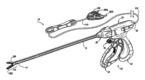
Referring now to FIG. 1A, a bipolar forceps 10 is shown for use
with various surgical procedures. Forceps 10 generally includes a housing 20,
a handle assembly 30, a rotating assembly 80, an activation assembly 70 and
an electrode assembly 110 which mutually cooperate to grasp and seal tissue
600 (See FIGS. 5A-5C). Although the majority of the figure drawings depict a
bipolar forceps 10 for use in connection with endoscopic surgical procedures,
an
open forceps 200 is also contemplated for use in connection with traditional
open surgical procedures and is shown by way of example in FIG. 1 B and is
described below. For the purposes herein, either an endoscopic instrument or
an open instrument may be utilized with the electrode assembly described
herein. Obviously, different electrical and mechanical connections and
considerations apply to each particular type of instrument, however, the novel
aspects with respect to the electrode assembly and its operating
characteristics
remain generally consistent with respect to both the open or endoscopic
designs.
More particularly, forceps 10 includes a shaft 12 which has a distal
end 14 dimensioned to mechanically engage a jaw assembly 110 and a
proximal end 16 which mechanically engages the housing 20. The shaft 12 may
be bifurcated at the distal end 14 thereof to receive the jaw assembly 110.
The
proximal end 16 of shaft 12 mechanically engages the rotating assembly 80 to
facilitate rotation of the jaw assembly 110. In the drawings and in the
6
CA 02520416 2005-09-21
descriptions which follow, the term "proximal", as is traditional, will refer
to the
end of the forceps 10 which is closer to the user, while the term "distal"
will refer
to the end which is further from the user.
Forceps 10 also includes an electrical interface or plug 300 which
connects the forceps 90 to a source of electrosurgical energy, e.g., an
electrosurgical generator 350 (See FIG. 3B). Plug 300 includes a pair of prong
members 302a and 302b which are dimensioned to mechanically and
electrically connect the forceps 10 to the electrosurgical generator 350. An
electrical cable 310 extends from the plug 300 to a sleeve 99 which securely
connects the cable 310 to the forceps 10. Cable 310 is internally divided
within
the housing 20 to transmit electrosurgicai energy through various electrical
feed
paths to the jaw assembly 110 as explained in more detail below.
Handle assembly 30 includes a fixed handle 50 and a movable
handle 40. Fixed handle 50 is integrally associated with housing 20 and handle
40 is movable relative to fixed handle 50 to actuate a pair of opposing jaw
members 280 and 282 of the jaw assembly 110 as explained in more detail
below. The activation assembly 70 is selectively movable by the surgeon to
energize the jaw assembly 110. Movable handle 40 and activation assembly 70
are typically of unitary construction and are operatively connected to the
housing 20 and the fixed handle 50 during the assembly process.
As mentioned above, jaw assembly 110 is attached to the distal
end 14 of shaft 12 and includes a pair of opposing jaw members 280 and 282.
Movable handle 40 of handle assembly 30 imparts movement of the jaw
members 280 and 282 about a pivot pin 119 from an open position wherein the
jaw members 280 and 282 are disposed in spaced relation relative to one
another for approximating tissue 600, to a clamping or closed position wherein
the jaw members 280 and 282 cooperate to grasp tissue 600 therebetween
(See FIGS. 5A-5C).
It is envisioned that the forceps 10 may be designed such that it is
fully or partially disposable depending upon a particular purpose or to
achieve a
particular result. For example, jaw assembly 110 may be selectively and
releasably engageable with the distal end 14 of the shaft 12 and/or the
proximal
end 16 of shaft 12 may be selectively and releasably engageable with the
7
CA 02520416 2005-09-21
housing 20 and the handle assembly 30. In either of these two instances, the
forceps 10 would be considered "partially disposable" or °reposable",
i.e., a new
or different jaw assembly 110 (or jaw assembly 110 and shaft 12) selectively
replaces the old jaw assembly 110 as needed.
Referring now to FIGS. 1 B and 2, an open forceps 200 includes a
pair of elongated shaft portions 212a each having a proximal end 216a and
216b, respectively, and a distal end 214a and 214b, respectively. The forceps
200 includes jaw assembly 210 which attaches to distal ends 214a and 214b of
shafts 212a and 212b, respectively. Jaw assembly 210 includes opposing jaw
members 280 and 282 which are pivotabfy connected about a pivot pin 219.
Each shaft 212a and 212b includes a handle 217a and 217b
disposed at the proximal end 216a and 216b thereof which each define a finger
hole 218a and 218b, respectively, therethrough for receiving a finger of the
user.
As can be appreciated, finger holes 218a and 218b facilitate movement of the
shafts 212a and 212b relative to one another which, in turn, pivot the jaw
members 280 and 282 from an open position wherein the jaw members 280 and
282 are disposed in spaced relation relative to one another for approximating
tissue 600 to a clamping or closed position wherein the jaw members 280 and
282 cooperate to grasp tissue 600 therebetween. A ratchet 230 is included for
selectively locking the jaw members 280 and 282 relative to one another at
various positions during pivoting.
Each position associated with the cooperating ratchet interfaces
230 holds a specific, i.e., constant, strain energy in the shaft members 212a
and
212b which, in tum, transmits a specific closing force to the jaw members 280
and 282. It is envisioned that the ratchet 230 may include graduations or
other
visual markings which enable the user to easily and quickly ascertain and
control the amount of closure force desired between the jaw members 280 and
282.
One of the shafts, e.g., 212b, includes a proximal shaft connector
(flange 221 which is designed to connect the forceps 200 to a source of
electrosurgical energy such as an electrosurgical generator 350 (FIG. 3B).
More particularly, flange 221 mechanically secures electrosurgical cable 310
to
the forceps 200 such that the user may selectively apply electrosurgical
energy
8
CA 02520416 2005-09-21
as needed. The proximal end of the cable 310 includes a similar plug 300 as
described above with respect to FIG. 1A. The interior of cable 310 houses a
pair of leads which conduct different electrical potentials from the
electrosurgical
generator 350 to the jaw members 280 and 282 as explained below with respect
to FIG. 2.
The jaw members 280 and 282 are generally symmetrical and
include similar component features which cooperate to permit facile rotation
about pivot 219 to effect the grasping of tissue 600. Each jaw member 280 and
282 includes a non-conductive. tissue contacting surface 284 and 286,
respectively, which cooperate to engage the tissue 600 during treatment.
As best shown in FIG. 2, the various electrical connections of the
electrode assembly 210 are typically configured to provide electrical
continuity to
an array of electrode micro-sealing pads 500 of disposed across one or both
jaw
members 280 and 282. The electrical paths 416, 426 or 516, 526 from the
array of electrode micro-sealing pads 500 are typically mechanically and
electrically interfaced with corresponding electrical connections (not shown)
disposed within shafts 212a and 212b, respectively. As can be appreciated,
these electrical paths 416, 426 or 516, 526 may be permanently soldered to the
shafts 212a and 212b during the assembly process of a disposable instrument
or, alternatively, selectively removable for use with a reposable instrument.
As best shown in FIGS. 4A-4C, the electrical paths are connected
to the plurality of electrode micro-sealing pads 500 within the jaw assembly
210.
More particularly, the first electrical path 526 (i.e., an electrical path
having a
first electrical potential) is connected to each ring electrode 522 of each
electrode micro-sealing pad 500. The second electrical path 516 (i.e., an
electrical path having a second electrical potential) is connected to each
post
electrode 522 of each electrode micro-sealing pad 500.
The electrical paths 516 and 526 typically do not encumber the
movement of the jaw members 280 and 282 relative to one another during the
manipulation and grasping of tissue 400. Likewise, the movement of the jaw
members 280 and 282 do not unnecessarily strain the electrical paths 516 and
526 or their respective connections 517, 527.
9
CA 02520416 2005-09-21
As best seen in FIGS. 2-5C, jaw members 280 and 282 both
include non-conductive tissue contacting surfaces 284 and 286, respectively,
disposed along substantially the entire longitudinal length thereof (i.e.,
extending
substantially from the proximal to distal end of each respective jaw member
280
and 284). The non-conductive tissue contacting surfaces 284 and 286 may be
made from an insulative material such as ceramic due to its hardness and
inherent ability to withstand high temperature fluctuations. Alternatively,
the
non-conductive tissue contacting surfaces 284 and 286 may be made from a
material or a combination of materials having a high Comparative Tracking
Index (CTI) in the range of about 300 to about 600 volts. Examples of high CTI
materials include nylons and syndiotactic polystryrenes such as QUESTRA~
manufactured by DOW Chemical. Other materials rnay also be utilized either
alone or in combination, e.g., Nylons, Syndiotactic-polystryrene (SPS),
Polybutylene Terephthalate (PBT), Polycarbonate (PC), Acrylonitrile Butadiene -
Styrene (ABS), Polyphthalamide (PPA), Polymide, Polyethylene Terephthalate
(PET), Polyamide-imide (PAI), Acrylic (PMMA), Polystyrene (PS and HIPS),
Polyether Sulfone (PES), Aliphatic Polyketone, Acetal (POM) Copolymer,
Polyurethane (PU and TPU), Nylon with Polyphenylene-oxide dispersion and
Acrylonitrile Styrene Acrylate. Typically, the non-conductive tissue
contacting
surfaces 284 and 286 are dimensioned to securingly engage and grasp the
tissue 600 and may include serrations (not shown) or roughened surfaces to
facilitate approximating and grasping tissue.
It is envisioned that one of the jaw members, e.g., 282, includes at
least one stop member 235a, 235b (FIG. 2) disposed on the inner facing
surface of the sealing surfaces 286. Alternatively or in addition, one or more
stop members 235a, 235b may be positioned adjacent to the non-conductive
sealing surfaces 284, 286 or proximate the pivot 219. The stop members 235a,
235b are typically designed to define a gap "G" (FIG. 5B) between opposing jaw
members 280 and 282 during the micro-sealing process. The separation
distance during micro-sealing or the gap distance "G" is within the range of
about 0.001 inches 00.03 millimeters) to about 0.006 inches (0.016
millimeters). One or more stop members 235a, 235b may be positioned on the
distal end and proximal end of one or both of the jaw members 280, 282 or may
CA 02520416 2005-09-21
be positioned between adjacent electrode micro-sealing pads 500. Moreover,
the stop members 235a and 235b may be integrally associated with the non-
conductive tissue contacting surfaces 284 and 286. It is envisioned that the
array of electrode micro-sealing pads 500 may also act as stop members for
regulating the distance "G" between opposing jaw members 280, 282 (See FIG.
4C).
As mentioned above, the effectiveness of the resulting micro-seal
is dependent upon the pressure applied between opposing jaw members 280
and 282, the pressure applied by each electrode micro-sealing pad 500 at each
micro-sealing site 620 (FIG. 4C), the gap "G" befween the opposing jaw
members 280 and 282 (either regaled by a stop member 235a, 235b or the
array of electrode micro-sealing pads 500) and the control of the
electrosurgical
intensity during the micro-sealing process. Applying the correct force is
important to oppose the walls of the tissue; to reduce the tissue impedance to
a
low enough value that allows enough current through the tissue; and to
overcome the forces of expansion during tissue heating in addition to
contributing towards creating the required end tissue thickness which is an
indication of a good micro-seal. Regulating the gap distance and regulating
the
electrosurgical intensity ensure a consistent seal quality and reduce the
likelihood of collateral damage to surrounding tissue.
As best shown in FIG. 2, the electrode micro-sealing pads 500 are
arranged in a longitudinal, pair-like fashion along the tissue contacting
surtaces
286 andlor 284. Two or more micro-sealing pads 500 may extend transversally
across the tissue contacting surface 286. FIGS. 3A and 3B show one
embodiment of the present disclosure wherein the electrode micro-sealing pads
500 include a ring electrode 422 disposed on one jaw members 282 and a post
electrode 412 disposed on the other jaw member 280. The ring electrode 422
includes an insulafing material 424 disposed therein to form a ring electrode
and
insulator assembly 420 and the post electrode 422 includes an insulating
material disposed therearound to form a post electrode and insulator assembly
430. Each post electrode assembly 430 and the ring electrode assembly 420 of
this embodiment together define one electrode micro-sealing pad 400.
Although shown as a circular-shape, ring electrode 422 may assume any other
11
CA 02520416 2005-09-21
r
annular or enclosed configuration or alternatively parfially enclosed
configuration such as a C-shape arrangement.
As best shown in F1G. 3B, the post electrode 422 is concentrically
centered opposite the ring electrode 422 such that when the jaw members 280
and 282 are closed about the tissue 600, electrosurgical energy flows from the
ring electrode 422, through tissue 600 and to the post electrode 412. The
insulating materials 414 and 424 isolate the electrodes 412 and 422 and
prevent stray current tracking to surrounding tissue. Alternatively, the
electrosurgical energy may flow from the post electrode 412 to the ring
electrode 422 depending upon a particular purpose.
FIGS. 4A-4.C show an alternate embodiment of the jaw assembly
210 according to the present disclosure for micro-sealing tissue fi00 wherein
each electrode micro-sealing pad 500 is disposed on a single jaw member, e.g.,
jaw member 280. More particularly and as best illustrated in FIG. 4B, each
electrode micro-sealing pad 500 consists of an inner post electrode 512 which
is
surrounded by an insulative material 514, e.g., ceramic. The insulative
material
514 is, in turn, encapsulated by a ring electrode 522. A second insulative
material 535 (or the same insulative material 514) may be configured to encase
the ring electrode 522 to prevent stray electrical currents to surrounding
tissue.
The ring electrode 522 is connected to the electrosurgical
generator 350 by way of a cable 526 (or other conductive path) which transmits
a first electrical potential to each ring electrode 522 at connection 527. The
post electrode 512 is connected to the eiectrosurgical generator 350 by way of
a
cable 516 (or other conductive path) which transmits a second electrical
potential to each post electrode 522 at connection 517. A controller 375 (See
FIG. 4B) may be electrically interposed befinreen the generator 350 and the
electrodes 512, 522 to regulate the electrosurgical energy supplied thereto
depending upon certain electrical parameters, current impedance, temperature,
voltage, etc. For example, the instrument or the controller may include one or
more smart sensors (not shown) which communicate with the electrosurgical
generator 350 (or smart circuit, computer, feedback loop, etc.) to
automatically
regulate the electrosurgical intensity (waveform, cun-ent, voltage, etc.) to
enhance the micro-sealing process. The sensor may measure or monitor one
12
CA 02520416 2005-09-21
or more of the following parameters: tissue temperature, tissue impedance at
the micro-seal, change in impedance of the tissue over time andlor changes in
the power or current applied to the tissue over time. An audible or visual
feedback monitor (not shown) may be employed to convey information to the
surgeon regarding the overall micro-seal quality or the completion of an
effective
tissue micro-seal.
Moreover, a PCB circuit of flex circuit (not shown) may be utilized
to provide information relating to the gap distance (e.g., a proximity
detector
may be employed) between the two jaw members 280 and 282, the micro-
sealing pressure between the jaw members 280 and 282 prior to and during
activation, load (e.g., strain gauge may be employed), the tissue thickness
prior
to or during activation, the impedance across the tissue during activation,
the
temperature during activation, the rate of tissue expansion during activation
and
micro-sealing. It is envisioned that the PCB circuit may be designed to
provide
electrical feedback to the generator 350 relating to one or more of the above
parameters either on a continuous basis or upon inquiry from the generator
350.
For example, a PCB circuit may be employed to control the power, current
and/or type of current waveform from the generator 350 to the jaw members
280, 282 to reduce collateral damage to surrounding tissue during activation,
e.g., thermal spread, tissue vaporization andlor steam from the treatment
site.
Examples of a various control circuits, generators and algorithms which may be
utilized are disclosed. in U.S. Patent No 6,228,080 and U.S. Application
Serial
No. 10/073,761 the entire contents of both of which are hereby incorporated by
reference herein.
In use as depicted in FIGS. 5A-5C, the surgeon initially
approximates the tissue (FIG. 5A) between the opposing jaw member 280 and
282 and then grasps the tissue 600 (FIG. 5B) by actuating the jaw members
280, 282 to rotate about pivot 219. Once the tissue is grasped, the surgeon
selectively activates the generator 350 to supply electrosurgical energy to
the
array of the electrode micro-sealing pads 500. More particularly,
electrosurgical
energy flows from the ring electrode 522, through the tissue 600 and to the
post
electrode 512 (See FIGS. 4B and 4C). As a result thereof, an intermittent
pattern of individual micro-seals 630 is created along and across the tissue
600
13
CA 02520416 2005-09-21
(See FIG. 5C). The arrangement of the micro-sealing pads 500 across the
tissue only seals the tissue which is between each micro-sealing pad 500 and
the opposing jaw member 282. The adjacent tissue remains viable which, as
can be appreciated, allows blood and nutrients to flow through the sealing
site
620 and between the individual micro-seals 630 to promote tissue healing and
reduce the chances of tissue necrosis. By selectively regulating the closure
pressure "F", gap distance "G", and electrosurgical intensity, effective and
consistent micro-seals 630 may be created for many different tissue types.
It is further envisioned that selective ring electrodes and post
electrodes may have varying electric potentials upon activation. For example,'
at or proximate the distal tip of one of the jaw members, one or a series of
electrodes may be electrically connected to a first potential and the
corresponding electrodes (either on the same jaw or perhaps the opposing jaw)
may be connected to a second potential. Towards the proximal end of the jaw
member, one or a series of electrodes may be connected to a third potential
and
the corresponding electrodes connected to yet a fourth potential. As can be
appreciated, this would allow different types of tissue sealing to take place
at
different portions of the jaw members upon activation. For example, the type
of
sealing could be based upon the type of tissues involved or perhaps the
thickness of the tissue. To seal larger tissue, the user would grasp the
tissue
more towards the proximal portion of the opposing jaw members and to seal
smaller tissue, the user would grasp the tissue more towards the distal
portion
of the jaw members. It is also envisioned that the pattern and/or density of
the
micro-sealing pads may be configured to seal different types of tissue or
thicknesses of tissue along the same jaw members depending upon where the
tissue is grasped between opposing jaw members.
From the foregoing and with reference to the various figure
drawings, those skilled in the art will appreciate that certain modifications
can
also be made to the present disclosure without departing from the scope of the
same. For example, it is envisioned that by making the forceps 100, 200
disposable, the forceps 100, 200 is less likely to become damaged since it is
only intended for a single use and, therefore, does not require cleaning or
sterilization. As a result, the functionality and consistency of the vital
micro-
14
CA 02520416 2005-09-21
sealing components, e.g., the conductive micro-sealing electrode pads 500, the
stop members) 235a, 235b, and the insulative materials 514, 535 will assure a
uniform and quality seal.
Experimental results suggest that the magnitude of pressure
exerted on the tissue by the micro-sealing pads 112 and 122 is important in
assuring a proper surgical outcome, maintaining tissue viability. Tissue
pressures within a working range of about 3 kglcm2 to about 16 kg/cm2 and,
more particularly, within a working range of 7 kg/cmz to 13 kg/cm2 have been
shown to be effective for micro-sealing various tissue types and vascular
bundles.
In one embodiment, the shafts 212a and 212b are manufactured
such that the spring constant of the shafts 212a and 212b, in conjunction with
the placement of the interfacing surtaces of the ratchet 230, will yield
pressures
within the above working range. In addition, the successive positions of the
ratchet interfaces increase the pressure between opposing micro-sealing
surfaces incrementally within the above working range.
It is envisioned that the outer surface of the jaw members 280 and
282 may include a nickel-based material or coating which is designed to reduce
adhesion between the jaw members 280, 282 (or components thereof) with the
surrounding tissue during activation and micro-sealing. Moreover, it is also
contemplated that other components such as the shaft portions 212a, 212b and
the rings 217a, 217b may also be coated with the same or a different "non-
stick"
material. Typically, the non-stick materials are of a class of materials that
provide a smooth surface to prevent mechanical tooth adhesions.
It is also contemplated that the tissue contacting portions of the
electrodes and other portions of the micro-sealing pads 400, 500 may also be
made from or coated with non-stick materials. When utilized on these tissue
contacting surfaces, the non-stick materials provide an optimal surface energy
for eliminating sticking due in part to surface texture and susceptibility to
surface
breakdown due electrical effects and corrosion in the presence of biologic
tissues. It is envisioned that these materials exhibit superior non-stick
qualities
over stainless steel and should be utilized in areas where the exposure to
pressure and electrosurgical energy can create localized "hot spots" more
CA 02520416 2005-09-21
susceptible to tissue adhesion. As can be appreciated, reducing the amount
that the tissue "sticks" during micro-sealing improves the overall efficacy of
the
instrument.
The non-stick materials may be manufactured from one (or a
combination of one or more) of the following "non-stick" materials: nickel-
chrome, chromium nitride, MedCoat 2000 manufactured by The Electrolizing
Corporation of OHIO, Inconel 600 and tin-nickel. Inconel 600 coating is a so-
called "super alloy" which is manufactured by Special Metals, Inc. located in
Conroe Texas. The alloy is primarily used in environments which require
resistance to con-osion and heat. The high Nickel content of Inconel 600 makes
the material especially resistant to organic corrosion. As can be appreciated,
these properties are desirable for bipolar electrosurgical instruments which
are
naturally exposed to high temperatures, high RF energy and organic matter.
Moreover, the resistivity of Inconel 600 is typically higher than the base
electrode material which further enhances desiccation and micro-seal quality.
One particular class of materials disclosed herein has
demonstrated superior non-stick properties and, in some instances, superior
micro-seal quality. For example, nitride coatings which include, but not are
not
limited to: TiN, ZrN, TiAIN, and CrN are preferred materials used for non-
stick
purposes. CrN has been found to be particularly useful for non-stick purposes
due to its overall surface properties and optimal performance. Other classes
of
materials have also been found to reducing overall sticking. For example, high
nickel/chrome alloys with a Ni/Cr ratio of approximately 5:1 have been found
to
significantly reduce sticking in bipolar instrumentation.
It is also envisioned that the micro-sealing pads 400, 500 may be
arranged in many different configurations across or along the jaw members 280,
282 depending upon a particular purpose. Moreover, it is also contemplated
that a knife or cutting element (not shown) may be employed to sever the
tissue
600 ~ between a series of micro-sealing pads 400, 500 depending upon a
particular purpose. The cutting element may include a cutting edge to simply
mechanically cut tissue 600 andlor may be configured to electrosurgically cut
tissue 600.
16
CA 02520416 2005-09-21
FIG. 6 discloses a resulting tissue seal sealed by an
electrosurgical forceps according to the prior art showing a potentially
weaker
seal area due to entry of fluid into the seal perimeter during sealing. More
particularly, tissue 600 of a lumen 602 of a patient's body such as the large
or
small intestines or any other passage or vessel is subject to a tissue seal
604
performed by an electrosurgical forceps of the prior art (not shown). The
tissue
seal 604 is typically formed utilizing radiofrequency (Rf=) energy. The lumen
602
has an approximate centerline axis X-X'. The seal 604 has a perimeter
generally of four contiguous sides 604a, 604b, 604c and 604d and a central
portion 606. Two sides 604a and 604c extend in a direction generally
orthogonal to the centerline axis X-X' of the lumen 602 and parallel to each
other, while the two sides 604b and 604d extend in a direction generally
parallel
to the centerline axis X-X'. It has been determined that during sealing, fluid
608 -
may enter at a side of the perimeter such as side 604a and propagate to the
central portion 606 of the tissue seal 604. A weaker seal may develop as a
result of increased fluid in a particular tissue area.
FIG. 7A illustrates one embodiment of a jaw member 720 of an
electrode assembly 700 for use with an electrosurgical forceps which includes
an electrode 721 with a plurality of slots or channels 732a and 732b. More
particularly, electrode 721 of jaw member 722 of electrode assembly 700
includes a substantially longitudinal, planar, tissue engaging surface 730
which
has at least first channel 732a, and typically includes a second channel 732b.
Each channel 732a and 732b is disposed in a lengthwise direction from a
proximal end 705 to a distal end 706 of the electrode 721 so as to divide
surtace
730 into at least two substantially longitudinal surfaces 730a and 730c. A
third
substantially longitudinal surface 730b is disposed between channels 732a and
732c.
FIG. 7B shows upper jaw member 710 of electrode assembly 700.
More particularly, upper jaw member 710 is similar to jaw member 720 and
includes a corresponding electrode member 711 which has a substantially
longitudinal, planar, tissue engaging surface 740. Jaw members 710 and 720
are pivotably connected around a pivot pin 719, and are movable from an open
position wherein the jaw members 710 and 720 are disposed in spaced relation
17
CA 02520416 2005-09-21
relative to one another for manipulating tissue 600, to a clamping or closed
position wherein the jaw members 710 and 720 cooperate to grasp tissue 600
therebetween. Jaw members 710 and 720 operate in an analogous manner as
described previously with respect to jaw members 280 and 282 (See FIGS. 5A-
5C).
Surface 740 includes at least a first channel 742a and typically
includes a second channel 742b. Each channel 742a and 742b is disposed in a
lengthwise direction from a proximal end 705 to a distal end 706 of the
electrode
710 so as to divide surface 740 into surfaces 740a, 740b, and 740c. Surface
730 of jaw member 720 and surface 740 of jaw member 710 are configured so
that channels 742a and 742b substantially correspond to channels 732a and
732b, and consequently, so that the surfaces 730a, 730b and 730c substantially
correspond with or are in vertical registration with surfaces 740a, 740b and
740c.
The corresponding or counterpart channels 732a and 742a, and
the corresponding or counterpart channels 732b and 742b form a plurality of
corresponding or counterpart electrode surfaces 730a and 740a, 730b and
740b, and 730c and 740c which form tissue seals characterized by potential
tissue fluid flow paths. It is envisioned that arranging the electrodes 711
and
721 in this fashion will impede the flow of tissue fluid during the sealing
process
which allows a stronger seal to develop. In other words, the envisioned
electrode 711 and 721 arrangement with channels 732a-732c and 742a-742c
inhibits the flow of fluid through the tissue seal, thereby increasing the
structural
integrity of the tissue seal and decreasing the probability of tissue seal
rupture.
FIG. 8A illustrates a jaw member 820 of an electrosurgical forceps
having an electrode arrangement in accordance with yet another embodiment of
the present disclosure. More particularly, an electrode 821 of jaw member 820
of an electrode assembly 800 includes a substantially longitudinal, planar,
tissue engaging electrode surface 830 which has a plurality of longitudinal
and
transverse or traversally oriented channels 832a and 832b and 834a to 834c,
respectively, which extend lengthwise from proximal end 805 to distal end 806
and across the jaw member 820.
18
CA 02520416 2005-09-21
Referring to FIG. 8B, jaw member 810 includes or is characterized
by a similar arrangement. An electrode 811 of jaw member 810 of electrode
assembly 800 has a substantially longitudinal, planar tissue engaging surface
840 which includes longitudinal channels 842a and 842b and transverse
channels 844a to 844c.
Jaw member 810 and jaw member 820 are pivotably connected
around pivot pin 819 such that jaw members 810 and 820 are movable from an
open position wherein the jaw members 810 and 820 are disposed in spaced
relation relative to one another for manipulating tissue 600, to a clamping or
closed position wherein the jaw members 810 and 820 cooperate to grasp
tissue 600 therebetween in a similar manner to jaw members 280 and 282 (see
FIGS. 5A-5C).
Much like the electrode arrangement of FIGS. 7A and 7B, the
electrode tissue engaging surface pattern and channels of each jaw member
810 and 820 are arranged to complement each other to produce a uniform and
effective seal. It is envisioned that the fluid path during sealing will be
impeded
such that a uniform, reliable and effective seal will develop upon activation
of
the electrodes 811 and 821.
FIG. 9A illustrates a jaw member 920 of an electrosurgical forceps
in accordance with still another embodiment of the present disclosure. More
particularly, an electrode 921 of jaw member 920 of an electrode assembly 900
has a substantially longitudinal, planar, tissue engaging electrode surtace
930.
The electrode 921 includes a proximal end 905 and a distal end 906 and is
bounded by first and second lateral side edges 970 and 972, respectively. The
surface 930 includes a first group 931 of substantially longitudinal slots 932
and
934 aligned in a column oriented from the proximal end 905 to the distal end
906. In one embodiment, the surface 930 includes a second group 941 of
substantially longitudinal slots 942, 944 and 946 aligned in a column oriented
from the proximal end 905 to the distal end 906. The first group 931 and the
second group 941 are disposed on the jaw surface 930 such that the slots 932
and 934 are staggered with respect to the slots 942, 944 and 946.
Referring to FIG. 9B, jaw member 910 includes or is characterized
by a similar arrangement. An electrode 911 of jaw member 910 of an electrode
19
CA 02520416 2005-09-21
assembly 900 has a substantially longitudinal, planar, tissue engaging
electrode
surface 950 which includes a first group 951 of substantially longitudinal
slots
952 and 954 aligned in a column oriented from a proximal end 907 to a distal
end 908. The electrode 911 is bounded by lateral side edges 974 and 976. In
one embodiment, the surtace 950 includes a second group 961 of substantially
longitudinal slots 962, 964 and 966 aligned in a column oriented from the
proximal end 907 to the distal end 908. The first group 951 and the second
group 961 are disposed on the jaw surface 950 such that the slots 952 and 954
are staggered with respect to the slots 962, 964 and 966. Furthermore, the
first
group 931 corresponds with or is in vertical registration with first group
951.
Similarly, the second group 941 corresponds with or is in vertical
registration
with second group 961. The embodiments are not limited in this context.
Jaw member 910 and jaw member 920 are pivotably connected
around pivot pin 919 such that jaw members 910 and 920 are movable from an
open position wherein the jaw members 910 and 920 are disposed in spaced
relation relative to one another for manipulating tissue 600, to a clamping or
closed position wherein the jaw members 910 and 920 cooperate to grasp
tissue 600 therebetween in a similar manner to jaw members 280 and 282 (see
FIGS. 5A-5C).
Yet again, the staggered slot arrangement forms a tissue seal
characterized by a plurality of potential flow paths. Much like the electrode
arrangements of FIGS. 7A and 7B, and 8A and 8B, the electrode tissue-
engaging surface patterns and channels of each jaw member 910 and 920 are
arranged to complement each other to produce a uniform and effective seal. It
is envisioned that the fluid path during sealing will be impeded such that a
uniform, reliable and effective seal will develop upon activation of the
electrodes
911 and 921.
FIGS. 10A and 10B show another example of an electrode
arrangement across the surface of a jaw member 1020. More particularly,
electrode 1021 includes one or more arrays of tissue-engaging surfaces 1032,
1042 and 1052 which are patterned across the jaw surface 1030 to impede fluid
flow during activation which is believed to result in a stronger and more
reliable
seal. In the particular tissue-engaging surface arrangement of FIGS. 10A and
CA 02520416 2005-09-21
10B, a similar pattern is envisioned wherein arrays 1032, 1042 and 1052 are
disposed within groups to define slots or flow restricting areas 1031a through
1031f similar to previously described FIGS. 9A and 9B above. Jaw housing
1030 is made typically from an electrically and thermally insulating material
such as a temperature resistant plastic or a ceramic or a cool polymer which
thermally conducts heat but which is an electrical insulator. Housing 1030
includes an inwardly facing surface 1025 which supports the various arrays of
tissue engaging surfaces 1032, 1042 and 1052.
The arrays 1032, 1042 and 1052 are staggered along the length
and width of the jaw surface 1025 with respect to one another. It is believed
that this electrode arrangement will further impede fluid flow during
electrode
activation by forcing fluid flow to occur substantially around the electrodes
and
substantially through slots or flow restricting areas 1031a through 1031f -
between the array of surfaces 1032, 1042 and 1052, resulting in a more
reliable
seal. It is also envisioned that other staggered patterns with a greater or
lesser
number of surface arrays may be employed to strengthen a tissue seal
depending upon a particular tissue type.
With particular respect to FIG. 10A, the tissue-engaging surfaces
within the arrays 1032, 1042, and 1052 are arranged such that the electrode
1021 carries an electrical potential from generator 350 through lead or leads
1060 to tissue upon electrical activation. It is also envisioned that each
tissue-
engaging surface of each array of tissue-engaging surtaces may be individually
connected to the generator 350. Commonly owned, concurrently filed U.S.
Patent Application Serial No. (attorney docket no.: 2886 PCT CIP
(203-3427 PCT CIP)) by Odom et al entitled "BIPOLAR FORCEPS WITH
MULTIPLE ELECTRODE ARRAY END EFFECTOR ASSEMBLY" discusses
several advantages and ways to connect one or more electrodes to accomplish
various surgical purposes.
In one embodiment, FIG. 10B shows opposing arrays of tissue-
engaging surtaces 1032 and 1033 of jaw members 1020 and 1010, respectively,
each connected to a corresponding common element, e.g., conductive
electrodes or plates 1021 and 1031, respectively. Each conductive plate 1021
and 1031 carries a different electrical potential through a series of
conductive
21
CA 02520416 2005-09-21
connections 1072 and 1082 to each respective array 1032 and 1033. As can be
appreciated, it is envisioned that arranging the an-ays in this fashion
facilitates
manufacturing such that arrays 1032 and 1033 and conductive plates 1021 and
1031 may be held in a die or support toot which the outer housings 1030 and
1040 are overmolded.
The jaw members 1010 and 1020, which are pivotably connected
at or in the vicinity of their proximal ends 1005 and 1007 around a pivot pin
1019, from an open position wherein the jaw members 1010 and 1020 are
disposed in spaced relation relative to one another for approximating tissue
600,
to a clamping or closed position wherein the jaw members 1010 and 1020
cooperate to grasp tissue 600 therebetween in a similar manner to jaw
members 280 and 282 (see FIGS. 5A-5C).
It is envisioned that the tissue engaging surfaces 730, 830, 930,
1030 and 740, 840, 940 and 1040 of the electrodes are disposed as a series of
longitudinal strips extending from a proximal end of each jaw member to a
distal
end thereof, the first and second strips being substantially offset relative
to one
another.
It is also contemplated that the various aforedescribed electrode
arrangements may be configured for use with either an open forceps as shown
in FIG. 1B or an endoscopic forceps as shown in FIG. 1A. One skilled in the
art
would recognize that different but known electrical and mechanical
considerations would be necessary and apparent to convert an open instrument
to an endoscopic instrument to accomplish the same purposes as described
herein.
While several embodiments of the disclosure have been shown in
the drawings, it is not intended that the disclosure be limited thereto, as it
is
intended that the disclosure be as broad in scope as the art will allow and
that
the specification be read likewise. Therefore, the above description should
not
be construed as limiting, but merely as exemplifications of preferred
embodiments. Those skilled in the art will envision other modifications within
the scope and spirit of the claims appended hereto.
22