Note: Descriptions are shown in the official language in which they were submitted.
CA 02520508 2005-09-23
WO 2004/084761 PCT/IL2004/000259
1
HYBRID INTERLOCKING PROXIMAL FEMORAL
FRACTURE FIXATION
FIEL~ OF THE INVEI~TfON
The present invention relates to fraciure fixation. f'Ulore particularly, the
present invention relates to hybrid interlocking proximal femoral fracture
fixation.
15
BACKGROUND OF THE INVENTION
Internal fixation with nails and plates is a well-known surgical procedure
used in orthopaedics and traumatology for stabilization of proximal femoral
fractures. This procedure is considered as a classical open major surgery
carrying out several possibilities of serious complications. It was considered
in
the past that rigidity of the fracture fixation site is advantageous,
therefore,
many of the available internal fixation devices are built so as to eliminate
all
movements (except of sliding possible motion) at the fracture site. It is now
generally accepted that some micro-movements at the fracture site are
essential for better fracture healing and even for stimulating callus
formatio~~a:
However, this conception is not valid for intracapsular femoral neck
fractures.
Internal fixation bears many disadvantages including the fact that the
surgery is highly expensive and complex, which may be complicated by
significant blood loss and infection. There is a lack of ability to perform
post-
operative re-fixati~n, the morbidity and mortality rates are high and as a
consequence of the surgery, there is a prolonged hospitalization related to
peri-operative complications. The death rates following internal fixation in
CA 02520508 2005-09-23
WO 2004/084761 PCT/IL2004/000259
2
cases of subcapital (intracapsular) fractures are intimidating: 3% in the
hospital, 25% at one year and additional 40% at two years following the
surgery. 30% experience avascular necrosis, 43% non-union and 50%
experience peri-operative - postoperative local and systemic complications.
The data is collected from Clinical Grthopaedics and Related Research
343:22-23, 1993; Clinical ~rthopaedics and Related Research 399:119-123,
2002. These papers are incorporated herein as references; however, similar
results were established and published in many other scientific reviewed
publications. The consequences of interkrochanteric-pertrochanteric
(extracapsular) fractures are no less frightening. 15% experience fixation
failure, 10% dies at one year; 20% at two year, 20% complicated with
infection, and 30% with mal-union. Similar consequences are found in
subtrochanteric fractures.
External fixation using nails and screws connected to the femoral head,
neck, and shaft through an external device provides the possibility to
stabilize
the fracture. This procedure is done using minimal invasive interventional
surgery.
Clinical evidences clearly indicate that stabilization of a peritrochanteric
femoral neck fracture by external fixation markedly reduces mortality, reduces
the incidence of severe complications and improve fracture outcomes at the
immediate postoperative time in comparison with the classical internal
fixation.
External fixation has other advantages such as decreased length of
hospitalization and medical costs, reduces post-operative fracture pain,
facilitates the access to the patient nursering care, reduces need for forced
recumbency as well as risk of pressure sores, pulmonary embolism,
pulmonary infection etc. External fixation is a safe and reliable method of
achieving osseous stability in trochanteric femoral fractures. Generally,
external fixation imparts versatility, ease to apply with minimal operative
time,
bleeding and tissue injury.
A percutaneous connection of a fractured upper part of the femur is
disclosed in US patent no. 5,429,641. Another example of an external
trochanter splint is disclosed in US patent no. 5,723,096. European patent
application EP 0940124A1 teaches an external fixation device with
changeable angle for trochanteric fractures. The devices that are disclosed
CA 02520508 2005-09-23
WO 2004/084761 PCT/IL2004/000259
3
herein as references as well as other similar devices for external fixation of
trochanteric femoral fractures have many disadvantages and complications.
One of the dangerous occurrences is the penetration of the neck screws into
the acetabulum due to severe osteoporosis. Other disadvantages are
hardware failure, the device is fiaced in a lateral posterior bulging position
that
is uncomfortable, and there is an immense difficulty in the supine or sitting
position of the patient.
It is a long felt need to provide an external device that eliminates the
severe disadvantages of the available devices for eazternal fixation of
peritrochanteric fractures, which is one of the fixations that results in
several
complications.
As for the internal fixation, specially designed screws were developed,
for example a screw member that is disclosed in PCT application published as
WO00/67653, an intramedullary cavity nail disclosed in EP 0853923, or an
anchor that is disclosed in US patent application 2002/0143333. Another
commercially available fixation is sold by Fixano s.a. by the commercial name
osteosynthesis of unstable femoral neck fractures by D.S.S. system (double
sliding screws).
SUMMARY OF THE INVENTION
It is an object of the present invention to provide a hybrid interlocking
proximal femoral fracture fixation that enables minimal invasive fracture
fixation. In this way, early callus formation in extracapsular fractures
occurs.
It is another object of the present invention to provide an internal fixation
for femoral neck intracapsular fractures that exhibits continuous compression
by sliding properties.
It is yet another object of the present invention to provide a hybrid
interlocking proximal femoral fracture fixation that combines a new and unique
internal nail and an external non-rigid fixator, for extracapsular fractures.
An additional object of the present invention is to provide a fixation
technique that is easy to use, and requires relatively short and minimal
procedure for the surgeon.
CA 02520508 2005-09-23
WO 2004/084761 PCT/IL2004/000259
4
It is therefore provided in accordance with one aspect of the present
invention, an internal fixation for fixing an intracapsular fracture of a
femoral
neck, comprising:
a tubular member having a sharp end and a blunt end;
at least one passage provided in said t~abLrlar member
wherein said at least one passage extends from said sharp end
to said blunt end;
at least one screw adapted to pass through said at least
one passa~c~e and e~ztend outwardly beyond said sharp end;
thread provided in a portion of said at least one screw that
is adapted to extend beyond said sharp end;
screw head is provided in said at least one screw so as to
prevent said at least one screw from being fully inserted into
said at least one passage;
Whereby after said tubular member is implanted in the bone crossing in
about 1 or 2 milimeters the fracture line, said at least one screw is
inserted through said at least one passage so that compression of
fragments of the bone is maintained in order to facilitate the healing
process.
Furthermore, in accordance with another preferred embodiment of the
present invention, said tubular member is a hollow tube.
Furthermore, in accordance with another preferred embodiment of the
present invention, said hollow tube is provided with a profile such as a
circular, oval, triangular, or rectangular profile.
Furthermore, in accordance with another preferred embodiment of the
present invention, said hollow tube is provided with holes.
Furthermore, in accordance with another preferred embodiment of the
present invention, said hollow tube can be filled with bone grafting materials
so as to promote bone healing.
Furkhermore, in accordance with another preferred embodiment of the
present invention, said tubular member combined with said at least one screw
perform compression and sliding motion.
CA 02520508 2005-09-23
WO 2004/084761 PCT/IL2004/000259
Furthermore, in accordance with another preferred embodiment of the
present invention, three lag screws are provided to correspond three
passages that are provided in said tubular member.
Furthermore, in accordance with another preferred embodiment of the
5 present invention, said tubular member is inserted to the femoral neclz in
an
angle of about 95-1 ~ 0 degrees in respect with an axial line of the femoral
shaft so that an inferior screw of said three lag screws is positioned in a
direction of an the inferior quadrant of the femoral head so as to slightly
touch
a the strong cortical bone of a calcar femori of the femoral neck.
Furthermore, in accordance with another preferred embodiment of the
presenfi invention, said three screws are adapted to penetrate an inferior
quadrant of the femoral head, preferable distally of the teres ligament
vascularity.
Furthermore, in accordance with another preferred embodiment of the
present invention, wherein said tubular member is provided with bone
substitutes allowing bone grafting into said tubular member.
Furthermore, in accordance with another preferred embodiment of the
present invention, said fixation is further interlocked with external fixator.
It is further provided in accordance with another aspect of the present
invention, a hybrid interlocking fixation apparatus for fixating a fracture in
the
femoral neck or the peritrochanteric region, the apparatus comprising:
a tubular member having a sharp end and a blunt end;
at least one passage provided in said tubular member
wherein said at least one passage extends from said sharp end
to said blunt end;
at least one screw adapted to pass through said at least
one passage and extend outwardly beyond said sharp end;
at least two bores are laterally provided on said tubular
member wherein said at least two bores are provided on
opposite sides of said tubular member;
at least four pin screws wherein at least two pin screws are
adapted to interlock said tubular member and said at least two
pin screws and at least two pin screw are nailed in a distal bone
CA 02520508 2005-09-23
WO 2004/084761 PCT/IL2004/000259
6
fragment so as to assure stability of the tubular member within
the bone;
a connecting member adapted to secure said at least two
pin screws together;
whereby said tubular member is implanted in the femoral neck, said at
least one screw is inserted through said at least one passage so that
compression and sliding of the fractured fragments is maintained, at
least one pin screw is nailed through said tubular member through said
at least two bores and at least two pin screw is pined in said distal bone
fragment wherein the pin screws are interconnected by said connecting
member in order to facilitate the healing process and wherein said
connecting member's connections can be corrected post-operatively.
Furthermore, in accordance with another preferred embodiment of the
present invention, three passages are provided in said tubular member.
Furthermore, in accordance with another preferred embodiment of the
present invention, three screws are provided to pass through said three
passages.
Furthermore, in accordance with another preferred embodiment of the
present invention, six bores are provided in said tube wherein said six bores
are organized so that three of the six bores are provided opposite other three
of said six bores and wherein three pin screws are adapted to be inserted
through said six bores from one side of said tubular member to another side.
Furthermore, in accordance with another preferred embodiment of the
present invention, said connecting member comprises two clamps and
rotating screwing rods, and wherein one clamp clamps the screw pins that are
nailed to the distal bone fragment and a second clamp clamps the screw pins
that are screwed into said tubular member.
Furthermore, in accordance with another preferred embodiment of the
present invention, a distance between said one clamp and said second clamp
is changeable by rotation of said rotating screwing rods that are connected to
each clamp by two bolts having spherical head wherein said rotating screwing
rods are screwed onto said two bolts.
CA 02520508 2005-09-23
WO 2004/084761 PCT/IL2004/000259
7
Furthermore, in accordance with another preferred embodiment of the
present invention, malpositioning of the hybrid interlocking fixation is
corrected
in intra and post-operative period.
Furthermore, in accordance with another preferred embodiment of the
present invention, said tubular member is pr~vided with a plurality ~f small
wall holes.
Furthermore, in accordance with another preferred embodiment of the
present invention, the apparatus is provided with radiolucent materials or
other metals.
Furthermore, in accordance with another preferred embodiment of the
present invention, the apparatus can be disposable.
Furthermore, in accordance with another preferred embodiment of the
present invention, said tubular member is provided with bone substitutes
allowing bone grafting into said tubular member.
And in accordance with yet another aspect of the present invention, it is
provided a method for fixing an intracapsular fracture of a femoral neck, the
method comprising:
providing an internal fixator comprising
a tubular member having a sharp end and a blunt end;
at least one passage provided in said tubular member
wherein said at least one passage extends from said sharp
end to said blunt end;
at least one screw adapted to pass through said at
least one passage and extend outwardly beyond said sharp
end;
thread provided in a portion of said at least one screw
that is adapted to extend beyond said sharp end;
screw head is provided in said at least one screw so
as to prevent said at least one screw from being fully
inserted into said a~fi least one passage;
perForming 2-3 cm long skin incision at the trochanter region;
inserting said tubular member to the femoral neck in an angle
of about 95-110 degrees in respect with an axial line of the femoral
shaft so that an inferior screw of said at least one lag screws is
CA 02520508 2005-09-23
WO 2004/084761 PCT/IL2004/000259
8
adapted to be positioned in a direction of an inferior quadrant of the
femoral head so as to slightly touch a strong cortical bone of the
calcar femori, wherein said tubular member is crossing in about 1 or
2 milimeters the fracture line;
screwing said at least one lag screw.
Furkhermore, in accordance with another preferred embodiment of the
present invention, the method further comprising:
providing at least two bores on said tubular member wherein
said at least two bores are provided on opposite sides of said
tubular member;
interlocking at least two pin screws in said at least two bores;
nailing at least two pin screws in a distal bone fragment;
providing a connecting member adapted to secure the pin
screws together.
Additionally, in accordance with another preferred embodiment of the
present invention, the method further comprising correction, reduction and
fixation in intro and post-operative period.
BRIEF DESCRIPTION OF THE FIGURES
In order to better understand the present invention and appreciate its
practical applications, the following Figures are attached and references
herein. Like components are denoted by like reference numerals.
It should be noted that the figures are given as examples and preferred
embodiments only and in no way limit the scope of the present invention as
defined in the appending Description and Claims.
Figure 1 illustrates an isometric view of a hybrid interlocking proximal
femoral fracture fiazation in accordance with a preferred
embodiment of the present invention, and its positioning in a
femoral bone.
CA 02520508 2005-09-23
WO 2004/084761 PCT/IL2004/000259
9
Figures 2a-c illustrate views of a nail-cage implant in accordance with a
preferred embodiment of the present invention.
Figure 3 illustrates an isometric view of a nail cage implant in accordance
with another preferred embodiment of the present invention.
Figure 4 illustrates an isometric view of a connecting member in
accordance with a preferred embodiment of the present
invention, connecting the external fia~ation.
Figure 5 illustrates an exploded view of the hybrid interlocking proximal
femoral fracture fixation shown in Figure 1.
Figure 6 illustrates an isometric view of the nail-cage implant fixed in an
internal fixation in accordance with a preferred embodiment of
the present invention.
DETAILED DESCRIPTION OF THE INVENTION AND THE FIGURES
The present invention provides a new and unique femoral fracture
fixation that comprises an internal fixator and an external fixator.
Basically, the
internal fixator for an intracapsular fracture of a femoral neck comprises a
tubular member having a sharp end and a blunt end. The tubular member can
be a hollow member or a solid member. At least one passage extending from
the sharp end to the blunt is provided in the tubular member wherein the
passages are adapted to accommodate lag screws. The screws are adapted
to extent outwardly beyond said sharp end and into the bone so that
compression of the fracture is maintained in order to facilitate the healing
process, therefore, the portion of the screws that extend beyond the sharp
end is provided with a thread. It is preferable to provide 3 passages and
corresponding lag screws. The tubular member will be referred in this text
also
as a nail-cage. The nail-cage and the lag screws can be used as "stand
alone" as a novel and unique internal fixator of intracapsular femoral neck
CA 02520508 2005-09-23
WO 2004/084761 PCT/IL2004/000259
fractures and as an alternative for the classic cannulated screws, allowing
bone grafting into the nail-cage. The nail-cage and the lag screws are mainly
intended for Garden 1-2 and 3 subcapital femoral fractures.
The nail-cage is inserted into the bone in a minimal invasive surgery. The
5 nail-cage is inserted preferably through a 2-3 cm lone shin incision. The
external parks of the fixator are suitable to be removed in the out-patient
follow
up, without anesthesia. The external parts of the device are preferably made
of titanium or other radiolucent materials such as aluminum 70 that is
approved by F~~ and are optionally disposable.
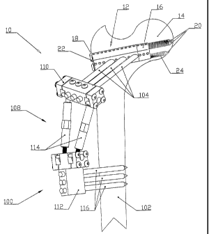
10 Reference is now made to Figure 1 illustrating an isometric view of a
hybrid interlocking proximal femoral fracture fixation in accordance with a
preferred embodiment of the present invention, and its positioning in a
femoral
bone. The femoral bone and especially its neck are subjected to severe
injuries that are difficult to fixate. Generally, the fracture fixation device
of the
present invention is a combination of internal and external fixation by
interlocking system that connects the fractured upper fragment to the femoral
shaft by a modular fixator. It is possible to correct the postoperative
positioning in any case of varus or valgus deformity, rotation angulation
shortening by distraction, compression, rotation, angulation, or translation.
Causes for malpositioning can be related to unskilled operative technique as
well as to low bone quality.
Hybrid device 10 comprises a nail-cage 12 that is adapted to be
implanted inside the femoral neck 14.. Nail-cage 12 is preferably a hollow
tubular member having a sharp end 16 and a blunt end 18. Optionally,. the
member can be a full structure instead of hollow. The hollow in nail-cage 12
allows drainage through the center hole of the nail so as to reduce the high
pressure intracapsular hemarthrosis that develops from the fracture
hemorrhage. The drainage reduces damage to the poor remaining vascularity
and consequently reduces one of the common causes of avascular necrosis
in the femoral head. Nail-cage 12 is inserted while crossing the fracture line
by
about 1-2 mm and allows bone grafting into the cage with autograft, allograft
or other bone substitute.
Preferably three compression screws 20 (lag screws) are adapted to
pass through nail-cage 12 from its blunt end 18 to its sharp end 16 so that
the
CA 02520508 2005-09-23
WO 2004/084761 PCT/IL2004/000259
11
screws are extended proximally through the sharp end and screwed inside the
bone. The extension of the screws beyond nail-cage 12 provides minimal
metal volume penetration to the femoral head using only the three lag screws
up to the subchondral bone. iVlinimal metal volume penetration prevents or
avoids further damage to the vascularity in the femoral head. Each
compression screw 20 is provided with a screw head 22 that prevents the
screw from totally advancing into nail-cage 12. Compression screws 20 are
used to compress the fracture so as to assure fast healing. The nail cage
combined with the compression screws are adapted to perform sliding motion
as well as compression. The compression is performed from blunt end 18 of
the nail and the screw heads, anchoring the lateral trochanteric cortical bone
around the blunt end of the nail-cage. The extended portion of compression
screws 20 is provided with thread 24 so as to facilitate the compression
process. Nail-cage 12 and compression screws 20 provide the possibility to
introduce chip bone grafting up to the fracture line due to the nail's unique
and
special structure. In the case the nail-cage is hollow, the interior of nail-
cage
12 can be filled with bone grafting materials so as to promote rapid bone
healing.
Nail-cage 12 is inserted into the femoral neck by minimal invasive
procedure. The optimal angle of penetration of the nail-cage and the screws is
between 100 to 110 degrees in respect of the femoral axis.. In this way, the
inferior screw of compression screws 20 is positioned so that it slightly
touches the strong cortical bone of the calcar femori. All three compression
screws 20 penetrate only the inferior half of the femoral head, preferable
distally of the teres ligament vascularity, avoiding damage to the capilar
spongeous intraosseous circulation and further reduce the possibility of
avascular necrosis.
External fixator 100 connects nail-cage 12 to the distal bone fragment
102. External fixator 100 comprises preferably six pin-screws from which three
pin-screws 104. are pined into nail-cage 12 and additional three pin screws
106 are pined in distal bone fragment 102. All six pin-screws, 104 and 106,
are interconnected in a connecting member 108 comprising an upper clamp
110, which clamps pin-screws 104, and a lower clamp 112, which clamps pin-
screws 106. Connecting member 108 further comprises preferably two
CA 02520508 2005-09-23
WO 2004/084761 PCT/IL2004/000259
12
rotating screwing rods 114 that connect upper clamp 110 and lower clamp
112. rotating screwing rods 114 have changeable length and connect the
clamps in a manner that allows a certain degree of freedom in the positioning
of the clamps in respect with each other. .4 comprehensive description is
provided herein after.
Deference is now made to Figures 2a-c illustrating views of a nail-cage
implant in accordance with a preferred embodiment of the present invention.
The nail cage implant is adapted to be incorporated in the hybrid
infierlocking
fiazation of the present invention. bail-cage 12 is a hollow tubular member
having a triangular profile that is provided with sharp end 16 and blunt end
18
and a hollow 55 that passes through the nail. Nail-cage 12 is adapted to be
inserted to the femoral neck by forcing it into the bone through sharp end 18.
Nail-cage 12 is preferably made of a biocompatible metal such as titanium.
The nail-cage can be left in place in case of bone grafting or removed after
the
bone is healed; however, the nail-cage as well as the compression screws
that pass through it may be made from a biodegradable material.
It is preferable to provide the wall of nail-cage 12 with a plurality of
relatively small holes 54 so as to allow the bone to grow into the nail-cage
12
in order to allow contact and bone growing together with the grafting
materials. It is optional to introduce autologous bone graft or
osteoconductive-
osteoinductive materials into nail-cage 12 (not shown in the Figures) so as to
encourage bone grow.
Nail-cage 12 is provided with passages and preferably three passages
56 that pass through the wall of the tube and extend from sharp end 16 to
blunt end 18. Passages 56 are provided with openings at both the sharp end
and the blunt end through which compression lag screws 20 (shown in Figure
1 ) may be screwed after the implantation of nail-cage 12. The nail cage of
the
present invention has the properties of compression and sliding when it is
combined with the three lag screws.
Deference is now made to Figure 6 illustrating illustrates an isometric
view of the nail-cage implant fixed in an internal fixation in accordance with
a
preferred embodiment of the present invention. mail-cage 12 is implanted
within a femoral bone 13 while three lag screws 20 are interlocked within nail-
cage 12.
CA 02520508 2005-09-23
WO 2004/084761 , PCT/IL2004/000259
13
Returning to Figure 2, nail-cage 12 is further provided with bores, and
preferably six bores wherein three bores 58 are provided in a row on one side
of the tube while the other three bores (can not be seen in the Figures) are
provided in a row opposite three bores 58. both opposite rows of bores are
shifted in respect with each other, e.g. one row is closer to blunt end 18 and
three bores 58 are closer t~ sharp end 16. Ail bores are provided with an
external thread so as to allow compatible screws to be screwed through them.
The bores are adapted to receive three pin screws 104 of the external fixation
(shown in Figure 1) to be screwed through them from one side to the other
while maintaining a predetermined angle between the nail cage and the pin
screws.
Reference is now made to figure 3 illustrating an isometric view of a nail
cage implant in accordance with another preferred embodiment of the present
invention. Nail-cage 200 is a hollow tubular member having a circular profile.
Similarly to the nail-cage shown herein before, nail-cage 200 is provided with
a sharp end 16 adapted to be pushed into the bone and a blunt end 18.
preferably three passages 56 adapted to receive lag screws (the screws are
not shown in the Figure).
Returning to Figure 7, it is clearly shown that three compression screws
20 are inserted through passages 56 and three pin screws 104 are inserted
through six lateral bores 58 and 60. As mentioned herein before, pin screws
104 are interconnected to pin screws 106 that are nailed into distal bone
fragment 102. Pin screws 104 are connected to nail-cage 12 in an angle that
directs the external fixation to anterior and lateral directions. In this way,
the
external fixation protrudes in the anterolateral proximal part of the
patient's
thigh, without disturbing the hip flexion or the lying supine patient
position.
It is important to notice that the nail-cage can be implanted in order to
fixate a fracture in the femoral neck using the compression fag screws
wifihout
employing the external fixation as shown in Figure 6.
Reference is now made to Figure 4 an isometric view of a connecting
member in accordance with a preferred embodiment of the present invention,
connecting the external fixation. The connecting member is a part of the
external fixation of the present invention. Connecting member 108 is adapted
to externally fixate and support the nail-cage that is implanted within the
CA 02520508 2005-09-23
WO 2004/084761 PCT/IL2004/000259
14
fiemoral neck. Connecting member 108 is designed so as to allow intra and
post-operative corrections. Therefiore, connecting member 108 comprises an
upper clamp 110, which clamps pin-screws 104, and a lower clamp 112,
which clamps pin-screws 106 (the pin-screws are not shown in Figure 3).
S connecting menlber 108 further comprises prefierably two rotating screwing
rods 114 that connect upper clamp 110 and lower clamp 112.
Upper clamp 710 is provided with three bores 150 that are compatible to
allow pin screws 104 to pass through them. Pin screws 104 are fastened in
bores 150 Casing three Allen screws 152 (the pin screws are not shown in
Figure 3). 1n a similar manner, a portion of lower clamp 112 is provided with
three bores 154 through which pin screws 106 (not shown in the figure) can
be inserted and fiastened using three Allen screws 156.
rotating screwing rods 114 that connect upper clamp 110 and lower
clamp 112 are preferably tubes provided with inner threads and preferably
1 S hexagonal aid 158 that facilitates rotation of rotating screwing rods 114
so as
to adjust the distance and the angles between upper clamp 110 and lower
clamp 112. rotating screwing rods 114 are provided with bolts 160 that are
screwed inside the tubes through all four openings of the tubes. Each bolt 160
is provided with a spherical head 162 wherein the spherical head is firmly
held
in the corresponding clamp. Rotating screwing rods 114 about their elongated
axis when the bolts heads are held in the clamps changes the distance
between the two clamps so as to enable changes in the orientation of the
external fixation post-operatively. Each bolt head 162 is held in the clamp in
a
recess that is adapted to be compressed onto the bolt head using Allen
2S screws 164. When the physician wants to change the angle of rotating
screwing rods 114 in regard with the clamps, he releases Allen screws 164 so
that the spherical heads can rotate in the clamp's recess. In the right
position,
the physician fastens Allen screws 164. It is important to notice the minimal
use of parts so as to facilitate the work of the surgeon with the device of
the
present invention.
Reference is now made to Figure 4 illustrating an exploded view of the
hybrid interlocking proximal femoral firacture fiixation shown in Figure 1. ft
is
clearly shown that the amount of parts that comprise the apparatus of the
present invention is minimal and effective. Nail cage 12 is adapted to be
CA 02520508 2005-09-23
WO 2004/084761 PCT/IL2004/000259
implanted in the femoral neck and receive compression screws 20. There are
cases in which the internal fixation is enough and there is no need in the
external fixation. In cases the external fixation is required; pin screws 104
are
inserked through nail cage 12 while pin screws 106 are nailed into the distal
5 bone fragment. The pins are interconnected in the connecting member that
comprises clamps 110 and 106 as well as rotating screwing rods 11q.. ~ne of
the important features of the connecting member of the present invention is in
its versatility using only ~411en screws and length changes in the rotating
screwing rods.
10 The device of the present invention is intended to be used for
intracapsular and extracapsular (including subtrochanteric) femoral fractures.
One of the important features of the hybrid interlocking fixation apparatus
of the present invention is that the procedure of the present invention is of
minimal invasive surgery instrumentations, mainly based on using multiple
15 tissues penetrations by pin-screws. This fact facilitates the use of
robotics for
fast, exact, and controlled operative steps.
The external parts of the hybrid interlocking fixation apparatus are
suitable for removal in the outpatient follow-up without anesthesia. The
internal nail-cage and screws may be left in place. The nail-cage and screws
can be made of bio-resorbable materials that are resorbed after some time.
The parts of the fixator that are inserted into the body can be made with
radiolucent materials so as to facilitate the x-ray imaging follow-up of the
bone
healing process.
It is optional to provide the device of the present invention with
disposable parts so as to reduce the costs.
It should be clear that the description of the embodiments and attached
Figures set forth in this specification serves only for a better understanding
of
the invention, without limiting its scope as covered by the following Claims.
It should also be clear that a person skilled in the art, after reading the
present specification can make adjustments or amendments to the attached
Figures and above described embodiments that would still be covered by the
following Claims.