Note: Descriptions are shown in the official language in which they were submitted.
CA 02520658 2005-09-27
WO 2004/088276 PCT/US2004/009191
A METHOD AND KIT FOR DETECTING THE EARLY ONSET
OF RENAL TUBULAR CELL INJURY
BACKGROUND OF THE INVENTION
[0001 ] Acute renal failure (ARF) secondary to a renal tubular cell injury,
including an
ischemic injury or a nephrotoxic injury remains a common and potentially
devastating
problem in clinical medicine and nephrology, with a persistently high rate of
mortality and
morbidity despite significant advances in supportive care. Pioneering studies
over several
decades have illuminated the roles of persistent vasoconstriction, tubular
obstruction, cellular
structural and metabolic alterations, and the inflammatory response in the
pathogenesis of
ARF. While these studies have suggested possible therapeutic approaches in
animal models,
translational research efforts in humans have yielded disappointing results.
The reasons for
this may include the multifaceted response of the kidney to ischemic injury
and nephrotoxins,
and a paucity of early biomarkers for ARF with a resultant delay in initiating
therapy.
[0002] An individual is considered to have acute renal failure when the
patient's serum
creatinine value either: (1) increased by at least 0.5 mg/dL when the baseline
serum
creatinine level was less than 2.0 mg/dL; (2) increased by at least 1.5 mgldL
when the
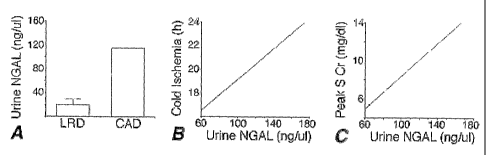
baseline serum creatinine level was greater than or equal to 2.0 mg/dL; or (3)
increased by at
least 0.5 mg/dL, regardless of the baseline serum creatinine level, as a
consequence of
exposure to radiographic agents.
[0003] It is believed that introduction of therapy early in the disease
process will reduce the
mortality rate associated with ARF and shorten the time for treatment of
various types of
renal tubular cell injuries, including, but not limited to, ischemic and
nephrotoxic renal
injuries. The identiftcation of a reliable, early biomarker for a renal
tubular cell injury would
1
CA 02520658 2005-09-27
WO 2004/088276 PCT/US2004/009191
be useful to facilitate early therapeutic intervention, and help guide
pharmaceutical
development by providing an indicator of nephrotoxicity.
[0004] The traditional laboratory approach for detection of renal disease
involved
determining the serum creatinine, blood urea nitrogen, creatinine clearance,
urinary
electrolytes, microscopic examination of the urine sediment, and radiological
studies. These
indicators are not only insensitive and nonspecific, but also do not allow for
early detection of
the disease. Indeed, while a rise in serum creatinine is widely considered as
the "gold
standard" for the detection of ARF, it is now clear that as much as 50~/0 of
the kidney
function may already be lost by the time the serum creatinine changes.
[0005] A few urinary biomarkers for ischemic renal injury have been earlier
described,
including kidney injury molecule-1 (KIM-1) and cysteine rich protein 61
(Cyr61). KIM-1 is
a putative adhesion molecule involved in renal regeneration. In a rat model of
ischemia-
reperfusion injury, KIM-1 was found to be upregulated 24-48 hours after the
initial insult,
rendering it a reliable but somewhat late marker of tubular cell damage.
Recent studies have
shown that KIM-1 can be detected in the kidney biopsy and urine of patients
with ischemic
acute tubular necrosis. However, this detection was documented in patients
with established
ischemic renal damage, late in the course of the illness. The utility of
urinary KIM-1
measurement for the detection of early ARF or subclinical renal injury has
thus far not been
validated.
[0006] The protein Cyr61 was found to be a secreted cysteine-rich protein that
is detectable
in the urine 3-6 hours after ischemic renal injury in animal models. However,
this detection
required a bioaffinity purification and concentration step with heparin-
sepharose beads,
followed by a Western blotting protocol. Even after bioaffinity purification
several non-
specific cross-reacting peptides were apparent. Thus, the detection of Cyr61
in the urine is
problematic with respect to specificity as well as the cumbersome nature of
the procedure.
2
CA 02520658 2005-09-27
WO 2004/088276 PCT/US2004/009191
[0007] Therefore, there remains an urgent need to identify improved biomarkers
for early
ischemic and nephrotoxic renal injuries.
SUMMARY ~F THE IIVVENTI~N
[0008] The present invention relates to a method for the detection of a renal
tubular cell
injury in a mammal, comprising the steps of: 1) obtaining a urine sample from
a mammalian
subject; 2) contacting the urine sample with an antibody for a renal tubular
cell injury
biomarker, the renal tubular cell injury biomarker comprising NGAL, to allow
formation of a
complex of the antibody and the renal tubular cell injury biomarker; and 3)
detecting the
antibody-biomarker complex.
[0009] The invention relates to a method of monitoring the effectiveness of a
treatment for
renal tubular cell injury comprising the steps of: 1) providing a treatment to
a mammalian
subject experiencing ischemic renal injury; 2) obtaining at least one post-
treatment urine
sample from the subject; and 3) detecting for the presence of a biomarlcer for
renal tubular
cell injury in the post-treatment urine sample.
[0010] The invention further relates to a kit for use in detecting the
presence of an immediate
or early onset biomarker for renal tubular cell injury in the urinary fluid of
a subject,
comprising: 1) a means for acquiring a quantity of a urine sample; 2) a media
having affixed
thereto a capture antibody capable of complexing with an renal tubular cell
injury biomarlcer,
the biomarker being NGAL; and 3) an assay for the detection of a complex of
the renal
tubular cell injury biomarker and the capture antibody.
[0011] The invention also relates to a competitive enzyme linked immunosorbent
assay
(ELISA) kit for determining the renal tubular cell injury status of a
mammalian subject,
comprising a first antibody specific to a renal tubular cell injury biomarker
to detect its
presence in a urine sample of the subject.
3
CA 02520658 2005-09-27
WO 2004/088276 PCT/US2004/009191
[0012] The invention further relates to a method of identifying the extent of
a renal tubular
cell injury caused by an event, comprising: 1) obtaining at least one urine
sample from a
mammalian subject; 2) detecting in the urine sample the presence of a
biomarker for renal
tubular cell injury; and 3) determining the extent of renal tubular cell
injury based on the time
for onset of the presence of IRI biomarkcr in the urine sample, relative to
the time of the
event.
[0013] The present invention further relates to a method for the detection of
a renal tubular
cell injury in a mammal, comprising the steps of: 1) obtaining a urine sample
comprising up
to 1 milliliter of the first urine from a mammalian subject following a
suspected renal tubular
cell injury; 2) contacting the urine sample with an antibody for a biomarker
for renal tubular
cell injury, to allow formation of a complex of the antibody and the
biomarlcer; and 3)
detecting the antibody-biomarlcer complex.
[0014] A preferred renal tubular cell injury biomarker is NGAL.
BRIEF DESCRIPTION OF THE DRAWINGS
[0015] Figure 1 shows induction of mouse kidney NGAL mRNA following ischemia.
Top
panel shows a representative RT-PCR with primers for mouse actin and NGAL,
using RNA
extracted from kidneys of control (C) mice or after various reperfusion
periods as shown
(hours). Lane M contains a molecular weight standard marker. Bottom panel
shows the fold
increase in NGAL mRNA expression at various time points from control (CON).
Values
obtained by microarray (solid line) vs RT-PCR (dotted line) are means +/- SD
from at least 3
experiments.
[0016] Figure 2A shows induction of mouse kidney NGAL protein following
unilateral
ischemia. Top panel shows a representative Western blot with whole kidney
samples
obtained from control (Con) mice or after reperfusion periods as shown
(hours), probed with
4
CA 02520658 2005-09-27
WO 2004/088276 PCT/US2004/009191
a polyclonal antibody to NGAL or a monoclonal antibody to tubulin (to
demonstrate equal
protein loading). Molecular weight markers are to the left. Bottom panel shows
the fold
increase in NGAL protein expression at various time points from control (CON).
Values
obtained by densitometry are means +/- SD from at least 3 experiments.
[0017] Figure 2B shows induction of mouse kidney NGAL protein following
bilateral
ischemia. Top panel shows a representative Western blot with whole kidney
samples
obtained from control (Con) mice or after reperfusion periods as shown
(hours), probed with
a polyclonal antibody to NGAL or a monoclonal antibody to tubulin (to
demonstrate equal
protein loading). Molecular weight markers are to the left. Bottom panel shows
the fold
increase in NGAL protein expression at various time points from control (CON).
Values
obtained by densitometry are means +/- SD from at least 3 experiments.
[0018] Figure 3 shows induction of mouse kidney NGAL protein following
ischemia.
Representative immunohistochemistry results on frozen sections of mouse
kidneys obtained
from control mice or after varying periods of reflow as shown (hours), probed
with a
polyclonal antibody to NGAL. "G" denotes a glomerulus. The panel on the
extreme right is
a 100X magnification, and the other panels are at 20X.
[0019] Figure 4A shows early detection of NGAL protein in the urine in mice
with unilateral
ischemic ARF. Representative Western blot of unprocessed urine samples (1-2 pl
per lane,
normalized for creatinine content) obtained at reperfusion periods as shown
(hours),
following unilateral renal artery clamping. Molecular weight marleers are
shown on the right.
Blots were probed with NGAL (top) or ~2-microglobulin (Beta2-M) (middle).
Urinary N-
acetyl- ~-D-glucosaminidase (NAG) determinations at various reperfusion
periods as
indicated, from five animals for five animals. Values are means +/- SD. *P <
0.05 versus
control at each time period, ANOVA.
CA 02520658 2005-09-27
WO 2004/088276 PCT/US2004/009191
[0020] Figure 4B shows early detection of NGAL protein in the urine in mice
with bilateral
ischemic ARF. Representative Western blot of unprocessed urine samples (1-2 pl
per lane,
normalized for creatinine content) obtained at reperfusion periods as shown
(hours),
following bilateral renal artery clamping. Molecular weight markers are shown
on the right.
Blots were probed with NGAL (top) or j32-microglobulin (l3eta2-M) (middle).
Urinary N-
acetyl- (3-D-glucosaminidase (NAG) determinations at various reperfusion
periods as
indicated, from five animals for eight animals. Values are means +/- SD. ~P <
0.05 versus
control at each time period, ANOVA.
[0021] Figure 5 shows detection of NGAL protein in the urine from mice with
subclinical
renal ischemia. Representative Western blot of unprocessed urine samples (1-2
p.l per lane,
normalized for creatinine content) obtained at reperfusion periods as shown
(hours),
following 5, 10, or 20 min of bilateral renal artery clamping. Molecular
weight markers are
shown on the left. These animals displayed normal serum creatinines at 24 h of
reflow.
[0022] Figure 6 shows early detection of NGAL protein in the urine in rats
with ischemic
ARF. Representative Western blot of unprocessed urine samples (1-2 pl per
lane, normalized
for creatinine content) obtained at reperfusion periods as shown (hours),
following 30 min of
bilateral renal artery clamping in rats. Molecular weight markers are shown on
the left.
These animals displayed a significant increase in serum creatinine at 24 h of
reflow.
[0023] Figure 7 shows induction of NGAL mRNA following ischemia in vitro. Top
panel
shows a representative RT-PCR with primers for human NGAL, using RNA extracted
from
renal proximal tubular epithelial cells (RPTEC) after various periods of
partial ATP depletion
as shown (hours). Lane M contains a 100 by DNA ladder. The middle panel shows
the fold
increase in NGAL mRNA expression at various time points from control (0),
normalized for
glyceeraldehyde-3-ohosphate dehydrogenase (GAPDH) expression. Values shown are
means
+/- SD from at least 3 experiments at each point. The bottom panel shows a
representative
6
CA 02520658 2005-09-27
WO 2004/088276 PCT/US2004/009191
Western blot (of three separate experiments) with RPTEC samples after various
periods of
partial ATP depletion as shown, obtained from equal amounts of cell pallets
(Pel) or the
culture medium (Sup), probed with a polyclonal antibody to NGAL. Molecular
weight
markers are to the left.
[0024] Figure 8A shows early detection of NGAL protein in the urine was
detected in mice
with cisplatin-induced injury. Representative Western blots on unprocessed
urine samples
(1-2 pl per lane, normalized for creatinine content) obtained at days as shown
following
cisplatin administration, probed with antibody for (3-2-microglobulin (top
panel) and NGAL
(middle panel). Molecular weight markers are shown on the left.
[0025] Figure 8B shows urinary NAG determinations at various days after
cisplatin
administration (n=4) in Figure 8A. Values are means +/- SD. *P < 0.05 versus
day 0.
[0026] Figure 9 shows that cisplatin administration results in tubule cell
necrosis and
apoptosis. Hematoxylin-eosin stain showed tubular dilatation, luminal debris,
and flattened
epithelium in cisplatin-treated kidneys (top center panel). At high power, a
tubule marked
with an asterisk displayed condensed intensely-stained nuclei (arrow),
indicative of apoptosis
(top right panel). TIJNEL staining showing TLJNEL-positive nuclei in cisplatin-
treated
kidneys (bottom center panel). At high power, the tubule indicated by an
asterisle displayed
condensed, fragmented nuclei (arrow) characteristic of apoptosis (bottom right
panel). Panels
labeled High Power are at 100X magnification, and the others are at 20X.
Results in control
mice are shown in top and bottom left panels.
[0027] Figure 10 shows that cisplatin administration results in rapid
induction of kidney
NGAL. Representative Western blots of kidney lysates from mice treated with
intraperitoneal cisplatin (20 p.g/kg) and obtained at various time points as
indicated (hours),
probed with a polyclonal antibody to NGAL or a monoclonal antibody to tubulin.
Molecular
weight markers are to the left.
7
CA 02520658 2005-09-27
WO 2004/088276 PCT/US2004/009191
[0028] Figure 11 shows that cisplatin administration results in rapid
induction of NGAL in
tubule epithelial cells. Representative immunohistochemistry results on frozen
kidney
sections from mice treated with intraperitoneal cisplatin (20 p.g/kg) and
obtained at various
time points as indicated (hours), probed with a polyclonal antibody to NGAL.
G9 glomerulus.
Panel labeled HP is at 100 magnification, and the others are at 20~.
[0029] Figure 12 shows that administration of 20 p,g/kg cisplatin results in
rapid appearance
of NGAL in the urine. Representative Western blot (upper panel) of unprocessed
urine
samples (3-5 pl/lane, normalized for creatinine content) obtained before or at
various time
points following cisplatin injections as shown. The same urine samples were
analyzed for
NAG excretion (center panel), and serum from the same animals subjected to
creatinine
measurement (bottom panel). *P<0.05 versus control.
[0030] Figure 13 shows that administration of 5 p,g/kg cisplatin results in
rapid appearance of
NGAL in the urine. Representative Western blot (upper panel) of unprocessed
urine samples
(3-5 p,l/lane, normalized for creatinine content) obtained before or at
various time points
following cisplatin injections as shown. The same urine samples were analyzed
for NAG
excretion (center panel), and serum from the same animals subjected to
creatinine
measurement (bottom panel). *P<0.05 versus control.
[0031] Figure 14 shows quantitation of urinary NGAL following cisplatin.
Coomassie Blue
(CB) staining (top left panel) and Enhanced Chemiluminescence (ECL) analysis
of known
quantities of recombinant purified NGAL (top right panel). Quantitation of
urinary NGAL
excretion at various time points following cisplatin 20 ~g/kg or 5 ~glkg,
determined by
densitometric analysis of Western blots and comparisons with Western blots of
defined
standards of purified NGAL performed under identical conditions.
[0032] Figure 15 shows in panel A the measurement of urine NGAL in patients
with
cadaveric kidney transplants (CAD, n=4) versus living related donor
transplants (LRD, n = 6)
8
CA 02520658 2005-09-27
WO 2004/088276 PCT/US2004/009191
(p < 0.005). Panel B shows a correlation between cold ischemia time and
urinary NGAL in
CAD (p < 0.001, r = 0.98, Spearman analysis). Panel C shows a correlation
between peak
serum creatinine and urinary NGAL in CAD (p < 0.001, r =0.96, Spearman
analysis).
[0033] Figure 16 shows in panel A the results of serial measurements of
urinary NGAL in
patients following open heart surgery, plotted against post bypass time in
hours (n=15).
Panel B shows a correlation beW een bypass time and the 2 hour urinary NGAL in
patients
who developed ARF (n = 5) (p< 0.01, r = 0.76, Spearman analysis). Panel C
shows a
correlation between changes in serum creatinine and the 2 hour urinary NGAL in
patients
who developed ARF (p < 0.01, r = 0.66, Spearman analysis).
DETAILED DESCRIPTION OF THE INVENTION
[0034] Throughout this application, various publications and unpublished
manuscripts are
referred to within parentheses. Disclosures of the publications in their
entireties are hereby
incorporated by reference into this application to more fully describe the
state of the art to
which this invention pertains. Full bibliographic citation for these
references can be found at
the end of this application, preceding the claims.
[0035] The present invention provides a method and kit for assaying the
presence of a renal
tubular cell injury biomarker present in the urine of a subject at the early
onset of renal
tubular cell injury. Early detection of the onset of the injury can reduce the
time for treatment
of the injury, and can reduce the risk of developing clinical acute renal
failure (ARF). The
renal tubular cell injury can include, but is not limited to, ischemic renal
injury (IRI) or
nephrotoxic renal injury (NRI).
[0036] A simple point-of care kit that uses principles similar to the widely-
used urine
pregnancy testing kits, for the rapid detection of urinary NGAL at the bedside
will allow the
clinician to rapidly diagnose ARF, and to rapidly institute proven and
effective therapeutic
9
CA 02520658 2005-09-27
WO 2004/088276 PCT/US2004/009191
and preventive measures. The use of the kit can represent the standard of care
for all patients
who are at risk of developing ARF, including use in cardiac surgery, kidney
transplantation,
stroke, trauma, sepsis, dehydration, and nephrotoxins (antibiotics, anti-
inflammatory agents,
radio-contrast agents, and chemotherapeutic agents). In current clinical
practice, when ARF
occurs in the setting of these predisposing conditions, the diagnosis is very
delayed, and the
associated mortality and morbidity unacceptably high. Ironically, even
tragically, effective
preventive and therapeutic measures are widely available, but almost never
administered in a
timely manner due to the lack of early biomarlcers of ARF. It is anticipated
that multiple
serial measurements of NGAL will be become indispensable not only for
diagnosing and
quantifying the initial kidney injury, but also for following the response to
early treatment,
and for predicting long term outcome.
[0037] The biomarker for renal tubular cell injury (which will also be
referred to as RTCI
biomarker) can be an immediate RTCI biomarlcer, such as NGAL, which can appear
in the
urine within 2 hours of the onset of renal tubular cell injury. An immediate
RTCI biomarker
can, as in the case of NGAL, be present in the first urine output of a subject
immediately after
the onset of renal tubular cell injury. The RTCI biomarlcer can also be an
early-onset RTCI
biomarker that can appear within the first 24 hours of the onset of renal
tubular cell injury.
As such, NGAL is also an example of an early-onset RTCI biomarker.
[0038] An effective RTCI biomarlcer is typically a secreted protein, whereby
it can be
excreted by the leidney into the urine. An effective RTCI biomarker is also
typically a
protease-resistant protein, such as NGAL. Nevertheless, an RTCI biomarker can
also be a
protease-sensitive protein, so long as stable fragments of the protein can be
detected in the
urine, such as by antibodies as described hereinafter for NGAL.
CA 02520658 2005-09-27
WO 2004/088276 PCT/US2004/009191
[0039] The RTCI biomarker can be an ischemic renal injury biomarker (IRI
biomarlcer), a
nephrotoxic renal injury biomarker (NRI biomarlcer), or a mixture thereof.
NGAL is an
example of both an IRI biomarker and an NRI biomarker.
[0040] The method of the invention can be used to detect the onset of renal
tubular cell
injury, and to monitor the treatment thereof, for a wide variety of events
that can include all
varieties of diminished blood supply to the kidneys, impaired heart function,
surgical
procedures, patients in intensive care units, and the administration of
pharmaceuticals,
radiocontrast dyes, or other medicament substances to a subject. The renal
tubular cell injury
can be an ischemic renal injury, a nephrotoxic renal injury, or other injury
that affects the
tubular cells of the kidney. The event can include administration or ingestion
of a large, and
wide variety of nephrotoxins, including, but not limited to cancer
chemotherapy (cisplatin,
cyclophosphamide, isosfamide, methotrexate), antibiotics (gentamicin,
vancomycin,
tobramycin), antifungal agents (amphotericin), anti-inflammatory agents
(NSAIDs),
immunosuppressants (cyclosporine, tacrolimus), and radiocontrast agents. The
method can
be used to evaluate the nephrotoxisity of both newly-developed and well-known
compounds.
[0041] The invention also provides a method and a kit for assessing the extent
of renal injury
based on a proportional relationship between the extent of injury, which can
range from the
very onset of renal tubular cell injury, to clinical ARF, with the quantity of
NGAL present in
the urine passing from the subject. The invention provides a means for a
clinician to estimate
the degree of renal injury at an initial assessment, and to monitor the change
in status of the
injury (worsening, improving, or remaining the same) based on the detected
amount of
NGAL in the urine.
[0042] Typically, the clinician would establish a protocol of collecting and
analyzing a
quantity of fresh urine sample from the patient at selected intervals.
Typically the sample is
obtained intermittently during a prescribed period. The period of time between
intermittent
11
CA 02520658 2005-09-27
WO 2004/088276 PCT/US2004/009191
sampling may be dictated by the condition of the subject, and can range from a
sample each
24 hours to a sample taken continuously, more typically from each 4 hours to
each 30
minutes.
[0043] Using the methods and techniques described herein, both a qualitative
level of the
RTCI biomarker present in the urine can be analyzed and estimated, and a
quantitative level
of RTCI biomarker present in the urine can be analyzed and measured. The
clinician would
select the qualitative method, the quantitative method, or both, depending
upon the status of
the patient. Typically, the quantity of urine to be collected is less than 1
milliliter, and more
typically less than 10 p.l. A typical sample can range from about 1 p,l to
about 1 ml.
Typically the larger quantities of urine sample (about 1 ml) are used for
quantitative assays.
Typically, these small amounts of urine are easily and readily available from
clinical subjects
who are either prone to developing ARF, or have developed ARF.
[0044] Once an indication of renal tubular cell injury or acute renal failure
has been detected,
and intervention and treatment of the disease or condition has commenced, the
clinician can
employ the method and lcit of the invention to monitor the progress of the
treatment or
intervention. Typically, one or more subsequent post-treatment urine samples
will be taken
and analyzed for the presence of the RTCI biomarker as the treatment of the
renal injury
commences and continues. The treatment is continued until the presence of the
RTCI
biomarlcer in subsequent post-treatment urine samples is not detected. As the
treatment and
intervention ameliorate the condition, the expression of RTCI biomarleer, and
its presence in
the urine, will be correspondingly reduced. The degree of amelioration will be
expressed by
a correspondingly reduced level of RTCI biomarker, such as NGAL, detected in a
sample.
As the renal injury nears complete healing, the method can be used to detect
the complete
absence of the RTC1 biomarker, signaling the completion of the course of
treatment.
12
CA 02520658 2005-09-27
WO 2004/088276 PCT/US2004/009191
[0045] Both monoclonal and polyclonal antibodies that bind an RTCI biomarlcer
are useful in
the methods and kits of the present invention. The antibodies can be prepared
by methods
known in the art. Monoclonal antibodies for a preferred RTCI biomarker, NGAL,
are
described, for example, in "Characterization of two ELISAs for NGAL, a newly
described
lipocalin in human neutrophils", Lars I~jeldsen et al., (1996) Journal of
Immunological
Methods, Vol. 19~, 155-16, herein incorporated by reference. Examples of
monoclonal
antibodies for NGAL can be obtained from the Antibody Shop, Copenhagen,
Denmark, as
HYB-211-O1, HYB-211-02, and NYB-211-O5. Typically, HYB-211-O1 and HYB-211-02
can be used with NGAL in both its reduced and unreduced forms. An example of a
polyclonal antibody for NGAL is described in "An Iron Delivery Pathway
Mediated by a
Lipocalin", Jun Yang et al., Molecular Cell, (2002), Vol. 10, 1045-1056,
herein incorporated
by reference. To prepare this polyclonal antibody, rabbits were immunized with
recombinant
gel-filtered NGAL protein. Sera were incubated with GST-Sepharose 4B beads to
remove
contaminants, yielding the polyclonal antibodies in serum, as described by the
applicants in
Jun Yang et al., Molecular Cell (2002).
[0046] Typically, the step of detecting the complex of the capture antibody
and the RTCI
biomarker comprises contacting the complex with a second antibody for
detecting the
biomarker.
[0047] The method for detecting the complex of the RTCI biomarlcer and the
primary
antibody comprises the steps of: separating any unbound material of the urine
sample from
the capture antibody-biomarker complex; contacting the capture antibody-
biomarker complex
with a second antibody for detecting the RTCI biomarker, to allow formation of
a complex
between the RTCI biomarker and the second antibody; separating any unbound
second
antibody from the RTCI biomarker-second antibody complex; and detecting the
second
antibody of the RTCI biomarker-second antibody complex .
13
CA 02520658 2005-09-27
WO 2004/088276 PCT/US2004/009191
[0048] A kit for use in the method typically comprises a media having affixed
thereto the
capture antibody, whereby the urine sample is contacted with the media to
expose the capture
antibody to NGAL contained in the sample. The kit includes an acquiring means
that can
comprise an implement, 5LlCh as a spatula or a simple stick, having a surface
comprising the
media. The acquiring means can also comprise a container for accepting the
urine sample,
where the container has a urine-contacting surface that comprises the media.
In another
typical embodiment, the assay for detecting the complex of the RTCI biomarker
and the
antibody can comprise an ELISA, and can be used to quantitate the amount of
NGAL in a
urine sample. In an alternative embodiment, the acquiring means can comprise
an implement
comprising a cassette containing the media.
[0049] Early detection of the RTCI biomarker can provide an indication of the
presence of
the protein in a urine sample in a short period of time. Generally, a method
and a kit of the
present invention can detect the RTCI biomarker in a sample of urine within
four hours, more
typically within two hours, and most typically within one hour, following
renal tubular cell
injury. Preferably, the RTCI biomarker can be detected within about 30 minutes
following
renal tubular cell injury.
[0050] A method and kit of the present invention for detecting the RTCI
biomarker can be
made by adapting the methods and kits known in the art for the rapid detection
of other
proteins and ligands in a biological sample. Examples of methods and kits that
can be
adapted to the present invention are described in US Patent 5,656,503, issued
to May et al. on
August 12, 1997, US Patent 6,500,627, issued to O'Conner et al. on December
31, 2002, US
Patent 4,870,007, issued to Smith-Lewis on September 26, 1989, US Patent
5,273,743, issued
to Ahlem et al. on December 28, 1993, and US Patent 4,632,901, issued to
Vallcers et al. on
December 30, 1986, all such references being hereby incorporated by reference.
14
CA 02520658 2005-09-27
WO 2004/088276 PCT/US2004/009191
[0051 ] A rapid one-step method of detecting the RTCI biomarker can reduce the
time for
detecting the renal tubular cell injury. A typical method can comprise the
steps of: obtaining
a urine sample suspected of containing the RTCI biomarker; mixing a portion of
the sample
with a detecting antibody which specifically binds to the RTCI biornarker, so
as to initiate the
binding the detecting antibody to the RTCI biomarker in the sample; contacting
the mixture
of sample and detecting antibody with an immobilized capture antibody which
specifically
binds to the RTCI biomarker, which capture antibody does not cross-react with
the detecting
antibody, so as to bind the detecting antibody to the RTCI biomarker, and the
RTCI
biomarker to the capture antibody, to form a detectable complex; removing
unbound
detecting antibody and any unbound sample from the complex; and detecting the
detecting
antibody of the complex. The detectable antibody can be labeled with a
detectable marker,
such as a radioactive label, enzyme, biological dye, magnetic bead, or biotin,
as is well
known in the art.
[0052] To identify potential genes and their proteins that may accompany and
mark the
earliest onset of renal tubular cell injuries, such as ischemic and
nephrotoxic renal injuries, a
cDNA microarray assay can be used to detect which of a large number of
potential gene
targets are markedly upregulated. Utilizing this screening technique,
neutrophil gelatinase-
associated lipocalin (NGAL) was identified as a gene whose expression is
upregulated more
than 10 fold within the first few hours following an ischemic renal injury in
a mouse model.
[0053] NGAL belongs to the lipocalin superfamily of over 20 structurally
related secreted
proteins that are thought to transport a variety of ligands within a (3-
barreled calyx. Human
NGAL was originally identified as a 25 kDa protein covalently bound to
gelatinase from
human neutrophils, where it represents one of the neutrophil secondary granule
proteins.
Molecular cloning studies have revealed human NGAL to be similar to the mouse
24p3 gene
first identified in primary cultures of mouse kidneys that were induced to
proliferate. NGAL
CA 02520658 2005-09-27
WO 2004/088276 PCT/US2004/009191
is expressed at very low levels in other human tissues, including kidney,
trachea, lungs,
stomach, and colon. NGAL expression is markedly induced in stimulated
epithelia. For
example, it is upregulated in colonic epithelial cells in areas of
inflammation or neoplasia, but
is absent from intervening uninvolved areas or within metastatic lesions. NGAL
concentrations are elevated in the serum of patients with acute bacterial
infections, the
sputum of subjects with asthma or chronic obstructive pulmonary disease, and
the bronchial
fluid from the emphysematous lung. In all these cases, NGAL induction is
postulated to be
the result of interactions between inflammatory cells and the epithelial
lining, with
upregulation of NGAL expression being evident in both neutrophils and the
epithelium.
[0054] It is believed that the detected NGAL induction represents a novel
intrinsic response
of the kidney proximal tubule cells to renal tubular cell injuries, including
both ischemic and
nephrotoxic injuries, and is not derived merely from activated neutrophils.
First, the response
is rapid, with NGAL appearing in the urine within 2 hours of the injury with
the very first
urine output following renal artery occlusion, while renal neutrophil
accumulation in this
model of ischemic ARF is usually first noted at 4 hours after injury. Second,
the temporal
patterns of NGAL induction and neutrophil accumulation are divergent. NGAL
mRNA and
protein expression was maximally noted at 12 hours of reflow, whereas
neutrophil
accumulation peaks at 24 hours by which time NGAL expression has significantly
declined.
Third, no NGAL-expressing neutrophils were detectable by immunofluorescence in
the
kidney samples examined (Figure 3). Fourth, NGAL mRNA and protein induction
was
documented to occur in cultured human proximal tubule cells following in vitro
ischemia,
with NGAL secreted into the culture medium within 1 hour of ATP depletion, in
a system
where neutrophils are absolutely absent. Nevertheless, some contribution from
infiltrating
neutrophils to the observed NGAL upregulation may have occurred. It is
possible that
CA 02520658 2005-09-27
WO 2004/088276 PCT/US2004/009191
upregulation of NGAL in renal tubule cells may be induced by local release of
cytolcines
from neutrophils trapped in the microcirculation early after ischemic injury.
[0055] An adequate explanation for the induction of NGAL by stimulated
epithelia has been
lacking, and whether NGAL is protective or proximate to injury or even an
imiocent
bystander remains unclear. Recent evidence suggests that, at least in a subset
of cell types,
NGAL may represent a pro-apoptotic molecule. In the mouse pro-B lymphocytic
cell line,
cytokine withdrawal resulted in a marked induction of NGAL as well as onset of
apoptosis.
Purified NGAL produced the same pro-apoptotic response as cytokine
deprivation, including
activation of Bax, suggesting that NGAL is proximate to programmed cell death.
NGAL has
also been linked to apoptosis in reproductive tissues. Epithelial cells of the
involuting
mammary gland and uterus express high levels of NGAL, temporally coinciding
with a
period of maximal apoptosis. It is likely that.NGAL regulates a subset of cell
populations by
inducing apoptosis. Stimulated epithelia may upregulate NGAL in order to
induce apoptosis
of infiltrating neutrophils, thereby allowing the resident cells to survive
the ravages of the
inflammatory response. Alternatively, epithelial cells may utilize this
mechanism to regulate
their own demise. However, it is interesting to note that induction of NGAL
following renal
ischemia-reperfusion injury occurs predominantly in the proximal tubule cells,
and apoptosis
under the same circumstances is primarily a distal tubule cell phenomenon.
[0056] Other recent studies have revealed that NGAL enhances the epithelial
phenotype.
NGAL is expressed by the penetrating rat ureteric bud, and triggers
nephrogenesis by
stimulating the conversion of mesenchymal cells into kidney epithelia. Another
lipocalin,
glycodelin, has been shown to induce an epithelial phenotype when expressed in
human
breast carcinoma cells. These findings are especially pertinent to the mature
kidney, in which
one of the well-documented responses to ischemic injury is the remarkable
appearance of
dedifferentiated epithelial cells lining the proximal tubules. An important
aspect of renal
CA 02520658 2005-09-27
WO 2004/088276 PCT/US2004/009191
regeneration and repair after ischemic injury involves the reacquisition of
the epithelial
phenotype, a process that recapitulates several aspects of normal development.
This suggests
that NGAL may be expressed by the damaged tubule in order to induce re-
epithelialization.
Support for this notion derives from the recent identification of NGAL as an
iron transporting
protein that is complementary to transferrin during nephrogenesis. It is well
known that the
delivery of iron into cells is crucial for cell growth and development, and
this is presumably
critical to postischemic renal regeneration just as it is during ontogeny.
Since NGAL appears
to bind and transport iron, it is also likely that NGAL may serve as a sink
for iron that is shed
from damaged proximal tubule epithelial cells. Because it has been observed
that NGAL can
be endocytosed by the proximal tubule, the protein could potentially recycle
iron into viable
cells. This might stimulate growth and development, as well as remove iron, a
reactive
molecule, from the site of tissue injury, thereby limiting iron-mediated
cytotoxicity.
[0057] NGAL is a novel urinary biomarker for cisplatin-induced nephrotoxic
renal injury that
is more sensitive than previously described biomarkers. One example is kidney
injury
molecule-1 or KIM-1, a putative adhesion molecule involved in renal
regeneration. In a rat
model of cisplatin nephrotoxicity, KIM-1 was qualitatively detectable 24-48
hours after the
initial insult, rendering it a somewhat late marker of tubular cell damage. In
contrast, NGAL
is readily and quantitatively detected within 3 hours following cisplatin
administration in
doses known to result in renal failure. In addition, urinary NGAL detection
precedes the
appearance of other markers in the urine such as NAG. Appearance of NGAL in
the urine
also precedes the increase in serum creatinine that is widely used to diagnose
nephrotoxic
renal failure.
[0058] Urinary NGAL is evident even after mild "sub-clinical" doses of
cisplatin, in spite of
normal serum creatinine levels. Thus, the invention has important implications
for the clinical
management of patients on cisplatin therapy. The efficacy of cisplatin is dose
dependent, but
CA 02520658 2005-09-27
WO 2004/088276 PCT/US2004/009191
the occurrence of nephrotoxicity frequently hinders the use of higher doses to
maximize its
antineoplastic potential. Nephrotoxicity following cisplatin treatment is
common and may
manifest after a single dose with acute renal failure. Although several
therapeutic maneuvers
have proven to be efficacious in the treatment of cisplatin-induced
nephrotoxicity in animals,
successful human experiences have remained largely anecdotal. ~ne reason for
this may be
the lack of early markers for nephrotoxic acute renal failure, and hence a
delay in initiating
therapy. In current clinical practice, acute renal injury is typically
diagnosed by measuring
serum creatinine. However, it is well known that creatinine is an unreliable
and delayed
indicator during acute changes in kidney function. First, serum creatinine
concentrations may
not change until about 50% of kidney function has already been lost. Second,
serum
creatinine does not accurately depict kidney function until a steady state has
been reached,
which may require several days. Thus, the use of serum creatinine measurements
impairs the
ability to both detect and quantify renal damage during the early phases of
renal injury.
However, animal studies have suggested that while nephrotoxic acute renal
failure can be
prevented and/or treated, there is a narrow "window of opportunity" to
accomplish this, and
treatment must be instituted very early after the initiating insult. The lack
of early biomarkers
of renal injury has impaired the ability of clinicians to initiate potentially
effective therapies
in a timely manner. The use of NGAL in an assay system would also be of value
for testing
existing or emerging therapeutic or preventive interventions, and for the
early evaluation of
the nephrotoxic potential of other pharmaceutical agents. NGAL detection is a
novel, non-
invasive, early urinary biomarker for cisplatin-induced kidney damage. Early
detection may
enable clinicians to administer timely therapeutic interventions, and to
institute maneuvers
that prevent progression to overt nephrotoxic renal failure.
[0059] It has been found that NGAL was easily and rapidly detected as
relatively clean
immunoreactive peptides in Western blots with as little as 1 ~,1 of the very
first unprocessed
CA 02520658 2005-09-27
WO 2004/088276 PCT/US2004/009191
urine output following renal ischemia in both mice and rats. Furthermore,
urinary NGAL
was evident even after very mild "subclinical" renal ischemia, despite normal
serum
creatinine levels. Urinary NGAL detection also far preceeded the appearance of
traditional
markers in the urine, including (32-microglobulin and NAG.
[0060] The upregulation and urinary excretion of NGAL may represent a rapid
response of
renal tubule cells to a variety of insults, and the detection of NGAL in the
urine may
represent a widely applicable noninvasive clinical tool for the early
diagnosis of tubule cell
inj ury.
[0061 ] NGAL is a sensitive, noninvasive urinary biomarker for renal tubular
cell injuries,
including renal ischemia and nephrotoxemia. The examination of the expression
of NGAL in
the urine of patients with acute, mild and early forms of renal tubular cell
injury, using the
rapid and simple detection methods and kits of the invention, can alert and
enable clinicians
to institute timely interventional efforts in patients experiencing acute
renal failure, and to
alert clinicians to institute maneuvers aimed at preventing progression in
patients with subtle,
subclinical renal tubular cell injuries (such as a nephrotoxins, kidney
transplants, vascular
surgery, and cardiovascular events) to overt ARF.
[0062) In the United States alone, there are approximately 16,000 kidney
transplants
performed every year. This number has been steadily increasing every year.
About 10,000 of
these are cadaveric kidney transplants, and are at risk for ARF. Each of these
patients would
benefit enormously from serial NGAL measurements, which could represent
routine care.
[0063] Ischemic renal injury has also been associated with open heart surgery,
due to the
brief interruption in blood flow that is inherent in this procedure. The
number of open heart
surgeries performed annually can be estimated. In any moderately busy adult
hospital,
approximately 500 such operations are performed every year. Given that there
are at least 400
such moderately busy hospitals in the United States alone, one can
conservatively estimate
CA 02520658 2005-09-27
WO 2004/088276 PCT/US2004/009191
that 200,000 open heart surgeries are performed every year. Again, serial NGAL
measurements would be invaluable in these patients, and would represent the
standard of
care.
EXPE1~IMENTAL PROCEDLJI2ES
1. Mouse models of renal ischemia-reperfusion injury:
[0064] We utilized well-established murine models of renal ischemia-
reperfusion injury, in
which the structural and functional consequences of brief periods of renal
ischemia have been
previously documented (3-7). Briefly, male Swiss-Webster mice (laconic Farms,
Germantown, NY) weighing 25-35 g were housed with 12:12 hour light:dark cycle
and were
allowed free access to food and water. The animals were anesthetized with
sodium
pentobarbital (50 mg/kg intraperitoneally), and placed on a warming table to
maintain a rectal
temperature of 37°C. Three separate protocols were employed: (a)
unilateral ischemia, (b)
bilateral ischemic with ARF, and (c) bilateral mild subclinical ischemia. For
the first set of
(unilateral ischemia) experiments, the left renal pedicle was occluded with a
non-traumatic
vascular clamp for 45 min, during which time the kidney was kept warm and
moist. The
clamp was then removed, the kidney observed for return of blood flow, and the
incision
sutured. The mice were allowed to recover in a warmed cage. After 0, 3, 12, or
24 hours of
reperfusion, the animal was re-anesthetized, the abdominal cavity was opened,
and blood
obtained via puncture of the inferior vena cava for measurement of serum
creatinine by
quantitative colorimetric assay kit (Sigma, St. Louis, MO). The mice were
killed with
intraperitoneal pentobarbital. The left ventricle was then perfused with 10 ml
of 1X PBS, and
then with 10 ml of 4°1° paraformaldehyde in PBS to achieve in
situ fixation of the kidneys.
Both kidneys were harvested (the right kidney served as a control for each
animal). At least
three separate animals were examined at each of the reflow periods. One half
of each kidney
CA 02520658 2005-09-27
WO 2004/088276 PCT/US2004/009191
was snap frozen in liquid nitrogen and stored at -70°C until further
processing; a sample was
fixed in formalin, paraffin-embedded, and sectioned (4 ~,m). Paraffin sections
were stained
with hematoxylin-eosin and examined histologically. The clamped kidneys
displayed the
characteristic morphologic changes resulting from ischemia-reperfusion injury,
as previously
published by others (3-6) and us (2). The other half of each kidney was
embedded in ~CT
compound (Tissue-Tek) and frozen sections (4 ~,m) obtained for
immunohistochemistry.
[0065] For the second set of (bilateral ischemia) experiments, both kidneys
were clamped for
30 min, and examined as various reflow periods as detailed above. This group
of eight
animals was designed to represent ARF, and displayed a significant elevation
in serum
creatinine at 24 hours following the injury.
[0066] For the third set of (bilateral mild subclinical ischemia) experiments,
both kidneys of
separate animals were clamped for 5, 10, or 20 min only, and examined at
various reperfusion
periods as above. This very mild degree of injury was designed to simulate
subclinical renal
ischemia, and mice in this group did not display any elevations in serum
creatinine measured
at 24 hours following the injury.
2. Rat model of renal ischemia-reperfusion injury:
[0067] We utilized well-established rodent models of renal ischemia-
reperfusion injury (2).
Briefly, male Sprague-Dawley rats weighing 200-250 g (Taconic Farms,
Germantown, NY)
were anesthetized with ketamine (150 ~.g/g) and xylazine (3 ~,g/g), and
subjected to bilateral
renal artery occlusion with microvascular clamps for 30 min as detailed in the
mouse
protocol. Timed urine collections were obtained at 3, 6, 9, 12 and 24 h of
reperfusion, and
blood was collected for creatinina measurement at the time of killing (24 h).
3. RNA isolation:
CA 02520658 2005-09-27
WO 2004/088276 PCT/US2004/009191
[0068] Mouse whole kidney tissues (or cultured human proximal tubule cells,
see below)
were disrupted with a Tissue Tearor (Biospec Products, Racine, WI). Total RNA
from
control and ischemic kidneys was isolated using the RNeasy Mini I~it (Qiagen,
Valencia,
CA), and quantitated by spectrophotometry.
4. Microarray Procedures:
[0069] Detailed descriptions of microarray hardware and procedures have been
previously
published (3). Briefly, for each experiment, 100 pag of purified total mouse
kidney RNA was
reverse transcribed with Superscript II reverse transcriptase (Life
Technologies, Rockville,
MD) in the presence of Cy3-dUTP (Amersham, Piscataway, NJ) for controls and
Cy5-dUTP
for ischemic samples. The cDNA samples were purified using a Microcon YM-50
filter
(Millipore, Madison, WI), and hybridized to microarray slides containing 8,979
unique
sequence-verified mouse probes (3). Three separate animals were examined for
each of the
reflow periods, and at least two independent microarray experiments were
performed for each
of the animals. The array slides were scanned using a microarray scanner
(GenePix 4000B,
Axon Instruments, Foster City, CA) to obtain separate TIFF images for Cy3 and
Cy5
fluorescence. The signal intensities for Cy3 and Cy5 were determined for
individual genes
using the GenePix Pro 3.0 data extraction software (Axon Instruments). Quality
control and
data analysis was completed as previously described (3).
5. Semi-quantitative Reverse Transcription-Polymerase Chain Reaction (RT-PCR):
[0070] An equal amount (1 ~,g) of total RNA from control and experimental
mouse lcidneys
was reverse transcribed with Superscript II reverse transcriptase (Life
Technologies) in the
presence of random hexamers according to the manufacturer's instructions. PCR
was
accomplished using a kit (Roche, Indianapolis, IN) and the following primers:
CA 02520658 2005-09-27
WO 2004/088276 PCT/US2004/009191
Mouse NGAL sense 5'-CACCACGGACTACAACCAGTTCGC-3' ;
Mouse NGAL antisense 5'-TCAGTTGTCAATGCATTGGTCGGTG-3' ;
Human NGAL sense 5'-TCAGCCGTCGATACACTGGTC-3' ; and
Human NGAL antisense 5'-CCTCGTCCGAGTGGTGAGCAC-3'.
[0071 ] Primer pairs for mouse and human [3-actin and glyceraldehyde-3-
phosphate
dehydrogenase (GAPDH) were obtained from Clontech (La Jolla, CA). Mock
reactions
devoid of cDNA served as negative controls. PCR products were analyzed by
agarose gel
electrophoresis followed by staining with ethidium bromide, and quantitated by
densitometry.
Fold changes in NGAL mRNA expression in ischemic versus control kidneys were
expressed
following normalization for (3-actin or GAPDH amplification.
6. Immunohistochemistry:
[0072] Frozen sections were permeabilized with 0.2% Triton X-100 in PBS for 10
min,
blocked with goat serum for 1 hr, and incubated with primary antibody to NGAL
(1:500
dilution) for 1 hr. Slides were then exposed for 30 min in the dark to
secondary antibodies
conjugated with Cy5 (Amersham, Arlington Heights, IL), and visualized with a
fluorescent
microscope (Zeiss Axiophot) equipped with rhodamine filters.
[0073] For co-localization of NGAL with Rabll, serial sections were first
incubated with
NGAL antibody or a monoclonal antibody to Rabll (1:500 dilution; Transduction
Laboratories), then with secondary antibodies conjugated with either Cy5 (for
NGAL) or Cy3
(for Rabll) and visualized with rhodamine or fluorescein filters,
respectively. For co-
localization of NGAL with proliferating cell nuclear antigen (PCNA), sections
were co-
incubated with NGAL antibody and a monoclonal antibody to PCNA (1:500
dilution;
Upstate), and was detection accomplished by immunoperoxidase staining
(ImmunoCruz
Staining System, Santa Cruz Biotechnology). For the TUNEL assay, we used the
ApoAlert
CA 02520658 2005-09-27
WO 2004/088276 PCT/US2004/009191
DNA Fragmentation Assay I~it Clontech). Paraffin sections were deparaffmized
through
xylene and descending grades of ethanol, fixed with 4% formaldehyde/PBS for 30
min at
4°C, permeabilized with proteinase I~ at room temperature for 15 min
and 0.2% triton X-
100/PBS for 15 min at 4°C, and incubated with a mixture of nucleotides
and TdT enzyme for
60 min at 37°C. The reaction was terminated with 2X SSC, and the
sections washed with
PBS and mounted with Crystal/mount (Biomeda, Foster City, CA). TUNEL-positive
apoptotic nuclei were detected by visualization with a fluorescence
microscope.
7. Urine Collection:
[0074] Mice or rats were placed in metabolic cages (Nalgene, Rochester, NY),
and urine
collected before and every hour after bilateral renal artery occlusion. Urine
samples were
centrifuged at 5000 x g to remove debris, and the supernatant analyzed by
Western blotting.
Urinary creatinine was measured by quantitative colorimetric assay kit (Sigma)
to normalize
samples for urinary NGAL determination. A colorimetric assay kit for the
determination of
N-acetyl-(3-D-glucosaminidase (NAG) in the urine was obtained from Roche.
8. Cell Culture:
[0075] Human renal proximal tubular epithelial cells (RPTEC) were obtained
from Clonetics
(San Diego, CA). Cells were grown in Renal Epithelial Cell Basal Medium
supplemented
with REGM complex (0.5 ~.lhnl hydrocortisone, 10 pg/ml hEGF, 0.5 ~g/ml
epinephrine, 6.5
pg/ml triiodothyronine, 10 ~ghnl transferrin, 5 ~,g/ml insulin, 1 ~g/ml
gentamicin sulfate, and
2% FBS), as recommended by the manufacturer.
9. Mild ATP depletion of cultured cells:
CA 02520658 2005-09-27
WO 2004/088276 PCT/US2004/009191
[0076] We modified previously described protocols of in vitro ischemia by ATP
depletion
with inhibitors of oxidative phosphorylation (8, 9). On the second day post-
confluence,
RPTEC cells were incubated with 1 ~.m antimycin A (Sigma) for varying periods
of time up
to 6 h. We have previously showxl that this results in mild partial reversible
ATP depletion,
and no loss of cell viability, in other types of cultured renal epithelial
cells such as MT~CI~ (8)
and 786-O (9) cells. ATP levels were monitored using a luciferase-based assay
kit (Sigma),
and expressed as a percentage of control values. Cells were harvested at
various time points
of ATP depletion, and analyzed for NGAL mRNA expression by RT-PCR and NGAL
protein
expression by Western analysis. The secretion of NGAL into the culture medium
was also
monitored.
10. Mouse Model of Cisplatin Nephrotoxicity
[0077] We utilized a well-established murine model in which the structural and
functional
consequences of cisplatin-induced nephrotoxicity have been previously
documented (12-14,
18). Briefly, male Swiss-Webster mice (Taconic Farms, Germantown, NY) weighing
25-30 g
were housed with 12:12 hour light:dark cycle and were allowed free access to
food and water.
Mice were given a single intraperitoneal injection of cisplatin, in the dose
of either 5 ~.g/kg or
20 ~.g/kg body weight. It has been previously shown that the larger dose
results in tubule cell
necrosis and apoptosis, and impaired renal function within 3-4 days after the
cisplatin
injection (12-14, 18). Animals were placed in metabolic cages (Nalgene,
Rochester, NY), and
urine collected before and at various time points (3, 12, 24, 48, 72 and 96 h)
following
cisplatin. At similar time points, the animals were anesthetized with sodium
pentobarbital (50
mg/kg intraperitoneally), the abdominal cavity opened, and blood obtained via
puncture of
the inferior vane cave for measurement of serum creatinine using a
quantitative colorimetric
assay kit (Sigma, St. Louis, MO). The mice were sacrificed, the kidneys
perfusion fixed in
CA 02520658 2005-09-27
WO 2004/088276 PCT/US2004/009191
situ with 4% paraformaldehyde in PBS, and both lcidneys harvested. One half of
each kidney
was snap frozen in liquid nitrogen and stored at -70° C until further
processing; a sample was
fixed in formalin, paraffin-embedded, and sectioned (4 mm). Paraffin sections
were stained
with hematoxylin-eosin and subjected to the TIJNEL assay. The rest was
processed for
Western blotting. Whole kidneys were homogenized in ice-cold lysis buffer (20
mM Tris, pH
7.4~, 250 mM sucrose, 150 mM NaCI, 1% NP-40, and 1X Complete° protease
inhibitors)
using a Polytron homogenizer. The homogenates were incubated on ice for 30
min,
centrifuged at 1,000 x g for 5 min at 4° C to remove nuclei and
cellular debris, and analyzed
for protein content by the Bradford assay (Bio-Rad, Hercules, CA). The other
half of each
kidney was embedded in OCT compound (Tissue-Tek) and frozen sections (4 ~.m)
obtained
for immunohistochemistry.
11. Expression, purification, and Western Bloltting of recombinant marine NGAL
[0078] Full length mouse NGAL cDNA was cloned into the pGEX expression vector
(Pharmacia, Nutley, NJ), expressed as a fusion protein with glutathione-S-
transferase (GST)
in bacteria, and purified using glutathione-sepharose columns (Amersham)
followed by
thrombin cleavage as previously described (16, 19, 20). Proteins were analyzed
by SDS-
PAGE followed by Coomassie blue staining or by Western blotting with a
polyclonal
antibody to NGAL. Protein concentrations were determined using the Bradford
assay (Bio-
Rad, Hercules, CA).
12. Quantitation of urinary NGAL by Western Blotting
[0079] The amount of NGAL in the urine was determined by comparison with
defined
standards of recombinant purified NGAL. Densitometric analysis of Western
blots using
CA 02520658 2005-09-27
WO 2004/088276 PCT/US2004/009191
known concentrations of recombinant NGAL and known volumes of urine were
performed
under identical conditions of transfer and exposure.
[0080 All chemicals were purchased from Sigma unless otherwise specified. For
Western
blotting, protein concentrations were determined by the Eradford assay (l3io-
Rad, I~ercules,
CA), and equal amounts of total protein were loaded in each lane. Monoclonal
antibody to a-
tubulin (Sigma) was used at 1:10,000 dilution for confirmation of equal
protein loading, and
polyclonal antibody to NGAL was used at 1:500 (15), unless otherwise
specified.
Immunodetection of transferred proteins was achieved using enhanced
chemiluminescence
(Amersham), unless otherwise specified.
EXAMPLE 1
[0081] NGAL is a small protease-resistant, secreted polypeptide that is
detectable in the
urine. The marked upregulation of NGAL mRNA and protein levels has been shown
in the
early post-ischemic mouse kidney. NGAL protein expression was detected
predominantly in
proximal tubule cells, in a punctate cytoplasmic distribution reminiscent of a
secreted protein.
Indeed, NGAL was easily and rapidly detected in the urine (in the very first
urine output)
following ischemic injury in both mouse and rat models of ARF, at which time
no leukocytic
infiltration of the kidney was observed. The origin of NGAL from tubule cells
was fiwther
confirmed in cultured human proximal tubule cells subjected to in vitro
ischemic injury,
where NGAL mRNA was markedly and promptly induced in the cells, and NGAL
protein
readily detectable in the culture medium within one hour of mild ATP
depletion. Our results
indicate that NGAL may represent a novel early urinary biomarker for ischemic
renal injury.
Identification of novel genes upregulated early after renal ischemia-
reperfusion injury:
CA 02520658 2005-09-27
WO 2004/088276 PCT/US2004/009191
[0082] A genome-wide search for transcripts induced soon after renal ischemia-
reperfusion
injury in a mouse model identified seven early biomarkers. Three separate mice
were
examined at each of the reperfusion periods (3, 12, and 24 h), and at least
two separate
microarray experiments were performed for each animal examined. A comparison
of the
transcriptome profiles of control and ischemic kidneys yielded a small subset
of seven genes
that were consistently induced greater than 10-fold. ~ne of these transcripts,
cysteine rich
protein 61 (Cyr61), has very recently been confirmed to be induced by renal
ischemia (1).
Surprisingly, the behavior of the other six differentially expressed genes is
novel to the ARF
literature. We chose to further characterize one of these previously
unrecognized genes,
namely neutrophil gelatinase-associated lipocalin (NGAL).
Characterization of the Animal Models of Early Renal Failure:
[0083] Ischemia-reperfusion injury murine models were used in which the
structural and
functional consequences of brief periods of renal ischemia have been
documented (3-7). The
characteristic histopathologic features of ischemic injury were readily
evident in the 24-h
reperfusion samples after both unilateral (45 min) and bilateral (30 min)
ischemia. These
included a loss of brush border membranes, tubular dilation, flattened tubular
epithelium,
luminal debris, and an interstitial infiltrate (Figure 1). The presence of
apoptotic cells was
documented using the TUNEL assay. Apoptosis was predominantly localized to
distal
tubular cells and ascending limb of Henle's loop, both in detached cells
within the lumen as
well as attached cells. Occasional proximal tubular cells were also apoptotic,
but the
glomeruli were essentially devoid of apoptosis. No TUNEL-positive cells were
detected in
the control kidneys or in the ischemic samples where TdT was omitted (not
shown). The
above histologic and apoptotic changes were absent from kidneys subjected to
milder degrees
of ischemia (5, 10, or 20 min of bilateral ischemia; not shown). The serum
creatinine levels
CA 02520658 2005-09-27
WO 2004/088276 PCT/US2004/009191
were reflective of the histopathologic changes observed. Thus, mice with
unilateral renal
ischemia or mild degrees of subclinical bilateral ischemia displayed serum
creatinine levels
that were indistinguishable from control animals, whereas mice with bilateral
ischemia for 30
min showed a significant elevation of serum creatinine (Figure 1).
NGAL mRNA is markedly induced in the early post-ischemic kidney:
[0084] By microarray analysis, NGAL was found to be consistently induced 3.2 +
0.5 fold,
11.1 ~ 1.2 fold, and 4.3 + 0.6 fold at 3, 12, and 24 h of reperfusion in the
ischemic mouse
kidney when compared to the control kidneys from the same animal (mean +/- SD
from three
animals at each time point). This finding was confirmed by semi-quantitative
RT-PCR, using
a normalization protocol with both [i-actin and GAPDH. No significant changes
in mRNA
expression of either (3-actin or GAPDH were noted at any of the reperfusion
periods
examined, as previously described (3). However, using mouse-specific primers,
we detected
a significant upregulation of NGAL mRNA expression (4.1 ~ 0.5 fold, 9 ~ 0.6
fold, and 4.2 ~
0.4 fold at 3, 12, and 24 h of reperfusion respectively, where values
represent mean +/- SD
from three separate animals). These results are illustrated in Figure 1, and
are in overall
agreement with the changes detected by transcriptome analysis.
NGAL protein is markedly over-expressed in the proximal tubules of early
ischemic mouse
kidneys
[0085] The post-ischemic expression of NGAL protein in the kidney parallels
that of the
mRNA. By Western analysis, NGAL was just detectable as a 25 lcDa
immunoreactive
peptide in control mouse kidneys. The identity of this band as NGAL was
established in a
separate set of experiments, where pre-incubation of the primary antibody with
recombinant
mouse lipocalin completely blocked this immunoreactivity (not shown). In the
unilateral
CA 02520658 2005-09-27
WO 2004/088276 PCT/US2004/009191
ischemic experiments, NGAL expression was induced 3-4 fold by densitometry in
the
ischemic kidney from three separate animals within 3 h of injury, as shown in
Figure 2, Panel
A. This response was dramatically enhanced in the bilateral ischemia
experiments from eight
separate animals. NGAL in these mice was induced threefold after 3 h of
reperfusion, peaked
at greater than 12-fold in the 24-h samples, and declined to normal levels by
the 72-h
recovery period (Figure 2, Panel B).
[0086] Using immunohistochemical techniques, NGAL protein was barely
detectable in
control mouse kidneys, but is upregulated predominantly in proximal tubules
within 3 h of
ischemia as illustrated in Figure 3. Identification of proximal tubules in
these sections was
based on the presence of a brush border membrane, ratio of nuclear to cell
size, and cellular
morphology. The induced NGAL appeared in a punctate cytoplasmic distribution
within
proximal tubule cells, reminiscent of a secreted protein. This pattern of
expression was
identical in both unilateral and bilateral models of ischemia-reperfusion
injury, and was
consistently evident in every animal studied. The glomeruli were devoid of
NGAL
expression, and no NGAL-expressing neutrophils were evident. Because NGAL has
been
shown in cultured Wilms tumor kidney cells to co-localize at least in part
with endosomes
(11), the distribution of NGAL and Rabll (a marker of late recycling
endosomes) was
examined in serial kidney sections. Merged images showed a significant co-
localization of
NGAL with Rabl l (not shown). To determine the functional significance of
enhanced NGAL
expression after ischemia, serial kidney sections were examined for NGAL
expression,
TUNEL-positive nuclei, or PCNA-positive nuclei. Whereas tubule cells
overexpressing
NGAL were not TUNEL-positive (not shown), a significant co-localization of
NGAL and
PCNA was evident in the proliferating and regenerating cells at the 48-h
reflow period (not
shown).
CA 02520658 2005-09-27
WO 2004/088276 PCT/US2004/009191
NGAL protein is easily detected in the urine immediately after induction of
ARF in mice:
[0087] This experiment demonstrates the utility of detecting urinary NGAL as
an early
noninvasive biomarker of ischemic renal injury. Using urinary creatinine
concentrations to
equalise for sample loading, NGAL was absent from the urine prior to ischemia.
In striking
contrast, NGAL was manifest as a 25 kDa band within 2 h of the injury (in the
very first urine
output following ischemia) in all animals examined, as shown in Figures 4A and
4B. The
identity of this band as NGAL was established in a separate set of
experiments, where pre-
incubation of the primary antibody with recombinant mouse lipocalin completely
blocked
this immunoreactivity (not shown). NGAL was easily detectable in as little as
1 ~1 of
unprocessed urine by Western analysis, and persisted for the entire duration
examined (24 h
of reperfusion). We then compared urinary NGAL excretion with that of
previously
established markers of injury, such as (32-microglobulin and NAG. Whereas
urinary NGAL
was evident within 2 h of ischemia, (32-microglobulin was detectable in the
same urinary
samples only after 12 h of unilateral (Figure 4, Panel A) and 8 h of bilateral
ischemia (Figure
4, Panel B). Similarly, urinary NAG excretion was significantly increased only
after 12 h of
unilateral (bottom panel of Figure 4, Panel A) and 8 h of bilateral ischemia
(bottom panel of
Figure 4, panel B) when compared with nonischemic control animals.
NGAL protein is easily detected in the urine even after mild renal ischemia in
mice:
[0088] In order to determine the sensitivity of urinary NGAL detection in the
absence of
overt ARF, we employed protocols whereby separate sets of mice were subjected
to only 5,
10, or 20 min of bilateral renal artery occlusion. These studies were designed
to assess
urinary NGAL excretion following mild subclinical renal ischernia. Serum
creatinine
measured after 24~ h of reflow was within normal limits in all these mice.
Strikingly, NGAL
was easily detected in as little as 1 ~,1 of unprocessed urine in these
animals (Figure 5),
CA 02520658 2005-09-27
WO 2004/088276 PCT/US2004/009191
although its appearance was somewhat delayed compared to animals with ARF.
Thus, while
30 min of bilateral ischemia resulted in urinary NGAL excretion within 2 h
(Figure 4), mice
with 20 or 10 min of bilateral ischemia manifested urinary NGAL after 4 h, and
those with 5
min of ischemia excreted NGAL only after 6 h (Figure 5). Thus, the appearance
NGAL in
the urine appears to be related to the dose and duration of renal ischemia.
EXAMPLE 2
NGAL protein is easily detected in the urine immediately after induction of
ARF in rats:
[0089] Since a debate exists regarding species differences in the responses to
renal artery
occlusion (10), we next examined the behavior of NGAL in a different animal
model, namely
a well-established rat model of renal ischemia-reperfusion injury. Using
urinary creatinine
concentrations to equalize for sample loading, NGAL was absent from the urine
prior to rat
renal ischemia. In marked contrast, NGAL was manifest as a 25 kDa
immunoreactive
peptide within 3 h of the injury (in the very first urine output following
ischemia), as shown
in Figure 6. In comparison, the serum creatinine in this model of ischemic
injury was
elevated only after 24 h of reperfusion (not shown). Once again, NGAL was
detectable in 1
~.l of unprocessed urine and persisted for the entire duration examined (24 h
of reperfusion).
EXAMPLE 3
NGAL mRNA is induced in cultured human proximal tubule cells after early mild
ischemia:
[0090] In order to confirm the origin of NGAL from ischemic proximal tubule
cells, we
modified previously described protocols of in vitro ischemia by ATP depletion
in cultured
human proximal tubule cells (RPTEC). Incubation in 1 ~,m antimycin resulted in
a mild
partial ATP depletion to about 83 ~ 3% of control within 1 h, with a more
gradual decrease to
about 75 ~ 3% of control by 6 h (mean +/- SD from four experiments). No
morphological
CA 02520658 2005-09-27
WO 2004/088276 PCT/US2004/009191
consequences of this mild ATP depletion were discernible. NGAL mRNA was just
detectable in resting cells. However, following partial ATP depletion, a rapid
and duration-
dependent induction of NGAL mRNA was evident by RT-PCR, as shown in Figure 7.
NGAL protein is easily detected in the medium after early ischemia in vitro:
[0091] We next examined NGAL protein expression in RPTEC cells and the culture
medium
following mild ATP depletion. NGAL protein was detectable in control RPTEC
cells, and its
expression increased after ATP depletion in a duration-dependent manner, as
shown in Figure
7. No NGAL immunoreactive protein was found in the culture medium from control
cells,
but NGAL was easily detectable within 1 hour of mild ATP depletion. Further
increases in
NGAL protein abundance were noted related to the duration of ATP depletion.
These results
suggest that the induced NGAL protein is rapidly secreted into the medium,
analogous to the
swift appearance of NGAL in the urine following renal ischemia in vivo.
EXAMPLE 4
NGAL protein is easily detected in the urine early after mild renal
nephrotoxemia in mice:
[0092] To determine whether nephrotoxemia results in the expression of the
NGAL protein
in the urine, thereby suggesting its utility as an early noninvasive
biomarlcer of nephrotoxic
renal injury, cisplatin-induced nephrotoxemia was induced in mice. In an
established mouse
model of cisplatin nephrotoxicity, NGAL was easily detected in the urine
within 1 d of
cisplatin administration (Figure 8A, bottom track). In contrast, urinary (32-
microglobulin was
barely detectable after 2 d and could not be reliably detected until day 4 to
5 after cisplatin
(Figure ~, Panel A, top track). Similarly, increased urinary NAG excretion was
not evident
until days 4 and 5 after cisplatin administration (Figure 8, Panel B).
CA 02520658 2005-09-27
WO 2004/088276 PCT/US2004/009191
Cisplatin nephrotoxicity is characterized by apoptosis and necrosis in renal
tubule cells:
[0093] Mice were given a single intraperitoneal injection of cisplatin, in the
dose of either 5
mg/kg or 20 mg/kg body weight. Results in control mice and those receiving the
larger dose
of cisplatin are shown in Figure 9. 'The larger dose resulted in tubule cell
necrosis, as
evidenced by the presence of tubular dilatation, luminal debris, and flattened
epithelium in
sections stained with hematoxylin-eosin (upper center panel). Also documented
were tubule
cells undergoing programmed cell death, indicated by condensed intensely-
stained nuclei
(upper right panel). This was confirmed by TLJNEL assay, which showed the
condensed,
fragmented nuclei characteristic of apoptosis (lower center and right panels).
No necrosis or
apoptosis was detected in the control kidneys (upper and lower left panels).
Kidneys from
mice treated with the smaller dose of cisplatin also appeared normal, similar
to untreated
controls (not shown). Figure 9 is representative of 5 separate experiments.
NGAL protein is rapidly induced in kidney tubules by cisplatin:
[0094] Since NGAL is induced following ischemic injury to the kidney (17), the
response to
cisplatin-induced nephrotoxic damage was determined. By Western analysis, NGAL
was
barely detectable in kidney lysates from control mice, but was rapidly induced
within 3 hours
of cisplatin administration (20 mg/kg), as illustrated in Figure 10. There was
a duration-
dependent increase in kidney NGAL expression, with a peak at 48 hours and a
persistent
upregulation for up to 96 hours. These results were confirmed by
immunofluorescence
staining, shown in Figure 11. Kidneys harvested at 3 (3h) (top right panel)
and 12 (12h)
(bottom left panel) hours after cisplatin injection showed induction of NGAL
protein. Also
shown in Figure 11 is a high power magnification image of the section
harvested at 12 hours
(HP) (bottom right panel). The arrow on the bottom left panel indicates the
region shown in
the HP image. NGAL was induced within 3 hours of cisplatin injection,
predominantly in
CA 02520658 2005-09-27
WO 2004/088276 PCT/US2004/009191
proximal tubule cells, but was absent in cells from control mice (Con) (top
left panel).
Identification of proximal tubules in these sections was based on the presence
of a brush
border membrane, ratio of nuclear to cell size, and cellular morphology. The
induced NGAL
appeared in a punctate cytoplasmic distribution within proximal tubule cells,
reminiscent of a
secreted protein. The induced NGAL appeared in a punctate cytoplasmic
distribution within
proximal tubule cells, reminiscent of a secreted protein. This pattern of
expression was
similar to that observed in mouse models of ischemia-reperfusion injury (17).
The glomeruli
were devoid of NGAL expression, and no NGAL-expressing neutrophils were
evident.
Figure 11 represents 5 animals at each time point.
NGAL protein is easily detected in the urine after high-dose cisplatin:
[0095] NGAL protein was detected in the urine following high dose cisplatin
(20 mg/kg),
thereby demonstrating its utility as an early noninvasive biomarker of
nephrotoxic renal
injury. Using urinary creatinine concentrations to equalize for sample
loading, NGAL was
essentially absent from the urine prior to ischemia. In striking contrast,
urinary NGAL was
easily detected within 3 hours of cisplatin injury (20 ~g/kg) in all animals
examined, as
shown in Figure 12 (top panel). The identity of this band as NGAL was
established in a
separate set of experiments, in which pre-incubation of the primary antibody
with
recombinant mouse lipocalin completely blocked this immunoreactivity (not
shown). NGAL
was easily detectable in as little as 5 ~,l of unprocessed urine by Western
analysis. There was
a duration-dependent increase in urinary NGAL excretion, with a peak at 4~
hours and a
persistent upregulation for up to 96 hours. We then compared urinary NGAL
excretion with
that of previously established markers of injury such as NAG. Whereas urinary
NGAL was
evident within 3 hours of cisplatin, urinary NAG excretion was significantly
increased only
after 96 hours of injury (center panel). Furthermore, assessment of renal
function by serum
CA 02520658 2005-09-27
WO 2004/088276 PCT/US2004/009191
creatinine measurements showed a significant change only after 96 hours of
cisplatin (bottom
panel). The figure represents five independent experiments at each time point.
NGAL protein is detected in the urine even after low dose cisplatin:
[0096] Separate sets of mice were subjected to only 5 ~,g/kg of cisplatin
injections in order to
determine the sensitivity of urinary NGAL detection following sub-clinical
nephrotoxic
injury, shown in Figure 13. NGAL was detectable in as little as 5 ~,l of
unprocessed urine in
these animals (top panel), although its appearance appeared to be
quantitatively less
compared to animals with 20 ~.g/kg cisplatin (Figure 12, top panel). Thus, the
appearance
NGAL in the urine correlates with the dose of nephrotoxin. Whereas urinary
NGAL excretion
was evident within 3 hours of cisplatin, urinary NAG excretion in this group
of animals was
not significantly increased even after 96 hours of injury (center panel).
Furthermore,
assessment of renal function by serum creatinine measurements showed that
serum creatinine
was not significantly altered even after 96 hours of low-dose cisplatin
(bottom panel). This
example demonstrates that NGAL is a more sensitive marker of renal
nephrotoxcicity than
ones currently in use.
Urinary NGAL excretion following cisplatin is dose- and duration-dependent:
[0097] Urinary NGAL excretion was quantitated to determine its utility as an
indicator of the
severity of a renal injury following cisplatin administration, shown in Figure
14. This
required the expression and purification of known quantities of NGAL for use
as a standard.
Analysis of recombinant NGAL protein by SDS-PAGE followed by Coomassie blue
staining
showed a single protein band of the appropriate sire (top left panel). Western
blotting of
aliquots of known concentration revealed a linear increase in signal intensity
at the 3-100
ng/ml range (top right panel). The amount of NGAL in the urine was then
determined by
CA 02520658 2005-09-27
WO 2004/088276 PCT/US2004/009191
comparison with these defined standards of recombinant purified NGAL.
Following 20
~,g/kg cisplatin, there was a duration-dependent increase in urinary NGAL
excretion (bottom
panel). A similar, although somewhat blunted, response was evident in animals
treated with
cisplatin doses resulting in sub-clinical nephrotoxic injury.
EXAMPLE 5
[0098] Urine samples were obtained from patients two hours after kidney
transplantation,
which is a predictable human model of ischemic renal injury, shown in Figure
15. Patients
(n=4) receiving cadaveric kidneys that are stored on ice for a period of time
prior to
implantation, had increased urinary NGAL that was easily quantified by Western
blot and
ELISA, compared to patients (n=6) receiving kidneys from living related donors
(panel A).
There , was a significant correlation between urinary NGAL and cold ischemia
time,
indicating that NGAL excretion is proportional to the degree of renal injury
(panel B) (r
=0.98, Spearman analysis). There was also a significant correlation between
urinary NGAL
and the peak serum creatinine (panel C). (r =0.96, Spearman analysis). It is
important to
emphasize that urinary NGAL measured within two hours of transplantation was
predictive
of ARF as reflected by serum creatinine peak, which occurred several days
later. Urine from
normal human controls or from patients with chronic renal failure contained
almost
undetectable amounts of NGAL, indicating that upregulation of urinary NGAL is
specific to
acute renal injury (not shown). Also, urine from patients with urinary tract
infections and
kidney transplant rejection (two neutrophil-related disorders) contained only
minimal
quantities of NGAL (not shown), easily distinguishable from the significantly
greater
quantities in cadaveric kidney transplants (>100 ng/ml). These data
demonstrate that NGAL
is a novel early urinary biomarker fox acute renal injury following kidney
transplantation.
CA 02520658 2005-09-27
WO 2004/088276 PCT/US2004/009191
EXAMPLE 6
[0099] Serial urine samples were obtained prospectively from fifteen patients
after open heart
surgery, with results shown in Figure 16. Urinary NGAL was quantified by
Western blot and
ELISA and found to be elevated in five of these fafteen patients (panel A).
Each line
represents one patient. The % change in serum creatinine from baseline is
shown on the
right of panel A. The same five patients developed post-operative acute renal
failure,
defined as a 50% or greater increase in serum creatinine from baseline,
yielding an incidence
rate of about 33%. In the 10 patients who did not develop acute renal failure,
there was small
early increase in urinary NGAL excretion (2 hour values of 6.0~2.0 ng/mg
creatinine) that
rapidly normalized to almost wdetectable levels within 12 hours post surgery
(panel A). In
marked contrast, patients who subsequently developed acute renal failure
displayed a greater
than 10-fold increase in the 2 hour value for urinary NGAL (75~10 ng/mg
creatinine), and a
greater than 20-fold increase in the 4 hour value for urinary NGAL (120~12
ng/mg
creatinine). There was a correlation between the quantity of urinary NGAL and
cardiopulmonary bypass time, indicating that NGAL excretion is proportional to
the degree
of renal injury (panel B). (r=0.76, Spearman analysis) There was also a
significant
correlation between urinary NGAL and the peak serum creatinine (panel C).
(r=0.66,
Spearman analysis) It is important to once again emphasize that urinary NGAL
measured
within two hours of cardiac surgery was predictive of ARF as reflected by
serum creatinine
peals, which occurred several hours or even days later. These data show that
NGAL is a novel
early urinary biomarker for acute renal injury following open heart surgery,
and its
quantitation is predictive of acute renal failure in this highly susceptible
population.
[0100] While the invention has been described in conjunction with preferred
embodiments,
one of ordinary skill after reading the foregoing specification will be able
to effect various
CA 02520658 2005-09-27
WO 2004/088276 PCT/US2004/009191
changes, substitutions of equivalents, and alterations to the subject matter
set forth herein.
Hence, the invention can be practiced in ways other than those specifically
described herein.
It is therefore intended that the protection herein be limited only by the
appended claims and
equivalents thereof.
REFERENCE
1. Muramatsu Y, Tsujie M, Kohda Y, Pham B, Perantoni A~, Zhao H, Jo S-K, Yuen
PST,
Craig L, Hu X, Star RA: Early detection of cysteine rich protein 61 (CYR61,
CCNl) in urine
following renal ischemia reperfusion injury. Kidney Int 62:1601-1610, 2002
2. Yoshida T, Kurelia M, Beato F, Min H, Ingelfmger JR, Stears RL, Swinford
RD, Gullans
SR, Tang S-S: Monitoring changes in gene expression in renal ischemia-
reperfusion in the
rat. Kidney Int 61:1646-1654, 2002
3. Supavekin S, Zhanh W, Kucherlapati R, Kaskel FJ, Moore LC, Devarajan P:
Differential
gene expression following early renal ischemia-reperfusion. Kidney Int 63:1714-
1724, 2003.
4. Nogae S, Miyazaki M, Kobayashi N, Saito T, Abe K, Saito H, Nakane PK,
Nalcanishi Y,
Koji T: Induction of apoptosis in ischemia-reperfusion model of mouse kidney:
Possible
involvement of Fas. J Am Soc Nephrol 9:620-631, 1998
5. Daemen MARL, Van de Ven MWCM, Heineman E, Buurman WA: Involvement of
endogenous interleukin-10 and tumor necrosis factor-~ in renal ischemia-
reperfusion injury.
Transplantation 67:792-800, 1999
6. Kelly KJ, Plotkin Z, Dagher PC: Guanosine supplementation reduces apoptosis
and
protects renal function in the setting of ischemic injury. J Clin Invest
108:1291-1298, 2001
7. Burne NJ, Rabb H: Pathophysiological contributions of fucosyltransferases
in renal
ischemia reperfusion injury. J Immunol 169:2648-2652, 2002
CA 02520658 2005-09-27
WO 2004/088276 PCT/US2004/009191
8. Feldenberg LR, Thevananther S, del Rio M, De Leon M, Devarajan P: Partial
ATP
depletion induces Fas- and caspase-mediated apoptosis in MDCK cells. Am J
Physiol 276
(Renal physiol 45):F837-F846, 1999
9. Devarajan P, De Leon M, Talasa~an F, Schoenfeld AR, Davidowit~ EJ, Burk RD:
The von
Hippel-Lindau gene product inhibits renal clell apoptosis via Bcl-2-dependent
pathways. J
Biol Chem 276:40599-40605, 2001
10. Molitoris BA, Weinberg JM, Venkatachalam MA, Zager RA, Nath KA, Goligorsky
MS:
Acute renal failure II. Experimental models of acute renal failure: imperfect
but
indispensable. Am J Physiol Renal Physiol 278:F1-F12, 2000
11. Kjeldsen L, Lowland JB, Borregaard N: Human neutrophil gelatinase-
associated lipocalin
and homologous proteins in rat and mouse. Biochim Biophys Acta 1482:272-283,
2000
12. Megyesi J, Safirstein RL, Price PM: Induction of p21WAF1/CIP/SD1 in kidney
tubule cells affects the course of cisplatin-induced acute renal failure. J
Clin Invest 101: 777-
782, 1998
13. Shiraishi F, Curtis LM, Truong L, Poss K, Visner GA, Madsen K, Nick HS,
Agarwal A:
Heme oxygenase-1 gene ablation or expression modulates cisplatin-induced renal
tubular
apoptosis. Am J Physiol 278: F726-F736, 2000
14. Ramesh G, Reeves WB: TNF-oc mediates chemokine and cytokine expression and
renal
injury in cisplatin nephrotoxicity. J Clin Invest 110: 835-842, 2002
15. Muramatsu Y, Tsujie M, Kohda Y, Pham B, Perantoni AO, Zhao H, Jo S-K, Yuen
PST,
Craig L, Hu X, Star RA: Early detection of cysteine rich protein 61 (CYR61,
CCN1) in urine
following renal ischemic reperfusion injury. Kidney Int 62: 1601-1610, 2002
16. Yang J, Goet~ D, Li J-Y, Wand W, Mori K, Setlilc D, Du T, Erdjmnent-
Bromage H,
Tempst P, Strong R, Barasch J: An iron delivery pathway mediated by a
lipocalin. Mol Cell
10:1045-1056, 2002
CA 02520658 2005-09-27
WO 2004/088276 PCT/US2004/009191
17. Mishra J, Ma Q, Prada A, Mitsnefes M, Zahedi K, Yang J, Barasch J,
Devarajan P:
Identification of NGAL as a novel urinary biomarker for ischemic injury. J Am
Soc Nephrol
14:2534-2543, 2003
18. Tsuruya K, Ninomiya T, Tolcumoto M, Hirakawa M, Masutani K, Taniguchi M,
Fukuda
K, Lanai H, Kishihara K, Hirakata H, Iida M: Direct involvement of the
receptor-mediated
apoptotic pathways in cisplatin-induced renal tubularcell death. Kidney Int
63: 72-82, 2003
19. Bundgaard J, Sengelov H. Borregaard N, Kjeldsen L: Molecular cloning and
expression
of a cDNA encoding Ngal: a lipocalin expressed in human neutrophils. Biochem
Biophys Res
Common 202: 1468-1475, 1994
20. Del Rio M, Imam A, De Leon M, Gomez G, Mishra J, Ma Q, Parikh S, Devarajan
P: The
death domain of kidney ankyrin interacts with Fas and promotes Fas-mediated
cell death in
renal epithelia. J Am Soc Nephrol 15:41-51, 2004