Note: Descriptions are shown in the official language in which they were submitted.
CA 02520722 2005-09-28
WO 2004/085631 PCT/CA2004/000456
-1-
TITLE: Canine Embryonic Stem Cells
FIELD OF THE INVENTION
The invention relates to the field of ira vitr~ culture of stem cells and
methods of producing the cells.
More particularly the invention relates to canine embryonic stem cells and
cell lines.
BACI~GR~ITI'~1D ~F THE Il'~TVEI'~TTI~I'~T
The establishment of embryonic stem (ES) cell lines has brought great promise
and opportunities
for regenerative medicine and pharmaceutical research. Embryonic stem cells
are derived from embryonic
sources and are pluripotent i.e. they possess the capability of developing
into a wide variety of different cell
types, and tissues and organs.
Procedures for the development of embryonic stem cell lines for different
species poses challenges
due to differences in the pattern of embryonic development in different
species. Different strategies are
required in order to prepare embryonic stem cells frown a particular species.
More specifically, careful timing
is required in the isolation of embryonic stem cells from a species so that
the inner cell mass (ICM) cells
remain pluripotent and are not influenced by differentiation elements. Culture
strategies also have to be
defined to sufficiently allow expansion of ES cells and prevent ES cell
differentiation in order to establish
the cell lines.
Pluripotent embryonic stem cell lines have been derived from preimplantation
embryos of mice
(Evans et al, Nature 292:154-139 1981; Martin, Proc. Natl. Acad. Sci. USA
78:7634-7638, 1981) and several
domestic and laboratory animal species (Evans et al, Theriogenology 33(1): 125-
128, 1990; Notarianni et al,
J. Reprod. Fertil. 41 (Suppl.) 51:-56, 1990; Giles et al., Mol. ReprOd. Dev.
33:4I8-431, 1992; Sukoyan et al.,
Mol. Reprod. Deve. 36: 424-433; 1993; Sukoyan et al., Mol. Reprod. Dev. 33:418-
431, 2992; Sukoyan, et
al., Mol Reprod. Dev. 36:148-158, 1993, Iannaccone et al Dev. Biol. I63: 288-
292, 1994; US Patent
Application 20020187549). Pluripotent embryonic stem cell lines have also been
described for primates and
humans (US 6,331,406; US 20030008392; US 20020160509).
To date, there have been no reports for the establishment of canine embryonic
cells or cell lines.
Methods which would allow production of canine embryonic cells and cell lines
would permit easier study of
canine development, provide a preclinical model for the development of human
therapies, permit the
development of conditions for in vitro differentiation of ES cells to cell
derivatives of all three embryonic germ
layers , and the use of canine cell lines would enable the development of cell
cultures for transplantation,
development of procedures for cloning purebred dogs, and the development of
transgenic animals, in particular
animal models of disease.
The citation of any reference herein is not an admission that such reference
is available as prior art to
the instant invention.
SUMMARY OF THE INVENTION
Applicants were able to define conditions for isolation of embryos and stem
cells from canines and
for establishing canine embryonic stem cells and cell lines. More
particularly, Applicants identified
appropriate culture conditions and determined embryonic developmental stages
that enable maintenance and
expansion of canine embryonic stem cells.
CA 02520722 2005-09-28
WO 2004/085631 PCT/CA2004/000456
-2-
The present invention provides cells exhibiting a canine embryonic stem cell
phenotype, and cell
lines comprising cells exhibiting a canine embryonic stem cell phenotype.
The invention further relates to a purified preparation comprised or enriched
for canine embryonic
stem cells that are capable of indefinite proliferation iai vitr~ in an
undifferentiated state. A preparation of
canine embryonic stem cells may also be characterised by being immunoreactive
with markers for
embryonic stem cells, preferably canine embryonic stem cells.
Canine embryonic stem Bells of the invention may be induced to differentiate
into cells of a variety
of lineages ira vitro or iaa vivo. In an embodiment, tlae invention relates to
a purified canine embryonic stem
cell preparation of the invention (preferably cultured iaa vitr~) induced to
differentiate into cells of various
lineages. A differentiated cell preparation is characterised by expression of
genetic markers of various cell
lineages
In an embodiment, the invention provides cells differentiated ira
vitr°o from a canine embryonic stem
cell of the invention. In addition, a committed progenitor cell capable of
giving rise to a mature somatic cell
is provided.
Embryonic stem cells or cells differentiated or derived therefrom according to
the invention can be
cultured either transiently or maintained as a cell line. Thus, the present
invention also relates to a cell line
comprising canine embryonic stem cells, or cells differentiated or derived
therefrom.
Cells, cell lines, and cell preparations of the invention may be derived from
or comprised of cells
that have been genetically modified either in nature or by genetic engineering
techniques ira vivo or in vitro.
In an aspect of the invention a method is provided for producing canine
embryonic stem cell lines
that exhibit a canine embryonic cell phenotype.
The invention relates to a method for obtaining a purified canine embryonic
cell line, comprising
the steps of culturing inner cell mass (ICM) cells from a canine embryo under
conditions to promote
proliferation of undifferentiated cells. The method may additionally comprise
inducing differentiation of the
stem cells.
In an aspect of the invention a method is provided for obtaining cells
exhibiting a canine embryonic
stem cell phenotype. Cells exhibiting a canine embryonic stem cell phenotype
may be isolated by (a)
obtaining a canine embryo; (b) culturing inner cell mass (ICM) cells from the
canine embryo under
conditions which promote proliferation of undifferentiated stem cells; and (c)
recovering stem cells.
In an aspect of the invention, the method comprises (a) isolating a canine
embryo, (b) culturing the
embryo in the presence of a feeder layer and one or more proliferation agents,
(c) removing a blastocyst
outgrowth and transferring to fresh feeder layers, and (d) selecting embryonic
stem like cell colonies and
subculturing the colonies. The invention also contemplates cell preparations
or lines derived at all stages of
development under the same culture conditions.
In an embodiment of the invention, a method of producing cells exhibiting a
canine embryonic stem
cell phenotype is provided comprising: (a) obtaining a canine embryo at a
morula to expanded blastocyst
stage; (b) removing inner cell mass (ICM) cells from the canine embryo; (c)
culturing ICM cells in the
presence of a feeder layer and one or more proliferation agent to promote
proliferation of undifferentiated
stem cells; and (c) recovering stem cells. The method may additionally
comprise removing an outgrowth
CA 02520722 2005-09-28
WO 2004/085631 PCT/CA2004/000456
-3-
comprising ES-like cell colonies, dissociating the outgrowth, transferring to
fresh feeders for expansion of
colony numbers and selecting embryonic stem cell like colonies and culturing
the colonies.
Stem cells obtained using a method of the invention may be passaged for
several months in culture.
The invention also contemplates embryonic stem cells isolated from in vitr~
treatment of canine
blastocysts. The invention further contemplates canine embryonic stem sells
pi°oduced by a method of the
invention. The resulting stem sells preferably resemble canine embryonic cells
in morph~logy, biochemical
histotype and in pluripotencty.
The invention also provides canine transgenic cells, cell lines, or tissues
using the canine embryonic
stem cells of the invention.
Stem cells of the invention may be used in genetic transformation techniques
and may be used in
the creation of embryos and to produce a genetically transformed animal by
embryo transfer. Thus, the
invention further provides an embryo (preferably an early stage embryo, for
example, a morula to expanded
blastocyst) to which has been introduced one or more canine embryonic stem
cells of the invention; an
embryonic stem cell to which has been introduced by nuclear transfer a nucleus
of an embryonic stem cell of
the invention; and a chimeric animal which is the progeny of such a blastocyst
or embryonic stem cell.
In an aspect the invention provides a method comprising introducing by nuclear
transfer into an
embryonic cell a nucleus of a stem cell of the invention.
In another aspect the invention provides a method comprising introducing to
the uterus of a pseudo
pregnant foster mother animal a viable embryo obtained using a blastocyst
comprising one or more stem
cells according to the invention, or an embryonic cell comprising a nucleus of
a stem cell according to the
invention.
The invention still further provides cells that exhibit a canine embryonic
cell phenotype or stem
cells derived therefrom of restricted developmental lineage for
transplantation.
The invention also provides pharmaceutical products produced by the cells,
cell lines, or cell
preparations of the present invention, or mitotic or differentiated cells that
are progeny of the cells.
Cells, cell lines, and cell preparations of the invention may be used in both
cell therapies and gene
therapies aimed at alleviating disorders and diseases. The invention
contemplates a method of treating a
subject with a condition comprising transferring to a patient an effective
amount of cells of the invention.
The cells, cell lines, and cell preparations of the invention may be used as
immunogens (or
tolerizing agents) that are administered to a heterologous recipient.
The cells, cell lines, and cell preparations of the invention may be used to
prepare model systems of
disease, in particular canine and human diseases. The cells, cell lines, and
cell preparations of the invention
can also be used to produce growth factors, hormones, etc.
The invention also contemplates a pharmaceutical composition comprising cells,
cell lines, and cell
preparations of the invention, and a pharmaceutically acceptable carrier,
excipient, or diluent. r~
pharmaceutical composition may include a targeting agent to target cells to
particular tissues or organs.
Dells, cell lines, and cell preparations of the invention may be used to
screen for potential
therapeutics that modulate development or activity of such cells or cells
differentiated therefrom.
CA 02520722 2005-09-28
WO 2004/085631 PCT/CA2004/000456
-4-
In an aspect, the invention provides a method for screening compounds
including small molecules
that affect the function of cells of the invention. The method includes
incubating components comprising a
test compound and at least one cell of the invention under conditions
sufficient to allow the components to
interact; and determining the effect of the compound on a function of a cell
before and after incubating with
the test compound. I-~ function of a cell of the invention may be modulated
(e.g. inhibited or stimulated) by
the test compound. )3y way of example, cell differentiation, gene expression,
production of growth factors,
response to growth factors, and cell membrane permeability may be modulated.
The invention also relates to a method for conducting a regenerative medicine
business. Still further
the invention relates to a method for conducting a stem cell business
involving identifying agents that affect
the proliferation, differentiation, function, or survival of canine embryonic
stem cells of the invention. P~n
identified agents) can be formulated as a pharmaceutical preparation, and
manufactured, marketed, and
distributed for sale.
In another aspect, the invention contemplates methods for influencing the
proliferation,
differentiation, or survival of cells of the invention by contacting the cells
with a test agent.
The invention also contemplates a method of treating a subject comprising
administering an
effective amount of an agent identified in accordance with a method of the
invention to a patient with a
disorder affecting or involving the proliferation, differentiation, function,
or survival of cells of the
invention.
The invention also contemplates a method for conducting a drug discovery
business comprising
identifying factors or agents that influence the proliferation,
differentiation, function, or survival of cells of
the invention, and licensing the rights for further development.
The invention further contemplates a method of providing drug development
wherein cells of the
invention or mitotic or differentiated progeny thereof are used as a source of
biological components of cells
in which one or more of these biological components are the targets of the
drugs that are being developed.
The invention also relates to methods of providing a bioassay.
In an aspect, the invention features a kit including cells generated using a
method of the invention,
or a mitotic or differentiated cells that are progeny of the cells.
The invention is also directed to a kit for transplantation of cells
comprising a flask with medium
and cells of the invention.
The invention also relates to a method of using the cells, cell lines, and
cell preparations in rational
drug design.
In an aspect, the invention relates to a kit for rational drug design
comprising cells obtained by a
method of the invention. In an embodiment, the kit comprises cells and
instructions for their use in toxicity
assays.
Still another aspect of the invention is a lcit for producing cells of the
invention, or for producing an
expanded stem cell preparation.
The invention also provides primers that hybridize to an ~ct4 canine
nucleotide sequence. In
particular, the invention provides a primer comprising the sequence of SEQ I17
M~. 1, 2, 5 or 6.
CA 02520722 2005-09-28
WO 2004/085631 PCT/CA2004/000456
-5-
These and other aspects, features, and advantages of the present invention
should be apparent to
those skilled in the art from the following drawings and detailed description.
DESC1~IPTIOhI OF THE I~I~WII~1GS
The invention will now be described in relation to the drawings in which:
Figure 1 are photographs of (A) an embryo-derived outgrovrth 5-10 days after
the zone pellucida
was cut open with a fine blade;) Low (B) and lxigh power (C) magniftcation of
canine ES colonies at passage
6.
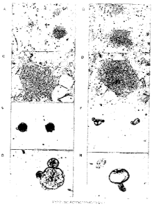
Figure 2 illustrates the morphology of an established Canine ES cell line. A
phase contrast image of
undifferentiated ES-like colonies well distinguished from the MEFs (A and B)
and a higher magnification
view (C and D) of tightly packed colony and cells with prominent nucleoli. At
least tyro phenotypically
distinct canine ES colonies can be identified: (A) Small, spherical, 3-D-like
ES colonies and (B) large
colonies with a more flattened appearance with well defined peripheral edges.
Transfer of single cell
suspensions or small clumps of cells from ES colonies to a sparse layer of
MEFs resulted in the formation of
structures resembling embryoid bodies (EBs) (E). (F) Low and (G and H) high
magnification of cystic
formations developing one week after transfer to non-coated culture dishes.
Figure 3 illustrates the optimization of canine Oct4 RT-PCR and nested PCR. (A-
C) Oct4 PCR
amplification of cDNA generated from total RNA isolated from two early passage
canine ES cell lines,
murine ES cells and murine trophoblast stem (TS) cells. (A) PCR amplification
of canine Oct4 in early
passage ES cells using primer pairs Oct4S1 and Oct4Rl. (B) PCR amplification
of murine Oct4 sequences
in ES and TS cell lines using Oct4 specific primers Oct4S1 and Oct4Rl. (C)
Nested PCR using Oct4S1 and
Oct4A1 primer pairs and the PCR generated Oct4 fragment generated in figure A.
(D-E) DNA sequence
analysis of the canine Oct4 fragment amplified by nested PCR.
Figure 4 shows Oct 4 expression in canine embryonic stem cells. Lane 1 -2:
Canine ES cells: Line
1, passage 1; Lane 3-4: Canine ES cells, Line 1, passage 10: Lane 5- ES cells,
Line 1 passage 10 (RNA
only) Lane 6. Negative control (H20).
Figure 5 shows (A) SSEA-4 and (B) TRA-1-60 expression in cells of canine ES
colonies.
Figure 6 Alkaline phosphatase expression in canine and murine ES cells. (A)
Unstained mouse ES
cells; (B) Mouse ES colony, and (C) Canine ES colony stained for expression of
alkaline phosphatase.
Figure 7 depicts the hatching of cells of the inner cell mass of canine
blastocysts on canine feeder
cells. The canine feeder layer supported the hatching of expanded canine
blastocysts but were unable to
support canine ES cell proliferation in an undifferentiated state.
Figure 8 shows the generation of canine ES cells on mouse feeder cells. Day 0:
Morula; Day 1:
Blastocyst showing cells of inner cell mass and blastocoel; Day 5: Expanded
blastocyst; Day 12: Hatching;
Day 19: Hatched ES cells before transfer to fresh MEFs; Day 27: ES colony
growing on MEFs; Day 30: ES
colonies growing on gelatinized plate with canine embryo-derived trophoblast-
like cells; Day 30: High
power magnification of ES colony.
Figure 9 shows the ire vitr~ differentiation of canine ES cells to endothelial
and neuronal cells. (A)
Differentiation of EBs to endothelial cells as indicated by morphological
appearance and reactivity to the
CA 02520722 2005-09-28
WO 2004/085631 PCT/CA2004/000456
-6-
endothelial cell specific antigen, CD31. (B) Irr vitro differentiation of EBs
to neuronal cells identified on the
basis of morphological appearance.
DETAILED DESCRIPTI~1~T OF A PREFERRED EI~iBODITVIEraIT
In accordance with the present invention there may be employed conventional
molecular biology,
microbiology, and recombinant DNA techniques within the skill of the art. Such
techniques are explain ed
fully in the literature. See for example, Sambrook, Fritsch, ~c Maniatis
(Molecular Cloning: A Laboratory
Manual, Second Edition (1989) Cold Spring Harbor Laboratory Press, Cold Spring
Harbor, N.Y); DNA
Cloning: A Practical Approach, Volumes I and II (D.N. Glover ed. 1985);
Oligonucleotide Synthesis (M..J.
Gait ed. 1984); Nucleic Acid Hybridization B.D. Hames ~c S.J. Higgins eds.
(1985); Transcription and
Translation B.D. Hames ~ S.J. Higgins eds (1984); Animal Cell Culture I2.I.
Freshney, ed. (1986);
Immobilized Cells and enzymes IRL Press, (1986); and B. Perbal, A Practical
Guide to Molecular Cloning
(1984). The invention may also employ standard methods in immunology known in
the art such as described
in Stites et al. (eds) Basic and Clinical Immunology, 8'1' Ed., Appleton &
Lange, Norwalk, Conn. (1994) and
Mishell and Shigi (eds), Selected Methods in Cellular Immunology, W.H. Freeman
and Co., New York
(1980).
For convenience, certain terms employed in the specification and claims are
collected here.
"Subject" or "patient" refers to an animal, preferably a mammal, to whom
treatment, inclufing
prophylactic treatment, with the cells, cell preparations, and compositions of
the present invention, is
provided. For treatment of those conditions or disease states that are
specific for a specific animal, the terms
refer to that specific animal. In particular, the terms refer to a canine. The
terms also include humans,
domestic animals including horses, cows, sheep, poultry, fish, pigs, cats, and
zoo animals.
"Pluripotent" refers to cells which retain the developmental potential to
differentiate into a variety
of cell lineages including the germ line.
"Canine embryonic stem cell phenotype" is used to describe cells which are
undifferentiated and
which are visually distinguished from other adult cells of canines.
"Cell line" refers to cultured cells that can be passaged at least one time
without terminating. The
invention contemplates cell lines that can be passaged at least 1, 2, 5, 10,
15, 20, 30, 40, 50, 60, 80, 100, and
200 times.
"Effective amount" refers to concentrations of components such as cells,
preparations, or
compositions effective for producing an intended result including treating a
disease or condition with cells,
preparations, and compositions of the invention, or for effecting a
transplantation of cells within a subject to
be treated.
The terms "administering" or "administration" refers to the process by which
cells, preparations, or
compositions of the invention are delivered to a subject for treatment
purposes. Cells, preparations, or
compositions may be administered a number of ways including parenteral (e.g.
intravenous and intraarterial
as well as other appropriate parenteral routes), oral, subcutaneous,
inhalation, or transdermal. Cells,
preparations, and compositions of the invention are administered in accordance
with good medical practices
taking into account the subject's clinical condition, the site and method of
administration, dosage, patient
age, sex, body weighi, and other factors known to physicians.
CA 02520722 2005-09-28
WO 2004/085631 PCT/CA2004/000456
_7_
"Transplanting", "transplantation", "grafting" and "graft" are used to
describe the process by which
cells, preparations, and compositions of the invention are~delivered to the
site within the subject where the
cells are intended to exhibit a favorable effect, such as repairing damage to
a subject's tissues, treating a
disease, injury or trauma, or genetic damage or environmental insult to an
organ or tissue caused by, for
example an accident or other activity. Cells, preparations, and compositions
may also be delivered in a
remote area of the body by any mode of administration relying on cellular
migration to the appropriate area
in the body to effect transplantation.
"Enriched" refers to a population of cells or a method which is at least 20+%,
30+%, 4.0+%, 50+°/~,
60+%, 70+%, 80+%, 85+%, 90+%, or 95+% effective, more preferably at least 98+%
effective, most
preferably 99+% effective. Therefore, a method that enriches for a given Bell
population, enriches at least
about 20+%, 30+%, 40+%, 50+%, 60+°/~, 70+%, 80%, 85%, 90%, or 95% of
the targeted cell population,
most preferably at least about 98% of the cell population, most preferably
about 99% of the cell population.
"Isolated" or "purifted" refers to altered "by the hand of man" from the
natural state i.e. anything
that occurs in nature is defined as isolated when it has been removed from its
original environment, or both.
In an aspect, a population or composition of cells is substantially free of
cells and materials with which it
may be associated in nature. By substantially free or substantially purified
is meant at least 50% of the
population are the target cells, preferably at least 70%, more preferably-at
least 80%, and even more
preferably at least 90% are free of other cells. Purity of a population or
composition of cells can be assessed
by appropriate methods that are well lrnown in the art.
"Gene therapy" refers to the transfer of new genetic information into cells
for the therapeutic
treatment of diseases or disorders. A foreign gene is transferred into a cell
that proliferates to introduce the
transferred gene throughout the cell population. Therefore, cells and
compositions of the invention may be
the target of gene transfer, since they may produce various lineages that will
potentially express the foreign
gene.
The term "embryo" as used herein refers to a developing cell mass that has not
implanted into the
uterine membrane of a maternal host. The term may refer to a fertilized
oocyte, a pre-blastocyst stage
developing cell mass, a blastocyst, andlor any other developing cell mass that
is at a stage of development
prior to implantation. Cells, cell lines, and cell preparations of the
invention may be isolated from and/or
arise from an embryo. An embryo can correspond to multiple stages of cell
development. The invention
preferably contemplates an early stage embryo in particular, an embryo at a
morula to expanded blastocyst
stage.
"Morula" refers to the structure during embryonic development comprising 8 or
more cells.
The term "blastocyst" used herein refers to the structure during early
embryonic development
comprising an inner cluster of cells, the inner cell mass (ICM), which gives
rise to the embryo, and an outer
layer, the tl~ophectoderm, which gives rise to extra-embryonic tissues. In
particular, cells from the ICM of an
early or expanded blasotocyst may be used in the present invention. In a
preferred embodiment, cells from a
blastocyst obtained 9-14. days, more preferably 10-11 days, post ovulation are
utilized in the invention.
CA 02520722 2005-09-28
WO 2004/085631 PCT/CA2004/000456
_g_
Stem Cells and Cell Lines
The present invention provides cells exhibiting a canine embryonic stem cell
phenotype, and a cell
line comprising cells exhibiting a canine embryonic stem cell phenotype.
In an embodiment, the present invention relates to a pluripotent canine stem
cell line. In another
embodiment, the invention relates to a purified preparation comprising, or
enriched for, canine embryonic
stem cells that are capable of indefinite proliferation ira vitr~ in an
undifferentiated state.
Proliferation irt viv~ may include cultivation of the stem cells for prolonged
periods where the cells
are substantially maintained in an undifferentiated state. The
undifferentiated cells may be capable of
maintaining an undifferentiated state when cultured in the presence of a
feeder layer. In a preferred aspect
the feeder layer does not induce extraembryonic differentiation or cell death.
A preparation of canine embryonic stem cells of the invention may also be
characterized by being
immunoreactive with markers for canine embryonic stem cells. In an embodiment,
the stem cells express
genetic markers of canine embryonic stem cells, including but not limited to
Oct-4, SSEA-4, TltA-1-60, and
alkaline phosphatase.
The canine embryonic stem cells of the invention may be characterized as
distinct from embryonic
stem cells from other species. In particular, canine embryonic stem cells may
be characterized as more
closely resembling human than marine embryonic stem cells in their morphology,
expression of cell surface
antigens, growth rates, and passage requirements.
The canine ES cells of the invention preferably have the potential to
differentiate in vitro when
subjected to differentiating conditions. Most preferably the stem cells have
the capacity ~to differentiate in
vitro into derivatives of the three embryonic germ layers. The ability of the
canine embryonic stem cells to
differentiate ira vitro into a variety of cell types including the ability to
differentiate into embryonic and more
highly differentiated cell types, may be tested by methods known in the art.
For example, to induce
differentiation in monolayer cultures, cells may be cultured without passage
onto a fresh feeder layer.
Differentiation may be induced in suspension culture by passing the cells onto
a gelatinized plate to
eliminate possible contamination by fibroblasts.
The invention therefore also relates to a purified canine embryonic stem cell
preparation of the
invention (preferably cultured ira vitr~) induced to differentiate into cells
of various lineages. A differentiated
cell preparation is characterized by expression of genetic markers of various
cell lineages
In an embodiment, the invention provides cells differentiated in vitro from an
undifferentiated
canine embryonic stem cell. In addition, a committed progenitor cell capable
of giving rise to a mature
somatic cell is provided. Preferably, undifferentiated cells are capable of
differentiating into extraembryonic
and embryonic lineages under differentiating conditions. In particular, the
cells of the invention are capable
of differentiating into cells derived from mesoderm, endoderm, and ectoderm
germ layers when the cells are
injected into an immunocompromised host.
A cell preparation of the invention may be derived from or comprised of cells
that have been
genetically modified either in nature or by genetic engineering techniques iat
viv~ or in vita-~.
Cell preparations or cell lines of the invention can be modified by
introducing mutations into genes
in the cells or by introducing transgenes into the cells. Insertion or
deletion mutations may be introduced in a
CA 02520722 2005-09-28
WO 2004/085631 PCT/CA2004/000456
-9-
cell using standard techniques. A transgene may be introduced into cells via
conventional techniques such as
calcium phosphate or calcium chloride co-precipitation, DEAE-dextran-mediated
transfection, lipofection,
electroporation, or microinjection. Suitable methods for transforming and
transfecting cells can be found in
Sambrook et al. (Molecular Cloning: A Laboratory Manual, 2nd Edition, Cold
Spring Harbor Laboratory
press (1989)), and other laboratory te~~tbo~ks. By way of example, a transgene
may be introduced into cells
using an appropriate expression vector including but not limited to cosmids,
plasmids, or modified viruses
(e.g. replication defective retroviruses - including lend- and onco-retrovial
vectors, adenoviruses and adeno-
associated viruses). Transfection is easily and efficiently obtained using
standard meth~ds including
culturing the cells on a monolayer of virus-producing cells (Van der Putten,
Proc IMatl Acad Sci U S A. 1985
Sep;82(18):6148-52; Stewart et al. (1987) EM13~ J. 6:383-388).
A gene encoding a selectable marker may be integrated into cells of a cell
preparation of the
invention. For example, a gene encoding a protein such as /3-galactosidase,
chloramphenicol
acetyltransferase, firefly luciferase, or a fluorescent protein marker may be
integrated into the cells.
Examples of fluorescent protein markers are the Green Fluorescent Protein
(GFP), and variants thereof.
Method of Producing Stem Cells
The invention relates to a method for obtaining purified canine embryonic
cells comprising the step
of culturing ICM cells from a canine embryo under conditions that promote
proliferation of undifferentiated
cells. In an embodiment, the cells are cultured in the presence of a feeder
layer (e.g. a fibroblast layer or a
medium conditioned by fibroblasts), and one or more proliferation agent.
A method for obtaining canine embryonic stem cells of the invention may
additionally comprise
expanding or maintaining canine embryonic stem cells, and/or inducing
differentiation of the stem cells, by
for example, removing the feeder layer.
In an aspect the invention provides a method of obtaining cells exhibiting a
canine embryonic stem
cell phenotype. Cells exhibiting a canine embryonic stem cell phenotype may be
isolated by (a) obtaining a
canine embryo; (b) culturing inner cell mass (ICM) cells from the canine
embryo under conditions which
promote proliferation of undifferentiated stem cells; and (c) recovering stem
cells. The conditions that
promote proliferation of undifferentiated stem cells (i.e. prevent
differentiation of stem cells) differ from the
requirements for other species.
In an embodiment of the invention, a method of producing cells exhibiting a
canine embryonic stem
cell phenotype is provided comprising: (a) obtaining a canine embryo at a
morula to expanded blastocyst
stage; (b) removing inner cell mass (ICM) cells of the blastocyst; (c)
culturing ICM cells in the presence of a
feeder layer to promote proliferation of undifferentiated stem cells; and (c)
recovering stem cells.
In an aspect of the invention there is provided a method of preparing a
preparation enriched for
undifferentiated canine embryonic stem cells comprising:
(a) obtaining a fertilized canine embryo;
(b) removing inner cell mass (ICM) cells from the embryo;
(c) culturing ICM cells under conditions which do not induce differentiation
and promote
proliferation of undifferentiated cells; and
(d) recovering stem cells.
CA 02520722 2005-09-28
WO 2004/085631 PCT/CA2004/000456
-10-
In an embodiment of the invention, a method for obtaining canine embryonic
stem cells is provided
comprising:
(a) growing embryos from canines in the presence of a feeder layer;
(b) removing ICM cells of the embryos either after spontaneous hatching or
after mechanical
removal of the zone pellucida;
(c) growing the cells in the presence of a feeder layer;
(d) selecting stem cell colonies by morphological characteristics; and
(e) culturing the selected stem cells.
In an embodiment of the invention, the method comprises obtaining a canine
embryo, culturing the
embryo in the presence of a feeder layer and proliferation agents, removing a
blastocyst outgrowth and
transferring the outgrowth to a fresh feeder layer. After establishment of the
culture of undifferentiated cells,
undifferentiated ES colonies are selected, dissociated by mechanical
manipulation or enzymatic digestion,
and transferred to fresh cultures for propagation. The invention also
contemplates cell preparations or lines
derived at all stages of development under the same culture conditions.
1 S The method may further comprise passaging the selected stem cells onto
fresh tissue culture growth
medium at intervals to prevent differentiation of the cells and to maintain a
cell line in culture. Cell
passaging may involve the steps of (1) releasing cells from a feeder layer and
disassociation of these cells,
and (2) placing the cells in media suitable for further cell proliferation. In
an embodiment, cells are passaged
by releasing cells from a surface using an enzymatic treatment. Cells that are
released can then be diluted
and transferred to fresh culture medium.
Canine embryos may be derived or isolated from any canine species. Canine
species may include
purebred species and species used as disease models or associated with
congenital, single or multigene
defects or disorders including hip dyspasia, and congenital heart defects.
Suitable species include but not
limited to a beagle, Doberman Pinscher, Ibizan Hound, Samoyed, Saluki,
Maltese, Leonburger, and poodle.
The canine embryos are harvested to provide maximum recovery and in vitro
maturation and hatching of
embryos. In an embodiment, the embryos are harvested after insemination or
post ovulation.
Mutant or transgenic blastocysts may be used to prepare a cell preparation or
cell line of the
invention. Cells used to prepare a cell preparation or cell line of the
invention can be engineered to contain a
selectable marker or they may be genetically altered using techniques well
knowmin the art.
A canine embryo (e.g. morula or bastocysts) used in a method of the invention
may be maintained
in culture under conditions permitting expansion of canine embryonic stem
cells. Embryos may be cultured
in the presence of a feeder layer. The feeder layer may be a confluent
fibroblast layer, including primary
mouse embryonic fibroblast (EMFI) cells or canine embryonic fibroblast like-
cells. Embryonic fibroblasts
may be obtained from 12 day old fetuses from outbred mice, but other strains
may be used as an alternative.
STO cells (i.e. a permanent line of irradiated mouse Fibroblasts) can also be
used as a feeder layer. The
feeder layer may also comprise medium conditioned by primary embryonic
iibroblast cells.
The conditions which promote proliferation of undifferentiated stem cells may
involve culturing the
cells in the presence of one or more proliferation agents including growth
factors, chemicals or cytokines.
The proliferation agents may be canine or human in origin, or may be derived
from other mammalian species
CA 02520722 2005-09-28
WO 2004/085631 PCT/CA2004/000456
-11-
active on canine cells. The following are representative examples of
proliferation agents which may be
employed in the present invention: all members of the fibroblast growth factor
(FGF) family including FGF-
4 and FGF-2, epidermal growth factor (EGF), stem cell factor (SCF),
thrombopoietin (TP~), FLT-3 ligand,
neural growth factor (NGF), VEGF, Granulocyte-Macrophage Growth Factor (GM-
CSF), IiGF, Iiox family,
Notch, leukemia inhibitor factor (LIF), cardiotrophin 1 (CT-1), ciliary
neurotroplxic factor (CNTF),
oncostatin M (~SM), and any member of the interleukin (IL) family, including
IL-6, IL-11, and IL-12.
Proliferation agents may be used in combination with equal molar or greater
amounts of a
glycosaminoglycan such as heparin sulfate.
Proliferation agents may be commercially available or can be produced by
recombinant DNA
techniques and purified to various degrees. For example, growth factors are
commercially available from
several vendors such as, for example, Genzyme (Framingham, Mass.), Genentech
(South San Francisco,
Calif.), Amgen (Thousand ~aks, Calif.), I~~cD Systems (Minneapolis, Minn.) and
Immunex (Seattle, Wash.).
Some proliferation agents may be purified from culture media of cell lines by
standard biochemical
techniques. Thus, it is intended that molecules having similar biological
activity as wild-type or purified
proliferation agents (e.g., recombinantly produced or mutants thereof) are
intended to be used within the
spirit and scope of the invention.
An effective amount of a proliferation agent is used in the culture medium.
The proliferation agents
are typically applied at sufficient intervals to maintain high proliferation
levels and maintenance of a stem
cell phenotype.
The culture medium used in the methods of the invention may comprise any
medium that supports
embryonic stem cells. The medium may be conditioned medium, non-conditioned
medium, or embryonic
stem cell medium. Examples of suitable conditioned medium include IMDM,
DME1VI, or aMEM,
conditioned with embryonic fibroblast cells (e.g. canine embryonic fibroblast
cells, human embryonic
fibroblast cells or mouse embryonic fibroblast cells), or equivalent medium.
Examples of suitable non-
conditioned medium include Iscove's Modified Delbecco's Medium (IMDM), DMEM,
or aMEM, or
equivalent medium. The culture medium may comprise serum (e.g. canine serum,
bovine serum, fetal bovine
serum, calf bovine serum, horse serum, human serum, or an artificial serum
substitute [e.g. 1% bovine serum
albumin, 10 ~,g/ml bovine pancreatic insulin, 200 ~g/ml human transferrin,
10'4M [3-mercaptoethanol, 2 mM
L-glutamine and 40 ~g/ml LDL (Low Density Lipoproteins)], or it may be serum
free. Preferably batch-
tested serums are used.
In an embodiment, the culture medium is serum free to provide cells that are
free of serum proteins
or biomolecules that may bind to the surface of the cells. Cells cultured in
such conditions may provide
somatic cells that have potential exposed novel antigenic sites. Such cells
may be useful as immunogens or
tolerizing agents for immune suppression. Thus, the invention provides a
cellular composition or mitotic or
differentiated cells therefrom that are isolated and maintained in serum-free
media.
In a preferred embodiment, the culture medium used for growth of embryonic
stem cells includes
ISO DMEM medium, preferably supplemented with serum (e.g. canine serum or
fetal bovine serum).
Embryos may be hatched spontaneously or manipulated mechanically to support
hatching. The zone
CA 02520722 2005-09-28
WO 2004/085631 PCT/CA2004/000456
-12-
pellucida surrounding the ICM may be removed using chemical (e.g. pronase,
acid Tyrodes solution) or
mechanical (e.g. needle, blade, laser dissection) methods. Preferably
mechanical methods are employed.
A method of the invention may involve treating the canine embryo to dislodge
the trophectoderm of
the embryo or porkion thereof. Methods suitable for removing the trophectoderm
include mechanical
methods and imanuno-surgery. The embryo (or blastocyst devoid of zone
pellucida) may be txwated wraith
antibody or antiserum specific for trophectoderm epitopes and/or with
complement.
Outgrowths or inner cell mass cells comprising ES-like cells may be removed
and cultured in the
presence of a feeder layer as described herein. The cells may be cultured for
a sufficient period of time to
establish the undifferentiated stem cells.
Once established the undifferentiated stem cells may be propagated, expanded,
and maintained.
Thus, a method for preparing canine embryonic stem cells may further include
removing the stem cells to
another feeder layer and culturing the stem cells for a period sufficient to
obtain proliferation of an enriched
preparation of morphologically undifferentiated stem cells. In order to expand
and maintain the
undifferentiated cells, cultured stem cells may be dissociated from the
culture (e.g. using enzymatic or
mechanical means) and cultured on fresh media. Cells may be regularly sub-
cultured (e.g. every 2- 7 days)
A method of the invention may further comprise preserving the canine embryonic
stem cells or cell
lines by preservation methods such as cryopreservation. Examples of suitable
cryopreservation methods are
those that are highly efficient for use with embryos such as vitrification, in
particular the Open Pulled Straw
(OPS) vitrification method.
A method of the invention may still further comprise inducing differentiation
of the canine
embryonic stem cells as described herein. The method may involve culturing the
stem cells under conditions
that promote differentiation (e.g. cell or tissue-specific differentiation).
The method may facilitate the
derivation of committed lineage progenitor cells which are no longer
pluripotent but may give rise to cells of
a variety of lineages.
Aunlications
Cells from the cell preparations may be introduced into a blastocyst or
aggregated with an early
stage embryo to produce chimeric conceptuses. A chimeric conceptus may be
allowed to grow to term, or
sacrificed during gestation to observe the contribution of the stem cells. The
conceptuses can be engineered
to carry selectable markers or genetic alterations. Cell lines can be derived
from the chimeric conceptuses.
Therefore, the invention further provides a chimeric conceptus derived from a
purified preparation of the
invention.
Cells of the invention may be used to repopulate an embryo of the same species
thus giving rise to a
chimeric animal, particularly a chimeric animal in which some or all of the
germ cells are derived from the
cultured cells. The embryonic stem cells may have been genetically modified or
selected for genetic
modification in culture.
The invention can provide for the derivation of canine embryonic stem cells
from embryos carrying
a particular genetic background or specific mutations. For example, the
embryos can be derived from high-
pedigree canines.
The cells, cell lines, sell preparations, chimeric conceptuses, embryos and
chimeric animals of the
CA 02520722 2005-09-28
WO 2004/085631 PCT/CA2004/000456
-13-
invention may be used to screen for potential therapeutics that modulate
development or activity e.g.
proliferation. In particular, the cell preparations and chimeric embryos may
be subjected to a test substance,
and the effect of the test substance may be compared to a control (e.g. in the
absence of the substance) to
determine if the test substance modulates development or activity. Selected
substances may be useful in
regulating canine embryonic stem cells or progeny thereof ifa viv~ and they
may be used to treat various
conditions requiring regulation of such cells.
The cells and cell preparations of the invention may be used to prepare model
systems of disease or
conditions. Canines develop similar diseases as humans and the clinical
presentations are similar. Thus,
canine models offer very useful models for studying disease and identifying
potential therapeutics. Canine
models of human diseases can be created including but not limited to models
for glycogen storage disease,
muscular dystrophy, haemophilia, narcolepsy, thrombasthenia, Von Willebrand
Disease, osteogenesis,
nephritis, retinal atrophy, severe combined immunodeficiency disease,
hematopoietic and autoimmune
disorders, cancer, heart diseases, motor neuron diseases, and degenerative
bone and joint diseases, and
atherosclerosis.
Canines provide a powerful preclinical large animal model in biomedical
research, which
historically has been used successfully to move novel treatment modalities
into the clinic (reviewed by
Ostrander et al (3)). Breeding programs for the generation of canines with
distinctive phenotypes have led to
the production of closed breeding populations characterized by more than 400
inherited disorders.
Autosomal recessive and complex traits represent the largest proportion of
canine diseases, some of which
include hematopoietic and autoimmune disorders, cancer, heart diseases, motor
neuron diseases, and
degenerative bone and joint diseases. These naturally occurring canine
diseases provide powerful models for
genetic mapping and the assessment of the pathophysiology and novel treatments
of homologous diseases in
humans. Canines share many biochemical and physiologic characteristics with
humans and thus they more
accurately resemble human diseases than do their rodent counterparts. Their
short generation time and long
life span make them ideal for studying the lifetime effects of medical
manipulations. Canines are more
readily available, incur lower costs, are more disease-free and easier to work
with than nonhuman primates.
Compared with mice, the large size of canines is amenable to serial blood and
tissue sampling and
continuous intravenous infusions. Since canines closely approximate humans in
body weights, blood
volumes, and issues of tissue typing and clinical management, they have been
instrumental in the
development of human bone marrow transplantation and gene therapy protocols
(31-42). Large canine
breeds have also made valuable contributions to the development of treatment
modalities for cardiovascular
(43) and orthopaedic diseases (44, 45). The availability of canine ES cells as
described herein facilitates the
development of ES cell-based therapies for the treatment of inherited and
acquired human diseases.
The cells, cell preparations or cell lines of the invention can be used to
produce growth factors and
hormones. The cell preparations or cell lines of the invention can also be
used to produce therapeutics.
The canine embryonic stem cells of the invention may be induced to
differentiate into cells of a
variety of lineages, preferably cells that exhibit morphological,
physiological, functional, and/or
immunological features of somatic and germ cells. Cells from a differentiated
cell preparation may be
characterized by expression of genetic markers from a variety of cell lineages
(e.g. markers for muscle,
CA 02520722 2005-09-28
WO 2004/085631 PCT/CA2004/000456
- 14-
neural, adipocyte, osteoclast, osteoblast, endothelial, hematopoietic,
astrocytes, pancreatic cells, retinal cells,
renal cells, connective tissue cells, and hepatocytes), or physiological,
immunological or functional
characteristics of cells of a variety of lineages. For example, cells can be
screened for expression of tissue
specific markers such as IYIyo-I~ (muscle), FLK-1 (endothelial), filial
fibrillary acidic protein (astrocytes),
glucagon (alpha-~ cells), insulin (islet-(3 cells), s~matostatin (islet-b),
pancreatic polypeptide (islet-PP cells),
cytokeratins (CIA), mucin IvIUCl, carbonic anyhydrase II, and carbohydrate
antigen 19.1 (ductal cells), and
NESTIN (neural).
Differentiated cells can be used to prepare a cDNA library relatively
unc~ntaminated with cDNA
preferentially expressed in cells from other lineages, and they can be used to
prepare antibodies that are
specific for particular markers of somatic cells.
After differentiation of the cells into selected somatic cells as described
herein, the cells may be
separated to obtain a population of cells largely consisting of somatic cells.
This may be accomplished by
positive selection of somatic cells using antibodies to identify tissue
specific cell surface markers or negative
selection using ES cell specific markers.
A cell preparation or cellular composition of the invention may be genetically
engineered in such a
manner that they or cells derived therefrom produce, irz vitro or irz vivo,
polypeptides, hormones and proteins
not normally produced in the cells in biologically significant amounts, or
produced in small amounts but in
situations in which regulatory expression would lead to a therapeutic benefit.
For example, the cells could be
engineered with a gene that expresses a molecule that specifically inhibits
bone resorption, but does not
otherwise interfere with osteoclasts binding to bone, or the cells could be
engineered with a gene that
expresses insulin at levels compatible with normal injected doses.
Alternatively the cells could be modified
such that a protein normally expressed will be expressed at much lower levels.
These products would then be
secreted into the surrounding media or purified from the cells. The cells
formed in this way can serve as
continuous short term or long term production systems of the expressed
substance.
Thus, in accordance with this aspect of the invention, cells of the invention
can be modified with
genetic material of interest. The modified cells can be cultured in vitro
under suitable conditions so that they
differentiate into cells of specific lineages. The cells are able to express
the product of the gene expression or
secrete the expression product. These modified cells can be administered to a
target tissue where the
expressed product will have a beneficial effect.
In a further embodiment, the transduced cells of the invention can be induced
ira vivo to differentiate
into cells of specific lineages that will express the gene product. For
example, the transduced cells may be
administered to induce production of cells of specific lineages having the
transduced gene. The cells may be
administered in admixture with each other or separately and may be delivered
to a targeted area. The cells
can be introduced intravenously and home to the targeted area. Alternatively,
the cells may be used alone
and caused to differentiate iaz viv~.
Thus, genes can be introduced into cells which are then injected into a
recipient where the
expression of the gene will have a therapeutic effect. For example,
osteoclasts may be genetically engineered
to have reduced activity in. vivo. Appropriate genes would include those that
play a role in the regulation of
osteoporosis, in areas such as serum calcium responsiveness, estrogen
secretion and bone resorption. An
CA 02520722 2005-09-28
WO 2004/085631 PCT/CA2004/000456
-15-
insulin gene may be introduced into blood stem cells to provide a constant
therapeutic dose of insulin in the
bone marrow and peripheral blood.
The technology may be used to produce additional copies of essential genes to
allow augmented
expression by cells of certain gene products ira viv~. These genes can be, for
example, hormones, matrix
$ proteins, cell membrane proteins, cytolcines, adhesion molecules, or
"rebuilding" proteins important in tissue
repair.
The cell preparations and compositions of the invention can be used in a
variety of methods (e.g.
transplantation) and they have numerous uses in the field of medicine. They
may be used for the
replacement of body tissues, organs, components or structures which are
missing or damaged due to trauma,
age, metabolic or toxic injury, disease, idiopathic loss, or any other cause.
In particular, they may have
application in the study, prevention, and treatment of conditions such as
hemophilia, muscular dystrophy,
MPS-1, glycogen storage disease, narcolepsy, thrombasthenia, Von Willebrand
Disease, osteogenesis,
nephritis, retinal atrophy, severe combined immunodeficiency disease,
hematopoietic and autoimmune
disorders, cancer, heart diseases, motor neuron diseases, degenerative bone
and joint diseases, and
atherosclerosis.
Transplantation or grafting, as used herein, can include the steps of
isolating a cell preparation
according to the invention and transferring cells in the preparation into a
mammal or a patient.
Transplantation can involve transferring the cells into a mammal or a patient
by injection of a cell suspension
into the mammal or patient, surgical implantation of a cell mass into a tissue
or organ of the mammal or
patient, or perfusion of a tissue or organ with a cell suspension. The route
of transferring the cells may be
determined by the requirement for the cells to reside in a particular tissue
or organ and by the ability of the
cells to find and be retained by the desired target tissue or organ. Where the
transplanted cells are to reside in
a particular location, they can be surgically placed into a tissue or organ or
simply injected into the
bloodstream if the cells have the capability to migrate to the desired target
organ.
The invention may be used for autografting (cells from an individual are used
in the same
individual), allografting cells (cells from one individual are used in another
individual) and xenografting
(transplantation from one species to another). Thus, the cells, cell
preparations and cellular compositions of
the invention may be used in autologous or allogenic transplantation
procedures to improve a cell deficit or
to repair tissue.
In an aspect of the invention, the newly created cells, cell lines, and
preparations, can be used in
both cell therapies and gene therapies aimed at alleviating disorders and
diseases involving the cells or
progeny thereof. The invention obviates the need for human tissue to be used
in various medical and research
applications.
The cell therapy approach involves the use of transplantation of the newly
created cells, cell lines,
or preparations or cells differentiated therefrom, as a treatment for injuries
and diseases. The steps in this
application include: (a) producing cells or a cell line of the invention, or
differentiating cells therefrom, as
described herein; and (b) allowing the cells to form functional connections
either before or after a step
involving transplantation of the cells. The gene therapy approach also
involves cellular compositions
CA 02520722 2005-09-28
WO 2004/085631 PCT/CA2004/000456
-16-
comprising cells of the invention transfected with an appropriate vector
containing a cDNA for a desired
protein, followed by a step where the modified cells are transplanted.
In either a cell or gene therapy approach, therefore, cells of the present
invention, or cells or tissues
differentiated from the Bells can be transplanted in, or grafted to, a patient
in need. Thus, the cells or
differentiated cells therefrom can be used to replace the cells in a patient
in a cell therapy approach, useful in
the treatment of tissue injury, and diseases. These cells can be also used as
vehicles for tlae delivery of
specific gene products to a patient. ~ne example of how these newly created
cells or cells differentiated
therefrom can be used in a gene therapy method is in treating the effects of
Parkinson's disease. For example,
tyrosine hydrolase, a key enzyme in dopamine synthesis, may be delivered to a
subject via the
transplantation of cells of the invention that are capable of differentiating
into neuronal cells, or
transplantation of neuronal cells differentiated from the cells, which have
been transfected with a vector
suitable for the expression of tyrosine hydrolase.
The invention also provides a method of treating a subject with a condition
involving a somatic cell
of the invention comprising transferring a cell of the invention into the
subject, wherein the cell differentiates
into the somatic cell.
The invention also contemplates a pharmaceutical composition comprising cells,
a cell preparation,
or cell line of the invention, and a pharmaceutically acceptable carrier,
excipient, or diluent. The
pharmaceutical compositions herein can be prepared by Bp r se known methods
for the preparation of
pharmaceutically acceptable compositions which can be administered to
subjects, such that an effective
amount of the active substance is combined in a mixture with a
pharmaceutically acceptable vehicle.
Suitable vehicles are described, for example, in Remington's Pharmaceutical
Sciences (Remington's
Pharmaceutical Sciences, Mack Publishing Company, Easton, Pa., USA 1985). On
this basis, the
compositions include, albeit not exclusively, solutions of the cells, cell
preparations, or cell lines in
association with one or more pharmaceutically acceptable vehicles or diluents,
and contained in buffered
solutions with a suitable pH and iso-osmotic with the physiological fluids.
Still another aspect of the invention is a kit for producing cells of the
invention. The kit includes the
reagents for a method of the present invention for producing cells of the
invention.
In an aspect, cells, cell preparations, and cellular compositions disclosed
herein can be used for
toxicity testing for drug development testing. Toxicity testing may be
conducted by culturing cells, cell
preparations, and cell lines or cells differentiated therefrom in a suitable
medium and introducing a
substance, such as a pharmaceutical or chemical, to the culture. The cells or
differentiated cells are examined
to determine if the substance has had an adverse effect on the culture. Drug
development testing may be
done by developing derivative cell lines which may be used to test the
efficacy of new drugs. Affinity assays
for new drugs may also be developed from the cells, differentiated cells, or
cell lines.
Using a method of the invention it is possible to identify drugs that are
potentially toxic to canine
embryonic stem cells.
The cellular compositions of the invention may be used to screen for potential
therapeutics that
modulate development or activity of cells of the invention. In particular, the
cells of the invention may be
subjected to a test substance, and the effect of the test substance may be
compared to a control (e.g. in the
CA 02520722 2005-09-28
WO 2004/085631 PCT/CA2004/000456
-17-
absence of the substance) to determine if the test substance modulates
development or activity of the cells or
cells differentiated therefrom.
In an aspect of the invention a method is provided for using cells of the
invention to assay the
activity of a test substance comprising the steps of:
(a) exposing the cells to a test substance; and
(b) detecting the presence or absence of an effect of the test substance on
the survival of the
cells or on a morphological, fixnctional, or physiological characteristic
and/or molecular
biological property of the cells, whereby an effect altering cell survival, a
morphological,
functional, or physiological characteristic and/or a molecular biological
property of tlxe
cells indicates the activity of the test substance.
In another aspect a method is provided for using cells of the invention to
screen a potential new
drug to treat a disorder involving the cells comprising the steps of:
(a) exposing the cells to a potential new drug; and
(b) detecting the presence or absence of an effect of the potential new drug
on the survival of
the cells or on a morphological, functional, or physiological characteristic
and/or molecular
biological property of said cells, whereby an effect altering cell survival, a
morphological,
functional, or physiological characteristic and/or a molecular biological
property of the
cells indicates the activity of the potential new drug.
The invention also relates to the use of cells, cell lines, cell preparations,
and compositions in drug
discovery. The invention provides methods for drug development using the
cells, cell preparations, and
cellular compositions of the invention. Cells, cell preparations, cell lines,
and compositions of the invention
may comprise cells that secrete novel or known biological molecules or
components. In particular, culturing
in the absence of serum may provide cells that have minimal interference from
serum molecules and thus,
may be more physiologically and topologically accurate. Therefore, proteins
secreted by cells described
herein may be used as targets for drug development. In one embodiment, drugs
can be made to target
specific proteins on cells of the invention. Binding of the drug may promote
differentiation of cells into cells
of specific lineages. In another embodiment, drugs specific for regulatory
proteins of somatic cells may be
used to arrest growth of a particular type of cell. Any of the proteins can be
used as targets to develop
antibody, protein, antisense, aptamer, ribozymes, or small molecule drugs.
Agents, test substances, or drugs identified in accordance with a method of
the invention or used in
a method of the invention. include but are not limited to proteins, peptides
such as soluble peptides including
Ig-tailed fusion peptides, members of random peptide libraries and
combinatorial chemistry-derived
molecular libraries made of D- and/or L-configuration amino acids,
phosphopeptides (including members of
random or partially degenerate, directed phosphopeptide libraries), antibodies
[e.g. polyclonal, monoclonal,
humanized, anti-idiotypic, chimeric, single chain antibodies, fragments, (e.g.
Fab, F(ab)2, and Fab
expression library fragments, and epitope-binding fragments thereof)], nucleic
acids, ribozymes,
carbolxydrates, and small organic or inorganic molecules. An agent, substance
or drug may be an endogenous
physiological compound or it may be a natural or synthetic compound.
CA 02520722 2005-09-28
WO 2004/085631 PCT/CA2004/000456
-18-
The cells, cell preparations, and' cell lines disclosed herein can be used in
various bioassays. In an
embodiment, the cells are used to determine which biological factors are
required for proliferation or
differentiation. By using cells of the invention in a stepwise fashion in
combination with different biological
compounds (such as hormones, specific growth factors, etc.), one or more
specific biological compounds can
be found to induce differentiation to somatic cells. ~'Jther uses in a
bioassay for the cells are differential
display (i.e. mRNA differential display) and protein-protein interactions
using secreted proteins from the
cells. Protein-protein interactions can be determined with techniques such as
a yeast two-hybrid system.
Proteins from cells, cell preparations and cellular compositions of the
invention can be used to identify other
unknown proteins or other cell types that interact with the cells. These
unknown proteins may be one or
more of the following: growth factors, hormones, enzymes, transcription
factors, translational factors, and
tumor suppressors. Bioassays involving cells, cell preparations, and cell
lines of the invention, and the
protein-protein interactions these cells form and the effects of protein-
protein or cell-cell contact may be
used to determine how surrounding tissue contributes to proliferation or
differentiation of cells of various
lineages.
In an aspect of the invention cells of the invention may be used to repair
cell or tissue injury. They
may also be used in the treatment of genetic defects that result in
nonfunctional cells. The stem cells of the
invention may be transplanted directly to the site of defective cells in order
to rescue the defect or delivered
via the blood stream by injecting the cells into the vein. In addition, gene
therapy vectors may be integrated
into the stem cells followed by engraftment of these engineered cells to their
target tissues. The introduction
of gene therapy vectors requires cell proliferation. The successful long term
engraf3ment of the cells to the
target tissue requires they maintain a stem cell characteristic.
The cells, cell preparations, and cell lines of the invention may be used as
immunogens that are
administered to a heterologous recipient. Administration of cells obtained in
accordance with the invention
may be accomplished by various methods. Methods of administering cells as
immunogens to a heterologous
recipient include without limitation immunization, administration to a
membrane by direct contact (e.g. by
swabbing or scratch apparatus), administration to mucous membranes (e.g. by
aerosol), and oral
administration. Immunization may be passive or active and may occur via
different routes including
intraperitoneal injection, intradermal injection, and local injection. The
route and schedule of immunization
are in accordance with generally established conventional methods for antibody
stimulation and production.
Mammalian subjects and antibody producing cells therefrom may be manipulated
to serve as the basis for
production of mammalian hybridoma cell lines.
In an aspect the invention provides a culture system from which genes,
proteins, and other
metabolites involved in proliferation or differentiation of cells of various
lineages can be identified and
isolated. The cells in a culture system of the invention may be compared with
other cells (e.g. differentiated
cells) to determine the mechanisms and compounds that stimulate production of
cells of various lineages.
The cells of the invention can be used to screen for genes expressed in or
essential for
differentiation of canine embryonic stem cells. Screening methods that can be
used include Representational
Difference Analysis (RDA) or gene trapping with for example SA-lacZ. Gene
trapping can be used to induce
dominant mutations (e.g. by deleting particular domains of the gene product)
that affect differentiation or
CA 02520722 2005-09-28
WO 2004/085631 PCT/CA2004/000456
-19-
activity of cells of the invention and allow the identification of genes
expressed in or essential for
differentiation of these cells.
Cell preparations of the invention comprising hematopoietic cells may be used
for enhancing the
immune and hematopoietic system of a subject. The cell preparations will
facilitate enhancement or
reconstitution of the subject's immune and/or blood forming system.
In an aspect of the invention, the cells, cell lines, and cell preparations of
the invention are used in
the treatment of leukemia (e.g. acute myelogenous leukemia, chronic
myelogenous leukemia), lymphomas
(e.g. non-Hodgkin's lymphoma), neuroblastoma, testicular cancer, multiple
myeloma, melanomas, breast
cancer, solid tumors that have a stem cell etiology, or other cancers in which
therapy results in the depletion
of hematopoietic cells.
In another aspect of the invention, cells, cell lines, and compositions of the
invention, with or
without genetic modification to provide resistance to HIV, are used to treat
subjects infected with HIV-1 that
have undergone severe depletion of their hematopoietic cell compartment
resulting in a state of immune
deficiency.
Hematopoietic cells may also be transfected with a desired gene that can be
used for treatment of
genetic diseases. Hematopoietic cell-related genetic diseases can be treated
by grafting with cells transfected
with a gene that can make up for the deficiency or the abnormality of the gene
causing the diseases. For
example, a normal wild type gene that causes a disease such as a-thalassemia
(Mediterranean anemia), sickle
cell anemia, ADA deficiency, recombinase deficiency, recombinase regulatory
gene deficiency and the like,
can be transferred into the hematopoietic cells by homologous or random
recombination and the cells can be
grafted into a subject. Further, a preparation comprising normal hematopoietic
cells free from abnormalities
of genes (from a suitable donor) can be used for treatment.
Another application of gene therapy permits the use of a drug in a high
concentration, which is
normally considered to be dangerous, by providing drug resistance to normal
hematopoietic cells by
transferring a drug resistant gene into the cells. In particular, it is
possible to carry out the treatment using an
anticancer drug in high concentration by transferring a gene having drug
resistance against the anticancer
drug, e.g., a multiple drug resistant gene, into hematopoietic cells of the
invention.
Diseases other than those relating to the hematopoietic system can be treated
by using the
hematopoietic cells of the invention in so far as the diseases relate to a
deficiency of secretory proteins such
as hormones, enzymes, cytokines, growth factors and the like. A deficient
protein can be induced and
expressed by transferring a gene encoding a target protein into the
hematopoietic cells under the control of a
suitable promoter. The expression of the protein can be controlled to obtain
the same activity as that obtained
by the natural expression ira vivo.
It is also possible to insert a gene encoding a ribozyme, an antisense nucleic
acid or the like or
another suitable gene into the hematopoietic cells to control expression of a
specific gene product in the cells
or to inhibit susceptibility to diseases. For example, the hematopoietic cells
can be subjected to gene
modification to express an antisense nucleic acid or a ribozyme, which can
prevent growth of hematic
pathogens such as HIV, HTLV-I, HTLV-II and the like in hematopoietic cells.
The cells and cell preparations comprising hematopoietic cells of the
invention can be introduced in
CA 02520722 2005-09-28
WO 2004/085631 PCT/CA2004/000456
-20-
a vertebrate, which is a recipient of cell grafting, by, for example,
conventional intravenous administration.
The invention also relates to a method for conducting a regenerative medicine
business, comprising:
(a) a service for accepting and logging in samples from a client comprising
cells of the invention; (b) a
system for culturing Bells dissociated from the samples; (c) a cell
preservation system for preserving cells
generated by the system in (b) f~r later retrieval on behalf of the client or
a third party. The method array
further comprise a billing system for billing the client or a medical
insurance provider thereof.
The invention features a method for conducting a stem cell business comprising
identifying agents
which influence the proliferation, differentiation, or survival of cells of
the invention. Examples of such
agents are small molecules, antibodies, and extracellular proteins. Identified
agents can be profiled and
assessed for safety and efficacy in animals. In another aspect, the invention
contemplates methods for
influencing the proliferation, differentiation, or survival of cells of the
invention by contacting the cells with
an agent or agents identified by the foregoing method. The identified agents
can be formulated as a
pharmaceutical preparation, and manufactured, marketed, and distributed for
sale.
In an embodiment, the invention provides a method for conducting a stem cell
business comprising
(a) identifying one or more agents which affect the proliferation,
differentiation, function, or survival of cells
of the invention; (b) conducting therapeutic profiling of agents identified in
(a); or analogs thereof for
efficacy and toxicity in animals; and (c) formulating a pharmaceutical
composition including one or more
agents identified in (b) as having an acceptable therapeutic profile. The
method may further comprise the
step of establishing a distribution system for distributing the pharmaceutical
preparation for sale. The method
may also comprise establishing a sales group for marketing the pharmaceutical
preparation.
The invention also contemplates a method for conducting a drug discovery
business comprising
identifying factors that influence the proliferation, differentiation,
function, or survival of cells of the
invention, and licensing the rights for further development.
Having now described the invention, the same will be more readily understood
through reference to
the following examples which are provided by way of illustration, and are not
intended to be limiting of the
present invention.
EXAMPLE 1
Generation Of Canine Embryonic Stem Cells For Use In Animal Models Of Human
Disease
Pluripotent embryonic stem cells are undifferentiated cells that retain the
ability to proliferate
indefinitely and differentiate to cells of the three embryonic germ layers and
their derivatives. This unlimited
life span and wide developmental potential suggest that these cells have
enormous potential for both basic
research and clinical applications focused on regenerative medicine and tissue
engineering. Herein is
described the derivation of four canine embryonic stem (ES) cell lines.
Preimplantation embryos collected on
days 9-13 post-insemination were maintained on murine feeder layers under
conditions used for the
expansion of human ES cells. Canine ES cells have been maintained in an
undifferentiated state and have
undergone multiple ira vitr~ passages in culture and multiple rounds of
cryoperservation and thawing since
their derivation. Similar to human ES cells, canine ES cells express ~ct4~,
SSEA-4, TItA-1-60 and alkaline
phosphatase and do not express SSEA-1. Plating of ES cells at low density in
the absence of fibroblasts
resulted in their differentiation or death whereas low density seeding of ES
cells onto a sparse feeder layer
CA 02520722 2005-09-28
WO 2004/085631 PCT/CA2004/000456
-21-
resulted in the formation of embryoid bodies. Undifferentiated ES cells may be
maintained and expanded,
and canine ES cells have been differentiated to endothelial cells and neuronal
cells ifz vitro.
Mt~TEI~II~LS AI'~TD METHODS
I~lating and Embry~ Collccti~n
Fourteen female d~gs of mixed breeding were used in this study. Nine t~ 13
days post-.ovulation or
6 to 12 days after first mating, dogs were subjected to surgical procedure
under general anesthesia. Both
oviducts and the uterine horns were removed and each uterine horn was flushed
using warm Dulbecco's
Phosphate Buffered Saline (DPBS). After inspection embryos were transferred t~
CO~ independent medium.
On average, eight embryos were obtained per experiment (range: 0 t~ 16). A
total of 5~ embryos were
collected throughout the study. No embryos were recovered from three dogs.
In 5~itr~ Cexlture ~f C~Ilected Embryos and Ilerivati~n of ES Cells
Embryos were cultured in Knock Out Dulbecco's Modified Eagle's Medium (ISO
DMEM) or in a
1:1 mixture of Dulbecco's Modified Eagle's Medium and Ham's nutrient mixture F-
12 (DMEM/F12)
(Invitrogen). Complete ISO DMEM or DMEM/F12 media were supplemented with 0.1
mM [3-
mercaptoethanol (Sigma), 0.1 mM non-essential amino acids, 0.1 mM sodium
pyruvate, penicillin (100
IU/ml), streptomycin (50 ~g/ml) (Invitrogen), LIF (20ng/ml) (Chemicon) and 15%
FBS (Invitrogen). In
some experiments, 15% of serum replacement media (SRM) (Invitrogen) was used
in place of fetal bovine
serum (FBS). Embryos were maintained at 37°C or 38°C in 5% CO2.
Upon completion of optimization experiments, cultures were maintained in
expansion medium
prepared with Complete DMEM/F12 medium supplemented with 15% dog serum or FBS
at 38°C. To
prepare dog serum, 50 to 100 ml peripheral blood was obtained from dogs from
which embryos had been
harvested and centrifuged at 2000 rpm at room temperature for 30 min. Serum
was collected after
centrifugation, heat inactivated at 56°C for 1 hour and sterilized
using a 0.22 ~m filter .
Mouse Embryo Fibroblasts (MEFs) or Canine Embryo Fibroblast-like cells (CEF-
like) were used as
feeder layers. MEFs were prepared by procedures outlined in a laboratory
manual, "Manipulation of Mouse
Embryo", Cold Spring Harbor Laboratory Press-Second Edition. To establish CEF-
like feeders, canine
embryo-derived cells that underwent spontaneous differentiation and gave rise
to fibroblast looking cells
were cultured in DMEM/F12 supplemented media containing 15% FBS without LIF.
Once confluent, cells
were cultured under conditions identical to those described for the
established of MEFs.
All feeders were inactivated by exposure to 10 ~g/ml of mytomicin C or by y-
irradiation (4000
reds) as per the above mentioned laboratory manual.
Establishment and Maintenance of Canine ES Cell Lines
Embryos were cultured in expansion medium and allowed to hatch either
spontaneously or
manipulated mechanically to support hatching. Mechanical hatching was
accomplished by cutting the zone
pellucida surrounding the ICM with a fine scalpel blade. One or two openings
were cut through the zone
pellucida and trophectoderm while ensuring no damage to embryonic cells was
incurred. In some cases, the
zone pellucida was gently split and removed from the embryo using fine needles
and the released ICM
collected and plated onto fresh feeders. Five to 10 days after hatching,
embryo-derived outgrowths were
CA 02520722 2005-09-28
WO 2004/085631 PCT/CA2004/000456
-22-
disaggregated into small pieces by mechanical cutting and gentle dissociation
using a finely pulled glass
pipette. The clusters of ES-like cells were transferred at high density to
fresh MEFs and resulting colonies
further sub-cultured every 2-4 days by mechanical manipulation with or without
addition of 0.125% Disease
in Hanks' Balanced Salt Solution. Once established, ES cell lines were
passaged every 3 to 7 days by
exposure to 0.1% collagenase/dispase (Sigma) prepared in DMEM/F12 or 0.125%
Disease in Hanks'
Balanced Salt Solution for 45-60 min followed by a brief exposure to 0.02%
EDTA (Sigma). Cultures were
maintained at 38°C in 5% CO2. Canine ES cultures were passaged at high
density to maintain the
undifferentiated phenotype. Cultures were examined daily and complete or half
medium changes done on
alternate days.
Cell lines were cryopreserved in cryomedium containing either DMEM/F12
supplemented with
10% DMSO and 20% dog serum or 90% FBS and 10% DMSO. Cells were cryopreserved
under slow-
cooling conditions, initially stored at -80°C and subsequently
transferred to liquid nitrogen. ES cells were
recovered from cryopreservation by immersion of cryovials for 30-60 seconds in
a 37°C water bath. Cells
were washed in DMED/F12 supplemented with 30% dog serum or FBS and spun at
1000 rpm prior to
plating to remove DMSO. Cells were plated on irradiated MEFs in six-well
dishes in complete DMEM/F12
medium supplemented with 15% dog serum or FBS and hLIF.
Cloning and Sequencing of Canine Oct4
In order to design primers specific for canine Oct4, human, mouse and bovine
Oct4 homologous
genes were obtained by BLAST search and aligned using Vector NTI 7.1
(InforMax, Inc. USA). The Oct4R1
primer was derived from the marine Oct2 sequence while the, Oct4S1 and Oct4A1
primers were designed
based on the human Oct4 sequence. (Xu C, Inokuma MS, Denham J, Golds K, I~undu
P, Gold JD, Carpenter
MIA. Feeder-free growth of undifferentiated human embryonic stem cells. Nature
Biotechnology 2001 Oct;
19(10):971-4., 2001)
Oct4Rl: ACTCGAACCACATCCTTCTCTAGC[SEQ ID N0.:2]
Oct4Sl: CTTGCTGCAGAAGTGGGTGGAGGAA [SEQ ID N0.:3]
Oct4Al: CTGCAGTGTGGGTTTCGGGCA [SEQ ID N0.:4]
Total RNA was prepared from two early passage canine ES cell lines (ES1 and
ES2), marine ES
cells and marine TS cells using a Trizol kit (Invitrogen, USA). mRNA was
extracted using dynal beads
(Dynal A. S, Oslo, Norway). cDNA was generated with 100 ng of mRNA using oligo
dT and reverse
transcription using Superscript II reverse transcriptase (Invitrogen, USA).
The canine Oct4 cDNA was
amplified by PCR using primers, Oct4S1 and Oct4R1 for 45 cycles. The PCR
products were diluted and
used as template in a nested PCR reaction using primers, Oct4S1 and Oct4Al.
The PCR protocol consisted of a 3 minute denaturation at 94°C, and 45
cycles, each consisting of a
15 seconds denaturation phase at 94°C, a 30 second annealing period at
55°C and a 1 minute extension time
at 72°C. A final extension of 10 minutes at 72°C was included.
PCR products were cloned and sequenced to
derive the canine Oct4 sequence.
Canine Oct4 I~T-PCI~ of Canine ES-like Cell
The above derived canine Oct4 fragment was sequenced and the sequence used to
design primers
specific for canine Oct4.
CA 02520722 2005-09-28
WO 2004/085631 PCT/CA2004/000456
- 23 -
Dog-POU5F1-S1: TGACGACAACAAAAATCT [SEQ ID N0.:5]
Dog-POU5F1-A1: CAGGCATGTGTTCTCCAG [SEQ ID N0.:6]
Oct4 expression was assessed by reverse transcriptase polymerase chain
reaction (RT-PCR). cDNA
from canine ES like-cells was prepared as described above. Oct4 cDNA was PCR
amplified in reactions
consisting of a 3 minute denaturation at ~4°C, and 4~5 cycles, each
consisting of denaturation for 30 seconds
at 94°C, primer annealing at 50°C for 30 seconds and extension
at 72°C for 30 seconds . A final extension
for 7 minutes was at 72°C.
Immunotluorescence Labeling of Canine ES-like Cells
Canine ES colonies were grown in 4-well slide chambers. Before staining, the
medium was
discarded and the cells were rinsed with PBS. Cells were fixed in cooled
methanol at -20°C. After 10
minutes methanol was removed and the cell membrane was permeabilized with
cooled acetone for 1 min at
20°C. Slides were washed twice with 4.°C PBS for 5 minutes,
incubated 4 hours at 4°C in 8% BSA to block
cells and washed again.
Cells were incubated with primary antibody SSEA-1, SSEA-4 or TRA-1-60
(Chemicon
International) at the concentration of 15 pg/ml in PBS containing 2% BSA for 2-
4 hours or overnight and
slides were washed three times for 5 minutes with PBS supplemented with 2%
BSA. An anti-mouse IgG
(Fab Specific) FITC labeled antibody (Sigma) was used as a secondary antibody
(1:200 dilution). Secondary
labeling was done for 20 minutes in PBS supplemented with 2% BSA and slides
washed again as described
above. Excess PBS was removed, slides mounted with mounting medium and covered
with cover slips.
Antibody labeling was detected by confocal microscopy and results recorded.
Analysis of Alkaline Phosphatase Activity
Expression of alkaline phosphatase (AP) in ES colonies was detected treating
cells with BM Purple
AP substrate (Roche) at 4°C according to the manufacturer's protocols.
Mouse ES cells and their
differentiated progeny were used as positive and negative controls for AP
activity.
Is: Vitro Formation of Embryoid Bodies (EBs)
ES cell lines were plated at low density in the absence of fibroblasts on
gelatin-treated four-well
tissue culture plates in DMEM/F12 supplemented with 15% canine serum, 2mM
glutamax, 0.1 mM (3-
mercaptoethanol and 1% nonessential amino acids. EBs were visible 24-48 hours
after plating.
In l~itro Differentiation of Canine ES Cells to Endothelial Cells
Small clumps of cells obtained after routine passage or day 2-4 EBs were
plated on Collagene IV
(Sigma) treated tissue culture plates. Cells were cultured in alpha-MEM medium
supplemented with 10%
FBS, 0.1 mM (3-mercaptoethanol, penicillin (100 IU/ml) and streptomycin (50
p.g/ml). Visualization of
endothelial cells was performed by direct immunoflourescence using Anti-CD31:
RPE (PECAM-1, Serotec).
In l~itro Differentiation of Canine ES Cells to Neuronal Cells
Undifferentiated ES cells were plated into bacterial dish in ES cell medium
without hLIF. One p,M
all-trans retinoic acid (RA) was added to the medium. On day 4 cell aggregates
were replated on tissue
culture slides in medium lacking RA. Half of the medium was removed and
replaced with fresh media every
3-4 days and neuronal cell differentiation was assessed by morphological
appearance.
CA 02520722 2005-09-28
WO 2004/085631 PCT/CA2004/000456
-24-
In Vitro Differentiation of Canine ES Cells To Cardiomyocytes
Differentiation of ES cells was induced by removal of cells from MEF feeders
followed by
cultivation in suspension for the generation of three-dimensional
differentiating embryoid bodies (EBs). EBs
were transferred to tissue culture grade plates and after an additional
incubation period, cardiomyocyte tissue
within the EBs was identified by the presence of sp~ntane0usly contracting
areas.
Results and Discaassion
Optimirzation of Culture Conditions
To identify ifa vitro culture conditions that support the attachment and
hatching of canine embryos,
embryos harvested 9 to 13 days post ovulation were cultured under conditions
that support the maintenance
~f human or mouse ES cells. The results of these studies are summarized in
Table 1. A total of eight compact
morula stage embryos were collected and plated on fresh MEFs. When cultured in
DMEM/F12
supplemented with 15% FBS, two of two embryos attached and compact morula
developed t~ early and
expanded blastocysts. Initially the zona pellucida of harvested embryos was
dense and thick. However, in
vitro culture of embryos resulted in a thinning of the zona pellucida and
expansion of the number of cells of
the inner cell mass (ICM). After five to ten days in culture, blastocysts
hatched and large outgrowths of
embryo-derived IMC were detected within three to five days of hatching.
Similar results were achieved with
one of two embryos plated in KO DMEM supplemented with 15 % FBS.
Supplementation of DMEM/F12
media with serum replacement media (SIUVI) in place of FBS facilitated
attachment of two of four compact
morulae within 24 hr. However both embryos failed to develop beyond the
compact morulae stage.
The ability of canine embryo derived fibroblast like cells (CEF-like cells) to
provide feeder support
with respect to embryo attachment and expansion was tested. Although, CEF-like
cells enabled more rapid
expansion and hatching of embryos, cells derived from the hatched embryos did
not generate outgrowths
with an ES cell appearance. Therefore, optimal maintenance and maturation of
canine embryos occurred on
MEFs in medium supplemented with either canine serum or batch-tested FCS under
culture conditions
similar to those used for the establishment of human ES cell lines.
Embryonic Developmental Stages and Expansion In Vitro
Collected embryos were classified based on post-ovulation date and
developmental stage at the time
of collection. Classification of the embryos collected used in this study are
presented in Table 2. Four of the
embryos were at the 16-cell morula stage, nine were compact morulae, four were
early blastocysts, two were
expanded blastocysts and six were contracted, hatching blastocysts with an
expanded or broken zona
pellucida. Canine embryos developed to the compact morula stage by day ten
post-ovulation and by day 12-
13 the majority of embryos had hatched. In addition, the size and
developmental stage of embryos collected
from the same or different females often varied with respect to developmental
stage.
To further optimize the timing of embryo harvests resulting in the maximum
number of embryo
hatchings and in vitro expansion of ICM outgrowths, embryos were maintained in
culture and examined
daily. A summary ~f the results of iia vitro 'embryo development are presented
in Table 3. Thirteen of 13
embryos collected as compact morula stage and early/expanded blastocysts (days
10 and 11, respectively)
were viable in in vitro culture conditions. Embryos attached within 24 to 4~8
hours of being plated in culture,
and significant embry~ expanse0n and increase in ICM cell members were
observed f~llowing attachment.
CA 02520722 2005-09-28
WO 2004/085631 PCT/CA2004/000456
-25-
The spontaneous hatching and outgrowth formation was noted in 12 of the 13
compact Morula,
early/expanded blastocyst embryos plated. In contrast, of the twelve 16-cell
morula stage or hatching
blastocyst stage (days 9 and 12 to 13, respectively) embryos plated six
attached, with only three of these
showing minimal expansion within the next few days. Eventually, these embryos
decreased in size,showed
sib is ~f degeneration and did not survive the initial culture period. This
finding could be due t~ suboptimal
culture conditions required to support these stages of embryo development. In
addition, it is possible that
significant expansion of the ICM after blastocyst hatching did not occur irr
vitr~ due either to differentiation
occurring irr viv~ prior to embryo harvest or to irr vitr~ culture conditions.
Thus these studies demonstrated
that the optimal time for embryo harvest that assures maximum recovery and irr
vitr~ maturation of embryos
was between days 10-11 after insemination.
Expansi~n ~f ICM and Establishment ~f ES cell Line
Of the 26 embryos collected 10 to 12 days post-ovulation, three were compact
morulae, four were
early blastocysts and 14 were expanded blastocysts. Five embryos were at a
degenerated morula stage and
although plated under optimal culture conditions did not demonstrate further
in vitro maturation or expansion
of the ICM. Embryos were maintained in culture and examined daily for
attachment and expansion of ICM.
As presented in Table 4, embryo hatching occurred either spontaneously or by
mechanical manipulation.
Zona pellucida from the first group of embryos, consisting mainly of expanded
blastocysts, was
mechanically cut open to facilitate the spontaneous release, outgrowth and
expansion of cells of the ICM.
The ICM emerged from the embryo as a large compact colony growing under the
zona pellucida and
forming an outgrowth with undefined shape and clear borders (Figure 1).
Embryos from the second group
did not attach within 5 to 7 days in culture although in vitro maturation and
expansion of cell of the ICM
were observed. To facilitate hatching, the zona pellucida from these embryos
was gently split apart using
fine needles and completely removed from the culture. Released cells of the
ICM were collected and plated
onto fresh feeders. Small, compact ES cell-like colonies with distinct
boundaries were detected 3-5 days
after replating. A third group of embryos that was not manipulated
mechanically was subjected to regular
media replacement and allowed to hatch spontaneously. Five of 7 embryos from
this group hatched and
produced embryo-derived outgrowth but subsequent colonies grew slowly and
degenerated or differentiated
after two to four passages. In summary, of the 69 embryos collected in this
study, 24 hatched and produced
large outgrowths which were transferred onto fresh feeders five to ten days
after initiation of the cultures.
Transfer by mechanical cutting and gentle desegregation of colonies with or
without 0.125%
disease or 0.1% collagenase/dispase resulted in the establishment of colonies
with ES cell-like morphology
after several days. In contrast, exposure of embryo-derived outgrowths to
enzymes such as 0.05-0.25
trypsin, 1% disease or 0.1% collagenase type IV, increased cell death and loss
of undifferentiated ES cell
colonies. Two to 3 mechanical transfers were required to propagate sufficient
number of ES-like colonies. In
total, 12 independent ES cell-like lines were generated. However, eight were
subsequently lost during irr
vitr~ manipulation. The remaining four canine ES cell lines were passaged
every 3 to 7 days by exposure to
either 0.1% collagenase/dispase or 0.125% disease. Two phenotypically
distinguishable colony types were
detected. Some colonies grew as tightly packed bundles of cells with dark
nucleoli, distinct borders with a 3-
I2 appearance resembling mouse ES cells colonies. The second colony type were
larger with a more flattened
CA 02520722 2005-09-28
WO 2004/085631 PCT/CA2004/000456
-26-
appearance more reminiscent of human ES cell colonies. These colonies expanded
more rapidly than the
tightly packed 3-D colonies (Figure 2). Canine ES cells with an ES-like
morphology have been maintained
ifa vitr~ for at least 4 months. In general, the morphology of the ES colonies
and their ability to proliferate in
an undifferentiated state is likely related to the initial developmental stage
of the embryo from which the ES
cells are derived. Furthermore, expanded blast0cysts show the greatest
potential to generate ES cell-like
colonies. This may be due to an increased number of pluripotent cells in the
ICM at that stage of embryo
development.
Cryopreservation of ES cells
Canine ES cell lines were successfully cryopreserved and thawed. Removal of
DMSO after thawing
by washing in complete DMEM/F12 supplemented with either 30% dog serum or FCS
was critical for
survival of ES cells. Significant cell death and loss of ES-like colonies were
noted when cells were thawed
and plated in the presence of DMSO. . Cells were plated in sufficient volume
of medium to cover the bottom
of the culture dish and media was replaced after several hours. ES cells
surviving cryopreservatio and
thawing retained an undifferentiated canine ES phenotype with respect to cell
morphology and cellular
proliferation.
Expression of Embryonic Stem Cell Markers
Three canine ES cell lines were studied for expression of embryonic stem cell
antigens indicative of
an undifferentiated state. Antigen expression on canine ES cells was compared
with expression on murine
ES cells and expression profiles reported for human ES cells. Canine ES cells
expressed alkaline
phosphatase all-be-it at levels lower than that detected in murine ES cells.
AP activity has been demonstrated
in pluripotent stem cells of mouse and human origin (46, 47). Similar to human
and primate ES cells (48-53),
canine ES cells expressed the cell surface marker stage-specific embryonic
antigen-4 (SSEA-4) and TRA-1-
60 but did not react with the SSEA-1 antibody. In contrast, murine ES cells
expressed SSEA-1 but did not
react with the SSEA-4 antibody. A third canine ES cell line that had lost the
undifferentiated phenotype
during in vitro expansion, did not react with the SSEA-4 and TRA-1-60
antibodies.
Expression of murine and human Oct-4, a POU domain transcription factor, is
largely restricted to
plurip0tent stem cells of the inner cell mass(54-60). Oct-4 expression is
downregulated as these cells
differentiate to trophoblast stem cells and derivatives of the three embryonic
germ layers. Oct-4 expression
was examined by RT=PCR in undifferentiated canine ES cells at first passage,
after ten ira vitf-o passages, in
differentiated canine ES cells and in mouse ES and TS cells. A 119 by Oct-4
fragment, which was
confirmed by DNA sequencing, was detected in both early and late passage
undifferentiated canine ES cells
and in mouse ES cells. Oct4 expression was downregulated in murine TS cells
and expression was not
detected in differentiated canine ES cells. Thus Oct-4 expression, which is
essential for the maintenance of
an embryonic stem cell phenotype, confirms the undifferentiated phenotype of
the established canine ES cell
lines.
Generation of Embryoid bodies
Plating canine ES cells at low density or as single cell suspensions in the
absence of feeder layers
resulted in their differentiation or death. Transfer of single cell
suspensions or small clumps of cells from ES
cell colonies to a sparse layer of MEFs or gelatinised dishes resulted in the
formation of structures
CA 02520722 2005-09-28
WO 2004/085631 PCT/CA2004/000456
_27_
resembling embryoid bodies (EBs). After transfer to non-coated culture dishes,
EBs enlarged and developed
cystic formations (Figure 2). Embryoid bodies contain all tissue types and can
be further manipulated to
generate differentiated cells including skin, muscle, bone, and neurons. The
development of EBs from
undifferentiated canine ES cells to thus indicate that the canine ES cells
have retained the potential to
differentiate in vitro to multiple tissue types. EBs were differentiated to
neuronal cells and endothelial cells
(Figure 9). Undifferentiated ES cells were also differentiated into
cardiomyocytes as evidenced by beating or
pulsing (i.e. spontaneously contracting) cultures.
Summary
In 9 separate experiments embryos were collected on days 9-13 post-
insemination. An average of 8
viable embryos were obtained per experiment ranging from late moruhlearly
blastocysts (day ~) to
early/expanded blastocysts (day 11) or primarily hatched blastocysts (day 13).
Embryos were maintained on
feeder layers that were either mouse embryonic fibroblasts (MEFs) or canine
embryonic fibroblast-like cells
(CEFLs) under conditions used for the expansion of mouse or human ES cells.
Both MEFs and CEFLs supported the development of compact morula to early,
expanded and
hatched blastocysts. Optimal development of morula to the blastocyst stage
occurred in medium
supplemented with canine serum and under culture conditions similar to those
used for the generation of
human ES cell lines. Hatching of embryos from zona pellucida occurred either
spontaneously or by
mechanical cutting. Blastocysts spontaneously hatched 5 to 10 days after
initiation of cultures with cells of
the inner cell mass expanding and adhering to feeders. Three to 5 days after
hatching, inner cell mass
outgrowths were mechanically transferred to new feeders at high density with
large flat colonies appearing
5-7 days after transfer.
Canine ES cells have been maintained in vitro in an undifferentiated state for
five months. Plating
of ES cells in the absence of fibroblasts, at low density or as single cells
resulted in their differentiation or
death. Transfer of single cell suspensions or small clumps of cells from ES
colonies to a sparse layer of
MEFs resulted in the formation of embryoid bodies (EBs) with cystic formations
developing after transfer to
noncoated culture dishes.
Conclusions:
Results showed that canine embryos developed to the compact morula stage by
day 10 post-
ovulation and by day 12-13 the majority of embryos had hatched. In addition,
it was observed that the
developmental stage of harvested embryos from the same or different females
often varied.
It is important to collect embryos at 10-11 days post ovulation to assure
maximum embryo recovery
and in. vitro maturation.
Although, CEF-like cells enabled more rapid expansion and hatching, embryo-
derived ICM
outgrowths did not result in the appearance of cells with an ES phenotype.
(Figure 7). Optimal development
of canine embryos occurred using MEF feeder layers (Figure 8) and culture
conditions similar to those used
for the generation of human ES cell lines in media supplemented with either
batch tested FBS or canine
serum.
Embryo hatching was achieved either spontaneously or by mechanical
manipulation. However, only
outgrowths from manipulated embryos gave rise to ES-like cells.
CA 02520722 2005-09-28
WO 2004/085631 PCT/CA2004/000456
_28_
If mechanical cutting and gentle disaggregation into small pieces was used as
a transfer method,
colonies with ES cell-like morphology appeared several days after transfer.
Treatment of colonies with
0.125% dispase or 0.1% collagenase/dispase also facilitated the disaggregation
of colonies to small clusters
of cells, and expansion of ES cell colonies in an undifferentiated state.
However, exposure of embryo-
derived outgrowths to enzymes such as 0.05% trypsin, 0.25 °/~ trypsin,
1°/~ dispase or 0.1% collagenase type
Ice, increased cell death and inhibited the development and propagation of ES
cell-like colonies. Similar to
human ES cell lines, canine ES cells must be passaged at a very high density
for maintenance of the
undifferentiated phenotype.
Characterization of canine ES cell colonies indicated that canine ES cells
express the ~ct-4~
transcription factor and cell surface antigens including SSEA-4 and Tl~-1-60.
Low-level AP expression
was also seen and expression of SSEA-1 was not detected (Figures 3 -6). These
findings indicate that the
canine ES cells could be maintained in an undifferentiated state during
multiple in vitro passages in culture.
Moreover, the phenotype of canine ES cells is more similar to human than
murine ES cells
Canine ES cell lines have been cryopreserved and successfully recovered and
expanded after
cryopreservation.
Transfer of single cell suspensions or small clumps of cells from ES colonies
to a sparse layer of MEFs or
gelatinized dishes resulted in the formation of structures resembling embryoid
bodies (EBs). After transfer to
non-coated culture dishes, EBs developed cystic formations. Canine ES cells
can also be differentiated izz
vitf~o to endothelial and neuronal cells.
The present invention is not to be limited in scope by the specific
embodiments described herein,
since such embodiments are intended as but single illustrations of one aspect
of the invention and any
functionally equivalent embodiments are within the scope of this invention.
Indeed, various modifications of
the invention in addition to those shown and described herein will become
apparent to those skilled in the art
from the foregoing description and accompanying drawings. Such modifications
are intended to fall within
the scope of the appended claims.
All publications, patents and patent applications referred to herein are
incorporated by reference in
their entirety to the same extent as if each individual publication, patent or
patent application was
specifically and individually indicated to be incorporated by reference in its
entirety. All publications,
patents and patent applications mentioned herein are incorporated herein by
reference for the purpose of
describing and disclosing the domains, cell lines, vectors, methodologies etc.
which are reported therein
which might be used in connection with the invention. Nothing herein is to be
construed as an admission that
the invention is not entitled to antedate such disclosure by virtue of prior
invention.
It must be noted that as used herein and in the appended claims, the singular
forms "a", "an", and
"the" include plural reference unless the context clearly dictates otherwise.
Thus, for example, reference to "a
host cell" includes a plurality of such host cells, reference to the
"antibody" is a reference to one or more
antibodies and equivalents thereof known to those skilled in the art, and so
forth.
Below full citations are set out for the references referred to in the
specification.
CA 02520722 2005-09-28
WO 2004/085631 PCT/CA2004/000456
-29-
Table 1
Identificati~n ~f Cnlteare Media Supp~rting Canine Embry~ Attachment and
1=Iateliin~
Caalture C~nditi~n~ I'Jnmber
~i' Embry~~
fr~an D~~
1
TOTAL ATTACHED
HATCHED
DMET~I/F12 + F1IS 2 2/2 2/2
KO DMEM + F13S 2 2/2 ~~ I
DMEM/F12 + SR 2 0/2 0/2
KO DMEM + SR 2 2/2 0/2
CA 02520722 2005-09-28
WO 2004/085631 PCT/CA2004/000456
-30-
Table 2
Embryonic Ilevelopmental Stage at the Time of Embryo Harvest
Days 9 10 11 12-13 Total #
Post-ovulationIlog 5 Ilog I~og 3 I~og 2 ~f embryos
4
16-cell Embryo4 4
Compact T~Ioreala 9 8
Early Blastocyst 4 4
Expanded
2 2
Blastocyst
Hatching Blastocyst 6 6
Total # of 4 9 4 8 25
embryos
CA 02520722 2005-09-28
WO 2004/085631 PCT/CA2004/000456
-31-
Table 3
Embryonic Developmental Stage Optimal for In Vitro Expansion
I'~ of
Embryos
Day POSt-Embryonic Developmental
~vulationStage
Total
Attached
Expanded
Hatched
16-cell Embryo
9 4 '!~ 0/4. 0/4
Compact Nlornla 9 7/~ 9/9 9/9
Early/Expanded
11 4 4/4 3~4 3~4
Blastocysts
Expanded Hatching
,
12-13 8 3/8 0/8 0l8
Blastocysts
CA 02520722 2005-09-28
WO 2004/085631 PCT/CA2004/000456
-32-
Table 4
Hatching and Expansion of ICMs
Conapaet L~dor~al~Early l~la~tosg~stE~~p~nded
~I~~toeg~st
bona Pellueida2 0 5
Cut-Open
~onc; Pellueida~ i 2
Removed
l~TO Manipulatioei1 2 4
CA 02520722 2005-09-28
WO 2004/085631 PCT/CA2004/000456
-33-
References:
1. Evans, M. J. & Kaufman, M. H. (1981) Nature 292, 154-6.
2. Andrews, P. W., Damjanov, L, Simon, D., Ranting, G. S., Carlin, C.,
Dracopoli, N. C. Vie. Fogh, J.
(1984) Lab Invest 50, 147-62.
3. Bradley, A., Evans, M., Kaufman, I~lf. H. ~ Robertson, E. (1984) Nature
309, 255-6.
4. Keller, G. M. (1995) Curr Opin Cell Biol 7, 862-9.
5. Maltsev, V. A., Rohwedel, J., Hescheler, J. ~c. Wobus, A. M. (1993) Mech
Dev 44~, 41-50.
6. Lee, S. H., Lumelsky, N., Studer, L., Auerbach, J. M. ~c McKay, R. D.
(2000) Nat Biotechnol 18,
675-9.
7. Assady, S., Maor, G., Amit, M., Itskovitz-Eldor, J., Skorecki, K. L. Vie.
Tzukerman, M. (2001)
Diabetes 50, 1691-7.
8. Lumelsky, N., Blondel, O., Laeng, P., Velasco, L, Ravin, R. 8~ McKay, R.
(2001) Science 292,
13 89-94.
9. Jones, E. A., Tosh, D., Wilson, D. L, Lindsay, S. & Forrester, L. M. (2002)
Exp Cell Res 272, 15-
22.
10. Hamazaki, T., Iiboshi, Y., Oka, M., Papst, P. J., Meacham, A. M., Zon, L.
I. & Terada, N. (2001)
FEBS Lett 497, 15-9.
11. Sottile, V., Thomson, A. & McWhir, J. (2003) Cloning Stem Cells 5, 149-55.
12. Zur Nieden, N. L, Kempka, G. & Ahr, H. J. (2003) Differentiation 71, 18-
27.
13. Buttery, L. D., Bourne, S., Xynos, J. D., Wood, H., Hughes, F. J., Hughes,
S. P., Episkopou, V. &
Polak, J. M. (2001) Tissue Eng 7, 89-99.
14. Dani, C. (1999) Cells Tissues Organs 165, 173-80.
15. Dani, C., Smith, A. G., Dessolin, S., Leroy, P., Staccini, L., Villageois,
P., Darimont, C. & Ailhaud,
G. (1997) J Cell Sci 110, 1279-85.
16. Thomson, J. A., Itskovitz-Eldor, J., Shapiro, S. S., Waknitz, M. A.,
Swiergiel, J. J., Marshall, V. S.
& Jones, J. M. (1998) Science 282, 1145-7.
17. Reubinoff, B. E., Pera, M. F., Fong, C. Y., Trounson, A. & Bongso, A.
(2000) Nat Biotechnol 18,
399-404.
18. Kaufman, D. S., Hanson, E. T., Lewis, R. L., Auerbach, R. & Thomson, J. A.
(2001) Proc Natl
Acad Sci U S A 98, 10716-21.
19. Odorico, J. S., Kaufman, D. S. & Thomson, J. A. (2001) Stem Cells 19, 193-
204.
20. Li, F., Lu, S., Vida, L., Thomson, J. A. & Honig, G. R. (2001) Blood 98,
335-42.
21. Zhang, S. C., Wernig, M., Duncan, I. D., Brustle, O. & Thomson, J. A.
(2001) Nat Biotechnol 19,
1129-33.
22. He, J. Q., Ma, Y., Lee, Y., Thomson, J. A. & Kamp, T. J. (2003) Circ Res
93, 32-9.
23. Kaufinan, D. S. ~ Thomson, J. A. (2002) J Anat 200, 243-8.
24. Reubinoff, R. E., Itsykson, P., Turetsky, T., Pera, M. F., Reinhartz, E.,
Itzik, A. ~e Ben-Hur, T.
(2001) Nat Biotechnol 19, 1134-40.
25. Pera, M. F. (2001) Curr Opin Genet Dev 11, 595-9.
CA 02520722 2005-09-28
WO 2004/085631 PCT/CA2004/000456
-34-
26. Verfaillie, C. M., Pera, M. F. & Lansdorp, P. M. (2002) Hematology (Am Soc
Hematol Educ
Program), 369-91.
27. Thomson, J. A., Kalishman, J., Golos, T. G., burning, M., Harris, C. P.,
Becker, R. A. l~z Heam, J.
P. (1995) Proc Natl Acad Sci U S A 92, 7844-8.
28. Thomson, J. A., Kalishman, J., Golos, T. G., burning,
M., Harris, C. P. ~z Hearn, J. P. (1996) Biol
Reprod 55, 254-9.
29. Suemori, H., Tada, T., Torii, R., Hosoi, Y., Kobayashi,
K., Imahie, H., Kondo, Y., Iritani, A. ~c
Nakatsuji, N. (2001) Dev Dyn 222, 273-9.
30. Ostrander, E. A., Galibert, F. ~c Patterson, D. F. (2000)
Trends Genet 16, 117-24.
31. Kolb, H. J., Guenther, W., Gyurkocza, B., Hoetzl, F.,
Simoes, B., Falk, C., Schleuning, M. ~c
Ledderose, G. (2003) Transplantation 75, 26S-315.
32. Georges, G. E. ~c Storb, R. (2003) Int J Hematol 77,
3-14.
33. Maris, M. & Storb, R. (2002) Cancer Treat Res 110, 149-75.
34. Lutzko, C., Omori, F., Abrams-Ogg, A. C., Shull, R.,
Li, L., Lau, K., Ruedy, C., Nanji, S., Gartley,
C., Dobson, H., Foster, R., Kruth, S. & Dube, I. D.
(1999) Hum Gene Ther 10, 1521-32.
35. Lutzko, C., Kruth, S., Abrams-Ogg, A. C., Lau, K., Li,
L., Clark, B. R., Ruedy, C., Nanji, S., Foster,
R., Kohn, D., Shull, R. & Dube, I. D. (1999) Blood 93,
1895-905.
36. Shull, R., Lu, X., Dube, L, Lutzko, C., Kruth, S., Abrams-Ogg,
A., Kiem, H. P., Goehle, S.,
. Schuening, F., Millan, C. & Carter, R. (1996) Blood
88, 377-9.
37. Bienzle, D., Abrams-Ogg, A. C., Kruth, S. A., Ackland-Snow,
J., Carter, R. F., Dick, J. E., Jacobs,
R. M., Kamel-Reid, S. & Dube, I. D. (1994) Proc Natl
Acad Sci U S A 91, 350-4.
38. Abrams-Ogg, A. C., Kruth, S. A., Carter, R. F., Dick,
J. E., Valli, V. E., Kamel-Reid, S. & Dube, I.
D. (1993) Can J Vet Res 57, 79-88.
39. Abrams-Ogg, A. C., Kruth, S. A., Carter, R. F., Valli,
V. E., Kamel-Reid, S. & Dube, I. D. (1993)
Am J Vet Res 54, 635-42.
40. Carter, R. F., Abrams-Ogg, A. C., Dick, J. E., Kruth, S. A., Valli, V. E.,
Kamel-Reid, S. & Dube, I.
D. (1992) Blood 79, 356-64.
41. Meertens, L., Zhao, Y., Rosic-Kablar, S., Li, L., Chan, K., Dobson, H.,
Gartley, C., Lutzko, C.,
Hopwood, J., Kohn, D., Kruth, S., Hough, M. R. & Dube, I. D. (2002) Hum Gene
Ther 13, 1809-
20.
42. Lutzko, C., Meertens, L., Li, L., Zhao, Y., Abrams-Ogg, A., Woods, J. P.,
Kruth, S., Hough, M. R.
& Dube, I. D. (2002) Exp Hematol 30, 801-8.
43. Moise, N. S. (1999) Cardiovasc Res 44, 37-46.
44. Smith, G. K. '(1997) J Am Vet Med Assoc 210, 1451-7.
45. Roush, J. K. (1993) Vet Clin North Am Small Anim Pract 23, 855-68.
4~6. Benham, F. J., Andrews, P. W., Knowles, B. B., Bronson, D. L. ~ Harris,
H. (1981) Dev Biol 88,
279-87.
47. Andrews, P. W., Meyer, L. J., Bednarz, K. L. &c Harris, H. (1984)
Hybridoma 3, 33-9.
48. Badcock, G., Pigott, C., Goepel, J. ~c Andrews, P. W. (1999) Cancer Res
59, 4715-9.
CA 02520722 2005-09-28
WO 2004/085631 PCT/CA2004/000456
-35-
49. Andrews, P. W., Gonczol, E., Fenderson, B. A., Holmes, E. H., O'Malley,
G., Hakomori, S. &
Plotkin, S. (1989) J Exp Med 169, 1347-59.
50. Andrews, P. W., Damjanov, L, Simon, D. & Dignazio, M.
(1985) Differentiation 29, 127-35.
51. Andrews, P. W., Banting, G., Damjanov, L, Arnaud, D.
~c Avner, P. (1984.) Hybridoma 3, 34.7-61.
52. Henderson, J. I~., Draper, J. S., Baillie, H. S., Fishel,
S., Thomson, J. A., Moore, H. ~c Andrevrs, P.
W. (2002) Stem Cells 20, 329-37.
53. Draper, J. S., Pigott, C., Thomson, J. A. Vie. Andrews,
P. W. (2002) J Anat 200, 249-58.
54. Buehr, M., Nichols, J., Stenhouse, F., Mountford, P.,
Greenhalgh, C. J., I~antachuvesiri, S.,
Brooker, G., Mullins, J. P~ Smith, A. G. (2003) Biol
Reprod 68, 222-9.
55. Fuhrmann, G., Sylvester, I. ~ Scholar, H. R. (1999)
Cell Mol Biol (Noisy-le-grand) 45, 717-24.
56. Hansis, C., Grifo, J. A. ~ ICrey, L. C. (2000) Mol Hum
Reprod 6, 999-1004.
57. ICirchhof, N., Carnwath, J. W., Lemma, E., Anastassiadis,
K., Scholar, H. ~ Niemann, H. (2000)
Biol Reprod 63, 1698-705.
58. Palmieri, S. L., Peter, W., Hess, H. & Scholar, H. R.
(1994) Dev Biol 166, 259-67.
59. Nichols, J., Zevnik, B., Anastassiadis, I~., Niwa, H.,
Klewe-Nebenius, D., Chambers, L, Scholar, H.
& Smith, A. (1998) Cell 95, 379-91.
60. Boiani, M., Eckardt, S., Scholar, H. R. & McLaughlin,
I~. J. (2002) Genes Dev 16, 1209-19.
CA 02520722 2005-09-28
WO 2004/085631 PCT/CA2004/000456
1/1
Sequence Listing
SEQ ID NO. 1
Oct4F1
GTTCTCTTTGGAAAGGTGTTCAGC
SEQ ID NO. 2
Oct4R1
ACTCGAACCACATCCTTCTCTAGC
SEQ ID NO. 3
Oct4S 1
CTTGCTGCAGAAGTGGGTGGAGGAA
SEQ ID NO. 4
Oct4A1
CTGCAGTGTGGGTTTCGGGCA
SEQ ID NO. 5
Dog-POUSF 1-S 1
TGACGACAACAAAAATCT
SEQ ID NO. 6
Dog-POUSF1-A1
CAGGCATGTGTTCTCCAG