Note: Descriptions are shown in the official language in which they were submitted.
CA 02520741 2005-09-28
WO 2004/089244 PCT/US2004/010277
1
DYNAMIC FIXATION DEVICE AND METHOD OF USE
FIELD OF THE INVENTION
This invention relates generally to securement devices and, more particularly,
to a
flexible rod or device along a portion thereof that is capable of flexibly
securing vertebrae
together.
BACKGROUND OF THE INVENTION
The lumbar spine absorbs a remarkable amount of stress and motion during
normal
activity. For the majority of the population, the healing response of the body
is able to stay
ahead of the cumulative effects of injury, wear, and aging, and yet still
maintain stability with
reasonable function. In some cases, however, the trauma or stress exceeds the
ability of the
body to heal, leading to local breakdown and excessive wear, and frequently
also leads to
local instability. Accordingly, degenerative change with age superimposed on
baseline
anatomy in the lumbar spine lead to problems including instability, pain and
neurologic
compromise in some patients. In some cases, the local anatomy may not provide
the same
protection to the motion segment, thereby aggravating this breakdown. Although
rehabilitation, conditioning, the limitation of stress, and time to recover
are effective
treatments for most patients, there is a significant failure rate with
persistent pain, disability
~0 and potential neurologic deficit.
Referring now to Figs. 1, and 2, two side views of a pair of adjacent
vertebral bodies
are shown. Figure 1 illustrates two vertebra V, and Vz of the spine in a
neutral position. As
shown in Fig. 2, when a person leans forwards, the spine undergoes flexion.
The anterior
portion of the spine comprises a set of generally cylindrically shaped bones
which are stacked
one on top of the other. These portions of the vertebrae are referred to as
the vertebral bodies
VB1 and VB2, and are each separated from the other by the intervertebral discs
D. The
pedicles P1 and PZ comprise bone bridges which couple the anterior vertebral
body VB to the
posterior portion of each vertebra. At each intervertebral joint or disc D,
flexion involves a
combination of anterior sagittal rotation and a small amplitude anterior
translation.
The intervertebral joint is a complex structure comprising an intervertebral
disk
anteriorly, and paired zygapophyseal joints posteriorly. The disk functions as
an elastic
support and connection between the vertebra, and allows for flexion and
extension of the
spine, as well as limited rotation and translation. The zygapophyseal joints
and associated
CA 02520741 2005-09-28
WO 2004/089244 PCT/US2004/010277
2
anatomy allow for significant flexion and extension while providing
constraints in translation
and rotation.
The primary bending motion in the lumbar spine is flexion and extension in an
anterior/posterior plane. This occurs in the range approximating 10-15 degrees
of flexion and
extension. In a young or normal lumbar spine, this motion occurs about an axis
in the mid
to posterior portion of the disk. This is associated with a distraction or
subluxation of the
facet joints or posterior elements of 10-15 mm. This occurs not about a pure
axis, but about
a neutral zone, or a centroid of rotation associated with the lumbar disk. The
normal
elasticity of the disk, joints and ligaments, and the degree of play or
freedom associated with
these joints, as well as the nature of the loads applied to the spine
contribute to the size of this
region of rotation. In some cases, the recurrent loads and motion on the disk
and associated
trauma to disk and motion segment exceed the natural rate of healing or repair
of the body.
In this situation, there is breakdown in the motion segment associated with
loss of the normal
axis of rotation. As increasing subluxation occurs with segmental motion,
there is a dramatic
shift in the axis of rotation with displacement occurring within the disk
space or frequently
to some point outside of the disk. Therefore, in the situation of a failing
motion segment,
there is breakdown in the centroid of rotation with associated translation of
the vertebral
segments. This translation is allowed by both breakdown occurring in the disk
and instability
associated with both wear and degeneration of the zygapophyseal joints. The
underlying
anatomy of the motion segment and joints allows for significantly greater
stress on the disc
and contributes to degeneration both in the disk and joints.
Traditionally, surgical treatment has been directed at treating neural
compromise, or
if the pain, instability, or risk of instability is considered sufficient, a
segmental fusion has
been considered. More recently, stabilization procedures have been tried over
the past
several years including artificial disks and ligaments and elastomeric
constructs to protect the
spine. Arthroplasty techniques to maximize function and reduce the dynamic
effects on
adjacent segments are a more recent approach with less follow-up as to long-
term results.
A challenge in designing such a system is constraining motion in a normal
physiologic range.
Current spinal fixation systems offer several drawbacks. Rigid fusion
constructs do
not allow relative movement between the vertebrae that are fused using a
construct
comprising a pedicle screw, connector mechanism, and rigid rod. Furthermore,
rigid
CA 02520741 2005-09-28
WO 2004/089244 PCT/US2004/010277
3
implants are known to create significant amounts of stress on the components
of the
construct, including the pedicle screws and the rod, as well as the bone
structure itself. These
stresses may even cause the rigid rod to break. In addition, the stresses
transferred to the
pedicle screws may cause the screws to loosen or even dislodge from the
vertebrae, thereby
causing additional bone damage.
Spinal fusion surgery is a method of fusing at least two mobile segments of
the spine
to knit them together as one unit and eliminate motion between the segments. A
dynamic
fixation device is a quasi-flexible, semi-rigid fixation construct that allows
some measure of
motion between the vertebrae attached to the dynamic fixation device. Dynamic
fixation of
the lumbar spine provides means of protecting lumbar structures and allows for
healing
without proceeding to a lumbar arthrodesis. The constraints on such a system
are in some
ways different than for a rigid or near rigid construct, such as that used for
fusion.
At the present time, pedicle fixation is an accepted method of fixing to the
spine. In
the situation of a lumbar fusion, a relatively rigid construct is appropriate
to stabilize the
spine and allow healing of the bony structures. In the situation of providing
protection to the
lmnbar structures, a flexible system is appropriate to limit but not stop the
motion of lumbar
elements. The flexible elements in such a system need to accomplish several
obj ectives. The
primary objective is to allow physiologic motion of the spine, while
protecting against
excessive or non-physiologic movement. A secondary consideration is to protect
the pedicle
fixation from undue stress that could loosen the fixation at its bony
interface.
Artificial disks may replace a failing disk and approximate a normal centroid
or axis
of rotation; however, placement of such a device is technically demanding and
replaces the
normal disk with a mechanical replacement with uncertain long-term results.
The artificial
disk will be subject to wear without the healing potential of the body to heal
itself.
It is also desirable with some patients to have a spinal implant system that
allows the
vertebral column to settle naturally under the weight of the human body. Human
bone heals
more readily under some pressure. In a rigid spinal implant system, the
patient's spinal
column may be unnaturally held apart by the structure of the implant. It is
possible that this
stretching of the vertebrae, in relation to one another, results in delayed or
incomplete healing
of the bone.
CA 02520741 2005-09-28
WO 2004/089244 PCT/US2004/010277
4
Posterior devices placed with pedicle fixation may provide some stabilization,
however, the natural motion of such devices does not necessarily act to mimic
normal
physiology. In a healthy lumbar spine the axis of rotation or neutral area for
motion is
situated near the inferior posterior third of the lumbar disk. A desirable
artificial system
would closely approximate physiologic motion. However, to date, posterior
systems have
failed to address these concerns.
Several existing patents disclose fusion devices having at least some partial
ability
to flex. For example, U.S. PatentNo. 5,415,661 discloses a device that
includes a curvilinear
rod. The curvilinear shape is designed to provide a specified amount of
flexibility, such that
the implant supposedly restores normal biomechanical function to the vertebrae
of the spine
receiving the implant. However, the '661 patent does not disclose a device
having structure
other than a curvilinear shape that has a radius of curvature of between 0 to
1 ~0 degrees. In
addition, the '661 patent does not disclose the concept of providing an
anteriorly projected
pivot point that models the natural articulation of the subject vertebrae by
using a structure
that provides a virtual rotation zone substantially identical to the rotation
zone provided by
the patient's vertebrae. In addition, as seen in Fig. 3 of the '661 patent,
the device disclosed
in the '661 patent utilizes a body 4 having a central section 10 having an
anteriorly oriented
position relative to its ends 6a, 6b.
U.S. Patent No. 6,293,949 also discloses a flexible spinal stabilization
device that
includes a longitudinal portion that includes a series of shapes that have an
accordion
appearance. The device disclosed in the '949 patent is intended for use along
the cervical
vertebrae, and it is intended to be installed along the anterior side of the
vertebrae.
U.S. Patent No. 6,440,169 discloses a device that attaches to the spinous
processes
of two vertebrae and has a leaf spring that allows the device to compress and
then recover
spontaneously after the stress has ceased. However, the '169 patent does not
address a
construct that includes an anteriorly projected pivot point that allows the
vertebrae to
articulate when the spine undergoes flexion.
W view of the above, there is a long felt but unsolved need for a method and
system
that avoids the above-mentioned deficiencies of the prior art and that
provides an effective
system that is relatively simple to employ and requires minimal displacement
or removal of
bodily tissue.
CA 02520741 2005-09-28
WO 2004/089244 PCT/US2004/010277
SUMMARY OF THE INVENTION
The present invention provides a device that can be implanted and that
provides for
a specified amount of forward bending motion, thereby allowing anterior
sagittal rotation
between the vertebrae that receive the implant. Reference is hereby made for
the
5 incorporation of the conventional descriptive terms of motion and other
content presented
in Clinical Anatomy of the Lumbar Spine and Sacrum by Nikolai Bogduk, third
edition,
published by Churchill Livingstone, 1999. Although anterior sagittal rotation
or flexion
between vertebrae is normal, significant anterior sagittal translation or
sliding motion
between vertebrae is not. Thus, by allowing some amount of rotational motion
while
protecting against translation, the patient's condition or injury can be
protected, thus
promoting the healing process, while subsequently providing some ability to
rotate one
vertebra relative to an adjacent vertebra, thereby allowing for improved
spinal motion
following surgery and recovery. Accordingly, as described herein, various
implants,
including a number of rod configurations having flexible portions are
presented that provide
a device having the ability to elongate and bend. Thus, it is a first aspect
of the present
invention to provide a device that elongates, and a second aspect of the
present invention to
provide a device that bends. More particularly, present invention is a dynamic
fixation
device that includes a flexible rod portion, wherein the flexible rod portion
can include one
or more of the following: a thin section of rod, a curvilinear rod portion, a
geometric shape,
and a hinge portion. These dynamic fixation devices are constructed of a
material of an
appropriate size, geometry, and having mechanical properties such that they
bend, thus
allowing the vertebrae associated with the implant to rotate relative to one
another, similar
to the movement of a natural spine.
The normal instantaneous axis of rotation of the lumbar spine occurs typically
near
the lower posterior third of the disk. Conventional pedicle fixation of the
spine typically
places the fixation rod or plate at the dorsal aspect of the apophyseal joint
or posterior to the
joint. Therefore, it is appropriate to consider a construct that effectively
shifts this rotation
point anteriorly toward the physiologic axis.
A group of geometries exist, which if applied to a posterior device, will
constrain the
subluxation of the segment and maintain the rotation in or close to the normal
zone or axis
of rotation. The indication for use is to constrain the stresses and motion
within a range
CA 02520741 2005-09-28
WO 2004/089244 PCT/US2004/010277
6
which will allow the body's normal healing response to maintain adequate
competence in the
motion segment to avoid development of instability or neurologic deficit and
minimize pain
or arthritis. The important features allow for maintenance of physiologic
motion without the
abnormal subluxation or translation that are associated with a degenerating
disk and
contribute to further degeneration. Thus, it is a separate aspect of the
invention to provide
a construct that limits excessive subluxation or translation.
Although the motion is complex related to the range of stresses which maybe
applied,
it is nonetheless possible to provide a device so that while in compression,
movement is axial
or accompanied by slight dorsal translation, and that while in flexion allows
both separation
of posterior elements and slight ventral translation allowing rotation about
the posterior
portion of the disk.
Accordingly, it is an aspect of the present invention to provide a device that
allows
for some limited motion, thereby decreasing the stresses placed on the various
component
parts of the implant, as well as the affected vertebrae. It is a further
aspect of the present
invention to provide a device whose motion is designed to model the bending
motion of the
spine. Several separate embodiments of the present invention accomplish such
tasks.
It is a separate aspect of the present invention to provide a construct that
geometrically accommodates the human spinal anatomy, while providing a
structural member
that provides an anteriorly projected zone of rotation.
In a first embodiment, an implantable elastomeric material may be used, or a
surgically implantable alloy can be used that is appropriately shaped and
thinned to function
as a spring and/or pivot. Appropriate shaping and contouring the flexible rod
portion allows
the flexible rod portion material to function in its elastic range and avoid
stress failure.
Additionally, this aspect of the invention allows control of how the motion
occurs. More
particularly, this feature provides a virtual axis of rotation not necessarily
centered at the rod,
thereby allowing the implant to more closely approximate the normal physiology
of the spine.
. Thus, in the first embodiment provided herein, thinning and/or flattening a
rod will allow
simple flexion to occur. As the flattened segment is lengthened, progressively
more
translation may be allowed.
In a second embodiment presented herein, use of a more complex curve on the
flexible rod portion allows both flexion and controlled translation, as well
as axial settling
CA 02520741 2005-09-28
WO 2004/089244 PCT/US2004/010277
7
in the event of an axial load on the spine. Controlling areas of thinning
along the curve
allows for controlling how the flexible rod portion bends when loaded. In
addition, variable
adjustment of thinning along the curve provides the ability to control
translation, and thereby
fine tuning of the effective axis of rotation. Furthermore, creating a curved
rather than flat
section allows for modification capability to selectively vary the bending
characteristics in
flexion versus extension, thus allowing a physician to control segmental
shifts.
In yet a separate embodiment, a double center section is used to provide
additional
control of rotation, or allow for translation without rotation. The double
center section
includes a arcuate member and an inverted T-shaped member. The members are
appropriately thinned or flattened sufficiently to allow controlled bending in
flexion. Thus,
the dual members may take on a variety of different shapes to achieve the
appropriate
bending characteristics.
For the above described devices, first and second rod arms are attached to
either end
of the flexible construct, with the other end of the rod arms attached to
connectors, which in
turn are connected to pedicle screws that are inserted into vertebrae of the
spine. During
flexion and extension each vertebra exhibits an arcuate motion in relation to
the vertebra
below. The center of the arc lies below the moving vertebra. The dynamic
fusion device
provides a device for allowing movement of the vertebrae, with a forwardly or
anteriorly
projected pivot location that models and substantially aligns with the actual
pivot point of
rotation for the vertebrae to which the device is attached. Accordingly, the
dynamic fusion
device provides a bendable rod for fusion that mimics the movement of the
vertebrae of the
spine.
The dynamic portions of the various embodiments of the present invention
lengthen
as they are elongated and shorten as they compressed. This characteristic
allows the devices
to be implanted in the spine with a pedicle screw system, and while the actual
construct is
positioned well dorsal in the spine, it will allow the spine to function as
though there were
a flexible construct in the anterior column of the spine.
In use, a problematic spinal disc is initially identified by a physician.
During surgery,
an incision is made through the skin and muscle overlying the implant location
of the spine.
Then a first pedicle screw is inserted into a first vertebra and a second
pedicle screw is
inserted into a second vertebra. The surgeon then attaches the dynamic
fixation device to the
CA 02520741 2005-09-28
WO 2004/089244 PCT/US2004/010277
pedicle screws using either an adjustable connector or an end connector that
is integrally
formed as a part of the dynamic fixation device.
Additional advantages of the present invention will become readily apparent
from the
following discussion, particularly when taken together with the accompanying
drawings.
BRIEF DESCRIPTION OF THE DRAWINGS
Fig. 1 is a side perspective view of two vertebra in a neutral position;
Fig. 2 is a side perspective view of the two vertebra shown in Fig. 1 in a
condition of
flexion;
Fig. 3 is a side elevation view of a first embodiment of a dynamic fixation
device used
in conjunction with pedicle screws;
Fig. 4 is a cross-sectional view of a first end of the rod portion of the
device shown
in Fig. 3;
Fig. 5 is a side elevation view of a modified version of the first embodiment
shown
in Fig. 3;
Fig. 6 is a side elevation view of a yet a different modified version of the
first
embodiment shown in Fig. 3;
Fig. 7 is a side elevation view of still a yet a different modified version of
the first
emb~diment shown in Fig. 3;
Figs. Sa-~h depict cross-sectional views of various potential center sections;
Fig. 9 illustrates a separate embodiment of a dynamic fixation device;
Fig. 10 illustrates a separate embodiment of a dynamic fixation device; and
Figs. l la-l if depict cross-sectional views of various potential center
sections.
DETAILED DESCRIPTION OF THE PREFERRED EMBODIMENT
While the present invention will be described more fully hereinafter with
reference
to the accompanying drawings, in which particular embodiments and methods of
implantation are shown, it is to be understood at the outset that persons
skilled in the art may
modify the invention herein described while achieving the functions and
results of this
invention. Accordingly, the descriptions which follow are to be understood as
illustrative and
CA 02520741 2005-09-28
WO 2004/089244 PCT/US2004/010277
9
exemplary of specific structures, aspects and features within the broad scope
of the present
invention and not as limiting of such broad scope.
As noted above, at each intervertebral joint or disc D, flexion involves a
combination
of anterior sagittal rotation and a small amplitude anterior translation. The
various
embodiments of the present invention allow for controlled rotation while
limiting translation
within an acceptable, normal physiological range.
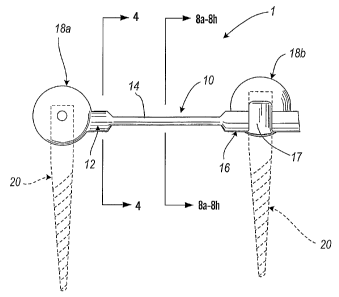
Referring now to Fig. 3, a first embodiment of a dynamic fixation system 1 is
illustrated. The dynamic fixation device 1 includes a rod portion 10 having a
first end 12,
a center section 14, and a second end 16. First end 12 and second end 16 of
rod portion 10
are preferably connected to connectors 18a, l 8b that, in turn, are connected
to pedicle screws
20, where pedicle screws 20, shown in dashed lines, are inserted into the
pedicles of
vertebrae when the device is used to fixate vertebrae. In one example of this
embodiment,
as shown in Fig. 3, rod portion 10 is interconnected at first end 12 to
connector 18a.
Connector 18a located at first end 12 is of the type that is integrally formed
as part of rod
portion 10. Alternately, a connector may be a separate type of connector that
can be
selectively positioned along the length of rod portion 10. For example,
connector 18b at
second end 16 of rod portion 10 is selectively adjustable and may be
interconnected to rod
portion 10 at a plurality of positions along second end 16 by slidably
adjusting the location
of second end 16 within band 17 of connector 18b prior to tightening of
comiector 18b to
interlock the position of second end 16 within connector 18b.
The center section 14 may have a constant cross-sectional area as shown in
Fig. 3.
Alternately, as shown in Fig. 3, the cross-section may vary along the length
of rod portion 10.
Fig. 3 shows the rod portion 10 having a center section 14 with a smaller
cross-sectional
width than the cross-sectional width of rod portion 10 at first end 12 or
second end 16. In
one example of this embodiment, rod portion 10 has a circular cross-section at
first end 12
and a circular cross-section at second end 16. Fig. 4 depicts one possible
cross-section of rod
portion 10 at first end 12. As shown in Fig. 3, this is the same cross-section
as that located
at second end 16, and is typically about 5mm in diameter.
Referring now to Fig. 5, dynamic fixation device 1' illustrates a modification
of the
first embodiment wherein the cross-sectional area varies along the length of
center section
14 between first end 12 and second end 16. As shown in Fig. 5, a continuously
varying
CA 02520741 2005-09-28
WO 2004/089244 PCT/US2004/010277
cross-sectional area may be used wherein the cross-sectional profile varies
continuously
along the length of center section 14. More specifically, Fig. 5 depicts one
example of this
modification to the first embodiment wherein the width of the center section
varies from its
widest diameter at first end 12 and/or second end 16, and gradually thins to
about the center
5 of center section 14.
Referring now to Fig. 6, the cross-sectional profile of center section 14 may
vary at
discrete points. Fig. 6 depicts yet a different modification of the first
embodiment. Dynamic
Fxation device 1"' illustrates an example of such a variable profile, wherein
a stepwise
variable cross-sectional area is provided along center section 14. As shown in
Fig. 6, center
10 section 14 can include a first width at first end 12 and second end 16, a
second width at
intermediate region 21, and a third width at center region 22.
Referring now to Fig. 7, in yet an alternate modification, dynamic fixation
device l iv
includes a center section 14 that resembles a twisting ribbon. Center section
14 can be
uniform or variable in its width, and is twisted along its length.
The above described alternative configurations offer different bending
characteristics,
such as the ability to allow a measure of twisting rotation as opposed to only
pure bending.
Depending upon a patient's circumstances, the attending physician may desire
incorporating
an implant with one of these different profiles to provide dynamic fixation of
the patient's
vertebrae.
Referring now to Figs. ga-~h, without limitation, the cross-section of center
section
14 of rod portion 10 can be of a number of different shapes, and those shapes
may vary in
cross-sectional area. Preferably, center section 14 has a thickness of about 2
to 3 mm, with
a width of about Smm. However, the dimensions will vary depending upon the
specific
design necessary for a specific patient. More particularly, the dimensions of
center section
14 will likely be thicker for a large heavy man, as opposed to that needed for
a small petite
woman. Furthermore, the type of material used to construct center section 14
will also
impact the required dimensions of center section 14. Rod portion 10 can be
made of a variety
of materials, preferably metals or materials demonstrating resilient
characteristics, and more
preferably, a titanium alloy or surgical stainless steel. In addition,
combinations or layers of
materials may be used. For example, center section 14 can be formed within its
center of
materials) having resilient or rubber like qualities, with a flexible metallic
wrapping
CA 02520741 2005-09-28
WO 2004/089244 PCT/US2004/010277
11
sufficiently thick to substantially resist translational motion. Such a
configuration allows
rotational bending and elongation during flexion while preventing the discs
from exceeding
normal physiologic limits of translational motion. Since different materials
have different
strength and resilient properties, the type of material used will, in part,
dictate the dimensions
of the rod portion required to achieve a certain function in a specific
patient.
As shown in Fig. 8a, the cross-section of center section 14 of rod portion 10
may be
that of an elongated ellipse. Alternately, as shown in Fig. 8b, the cross-
section of center
section 14 may be that of a flattened rectangle. In yet an alternate
variation, the center section
14 may resemble a bow-tie, as shown in Fig. 8c, or a flattened hexagon as
shown in Fig. 8d.
Figure 8e depicts a center section 14 having a circular cross-section, but one
that is
sufficiently small such that is provides the ability to flex or bend. Figures
8f 8h depict cross
sections with variable widths, a feature shared with the structure shown in
Fig. 8c. Fig. 8h
is a crescent shaped center section 14. Therefore, center section 14 can be of
a variety of
different shapes and yet still provide the necessary flexibility to allow for
controlled, limited
bending of the spine.
Appropriate shaping and contouring of the center section 14 allows rod portion
10 to
function in its elastic range, and avoid stress failure. Furthermore, the
center section 14
provides a virtual axis of rotation not necessarily centered at rod portion
10, thereby allowing
the implant to more closely approximate the normal physiology of the spine.
Referring now to Fig. 9, a separate embodiment of the dynamic fixation device
is
illustrated. The dynamic fixation device 24 shown in Fig. 9 includes an
inverted T-shaped
spring within central region 14. As with the dynamic fixation device 1 shown
in Fig. 3, first
end 12 and second end 16 of rod portion 10 are interconnected t~ connectors
18a and 18b,
respectively, that are, in turn, connected to pedicle screws 20 that are
installed in the pedicles
of vertebrae. As with dynamic fixation device 1, the connectors 18a and 18b
used with
dynamic fixation device 24 may be formed as an integral part of the device 24,
or they can
be separate, thereby providing adjustability at first end 12 and second end
16. In addition to
having a center section 14 that has a relatively thin cross-section that can
function in an
elastic range yet avoid stress failure as described above, the center section
14 has a shape that
is non-linear, as depicted in Fig. 9.
CA 02520741 2005-09-28
WO 2004/089244 PCT/US2004/010277
12
Center section 14 preferably includes at least two bends, and more preferably,
a series
of bends that add a further spring effect. As noted above, rod portion 10 of
the dynamic
fixation device 24 depicted in Fig. 9 includes an inverted T-shaped region
within center
section 14. More particularly, dynamic fixation device 24 includes a first
pair of reverse
bends 26a and 26b and a second set of reverse bends 28a and 28b. Each reverse
bend 26a,
26b, 28a, and 28b in the rod portion 10 is greater than about 90 degrees, and
more preferably,
each reverse bend is more than about 135 degrees and up to about 180 degrees.
That is, rod
portion 10 bends at bend 26a at least about 135 degrees and up to about 180
degrees before
initiating bend 28a, which also bends at least about 135 degrees and up to
about 180 degrees.
Reverse bends 26b and 28b are the opposite, but similar in curvature to the
bends 26a and
28a, respectively.
The modified dynamic fixation device 24 shown in Fig. 9 helps dampen an axial
compression load between the vertebrae interconnected by the device. This
construct not
only allows for bending between the vertebrae, but also provides a dampening
effect for
compression loading that occurs between the vertebrae. The inverted T-shaped
region of
center section 14~ shifts the axis of rotation forward, or anteriorly toward
the physiologic axis.
This allows some axial loading of the spine without unduly stressing the
pedicle screw to
bone interface.
Similar to dynamic fixation device 1, the center section 14 of dynamic
fixation device
24 can have a variety of different cross-sections. The center sections 14
shown in Figs. 8a-8h
present a number of the possible cross-sections that can be used to construct
dynamic fixation
device 24.
Referring now to Fig. 10, a separate embodiment of a dynamic fixation device
30 is
shown. Dynamic fixation device 30 features the ability to provide a device
that allows
bending, as well as dampening of compression loads, while at the same time
providing
increased stability. Accordingly, depending upon a patient's attributes,
including physical
size, age, bone density, and level of activity, the device depicted in Fig. 10
may be more
suitable for certain patients.
The functional aspects of the dynamic fixation device 30 are achieved by
providing
dual central members 32a and 32b. First central member 32a includes an
inverted T-shaped
region similar to that previously described, and as depicted in Fig. 9. In
addition, dynamic
CA 02520741 2005-09-28
WO 2004/089244 PCT/US2004/010277
13
fixation device 30 features a second central member 32b that is an arcuate
shaped thin
section.
The combination of two central members 32a and 32b may be modified in
orientation
depending upon the patient's needs. More particularly, the arcuate shaped
member may be
positioned above (not shown) the inverted T-shaped member or adjacent (not
shown) the T
shaped member, and not necessarily under the T-shaped member as depicted in
Fig. 10.
Different orientations provide different characteristics in bending and in
compression, as well
as in torsion. Thus, various configurations of multiple member dynamic
fixation devices are
appropriate for addressing specific patient's needs, as the cases may dictate.
Furthermore,
two T-shaped members in various orientations may be used in contrast to one
acuate member
and one inverted T-shaped member. Likewise, two acuate members may also be
used in
combination, to include arcuate members stacked like spoons, arcuate members
oriented 1 ~0
degrees to each other, or arcuate members disposed 90 degrees to each other.
For the embodiment depicted in Fig. 10, various cross-sections for each
central
member 32a and 32b are possible. Several, but not all possible cross-sectional
views are
depicted in Fig. lla-llf. Two elongated elliptical members are depicted in
Fig. lla.
Alternately, central members 32a, 32b may take the form of one elongated
elliptical member
and one flattened rectangle, as depicted in Fig. l lb. In yet an alternate
combination, a
relatively small circular member may be used in combination with a flattened
hexagonal
member, as depicted in Fig. 11 c. Alternately, a flattened rectangular member
may be used
in combination with a bow tie-shaped member, as depicted in Fig. 11 d. ~ther
combinations
of shapes for central members 32a and 32b not listed here are within the scope
of the
invention.
In yet a separate embodiment, a dynamic fixation device can utilize a coil
portion (not
shown) for providing a mechanism for allowing the rod to bend. In an alternate
design of this
embodiment, a composite material is used to serve as a bendable portion.
Whether a coil or
composite material is used to form a bendable portion, this embodiment
preferably utilizes
a mechanism for preventing reverse bending, or posterior sagittal rotation.
For example, a
separate stiffener may be provided on the posterior side of the coil portion,
thereby allowing
the device to bend in a forward direction, allowing anterior sagittal
rotation, but substantially
limiting or preventing bending in a reverse direction, thereby preventing
posterior sagittal
CA 02520741 2005-09-28
WO 2004/089244 PCT/US2004/010277
14
rotation. Furthermore, multiple stiffeners may be used to limit lateral
rotation. That is,
additional stiffeners may be incorporated that substantially limit or prevent
left or right
coronal rotation.
The nature of the coil may be a single winding, a double winding, or it may
contain
a plurality of windings. In one preferred embodiment, a helix-shaped coil is
provided. Coils
uncoil when stressed. Composites have physical properties that mimic coiling
and uncoiling
depending upon the loading conditions. Coils may be used in combination with
composite
materials, and in combination with stiffeners of various orientations.
In a typical use to span two vertebra, the total length of the dynamic
fixation devices
1, 24, and 30 may be approximately 25 to 30mm. For a dynamic fixation device
spanning
one joint, it will expand up to approximately 5 to l Omm in length, and will
rotate forward up
to between 5 to 10 degrees to accommodate flexion of the spine. Obviously,
different size
dynamic fixation devices may be used to accommodate the specific needs of each
individual
patient. More particularly, a relatively large dynamic fixation device may be
needed for a
large man, while a relatively small dynamic fixati~n device may be needed for
a smaller
patient, such as child or a petite woman. I~owever, a limited number of sizes
may provide
adequate coverage for the majority of the patient population. For any given
device, a
potential elongation of the dynamic fixation device of approximately 20% is
anticipated.
The dynamic Taxation devices can be used to flexibly fuse a plurality of
vertebra.
l~lternatively, the dynamic fixation devices can be located at specific points
where bending
of the spine is desired, while a rigid rod may be used at other locations
desired by the
physician.
The structures of the present invention are made from one or more materials
that
possesses the appropriate strength characteristics necessary to withstand
loading from the
human body when used in medical applications. In addition, the materials are
compatible
with the human body. Preferably, materials include ceramics, plastics, metals,
or carbon fiber
composites. More preferably, the materials are made from titanium, a titanium
alloy, or
stainless steel.
Devices disclosed herein can also be made of thermal memory materials or
materials
that possess different elastic properties at varying temperatures. In this
aspect of the
invention, the subject components) may be heated or cooled to a desired
temperature,
CA 02520741 2005-09-28
WO 2004/089244 PCT/US2004/010277
implanted, then subsequently allowed to cool or warm to the temperature of the
ambient
conditions that will exist during the usage period for the subj ect device,
namely, normal body
temperature.
It is to be understood that the present invention has application to medical
devices
5 other than spinal implants. Furthermore, it is understood that the, present
invention has
application outside the medical field. The dynamic fixation device of the
present invention
is not limited to medical implants. The device could be used in seismic
dampening
applications. Alternatively, the present invention could be used to secure any
two objects,
such as in linking mechanisms, and has application to any type of mechanical
device with a
10 moving connection. Other applications, byno means exhaustive, may include
connecting any
articulated device, such as an implement connection to a tractor. It may also
be used in
heretofore static type connection applications, such as attaching an antenna
to a base
structure. ~ne of skill in various of the construction arts will appreciate
how to make and
use the present invention in view of the guidance provided herein (with
respect to a surgical
15 application) and in view of the figures set forth herein.
While various embodiments of the present invention have been described in
detail,
it is apparent that modifications and adaptations of those embodiments will
occur to those
skilled in the art. However, it is to be expressly understood that such
modifications and
adaptations are within the spirit and scope of the present invention, as set
forth in the
following claims.