Note: Descriptions are shown in the official language in which they were submitted.
CA 02520932 2005-09-29
WO 2004/087152 PCT/IB2004/000867
-1-
DOSAGE F0121~iS COINiPI2ISING AG013736
This application claims the benefit of U.S. Provisional Application No.
60/460,695, filed
April 3, 2003, and U.S. Provisional Application No. 60/491,771, filed July 31,
2003, the disclosures
of which are incorporated herein by reference in their entireties.
Background of the Invention
This invention relates to VEGFR inhibitors that are useful in the treatment of
abnormal cell
growth, such as cancer, in mammals. This invention also relates to a method of
using such
compounds in the treatment of abnormal cell growth in mammals, especially
humans, and to
pharmaceutical compositions containing such compounds.
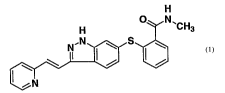
The compound 6-[2-(methylcarbamoyl)phenylsulfanyl]-3-E-[2-(pyridin-2-
yl)etheny1] indazole,
represented by formula 1
H
~'CH3
is a potent and selective inhibitor of VEGFR/PDGFR tyrosine kinases with broad
preclinical activity
in xenograft models of colon, melanoma, breast and lung cancer. (Hu-Lowe D,
Heller, D, Brekken
J, Freley R, Amundson l~, Haines l~i, Troche G, l~im 1°°,
Gon~aleG D, Herrman M, Batugo M, Veleich
S, I<ania R, McTigue M, Gregory S, Bender S, Shalinsky D., Pharmacological
Activities ~f
AG013736, a Small Molecule Inhibitor ~f VEGF/PDGF Receptor Tyrosine 4~inases;
Proc. Am.
Assoc. Cancer Res. 2002: abstract #5357). Preclinical tumor vascular response
assessed using
dynamic contrast enhanced MRI (dceMRl) has been shown fio correspond with
tumor growth index.
(Wilmes LJ, Hylton NM, V\lang D, Fleming LM Gibbs J, trim Y, Dillon R, Brasch
RC, Park J~IV, Li FC-
L, Henry RG, Partridge SC, Shalinsky DR, Hu-Lowe D, McShane TM, and
Pallavicini MG.,
AG013736, a Novel VEGFR TIC Inhibitor, Suppresses Tumor Growth and Vascular
Permeability in
Human BT474 Breast Cancer ?Cenografts in Nude Mice' ; Proc. Am. Assoc. Cancer
Res. 2003:
Abstract #3772.)
CA 02520932 2005-09-29
WO 2004/087152 PCT/IB2004/000867
-2-
Summay of the Invention
The invention provides dosage forms and methods of treatment using a compound
of formula
1:
H
~ N'CH3
.N S
\ N
v v
iN
1
which can be systematically named as 6-[2-(methylcarbamoyl)phenylsulfanyl]-3-E-
[2-(pyridin-2-
yl)ethenyl]indazole.
In one embodiment, the invention provides a dosage form for administration to
a mammal,
the dosage form comprising the compound of formula 1, a pharmaceutically
acceptable salt, solvate
or prodrug thereof, or a mixture thereof, in an amount effective to provide a
24-hour AUC blood
plasma value of no more than 4500 ng~hr/mL of the compound of formula 1 or
active metabolites
thereof, afker administration to the mammal. 24.-hour AUC blood plasma values
can be determined as
described in the ~etaited ~escription herein.
In specific aspects of this embodiment, the upper limifi of the 24-hour AUC
blood plasma
value is no more than 4000 ng~hrlmL or no more than 3000 ng~hr/mL or no more
than 2500 ng°hrlrrrL
or no more than 2000 ng~hrlmL or no more than 1500 ng~hr/mL or no more than
1000 ng~hr/mL or no
more than 800 ng~hr/mL or no more than 700 ng~hr/mL. Preferably, and in
combination with any of
the recited upper limits, the 24-hour AUC blood plasma value is at least 10
ng~hr/mL or at least 25
ng~hrlmL or at least 50 ng~hr/mL or at least 75 ng°hr/mL or at least
100 ng~hr/mL or at least 125
ng~hr/mL. Contemplated ranges of 24-hour AUC blood plasma values include
ranges from any of the
recited lower limits to any of the recited upper limits. Specific, non-
limiting examples of preferred
ranges include from 25 to 4500 ng~hr/mL, 50 to 2500 ng~hr/mL, 75 to 1000
ng~hr/mL, 100 to 800
ng~hr/mL, and 125 to 700 ng~hr/mL.
In another embodiment, fihe invention provides a dosage form comprising the
compound of
formula 1 as defined above, a pharmaceutically acceptable salt, solvate or
prodrug thereof, or a
mixture thereof, in an amount of no more than 30 mg. It should be appreciated
that when all or part
of the compound is in the dosage form as a salt, solvate or prodrug, the
amount is the equivalent
amount of the compound of formula 1, which is readily calculated by one
sleilled in the art based on
molar masses.
In specific aspects of this embodiment, the upper limit of the amount is no
more than 20
mg or no more than 15 mg or no more than 12 mg or no more than 10 mg or no
more than 8 mg or
no more than 7 mg. Preferably, and in combination with any of the recited
upper limits, the amount
is at least 0.5 mg or at least 1 mg or at least 1.5 mg or at least 2 mg or at
least 2.5 mg or at least 3
mg. Contemplated ranges include ranges from any of the recited lower limits to
any of the recited
CA 02520932 2005-09-29
WO 2004/087152 PCT/IB2004/000867
-3-
upper limits. Specific, non-limiting examples of preferred ranges include from
0.5 to 30 mg, 1 to 20
mg, 1.5 to 15 mg, 2 to 10 mg, 2.5 to 8 mg, and 3 to 7 mg.
The invention further provides a method of treating abnormal cell growth in a
mammal,
including a human, by administering to the mammal the compound of formula 1 as
defined above, a
pharmaceutically acceptable salt, solvate or prodrug thereof, or a mixture
thereof, in an amount
efFective to provide a 24-hour AUC blood plasma value of no more than 4500
ng~hr /mL of the
compound of formula 1 or active metabolites thereof, after administration to
the mammal. 24-hour
AUC blood plasma values can be determined as described in the Detailed
Description herein.
In specific aspects of this embodiment, the upper limit of the 24-hour AUC
blood plasma
value is no more than 4000 ng~hr/mL or no more than 3000 ng~hr/mL or no more
than 2500
ng~hr/mL or no more than 2000 ng~hr/mL or no more than 1500 ng~hr/mL or no
more than 1000
ng~hr/mL or no more than 800 ng~hr/mL or no more than 700 ng~hr/mL.
Preferably, and in
combination with any of the recited upper limits, the 24-hour AUC blood plasma
value is at leasfi 10
ng~hr/mL or at least 25 ng~hr/mL or at least 50 ng~hr/mL or at least 75
ng~hr/mL or at least 100
ng~hr/mL or at least 125 ng~hr/mL. Contemplated ranges of 24-hour AUC blood
plasma values
include ranges from any of the recited lower limits to any of the recited
upper limits. Specific, non-
limiting examples of preferred ranges include from 25 to 4.500 ng~hr/mL, 50 to
2500 ng~hr/mL, 75 to
1000 ng~hr/mL, 100 to 800 ng~hr/mL, and 125 to 700 ng~hr/mL.
The invention further provides a method of treating abnormal cell growth in a
mammal,
including a human, by administering to the mammal the compound of formula ~ as
defiined above,
a pharmaceutically acceptable salt, solvate or prodrug thereof, or a mixture
thereof, in an amount
of no more than 30 mg per dose. It should be appreciated that when all or part
of the compound is
in the dosage form as a salt, solvate or prodr~ao~, the amount is the
equivalent amount of the
compound of formula 1, which is readily calo~alated by one skilled in the art
based on molar
masses.
In specific aspects of this embodiment, the upper limit of the amount is no
more than 20
mg or no more than 15 mg or no more than 12 mg or no more than 10 mg or no
more than 8 mg or
no more than 7 mg. Preferably, and in combination with any of the recited
upper limits, the amount
is at least 0.5 mg or at least 1 mg or at least 1.5 mg or at least 2 mg or at
least 2.5 mg or at least 3
mg. Contemplated ranges include ranges from any of the recited lower limits to
any of the recited
upper limits. Specific, non-limiting examples ofi preferred ranges include
from 0.5 to 30 mg, 1 to 20
mg, 1.5 to 15 mg, 2 to 10 mg, 2.5 to 8 mg, and 3 to 7 mg.
In a specific embodiment of any of the inventive methods described herein, the
abnormal cell
growth is cancer, including, but not limited to, lung cancer, bone cancer,
pancreatic cancer, skin
cancer, cancer of the head or neck, cutaneous or intraocular melanoma, uterine
cancer, ovarian
cancer, rectal cancer, cancer of the anal region, stomach cancer, colon
cancer, breast cancer, uterine
cancer, carcinoma of the fallopian tubes, carcinoma of the endometrium,
carcinoma of the cervix,
carcinoma of the vagina, carcinoma of the vulva, Hodgkin's Disease, cancer of
the esophagus, cancer
of the small intestine, cancer of the endocrine system, cancer of the thyroid
gland, cancer of the
parathyroid gland, cancer of the adrenal gland, sarcoma of soft tissue, cancer
of the urethra, cancer of
CA 02520932 2005-09-29
WO 2004/087152 PCT/IB2004/000867
-4-
the penis, prostate cancer, chronic or acute leukemia, lymphocytic lymphomas,
cancer of the bladder,
cancer of the kidney or ureter, renal cell carcinoma, carcinoma of the renal
pelvis, neoplasms of the
central nervous system (CNS), primary CNS lymphoma, spinal axis tumors, brain
stem glioma,
pituitary adenoma, or a combination of one or more of the foregoing cancers.
In another embodiment
of said method, said abnormal cell growth is a benign proliferative disease,
including, but not limited
to, psoriasis, benign prostatic hypertrophy or restinosis.
In another embodiment, the invention provides a method of inhibiting PDGFR BB
mediated
cancer cell migration in a mammal, by administering to the mammal a
therapeutically acceptable
amount of the compound of formula 1.
In another embodiment, the invention provides a method of inhibiting c-KIT
activity in a
mammal, by administering to the mammal a therapeutically acceptable amount of
the compound of
formula 1.
In further specific embodiments of any of the inventive methods described
herein, the
method further comprises administering to the mammal an amount of one or more
substances
selected from anti-tumor agents, anti-angiogenesis agents, signal transduction
inhibitors, and
antiproliferafiive agents, which amounts are together effective in treating
said abnormal cell growth.
Such substances include those disclosed in PCT publication nos. W~ 00/33715,
W~ 00/35715,
1~~ 00/35717, V~~ 00/35713, W~ 00/3371 g, ~~ 00/33730, ~~ 00/33565, W~
OOI37107 and
1l~/~ 00/33736, the disclosures of which are incorporated herein by reference
in their entireties.
E~eamples of anti-tumor agents include mitotic inhibitors, for e~3ample ~rinca
alkaloid
derivatives such as vinblastine vinorelbine, vindescine and vincristine;
colchines allochochine,
halichondrine, N-benzoyltrimethyl-methyl ether colchicinic acid, dolastatin
10, maystansine, rhizoxine,
taxanes such as paclitaxel (TaxolT""), docetaxel (Taxotere~"), 2'-N-[3-
(dimethylamino)propyl]glutaramate (Ta~~oIT"'° derivatie~e),
thiocholchicine, trityl cysteinr~, teniposide,
~5 methotrexate, azathioprine, fluorouricil, cytosine arabinoside, ~'~'-
difluorodeo~~cytidine (gemcitabine),
adriamycin and mitamycin. Alkylating agents, for example cis-platin,
carboplatin oxiplatin, iproplatin,
Ethyl ester of N-acetyl-DL-sarcosyl-L-leucine (Asaley or Asalex), 1,4-
cyclohexadiene-1,4-dicarbamic
acid, 2,5 -bis(1-azirdinyl)-3,6-dioxo-, diethyl ester (diaziquone), 1,4-
bis(methanesulfonyloxy)butane
(bisulfan or leucosulfan) chlorozotocin, clomesone,
cyanomorpholinodoxorubicin, cyclodisone,
dianhydroglactitol, fluorodopan, hepsulfam, mitomycin C, hycantheonemitomycin
C, mitozolamide, 1-
(2-chloroethyl)-4-(3-chloropropyl)-piperazine dihydrochloride,
piperazinedione, pipobroman,
porfiromycin, spirohydantoin mustard, teroxirone, tetraplatin, thiotepa,
triethylenemelamine, uracil
nitrogen mustard, bis(3-mesyloxypropyl)amine hydrochloride, mitomycin,
nitrosoureas agents such as
cyclohexyl-chloroethylnitrosourea, methylcyclohexyl-chloroethylnitrosourea 1-
(2-chloroethyl)-3-(2,6-
dioxo-3-piperidyl)-1-nitroso-urea, bis(2-chloroethyl)nitrosourea,
procarbazine, dacarbazine, nitrogen
mustard-related compounds such as mechloroethamine, cyclophosphamide,
ifosamide, melphalan,
chlorambucil, estramustine sodium phosphate, strptozoin, and temozolamide. DNA
anti-metabolites,
for example 5-fluorouracil, cytosine arabinoside, hydroxyurea, 2-[(3hydroxy-2-
pyrinodinyl)methylene]-
hydrazinecarbothioamide, deoxyfluorouridine, 5-hydroxy-2-formylpyridine
thiosemicarbazone, alpha-
2'-deoxy-6-thioguanosine, aphidicolin glycinate, 5-azadeoxycytidine, beta-
thioguanine deoxyriboside,
CA 02520932 2005-09-29
WO 2004/087152 PCT/IB2004/000867
-5-
cyclocytidine, guanazole, inosine glycodialdehyde, macbecin II,
pyrazolimidazole, cladribine,
pentostatin, thioguanine, mercaptopurine, bleomycin, 2-chlorodeoxyadenosine,
inhibitors of
thymidylate synthase such as raltitrexed and pemetrexed disodium, clofarabine,
floxuridine and
fludarabine. DNAIRNA antimetabolites, for example, L-alanosine, 5-azacytidine,
acivicin, aminopterin
and derivatives thereof such as N-[2-chloro-5-[[(2, 4-diamino-5-methyl-6-
quinazolinyl)methyl]amino]benzoyl]-L-aspartlc acid, N-[4-[[(2, 4-diamino-5-
ethyl-6-
quinazolinyl)methyl]amino]benzoyl]-L-aspartic acid, N -[2-chloro-4-[[(2, 4-
diaminopteridinyl)methyl]amino]benzoyl]-L-aspartic acid, soluble Baker's
antifol, dichloroallyl lawsone,
brequinar, ftoraf, dihydro-5-azacytidine, methotrexate, N-(phosphonoacetyl)-L-
aspartic acid
tetrasodium salt, pyrazofuran, trimetrexate, plicamycin, actinomycin D,
cryptophycin, and analogs
such as cryptophycin-52 or, for example, one of the preferred anti-metabolites
disclosed in European
Patent Application No. 239362 such as N-( _5-L-(3,4-dihydro-2-methyl-4-
oxoquinazolin-6-ylmethyl)-N-
methylamino]-2-thenoyl)-L-glutamic acid; growth factor inhibitors; cell cycle
inhibitors; intercalating
antibiotics, for example adriamycin and bleomycin; proteins, for example
interferon; and anti-
hormones, for example anti-estrogens such as NolvadexTM (tamoxifen) or, for
example anti-
androgens such as CasodexTM (4'-cyano-3-(4-tluorophenylsulphonyl)-2-hydroxy-2-
methyl-3'-
(trifluoromethyl)propionanilide). Such conjoint fireatment may be achieved by
way of the
simultaneous, sequential or separate dosing of the individual components of
the treatment.
Anti-angiogenesis agents include MMP-2 (matrbe-metalloproteinase 2)
inhibitors, MMP-9
(matrix-metalloprotienase 9) inhibitors, and C~?Z-II (cyclooxygena~se II)
inhibitors. E~;a~mples of
useful COX-II inhibitors include CELEBREXT"" (alecoxib), valdecoxib, and
rofecoxib. Examples of
useful matrix metalloproteinase inhibitors are described in WQ 96133172
(published ~ctober 24,
1996), W~ 96/27583 (published March 7, 1996), European Patent Application No.
97304971.1 (filled
July 8, 1997), European Patent Application No. 9930817.2 (filed ~ctober 29,
1999), W~ 98/07597
(published February 26, 1998), WO 98/03516 (published January 29, 1998), WO
98134.918 (published
August 13, 1998), W~ 98/34915 (published August 13, 1998), WO 98/33768
(published August 6,
1998), WO 98/30566 (published July 16, 1998), European Patent Publication
606,046 (published July
13, 1994), European Patent Publication 931,788 (published Jtaly 28, 1999), W~
90/05719 (published
May 331, 1990), WO 99/52910 (published ~ctober 21, 1999), W~ 99/52889
(published ~ctober 21,
1999), WO 99/29667 (published June 17, 1999), PCT International Application
No. PCT/IB98/01113
(filed July 21, 1998), European Patent Application No. 99302232.1 (filed March
25, 1999), Great
Britain patent application number 9912961.1 (filed June 3, 1999), United
States Provisional
Application No. 60/148,464 (filed August 12, 1999), United States Patent
5,863,949 (issued January
26, 1999), United States Patent 5,861,510 (issued January 19, 1999), and
European Patent
Publication 780,386 (published June 25, 1997), all of which are herein
incorporated by reference in
their entirety. Preferred MMP-2 and MMP-9 inhibitors are those that have
little or no activity inhibiting
MMP-1. More preferred, are those that selectively inhibit MMP-2 and/or MMP-9
relative to the other
matrix-metalloproteinases (i.e. MMP-1, MMP-3, MMP-4, MMP-5, MMP-6, MMP-7, MMP-
8, MMP-10,
MMP-11, MMP-12, and MMP-13).
CA 02520932 2005-09-29
WO 2004/087152 PCT/IB2004/000867
-6-
Examples of MMP inhibitors include AG-3340, RO 32-3555, RS 13-0830, and the
compounds recited in the following list:
3-[[4-(4-fluoro-phenoxy)-benzenesulfonyl]-(1-hydroxycarbamoyl-cyclopentyl)-
amino]-
propionic acid;
3-exo-3-[4-(4-fluoro-phenoxy)-benzenesulfonylamino]-8-oxa-bicyclo[3.2.1]octane-
3-
carboxylic acid hydroxyamide;
(2R, 3R) 1-[4-(2-chloro-4-fluoro-benzyloxy)-benzenesulfonyl]-3-hydroxy-3-
methyl-
piperidine-2-carboxylic acid hydroxyamide;
4-[4-(4-fluoro-phenoxy)-benzenesulfonylamino]-tetrahydro-pyran-4-carboxylic
acid
hydroxyamide;
3-[[4-(4-fluoro-phenoxy)-benzenesulfonyl]-(1-hydroxycarbamoyl-cyclobutyl)-
amino]-
propionic acid;
4-[4-(4-chloro-phenoxy)-benzenesulfonylamino]-tetrahydro-pyran-4-carboxylic
acid
hydroxyamide;
3-[4-(4-chloro-phenoxy)-benzenesulfonylamino]-tetrahydro-pyran-3-carboxylic
acid
hydroxyamide;
(2R, 3R) 1-[4-(4-filuoro-2-methyl-benzylo~ay)-benzenesulfonyl]-3-hydroary-3-
methyl-
piperidine-2-carboxylic acid hydroxyamide;
3-[[4-(4-fluoro-phenoxy)-benzenesulfonyl]-(1-hydroxycarbamoyl-1-methyl-ethyl)-
amino]-
propionic acid;
3-[[4-(4-fluoro-phenoxy)-benzenesulfonyl]-(4-hydroxycarbamoyl-tetrahydro-pyran-
4-yl)-
amino]-propionic acid;
3-exo-3-[4-(4-chloro-phenoxy)-benzenesulfonylamino]-8-oxa-bicyclo[3.2.1
]octane-3-
oarbo~ylic acid hydronyamide;
3-endo-3-[4._(q._fluoro-phenoxy)-benzenesulfonylamino]-8-ob<a-
bicyclo[3.2.1]octane-3-
carboxylic acid hydroxyamide; and
3-[4-(4-fluoro-phenoxy)-benzenesulfonylamino]-tetrahydro-furan-3-carboxylic
acid
hydroxyamide;
and pharmaceutically acceptable salts, solutes and prodrugs of said compounds.
Examples of signal transduction inhibitors include agents that can inhibifi
EGFR (epidermal
growth factor recepfior) responses, such as EGFR antibodies, EGF antibodies,
and molecules that
are EGFR inhibitors; VEGF (vascular endothelial growth factor) inhibitors; and
erbB2 receptor
inhibitors, such as organic molecules or antibodies that bind to the erbB2
receptor, for example,
HERCEPTINTM (Genentech, Inc. of South San Francisco, California, USA).
EGFR inhibitors are described in, for example in WO 95/19970 (published July
27, 1995),
WO 98/14451 (published April 9, 1998), WO 98/02434 (published January 22,
1998), and United
States Patent 5,747,498 (issued May 5, 1998). EGFR-inhibiting agents include,
but are not limited
to, the monoclonal antibodies C225 and anti-EGFR 22Mab (ImClone Systems
Incorporated of New
York, New York, USA), the compounds ZD-1839 (AstraZeneca), BIBX-1382
(Boehringer
Ingelheim), MDX-447 (Medarex Inc. of Annandale, New Jersey, USA), and OLX-103
(Merck & Co.
CA 02520932 2005-09-29
WO 2004/087152 PCT/IB2004/000867
-7-
of Whitehouse Station, New Jersey, USA), VRCTC-310 (Ventech Research) and EGF
fusion toxin
(Seragen Inc. of Hopkinton, Massachusetts).
VEGF inhibitors, for example SU-5416 and SU-6668 (Sugen Inc. of South San
Francisco,
California, USA), can also be combined or co-administered with a compound of
formula 1. VEGF
inhibitors are described in, for example in WO 99/24440 (published May 20,
1999), PCT
International Application PCT/IB99/00797 (filed May 3, 1999), in WO 95/21613
(published August
17, 1995), WO 99/61422 (published December 2, 1999), United States Patent
5,834,504 (issued
November 10, 1998), WO 98/50356 (published November 12, 1998), United States
Patent 5,883,113
(issued March 16, 1999), United States Patent 5,886,020 (issued March 23,
1999), United States
Patent 5,792,783 (issued August 11, 1998), WO 99/10349 (published March 4,
1999), WO 97/32856
(published September 12, 1997), WO 97122596 (published June 26, 1997), WO
98/54093 (published
December 3, 1998), WO 98/02438 (published January 22, 1998), WO 99/16755
(published April 8,
1999), and WO 98/02437 (published January 22, 1998), all of which are herein
incorporated by
reference in their entirety. Other examples of some specific VEGF inhibitors
are IM862 (Cytran Inc.
of Kirkland, Washington, USA); anti-VEGF monoclonal antibody bevacizumab
(Genentech, Inc. of
South San Francisco, California); and angiozymeT"", a synthetic ribozyme from
Ribozyme (Boulder,
Colorado) and Chiron (Emeryville, California).
ErbB2 receptor inhibitors, such as GW-282974. (Gla~zo Wellcome ploy, and the
monoclonal
antibodies AR-209 (Aronex Pharmaceuticals Inc. of The Woodlands, Texas, USA)
and 2B-1
(Chiron), may be administered in combination with a compound of formula ~.
Such erbB2 inhibitors
include those described in WO 98102434 (published January 22, 1998), WO
99/35146 (published
July 15, 1999), WO 99/35132 (published July 15, 1999), WO 98/02437 (published
January 22,
1998), WO 97/13760 (published April 17, 1997), WO 95/19970 (published Jtaly
27, 1995), United
States Patent 5,587,458 (issued December 2q., 1998), and United States Patent
5,877,305 (issued
March 2, 1999), each of which is herein incorporated by reference in its
entirety. ErbB2 receptor
inhibitors useful in the present invention are also described in United States
Provisional Application
No. 60/117,341, filed January 27, 1999, and in United States Provisional
Application No.
60/117,346, filed January 27, 1999, both of which are herein incorporated by
reference in their
entirety.
Other antiproliferative agents that may be used include inhibitors of the
enzyme farnesyl
protein transferase and inhibitors of the receptor tyrosine kinase PDGFr,
including the compounds
disclosed and claimed in the following United States patent applications:
09/221946 (filed
December 28, 1998); 09/454058 (filed December 2, 1999); 09/501163 (filed
February 9, 2000);
09/539930 (filed March 31, 2000); 09/202796 (filed May 22, 1997); 09/384339
(filed August 26,
1999); and 09/383755 (filed August 26, 1999); and the compounds disclosed and
claimed in the
following United States provisional patent applications: 60!168207 (filed
November 30, 1999);
60/170119 (filed December 10, 1999); 60/177718 (filed January 21, 2000);
60/168217 (filed
November 30, 1999), and 60/200834 (filed May 1, 2000). Each of the foregoing
patent applications
and provisional patent applications is herein incorporated by reference in
their entirety.
CA 02520932 2005-09-29
WO 2004/087152 PCT/IB2004/000867
_g_
The compound of formula 1 may also be used with other agents useful in
treating
abnormal cell growth or cancer, including, but not limited to, agents capable
of enhancing antitumor
immune responses, such as CTLA4 (cytotoxic lymphocite antigen 4) antibodies,
and other agents
capable of blocking CTLA4; and anti-proliferative agents such as other
farnesyl protein transferase
inhibitors. Specific CTLA4 antibodies that can be used in the present
invention include those
described in United States Provisional Application 60/113,647 (filed December
23, 1998) which is
herein incorporated by reference in its entirety.
In another embodiment, the invention provides a pharmaceutical composition
comprising the
compound of formula 1, or a pharmaceutically acceptable salt, solvate or
prodrug thereof, and a
therapeutically effective amount of docetaxel.
In another embodiment, the invention provides a method of treating abnormal
cell growth in a
mammal, including a human, by administering to the mammal the compound of
formula 1, or a
pharmaceutically acceptable salt, solvate or prodrug thereof, and a
therapeutically effective amount of
docetaxel. The compound of formula 1-and docetaxel can be administered
separately or in the same
composition, and can be administered on the same dosing schedule or on
different dosing schedules,
as desired.
Defiinitions
"Abnormal cell growth", as used herein, unless otherwise indicated, refers to
cell growth that
~0 is indr~pendent ofi normal regulatory mechanisms (e.g., loss of contact
inhibition). This includes the
abnormal growth of: (1) tumor cells (tumors) that proliferate by expressing a
mutated tyrosine kinase
or overexpression of a receptor tyrosine kinase; (2) benign and malignant
cells of other proliferative
diseases in which aberrant tyrosine leinase activation occurs; and (4) any
tumors that proliferate by
receptor tyrosine hinases.
~5 The term '°treating", as used herein, unless otherwise indicated,
means reversing, alleviating,
inhibiting the progress of, or preventing the disorder or condition to which
such term applies, or one or
more symptoms ofi such disorder or condition. The term "treatment", as used
herein, unless
otherwise indicated, refers to the act of treating as "treating" is defined
immediately above.
The phrase "pharmaceutically acceptable salts)", as used herein, unless
otherwise indicated,
30 includes salts of acidic or basic groups which may be present in a
compound. Compounds that are
basic in nature are capable of forming a wide variety of salts with various
inorganic and organic acids.
The acids that may be used to prepare pharmaceutically acceptable acid
addition salts of such basic
compounds are those that form non-toxic acid addition salts, i.e., salts
containing pharmacologically
acceptable anions, such as the acetate, benzenesulfonate, benzoate,
bicarbonate, bisulfate, bitartrate,
35 borate, bromide, calcium edetate, camsylate, carbonate, chloride,
clavulanate, citrate, dihydrochloride,
edetate, edislyate, estolate, esylate, ethylsuccinate, fumarate, gluceptate,
gluconate, glutamate,
glycolylarsanilate, hexylresorcinate, hydrabamine, hydrobromide,
hydrochloride, iodide, isothionate,
lactate, lactobionate, laurate, malate, maleate, mandelate, mesylate,
methylsulfate, mutate,
napsylate, nitrate, oleate, oxalate, pamoate (embonate), palmitate,
pantothenate,
CA 02520932 2005-09-29
WO 2004/087152 PCT/IB2004/000867
_g_
t
phospate/diphosphate, polygalacturonate, salicylate, stearate, subacetate,
succinate, tannate, tartrate,
teoclate, tosylate, triethiodode, and valerate salts.
The term "prodrug", as used herein, unless otherwise indicated, means
compounds that are
drug precursors, which following administration, release the drug in vivo via
some chemical or
physiological process (e.g., a prodrug on being brought to the physiological
pH is converted to the
desired drug form).
The subject invention also includes isotopically-labeled compounds, which are
identical to
those recited in Formula 1, but for the fact that one or more atoms are
replaced by an atom having
an atomic mass or mass number different from the atomic mass or mass number
usually found in
nature. Examples of isotopes that can be incorporated into compounds of the
invention include
isotopes of hydrogen, carbon, nitrogen, oxygen, phosphorus, sulfur, fluorine
and chlorine, such as
2H' 3H 13C 14C' 15N' 18~' 170 31P 32P' 35~' 18F and 36C1, respectively.
Compounds of the present
invention, prodrugs thereof, and pharmaceutically acceptable salts and
solvates of said compounds
or of said prodrugs which contain the aforementioned isotopes and/or other
isotopes of other
atoms are within the scope of this invention. Certain isotopically-labeled
compounds of the present
invention, for example those into which radioactive isotopes such as 3H and'4C
are incorporated,
are useful in drug and/or substrate tissue distribution assays. Tritiated,
i.e., 3H, and carbon-14., i.e.,
14~ isotopes are particularly preferred for their ease of preparation and
delectability. Further,
substitution with heavier isotopes such as deuterium, i.e., 2H, can afford
certain therapeutic
ad~rantages resulting from greater metabolic stability, for es;a~mple
increased in ~i~o half life or
reduced dosage requirements and, hence, may be preferred in some
circumsfiances. Isotopically
labeled compounds of Formula 1 of this invention and prodrugs thereof can
generally be prepared
by carrying out the procedures described for the non-labeled compound,
substituting a readily
available isotopically labeled reagent for a non-isolopically labeled reagent.
Srief Descriolion of the Drawing
Figure 1 shows metabolites of the compound of formula 1 identified in dogs
following a
single oral dose of the 14C-labeled compound.
Figure 2 shows metabolites of the compound of formula 1 identified in mice
following a
single oral dose of the 14C-labeled compound.
Detailed Description ~f The Invention
The compound of formula 1 can be prepared as described in U.S. Patent Nos.
6,531,491
and 6,534,524 (issued March 11, 2003 and March 1>3, 2003, respectively), which
are incorporated
herein by reference in their entireties. Certain starting materials may be
prepared according to
methods familiar to those skilled in the art and certain synthetic
modifications may be done according
to methods familiar to those skilled in the art.
The compound of formula 1 is capable of forming a wide variety of different
salts with various
inorganic and organic acids. Although such salts must be pharmaceutically
acceptable for
administration to mammals, it is often desirable in practice to initially
isolate the compound of formula
CA 02520932 2005-09-29
WO 2004/087152 PCT/IB2004/000867
-10-
1 from the reaction mixture as a pharmaceutically unacceptable salt and then
simply convert the latter
back to the free base compound by treatment with an alkaline reagent and
subsequently convert the
latter free base to a pharmaceutically acceptable acid addition salt. The acid
addition salts of the base
compounds of this invention are readily prepared by treating the base compound
with a substantially
equivalent amount of the chosen mineral or organic acid in an aqueous solvent
medium or in a
suitable organic solvent, such as methanol or ethanol. Upon careful
evaporation of the solvent, the
desired solid salt is readily obtained. The desired acid salt can also be
precipitated from a solution of
the free base in an organic solvent by adding to the solution an appropriate
mineral or organic acid.
Administration of the compound of formula 1 can be effected by any method that
enables
delivery of the compound to the site of action. These methods include oral
routes, intraduodenal
routes, parenteral injection (including intravenous, subcutaneous,
intramuscular, intravascular or
infusion), topical, and rectal administration.
The compound may, for example, be provided in a form suitable for oral
administration as a
tablet, capsule, pill, powder, sustained release formulation, solution,
suspension, for parenteral
injection as a sterile solution, suspension or emulsion, for topical
administration as an ointment or
cream or for rectal administration as a suppository. The compound may be in
unit dosage forms
suitable for single administrafiion of precise dosages. Preferably, dosage
forms include a conventional
pharmaceutical carrier or e~zcipient and the compound of formula 1 as an
active ingredient. In
addition, dosage forms may include other medicinal or pharmaceutical agents,
carriers, adjuvants,
~0 etc.
Exemplary parenteral administration forms include solutions or suspensions in
sterile
aqueous solutions, for example, aqueous propylene glycol or dextrose
solutions. Such dosage forms
can be suitably buffered, ifi desired.
Suitable pharmaceutical carriers include inert diluents or fillers, water and
various organic
solvents. The pharmaceutical composition may, if desired, contain additional
ingredients such as
filavorings, binders, excipients and the like. Thus for oral administration,
tablets containing various
excipients, such as citric acid may be employed together with various
disintegrants such 'as starch,
alginic acid and certain complex silicates and with binding agents such as
sucrose, gelatin and acacia.
Additionally, lubricating agents such as magnesium stearate, sodium lauryl
sulfate and talc are often
useful for tableting purposes. Solid compositions of a similar type may also
be employed in sofk and
hard filled gelatin capsules. Preferred materials therefor include lactose or
milk sugar and high
molecular weight polyethylene glycols. When aqueous suspensions or elixirs are
desired for oral
administration the active compound therein may be combined with various
sweetening or flavoring
agents, coloring matters or dyes and, if desired, emulsifying agents or
suspending agents, together
with diluents such as water, ethanol, propylene glycol, glycerin, or
combinations thereof.
In preferred embodiments of the dosage forms of the invention, the dosage form
is an oral
dosage form, more preferably, a tablet or a capsule.
In preferred embodiments of the methods of the invention, the compound of
formula 1 is
administered orally, such as, for example, using an oral dosage form as
described herein.
CA 02520932 2005-09-29
WO 2004/087152 PCT/IB2004/000867
-11-
The methods include administering the compound of formula 1 using any desire
dosage
regimen. In one specific embodiment, the compound is administered once per day
(quaque die, or
QD), preferably twice per day (bis in die, or BID), although more or less
frequent administration is
within the scope of the invention. The compound can be administered to the
mammal, including a
human, preferably in a fasted state (no food or beverage within 2 hours before
and after
administration). In a particularly preferred embodiment, the dosage is BID,
fasted.
Methods of preparing various dosage forms with a specific amount of the
compound of
formula 1 are known, or will be apparent, to those skilled in this art. For
examples, see Remingiton's
Pharmaceutical Sciences, Mack Publishing Company, Easter, Pa., 15th Edition
(1975).
AUC blood plasma values can be determined by directly measuring blood plasma
concentrations of the compound of formula one or active metabolites thereof,
such as by liquid
chromatography-tandem mass spectrometry (LC-MS/MS), at various time inteneals,
and calculating
the area under the plasma concentration versus time curve. Suitable methods
for calculating AUC
are well-known in the art, such as, for example, by using the trapezoidal
approximation,
A UC~o-~i = ~ t1+y t~ ~~~ .+- C's+i
=o
where n is the number of data points, and t; and C; are the time and
concentration (x and y values) of
the ~h data point. 24.-hour AUC values can be determined by normalizing
measured blood plasma
concentrations according to the dosing schedule. Sodium bisulfite is added as
a stabilizer in the
reconstitution solution for preparation of concentration standards.
The compound of formula 1 has advantageous properties relating to the
modulation and/or
inhibition of the kinase activity associated with VEGF-R, FGF-R, CDK
complexes, CHIC1, CSF-R,
andlor LCIZ.
~s shown in the e~gamples below, the compound of formula 1 is capable of
inducing
HUVEC apoptosis in vitro, inhibiting VEGF mediated Rakt and ef~~S
phosphorylation in HUVEC,
demonstrating a lasting inhibitory efFect on VEGFR-2 phosphorylation in HUVEC
after compound
withdrawal, and inhibiting PDGF BB induced cancer cell migration on matrix
protein fibronectin.
The compound of formula 1 may have activity against PDGFR-driven tumor
progression by
inhibiting migration and invasion.
The compound of formula 1 also demonstrates more efficacious activity in tumor
growth
inhibition when combined with TaxoIT"", more preferably docetaxel. More
significant tumor
regression was observed with the co-therapy than either agent alone.
The present invention is further directed to methods of modulating or
inhibiting protein
kinase activity, for example in mammalian tissue, by administering the
compound of formula 1.
The activity of the inventive compound as a modulator of protein kinase
activity, such as the activity
of kinases, may be measured by any of the methods available to those skilled
in the art, including
in vivo andlor in vitro assays. Examples of suitable assays for activity
measurements include those
described in Parast C. et al., Biochemistry, 37, 16788-16801 (1998); Jeffrey
et al., Nature, 376,
313-320 (1995); WIPO International Publication No. WO 97/34876; and WIPO
International
CA 02520932 2005-09-29
WO 2004/087152 PCT/IB2004/000867
-12-
Publication No. WO 96!14843. These properties may be assessed, for example, by
using one or
more of the biological testing procedures set out in the examples below.
The examples and preparations provided below further illustrate and exemplify
the dosage
forms and methods of the present invention. It is to be understood that the
scope of the present
invention is not limited in any way by the scope of the following examples.
Exam Ip a 1
The compound of formula 1 was tested for: (1) in vivo efficacy under several
scheduling:
std, weekend dose holiday and intermittent dosing; (2) efficacy when combined
with docetaxel in
xenograft models; (3) in vitro eNOS and Akt phosphorylation in endothelial
cells; (4) the
concentration of Nitro Oxide and related products in cell culture and in vivo
and (5) use of c-Kit
signal in the whole blood cells as a potential biomarker for the compound.
BIOLOGICAL TESTING; ENZYME ASSAYS
The stimulation of cell proliferation by growth factors such as VEFG, FGF, and
others is
dependent upon their induction of autophosphorylation of each of their
respective receptor's
tyrosine kinases. Therefore, the ability of a protein kinase inhibitor to
block cellular proliferation
induced by these growth factors is directly correlated with its ability to
block receptor
autophosphorylation. To measure the protein kinase inhibition activity of the
compounds, the
following constructs were devised.
VEGF-R2 Construct for Assay: This construct determines the ability of a test
compound to
inhibit tyrosine kinase acti~rity. A construct (VEGF-R2~50) of the cytosolic
domain of human
vascular endothelial growth factor receptor 2 (VEGF-R2) lacking the 50 central
residues of the 68
residues of the kinase insert domain was expressed in a baculoviruslinsect
cell system. Of the
1356 residues of full-length VEGF-R2, VEGF-R2~50 contains residues 806-939 and
990-1171, and
also one point mutation (E990V) within the lainase insert domain relative to
wild-type VEGF-R2.
Autophosphorylation of the purified construct was performed by incubation of
the enzyme at a
concentration of 4 p,M in the presence of 3 mM ATP and 40 mM MgCh in 100 mM
HEPES, pH 7.5,
containing 5°f° glycerol and 5 mM DTT, at 4 °C for 2 h.
After autophosphorylation, this construct
has been shown to possess catalytic activity essentially equivalent to the
wild-type
autophosphorylated kinase domain construct. See Parast et al., Biochemistry,
37, 16788-16801
(1998).
FGF-R1 Construct for Assay: The intracellular kinase domain of human FGF-R1
was
expressed using the baculovirus vector expression system starting from the
endogenous
methionine residue 456 to glutamate 766, according to the residue numbering
system of
Mohammadi et al., MoL Cell. BioL, 16, 977-989 (1996). In addition, the
construct also has the
following 3 amino acid substitutions: L457V, C488A, and C584S.
LCK Construct for Assay: The LCK tyrosine kinase was expressed in insect cells
as an N-
terminal deletion starting from amino acid residue 223 to the end of the
protein at residue 509, with
the following two amino acid substitutions at the N-terminus: P233M and C224D.
CHK-1 Construct for Assay: C-terminally His-tagged full-length human CHK-1 (FL-
CHK-1)
was expressed using the baculoviruslinsect cell system. It contains 6
histidine residues (6 x His-
CA 02520932 2005-09-29
WO 2004/087152 PCT/IB2004/000867
-13-
tag) at the C-terminus of the 476 amino acid human CHK-1. The protein was
purified by
conventional chromatographic techniques.
CDK2/Cyclin A Construct for Assay: CDK2 was purified using published
methodology
(Rosenblatt et al., J. MoG Biol., 230, 1317-1319 (1993)) from insect cells
that had been infected
with a baculovirus expression vector. Cyclin A was purified from E. coli cells
expressing full-length
recombinant cyclin A, and a truncated cyclin A construct was generated by
limited proteolysis and
purified as described previously (Jeffrey et al., Nature, 376, 313-320
(1995)).
CDK4/Cyclin D Construct for Assay: A complex of human CDK4 and cyclin D3, or a
complex of cyclin D1 and a fusion protein of human CDK4 and glutathione-S-
transferase (GST
CDK4), was purified using traditional biochemical chromatographic techniques
from insect cells
that had been co-infected with the corresponding baculovirus expression
vectors.
VEGF-R2 Assay: Coupled Specfrophotometrlc (FLVK P) Assay
The production of ADP from ATP that accompanies phosphoryl transfer was
coupled to
oxidation of NADH using phosphoenolpyruvate (PEP) and a system having pyruvate
kinase (PK)
and lactic dehydrogenase (LDH). The oxidation of NADH was monitored by
following the decrease
of absorbance at 340 nm (esao = 6.22 cm ~ mM-~) using a Beckman DU 650
spectrophotometer.
Assay conditions for phosphorylated VEGF-R2~50 (indicated as FLVI~-P in the
tables below) were
the following: 1 mM PEP; 250 pl~Ii NADH; 50 units of LDH/mL; 20 units of
PICImL; 5 mM DTT; 5.1
mM poly(E4Y~); 1 mM ATP; and 25 mM MgCh in 200 mM HEPES, pH 7.5. Assay
conditions for
unphosphorylated VEGF-82050 (indicated as FLVK in the tables) were the
following: 1 mf~'i PEP;
250 pM NADH; 50 units of LDH/mL; 20 units of PKImL; 5 mM DTT; 20 mM
poly(E4Y~); 3 mM ATP;
and 60 m.M MgCh and 2 mM MnCla in 200 mM HEPES, pH 7.5. Assays were initiated
with 5 to 40
nM of enzyme. K; values were determined by measuring enzyme activity in the
presence of varying
concentrations ofi test compounds. The data were analyzed using Enzyme
Peinetic and
Kaleidagraph software.
ELISA Assay: Formation of phosphogastrin was monitored using biotinylated
gastrin
peptide (1-17) as substrate. Biotinylated phosphogastrin was immobilized using
streptavidin coated
96-well microtiter plates followed by detection using anti-phosphotyrosine-
antibody conjugated to
horseradish per0xidase. The activity of horseradish peroxidase was monitored
using 2,2'-azino-di-
[3-ethylbenzathiazoline sulf0nate(6)] diammonium salt (ABTS). Typical assay
solutions contained:
2 pM biotinylated gastrin peptide; 5 mM DTT; 20 p,M ATP; 26 mM MgCIZ; and 2 mM
MnCl2 in 200
mM HEPES, pH 7.5. The assay was initiated with 0.8 nM of phosphorylated VEGF-
R2~50.
Horseradish peroxidase activity was assayed using ABTS, 10 mM. The horseradish
peroxidase
reaction was quenched by addition of acid (H2S04), followed by absorbance
reading at 405 nm. K;
values were determined by measuring enzyme activity in the presence of varying
concentrations of
test compounds. The data were analyzed using Enzyme Kinetic and Kaleidagraph
software.
FGF-R Assay: The spectrophotometric assay was carried out as described above
for
VEGF-R2, except for the following changes in concentration: FGF-R = 50 nM, ATP
= 2 mM, and
poly(E4Y1) = 15 mM.
CA 02520932 2005-09-29
WO 2004/087152 PCT/IB2004/000867
-14-
LCK Assay: The spectrophotometric assay wad carried out as described above for
VEGF-
R2, except for the following changes in concentration: LCK = 60 nM, MgCl2 = 0
mM, poly(E4Y1) _
20 mM.
CHK-1 Assay: The production of ADP from ATP that accompanies phosphoryl
transfer to
the synthetic substrate peptide Syntide-2 (PLARTLSVAGLPGKK) was coupled to
oxidation of
NADH using phosphoenolpyruvate (PEP) through the actions of pyruvate kinase
(PK) and lactic
dehydrogenase (LDH). The oxidation of NADH was monitored by following the
decrease of
absorbance at 340 nm (e340 = 6.22 cm ~ mM-~) using a HP8452 spectrophotometer.
Typical
reaction solutions contained: 4 mN PEP; 0.15 mM NADH; 28 units of LDH/mL; 16
units of PK/mL;
3 mM DTT; 0.125 mM Syntide-2; 0.15 mM ATP; 25 mM MgCh in 50 mM TRIS, pH 7.5;
and 400
mM NaCI. Assays were initiated with 10 nM of FL-CHK-1. K; values were
determined by
measuring initial enzyme activity in the presence of varying concentrations of
test compounds. The
data were analyzed using Enzyme Kinetic and Kaleidagraph software.
HUVEC Proliferation Assay: This assay determines the ability of a test
compound to inhibit
the growth factor-stimulated proliferation of human umbilical vein endothelial
cells ("HUVEC").
HUVEC cells (passage 3-4, Clonetics, Corp.) were thawed into EGM2 culture
medium (Clonetics
Corp) in T75 filasles. Fresh EGM2 medium was added to the flasks 24 hours
later. Four or five
days later, cells were exposed to another culture medium (F12K medium
supplemented with 10°/~
fetal bovine serum (FBS), 60 Ng/mL endothelial cell growth supplement (ECGS),
and 10 Ng/m
heparin). Ea2ponenti2~lly-growing HUVEC cells were used in experiments
thereafter. Ten to twelve
fihousand HUVEC cells were plated in 96-well dishes in 100 pL ofi rich,
culture medium (described
above). The cells were allowed to attach for 24 hours in this medium. The
medium was then
removed by aspiration and 115 iaL of starvation media (F12K+1% FBS) was added
to each well.
After 18 hours, 15 NL of test agent dissolved in 1% Di'~iSG in starvafiion
mediurn or this vehicle
alone was added into each treatment well; the final DMS~ concentration was 0.1
°d~. ~ne hour
lafier, 20u1 of 150ng/mL hrVEGF'65 in starvation media was added to all wells
except those
containing untreated controls; the final VEGF concentration was 20 ng/mL.
Cellular proliferation
was quantified 72 hours later by MTT dye reduction, at which time cells were
exposed for 4-5 hours
MTT (Promega Corp.). Dye reduction was stopped by addition of a stop solution
(Promega Corp.)
and absorbance at 570 and 630 nm was determined on a 98-well spectrophotometer
plate reader.
Cancer Cell Proliferation (MV522) Assay: To determine the whether a protein
kinases
inhibitor should have therapeutic usefulness in blocking angiogenesis for
treating cancer, it is
important to demonstrate the inhibitor does not non-specifically block
cellular proliferation in cells
that do not express the kinase receptor. This is done by performing
proliferation assays using
cancer cells. The protocol for assessing cellular proliferation in cancer
cells is similar to that used
for assessments in HUVEC cells. Two thousand lung cancer cells (line MV522,
acquired from
UCSD) were seeded in growth media (RPM11640 medium supplemented with 2 mM
glutamine and
10% FBS). Cells are allowed to attach for 1 day prior to addition of test
agents and /or vehicles.
Cells are treated simultaneously with the same test agents used in the HUVEC
assay. Cellular
proliferation is quantified by MTT dye reduction assay 72 hours after exposure
to test agents.
CA 02520932 2005-09-29
WO 2004/087152 PCT/IB2004/000867
-15-
C-Kit potency determination: NCI-H526 (ATCC) cells were used for determining
potency
against c-Kit by the inhibitor. The cells were grown to sub-confluency and
incubated in starvation
media for 18 hours. The inhibitor was added and the cells were incubated for
45 min at 37°C in the
presence of 2.3% albumin and 1mM NasV04 (Sigma). SCF, the c-Kit growth factor
was added to
the culture at a final concentration of 50 nglmL. Five minutes later the cells
were rinsed 2X with
cold PBS and lysed with lysis buffer (50 mM Tris, 150 mM NaCI, 1 mM PMSF, 1%
NP40, 1 mM
Na3VO4 and a protease inhibitor cocktail). Immunoprecipitation was performed
using 1 mg total
protein from each lysate, incubating over night at 4° with 4 Ng/mL
CD117 ab-3 (K45, Neomarkers).
The antibody complex was conjugated to protein A beads the following morning.
SDS PAGE and
Western Blot analysis was conducted using anti phosphotyrosine antibody 4610
(Upstate
Biotechnology) for phosphorylated receptors, or anti-c-Kit receptor antibody
sc-1493 (C-14, Santa
Cruz) at 1:1000. The blots were visualized by the chemiluminescent reagents
ECL Plus. A
phosphorimager (Storm 846, Molecular Dynamics) was used for the quantification
of the signals in
the blots.
The reduction of c-kit positive cell population in total peripheral blood
cells of an animal and
mammal may be used as a biomarker for activity of the compound of formula 1.
ENOS and Akt phosphorylation measurement: HUVEC (Clonetics) were used for
determining potency against eNOS and Akt by the inhibitor. The cells were
grown to sub-
confiluency and incubated in starvation media for 18 hours. The inhibitor was
added and the cells
were incubated for 45 min at 37 °C in the presence of 2.3% albumin and
lmfadi Na~V04 (Sigma).
VEGF was added to the culture medium at 50 ng/mL. Five minutes later the cells
were rinsed 2X
with cold PBS and lysed with lysis buffer (50 mM Tris, 150mM NaCI, 1mM PMSF,
1% NP40, 1mM
Na~V04 and a protease inhibitor cocktail). A total profiein of 30-4.Oug was
analyzed by the Western
method. eI~OS and f~kt Phosphorylation was assessed by using: Phospho-eNOS
(Ser 1177)
X9571 or Phospho-Akt (Ser 473) X9271 antibodies (both from Cell signaling).
Protein detection
was achieved by using: NOS3 (C-20) sc-654 (Santa Cruz) or Alet antibody X9272
(Cell Signaling).
All require an anti rabbit HRP linked secondary antibody used at 1:3000. The
blots were visualized
by the chemiluminescent substrate Super Signal West Dura (Pierce). An Alpha
Imager 8800 from
Alpha Innotech was used for the quantification of the signals in the blots.
Mouse PK Assay: The pharmacokinetics (e.g., absorption and elimination) of
drugs in mice
were analyzed using the following experiment. Test compounds were formulated
as a suspension
in a 0.5% CMC vehicle or as a solution in a 30:70 (PEG400:acidified H~0)
vehicle. This suspension
or solution was administered orally (p.o.) or intraperitoneally (i.p.) to the
C3H female mice (n=4).
Blood samples were collected via an orbital bleed at time points: 0 hour (pre-
dose), 0.5 hr, 1.0 hr,
2.0 hr, and 4.0 hr post dose. Plasma was obtained from each sample by
centrifugation at 2500
rpm for 5 min. Test compound was extracted from the plasma by an organic
protein precipitation
method. For each time bleed 50 pL of plasma was combined with 1.0 mL of
acetonitrile, vortexed
for 2 min. and then spun at 4000 rpm for 15 min. to precipitate the protein
and extract out the test
compound. Next, the acetonitrile supernatant (the extract containing test
compound) was poured
into new test tubes and evaporated on a hot plate (25 °C) under a steam
of NZ gas. To each tube
CA 02520932 2005-09-29
WO 2004/087152 PCT/IB2004/000867
-16-
containing the dried test compound extract 125 NL of mobile phase (60:40,
0.025 M NH4HZP04
+2.5 mL/L TEA:acetonitrile) was added. The test compound was resuspended in
the mobile phase
by vortexing and more protein was removed by centrifugation at 4000 rpm for 5
min. Each sample
was poured into an HPLC vial for test Compound Analysis on an Hewlett Packard
1100 series
HPLC with UV detection. From each sample, 95 NL was injected onto a Phenomenex-
Prodigy
reverse phase C-18, 150 x 3.2 mm column and eluted with a 45-50% acetonitrile
gradient run over
min. Test-compound plasma concentrations (Ng/mL) were determined by a
comparison to
standard curve (peak area vs. conc. pg/mL) using known concentrations of test
compound
extracted from plasma samples in the manner described above. Along with the
standards and
10 unknowns, three groups (n=4) of quality controls (0.25 pg/mL, 1.5 Ng/mL,
and 7.5 pg/mL) were run
to insure the consistency of the analysis. The standard curve had an R2> 0.99
and the quality
controls were all within 10 % of their expected values. The quantitated test
samples were plotted
for visual display using 4~aleidagraph software and their pharmacokinetic
parameters were
determined using WIN NONLIN software.
Human Liver Microsome (HLM) Assay: Compound metabolism in human liver
microsomes
was measured by LC-MS analytical assay procedures as follows. First, human
liver microsomes
(HLM) were thawed and diluted to 5 mglmL with cold 100 mM potassium phosphate
(f~P04.) huffier.
Appropriate amounts of ICPO4 buffer, NA~PH-regenerating solution (containing E-
NA~P, glucose-
6-phosphate, glucose-6-phosphate dehydrogenase, and MgCh), and HLM were
preincubated in 13
~~ 100 mm glass tubes at 37 C for 10 min. (3 tubes per test compound--
triplicate). Test compound
(5 NM final) was added to each tube to inifiiate reaction and was mixed by
gentle vortexing, followed
by incubation at 37 C. At t=0, 2 h, a 250-uL sample was removed from each
incubation tube to
separate 12 x 75 mm glass tubes containing 1 mL ice-cold acetonitrile with
0.05 paM reserpine.
Samples were centrifuged at x.000 rpm for 20 min. to precipitate profeins and
salt (Eecl:man
Allegra 61~R, S/N ALI~C98D08, /+534). Supernatant was transferred to new 12 x
75 mm glass tubes
and evaporated by Speed-Vac centrifugal vacuum evaporator. Samples were
reconstituted in 200
taL 0.1°/~ formic acid/acetonitrile (90/10) and vortexed vigorously to
dissolve. The samples were
then transferred to separate polypropylene microcentrifuge tubes and
centrifuged at 14000 x g for
10 min. (Fisher Micro 14, SIN M0017580). For each replicate (~1-3) at each
timepoint (0 and 2 h),
an aliquot sample of each test compound was combined into a single HPLC vial
insert (6 total
samples) for LC-MS analysis, which is described below.
The combined compound samples were injected into the LC-MS system, composed of
a
Hewlett-Packard HP1100 diode array HPLC and a Micromass Quattro II triple
quadruple mass
spectrometer operating in positive electrospray SIR mode (programmed to scan
specifically for the
molecular ion of each test compound. Each test compound peak was integrated at
each timepoint.
For each compound, peak area at each timepoint (n=3) was averaged, and this
mean peak area at
2 h was divided by the average peak area at time 0 hour to obtain the percent
test compound
remaining at 2 h.
In vitro HUVEC Apoptosis Assays
CA 02520932 2005-09-29
WO 2004/087152 PCT/IB2004/000867
-17-
Quantification of Apoptosis by ELISA: Apoptosis of HUVEC cells was measured
using Cell
Death Detection Elisa PLUS (catalog #1775425, Roche Biochemicals, Mannheim,
Germany) that
quantifies cytoplasmic histone-associated DNA fragments in cell lysates. The
procedure was
performed with minor modifications to the manufacture's instructions. Briefly,
Starved HUVEC cells
were treated with various concentrations of Compound A in the presence of VEGF
(20 ng/mL).
The cytosolic fraction of the cells at various time points was collected and
used as an antigen
source in a sandwich ELISA with a primary anti-histone mAb coated to the
microtiter plate and a
secondary anti-DNA mAb coupled to peroxidase. The number of apoptotic cells
was determined
by adding chromogenic peroxidase substrate and measuring the absorption with a
spectrophotometer at 405 nm (reference wavelength 490nm).
Visualization of Apoptosis by TUNEL: In situ detection of apoptotic cell was
carried out
using the TdT-mediated dUTP nick end labeling (TUNEL) technique. Briefly HUVEC
cells grown in
8 well Lab-Tek chamber slides were starved O/N and then treated for 6 hours
with various
concentrations of Compound A. The cells were then fixed in 4%
Paraformaldehyde, permeablized
with Triton X-100 and incubated for 1 hour in a mixture of terminal
transferase and nucleotides
including Fluorescein-dUTP (Deadend Fluorometric TUNEL system, Promega,
catalog # 63250)
in accordance with the manufacturer's instructions. The cells were
counterstained with Propidium
iodide (PI) solution. Positively stained Fluorescein and PI labeled cells were
visualized and
photographed by fluorescence microscopy.
PDGF mediated Cell f~611iar2~tion Assay: U87i~YG cells v~ere used in this
assay. Siu well
plates are pre-incubated overnight with 0.5 ng/mL Fibronectin. The following
day U87MG cells are
plated in each well and allowed to grow to confluence. The cells were
incubated overnight with
starvation media containing 0.1 % FBS. A ~1 cm scratch was made using a
pipette tip and the cells
washed with the starvation media. The plates ~~~ere then incubated with 0.5
nglmL Fibronectin for 1
hour and then washed again. The experimental media containing 100 ng/Ll rhPDGF
BB and
Compound A in the starvation media was introduced. Cells were photographed
between 0 and 15
hour and the migration was visualized.
Cellular VEGFR-2 and Downstream Molecule Phosphorylation Assay: HUVECs
(Clonetics)
were cultured to sub-confluency and incubated in starvation media (F121C plus
0.1°/~ FBS) for 18
hours. Compound A was added to the cells in the presence of 2.3°/~
albumin and 1mM Na3VO4
(Sigma). Forty-five minutes later, VEGF was added to the culture with a final
concentration of 50
ng/mL. Five minutes later the cells were rinsed with cold PBS and lysed with
lysis buffer (50 mM
Tris, 150 mM NaCI, 1 mM PMSF, 1 % NP40, 1 mM Na3V04 and a protease inhibitor
cocktail). One
milligram of total proteins from lysate was immunoprecipitated using anti-Flk-
1 C-1158 (Santa
Cruz). The antibody complex was conjugated to protein A beads and SDS
PAGE/Western analysis
was conducted. phosphorylated VEGFR-2 and the protein was detected by the anti
phosphotyrosine antibody 4610 (Upstate Biotechnology) and anti-Flk-1 C-20
(Santa Cruz),
respectively. For eNOS and Akt, the cells were treated the same as above.
Western analyses
were performed using a total of 30-40Ng proteins. eNOS and Akt phosphorylation
was probed by
using Phospho-eNOS (Ser 1177, #9571) or Phospho-Akt (Ser 473, #9271)
antibodies (Cell
CA 02520932 2005-09-29
WO 2004/087152 PCT/IB2004/000867
-18-
Signaling).' Proteins were assessed by using NOS3 C-20 (sc-654, Santa Cruz) or
Akt antibody
#9272 (Cell Signaling). HRP linked anti-rabbit IgG was used as the secondary
antibody. All blots
were visualized by the chemiluminescent substrate Super Signal West Dura
(Pierce). The signal
was quantified using an Alpha Imager 8800 from Alpha Innotech.
Washout Experiments: HUVEC cells were treated as described above. After
incubation
with Compound A (10 nM) for 45 min and stimulated with VEGF (50 ng/mL) for 5
min, the
supernatant was removed, washed and replace with the starvation media
containing VEGF and
Na3V04. The cells were further incubated for desired length of time before
lysed and processed
using immunoprecipitation and Western for phosphorylated and total VEGFR-2
(see above). In
another experiment, the cells were treated with VEGF for the entire length of
time as above and
VEGFR-2 phosphorylation and total VEGFR-2 at desired time points were assessed
similarly.
Signals during washout were quantified by densitometry. Intensities of maximum
stimulation (5
min) from each experiment was normalized to each other and the intensity of
phospho-VEGFR-2 at
each time point was compared across the two experiments, which allowed fio
determine VEGFR-2
phosphorylation recovery relative to cells that were untreated but VEGF-
stimulated.
Tumor Models: For the human MV522 (colon carcinoma) and MDA-MB-231 (breast
carcinoma ) models, athymic mine (n = 812) were implanted (s.c.) with 5 x 10⁶
cells/site; For the
marine Lewis Lung carcinoma model, tumor firagments (1-2 mm~) were trocar-
implanted in the right
filank of B6D2F1 mine. Dosing usually started on day-7 (MV522) or when average
tumor size
reached 150-200 mm3 (I~IDA-~iB-231).
The compound of formula 1 was formulated in 0.5% CMC/HZO and administered PO,
BID.
Docetaxel was formulated in 7% EtOH/3% Polysorbate/90% HBO and was dosed
weekly,
intravenously. Treatment usually lasted for 3-~. weeks. The geometric length
and widfih of the
tumor was measured three times per v~eele using an electronic caliper. Tumor
volume was
calculated as a product of 0.4 a~ Length z. (Width)~~. Data were reported as
mean ~ SEM. At end
of studies, tumors and tissues were resected, weighed and collected fior
analysis. Plasma was
collected for analysis of drug concentration.
Results are shown in Tables 1-3.
Table 1. Potency and Selectivity of Compound 1
Target ~ Enzymatic Activity,Receptor Phosphorylation,
K; (nM) ICSO (nM)a
VEGFR-2 (KDR) 1.1 0.25
VEGFR-1 (Flt-1) 8.3 1.2b
VEGFR-3 (Flt-4) nd 0.29
PDGFR-(3 1.3 2.5
c-Kit nd 2
FGFR-1 56 218
a measured by cell proliferation assays; " measured in the presence or z.s iv
aioumin oy irnrs; na: Not netermmea.
Other enzyme screened but were above limit for K; calculation are: cMet, LCK,
c-Src, FAK,
Pyk2, IRL, BTK, CDK1, CDK2, CDK4, PKA, PKC, PLK 2~td Chk1.
CA 02520932 2005-09-29
WO 2004/087152 PCT/IB2004/000867
-19-
Table 2. Study design for the co-administration of Compound 1 and docetaxel
in the MDA-MB-231 human breast cancer model.
'Compound 1 (mg/kg)aDocetaxelb Dose Selection Rationale
25 0 EDso
0 EDSo
1 0 low dose
0 20 70% MTD for mouse
0 10 talc. equiv. human MTD
0 2 low dose
25 20 tolerance and DDI
5 10 additivity and DDI
5 2 additivity and DDI
1 10 additivity and DDI
1 2 additivity and DDI
po, bid, daily; ° iv, onceiweetc
5 Table 3. Combination therapy of doceta~zel and Compound 1 produced
greater anti-tumor activity in MDA-MB-231 xenograft model.
Compound 1 Doceta~zelb PR''' CR
(mg/fcg)~
0 0 0 0
25 0 ~ 3 0
5 0 3
1 0
0 20 4
0 10 6
~ 2 0 0
25 20 12
5 10 10 2
1 10 7 2
5 2 0 0
1 2 0 0
po, bid, dauy; - iv, oncerweeK
The combination groups demonstrate the increased incidences of complete and
partial
tumor regression. Tumor growth rate was reduced to a greater degree when the
agents were
combined. The combination treatment was equally well tolerated than the single
agents alone.
CA 02520932 2005-09-29
WO 2004/087152 PCT/IB2004/000867
-20-
Example 2
The compound of formula 1, 6-[2-(methylcarbamoyl)phenylsulfanyl]-3-E-[2-
(pyridin-2-yl)
ethenyl]indazole, was administered in varying doses to patients with solid
tumors. Thirty patients
(13 male, 17 female) were treated using the compound of formula 1 in an oral
dosage, tablet form,
on a BID or QD schedule. Cycles were 28 days each. The specific tumor
diagnoses were breast
(11), thyroid (5), renal cell (5), lung (4) and other (5). Pharmacokinetic
data were measured by
liquid chromatography-tandem mass spectrometry (LC-MS/MS). Blood samples were
taken on day
of the cycle at times of'f hour, 1 hour, 2 hours, 4 hours, 8 hours and 12
hours from the time of
administration.
10 Pharmacokinetic results (day 15 mean values) are shown in Table 4. The
patients were
not fasted unless otherwise indicated. The numbers in parentheses are the
coefficient of variation
expressed as a percentage. In the Table, Cm~ is the maximum observed blood
plasma
concentration of the compound of formula 1, AUC (0-24) is the 24-hour AUC
blood plasma
concentration, and T~,2 is the half life as determined from a concentration
versus time plot. The
15 entry "# patients with PIC" indicates the number of patients for whom
pharmacokinetic data were
obtained.
Table 4
Dose Schedulea patio nts C~,~ AUC (0-24) T~,a
snd / (ng/mL) (ng~hr/mL) (hr)
hmount # patients
with Pl~
5 mg BID h/8 27.1 (3~) 257 (39) 2.2 (1B)
5 mg BID 8l6 54.5 (48) 311 (76) 2.7 (39)
fasted
15 mg OD 6f~ 78.~ (54) 797 (9~) 3.5 (48)
mg BID 4/3 129.4 (8~)1524 (87) 3.1 (51)
In addition, patients in the first cohort (n = 6) received individualized
doses ranging from 10
20 mg QD to 30 mg BID (P4~ not shown). Plasma exposures were higher (about
49~/~) and intra-
patient variability was reduced, in the fasted versus fed state. The maximum
tolerated dose (MTD)
at the present time has been determined to be 5 mg BID fasted. Dose-limiting
toxicities (DLTs) at
doses greater than the MTD were hypertension (HTN), seizure, elevated liver
function tests,
pancreatitis, apnea and stomatitis. In addition, 2 responding patients with
NSCLC had fatal
hemoptysis, one 3 weeks after stopping the compound treatment. Non-dose-
limiting proteinuria
was also observed. At doses less than or equal to the MTD, the DLT was limited
to grade 2
stomatitis in 1 patient. Non-dose-limiting HTN was observed in 7/14 patients
and was managed by
conventional hypertensive medications. Two durable partial responses by RECIST
criteria were
observed (in renal call and adenoid cystic tumor of the maxillary sinus) and
stable disease lasting
greater than or equal to 4 month (range 4-13+ months) in 5 patients of this
heavily pretreated
population. Using dceMRl, preliminary analysis of 21 patients was performed to
measure vascular
effected induced by the compound of formula 1 at baseline, and on days 2, 28
and 56. The
CA 02520932 2005-09-29
WO 2004/087152 PCT/IB2004/000867
-21-
percentage change in mean Ktraos (P.S. Tofts, G. Brix, D.L. Buckley, J.L.
Evelhoch, E. Henderson,
M.V. Knopp, H.B.W. Larsson, T. Lee, N.A. Mayr, G.J.M. Parker, R.E. Port, J.
Taylor and R.M.
Weisskoff, Estimating Kinetic Parameters from Dynamic Contrast-Enhanced T~-
Weighted MRI of a
Diffusable Tracer: Standardized Quantities and Symbols, Journal of Magnetic
Resonance Imaging,
10:223-232 (1999)) and initial area under the contrast intensity X time curve
(IAUC) was computed
for each index tumor (n = 1-4 per patient). A tumor vascular response was
defined as greater than
or equal to 50% decrease in baseline parameter values to day 2. Acute (day 2)
decreases in tumor
vascular response (greater than or equal to 50% decrease in K~"s and IAUC)
were observed in
6/18 evaluable patients, and 11/18 demonstrated a greater than or equal to 40%
decrease in both
K~°S and IAUC. Due to technical issues with the scans, 3/21 image sets
were not evaluable. This
example shows that the compound of formula 1 is a highly active agent as
manifested by clinical
response and acute tumor vascular changes.
Example 3
Following oral administration of a 30 mg free base/kg dose of ['4C]-labeled
compound of
formula 1 to intact or bile duct-cannulated beagle dogs, extensive metabolism
was observed.
Biotransformation pathways included oxygenation (mono- or di-),
glucuronidation, glucosylation,
and oa;~ygenation followed by either sulfation or glucosylation. Figure 1
shows the identifiied
metabolites. In plasma, M12 (an ~I-oxide) is the only metabolite detectable.
In urine, M5 (a
depyridinyl curb~d<ylic acid) is the major metabolite. The major biliary
metabolites include f~18 (a
sulfate) and M12. The chemical structure of the major fecal metabolite M1
remains unknown.
Excretion patterns for (~4C]-derived radioactivity in beagle dogs following a
single oral dose
of the compound were similar for males and females, with radioactivity
excreted primarily via feces.
i~iean recoveries for intact males were 85.5°/~ in feces and 5.3~/~ in
urine, compared to recoe9eries
of 80.9°/~ in feces and 7.0°/~ in urine for intact females. Bile
duct-cannulated male dogs e~zcreted a
relatively small fraction of radioactivity in bile (8.3°/~ recovery),
with additional radioactivity
recovered in feces (52.7%) and urine (11.3°/~). The combined total of
urinary and biliary
radioactivity firom bile duct-cannulated dogs suggests that approximately
20°/~ of administered
radioactivity underwent gastrointestinal absorption. The total mean recoveries
in all samples were
92.4°/~ and 92.6% for intact males and females, respectively, and
89.6°/~ for bile duct-cannulated
males. All metabolite profiling and structure elucidation were performed using
HPLC coupled in-line
with radio-HPLC detector ([3-RAM) and MS detection with electrospray (ESI) and
atmospheric
pressure chemical ionization (APCI) sources in positive or negative mode.
Example 4
The compound of formula 1 undergoes extensive metabolism in CD-1 mice
following single
oral administration of the [~4C]-labeled compound. A low percentage of
unchanged drug was
recovered in urine and feces, and a variety of phase I and phase II
metabolites were observed.
Biotransformation pathways included oxygenation (mono- or di-),
glucuronidation, glucosylation and
oxygenation followed by either glucuronidation or glucosylation. The
metabolites identified are
CA 02520932 2005-09-29
WO 2004/087152 PCT/IB2004/000867
-22-
shown in Figure 2. In plasma, unchanged drug and M12 (an N-oxide) represented
the two major
components. M7 (a glucuronide) represented the most significant metabolite in
both urine and
feces.
The majority of [~4C]-derived radioactivity was recovered in feces following a
single oral
administration of 50 mg free base/kg dose of [~4C]AG-013736 to male CD-1
mice.' Mean (n = 2)
recoveries of the radioactivity (% of dose) at 48 hours postdose were 65.8% in
feces and 12.7% in
urine. The rate of elimination of radioactivity in excreta was rapid with ~72%
of the dose recovered
within 24 hours postdose. Radioactivity profiling and structure
characterization of metabolites was
performed using LC-RAM-MS methods.
In addition to the metabolites shown in Figures 1 and 2, other known
metabolites include
the active des-methyl metabolite shown in formula 1a.
O NH2
H
N,N I j S
iN
1a
Example 5
Angiogenesis was assessed by measuring tumor microvessel density (I~I~D) using
immunohistochemistry. Frozen tumor sections were stained for vessel surface
marker CD-31 and
the amount of vessels in several fields of the tissue section were quantified
manually.
Studies demonstrated that PO BID administration of the compound of formula 1
for 2 to 3 weeks
reduced the number of blood e~essels in treated tumors by 70% compared with
the control tum~rs.
This decrease of microvessel density after treatment was observed across all
tumor models used,
including the LLC, Ml1522, and M24met. lNhen delivered confiinuously via an
osmofiic Alzet pump
in the LLC fiumor model, the compound of formula 1 produced a significant
growth inhibition. Data
firom 3 studies indicated that the maximum fiumor growth inhibition that can
be achieved by this
class of agent in the LLC model was 78°/~. At plasma concentrations as
low as 55~17 ng/mL
(N = 3), 90% of maximum growth inhibition was achieved. This concentration was
designated as
the biologically active concentration (BAC). The 50% maximum growth inhibition
was associated
with a plasma concentration of 28~11 ng/mL (N = 3). This concentration was
designated as the
minimal efficacious concentration (MEC). In 1 study group, 70% of MGI produced
by continuous
infusion of the compound was associated with an AUC(0-24) of 574 ng~hrlmL,
whereas in the
same study an AUC~o_24~ of 720 ng~h/mL after PO BID dosing resulted in a 40%
maximal growth
inhibition (MGI. These results suggest that antitumor efficacy seen in this
model is driven by trough
concentration and that in mice, a continuous low concentration of the compound
may be sufficient
to produce maximal antitumor efficacy.
The compound of formula 1 was efficacious as a single agent in the human
breast
carcinoma xenograft model MDA-MB-231. In preN,:.ration for an efficacy study
with the
CA 02520932 2005-09-29
WO 2004/087152 PCT/IB2004/000867
-23-
combination of the compound of formula 1 and docetaXel in this model, a
preliminary study in naive
nude mice was conducted to determine the effect of potential drug-drug
interactions on PK and
tolerability. Following IV administration of 15 or 30 mg/kg docetaxel once per
week for 3 weeks, a
decrease in body weight (7% and 11 %, respectively) compared with control was
identified in
docetaxel-treated animals. No difference in body weight was noted between
animals treated with
docetaxel alone and those given the combination of docetaxel and the compound
of formula
1 (30 mg/kg/day for 16 days; PO). Docetaxel administration did not affect the
AUC of the
compound of formula 1, whereas Cm~ values of AG-013736 were reduced
significantly in the
combination groups compared with the compound of formula 1 alone.
Histologic examination of selected tissues (liver, kidneys, heart, spleen,
stomach, small
and large intestines, ovaries, sternum, joint) revealed no target organ
effects in mice treated with
the compound of formula 1 as a single agent in this study. Changes noted in
docetaxel-treated
mice included ovarian follicular necrosis and minimal to mild bone marrow
hypocellularity. The
combined treatment of the compound of formula 1 and docetaxel did not
exacerbate the effect of
docetaxel on the ovary, but an increased intensity of bone marrow
hypocellularity was noted
(minimal to moderate) in animals given the compound of formula l/docetaxel
combination. In
addition, bone marrow hemorrhage was obsenaed in combination-treated animals,
IiOeely a
secondary effect of the increased intensity of hypocellularity.
the compound of formula 1 and docetaxel were combined for efficacy assessment
in the
MDA-I~1B-231 tumor model. The compound of formula 1 alone (25, 5, and 1
mg/l:g, PO, BID, gie~en
for 3 weeks) resulted in dose-dependent tumor growth inhibition. Docetaxel
alone (IV, weekly) at
20 and 10 mg/kg, but not 2 mg/kg, was also efficacious. It appeared that there
might be a
beneficial therapeutic interaction between the compound of formula 1 and
docetaa<el. This benefit
was more evident v~hen combining the agents at both the high and middle doses.
The incidences
of partial regression (16% to 97°/~ reduction in fiumor size) and
complete response in the high- and
middle-dose combination arms were much greater than those in the groups of
individual agent
alone at the same doses. Due to limited groups and relatively short time frame
of the study, this is
not a definitive finding. The compound of formula 1 was well-tolerated at all
doses. There was a
3% to 7°/~ decline of the average body weight in the high-dose
combination group (25 mg/kg
compound 1 and 20 mg/kg docetaxel) after the third dose of the
chemotherapeutic agent,
compared with all the other groups. Pharmacokinetic analysis demonstrated that
the AUC values
of the compound of formula 1 were not affected in the presence of docetaxel,
but values of Cm~
were reduced significantly in the combination groups compared with the
compound of formula
1 alone group.
Anti-tumor efficacy of the compound of formula 1 in combination with docetaxel
was
investigated in the LLC model. The LLC model is highly resistant to docetaxel.
At the reported
MTD (30 mg/kg weekly dose, iv) little tumor growth delay (TGD) was seen with
the cytotoxic agent
(TGD = 3.2 days). All mice were euthanized within 28 days of experiment due to
large primary
tumors. In contrast, single agent compound of formula 1 generated dose-
dependent and
statistically significant TGD (13.4 days at 10 mg/kg and 15.4 days at 30
mg/kg, PO, BID).
CA 02520932 2005-09-29
WO 2004/087152 PCT/IB2004/000867
-24-
However, the agent only delayed, but didn't stop, metastasis to the lung. The
TGD (20.4 days) of
the high dose combination group, but not the low dose combination group (TGD =
15.2 days), was
statistically different from either of the single agents alone (P = 0.0079 and
P = 0.254, respectively).
More animals (3/10) reached objective end point in the high dose combination
group, but not in the
low dose combination group. In conclusion, high dose combination therapy of
the compound of
formula 1 and docetaxel can generate greater delay of primary tumor growth and
metastasis than
either monotherapy alone, but it does not result in a complete cure.
One study using the MV522 tumor model demonstrated that a single daily (QD) 60
mg/kg
PO dose of the compound of formula 1 resulted in a similar tumor growth
inhibition effect as did
30 mg/kg PO, BID (p = 0.154). In addition, antitumor efficacy did not appear
to be compromised
when dosed PO, BID at 30 mg/kg for 5 consecutive days followed by 2 dosing
holidays, compared
with the daily PO BID using the same dose concentration (p = 0.223). These
results suggest that
in this nonclinical tumor model, it might be possible to give the compound of
formula 1 with either
QD or certain interim dosing scheduling and expect to achieve significant
antitumor efficacy.
The amount of time of receptor inhibition and concentrations of the compound
of formula 1
required to produce anti-tumor efficacy in the MV522 xenograft model were
investigated. The
results showed that with PO dosing (CAD or BID), an approximately 24-hour
daily exposure above
the EC~~ (5 ng/mL) was necessary for a > 50°/~ antitumor effiic2~cy. A
minimum of 4-hour daily
exposure at plasma concentration of >_ 40-60 ng/mL was necessary in order to
achieve a 90°/~
tumor growth inhibition. An e~sposure beyond the above threshold did not
~c~arrant additional
efficacy. There was a similar body weight loss in either the BID or the QD
group; both were under
5%. Thus, given the appropriate dose and time of exposure, the QD regimen may
be as effective
as the BID regimen.
It was also demonstrated that continuous ea~posure e~ia the Alzet pumps
generated greater
anfiitumor efficacy by the compound of formula 1 as compared with regular
periodic dosing.
Delivery by the pumps at 10 mg/mL produced a constant average systematic
exposure of 30
ng/mL, which resulted in tumor stasis. In contrast, safiurating doses (PO,
BID), which yielded
plasma concentrations of the compound of formula 1 above projected ECgo ,
could only generate
tumor growth delay. Thus, continuous systematic exposure of the compound of
formula 1
appeared to be more effective than the twice daily oral dosing regimen in
treating the tumor.
Anti-tumor efficacy of the compound of formula 1 using an intermittent dosing
regimen was
also studied. The treatment groups were as follows: daily dosing vehicle,
intermittent vehicle, daily
dose of 30 mg/kg (BID), and an intermittent dose of 30 mglkg. The intermittent
dosing schedule
was as follows: Cycle-1 (Days 12 ~ 18 - dosing on and Days 19 ~ 28 - dosing
off), and Cycle 2
(Days 29 ~ 36 - dosing on and Days 37 ~ 44 - dosing off). Dosing started when
the average tumor
size was 250 mm3; all were given AG-013736 (PO, BID). Overall, there was a
significant difference
between the intermittent and daily BID dosing, with the continual, daily
dosing regimen being more
effective in generating growth delay. For the intermittently dosed drug group,
tumors regained
normal growth rate within 3-4 days after dosing was stopped. However, tumor
growth inhibition
CA 02520932 2005-09-29
WO 2004/087152 PCT/IB2004/000867
-25-
resumed within 2 days of the Cycle-2 dosing. As expected, no regression was
seen in any of the
groups.
While the invention has been illustrated by reference to specific and
preferred
embodiments, those skilled in the art will recognize that variations and
modifications may be made
through routine experimentation and practice of the invention. Thus, the
invention is intended not
to be limited by the foregoing desc~'iption, but to be defined by the appended
claims and their
equivalents.