Note: Descriptions are shown in the official language in which they were submitted.
CA 02522133 2007-10-23
1
NOVEL GLUE FOR CARTILAGE REPAIR
FIELD OF INVENTION
The present invention is generally directed toward an implant and is more
specifically directed
toward a paste or gel implant material for a cartilage defect.
BACKGROUND OF THE INVENTION
Articular cartilage injury and degeneration present medical problems to the
general population
which are addressed by orthopedic surgeons. Every year in the United States,
over 500,000
arthroplastic or joint repair procedures are performed. These include
approximately 125,000 total hip
and 150,000 total knee arthroplastics and over 41,000 open arthroscopic
procedures to repair
cartilaginous defects of the knee.
In the knee joint, the articular cartilage tissue forms a lining which faces
the joint cavity on
one side and is linked to the subchondral bone plate by a narrow layer of
calcified cartilage tissue on
the other. Articular cartilage (hyaline cartilage) consists primarily of
extracellular matrix with a sparse
population of chondrocytes distributed throughout the tissue. Articular
cartilage is composed of
chondrocytes, type II collagen fibril network, proteoglycans and water. Active
chondrocytes are
unique in that they have a relatively low turnover rate and are sparsely
distributed within the
surrounding matrix. The collagens give the tissue its form and tensile
strength and the interaction
ofproteoglycans with water give the tissue its stiffness to compression,
resilience and durability. The
hyaline cartilage provides a low friction bearing surface over the bony parts
of the joint. If the lining
becomes worn or damaged resulting in lesions, joint movement may be painful or
severely restricted.
Whereas damaged bone typically can regenerate successfully, hyaline cartilage
regeneration is quite
limited because of it's limited regenerative and reparative abilities.
Articular cartilage lesions generally do not heal, or heal only partially
under certain biological
conditions due to the lack of nerves, blood vessels and a lymphatic system.
The limited reparative
capabilities of hyaline cartilage usually results in the generation of repair
tissue that lacks the structure
and biomechanical properties of normal cartilage. Generally, the healing of
the defect results in a
fibrocartilaginous repair tissue that lacks the structure and biomedical
properties of hyaline cartilage
and degrades over the course of time. Articular cartilage lesions are
frequently associated with
disability and with symptoms such as joint pain, locking phenomena and reduced
CA 02522133 2005-10-12
WO 2004/096983 PCT/US2004/010956
2
or disturbed function. These lesions are difficult to treat because of the
distinctive structure and
function of hyaline cartilage. Such lesions are believed to progress to severe
forms of
osteoarthritis. Osteoarthritis is the leading cause of disability and
impairment in middle-aged and
older individuals, entailing significant economic, social and psychological
costs. Each year,
osteoarthritis accounts for as many as 39 million physician visits and more
than 500,000
hospitalizations. By the year 2020, arthritis is expected to affect almost 60
million persons in the
United States and to limit the activity of 11.6 million persons.
There are many current therapeutic methods being used. None of these therapies
has
resulted in the successful regeneration of hyaline-like tissue that withstands
normal joint loading
and activity over prolonged periods. Currently, the techniques most widely
utilized clinically for
cartilage defects and degeneration are not articular cartilage substitution
procedures, but rather
lavage, arthroscopic debridement, and repair stimulation. The direct
transplantation of cells or
tissue into a defect and the replacement of the defect with biologic or
synthetic substitutions
presently accounts for only a small percentage of surgical interventions. The
optimum surgical
goal is to replace the defects with cartilage-like substitutes so as to
provide pain relief, reduce
effusions and inflammation, restore function, reduce disability and postpone
or alleviate the need
for prosthetic replacement.
Lavage and arthroscopic debridement involve irrigation of the joint with
solutions of
sodium chloride, Ringer or Ringer and lactate. The temporary pain relief is
believed to result from
removing degenerative cartilage debris, proteolytic enzymes and inflammatory
mediators. These
techniques provide temporary pain relief, but have little or no potential for
further healing.
Repair stimulation is conducted by means of drilling, abrasion arthroplasty or
microfracture. Penetration into the subchondral bone induces bleeding and
fibrin clot formation
which promotes initial repair, however, the tissue formed is fibrous in nature
and not durable. Pain
relief is temporary as the tissue exhibits degeneration, loss of resilience,
stiffness and wear
characteristics over time.
The periosteum and perichondrium have been shown to contain mesenchymal
progenitor
cells capable of differentiation and proliferation. They have been used as
grafts in both animal and
human models to repair articular defects. Few patients over 40 years of age
have obtained good
clinical results, which most likely reflects the decreasing population of
osteochondral progenitor
cells with increasing age. There have also been problems with adhesion and
stability of the grafts,
which result in their displacement or loss from the repair site.
Transplantation of cells grown in culture provides another method of
introducing a new cell
population into chondral and osteochondral defects. Carticel is a commercial
process to culture
CA 02522133 2005-10-12
WO 2004/096983 PCT/US2004/010956
3
a patient's own cartilage cells for use in the repair of cartilage defects in
the femoral condyle
marketed by Genzyrne Biosurgery in the United States and Europe. The procedure
uses
arthroscopy to take a biopsy from a healthy, less loaded area of articular
cartilage. Enzymatic
digestion of the harvested tissue releases the cells that are sent to a
laboratory where they are grown
for a period ranging from 2-5 weeks. Once cultivated, the cells are injected
during a more open
and extensive knee procedure into areas of defective cartilage where it is
hoped that they will
facilitate the repair of damaged tissue. An autologous periosteal flap with
cambium layer is used
to seal the transplanted cells in place and act as a mechanical barrier.
Fibrin glue is used to seal
the edges of the flap. This technique preserves the subchondral bone plate and
has reported a high
success rate. Proponents of this procedure report that it produces
satisfactory results, including the
ability to return to demanding physical activities, in more than 90% of
patients and that biopsy
specimens of the tissue in the graft sites show hyaline-like cartilage repair.
More work is needed
to assess the function and durability of the new tissue and determine whether
it improves joint
function and delays or prevents joint degeneration. As with the perichondrial
graft, patient/donor
age may compromise the success of this procedure as chondrocyte population
decreases with
increasing age. Disadvantages to this procedure include the need for two
separate surgical
procedures, potential damage to surrounding cartilage when the periosteal
patch is sutured in place,
the requirement of demanding microsurgical techniques, and the expensive cost
of the procedure
which is currently not covered by insurance.
Osteochondral transplantation or mosaicplasty involves excising all injured or
unstable
tissue from the articular defect and creating cylindrical holes in the base of
the defect and
underlying bone. These holes are filled with autologous cylindrical plugs of
healthy cartilage and
bone in a mosaic fashion. The osteochondral plugs are harvested from a lower
weight-bearing area
of lesser importance in the same joint. This technique, shown in Prior Art
Figure 2, can be
performed as arthroscopic or open procedures. Reports ofresults
ofosteochondral plug autografts
in a small number of patients indicate that they decrease pain and improve
joint function, however,
long-term results have not been reported. Factors that can compromise the
results include donor
site morbidity, effects of joint incongruity on the opposing surface of the
donor site, damage to the
chondrocytes at the articular margins of the donor and recipient sites during
preparation and
implantation, and collapse or settling of the graft over time. The limited
availability of sites for
harvest of osteochondral autografts restricts the use of this approach to
treatment of relatively small
articular defects and the healing of the chondral portion of the autograft to
the adjacent articular
cartilage remains a concern.
Transplantation of large allografts of bone and overlying articular cartilage
is another
CA 02522133 2005-10-12
WO 2004/096983 PCT/US2004/010956
4
treatment option that involves a greater area than is suitable for autologous
cylindrical plugs, as
well as for a non-contained defect. The advantages of osteochondral allografts
are the potential
to restore the anatomic contour of the joint, lack of morbidity related to
graft harvesting, greater
availability than autografts and the ability to prepare allografts in any size
to reconstruct large
defects. Clinical experience with fresh and frozen osteochondral allografts
shows that these grafts
can decrease joint pain, and that the osseous portion of an allograft can heal
to the host bone and
the chondral portion can function as an articular surface. Drawbacks
associated with this
methodology in the clinical situation include the scarcity of fresh donor
material and problems
connected with the handling and storage of frozen tissue. Fresh allografts
carry the risk of immune
response or disease transmission. Musculoskeletal Transplant Foundation (MTF)
has preserved
fresh allografts in a media that maintains a cell viability of 50% for 35 days
for use as implants.
Frozen allografts lack cell viability and have shown a decreased amount of
proteoglycan content
which contribute to deterioration of the tissue.
A number ofpatents in the prior art show the use of bone putty, pastes or gels
to fill bone
defects. U.S. Patent Number 5,290,558 issued March 1, 1994 discloses a
flowable demineralized
bone powder composition using an osteogenic bone powder with large particle
size ranging from
about 0.1 to about 1.2 cm. mixed with a low molecular weight polyhydroxy
compound possessing
from 2 to about 18 carbons including a number of classes of different
compounds such as
monosaccharides, disaccharides, water dispersible oligosaccharides and
polysaccharides.
A bone gel is disclosed in the U.S. Patent Number 5,073,373 issued December
17, 1991.
Bone lamellae in the shape of threads or filaments retaining low molecular
weight glycerol carrier
are disclosed in U.S. Patent Numbers 5,314,476 issued May 24, 1994 and
5,507,813 issued April
16, 1996 and the tissue forms described in these patents are known
commercially as the
GRAFTON Putty and Flex, respectively.
U.S. Patent Number 5,356,629 issued October 18, 1994 discloses making a rigid
gel in the
nature of a bone cement to fill defects in bone by mixing biocompatible
particles, preferably
polymethylmethacrylate coated with polyhydroxyethylmethacrylate in a matrix
selected from a
group which lists hyaluronic acid to obtain a molded semi-solid mass which can
be suitably worked
for implantation into bone. The hyaluronic acid can also be utilized in
monomeric form or in
polymeric form preferably having a molecular weight not greater than about one
million Daltons.
It is noted that the nonbioabsorbable material which can be used to form the
biocompatible
particles can be derived from xenograft bone, homologous bone, autogenous bone
as well as other
materials. The bioactive substance can also be an osteogenic agent such as
demineralized bone
powder, morselized cancellous bone, aspirated bone marrow and other autogenous
bone sources.
CA 02522133 2005-10-12
WO 2004/096983 PCT/US2004/010956
The average size of the particles employed is preferably about 0.1 to about
3.0 mm, more
preferably about 0.2 to about 1.5 mm, and most preferably about 0.3 to about
1.0 mm. It is
inferentially mentioned but not taught that particles having average sizes of
about 7,000 to 8,000
microns, or even as small as about 100 to 700 microns can be used.
U.S. Patent Number 4,172,128 issued October 23, 1979 discloses a demineralized
bone
material mixed with a carrier to reconstruct tooth or bone material by adding
a mucopolysaccharide
to a mineralized bone colloidal material. The composition is formed from a
demineralized coarsely
ground bone material, which may be derived from human bones and teeth,
dissolved in a solvent
forming a colloidal solution to which is added a physiologically inert
polyhydroxy compound such
as mucopolysaccharide or polyuronic acid in an amount which causes orientation
when hydrogen
ions or polyvalent metal ions are added to form a gel. The gel will be
flowable at elevated
temperatures above 35 C and will solidify when brought down to body
temperature. Example 25
of the patent notes that mucopolysaccharides produce pronounced ionotropic
effects and that
hyaluronic acid is particularly responsible for spatial cross-linking.
U.S. PatentNumber 6,030,635 issued February 29, 2000 andU.S. PatentNumber
6,437,018
issued August 20, 2002 are directed toward a malleable bone putty and a
flowable gel composition
for application to a bone defect site to promote new bone growth at the site
which utilize a new
bone growth inducing compound of demineralized lyophilized allograft bone
powder. The bone
powder has a particle size ranging from about- 100 to about 850 microns and is
mixed in a high
molecular weight hydrogel carrier which contains a sodium phosphate saline
buffer.
The use of implants for cartilage defects is much more limited. Aside from the
fresh
allograft implants and autologous implants, U.S. Patent Number 6,110,209
issued November 5,
1998 shows the use an autologous articular cartilage cancellous bone paste to
fill arthritic defects.
The surgical technique is arthroscopic and includes debriding (shaving away
loose or fragmented
articular cartilage), followed by morselizing the base of the arthritic defect
with an awl until
bleeding occurs. An osteochondral graft is then harvested from the inner rim
of the intercondylar
notch using a trephine. The graft is then morselized in a bone graft crusher,
mixing the articular
cartilage with the cancellous bone. The paste is then pushed into the defect
and secured by the
adhesive properties of the bleeding bone. The paste can also be mixed with a
cartilage stimulating
factor, a plurality of cells, or a biological glue. All patients are kept non-
weight bearing for four
weeks and used a continuous passive motion machine for six hours each night.
Histologic
appearance of the biopsies have mainly shown a mixture of fibrocartilage with
hyaline cartilage.
Concerns associated with this method are harvest site morbidity and
availability, similar to the
mosaicplasty method.
CA 02522133 2007-10-23
6
SUMMARY OF THE INVENTION
A cartilage implant material in paste or gel form for repairing articular
cartilage defects is
composed of milled allograft cartilage pieces in a bioabsorbable carrier.
Autologous chondrocyte in
an amount exceeding the number naturally occurring in hyaline cartilage for a
mature adult between
20 and 55 years of age may also be applied to the matrix. Additives maybe
applied to the mixture in
order to increase chondrocyte migration and proliferation. The implant
material can support the
addition of a variety of chondrogenic stimulating factors including, but not
limited to growth factors
(FGF-2, FGF-5, IGF-1, TGF-B, BMP-2, BMP-7, PDGF, VEGF), human allogenic or
autologous
chondrocytes, human allogenic or autologous bone marrow cells, stem cells,
demineralized bone
matrix, insulin, insulin-like growth factor-1, transforming growth factor-B,
interleukin I receptor
antagonist, hepatocyte growth factor, platelet-derived growth factor, Indian
hedgehog and parathyroid
hormone-related peptide or bioactive glue.
The implant material is placed in the lesion area and may be sealed with a
periosteum cap.
It is an object of the invention to provide an allograft implant material for
joints which
provides pain relief, restores normal function and will postpone or alleviate
the need for prosthetic
replacement.
It is also an object of the invention to provide a cartilage repair implant
material which is
easily placed in a defect area by the surgeon using an arthroscopic, minimally
invasive technique.
It is further an object of the invention to provide an allograft implant
material procedure
which is applicable for both partial and full thickness lesions.
It is yet another object of the invention to provide an allograft implant
material which
facilitates growth of hyaline cartilage.
It is an additional object of the invention to provide implant paste and gel
material
formulations that satisfy surgical requirements and are made from donated
human available allograft
tissue, some of which would otherwise be considered waste and thrown away.
In a broad aspect, then, the present invention relates to a sterile allograft
cartilage defect
implant material for use in human beings comprising milled allograft cartilage
pieces sized
less than 1 mm and lyophilized so that their water content ranges from about
0.1 % to about
8.0% in a bioabsorbable carrier.
In another broad aspect, the present invention relates to a sterile cartilage
defect
implant material comprising milled allograft articular cartilage pieces
ranging from 0.01 mm
CA 02522133 2009-03-13
6a
to 1.0 mm in size in a bioabsorbable carrier taken from a group consisting of
sodium
hyaluronate, hyaluronic acid and its derivatives, gelatin, collagen, chitosan,
alginate, buffered
PBS, Dextran or polymers and allogenic chondrocytes in an amount exceeding the
natural
occurrence of same in articular cartilage, said milled cartilage ranging form
about 15% to
about 50% by weight and said carrier ranging from about 85% to 50% by weight.
In another broad aspect, the present invention relates to use of a cartilage
defect
material for replacement in a cartilage defect, said cartilage defect material
comprising milled
allograft articular cartilage which has been lyophilized and mixed in a
bioabsorbable carrier,
said use being in conjunction with steps comprising: (a) cutting of a
patient's tissue at a site
of a cartilage defect to remove a diseased area of cartilage; (b) addition of
autologous cells
consisting of one or more of a group consisting of chondrocytes, bone marrow
cells, and stem
cells to a mixture of milled allograft cartilage in a bioabsorbable carrier;
(c) placement of a
mixture of milled allograft cartilage with added autologous cells in a
bioabsorbable carrier in
the cartilage defect area where cartilage has been removed; and (d) placement
of a cover over
the mixture of milled allograft cartilage in a bioabsorbable carrier to
contain the mixture in
cartilage defect site for a predetermined period of time.
These and other objects, advantages, and novel features of the present
invention will
become apparent when considered with the teachings contained in the detailed
disclosure
along with the accompanying drawings.
BRIEF DESCRIPTION OF THE DRAWINGS
Figure 1 shows the anatomy of a knee joint with a lesion;
Figure 2 shows a schematic mosaicplasty as known in Feczko et al.,
"Experimental
Results of Donor Site Filling for Autologous Osteochondral Mosaicplasty",
Arthroscopy: The
Journal of Arthroscopic and Related Surgery, Vol. 19, No. 7 (September 2003),
pp. 755-761;
and Jackson et al. "Cartilage Substitute: Overview of Basic Science &
Treatment Options",
Journal of American Academy of Orthopedic Surgeons, Vol. 9 (Jan./Feb. 2001),
pp 37-52; and
CA 02522133 2005-10-12
WO 2004/096983 PCT/US2004/010956
7
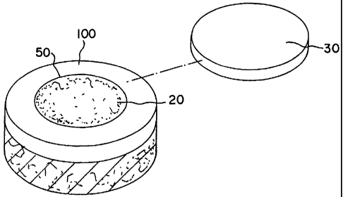
Figure 3 shows a schematic perspective view of cartilage defect material
placed in a defect
site with an exploded periosteum cap.
DESCRIPTION OF THE INVENTION
The terms "tissue" is used in the general sense herein to mean any
transplantable or
implantable tissue, the survivability of which is improved by the methods
described herein upon
implantation. In particular, the overall durability and longevity of the
implant are improved, and
host-immune system mediated responses, are substantially eliminated.
The terms "transplant" and "implant" are used interchangably to refer to
tissue, material or
cells (xenogeneic or allogeneic) which may be introduced into the body of a
patient to replace or
supplement the structure or function of the endogenous tissue.
The terms "autologous" and "autograft" refer to tissue or cells which
originate with or are
derived from the recipient, whereas the terms "allogeneic" and "allogratt"
refer to cells and tissue
which originate with or are derived from a donor of the same species as the
recipient. The terms
"xenogeneic" and "xenograft" refer to cells or tissue which originates with or
is derived from a
species other than that of the recipient.
The term "gel" refers to a mixture of minced or milled pretreated allograft
cartilage in a
biocomposite carrier having a viscosity which is less than and is less rigid
than a mixture of minced
or milled pretreated allograft cartilage in a biocompatible carrier referred
to by the terms "putty"
or "paste" and contains less cartilage by weight than putty or paste.
The present invention is directed towards a cartilage repair material and
method of
treatment. The preferred embodiment and best mode of the invention is shown in
Figure 3. In the
production of the invention, allograft hyaline cartilage is lyophilized
reducing its water content
and milled for ease in application.
After washes with sterile de-ionized (DI) water, the cartilage material was
frozen at -20
to -100 C preferably -70 C and lyophilized to reduce the water content
within the range of about
0.1% to about 8.0%. The cartilage is frozen with liquid nitrogen and ground
into particles.
A lesion or defect is removed by cutting a bore 50 or trimming a lesion in the
implant area
100 and filling the bore 50 or lesion area with a milled cartilage mixture 20
of paste or gel
consisting together with a biological carrier such as hyaluronic acid and its
derivatives, gelatin,
collagen, chitosan, alginate, buffered PBS, Dextran, or polymers and one or
more additives namely
chondrogenic stimulating factors including, but not limited to growth factors
(FGF-2, FGF-5, IGF-
CA 02522133 2005-10-12
WO 2004/096983 PCT/US2004/010956
8
1, TGF-(3, BMP-2, BMP-7, PDGF, VEGF), human allogenic or autologous
chondrocytes, human
allogenic cells, human allogenic or autologous bone marrow cells, human
allogenic or autologous
stem cells, demineralized bone matrix, insulin, insulin-like growth factor-1,
interleukin- 1 receptor
antagonist, hepatocyte growth factor, platelet-derived growth factor, Indian
hedgehog and
parathyroid hormone-related peptide.
Suitable organic glue material can be used to keep the viscous cartilage
mixture 20 fixed
in place in the implant area or to affix a periosteal cap 30 in place over the
surrounding hyaline
cartilage area 100. Suitable organic glue material can be found commercially,
such as for example;
TISSEEL or TISSUCOL. (fibrin based adhesive; Immuno AG, Austria), Adhesive
Protein
Sigma Chemical, USA), and Dow Corning Medical Adhesive B (Dow Coming, USA).
Example 1: A matrix of minced cartilage putty consisting of minced or milled
allograft
articular cartilage which has been lyophilized so that its water content
ranges from 0.1% to 8.0%
with a cartilage content ranging from 25% to 50% by weight is mixed with a
carrier of sodium
hyaluronate solution (HA) (molecular weight ranging from 7.0'x 105 to 1.2 x
106) or any other
bioabsorbable carrier such as hyaluronic acid and its derivatives, gelatin,
collagen, chitosan,
alginate, buffered PBS, Dextran, or polymers, the carrier ranging from 75% to
50% by weight. The
cartilage is milled to a size ranging from 0.01 mm to 1 mm. In gel form, the
minced cartilage which
has been lyophilized so that its water content ranges from 0.1% to 8.0%
ranging from 15% to 30%
by weight and the carrier ranges from 85% to 70% by weight. The particle size
of the cartilage
when milled is less than or equal to 1 mm dry in the previously stated range.
The cartilage pieces
can be processed to varying particle sizes and the HA or other carrier can
have different viscosities
depending on the desired consistency ofthe putty or paste. This cartilage
matrix can be deposited
into the cartilage defect arthroscopically and fit into the defect where it is
held in place by it's own
viscosity, mixed with fibrin glue or covered with a periosteal or
perichondrial flap, then sealed with
biological glue. As with the first two matrices, this matrix can support the
previously mentioned
chondrogenic factors.
Exam p le 2: A matrix of minced cartilage putty consisting of minced or milled
allograft
cartilage which has been lyophilized so that its water content ranges from 0.1
% to 8.0% ranging
from 25% to 50% by weight is mixed with a carrier of sodium hyaluronate
solution (HA) (7.0 x
105 to 1.2 x 10⁶)or any other bioabsorbable carrier such as hyaluronic acid
and its derivatives,
gelatin, collagen, chitosan, alginate, buffered PBS, Dextran, or polymers
ranging from 75% to 50%
by weight. In a gel form, the minced cartilage which has been lyophilized so
that its water content
ranges from 0.01% to 8.0% ranging from 15% to 30% by weight and the carrier
ranges from 85%
CA 02522133 2005-10-12
WO 2004/096983 PCT/US2004/010956
9
to 70% by weight. The particle size of the cartilage is less than or equal to
1 mm dry ranging from
0.01mm to lmm. The cartilage pieces can be processed to varying particle sizes
and the HA or
carrier can have different viscosities depending on the desired consistency of
the putty or paste.
Autologous or allogenic cells which have been grown outside the patient are
inserted by syringe
into the matrix before, during or after deposit of the cartilage matrix into
the defect area. Such
cells include allogenic or autologous bone marrow cells, stem cells and
chondrocyte cells. The
cellular density of the cells preferably ranges from about 1 x 10$ to 5 x 10$
or from about 100
million to about 500 million cells per cc of putty or gel mixture. This
composite material can be
injected into the cartilage defect arthroscopically and fit into the defect
where it is held in place by
it's own viscosity, or covered with a periosteal or perichondrial flap, then
sealed with biological
glue. As with the first matrix, this matrix can support the previously
mentioned chondrogenic
factors.
The operation of placing the cartilage composition in a cartilage defect,
comprises (a)
cutting a patient's tissue at a site of a cartilage defect to remove the
diseased area of cartilage; (b)
placing a mixture of milled allograft cartilage in a bioabsorbable carrier in
the defect area; and (
c) placing a periosteal cover over the mixture of the inserted milled
allograft cartilage in a
bioabsorbable carrier to contain the mixture in the defect area for a
predetermined period of time
to promote cartilage growth at the defect site. Alternate steps include the
addition of growth
factors, chondrocytes, bone marrow cells and stem cells.
The principles, preferred embodiments and modes of operation of the present
invention
have been described in the foregoing specification. However, the invention
should not be
construed as limited to the particular embodiments which have been described
above. Instead, the
embodiments described here should be regarded as illustrative rather than
restrictive. Variations
and changes may be made by others without departing from the scope of the
present invention as
defined by the following claims: