Note: Descriptions are shown in the official language in which they were submitted.
CA 02522198 2010-08-20
-1-
GUIDE WIRE HAVING BENDING SEGMENT
[0002] Field of the Invention
[0003] The present invention is related generally to a guide wire structure.
In one embodiment,
the invention is directed to a guide wire structure which can be inserted into
an interior
space within a human or animal body, such as the gastrointestinal (GI) tract
of a human
patient.
[0004] Background of the Invention
[0005] A physician typically accesses and visualizes tissue within a patient's
gastrointestinal
(GI) tract with a long, flexible endoscope. For the upper GI, a physician may
insert a
gastroscope into the sedated patient's mouth to examine and treat tissue in
the esophagus,
stomach, and proximal duodenum. For the lower GI, a physician may insert a
colonoscope through the sedated patient's anus to examine the rectum and
colon. Some
endoscopes have a working channel, typically about 2.5-3.5mm in diameter,
extending
from a port in the handpiece to the distal top of the flexible shaft. A
physician may insert
medical instruments into the working channel to help diagnose or treat tissues
within the
patient. Physicians commonly take tissue biopsies from the mucosal lining of
the GI tract
using a flexible, biopsy forceps through the working channel of the endoscope.
[0006] Insertion of a flexible endoscope, especially into the colon, can be
very time-consuming
and uncomfortable procedure for the patient, even when sedated with drugs. A
physician
often needs several minutes to push a flexible endoscope through the
convoluted sigmoid,
CA 02522198 2005-09-29
WO 2004/089455 PCT/US2004/009966
-2-
descending, transverse, and ascending portions of the colon. The physician may
diagnose
and/or treat tissues within the colon either during insertion or removal of
the endoscope.
The flexible endoscope may "loop" within the colon, such as at the sigmoid
colon or at
the splenic flexure of the colon, so that it becomes difficult to further
advance the
endoscope along the colon. When a loop is formed, the force exerted to push
the scope
stretches the mesentery and causes pain for the patient. Depending on the
anatomy of the
patient and the skill of the physician in manipulating the flexible endoscope,
some
portions of the colon may be unexamined, thus increasing the risk of
undiagnosed
disease.
[0007] Guide wires have been used to aid the introduction of catheters and
other instruments into
many sites in the human body. Many medical applications and specific designs
of guide
wires have been for cardiovascular use. There are, however, specific
challenges relates to
the use of guide wires in the GI tract, as opposed to the vascular system.
Thus, the bowel
is more tortuous, softer and generally of larger diameter. Furthermore, in the
case of the
small intestine and the colon, these are longer than most arteries or veins.
[0003] Summary of the Invention
[0009] In one embodiment, the present invention provides a guide wire
structure for use with a
medical device for insertion into a body lumen, such as the GI tract. The
guide wire
structure comprises a continuous, unitary wire comprising at least a first
segment, a
second segment, and a third segment disposed intermediate the first and second
segments.
The third segment has a bending moment of inertia less than a bending moment
of inertia
of the first segment and less than a bending moment of inertia of the second
segment.
The third segment can provide a flexible hinge for bending of the unitary
wire. By the
phrase "continuous, unitary wire" it is meant the portion of the wire
associated with the
third segment and adjacent portions of the first and second segments do not
include any
joints, junctures, or other connections (such as for instance welds, braze
joints, or solder
joints ), although the ends of the wire may include a joint or connection for
connecting
CA 02522198 2010-08-20
-3-
the wire to a handle or for other purposes. In one embodiment the wire is
formed of a
single material, such as a superelastic material. One suitable material from
which the
wire may be formed is Nitinol.
[0010] In one embodiment, the third segment has a cross-sectional area less
than the cross
sectional areas of the first segment and the second segment. The reduced cross-
sectonal
area of the third segment can be formed by grinding the outer diameter of the
wire to
form a reduced cross-sectional area third segment between first and second
segments
having a generally constant cross sectional area. The wire can have a circular
cross-
section, or alternatively, non-circular cross-sections. A generally conical
transistion
segment can extend from each end of the third segment to connect the third
segment to
the first and second segments.
[0011] Brief Description of the Drawings
[0012] The invention is described further below with reference to the
accompanying drawings,
in which:
[0013] Figure la shows an embodiment of guide wire structure as disclosed in
US Patent
No. 7288074.
[0014] Figure lb shows the structure of Figure la when one of its guide wires
is advanced
rightwardly and the other is held steady;
[0015] Figure lc shows the structure of Figure la after further righthand
advance of one of the
guide wires;
[0016] Figure 2 shows an example of a pattern of markings which may be
provided on the guide
wires to indicate their relative position to a physician;
[0017] Figure 3a to 3c show a guide wire structure advancing into the colon;
CA 02522198 2010-08-20
-4-
[0018] Figure 4 shows diagrammatically a handle for use in controlling
movement of guide
wires;
[0019] Figures 5a and 5b show successive stages in the use of a guide wire
structure in
conjunction with a bias tube;
[0020] Figures 6a and 6b show successive stages in the use of a cutting
catheter to sever the
junction between two guide wires;
[0021] Figure 7 shows two guide wire structures arranged in parallel;
[0022] Figures 8a to 8c illustrate diagrammatically the use of a guide wire
structure which has a
pivotal junction portion;
[0023] Figure 9 shows another guide wire structure described in US Patent No.
7288074.
[0024] Figure 10 illustrates an embodiment of the present invention in which a
guidewire cross
section is varied along its length to have a reduced cross section at a
location spaced from
the ends of the wire, such as at or close to the midpoint of length of the
guide wire.
[0025] Figure 11 shows the guide wire of Figure 10 bent into a generally U-
shaped
configuration for passage into a lumen such as the GI tract.
[0026] Figures 12a, b, and c show alternative embodiments in which different
cross-sections are
employed.
[0027] Figure 13 illustrates an embodiment of the guide wire of the present
invention being
advanced from the distal end of a medical device to form a loop forward of the
distal end
of the medical device.
CA 02522198 2010-08-20
-5-
[0028] Detailed Description
[0029] Figures 1-9 illustrate guide wire structures disclosed in US Patent No.
7288074.
10/409,270, incorporated herein by reference. Figures 10-13 illustrate a guide
wire
structure according to the present invention.
[0030] The structure of Figure 1 a comprises a first guide wire 1 and a second
guide wire 2, the
wires 1 and 2 being connected to one another by a junction 3 formed at the
leading ends
of the wires 1 and 2. Although the junction 3 is shown as being at the leading
ends, it
could alternatively be adjacent the leading ends. The length of the junction
need be no
more than is necessary to hold the leading ends securely together side by
side.
Depending on the nature of the junction, a length of as little as 5-10 mm may
be
sufficient, though a greater length may sometimes be preferable.
[0031] The guide wires 2 and 3 can be made of the materials conventionally
used for guide
wires, for example straight stainless steel wire, coiled stainless steel wire,
glass fiber, a
plastics material, or nitinol. Conveniently, a guide wire has a floppy tip,
i.e. a leading
end portion, typically 4-5 cm in length, of greater flexibility than the
remainder of the
guide wire, in order to reduce the risk of the leading end of the guide wire
causing
damage to the wall of the lumen through which it is passing. Where two such
conventional guide wires are joined together to produce the guide wire
structure of Figure
1, it can be these floppy tips, or parts thereof, which are joined together.
The length of
the junction can be less than the length of the floppy tips, so that some
length of floppy
material remains which is unaffected by the junction.
[0032] The whole or part of each of the guide wires may be coated to reduce
its coefficient of
friction, as is done with conventional guide wires. For example, guide wires
can be
coated with a low friction material such as silicone, or with a hydrophilic
material which
CA 02522198 2005-09-29
WO 2004/089455 PCT/US2004/009966
-6-
becomes slippery in use in a patient, or with both a low friction material
such as silicone
and hydrophilic material applied over the low friction material.
[0033] The junction 3 can be formed in any desired manner, provided the
resulting leading end
of the guide wire structure is not such as to damage the wall of the GI tract
or other body
lumen, nor cause undue pain when in contact therewith. For example, the
junction can be
made by gluing or welding the leading end portions together and then covering
those
portions with heat shrink tubing. Alternatively, the end portions could be
held together
by having a metal band crimped on to them, optionally enclosed by a cover made
of a
softer material.
[0034.] It is not essential for all the guide wires, or both the guide wires,
as the case may be, to be
of material which would normally be regarded as guide wire material. For
example, in
the case of a guide wire structure consisting of just two guide wires, one of
the guide
wires may be made of a thread, which is joined to the other guide wire by
being tied to it.
[0035] Another possibility would be to start with a single guide wire of twice
the required length
and fold it sharply back on itself, for example by crimping the folded wire
adjacent the
fold, so that it became, in effect, a pair of guide wires joined at the fold.
A guide wire
structure having an even number n of guide wires greater than two could be
formed by
folding half that number of guide wires.
[0036] The principle of operation of the guide wire structure can be seen by
comparing Figures
1b and 1c with Figure la. Figure lb shows the result of advancing the guide
wire 1
rightwardly, as indicated by the arrow, whilst holding the guide wire 2 still.
As indicated
CA 02522198 2005-09-29
WO 2004/089455 PCT/US2004/009966
-7-
in Figure lb, this causes the distal region of the guide wire structure to
curve in a
direction so that the advanced guide wire 1 is on the outside of the curve and
the still
guide wire 2 is on the inside of the curve. Continued advancement of guide
wire 1
beyond the position illustrated in Figure 1b, whilst continuing to hold guide
wire 2
steady, results in the formation of a loop in an end region of guide wire 1.
This is
illustrated in Figure lc, where the loop is denoted by reference numeral 4.
[0037] To enable the physician to easily advance one of the guide wires while
keeping the other
still, the guide wires can be received, at their ends remote from the junction
3, in a handle
which can be moved up and down the guide wires as they are advanced and
retracted.
The handle should allow precise regulation of the relative lengths of the two
guide wires.
It should also allow the introduction of the various catheters, imagers and
other
accessories, discussed in more detail below, giving accurate information on
their
relationship to the junction 3. The handle may be provided with a reversible
motor drive
which enables both guide wires to be driven. The motor drive itself may
provide data to
enable the user to monitor the lengths of the guide wires which have been fed
forward.
[0038] An example of a handle is illustrated in Figure 4. The illustrated
handle 40 comprises a
pistol grip 41 within which is mounted a pair of electric motors 42 (of which
one is
shown) powered either by a battery 43 or a mains supply 44. The motors are
controlled
by respective finger controls 45, one for each motor, each control having
forward, reverse
and stop positions. Each motor provides drive, via a respective gear, shown
diagrammatically at 46, to a respective belt or chain drive 47, each of which
propels a
respective guide wire 48 forwardly (or backwardly). A switch 47a is provided
to cause
the driving belts or chains to move away from the wires, to allow the wires to
be released,
for example at the conclusion of a procedure. A lock mechanism 49 is provided
to attach
the handle 40 to a catheter or to an accepting channel of an endoscope,
through which the
CA 02522198 2005-09-29
WO 2004/089455 PCT/US2004/009966
-8-
guide wire is to be driven. The guide wires are stored in a coiled plastics
tube 50, either
with both wires side by side in a single tube or each in its own tube. This
has the benefit
of keeping the guide wires clean, and avoiding the risk of their trailing on
to the floor.
Under some conditions this storage facility may be omitted.
[0039] The combined effect of the forms of behaviour illustrated in Figures lb
and lc enables
the guide wire structure to perform in a highly advantageous manner. Thus,
causing the
structure to become curved, as shown in Figure 1b, enables the physician to
steer the
leading end of the structure round bends in the lumen through which the
structure is
being advanced. The ability to form a loop, as illustrated in Figure lc,
enables the guide
wire structure to adopt as configuration in which it can be safely advanced
along the
lumen, without undue discomfort for the patient.
[0040] Furthermore, the presence of a loop at the leading end of the structure
rather than the tip
of a single wire, makes the structure more likely to follow the main course of
the lumen,
and less likely to inadvertently enter branches off it. Thus, in the case of
the gut, there
will be a much reduced tendency to enter, for example, diverticulae or the
orifice of the
appendix. However, the fact that the loop is not permanently present, and can
be
eliminated by putting the structure into the configuration shown in Figure la,
means that
the structure can easily, and without damage to itself, be passed along a very
narrow
passageway. It can therefore be passed, for example, along a channel of an
endoscope or
down a catheter, as is described further below. Also, when the guide wire
structure is not
in an endoscope or catheter, but is advancing directly along a patient lumen,
it is not
always desirable to do so with a loop at the front (for example if it has to
pass through a
small opening). Under such circumstances the guide wire structure is allowed
to revert to
the straight form shown in Figure la with both guidewires being advanced
aligned and in
unison.
[0041] Figures 3a to 3c show diagrammatically, and by way of example,
successive stages in
advancing the guide wire structure along a colon 30. It is shown being
introduced in
CA 02522198 2005-09-29
WO 2004/089455 PCT/US2004/009966
-9-
conjunction with a catheter 31 within which the whole guide wire structure is
slidably
received. The individual guide wires are denoted as wl and w2. Advancement
takes
place by alternately:
[0042] (a) pushing one wire forward while holding the other still; and
[0043] (b) pushing the catheter forwards as far as the position shows in
Figure 3c, or even
somewhat further.
[0044] It is desirable in endoscopic procedures to avoid, or at least reduce,
the use of X-ray
imaging to monitor what is taking place. With this in mind, the guide wires
are
preferably each provided with a pattern of markings, distributed along their
length, to
indicate how far each individual guide wire has been inserted. One such
pattern in shown
in Figure 2. As shown there, a pattern of markings in a given colour, and
similar in
nature to a bar code, is spaced along a first length (L,I), and then repeated
along
successive lengths (of which only L2 is shown) each time in a different
colour. Each of
the lengths could conveniently be of the order of 10cm. This provides a method
by
which the physician can easily see which of the guide wires is the further
advanced, and
by how much, and enable him, for example, to make the inserted lengths equal
and thus
eliminate any curve (Figure lb) or loop (Figure lc). Of course, many other
patterns of
marking, for example numerals or letters, could be used instead of that
illustrated, which
is given only as an example.
[0045] Additionally, or instead, the guide wire structure can be provided with
other forms of
position indication. It is known to provide a conventional guide wire with a
series of
miniature electrically conductive coils which surrounded the guide wire and
are spaced
along its length, the coils being connected to a source of electrical current,
whereby each
coil becomes a miniature electromagnet. Such coils can be provided on the
guide wires
used to form the guide wire structure shown. A sensing device outside the
patient is used
to detect the position of the coils within the patient, and thereby determine
the location of
the guide wires.
CA 02522198 2005-09-29
WO 2004/089455 PCT/US2004/009966
-10-
[0046] The path of the guide wire structure can be influenced by the use of a
catheter, which can
be passed over one or both of the two guide wires, when there are precisely
two, or over
one, some, or all of the guide wires, when there are more than two. In one
embodiment
the catheter has a curved tip, which allows the application of torque to bias
the forward
motion of the guide wire (or wires) over which it passes in any given
direction. The use
of a catheter in this way is illustrated in Figures 5a and 5b. Figures 5a and
5b show a pair
of guide wires 51 and 52 joined at a junction 53. Guide wire 51 is received
within a
catheter 54, referred to herein as a bias tube, the leading end portion of
which is so
formed as to have a curvature in it. The guide wire 51 with the bias tube, and
the guide
wire 52, are both received within an outer catheter 55. The ends of the
catheters 51 and
52 remote from their tips emerge from the catheter 55 to allow them to
selectively
advance and retract. The end of the bias tube 54 remote from the curved end
thereof
emerges from the outer catheter 55 at the user's end. As can be seen by
comparing the
state shown in Figure 5a with the subsequent state shown in Figure 5b, in
advancing both
the guide wires, but advancing guide wire 51 more than guide wire 52, the bias
tube helps
to ensure that the combined guide wire structure curves in the desired
direction. If it
were desired to cause the structure to advance in some other direction, this
could be
achieved by twisting the catheter 55 about its longitudinal axis, thus
altering the positions
of the guide wires relative to the lumen in which they are being advanced.
[0047] The purpose of the guide wire is, as its name indicates, to act as a
guide for some other
element. Accordingly, when the guide wire structure is in place some other
element is
then passed over it, or otherwise pushed or advanced along the guide wire.
[0048] As in the case of a catheter used to influence the path of a guide wire
structure during
passage of the guide wire structure along a lumen, a catheter introduced
subsequently can
pass over one or both of the guide wires, when there are precisely two, or
over one, some,
or all of the guide wires, when there are more than two. When the catheter is
passed over
both, or all, the guide wires, as the case may be, the leading end of the
catheter will be
free to pass beyond the leading end of the guide wire structure once it
reaches that point.
]E'kn-% Gl1 A A
CA 02522198 2005-09-29
WO 2004/089455 PCT/US2004/009966
-11-
If the catheter is not passed over both, or all, the guide wires, for example
if it is passed
over only one of two interconnected guide wires, the leading end of the
catheter will
normally be unable to pass beyond the connection between the guide wires. That
may be
desirable, for the purpose of ensuring that the leading end of the catheter
can be brought
to a position previously defined by the leading end of the guide wire
structure. It also has
the result, however, that if the guide wire structure is withdrawn, the
catheter must be
withdrawn with it.
[0049] If it is desired to enable the leading end of the catheter to pass
beyond the end of the
guide wire over which it is traveling, or to enable the catheter to remain in
position after
the guide wire has been withdrawn, this can be achieved by providing the
leading end of
the catheter with a cutting device. The use of such a catheter is illustrated
in Figures 6a
and 6b. Figures 6a and 6b show guide wires 61 and 62 connected by a junction
63 and
extending within an outer catheter 65. A cutting catheter 64 surrounds one of
the guide
wires, in this case the guide wire 61. The catheter 64 has a cutting tip (not
visible in
Figure 6a) which, when the catheter 64 is advanced over the guide wire 61,
severs the
junction 63. Figure 6b shows the severing operation partly completed.
[0050] The cutting catheter comprises a cylindrical cutting member 66 with a
circular cutting
edge 67 (visible in Figure 6b but not in Figure 6a) formed at its leading end.
When not in
use the cutting edge is shielded by a generally cylindrical sheath 68 which is
biased to a
forward protecting position by a compression spring 69 located between the
rearward end
of the sheath 68 and a stop 70 fixed to the end of the catheter. When the
cutting catheter
is pushed forwards, against the force of the spring 69, as it is in Figure 6b,
the cutting
edge 67 emerges from the sheath 68 and severs the junction 63. As soon as
severing is
completed the spring automatically causes the sheath 68 to move forwards,
covering the
cutting edge 67 and preventing it from harming the patient.
[0051] Once a sufficiently large guide wire loop has been formed in, say, the
gut, it becomes
possible to pull the gut backwards to some extent, using the friction between
the loop and
CA 02522198 2005-09-29
WO 2004/089455 PCT/US2004/009966
-12-
the wall of the gut. To do this, both guide wires are pulled backwards in
synchronism.
This provides a means for straightening the gut, and this in turn makes it
easier to
advance the guide wire structure further or, indeed, to advance other
structures (e.g.
endoscopes), and reduces the pain of the procedure, which is mainly caused by
stretching
nerve endings in the mesentery.
[0052] The above described concept of using a guide wire loop to straighten a
passageway, e.g.
the gut, can employ two guide wire structures operating in parallel. An
example of such
an embodiment is shown in Figure 7. This comprises two parallel catheters 72a
and 72b,
which are preferably connected together side by side in such a way as to allow
each to
move longitudinally with respect to the other. In the illustrated embodiment
the
connection is provided by a T-shaped stud 73 formed on catheter 72a which is
slidable in
a correspondingly shaped passageway 74 formed in catheter 72b and running
longitudinally along it. A single stud may be provided, or a plurality of
studs spaced
along the length of catheter 72a, or there may be a continuous stud running
along all or
part of the length of catheter 72a. Catheter 72a receives a first guide wire
structure 75a,
comprising a pair of wires wl and W2 joined at a junction 76a. Catheter 72b
receives a
guide wire structure 75b, comprising a pair of wires w3 and w4 joined at a
junction 76b.
[0053] The embodiment shown in Figure 7 can be used in a procedure which
employs the
following steps:
[0054] 1. Push the combination of catheters 72a and 72b into an appropriate
orifice, e.g. the
anus in the case of the colon, as far as they will go.
[0055] 2. Advance wire W3 as far as the loop which it forms is able to travel
(this is
substantially the configuration shown in Figure 7).
[0056] 3. Pull back on both catheters so that the loop in guide wire structure
75b straightens
the gut.
CA 02522198 2005-09-29
WO 2004/089455 PCT/US2004/009966
-13-
[0057] 4. Advance guide wire structure 75a in its unlooped form, i.e. wires wl
and W2,
through the catheter 72a as far as it will go (which should be past the loop
in guide wire
structure 75b).
[0058] 5. Advance catheter 72a over wl and w2 so that it is ahead of catheter
72b, while
catheter 72a, and the loop extending from the catheter, hold the gut in
position.
[0059] 6. Advance guide wire wl or guide wire W2 so that a loop is formed in
guide wire
structure 75a and advances in the gut.
[0060] 7. Withdraw whichever of wires W3 and W4 is the more forward of the
two, so as to
eliminate the loop in guide wire structure 75b.
[0061] 8. Advance catheter 72b so that it catches up with catheter 72a.
[0062] The above cycle is then repeated until the desired degree of
advancement has been
achieved.
[0063] A similar cycle of steps can be achieved by a modified form of the
embodiment of Figure
7, in which one or each of the two catheters 72a and 72b is replaced by a
suction catheter.
A suction catheter can be used to effect the above described straightening of
the gut by
pulling back on it while suction is being applied. The suction is only applied
during the
straightening step. Yet another modification is to replace one of the guide
wire structures
by a soft balloon, which can be inflated to engage the gut wall, and then
pulled back to
straighten the gut.
[0064] Many different devices can be passed over the guide wire structure, and
some examples
will now be given.
[0065] (a) A small imager (for example a CCD or CMOS chip) on a catheter could
be passed
along the guide wire or guide wires to the tip. This could optionally be
propelled along
the guide wire by a water jet or some other means of tip propulsion to reduce
the force
that has to be exerted outside the patient. A source of white or coloured
light could be
CA 02522198 2005-09-29
WO 2004/089455 PCT/US2004/009966
-14-
also introduced by the same means. This source could be in the form of light
emitting
diodes or could use fibre-optics. One of the wires could be optionally formed
out of a
fiberoptic bundle. It would be easier to take the optical signal through a
light-weight
insulated wire which could be incorporated into the guide wire or via a
separate wire in a
catheter. The imager could then convert the optical information to radiowaves
or
microwaves, to send the information to an aerial attached to, or adjacent to,
the exterior
of the patient.
[0066] (b) A separate soft catheter could be run over the guide wire to the
tip and this could
be used to introduce air from a controlled pump to inflate the viscus. Water
for rinsing
purposes could be passed through this catheter or through some other from a
water pump.
[0067] (c) A catheter could be passed over one of the guide wires, which would
provide a
channel through which biopsies could be performed. This is preferably done
after the
imager referred to in (a) above has been placed in position, so that the
imager can be used
to view the biopsy procedure. This catheter might have tip angulation
properties.
[0068] (d) A double lumen catheter could be passed over the double wire, which
might allow
the introduction of another wire of greater stiffness or with a curled tip to
allow the
movement of the device in a desired direction.
[0069] Once the guide wire, and the imager referred to in (a) above, have
reached the desired
location, an overtube could be passed, for example to the cecum. The guide
wire and the
imager could then be withdrawn and a conventional endoscope could be passed
through
the overtube to deliver therapy, for example removing a polyp or cancer.
[0070] A conventional endoscope could be introduced into a body lumen by
passing it over the
guide wire structure. However, a conventional endoscope may be too stiff for
this to be
possible, and the guide wire structure offers the possibility of, in effect,
constructing an
endoscope within a patient. To achieve this, a number of catheters, each
providing one or
more of the utilities normally provided a conventional endoscope, are
successively passed
over one or more of the guide wires, so that result is an assemblage of these
various
CA 02522198 2005-09-29
WO 2004/089455 PCT/US2004/009966
-15-
elements within the patient. A particular advantage of proceeding in this way
is that the
force required to advance each of the individual catheters is substantially
less than that
required to advance a complete conventional endoscope (e.g. a colonoscope or
an
enteroscope), since the latter is much stiffer and has much greater mass. It
is therefore
easier for the physician, and less uncomfortable for the patient, and is less
likely to cause
injury to the patient. Also, since the endoscope is then assembled element by
element,
the endoscope can have those facilities which are required for the particular
patient, and,
only those facilities, so that the endoscope is tailored to the requirements
of the medical
procedure being carried out. It will be understood that, for the purpose of
allowing in
situ assembly of a catheter, the guide wire structure should preferably
comprise more
than two guide wires, for example three or four guide wires.
[0071] Although a structure having more than two guide wires is particularly
useful for the
purpose discussed above of assembling an endoscope in situ, it may also have
value in
relation to the procedure for introducing the guide wire structure into a
lumen. This is
because the two-guide wire structure shown in Figures 1 a to 1 c allows
curvature in only
one plane, so that steering the structure in three dimensions requires the
user to twist the
structure about its longitudinal axis, for example by using a catheter to
which the
necessary torque can be applied. However, if more than two guide wires are
provided it
is possible to curve the structure in any plane; three guide wires are
sufficient for this
purpose.
[0072] Attention is now directed to Figures 8a to 8c, which illustrate the use
of a guide wire
structure 80 which comprises two guide wires 81 and 82 connected by a junction
portion
83. As can be seen, the junction portion 83 is pivotal about an axis located
at the
proximal end of the portion 83, so that, as shown in Figure 8a, it can pivot
to such an
extent that it lies flat along the distal end portion of guide wire 81. This
is advantageous
in that it makes possible, or makes easier, movement of the portion 83 within
a catheter
84, not only where there is no loop present (as in Figure 8c) but also when
there is (as
shown in Figure 8a). In this connection it is to be understood that the
diameter of the
CA 02522198 2010-08-20
-16-
catheter 84 would actually be substantially greater than that shown in these
Figures. It is
also to be understood that instead of being joined by a junction portion 83 of
significant
length, as illustrated, the guide wires could alternatively be joined by a
junction of
substantially no length, i.e. the ends of the guide wires could be connected
by a junction
consisting, at least in substance of just a pivot point.
[0073] Figure 9 shows yet another guide wire structure in which a similar
pivoting action can be
achieved. This comprises guide wires 91 and 92, having respective floppy tip
portions
91 a and 92a connected to one another by a thread or highly flexible wire 93.
This thread
or wire can be inserted into the portions 91a and 92a, or attached to their
surfaces.
[0074] Figures 10-13 illustrate a guide wire structure according to the
present invention. The
guide wire structure includes a continuous, unitary wire having a segment
(which can be
positioned generally in the middle portion of the wire), which segment has a
bending
moment of inertia which is lower than the bending moment of inertia of the
adjacent wire
segments. For instance, the wire can change in cross sectional shape or
dimension at a
location that is not a terminal end, so as to provide a bending hinge.
[0075] The bending moment of inertia for a circular cross-section can be
calculated as it r4/4,
where r is the radius of the cross-section. The bending moment of inertia for
a
rectangular cross-section can be calculated as bh3/12, where b is the base of
the rectangle
and h is the height. "Mechanics of Materials", A.C. Ugural, 1991, McGraw Hill
is related to bending of cross-sections.
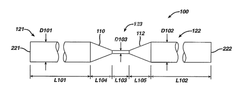
[0076] Figure 10 shows an embodiment of guide wire structure of the present
invention
comprising a continuous, unitary wire 100 that has varying cross sectional
area along a
portion of its length. In this embodiment, the wire 100 can have a first
segment 121
having a generally circular cross section of nominal diameter D101 and a
length Li01, a
second segment 122 having a generally circular cross-section of nominal
diameter D102
and a length L102, and a third segment 123 having a generally circular cross-
section of
nominal diameter D103 and length L103, The wire 100 can also include a tapered
CA 02522198 2005-09-29
WO 2004/089455 PCT/US2004/009966
-17-
transition segment 110 having a conical shape and a length L104 and extending
between
segment 121 and segment 123, and a tapered transition segment 112 having a
length
L105 and extending between segment 123 and segment 122.
[0077] The reduced diameter D 103 of the third segment 123 relative to the
diameter D 101 and
the diameter D 102 provides the third segment 123 with a bending moment of
inertia
which is lower than that of the segments 121 and 122. Accordingly, the wire
100 can
bend at the third segment 123 to provide a hinge, which hinge can encompass
the length
L103 of third segment 123, as well as some or all of the lengths L104 and L105
of
segments 110 and 112. In one embodiment, the hinge so formed can be an elastic
hinge.
[0078] The wire 100 with it's associated hinge can be used in the embodiment
as described
below, as well as in those methods disclosed with reference to Figures 1-9
above, without
the need for attaching or otherwise joining two wires or using different
materials.
[0079] In one embodiment, the diameters D101 and D102 can be between about
0.010 inch to
about 0.035 inch, and more particularly about 0.016 inch to about 0.020 inch.
The third
segment 123 can have a diameter D103 of between 0.005 inch and about 0.010
inch, and
in one embodiment D103 can be about 0.007 inch.
[0080] Each of L101 and L102 can be at least about 3 feet, and can be between
about 6 feet and
about 12 feet. The combined length lengths L101, L102, L103, L104, and L105
can be
between about 7 feet and about 25 feet. In one embodiment, the lengths L101
and L102
can be about equal, and their combined length can be at least about 20 feet.
Length L103
of the third segment can be between about 0.100 inch to about 0.500 inch, and
in one
embodiment can be about 0.300 inch. The length L104 and Length 105 can be
about
equal, and can each be about 2 inches. Modification of the cross section of
the wire 100
at a location intermediate the ends may be accomplished by any suitable
process, such as
by grinding, drawing, or stamping wire 100.
[0081] In one embodiment, the reduced cross-section of the third segment 123
can be formed by
centerless grinding. A reduced cross-section can be formed using a grinding
machine
CA 02522198 2005-09-29
WO 2004/089455 PCT/US2004/009966
-18-
such as a TF-9CPG System 2000 Guide Wire Profile Grinder available from Glebar
Company of Franklin Lakes, NJ.
[0082] The wire 100 can be enclosed in one or more low friction and/or
lubricous sleeves. In
Figure 11, the first wire segment 121 is enclosed in a sleeve 155, second wire
segment
122 is enclosed in a sleeve 159, and the third segment and the transition
segments are
enclosed in a sleeve 157. The sleeves 155, 157, and 159 can be formed of a low
friction
material, such as PTFE or polyester. Indicators can be associated with the
first and
second wire segments 121 and 122 so that the wire segments can be
distinguished when
viewed through a camera or other optical device associated with an endoscope
or other
medical device. For instance, the indicators can be,visual, and can employ
different
colors. In one embodiment, the sleeves 155 and 159 can be provided in
different colors
and/or with different patterns of markings. In Figure 13, sleeve 155 has a
pattern of
heavy, diagonally slanted marks, while sleeve 159 has a pattern of lighter,
non-slanted
markings. The marking colors and/or the background color of the sleeves can be
different to distinguish sleeve 155 from sleeve 159. Alternating stripes of
different colors
can also be used to distinguish sleeve 155 from sleeve 159, and thus segment
121 from
122 as viewed through a visualization device. Sleeve 157 can have yet another
color or
pattern of colors to provide a visual indication of the location of the third
segment 123.
[0083] Figure 11 illustrates the wire 100 bent at the third segment 123 to
form a narrow wire
loop for introduction into a body cavity. In Figure 11, the wire 100 is
illustrated with a
generally U-shaped bend 150 with a radius R110 so that the wire does not kink
upon
placement through a colonoscope working channel, does not form a sharp point
that can
damage tissue upon placement in a body lumen, and preferably does not
substantially
plastically deform. In one embodiment, the radius R110 can be about 0.75mm to
about
1.5mm, and more particularly about lmm.
[0084] Suitable biocompatible materials from which such a wire can be
constructed include
those mentioned from which the wires of Figures 1-9 can be formed, including
without
CA 02522198 2010-08-20
-19-
limitation a superelastic material such as nitinol. Other materials, such as
steel and alloys
can also be used. One suitable material from which wire 100 can be formed is
Nitinol
NDC SE508 available from Nitinol Devices and Components, a Johnson & Johnson
Company of Fremont, CA.
[0085] Figure 12 illustrates alternative embodiments in which the cross-
section of the narrowed
length 103 is not round. Different cross sectional shapes may be formed, such
as
changing a round wire to a flat rectangular cross section in Figure 12a, an
oval cross
section in Figure 12b, or a square cross section in Figure 12c. Other cross-
sectional
shapes, such as triangular, hexangonal, or other polygonal shapes may be
employed.
Preferential bending planes of certain cross sectional shapes can be used for
the purposes
of directing the U-loop of the wire. For instance, a rectangular, oval, or
triangular cross-
section can be employed to direct bending about a particular axis.
[0086] The guide wire structure with wire 100 of the present invention can be
used in place of
the wire configurations shown in Figures 1-9, as well as with the device
illustrated in US
Patent No. 7288074.
Figure 13 is a schematic illustration of the guide wire structure with
wire 100 in use with a medical device 300. Generally, medical device 300 can
be a
flexible endoscope, such as a flexible colonosocope, or a device such as is
shown in the
above referenced patent application.
[0087] Medical device 300 can include a handle 310, which is positioned
outside a patient, an
elongate flexible body portion 330, and a distal end 320 which can be
positioned in a
patient, such as in a patient's GI tract, with distal end 320 sized and shaped
to be
advanced in the GI tract. The medical device can also include a working
channel 350
extending through the body portion 330 and opening at the distal end 320 of
the device
300, a camera 360, light source 370, a camera lens wash nozzle/irrigation
nozzle 380, a
light source 392, and a light source 394. Suction can be provided through
working
channel 350, if desired.
CA 02522198 2005-09-29
WO 2004/089455 PCT/US2004/009966
-20-
[0088] The guide wire with wire 100 of the present invention can be positioned
within the
working channel 350 such that the U shaped bend in third segment 123 is
positioned in
the patient's body, and the ends of the guide wire extend through an access
opening of
the handle 310. In figure 13, the ends of the guide wire are indicated by
numeral 221
(associated with first segment 121) and numeral 222 (associated with second
segment
122).
[0089] The guide wire with wire 100 can be used generally as shown in Figures
3A-3C to
advance a device into a body lumen, such as the GI tract. In Figure 13, after
the U
shaped bend in the third segment 123 has been advanced through the working
channel
350, the first segment 121 is advanced through the working channel relative to
second
segment 122, so that the first segment 121 takes on a curvature having a
radius of
curvature greater than Radius of curvature 12110, as shown in Figure 13. To
advance the
distal end 320 further into the patient, the operator can pull proximally on
end 222 to
move third segment 123 back into the working channel 350, thereby leaving the
relatively
large radius of curvature loop in first segment 121 in the body lumen and
extending from
the distal end of the device 300. End 222 can then be held fixed, and end 221
can be
advanced distally toward handle 310 so that additional length of the first
segment 121 is
advanced distally out of working channel 350, thereby advancing the relatively
large
radius of curvature loop distally in the GI tract. Then, while holding end 222
stationary
with respect to handle 310, end 221 can be pulled in tension (proximally)
while
simultaneously pushing (distally) the elongate body portion 330 distally along
wire
segments 121 and 122 in working channel 350, so that the distal end 350 moves
forward
(distally) into the GI tract. Accordingly, the wire segments 121 and 122 serve
as a track
upon which the distal end 350 of device 300 can be advanced.
[0090] While the present invention has been illustrated by description of
several embodiments, it
is not the intention of the applicant to restrict or limit the spirit and
scope of the appended
claims to such detail. Numerous other variations, changes, and substitutions
will occur
to those skilled in the art without departing from the scope of the invention.
Moreover,
CA 02522198 2005-09-29
WO 2004/089455 PCT/US2004/009966
-21-
the structure of each element associated with the present invention can be
alternatively
described as a means for providing the function performed by the element. It
will be
understood that the foregoing description is provided by way of example, and
that other
modifications may occur to those skilled in the art without departing from the
scope and
spirit of the appended Claims.
END 5244