Note: Descriptions are shown in the official language in which they were submitted.
CA 02522408 2005-10-13
WO 2004/093962 PCT/US2004/008896
MEDICAL DEVICE WITH THERAPEUTIC AGENTS
FIELD OF THE INVENTION
The present invention relates to medical devices suitable for at least
partial implantation into a body. More specifically, the present invention
relates to medical devices with therapeutic agents. In preferred
embodiments, the invention relates to cannulae, such as catheters, with
therapeutic agents. The present invention also relates to a method of making
a medical device, and a method of establishing access to a vessel within a
body.
BACKGROUND OF THE INVENTION
Many types of medical devices are used in a variety of medical
procedures that include at least partial implantation into a body. When
implanted, medical devices can be in intimate contact with a variety of cells,
tissues, and body systems. For example, cannulae, such as catheters, are
used in a variety of medical procedures to introduce articles, such as stents,
into body vessels. Cannulae are also used to establish a communicative
passageway by which a body vessel can be accessed from the exterior of the
body. These cannulae are indispensable in procedures that require repeated
access to the vessel, such as hemodialysis procedures that include repeated
extracorporeal treatment of blood.
While implanted medical devices provide several advantages, they also
present an opportunity for infection. Indwelling medical devices, such as
indwelling cannulae used for access ports, are particularly susceptible to
infection due to their long term presence in the body. In essence, the cannula
provides a path from the external environment into the body along which
microorganisms can colonize, and eventually produce an infection.
The establishment of an infection can require intervention, such as
treatment with a therapeutic agent or even mechanical manipulation of the
medical device to remove the microorganisms. Even worse, the infection may
require removal and replacement of the medical device. Ultimately, the
presence of an infection may outweigh the benefits of the implantation.
I
CA 02522408 2005-10-13
WO 2004/093962 PCT/US2004/008896
Infections associated with indwelling medical devices are commonly
caused by bacteria or fungi. The most common organisms associated with
infections associated with indwelling devices are Staphylococcus epidermidis
and Staphylococci aureus. Candida albicans, a fungi, is another significant
cause of infections associated with these devices. No matter the
microorganism, establishment of infection requires colonization along the
surface of the medical device, which depends on a variety of factors,
including
the formation of glycocalyx and a fibrin sheath.
Glycocalyx is a polysaccharide produced by adherent microorganisms.
The glycocalyx allows the microorganisms to adhere to the surface, and
contributes to the formation of a biofilm around the medical device. In
addition to the glycocalyx formation, a fibrin sheath is often produced by the
host as a natural result of thrombogenesis. The fibrin sheath essentially
covers the surfaces of the indwelling device, and provides another agent onto
which microorganisms can adhere.
Considering the importance of implantable medical devices,
considerable attention has been directed toward preventing colonization
and/or infection on these articles. The art contains many examples of medical
devices that incorporate a variety of approaches that attempt to control
colonization and/or infection. For example, United States Patent
No. 5,688,516 to Raad et al. discloses medical devices coated with mixtures
of antibiotics and other therapeutic agents. Also, United States Patent
No. 5,624,704 to Darouiche et al. discloses medical devices impregnated with
antimicrobials.
As indicated above, the microorganisms commonly associated with
colonization and/or infection from implanted medical devices typically
originate from outside the body, such as on the skin, and progress into the
body along the path of the medical device. Once inside the body, the
microorganisms produce the glycocalyx that facilitates adherence, and the
body produces a fibrin sheath around the device that facilitates colonization
and establishment of an infection. Thus, two distinct processes are occurring
on two distinct portions of the medical device. Outside the body,
microorganisms gain access to the device and begin to proceed into the body.
Inside the body, microorganisms arriving from the external portion of the
2
CA 02522408 2005-10-13
WO 2004/093962 PCT/US2004/008896
device produce a glycocalyx to facilitate adherence, and the body produces
the fibrin sheath which further facilitates adherence. The prior art fails to
recognize the localization of these processes in the available devices
designed to prevent or inhibit colonization and/or infection.
BRIEF SUMMARY OF THE INVENTION
The present invention provides a medical device for at least partial
implantation in a body, such as a human body, comprising a main body
having a first end, a second end, and a length extending from the first end to
the second end. The medical device has first and second sections extending
along the length of the medical device. The first section is near the first or
distal end of the device and the second section is near the second or proximal
end of the device. The first section has a first therapeutic agent, and the
second section has a second therapeutic agent. Once implanted, the first
section is fully implanted in the body, and the second section is only
partially
implanted in the body. The second section is at least partially positioned
within a subcutaneous layer of the body, and may have a section that extends
outside of the body.
In a preferred embodiment, the main body comprises a cannula having
an interior surface and an exterior surface. The cannula defines a lumen.
Further, the medical device can include a separator that separates the first
section from the second section.
The first and second therapeutic agents can be associated with the first
and second sections, respectively, in a variety of manners. For example, the
agents can be impregnated into the main body of the medical device, or can
be posited onto the medical device. In a preferred embodiment, one or more
of the therapeutic agents is coated onto one or more surfaces of the medical
device. In a particularly preferred embodiment, the first therapeutic agent is
impregnated into the first section of the main body and the second therapeutic
agent is coated onto at least one surface of the second section of the main
body.
The first and second therapeutic agents can be any suitable type of
agent. Examples of suitable types of agents include, without limitation,
3
CA 02522408 2005-10-13
WO 2004/093962 PCT/US2004/008896
antiproliferatives, anticoagulants, antithrombotics, thrombolytics and/or
fibrinoiytics, and antimicrobials.
In a preferred embodiment, a first therapeutic agent comprises an
antiproliferative. Particularly preferable, the first therapeutic agent
comprises
paclitaxel, a paclitaxel derivative, or a paclitaxel pro-drug. Also
preferable, the
second therapeutic agent comprises one or more antimicrobials. The
antimicrobial can be an antibiotic, an antiseptic, and/or a disinfectant. In a
particularly preferred embodiment, the second therapeutic agent comprises a
blend of two or more antibiotics. A desirable blend includes rifampin and
minocycline.
The present invention also provides a method of making a medical
device for at least partial implantation. The method comprises providing a
main body having a first end, a second end, a length.extending from the first
end to the second end, a first section along the length, and a second section
along the length; exposing the first section to a solvent so that the first
sections swells; soaking the first section in a solution containing a first
therapeutic agent; drying the first section; and coating at least a portion of
the
second section with a second therapeutic agent.
The present invention also provides a method of establishing access to
a vessel of the body. The method comprises providing a medical device
comprising a cannula having a distal end, a proximal end, an interior surface,
an exterior surface, and defining a lumen. The cannula has a length
extending from the proximal end to the distal end, a first section extending
along the length with a first therapeutic agent, and a second section
extending
along the length with a second therapeutic agent. The method also includes
implanting the distal end of the cannula into the body so that the proximal
end
remains either substantially outside the body or in a subcutaneous layer, and
forming an interface between the distal end and the vessel. The interface can
be a direct insertion of the distal end into the vessel or an attachment of
the
distal end to the vessel, such as an anastomosis.
BRIEF DESCRIPTION OF THE DRAWINGS
Figure 1 is a schematic illustration of a medical device according to an
embodiment of the present invention.
4
CA 02522408 2005-10-13
WO 2004/093962 PCT/US2004/008896
Figures 1A, 1 B, 1 C and 1 D illustrate various cross-sectional shapes
and lumen configurations for devices according to the present invention.
Figure 2 is a cross-sectional view taken along line I-I in Figure 1.
Figure 3 is a cross-sectional view illustrating an embodiment of the
present invention.
Figure 4 is a cross-sectional view illustrating an embodiment of the
invention.
Figure 5 is a cross-sectional view illustrating an embodiment of the
invention.
Figure 6 is a cross-sectional view taken along line II-II in Figure 1.
Figure 7 is a cross-sectional view illustrating an embodiment of the
invention.
Figure 8 is a cross-sectional view illustrating an embodiment of the
invention.
Figure 9 is a cross-sectional view illustrating an embodiment of the
invention.
Figure 10 is a cross-sectional view of a medical device according to an
embodiment of the present invention.
Figure 11 is a schematic illustration of a medical device according to
the present invention transcutaneously implanted into a body.
Figure 12 is a schematic illustration of a medical device according to
the present invention implanted subcutaneously into a body.
Figure 13 is a schematic illustration of a medical device according to an
embodiment of the present invention.
Figure 14 is a schematic illustration of a medical device according to an
embodiment of the present invention.
Figure 15 is a schematic illustration of a medical device according to an
embodiment of the invention.
Figure 16 is a schematic illustration of a medical device according to an
embodiment of the invention.
DETAILED DESCRIPTI R! OF THE INVENTION
Medical devices according to the present invention can be any of a
variety of medical device types and configurations. The medical device need
CA 02522408 2005-10-13
WO 2004/093962 PCT/US2004/008896
only be at least partially implantable within a body. Examples of types of
medical devices that can be made according to the present invention include
leads, fasteners, and cannula, such as catheters.
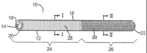
Figure 1 illustrates a medical device according to one embodiment of
the present invention. In this embodiment, the medical device 10 comprises a
cannula having a main body 12 and defining a lumen 14. The cannula 10 has
exterior 16 and interior 18 surfaces, a first or distal end 20, and a second
or
proximal end 22.
The length of the cannula extends from the first end 20 to the second
end 22. A first section 24 of the cannula 10 extends along a portion of the
length, and a second section 26 extends along another axially distinct portion
of the length. Both the first 24 and second 26 sections extend
circumferentially around the cannula 10 and axially along a respective portion
of the length. As illustrated in the figure, the first section 24 is
preferably near
the first end 20 and the second section 26 is preferably near the second end
22.
A first therapeutic agent 28 is associated with the cannula 10 at the first
section 24, and a second therapeutic agent 30 is associated with the cannula
at the second section 26. Both the first 28 and second 30 therapeutic
agents can be associated with the cannula 10 in a variety of manners.
Preferably, as illustrated in Figure 2, the first therapeutic agent 28
comprises
an impregnated agent 36 disposed in the material of the first section 24, such
as by bulk distribution, solvent swelling, or other suitable techniques. Also,
as
illustrated in Figure 6, the second therapeutic agent 30 preferably comprises
a
coating layer 38 posited on the external surface 16 of the second section 26
of the cannula 10.
Many alternative arrangements for the first 28 and second 30
therapeutic agents are within the scope 'of the present invention. For
example, Figures 3-5 illustrate alternative arrangements for the first
therapeutic agent 28 in relation to the first section 24 of the cannula 10,
and
Figures 7-9 illustrate alternative arrangements for the second therapeutic
agent 30 in relation to the second section 26 of the cannula 10. As
illustrated
in Figure 3, the first therapeutic agent 28 can comprise a coating layer 32
posited on the external surface 16 of the cannula 10. As illustrated in Figure
6
CA 02522408 2005-10-13
WO 2004/093962 PCT/US2004/008896
4, the first therapeutic agent 28 can comprise a coating layer 34 posited on
the internal surface 18 of the cannula 10. Also, as illustrated in Figure 5,
the
first therapeutic agent 28 can comprise a coating layer 32 posited on the
external surface 16 and a coating layer 34 posited on the internal surface 18.
The second therapeutic agent 30 can likewise be associated with the second
section 26 in similar ways. Thus, as illustrated in Figure 7, the second
therapeutic agent 30 can comprise an impregnated agent 42 disposed in the
material of the second section 26 of the cannula 10. As illustrated in Figure
8,
the second therapeutic agent 30 can comprise a coating layer 40 posited on
the interior surface 18 of the cannula 10. Furthermore, as illustrated in
Figure
9, the second therapeutic agent 30 can comprise a coating layer 38 posited
on the external surface 16 and a coating layer 40 posited on the internal
surface 18.
In addition to the various arrangements for each of the first 28 and
second 30 therapeutic agents, any suitable combination of arrangements, i.e.,
one for each agent 28, 30, can be utilized.
The main body 12 can be formed of any suitable material, and need
only be biocompatible and appropriate for the desired type of medical
procedure in which the device will be utilized. Preferred materials for the
main
body 12 include thermoplastic and thermoset materials. In particularly
preferred embodiments, silicone, a thermoset material, is utilized as the
material of the main body 12.
The cross-sectional shape of the medical device can be any shape
suitable for the types of procedures in which the device will be utilized. A
circular cross-sectional shape is particularly preferable in embodiments in
which the device comprises a cannula, such as that illustrated in FIGURE 1.
A circular cross-sectional shape maximizes space within the lumen 14 of the
cannula 10 while also providing a suitable shape for interfacing with a body
vessel. Furthermore, the medical device can have any suitable configuration
of lumen(s), and the chosen configuration will depend on the application for
which the device is used. Single and multi-lumen configurations can be
utilized. Figures 1A, 1 E, 1 C and 1 illustrate various suitable cross-
sectional
shapes and lumen configurations for use in medical devices 10 according to
the present invention.
7
CA 02522408 2005-10-13
WO 2004/093962 PCT/US2004/008896
The first 28 and second 30 therapeutic agents can be any suitable
agents, and need only provide the desired effects. Thus, the first therapeutic
agent 28, which is associated with the first section 24 near the first or
distal
end 20, need only have a negative effect on the formation of fibrin sheaths.
Also, the second therapeutic agent 30, which is associated with the second
section 26 near the second or proximal end 22, need only have an
antimicrobial effect.
Examples of suitable therapeutic agents for use as the first therapeutic
agent 28 include anticoagulants, antithrombotics, thrombolytics and/or
fibrinolytics, and antiproliferatives. The type of agent selected as the first
therapeutic agent 28 will depend on several factors, including the stage of
development of the fibrin sheath at which interference with further
development is desired. For example, antithrombotics, such as heparin,
hirudin, hirulog and PPACK, directly or indirectiy bind thrombin to prevent
polymerization of fibrin from fibrinogen, a necessary step in the coagulation
process. Anticoagulants, such as the glycoprotein Ilbllla inhibitors, attach
to
platelet receptors and block activation sites, thereby preventing their
degranulation and release of serotonin. Other anticoagulants block ADP
induced platelet aggregation, such as Ticlopidine and Clopidigrel. Still other
anticoagulants such as warfarin and coumadin inhibit the action of vitamin K
and the production of coagulation factors. Some anticoagulants, such as
aspirin, inhibit platelet aggregation by inhibiting Thromboxane A2.
Thrombolytics and/or fibrinolytics lyse or break down an organized
thrombus by activating plasmin, which breaks down fibrin. Examples of
suitable thrombolytics and/or fibronolytics include Tissue Plasminogen
Activator (tPA), Urokinase, and Streptokinase.
Certain matrix metalloproteinases, such as collagenase, can break
down the connective tissue of a formed fibrin sheath.
Examples of suitable antithrombotics include heparin, hirudin, hirulog,
and PPACK. Examples of suitable anticoagulants include glycoprotein Ilbllla
inhibitors, ticlopidine, clopidigrel, warfarin, coumadin, and aspirin.
Examples
of suitable thrombolytics and/or fibrinolytics include tPA, recombinant tPA,
urokinase, streptokinase, Tenecteplase, Alteplase, Activase, Lysatec,
Antistreplase, APSAC, Eminase, Retaplase, Retavase, Hannahpep (Indian
8
CA 02522408 2005-10-13
WO 2004/093962 PCT/US2004/008896
King Cobra venom), and Ancrod (Malayan pit viper venom). Examples of
suitable matrix metalloproteinases include collagenase. Other suitable agents
for the first therapeutic agent include olyeyloxyethyl phosphorylcholine.
Also, combinations of two or more agents can be used as the first
therapeutic agent 28.
In a preferred embodiment, the first therapeutic agent comprises an
antiproliferative. In a particularly preferred embodiment, the first
therapeutic
agent 28 comprises natural or synthetic paclitaxel, a derivative of
paclitaxel,
and/or a paclitaxel pro-drug.
Paclitaxel is a natural diterpere product isolated from the Pacific yew
tree (Taxus brevifolia). Paclitaxel is a member of the taxane family of
terpenes, and was first isolated by Wani et al. (J. Am. Chem. Soc., 93:2325,
1971). Paclitaxel has proven efficacious in the treatment of a variety of
neoplasms, and has been approved for use in the clinical treatment of breast
and ovarian cancer in the United States.
Paclitaxel functions as an antiproliferative agent; i.e., as an inhibitor of
cell replication. It is believed that paclitaxel inhibits replication by
inducing an
abnormal polymerization of tubulin. This results in stabilization of
microtubules and disruption of the cell division process, mitosis. Further,
paclitaxel inhibits smooth muscle cell proliferation both in vitro and in
vivo.
Paclitaxel can be used in medical devices of the present invention in its
basic form, as a derivative (see for example U.S. Patent No. 6,476,242 to
Kingston et al. for 2-AROYL4-ACYL PACLITAXEL ANALOGS; see also U.S.
Patent No. 6,441,025 to Li et al. for WATER SOLUBLE PACLITAXEL
DERIVATIVES), and/or as a-PRO-DRUG (i.e., a drug that yields paclitaxel
upon action by an appropriate agent, such as a naturally occurring enzyme;
see U.S. Patent No. 6,153,756 to Digenis et al. for SOLUBLE PRODRUGS
OF PACLITAXEL). Also, a preparation of paclitaxel can be utilized. Any
suitable preparation can be used, and should facilitate placement of the
paclitaxel into or on the medical device of the present invention, and should
allow its release from the medical device. Examples of suitable paclitaxel
preparations include those described in U.S. Patent No. 5,681,846 to Triysel
for EXTRUDED STABILITY FORMULATIONS FOR PACLITAXEL.
9
CA 02522408 2005-10-13
WO 2004/093962 PCT/US2004/008896
Considerable attention has been directed toward the effects of
paclitaxel on a variety of cell types and physiological processes. Paclitaxel
may arrest the migration of fibroblasts and smooth muscle cells, thereby
reducing or preventing connective tissue formation that often follows fibrin
sheath formation. It has also been found to decrease restenosis of human
coronary arteries following stent use.
The second therapeutic agent 30 can be any suitable antimicrobial
agent. As used herein, the term 'antimicrobial' means any agent that has
killing or growth inhibiting effects on one or more microorganisms.
Suitable classes of antimicrobials include antibiotics, disinfectants, and
antiseptics.
In a preferred embodiment, the second therapeutic agent 30 comprises
one or more antibiotics having activity against the common microorganisms
associated with colonization and/or infection with indwelling cannulae.
Examples of suitable classes of antibiotics include tetracyclines, rifamycins,
macrolides, penicillins, cephalosporins, other beta-lactam antibiotics,
aminoglycosides, chloramphenicol, sulfonamides, glycopeptides, quinolones,
fusidic acid, trimethoprim, metronidazole, clindamycin, mupirocin, polyenes,
azoles and beta-lactam inhibitors.
Examples of specific antibiotics that can be used as the second
therapeutic agent 30 include minocycline, rifampin, erythromycin, nafcillin,
cefazolin, imipenem, aztreonam, gentamicin, sulfamethoxazole, vancomycin,
ciprofloxacin, trimethoprim, metronidazole, clindamycin, teicoplanin,
mupirocin, azithromycin, clarithromycin, ofloxacin, lomefloxacin, norfloxacin,
nalidixic acid, sparfloxacin, pefloxacin, amifloxacin, enoxacin, fleroxacin,
temafloxacin, tosufloxacin, clinafloxacin, sulbactam, clavulanic acid,
amphotericin B, fluconazole, itraconazole, ketoconazole, and nystatin.
The second therapeutic agent 30 can comprise a combination of two or
more antimicrobials. In these embodiments, the two or more antimicrobials
can be located in or on discrete locations within the second section 26, or
the
two or more antimicrobials can be blended together and uniformly distributed
within or on the second section 26.
In a preferred embodiment, rifampin and minocycline are used as the
second therapeutic agent 30. The rifampin and minocycline preferably are
CA 02522408 2005-10-13
WO 2004/093962 PCT/US2004/008896
blended together and evenly distributed either in or on the second section 26.
In a particularly preferred embodiment, discussed below, blended rifampin
and minocycline are coated onto the surfaces of second section 26.
Figure 10 is a cross-sectional illustration of a medical device according
to a preferred embodiment of the present invention. In this embodiment, the
medical device comprises a cannula 10 having a main body 12 and defining a
lumen 14. The cannula 10 has an exterior 16 and an interior surface 18. The
cannula 10 has a first or distal end 20 and a second or proximal end 22, and a
length extending between the two ends 20, 22. A first section 24 extends
along a portion of the length, and a second section 26 extends along a
different portion of the length. Each of the first 24 and second 26 sections
preferably extends circumferentially around the cannula 10. A first
therapeutic
agent 28 comprises paclitaxel impregnated into the main body 12 of the first
section 24. A second therapeutic agent 30 comprises a blend of rifampin and
minocycline coated on the exterior 16 and interior 18 surfaces of the second
section 26.
In one application medical devices according to the present invention
can be used to establish access to a vessel within a body. As discussed
above, medical devices according to preferred embodiments of the invention
comprise cannulae that define a lumen. The distal end of the cannula can be
interfaced with a vessel to establish a communicative passageway between
the vessel and the lumen of the cannula. In this configuration, the medical
device is particularly well suited for allowing convenient access to the
vessel.
These devices can be used advantageously in procedures that require
repetitive access to the vessel, such as repetitive introduction of an agent
into
the blood stream or the repetitive extracorporeal treatment of blood, such as
in hemodialysis procedures.
The medical devices according to the present invention can be
completely implanted within the body, or only partially implanted within the
body. In each scenario, however, at least a portion of the second section of
the device remains within the subcutaneous space. Figure 11 illustrates a
schematic of a medical device 10 according to the present invention that is
transcutaneously implanted into a body. In this embodiment, the medical
device 10 traverses the skin through the epidermis 52, derma 54 and
11
CA 02522408 2005-10-13
WO 2004/093962 PCT/US2004/008896
subcutaneous 56 layers to a vessel 58. An interface 60 is formed between
the vessel 58 and the device 10. The interface defines a communicative
passageway between the vessel 58 and the lumen of the device 10. The
interface 60 can be a direct insertion of the distal end 20 of the device 10
into
the vessel 58, or can comprise an attachment of the distal end 20 to the
vessel 58, such as an anastomosis.
Because the device 10 is implanted transcutaneously, the device 10 in
this embodiment includes a portion 61 that remains external to the body. This
portion 61 provides the desired access to the lumen which is in
communication with the vessel 58. Thus, in this embodiment, the vessel 58
can be accessed without further disruption to the skin 50.
The second section 26, which includes the second therapeutic agent
30, preferably is positioned across the subcutaneous layer 56. As illustrated
in the Figure, the second section 26 can extend beyond the subcutaneous
layer and toward and through the derma 54 and epidermis 52. The first
section 24, which includes the first therapeutic agent 28, preferably is
positioned below the subcutaneous layer 56, and is preferably approximately
adjacent the interface 60.
Figure 12 illustrates a cannula 10 according to the present invention
that is completely implanted within a body. In this embodiment, the cannula
includes an access port 62. The access port 62 defines a chamber that
can receive a communicative member, such as a needle, for either
withdrawing fluid from or directing fluid into the vessel 58. Typically, the
access port 62 includes a section of resealable material 64 that prevents
escape of fluid from the cannula 10 when a communicative member is not
received by the access port 62. The resealable material can comprise silicon
or any other suitable material.
In this embodiment, the second section 26, and therefore the second
therapeutic agent 30, is completely contained within the subcutaneous layer
56. The first section 24, and therefore the first therapeutic agent 28, is
positioned below the subcutaneous layer 56 and is preferably adjacent the
interface 60 between the cannula 10 and the vessel 58.
Figure 13 illustrates another embodiment of the present invention. A
medical device according to this embodiment is identical to the embodiment
12
CA 02522408 2005-10-13
WO 2004/093962 PCT/US2004/008896
illustrated in Figure 1, except as detailed below. Thus, the medical device of
this embodiment comprises a cannula 10 that has a main body 12 and defines
a lumen 14. The cannula 10 has an exterior surface 16, an interior surface
18, a first or distal end 20, and a second or proximal end 22. The cannula 10
has a length that extends from the first end 20 to the second end 22. A first
section 24 extends along a portion of the length, and a second section 26
extends along a difFerent portion of the length. Each of the first 24 and
second 26 sections preferably extend circumferentially around the main body
12 of the cannula 10. A first therapeutic agent 28 is associated with the
first
section 24, and a second therapeutic agent 30 is associated with the second
section 26.
The cannula 10 of this embodiment includes a separator 70 that
spaces the first section 24 from the second section 26. The separator 70, in
addition to physically separating the first 24 and second 26 sections,
provides
a visual indicator of the transition between these sections, which can aid
fabrication and implantation procedures. The separator 70 can be any
suitable separator that provides a separation between the first 24 and second
26 sections. The separator 70 need only not interfere with implantation in the
body. Thus, as illustrated in Figure 13, the separator 70 preferably comprises
a portion of the main body 12 that has a reduced diameter as compared to the
diameters of the first 24 and second 26 sections.
Examples of other suitable separators include markers, such as bands
and dyes disposed within or on the main body 12 and other visual indicators.
Also, the separator 70 can comprise an altered region of the main body 12,
such as the reduced diameter section described above.
Figure 14 illustrates a medical device according to another
embodiment of the present invention. In this embodiment, the medical device
comprises a cannula 100 and includes first 102 and second 104 tubes. The
second tube 104 is positioned within a lumen 106 of the first tube 102. This
configuration forms an annular space 108 between the interior surface of the
first tube 102 and the exterior surface of the second tube 104. The first tube
102 can be formed of a porous material. In this embodiment, the first 110 and
second 112 therapeutic agents are associated with the first 114 and second
116 sections along the length of the cannula 100. The first therapeutic agent
13
CA 02522408 2005-10-13
WO 2004/093962 PCT/US2004/008896
110 can be positioned within the annular space 108 between the tubes 102,
104. The second therapeutic agent 112 can be positioned within or on the
first tube 102. The first therapeutic agent 110 escapes from the annular
space 108 and through the main body of the first tube 102 due to its porous
nature. A seai 113 can be positioned between a first end of the first tube 102
and a first end of the second tube 104 to prevent escape of the first
therapeutic agent 110 directly from the annular space 108.
Figure 15 illustrates a medical device according to another
embodiment of the present invention. The medical device according to this
embodiment comprises a cannula 200 and includes first 202 and second 204
tubes. The second tube 204 is positioned within a lumen 206 of the first tube
202. The second tube 204 also defines a lumen 208. An annular space 210
is formed between the interior surface of the first tube 202 and the exterior
surface of the second tube 204. An access line 214 provides communication
with the annular space 210. A seal 212 is positioned proximal to the access
line 214 and prevents fluid within the annular space 210 from moving up the
cannula away from the body. In this embodiment, the first cannula 202 is
preferably porous and the first therapeutic agent is preferably contained
within
the annular space 210 and escapes from the annular space 210 through the
first tube 202 due to its porosity. The access line 214 allows for replacement
of the first therapeutic agent that has escaped from the annular space 210
through the first tube 202. A seal (not illustrated) can close the annular
space
210 at the distal end of the device 200 to prevent escape of the first
therapeutic agent through the distal end. The second therapeutic agent can
be placed in the annular space 210 proximal to the seal 212, thereby being
separated from the first therapeutic agent. Similar to the first therapeutic
agent, the second therapeutic agent will escape from the annular space 210
through the first tube 202 due to its porosity. Alternatively, the second
therapeutic agent can be coated onto one or more surfaces of the first 202
and/or second 204 tubes. The lumen 208 of the second tube 204 is placed in
communication with a body vessel. This double tube structure allows for the
establishment of access to a body vessel and for the replenishment of the
first
therapeutic agent, which facilitates the use of the medical device as an
indwelling cannula.
14
CA 02522408 2005-10-13
WO 2004/093962 PCT/US2004/008896
Figure 16 illustrates a medical device according to another
embodiment of the invention. In this embodiment, the medical device
comprises a catheter 300 that includes first 302 and second 304 lumens. A
first section 306 of the catheter 300 is coated with paclitaxel, and a second
section 308 is coated with a blend of rifampin and minocycline. In this
embodiment, the separator 310 comprises a visual distinction between the
first 306 and second 308 sections. Also, the separator 310 defines a slight
increase in the diameter of the medical device. The separator 310 includes a
taper 312 from the smaller diameter of the first section 306 to the larger
diameter of the second section 308. The extracorporeal portion 312 of the
catheter includes various connectors 314, 316 that are in individual
communication with the first 302 and second 304 lumen, respectively.
The invention also includes medical devices having a single
therapeutic agent. In these embodiments, the medical devices are preferably
devices suitable for partial implantation in a body. Preferably, the devices
have a therapeutic agent in or on a section of the device that will be
implanted
in the body. For example, a hemodialysis catheter can be coated with an
antiproliferative agent, such as paclitaxel, along the portion of the device
that
will be implanted into the body. Alternatively, the therapeutic agent can be
distributed within the material of the device in the section that will be
implanted into the body. In these embodiments, no second therapeutic agent
is utilized.
The first and second therapeutic agents can be associated with the
respective portions of the medical device in any suitable manner. For
example, if an agent(s) is bulk distributed in the material of the device, a
swelling method can be utilized. Alternatively, the agent(s) can be added to a
melt of bulk material. Once extruded, the device will include the agent(s) in
the material. Also, if a coating layer is desired, the agent(s) can be dip-
coated, spray-coated, or coated onto the device using any other suitable
coating technique. Further, if different portions of the device have agents
associated in different manners (e.g., bulk distribution versus coating
layer), a
combination of suitable techniques can be utilized. For example, Paclitaxel
can be associated with a first portion by a swelling process, and
rifampin/minocycline can be associated with a second portion of the device by
CA 02522408 2005-10-13
WO 2004/093962 PCT/US2004/008896
a coating process. A separator between the first and second sections of
devices according to these embodiments, as described above, can
advantageously be used to isolate different techniques during fabrication.
Examples
Example 1-- Loading of Silicone Tubing Devices with Paclitaxel
Silicon tubing segments (approximately 0.8 mm i.d., 1.7 mm O.D., 50
mm length, 120 mg weight) cut from silicone catheter samples (5FR single
lumen) were swelled by soaking for approximately 20 hours in either freon or
hexane. The samples were then loaded with paclitaxel by soaking for
approximately 7 hours in one of the following solutions containing 4 mg/mI
paclitaxel: 100% ethanol, 50/50 / freon/ethanol, 50/50% hexane/ethanol.
After loading, the tubing segments were allowed to dry for approximately 24
hours. The amount of paclitaxel loaded into each segment was determined
by extracting the tubing in ethanol for approximately 12 hours, and assaying
the extract by HPLC.
The results are summarized in Table I below:
16
CA 02522408 2007-07-24
WO 2004/093962 PCT/US2004/008896
Sample priginal Swelling Paclitaxel Paclitaxel Loading Drying HPLC HPLC
Number Swelling Time Loading Loading Time Time Measured Measured
Solvent (hr) Solvent Solution (hr) (hr) Paclitaxel Paclitaxel
Concen- Mass Mass per
tration Total Length
(mglrrtl) (ug) (ug/cm)
1 Freon -20 1009% Ethanol 4 -24 4=1 0.8
2 Freon -20 100% Ethanol 4 --7 Z24 48 9.5
3 Freon -20 50% 4 ~ I ~24 94 18. 7
Freon/Ethanol
4 Freon -20 50% 4 -7 -24 76 '15.3
Freon/
Ethanol
7 Hexane -20 100% 4 -7 -24 55 11.0
Ethanol
8 Hexane -20 100% 4 -7 -24 34 6.8
Ethanol
9 Hexane -20 50% Hexane/ 4 -7 -24 66 13.2
Ethanol
12 Hexane -20 50% Hexane/ 4 -7 -24 71 14.1
Ethanol
AVG 61 12
STDS 19 4
On average the tubing segments yielded approximately 61 19 pg
paclitaxel. For comparison, 3.0 mm x 15 mm long VFIexPlus coronary stents,
which appeared effective in inhibiting restonosis in clinical trial studies,
were
loaded with approximately 60 pg paclitaxel.
17