Note: Descriptions are shown in the official language in which they were submitted.
CA 02522583 2005-10-18
WO 2004/093986 PCT/US2004/011244
CARDIAC RESYNCHRONIZATION VIA LEFT VENTRICULAR PACING
The invention relates to medical devices and, more particularly, to
implantable medical
devices used for cardiac pacing.
Many patients that suffer from congestive heart failure (CHF) develop a wide
QRS
complex resulting from a delayed activation of one of the ventricles in the
heart, and inter- and/or
intraventricular electrical-mechanical dysynchrony. This ventricular
"dysynchrony" may be
ZO caused by dilation of the heart, which disrupts the conductive pathways and
interferes with
depolarization sequences. Ventricular dysynchrony may worsen heart failure
symptoms.
In a classic case of ventricular dysynchrony, the right ventricle of the heart
activates first, and the
left ventricle activates at a later time. Delayed activation of the left
ventricle may be caused by a
particular disruption of the conductive pathways of the heart, referred to as
a left bundle branch
block (LBBB). A patient who has LBBB often experiences a reduction in cardiac
output because
of dysynchronous ventricular contraction. Moreover, in the case of LBBB,
different regions
within the left ventricle may not contract together in a coordinated fashion,
further reducing
cardiac output.
Patients having a wide QRS complex or having inter- and/or intraventricular
electrical-
mechanical dysynchrony often are treated with an implanted medical device,
such as a pacemaker,
that paces both ventricles. The implanted medical device senses or paces
atrial contractions, waits
a predetermined time (or atrioventricular (AV) delay) after each sensed or
paced atrial
contraction, and then paces both ventricles. The ventricles may be paced
simultaneously, or one
ventricle may be paced before another. This biventricular pacing is often
referred to as cardiac
resynchronization.
The invention is directed to techniques fox cardiac resynchronization. In
particular, the
invention is directed to techniques for synchronizing delivery of pacing
pulses to the left ventricle
with intrinsic right ventricular depolarizations. One exemplary situation in
which the invention
may be applied is the provision of cardiac resynchronization therapy to
patients with Ieft bundle
branch block (LBBB) who have adequate atrial-right ventricular conduction.
Implantable medical
devices employing these techniques may provide a more physiological interval
between atrial and
ventricular contractions, in the sense that the interval between the atrial
and ventricular
CA 02522583 2005-10-18
WO 2004/093986 PCT/US2004/011244
2
contractions is a function of an intrinsic, rather than paced, depolarization
of the right ventricle.
Further, implantable medical device employing this technique may consume less
power than
conventional devices that provide cardiac resynchronization therapy by
delivering pacing pulses
to both the right and left ventricles.
In order to deterniine the proper timing for delivery of pacing pulses to the
left ventricle,
an implantable medical device according to the invention measures an interval
between an
intrinsic or paced atrial depolarization and an intrinsic ventricular
depolarization. The intrinsic
ventricular depolarization may be an intrinsic right or left ventricular
depolarization. The
implantable medical device delivers pacing pulses to the left ventricle to
test a plurality of pacing
intervals determined based on the measured interval. A pacing interval is the
interval between an
atrial depolarization and delivery of a pacing pulse to the left ventricle.
The pacing intervals
tested may be within a range around the measured interval.
One of the pacing intervals is selected based on a measured characteristic of
an
electrogram that indicates ventricular synchrony. For example, the pacing
interval may be
selected based on measured QRS complex widths and/or Q-T intervals. The pacing
interval
selected may be the tested pacing interval that provides the shortest QRS
complex width or the
longest Q-T interval. In some embodiments, the selected pacing interval may be
an average of the
interval that provides the shortest QRS complex width and the pacing interval
that provides the
longest Q-T interval.
The implantable medical device paces the left ventricle based on the selected
pacing
interval. The implantable medical device may determine a difference between
the selected pacing
internal and the measured interval between the atrial depolarization and the
intrinsic ventricular
depolarization, and pace the left ventricle based on the difference. For
example, the intrinsic
ventricular depolarization may be a right ventricular depolarization, and
where the pacing interval
is equal to the measured interval, i.e, the left ventricular pace should be
delivered at the same time
as the intrinsic right ventricular depolarization, the implantable medical
device may pace the left
ventricle upon detection of subsequent intrinsic right ventricular
contractions.
Where the pacing interval is greater than the measured interval, i.e., the
left ventricular
pace should be delivered after the intrinsic right ventricular depolarization,
the implantable
medical device may pace the left ventricle based on a determined difference
between pacing
interval and the measured interval. In particular, the implantable medical
device paces the left
ventricle upon expiration of an interval that is initiated upon detection of
subsequent intrinsic
CA 02522583 2005-10-18
WO 2004/093986 PCT/US2004/011244
right ventricular depolarizations. The interval is equal to the determined
difference between the
selected pacing interval and the measured interval. Pacing the left ventricle
based on the
determined difference may allow an implantable medical device to maintain
ventricular
synchrony despite beat-to-beat changes in the interval between atrial
depolarizations and intrinsic
right ventricular depolarizations due to changes in patient activity level,
medication, or the life.
Where the pacing interval is less than the measured interval, i.e., the left
ventricular pace
should be delivered before the intrinsic right ventricular depolarization, the
implantable medical
device may, in order to maintain ventricular synchrony despite beat-to-beat
changes in the interval
between atrial depolarizations and intrinsic right ventricular
depolarizations, periodically
determine a current interval between an atrial depolarization and an intrinsic
right venh~icular
depolarization. The implantable medical device may then determine a current
pacing interval
based on the current measured interval. The current pacing interval may be the
difference
between the current measured interval and the previously determined difference
between the
previously determined pacing interval and the previous measured interval. The
implantable
medical device paces the left ventricle upon expiration of the current pacing
interval, which is
initiated upon detection of subsequent paced ox sensed atrial depolarizations.
In some embodiments, an implantable medical device according to the invention
may
include electrodes capable of sensing electrical activity within and
delivering pacing pulses to an
atrium, a right ventricle, and a left ventricle of a heart. In some
embodiments, an implantable
medical device may not include or may not use electrodes in the right
ventricle. In such
embodiments, the implantable medical device may detect an interval between an
atrial
depolarization and an intrinsic left ventricular depolarization, test pacing
intervals around the
measured interval, and select a pacing interval based on QRS complex widths
andJor Q-T
intervals. In such embodiments, the implantable medical device may determine a
difference
between the measured interval and the selected pacing interval, periodically
measure a current
interval between an atrial depolarization and an intrinsic left ventricular
depolarization, determine
a current pacing interval based on the current measure interval and the
difference, and pace
according to the current pacing interval.
hi one embodiment, the invention provides an implantable medical device to
provide
cardiac resynchronization therapy. The implantable medical device includes
electrodes to detect
electrical signals within and deliver pacing pulses to a heart of a patient
and a processor. The
processor measures an interval between an atrial depolarization of the heart
and an intrinsic
CA 02522583 2005-10-18
WO 2004/093986 PCT/US2004/011244
4
ventricular depolarization of the heart based on the detected signals. The
processor controls
delivery of pacing pulses to a left ventricle of the heart via the electrodes
at pacing intervals
determined based on the interval between the atrial depolarization and the
intrinsic ventricular
depolarization. The processor selects one of the pacing ix~ter~rals based on
an electrogram signal
representing signals detected by the electrodes. The processor may control
delivery of pacing
pulses to the left ventricle based on the selected one of the pacing
intervals.
In another embodiment, the invention is directed to a method for providing
cardiac
resynchronization therapy in which an interval between an atrial
depolarization and an intrinsic
ventricular depolarization is measured. Pacing pulses are delivered to a left
ventricle of a heart at
pacing intervals determined based on the interval between the atrial
depolarization and the
intrinsic ventricular depolarization. One of the pacing intervals is selected
based on an
electrogram signal that represents signals within the heart. Pacing pulses may
be delivered to the
left ventricle based on the selected one of the pacing intervals.
In still another embodiment, the invention provides a computer-readable medium
that
comprises program instructions. The program instructions cause a programmable
processor to
measure an interval between an atrial depolarization and an intrinsic
ventricular depolarization.
The instructions also cause a processor to control delivery of pacing pulses
to a left ventricle of a
heart at pacing intervals determined based on the interval between the atrial
depolarization and the
intrinsic ventricular depolarization. The instructions further cause a
processor to select one of the
pacing intervals based on an electrogram representing signals.within the
heart. The instructions
may cause the processor to control delivery of pacing pulses to the left
ventricle based on the
selected one of the pacing intervals.
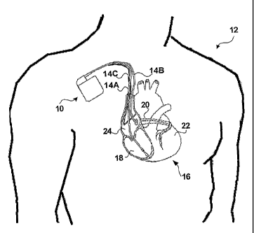
FIG. 1 is a conceptual diagram illustrating an exemplary implantable medical
device
implanted in a patient.
FIG. 2 is conceptual diagram further illustrating the implantable medical
device of FIG. 1
and the heart of the patient.
FIG. 3 is a functional block diagram of the implantable medical device of FIG.
1.
FIG. 4 is a timing diagram illustrating example electrogram (EGM) signals that
represent
electrical activity within the heart of the patient and illustrate techniques
for determining left
ventricular pace timing for cardiac resynchronization.
CA 02522583 2005-10-18
WO 2004/093986 PCT/US2004/011244
FIG. 5 is a flow diagram illustrating an example method that an implantable
medical
device may employ to deliver cardiac resynchronization therapy according to
the invention.
FIG. 6 is a flow diagram illustrating an example method that an implantable
medical
device may employ to determine left ventricular pace timing for cardiac
resynchronization.
FIG. 7 is flow diagram illustrating an example method that an implantable
medical device
may employ to pace the left ventricle based on the determined timing.
FIG. 8 is a flow diagram illustrating another example method that an
implantable medical
device may employ to determine left ventricular pace timing for cardiac
resynchronization.
FIG. 9 is flow diagram illustrating another example method that an implantable
medical
device may employ to pace the left ventricle based on the determined timing.
FIG. 1 is a conceptual diagram illustrating an exemplary implantable medical
device
(IMD) 10 implanted in a patient I2. IMD 10 may, as shown in FIG. l, take the
form of a multi-
chamber cardiac pacemaker. In the exemplary embodiment illustrated in FIG. 1,
IMD 10 is
coupled to leads 14A, 14B and 14C (collectively "leads 14") that extend into
the heart 16 of
patient 12
More particularly, right ventricular (RV) lead 14A may extend through one or
more veins
(not shown), the superior vena cava (not shown), and right atrium 24, and into
right ventricle 18.
Left ventricular (LV) coronary sinus lead 14B may extend through the veins,
the vena cava, right
atrium 24, and into the coronary sinus 20 to a point adjacent to the free wall
of left ventricle 22 of
heart I6. Right atrial (RA) lead 14C extends through the veins and vena cava,
and into the right
atrium 24 of heart 16.
Each of leads 14 includes electrodes (not shown), which IMD 10 may use to
sense
electrical signals attendant to the depolarization and repolarization of heart
16, and to provide
pacing pulses to heart 16. In some embodiments, IMD 10 may also provide
cardioversion or
defibrillation pulses via electrodes located on leads 14. The electrodes
located on leads 14 may be
unipolar or bipolar, as is well known in the art.
IMD 10 delivers cardiac resynchronization therapy to patient 12 via leads 14.
In
particular, as will be described in greater detail below, IMD 10 delivers
pacing pulses to left
ventricle 22 via lead 14B to synclmonize contractions of left ventricle 22
with contractions of right
ventricle I8 resulting from intrinsic depolarizations of right ventricle 18.
~ne exemplary situation
in which IMD 10 may be used is where patient 12 has left bundle branch block
(LBBB), but has
CA 02522583 2005-10-18
WO 2004/093986 PCT/US2004/011244
adequate physiological atrial-right ventricular conduction. By synchronizing
contraction of
ventricles I 8 and 22 through pacing of left ventricle 22 alone, IMD 10 may
provide a more
physiological interval between atrial and ventricular contractions in the
sense that the interval
between the atrial and ventricular contractions is a functi~n of an intrinsic,
rather than paced,
depolarization of the right ventricle. In addition, by pacing left ventricle
22 alone, IMD 10 may
consume less power than conventional devices that provide cardiac
resynchronization therapy by
delivering pacing pulses to both the right ventricle 18 and left ventricle 22.
IMD 10 determines the timing of delivery of pacing pulses to left ventricle 22
based on
one or more measured characteristics of an electrogram signal detected via one
or more of leads
14 that represents electrical activity within heart 16. The measured
characteristics indicate
synchrony of contractions of ventricles 18 and 22. For example, wider QRS
complex width
indicates Iess synchronous contraction of ventricles I 8 and 22. As another
example, short Q-T
intervals indicate increased sympathetic drive resulting from inadequate
cardiac output, which in
turn indicates dysynchrony of contraction of ventricles 18 and 22. Therefore,
IMD 10 may, for
example, select the left ventricular pace timing that results in the smallest
QRS complex width,
the largest Q-T interval, or the best combination of QRS complex width and Q-T
interval.
The configuration of IMD 10 and leads 14 illustrated in FIG 1 is merely
exemplary. IMD
10 may be coupled to any number of leads 14 that extend to a variety of
positions within or
outside of heart 16. Fox example, in some embodiments, IMD 10 may not be
coupled to a right
ventricular lead 14A. Further, lead 14C may extend to the left atrium of heart
16.
Some of leads 14 may be epicardial Ieads. Some electrodes used by IMD 10 to
sense electrical
activity of heart 16 need not be carried by leads 14 at all, but may instead
be integral with a
housing of IMD 10 (not shown). Further, IMD 10 need not be implanted within
patient 12, but
may instead be coupled with subcutaneous leads 14 that extend through the skin
of patient 12 to a
variety of positions within or outside of heart 16.
FIG. 2 is conceptual diagram further illustrating IMD 10 and heart 16 of
patient 12. Each
of leads 14 may include an elongated insulative lead body carrying a number of
concentric coiled
conductors separated from one another by tubular insulative sheaths. Located
adjacent distal end
of leads I4A, 14B and I4C are bipolar electrodes 30 and 32, 34 and 36, and 38
and 40
respectively. Electrodes 30, 34 and 38 may take the form of ring electrodes,
and electrodes 32, 36
and 4~0 may take the form of extendable helix tip electrodes m~unted
retractably Wlthlll 111SL1Iatlve
CA 02522583 2005-10-18
WO 2004/093986 PCT/US2004/011244
7
electrode heads 42, 44 and 46, respectively. Each of the electrodes 30-40 is
coupled to one of the
coiled conductors within the lead body of its associated lead 14.
Sense/pace electrodes 30, 32, 34, 36, 38 and 40 sense electrical signals
attendant to the
depolarization and repolarization of heart 16. The electrical signals are
conducted to IMD 10 via
leads 14. Senselpace electrodes 30, 32, 34, 36, 38 and 40 further may deliver
pacing to cause
depolarization of cardiac tissue in the vicinity thereof. IMD 10 may also
include one or more
indifferent housing electrodes, such as housing electrode 48, formed integral
with an outer surface
of the hermetically sealed housing 50 of IMD 10. Any of electrodes 30, 32, 34,
36, 38 and 40
may be used for unipolar sensing or pacing in combination with housing
electrode 48.
Leads 14A, 14B and 14C may also, as shown in FIG. 2, include elongated coil
electrodes
52, 54 and 56, respectively. IMD 10 may deliver defibrillation or
cardioversion shocks to heart
16 via defibrillation electrodes 52-56. Defibrillation electrodes 52-56 may be
fabricated from
platinum, platinum alloy or other materials known to be usable in implantable
defibrillation
electrodes, and may be about 5 cm in length.
FIG. 3 is a functional block diagram of IMD 10. As shown in FIG. 3, IMD 10 may
take
the form of a multi-chamber pacemalcer-cardioverter-defibrillator (PCD) having
a
microprocessor-based architecture. However, this diagram should be taken as
exemplary of the
type of device in which various embodiments of the present invention may be
embodied, and not
as limiting, as it is believed that the invention may be practiced in a wide
variety of device
implementations, including devices that provide caxdiac resynchronization
pacing therapies but do
not provide cardioverter and/or defibrillator functionality.
IMD 10 includes a microprocessor 60. Microprocessor 60 may execute program
instructions stored in a memory, e.g., a computer-readable medium, such as a
ROM (not shownn),
EEPROM (not shown), and/or RAM 62. Program instruction stored in a computer-
readable
medium and executed by microprocessor 60 control microprocessor 60 to perform
the functions
ascribed to microprocessor 60 herein. Microprocessor 60 may be coupled to,
e.g., to
communicate with and/or control, various other components of IMD 10 via an
address/data bus
64.
IMD 10 senses electrical activity within heart 16. Electrodes 30 and 32 are
coupled to amplifier
66, which may take the form of an automatic gain controlled amplifier
providing an adjustable
sensing threshold as a function of the measured R-wave amplitude. A signal is
generated on RV
out line 68 whenever the signal sensed between electrodes 30. and 32 exceeds
the present sensing
CA 02522583 2005-10-18
WO 2004/093986 PCT/US2004/011244
threshold. Thus electrodes 30 and 32 and amplifier 66 may be used to detect
intrinsic right
ventricular depolarizations.
Electrodes 34 and 36 are coupled to amplifier 70, which also may take the form
of an
automatic gain controlled amplifier providing an adjustable sensing threshold
as a function of
measured R-wave amplitude. A signal is generated on LV out line 72 whenever
the signal sensed
between electrodes 34 and 36 exceeds the present sensing threshold. Thus,
electrodes 34 and 36
and amplifier 70 may be used to detect intrinsic left ventricular
depolarizations.
Electrodes 38 and 40 are coupled to amplifier 74, which may take the form of
an automatic gain
controlled amplifier providing an adjustable sensing threshold as a function
of the measured P-
wave amplitude. A signal is generated on R.A out line 76 whenever the signal
between electrodes
38 and 40 exceeds the present sensing threshold. Thus, electrodes 38 and 40
and amplifier 74
may be used to detect intrinsic atrial depolarizations.
IMD 10 paces heart 16. Pacer timing/control circuitry 78 preferably includes
programmable digital counters which control the basic time intervals
associated with modes of
pacing. Circuitry 78 also preferably controls escape intervals associated with
pacing. For
example, IMD 10 may pace right atrium 24 via timing/control circuitry 78
triggering generation
of pacing pulses by pacer output circuit 84, which is coupled to electrodes 38
and 40. Pacer
timing/control circuitry 78 may triggex generation of pacing pulses for right
atrium 24 upon
expiration of an atrial escape interval.
As mentioned above, IMD 10 delivers pacing pulses to left ventricle 22 to
synchronize
contractions of left ventricle 22 with contractions of right ventricle 18
resulting from intrinsic
depolarizations of right ventricle 18. Pacer timing/control circuitry 78
triggers generation of
pacing pulses for left ventricle 22 by pacer output circuit 82, which is
coupled to electrodes 34
and 36. As will be described in greater detail below, circuitry 78 triggers
generation of pacing
pulses delivered to left ventricle 22 upon expiration of an interval that may
be timed from
detection of either an atrial or intrinsic right ventricular depolarization.
IMD 10 may also provide biventricular modes of cardiac resynchronization
therapy, or
non-resynchronization pacing modalities that require delivery of pacing pulses
to right ventricle
18, and may switch from a left ventricular cardiac resynchronization mode as
described herein to
one of these additional modes. Pacer timing/control circuitry 78 triggers
generation of pacing
pulses for right ventricle 18 by pacer output circuit 80, which is coupled to
electrodes 30 and 32.
CA 02522583 2005-10-18
WO 2004/093986 PCT/US2004/011244
9
Pacer timing/control circuitry 78 may trigger generation of pacing pulses for
right ventricle 18
upon expiration of an A-V or V-V escape interval, depending on the pacing
mode. .
Output circuits 80, 82 and 84 may be pulse generation circuits known in the
art, which
include capacitors and switches for the storage and delivery of energy as a
pulse. Pacer
S timing/control circuitry 78 resets escape interval counters upon detection
of R-waves or P-waves,
or generation ~f pacing pulses, and thereby controls the basic timing of
cardiac pacing functions.
Intervals defined by pacing circuitry 78 may also include refractory periods
during which sensed
R-waves and P-waves are ineffective to restart timing of escape intervals, and
the pulse widths of
the pacing pulses. The durations of these intervals are determined by
microprocessor 60 in
response to data stored in RAM 62, and are communicated to circuitry 78 via
address/data bus 64.
Pacer timing/control circuitry 78 also determines the amplitude of the cardiac
pacing pulses under
control of microprocessor 60.
Microprocessor 60 may operate as an interrupt driven device, and is responsive
to
interrupts from pacer timing/control circuitry 78 corresponding to the
occurrence of sensed P-
1S waves and R-waves and corresponding to the generation of cardiac pacing
pulses. Those
inten-upts axe provided via data/address 'bus 66. Any necessary mathematical
calculations to be
performed by microprocessor 60 and any updating of the values or intervals
controlled by pacer
timing/control circuitry 78 take place following such interrupts.
Microprocessor 60 determines the timing of delivery of pacing pulses to left
ventricle 22,
i.e., the internals used to pacer timing/control circuit 78 to trigger
generation of pacing pulses by
output circuit 82, based on one or more measured characteristics, e.g., QRS
complex width or Q-T
interval, of one or more electxogram signals that represent electrical
activity within heart 16. IMD
10 receives signals that represent electrical activity within heart 16, and
may digitally process the
signals to measure characteristics of the signals. Switch matrix 92 is used to
select which of the
2S available electrodes 30-40 and 48 axe coupled to wide band (O.S-200 Hz)
amplifier 94 for use in
digital signal analysis. As will be described in greater detail below, any of
a number of potential
combinations of these electrodes may be used, so long as the signal provided
by the combination
allows for identification and measurement of the desired characteristic.
Selection of electrodes is
controlled by microprocessor 60 via data/address bus 66, and,the selections
may be varied as
desired.
The analog signals derived from the selected electrodes and amplified by
amplifier 94 are
provided to multiplexer 96, and thereafter converted to a mufti-bit digital
signal by A/D converter
CA 02522583 2005-10-18
WO 2004/093986 PCT/US2004/011244
98. A digital signal processor (DSP) 100 may process the multi-bit digital
signals to measure
QRS complex widths andlor Q-T internals, as will be described in greater
detail below. In some
embodiments, the digital signal may be stored in RAM 62 under control of
direct memory access
circuit 102 for later analysis by DSP 100.
Although IMD 10 is described herein as having separate processors,
microprocessor 60
may perform both the functions ascribed t~ it herein and digital signal
analysis functions ascribed
to DSP 100 herein. Moreover, although described herein in the context of
microprocessor based
PCD embodiment IMD 10, the invention may be embodied in various implantable
medical
devices that include one or more processors, which may be microprocessors,
DSPs, FPGAs, or
10 other digital logic circuits. Further, in some embodiments, IMD 10 may not
include or utilize
DSP 100 to measure QRS complex widths and Q-T intervals. For example, IMD 10
may include
analog slope or threshold detecting amplifier circuits to identify the
beginning and end points of
QRS complexes or Q-waves and T-waves, as is known in the art. In such
embodiments of IMD
10, pacer timing/control circuit 78 may receive the output of these amplifier
circuits, and provide
an indication of the occurrence of these events to microprocessor 60 so that
microprocessor may
measure QRS complex widths and/or Q-T intervals.
IMD 10 may detect ventricular and/or atrial tachycardias or fibrillations of
heart 16 using
tachycardia and fibrillation detection techniques and algorithms known in the
art. For example,
the presence of a ventricular or atrial tachycardia or fibrillation may be
confirmed by detecting a
sustained series of short R-R or P-P intervals of an average rate indicative
of tachycardia, or an
unbroken series of short R-R or P-P intervals. IMD 10 is also capable of
delivering one or more
anti-tachycardia pacing (ATP) therapies to heart 16, and cardioversion and/or
defibrillation pulses
to heaxt 16 via one or more of electrodes 48, 52, 54 and 56.
Electrodes 48, 52, 54 and 56, are coupled to a cardioversion/defibrillation
circuit 90,
which delivers cardioversion and defibrillation pulses under the control of
microprocessor 60.
Circuit 90 may include energy storage circuits such as capacitors, switches
for coupling the
storage circuits to electrodes 48, 52, 54 and 56, and logic for controlling
the coupling of the
storage circuits to the electrodes to create pulses with desired polarities
and shapes.
Microprocessor 60 may employ an escape interval counter to control timing of
such cardioversion
and defibrillation pulses, as well as associated refractory periods.
FIG. 4 is a timing diagram illustrating example electrogram (EGM) signals that
represent
electrical activity within heart 16. Signal 110 is a right atrial EGM. IMD 10
may digitally
CA 02522583 2005-10-18
WO 2004/093986 PCT/US2004/011244
11
process atrial EGM 110 to measure a width 116 of QRS complex 118. Signal 110
may be
detected using electrodes 38 and 40 of RA lead 14C in a bipolar configuration,
or one of
electrodes 38 and 40 and housing electrode 48 in a unipolar configuration.
In general, it is preferred that IMD 10 digitally process signals that include
far-field QRS
complexes 118, such as right atrial EGM 110, to measure widths 116. Processing
these signals is
preferred because such signals include QRS complexes that are more 'bglobal"
in that they reflect
depolarization of both ventricles 18, 22, and thus the widths 116 of far-field
QRS complexes 118
more accurately reflect ventricular synchrony. In addition to atrial EGM
signal I 10, IMD 10 may
detect signals that include fax-field QRS complexes using two or more housing
electrodes 4~8.
Detecting cardiac signals via housing electrodes 48 may enable embodiments of
IMD 10 that do
not include an atrial lead.
In order to measure QRS complex width 116, DSP 100 first identifies far-field
QRS
complex 118 within signal 110. DSP 100 may identify QRS complex 118 within
signal 110 by
any methods known in the art. For example, DSP I00 may receive indications of
the occurrence
of an R-wave 120 or 122 from pacer timing/control circuit 78, and identify QRS
complex 118
based on these indications. As another example, DSP 100 may identify QRS
complex 118 by
detecting a number of threshold-crossings of the digital signal provided by
A/D converter 98, or
zero-crossings of tile first derivative of the digital signal occurring within
a time window. As yet '
another example, DSP 100 may detect QRS complexes within signals 110-114 using
techniques
described in commonly assigned U.S. Patent No. 6,029,087, to Wohlgemuth, and
titled "Cardiac
Pacing System With Improved Physiological Event Classification Based on DSP"
("Wohlgemuth
'087 Patent").
DSP 100 may measure width 116 as a period of time from a begimiing point 124
to an ending
point 126. DSP 100 may identify begimzing point 124 and ending point 126 as
threshold-
crossings of the digital signal or zero-crossings of the first derivative of
the digital signal.
Signals 112 and 114 are right and left ventricular EGMs, respectively, and may
be
detected via RV lead 14C and LV coronary sinus lead 14B, respectively. Signals
112 and 114
may be detected using bipolar electrode pairs 30, 32 and 34, 36, or one
electrode from each pair
and housing electrode 48 in a unipolar configuration.
IMD 10 may digitally process signal 114 to measure a Q-T interval 128. For
example,
DSP I00 may receive an indication of delivery of a pacing pulse 130 from pacer
tilninglcontrol
circuitry 78, and measure Q-T interval 128 as the period of time from pacing
pulse 130 to
CA 02522583 2005-10-18
WO 2004/093986 PCT/US2004/011244
12
detection of T-wave 132 within the digital signal provided by AlD converter
98. T-wave 132
may, for example, be detected using techniques described in the above-
referenced Wohlgemuth
'087 Patent.
For ease of illustration, only a portion of each of EGM signals 110-114
representing a
single cardiac cycle of heart 16 is shown in FIG. 4. However, it is understood
that DSP 100
measures multiple QRS complex widths and/or Q-T intervals over multiple
cardiac cycles. As
will be described in greater detail below, DSP 100 measures these values in
response to delivery
of pacing pulses 130 to left ventricle 22. 'The values for QRS complex widths
116 and/or Q-T
intervals 128 measured by DSP 100 may be stored in RAM 62 for later analysis
by
microprocessor 60. Microprocessor 60 analyzes the measured values to, for
example, identify the
smallest QRS complex width 116 or the largest Q-T interval 128.
In various embodiments of IMD 10, microprocessor 60 may measure intervals 134
between intrinsic and/or paced atrial depolarizations, e.g., P-waves 136, and
intrinsic right
ventricular depolarizations, e.g., R-waves 120. In other embodiments of IMD
10, microprocessor
60 may measure intervals 138 between P-waves I36 and intrinsic left
ventricular depolarizations,
e.g., R-waves 122. In either case, microprocessor 60 controls pacer
timing/control circuitry 78 to
test delivery of pacing pulses 130 at a variety of pacing intervals 140 timed
from P-wave 136.
Microprocessor 60 may control circuit to test pacing intervals 140 within a
range around either
interval I34 or interval 138, depending on the embodiment of IMD 10.
DSP 100 measures one or both of a QRS complex width 116 and Q-T interval 128
for
each pacing interval 140 tested. Microprocessor 60 selects the tested pacing
interval 140 that
microprocessor 60 determines provides the best synchronization between
contractions of right and
left ventricles 18 and 22, e.g., the pacing interval 140 that resulted in the
shortest QRS complex
width 116, the longest Q-T interval 128, or the average of the pacing
intervals 140 that resulted in
the shortest QRS complex width 116 and the longest Q-T interval 128,
respectively.
Microprocessor 60 then controls delivery of pacing pulses to left ventricle 22
based on the
selected pacing interval 140, as will be described in greater detail below.
FIG. 5 is a flow diagram illustrating an example method that IMD 10 may employ
to
deliver cardiac resynchronization therapy according to the invention. In
general, IMD 10, and
more particularly microprocessor 60 of IMD 10, determines a timing of left
ventricular pacing
that synchronizes the paced contractions of left ventricle 22 with
contractions of right ventricle 18
resulting from intrinsic depolarizations of right ventricle 18 (150).
Processor 60 determines the
CA 02522583 2005-10-18
WO 2004/093986 PCT/US2004/011244
13
timing of left ventricular pacing based on measured characteristics of
electrogram signals, e.g.,
QRS complex widths 16 andlor Q-T intervals 28. Processor 60 controls pacing of
left ventricle 22
based on the determined timing (52). Processor 60 periodically retests the
timing of left
ventricular pacing, e.g., hourly, daily, or mont111y, to account for longer-
term changes in the
condition of patient 12.
FIGS. 6-9 further illustrate the method of FIG. 5 accoxding to various
embodiments of the
invention. In particular, FIGS. 6 and 8 illustrate methods that may be
employed by IMD 10 to
determine the timing of left ventricular pacing for synchronization with
intrinsic right ventricular
depolarizations. FIG. 6 illustrates a method that may be employed by IMD 10 to
determine the
timing based on a measured interval between an atrial depolarization and an
intrinsic right
ventricular depolarization, e.g., interval 134 (FIG. 4). FTG. 8 illustrates a
method that may be
employed by IMD 10 to determine the timing based on a measuxed interval
between an atrial
depolarization and an intrinsic left ventricular depolarization, e.g.,
interval 138 (FIG. 4).
The method illustrated in FIG. 6 may be applied in situations where IMD 10 is
coupled to
a right ventricular lead 14A that includes electrodes for sensing electrical
activity in right
ventricle 18, such as bipolar electrodes 30 and 32. The method illustrated in
FIG. 8 may be
applied whether or not IMD 10 is coupled to right ventricular lead 14A,
requiring only that IMD
10 be coupled to left ventricular lead 14B for pacing and sensing left
ventricle 22, e.g., via
electrodes 34 and 36. FIGS. 7 and 9 illustrate methods for pacing left
ventricle 22 based on the
timing as determined according to the methods illustrated in FIGS. 6 and 8,
respectively.
As shown in FIG. 6, IMD 10 measures an interval 134 between an intrinsic or
pace atrial
depolarization, e.g., P-wave 136, and an intrinsic right ventricular
contraction, e.g. R-wave 120,
as described above (160). In some embodiments, IMD IO may measure a plurality
of such A-RV
intervals 134 and determine an average of the measured A-RV intervals. IMD I O
then delivers
pacing pulses to left ventricle 22 at a variety of pacing intervals 140
measured from an intrinsic or
paced P-wave 136 (162). IMD 10 may test pacing intervals 140 within a range
around the
determined A-RV interval 134.
IMD 10 then identifies the pacing interval 140 that provides synchronization
of left
ventricular pacing with intrinsic right ventricular contractions, as described
above (164). As
described above, IMD 10 may measure QRS complex widths I 16 and/or Q-T
intervals 128
corresponding to each pacing 121terval 140. IMD 10 selects one of the tested
pacing internals 140
based on the measured values. For example, IMD 10 may select the tested pacing
interval 140
CA 02522583 2005-10-18
WO 2004/093986 PCT/US2004/011244
14
which results in the smallest QRS complex Width 116 or longest Q-T interval
128. Where IMD
measures both, IMD 10 may select a pacing interval 140 by avexaging the pacing
intervals that
resulted in the smallest QRS complex width 116 or longest Q-T interval 128,
respectively.
IMD 10 calculates and stores the difference between the selected pacing
interval 140 and
the measured A-RV interval 134 (166) for use in pacing left ventricle 22, as
will be described in
greater detail with reference to FIG. 7. In some embodiments, A-RV intervals
134 may be
measured and pacing intervals 140 may be tested individually for paced and
intrinsic P-waves
136. In such embodiments, IMD 10 may calculate and store differences
determined using each of
paced and intrinsic P-waves I34, and apply the respective differences to pace
left ventricle 22
10 depending on whether a paced or intrinsic P-wave 134 has been detected.
FIG. 7 illustrates a method that may be employed by IMD 10 to pace left
ventricle 22
based on a calculated difference between the selected pacing interval 140 and
the measured A-RV
interval 134. If the difference is equal to zero (170), IMD 10 delivers pacing
pulses to left
ventricle 22 upon detection of intrinsic ventricular depolarizations, e.g., R-
waves 120 (172, 174).
If the difference is greater than zero (170), i.e., if the selected pacing
interval 140 is greater than
the measured A-RV interval 134, TMD 10 delivers pacing pulses to.Ieft
ventricle 22 upon
expiration of a counter initiated upon detection of intrinsic ventricular
depolarizations, e.g., R-
waves 120 (178, 180). The counter is set to measure an amount of time equal to
the determined
difference between the selected pacing interval 140 and the measured A-RV
interval 134.
Calculating the difference between the selected pacing intexval 140 and the
measured A-RV
interval I34, and pacing left ventricle 22 based on the difference, as opposed
to the selected
pacing interval 140, allows IMD 10 to maintain ventricular synchrony despite
beat-to-beat
variation in the A-RV interval.
If the difference is less than zero (170), i.e., if pacing pulses must be
delivered to left
ventricle 22 prior to intrinsic ventricular depolarizations to provide
ventricular synchrony, IMD
10 periodically determines a current A-RV interval 134 (184, 192), and
determines a current
pacing interval 140 as the sum of the current A-RV interval and the difference
(186, 192). IMD
10 may determine current A-RV intervals and current pacing intervals every 10,
20, 32 or 100
cardiac cycles, for example.
IMD 10 delivers pacing pulses to left ventricle 22 the current pacing interval
I40 after
detection of a paced or intrinsic P-wave 136 (188, 190). Periodically
determining current A-RV
intervals and current pacing intervals allows IMD 10 to maintain ventricular
synchrony despite
CA 02522583 2005-10-18
WO 2004/093986 PCT/US2004/011244
beat-to-beat variation in the A-RV interval and despite the necessity of
delivering pacing pulses to
left ventricle 22 prior to intrinsic right ventricular depolarizations. As
mentioned above, IMD 10
may periodically, e.g. hourly, weekly, or monthly, perform the method
illustrated in FIG. 6 to
recalculate the difference. Periodically recalculating the difference may
allow I1VID 10 to address
5 longer-term changes in the condition of patient 12.
FIG. 8 illustrates a method that may be employed by IMD 10 t~ determine the
timing
based on a measured interval 138 between a paced or intrinsic atrial
depolarization, e.g. a P-wave
136, and an intrinsic left ventricular depolarization, e.g., an intrinsic R-
wave 122 (FIG. 4~) (200).
A single such A-LVSENSE interval 138 may be measured, or an average of several
SLlch A-
10 LVSENSE intervals 138 may be determined.
IMD 10 then delivers pacing pulses to left ventricle 22 at a variety of pacing
intervals 140
measured from an intrinsic or paced P-wave 136 (202). IMD 10 may test pacing
intervals 140
within a range around the determined A-LVSENSE interval 138. IMD 10 identifies
the pacing
interval 140 that provides synchronization of left ventricular pacing with
intrinsic right ventricular
15 contractions, e.g., based on measured QRS complex widths 116 and/or Q-T
intervals 128, as
described above (204). IMD 10 calculates and stores a difference between the
selected pacing
interval 140 and the determined A-LVSENSE interval 138 (206). Separate
intervals and
differences may be determined for intrinsic and paced atrial depolarizations,
as described above.
FIG. 9 illustrates a method that may be employed by IMD 10 to pace left
ventricle 22
based on a calculated difference between a selected pacing interval 140 and
the determined A-
LVSENSE interval 138. IMD 10 periodically determines a current A-LVSENSE
interval 138
(210, 218), and determines a current pacing interval 140 as the sum of the
current A-LVSENSE
interval and the difference (2I2, 218). IMD 10 may determine current A-RV
intervals and current
pacing intervals every 10, 20, 32 or 100 cardiac cycles, for example. IMD 10
delivers pacing
pulses to left ventricle 22 upon expiration of a counter initiated upon
detection of a paced or
intrinsic P-wave 136 (214, 216). The counter is set to measure an amount of
time edual to the
determined current pacing interval 140.
A number of embodiments of the invention have been described. However, one
skilled in
the art will appreciate that the invention can be practiced with embodiments
other than those
disclosed. The disclosed embodiments are presented for purposes of
illustration and not
limitation, and the invention is limited only by the claims that follow.