Note: Descriptions are shown in the official language in which they were submitted.
WO 2004/094588 CA 02522669 2005-10-14PCT/US2003/012276
Postnatal stem cells and uses thereof
Statement of Government Rights
This invention was developed with the support of the Department of Health
and Human Services. The United States Government has certain rights in the
invention.
Field of the Invention
The invention generally relates to postnatal dental stem cells and methods for
their use. More specifically, the invention relates in one aspect to postnatal
dental
pulp stem cells, use of the cells to generate dentin, and differentiation of
the cells. In
another aspect, the invention relates to human postnatal deciduous dental pulp
multipotent stem cells, use of the cells to generate dentin, and
differentiation of the
cells.
Background of the Invention
Post-natal stem cells (meaning those present after birth) are unspecialized
cells
that can renew themselves extensively and develop into more mature cells
having
specialized functions. Stem cells may be induced under certain physiologic or
experimental conditions to become cells with special functions, such as the
beating
cells of the heart muscle, or the insulin-producing cells of the pancreas. The
process
by which a stem cell becomes a cell with special functions is known as
differentiation.
Differentiation can be induced through use of multiple signals that can
include
chemicals secreted by other cells, physical contact with neighboring cells,
and certain
molecules in the microenvironment. Thus, stem cells can be treated with
specific
signals to become specific types of cells having useful functions. These newly
differentiated cells can then be used to generate replacements for cells that
are lost
through normal wear and tear, injury, or disease. For example, stem cells show
promise for treating diseases such as Parkinson's disease, diabetes, and heart
disease.
Stem cells have multiple applications in medicine and dentistry. Accordingly,
new
sources of stern cells, and methods for their use are needed.
1
CA 02522669 2005-10-14
WO 2004/094588 PCT/US2003/012276
Summary of the Invention
Methods and materials are provided by the current invention that address the
aforementioned needs. The invention provides an isolated human postnatal
deciduous
dental pulp multipotent stem cell, a method to implant a bone-inducing cell
within an
organism, a method to implant a neural,cell within an organism, a method to
implant
an adipocyte within an organism, and a method to generate dentin.
The invention provides an isolated human postnatal deciduous dental pulp
multipotent stern cell. A human postnatal deciduous dental pulp multipotent
stem cell
can differentiate into a neural cell, an adipocyte, or an odontoblast. A human
postnatal deciduous dental pulp multipotent stem cell can be obtained from a
non-
exfoliated deciduous tooth. Preferably, a human postnatal deciduous dental
pulp
multipotent stem cell is obtained from an exfoliated deciduous tooth (SHED). A
human postnatal deciduous dental pulp multipotent stem cell can be stored for
later
use. A human postnatal deciduous dental pulp multipotent stem cell can be
grown in
tissue culture medium. Preferably, the tissue culture medium includes serum.
More
preferably, the tissue culture medium does not include serum. The tissue
culture
medium can include one or more growth factor. Preferably, the growth factor is
basic
fibroblast growth factor, epidermal growth factor, or both. The tissue culture
medium
can include a neuronal supplement. Preferably, the neuronal supplement is B27
supplement.
The invention provides a method to generate bone within an organism.
Generally, the method involves implanting a human postnatal deciduous dental
pulp
multipotent stem cell into an organism. Preferably the organism is a mammal.
More
preferably the organism is a human. The human postnatal deciduous dental pulp
multipotent stem cell may be obtained from one human and implanted into a
different
human. Preferably, the human postnatal deciduous dental pulp multipotent stem
cell
is obtained from, and implanted into the same human. The human postnatal
deciduous dental pulp multipotent stem cell may be expanded ex vivo prior to
being
implanted into the organism. Preferably the human postnatal deciduous dental
pulp
multipotent stem cell is induced prior to being implanted into the organism.
Preferably, the human postnatal deciduous dental pulp multipotent stem cell is
2
CA 02522669 2005-10-14
WO 2004/094588 PCT/US2003/012276
induced with BMP-4 or mineralizing induction. A human postnatal deciduous
dental
pulp multipotent stem cell that is not in combination with a carrier can be
implanted
into an organism. A human postnatal deciduous dental pulp multipotent stem
cell that
is in combination with a carrier can be implanted into an organism.
Preferably, the
carrier contains hydroxyapatite. More preferably, the carrier contains
tricalcium
phosphate. Most preferably, the carrier contains hydroxyapatite and tricalcium
phosphate. The human postnatal deciduous dental pulp multipotent stern cell
can
induce a recipient cell to differentiate into bone-forming cells. The method
of the
invention can be used to promote bone formation at a site of trauma within an
organism. The trauma may be produced by a physical injury. Preferably the
physical
injury is an accidental physical injury. More preferably, the physical injury
results
from a medical or dental procedure. Most preferably, the physical injury
results from
surgery. The trauma may be due to degenerative disease. Preferably the
degenerative
disease is osteoporosis.
The invention provides a method to produce neural tissue within an organism.
Generally, the method involves implanting a dental stem cell into an organism.
Preferably, the dental stem cell is a dental pulp stein cell. More preferably,
the dental
stem cell is a human postnatal deciduous dental pulp multipotent stem cell.
Preferably the organism is a mammal. More preferably the organism is a human.
The
dental stem cell can be implanted into tissue present within the organism.
Preferably
the tissue is neural tissue. The dental stem cell may be expanded ex vivo
prior to
being implanted into the organism. Preferably the dental stem cell is neuronal
induced prior to being implanted into the organism. A dental stem cell that is
not in
combination with a carrier can be implanted into an organism. A dental stem
cell that
is in combination with a carrier can be implanted into an organism.
The invention provides a method to produce adipose tissue within an
organism. Generally, the method involves implanting a dental stem cell into an
organism. Preferably, the dental stem cell is a dental pulp stem cell. More
preferably,
the dental stem cell is a human postnatal deciduous dental pulp multipotent
stem cell.
Preferably the organism is a mammal. More preferably the organism is a human.
The
dental stem cell may be expanded ex vivo prior to being implanted into the
organism.
Preferably the dental stem cell is adipogenesis induced prior to being
implanted into
3
CA 02522669 2005-10-14
WO 2004/094588 PCT/US2003/012276
the organism. A dental stem cell that is not in combination with a carrier can
be
implanted into an organism. A dental stem cell that is in combination with a
carrier
can be implanted into an organism.
The invention provides a method to generate dentin by implanting a dental
stem cell within an organism. The method can be used to generate dentin on pre-
existing dentin by contacting the pre-existing dentin with a dental stem cell
and
incubating the pre-existing dentin and the dental stem cell. Preferably, the
dental
stem cell is a dental pulp stem cell. More preferably, the dental stem cell is
a human
postnatal permanent tooth dental pulp stem cell. More preferably, the dental
stem cell
is a human postnatal deciduous dental pulp multipotent stem cell. Preferably,
the pre-
existing dentin is contacted with the dental stem cell in vitro. More
preferably, the
pre-existing dentin is contacted with the dental stem cells in vivo. The pre-
existing
dentin can be contained within a tooth. The dental stem cells can be obtained
from
the tooth of a mammal. Preferably, the dental stem cell is obtained from the
tooth of a
human. More preferably, the dental stem cell is obtained from a human
permanent
tooth. Most preferably, the dental stem cell is obtained from a human
deciduous
tooth. The pre-existing dentin can be from a mammal. Preferably, the pre-
existing
dentin is from a human. The pre-existing dentin and the dental stem cell can
be
obtained from different mammals. More preferably, the pre-existing dentin and
the
dental stem cell is obtained from the same mammal. Most preferably, the pre-
existing
dentin and the dental stem cell is obtained from the same human. The pre-
existing
dentin can be contacted with a formulation to produce treated dentin.
Preferably, the
pre-existing dentin is contacted with a formulation after the pre-existing
dentin is
contacted with a dental stem cell. More preferably, the pre-existing dentin is
contacted with a formulation before the pre-existing dentin is contacted with
a dental
stem cell. Preferably, the formulation is a base solution. More preferably,
the
formulation is an acid solution. Most preferably, the formulation is an acetic
acid
solution. The treated dentin can be washed with a fluid. Preferably, the fluid
is a
biological solvent. More preferably, the fluid is water. Even more preferably,
the
fluid is a biological buffer. Most preferably, the fluid is phosphate buffered
saline.
The pre-existing dentin can be contacted with a dental stem cell that is not
in
combination with a carrier. The pre-existing dentin can be contacted with a
dental
4
CA 02522669 2011-02-15
stem cell that is in combination with a carrier. Preferably, the carrier
contains
hydroxyapatite. More preferably, the carrier contains tricalcium phosphate.
Most
preferably, the carrier contains hydroxyapatite and tricalcium phosphate. The
method
of the invention can be used to generate dentin in response to trauma to a
tooth.
Preferably the trauma is erosion of the tooth. More preferably, the trauma
results
from dental treatment. Most preferably, the trauma results from a root canal
procedure.
Brief Description of the Drawings
Figure 1 is a diagram showing the preparation of a dentin scaffold, culturing
DPSCs,
and implanting a dentin/DPSC-complex into an immunocompromised mouse. (A)
Root tips from extracted third molars were cut off on the indicated cutting
line and the
root foramen was sealed with GelfoamTM. (13) Pulp tissue and a layer of pulpal
dentin
were removed from the root tip and then treated with 1% acetic acid for 10
minutes.
(C) After PBS washing, DPSCs were loaded onto the dentin surface of the pulpal
chambers and cultured at 37 C for 12 hours in 10 cm cell culture dishes with
15 ml of
culture medium covering the whole root tips. (D) DPSC/dentin complex was
implanted into immunocompromised mice subcutaneously.
=
Figure 2 shows the dentinogenesis of human DPSCs in vivo. (A) Implanted acid-
treated dentin vehicle without any cells, there was only connective tissue
around
dentin surface. (13) Skin fibroblasts were loaded on the acid-treated dentin
and
Cultured for 12 hours, then fibroblast/dentin complexes were implanted into
immunocompromised mice. There was connective tissue around the dentin surface
without any new dentin formation eight weeks after implantation. (C-E) Newly
formed dentin (ND) was associated with human dentin scaffold (Dentin) and pulp-
like
tissue containing odontoblasts (open triangles) blood vessel (BV) eight weeks
after
implantation of the DPSC/dentin complexes. The newly formed dentin may contain
trapped cellular components (white arrows in C and D) or only odontoblasts
responsible for a thin layer of dentin formation (ND in E). The pulp-like
tissue was
defined as a cell rich connective tissue containing blood vessels (By), red
blood cells,
and odontoblasts (open arrows) lining on the surface of newly formed dentin,
which
5
CA 02522669 2005-10-14
WO 2004/094588 PCT/US2003/012276
was different from regular connective tissue containing a limited number of
cells (C
and D). (F) DPSCs were implanted with HAJTCP as a carrier to show odontoblasts
(open arrows) were responsible for the newly formed dentin (ND) with tubule
structure (black arrows) on the surfaces of HA/TCP (HA). Sections were stained
with
hematoxylin and eosin. Original magnification is 40X.
Figure 3 shows the characterization of a DPSC/dentin implant. (A) DSP
immunohistochemical staining on fibroblast/dentin implant showed a positive
staining
on the peritubular dentin (black arrows). Connective tissue showed a negative
immunostaining for DSP antibody. (B) In DPSC/dentin implants, odontoblasts
(open
arrows) and cells trapped inside newly formed dentin (open triangles in ND)
were
immunoreactive for human DSP antibody. Human dentin scaffold (Dentin) showed a
DSP immunostaining on peritubular structures (black arrows). Pulp-like tissue
was
immunonegative for DSP antibody staining. (C) Immunohistochemical staining of
human-specific anti-mitochondria antibody showed human DPSCs differentiated
into
odontoblasts (open arrows) lining on the surfaces of newly generated dentin
(ND) and
also became dentinogenic cells (open arrows) trapped inside newly formed
dentin
(ND). (D) Negative control of immunohistochemical staining on DPSC/dentin
implant without primary antibody. Original magnification is 60X.
Figure 4 shows the expression of FGF and VEGF receptors in human DPSCs. (A and
B) FGF receptor 1 (A) and VEGF receptor 1 (B) expressed in cultured human
DPSCs
at 25 population doublings. Light grey (red in color photo) represents
positive
staining and medium grey (blue in color photo) shows nuclei staining of DAPI.
(C)
Western blot analysis confirmed the expression of these molecules in cultured
DPSCs.
Asterisks represent immunopositive bands. HSP90 was used to show protein
loading
per sample. (D-E) DPSC/dentin implants at eight weeks post-implantation,
dentinogenic cells (black arrows) trapped inside newly formed dentin (ND) were
immunopostive for FGF receptor 1 antibody (D) and VEGF receptor 1 antibody
(E).
However, odontoblasts were only immunoreactive for FGF receptor 1 antibody
(open
arrows in D). Original magnification is 40X for A-B and D-E.
6
CA 02522669 2005-10-14
WO 2004/094588 PCT/US2003/012276
Figure 5 illustrates isolation of SHED. (A) The exfoliated primary incisor
contained
dental pulp as shown (triangles). The dashed line shows the occlusion edge of
the
incisor. (B and C) Hematoxylin and eosin staining indicated dentin (D) and
pulp
(pulp) of exfoliated deciduous teeth. The pulp contained odontoblasts
(arrows), blood
vessels (open arrows), and connective tissues. The straight and curved dash
lines in
(B) represent the occlusion and resorbed root surfaces, respectively. (D)
Single
colonies were formed after SHED were plated at low density and cultured for
two
weeks. (E) SHED were capable of forming sphere-like clusters when cultured
with
the conditions described in the Methods. (F) The sphere-like clusters could be
dissociated by passage through needles and subsequently grew on 0.1% gelatin
coated
dishes. (G) The proliferation rates of SHED, BMSSCs, and DPSCs were assessed
by
bromodeoxyuridine (BrdU) incorporation for 12 hours. SHED showed a
significantly
higher proliferation rate in comparison with BMSSCs and DPSCs (*P<0.05;
Student t
test). (H) SHED were able to proliferate to over 140 population doublings,
which was
significantly higher (*P<0.05; Student t test) than BMSSCs and DPSCs.
Figure 6 shows that SHED possess stern cell characteristics. (A-E) The remnant
pulp
showed STRO-1 (open arrows in A) and CD146 (open arrows in B) immunopositive
staining for cells in perivascular areas. FACS analysis showed that ex vivo
expanded
SHED contained approximately 9% STRO-1 positive cells (C). SHED expressed
STRO-1 (D) and CD146 (E) (arrows). (F-I) SHED expressed osteogenic and
angiogenic markers ALP, MEPE, bFGF, and endostatin. (J and K) SHED were either
cultured with regular medium (J) or cultured with L-ascorbate-2-phosphate,
dexamethasone, and inorganic phosphate for 4 weeks (K). Alizarin red staining
showed mineralized nodule formation in the induction (K). (L) Western blot
analysis
showed an up-regulated expression of CBFA1, ALP, MEPE, BSP, and DSPP
following the induction as described herein. HSP90 was used to assess the
amount of
protein loaded per sample. (M) Human recombinant BMP-4 (300ng/ml, 24 hours)
was added to induce a significant up-regulation of CBFA1, Osterix, and
osteocalcin
(OC) in SHED as detected by semi-quantitative PCR.
7
CA 02522669 2005-10-14
WO 2004/094588 PCT/US2003/012276
Figure 7 shows implanted SHED in immunocompromised mice. (A and B) Eight
weeks after implantation, SHED were able to differentiate into odontoblasts
(open
arrows) that were responsible for the dentin-like structure (D) formation on
the
surfaces of hydroxyapatite tricalcium (HA) (A). The same field is shown for
human-
specific alu in situ hybridization indicating the human origin of odontoblasts
(open
arrows, B). The black dashed line represents interface between newly formed
dentin
(D) and HA/TCP (HA). (C) Immunohistochemical staining of anti-DSPP antibody
shows a positive staining on the regenerated dentin (black arrows). (D) In
contrast to
DPSC implants, newly generated bone (B) by host cells in the same SHED implant
shows no reactivity to the DSPP antibody. (E) Of 12 selected single-colony
derived
SHED strains, only three (25%) were capable of generating dentin in vivo.
Newly
formed dentin (arrows) was found to be adjacent to the surfaces of HA/TCP
carrier
(HA) and associated with connective tissue (CT). (F) Human-specific alu in
situ
hybridization showed that human cells (open arrows) were associated with
dentin
formation (D) and were residing within the connective tissue compartment (CT).
(G)
The remaining 75% (9 of 12) single-colony derived SHED strains were unable to
generate dentin in vivo. (H) In situ hybridization demonstrated that alu-
positive
human cells survived in the connective tissue compartment (CT) in the implants
in
which there was no odontogenesis. Human cells were also found to surround the
blood vessels (arrows). (I) 7 of 12 (58.4%) single-colony derived SHED lines
induced a very limited amount of bone formation (B) on the surface of HA/TCP
(HA). (J) 5 of 12 (41.6%) single-colony derived SHED lines were able to induce
significant amount of bone formation (B) on the surfaces of HA/TCP (HA). (K)
The
alu in situ hybridization showed human cells (arrows) aftached to the surfaces
of
HA/TCP (HA) at the initial site of bone formation (B). The black dash lines
represent
the interface between newly formed bone (B) and HA/TCP (HA). (L) In situ
hybridization studies showed the murine-specific pfl DNA probe reacting with
osteoblasts and osteocytes (arrows) associated with the new bone formation
(B).
Figure 8 illustrates neural differentiation of SHED. (A-H) Immunocytochemical
staining depicts SHED expressing Nestin, GFAP, Neurofilament M, CNPase, Beta
III
tubulin, GAD, NeuN. (I) Western blot analysis confirmed that SHED expressed
8
CA 02522669 2005-10-14
WO 2004/094588 PCT/US2003/012276
neural markers as described above. After four weeks of culture in the presence
of
B27 supplement, bFGF (40 ng/ml), and EGF (20 ng/ml) (Neural cliff. +),
expression
levels of beta III tubulin, GAD, and NeuN were up-regulated when compared with
regular culture conditions as described in the Methods (Neural diff-).
However,
expression levels of Nestin, GFAP, CNPase, and Neurofilament remained the same
following the treatment. (J-0) SHED may co-express neuronal markers including
beta III tubulin (J and L, green in color photo)/GAD (K and L, red in color
photo) and
beta III tubulin (M and 0; green in color photo)/ NFM (N and 0, red in color
photo).
The morphology of SHED showed elongated cell-cytoplasmic processes that
sometimes co-express neural markers (triangles) or only express individual
neural
marker (open arrows). (P-S) Toluidine blue (0.1%) staining depicting the
altered
morphology of SHED after induction with neural culture medium (P and 0,
arrows).
Immunopositive staining for anti MAP2 and Tau antibodies on dendrites and axon
(R
and S, arrows), respectively. Double staining experiments showing beta III
tubulin
positive cells were also detected in the same field (R, triangle, green in
color photo).
(T-W) SHED continued to express glial cell markers including Nestin (T, red in
color
photo), CNPase (U, red in color photo), GFAP (V, red in color photo), and
neurofilament (W, green in color photo) by immunocytostaining.
Figure 9 shows implantation of SHED into the brain. (A) Diagram indicating
injection of SHED into the dentate gyrus of the hippocampus. (B) SHED were
cultured in the neural differentiation medium as described in the Methods for
one
week, after which 5,000 cells in 0.5 ill PBS were injected into the dentate
gyrus of the
hippocampus of immunocompromised mice. After 10 days, the brain was fixed and
prepared for immunofluorescence staining with NFM and human-specific anti-
mitochondrial antibody. The anti-mitochdondrial antibody immunostaining showed
human SHED (arrows, middle panel, green in color photo) in the dentate gyms of
the
hippocampus with co-expression of neurofilament (arrows, left panel, red in
color
photo). In merged images, co-expression of human mitochondria and NFM showed
co-localization of antigen expression as indicated by arrows (yellow in color
photo).
Magnification 20X.
9
CA 02522669 2005-10-14
WO 2004/094588 PCT/US2003/012276
Figure 10 illustrates the adipogenic differentiation of SHED. Cultured SHED
formed
Oil red 0 positive lipid clusters following five weeks of induction in the
presence of
0.5 mM isobutylmethylxanthine, 0.5 [tM hydrocortisone, and 60 [tM indomethacin
(A). A significant up-regulation of PPARy2 and lipoprotein lipase (LPL) was
observed in the group induced with the adipogenic cocktail (Adip) as compared
to the
control group (Cont) by RT-PCR (B).
Detailed Description of the Invention
The invention includes human postnatal deciduous dental pulp multipotent
stem cells. It was surprisingly discovered that human deciduous teeth contain
progenitor cells that can give rise to diverse cell types (multipotent stem
cells). This
discovery was surprising because the presence of multipotent stern cells in
human
deciduous teeth has never been reported before. Rather, past studies were
conducted
with animals models having continuously erupting teeth, or were conducted with
fetal
material. Because human teeth do not continuously erupt, they are thought to
be
different from the animal models based on continuous tooth eruption.
Stem cells isolated from human exfoliated deciduous teeth have been
abbreviated herein as SHED (stem cells from human exfoliated deciduous tooth).
SHED are included within the group of human postnatal deciduous dental pulp
multipotent stem cells. SHED have been characterized as being highly
proliferative,
clonogenic cells capable of differentiating into a variety of cell types.
These cell
types include neuronal cells, adipocytes, and odontoblasts. SHED were also
found to
be able to induce bone formation, generate dentin, and survive in mouse brain.
SHED
have also been found to express neural markers. These stem cells derived from
exfoliated deciduous teeth are completely different from any previously
identified
stem cells. Whereas SHED cells were isolated from exfoliated deciduous teeth,
the
invention also includes multipotent cells obtained from deciduous teeth that
have not
exfoliated.
As described herein, SHED represent a novel population of postnatal stem
cells capable of extensive proliferation and multi-potential differentiation.
Deciduous
teeth may, therefore, be an ideal resource of stern cells to repair damaged
tooth
10
CA 02522669 2005-10-14
WO 2004/094588 PCT/US2003/012276
structures, induce bone regeneration, and possibly to treat neural tissue
injury or
degenerative diseases, and to create fat when needed.
The invention also includes methods to generate dentin on pre-existing dentin.
The method involves implanting dental stem cells onto pre-existing dentin. The
dental stem cells can be dental pulp stem cells, or be human postnatal
deciduous
dental pulp multipotent stem cells. It has been discovered that implanted
dental stem
cells are able to form reparative dentin directly on the surface of pre-
existing human
dentin. Pulp-like tissue was also associated with the newly formed reparative
dentin.
In addition, odontoblasts and dentinogenic cells trapped inside the newly
formed
reparative dentin were immunopositive for a human dentin sialoprotein (DSP)
antibody, and were shown by human-specific anti-mitochondrial staining to be
derived from the implanted human DPSCs. The DPSCs also expressed angiogenic
(blood vessel related) markers such as FGF receptor 1 and VEGF receptor 1. The
expression of these markers indicates that DPSCs may also be involved in the
creation
of a pulp-like microenvironment to support the newly regenerated dentin.
Accordingly, the first direct evidence to indicate that dental stem cells are
able to
generate reparative dentin on the surface of pre-existing human dentin is
presented
herein.
The newly discovered ability to generate reparative dentin on the surface of
pre-existing dentin represents a great technical advance because it provides
for the
restorative generation of dentin within a tooth. This in turn has great
practical value
because it allows a dental or medical practitioner to provide better care to a
patient in
need of such treatment. For example, current protocols used during the
performance
of a dental root canal call for the removal of material, such as dentin and
pulp, from
the inside of a tooth to create a void, and then filling the void with an
artificial
material. A major defect in these types of protocols is that they produce an
interface
between the artificial material and the natural tissues found in the tooth.
This
interface can lead to infection and pain, and may require a patient to undergo
further
painful treatment and incur additional cost. Application of the invention to a
root
canal procedure allows human dental pulp stem cells to be placed into the void
produced during the procedure. These cells will produce regenerative dentin on
the
surface of the pre-existing dentin, and will thereby avoid creating an
interface of an
11
WO 2004/094588 CA 02522669 2005-10-14PCT/US2003/012276
artificial material with the pre-existing dentin. Thus, it is thought that use
of the
method of the invention can reduce costs and pain associated with dental
treatment.
Definitions:
Abbreviations: Stein cells from human exfoliated deciduous teeth (SHED),
Bone marrow stromal stem cell (BMSSC), Dental pulp stem cell from a permanent
tooth (DPSC), phosphate buffered saline (PBS), bone morphogenic protein-4 (BMP-
4), dentin sialoprotein (DSP), vascular endothelial growth factor (VEGF),
basis
fibroblast growth factor (bFGF), epidermal growth factor (EGF), alkaline
phosphatase
(ALP), matrix extracellular phosphoglycoprotein (MEPE), glutamic acid
decarboxylase (GAD), glial fibrillary acidic protein (GFAP), neurofilament M
(NFM), neuronal nuclei (NeuN), 2'-3'-cyclic nucleotide-3'-phosphodiesterase
(CNPase).
An "acid solution" refers to a biocompatible liquid having a pH that is less
than 7Ø The concentration of acid in an acid solution can have a broad
range.
Generally, the acid solution can be used to contact the surface of pre-
existing dentin
to remove materials that are inhibitory to the regenerative formation of
dentin by
dental stem cells. Accordingly, those of skill in the art can readily
determine the
concentration of acid that may be used in an acid solution. For example, the
concentration can be between 0.01% and 100%, 1% and 10%, 1% and 5%, 0.5% and
2%, and values between the aforementioned ranges. Acid solutions within these
ranges can be prepared based on volume (acid) to volume (diluent) , mass
(acid) to
volume (diluent), or mass (acid) to mass (diluent), depending upon the methods
used
in the art to prepare a solution of a specific acid.
A "base solution" refers to a biocompatible liquid having a pH that is greater
than 7Ø The concentration of base in a base solution can have a broad range.
Generally, the acid solution can be used to contact the surface of pre-
existing dentin
to remove materials that are inhibitory to the regenerative formation of
dentin by
dental stem cells. Accordingly, those of skill in the art can readily
determine the
concentration of acid that may be used in an acid solution. For example, the
concentration can be between 0.01% and 100%, 1% and 10%, 1% and 5%, 0.5% and
12
CA 02522669 2005-10-14
WO 2004/094588 PCT/US2003/012276
2%, and values between the aforementioned ranges. Base solutions within these
ranges can be prepared based on volume (base) to volume (diluent) , mass
(base) to
volume (diluent), or mass (base) to mass (diluent), depending upon the methods
used
in the art to prepare a solution of a specific base.
A "biological buffer" refers to a fluid which contains a buffering component
which serves to maintain a constant pH. Numerous biological buffers are known
in
the art and have been described (Sambrook et al., Molecular Cloning: A
Laboratory
Manual, 3rd edition, Cold Spring Harbor Press, Cold Spring Harbor, N.Y.
(2001)).
Phosphate buffered saline is an example of a biological buffer.
A "biological solvent" is a biologically acceptable fluid that can be used to
wash away a formulation used to prepare treated dentin, and which allows
dental pulp
stem cells to grow on the treated dentin. One example of a biologically
acceptable
solvent could be an ethanol solution. Those of skill in the art can readily
determine
biological solvents by washing pre-existing dentin with a candidate biological
solvent,
and determining if dental pulp stem cells are able to grow on the washed pre-
existing
dentin.
The term "carrier" refers to a vehicle with which a stem cell can be mixed
prior to being implanted into an organism. Examples of carriers include, but
are not
limited to, gelatin, polyvinyl sponges, collagen matrices, and
hydroxyapatite/tricalcium phosphate ceramics. Carriers can be prepared in
numerous
forms. For example, carriers can be formed into blocks, powders, strips, and
the like.
Carriers are known in the art and have been described (Krebsbach et al.,
Transplantation, 63:1059 (1997)).
A "dental stem cell" refers to a postnatal stem cell that is isolated from a
human tooth. Dental stem cells can be isolated from apermanent tooth or a
deciduous
tooth.
The term "formulation" refers to a composition that can be used to prepare a
surface of pre-existing dentin, or a region into which stem cells will be
implanted, to
allow implantation of dental pulp stem cells. Such a formulation can be used
to
remove materials from a surface or region that may interfere with implantation
of a
stem cell. Examples of interfering materials include cells, cell signaling
molecules,
peptides, and the like. In one embodiment, a formulation may be a 1% (w/v)
acetic
13
CA 02522669 2005-10-14
WO 2004/094588 PCT/US2003/012276
acid solution. A formulation can be readily determined by applying a test
formulation
to the surface of pre-existing dentin and determining whether dental pulp
stern cells
are able to attach and grow.
A "human postnatal deciduous dental pulp multipotent stem cell" refers to a
stem cell that is isolated from a human deciduous tooth. Human postnatal
deciduous
dental pulp multipotent stem cells can be isolated from a deciduous tooth
prior to
exfoliation, or after exfoliation.
The term "isolated" means that a cell of the invention is not in the state
found
in nature. For example, the cell is sufficiently free of contaminants or other
cell types
with which a cell of the invention is naturally found. Moreover, an isolated
cell of the
invention may be present in a form that is sufficiently pure to be used
therapeutically
or for research. The term isolated does not require a cell of the invention to
be free of
all contaminants.
The term "mineralizing induction" refers to incubation of a stem cell in a
culture medium which promotes action of the stem cell on other cell types,
which
causes the other cell types to form bone. Although not bound by any theory,
the
induced stern cells are thought to secrete factors that act on other cell
types and
promote bone formation by the other cell types. For example, a stem cell fi-om
a
deciduous tooth (i.e. SHED) that has undergone mineralizing induction can
stimulate
a recipient cell to produce bone. An example of a medium that can be used for
mineralizing induction includes L-ascorbate-2-phosphate, dexamethasone, and
inorganic phosphate.
The term "neural induction" refers to incubation of a stem cell in a culture ,
medium that promotes differentiation of the stern cell into a neural cell. An
example
of a medium that can be used for neural induction includes Neurobasal A, B27
supplement, 1% penicillin, epidermal growth factor, and fibroblast growth
factor.
A "recipient cell" is a cell within an organism that becomes proximate to a
stem cell when the stem cell is implanted into the organism. A recipient cell
may be
in direct contact with an implanted stem cell, or not in direct contact with
the
implanted cell but still influenced by the implanted cell. For example, an
implanted
human postnatal deciduous dental pulp multipotent stem cell may cause a
recipient
cell to form bone without actually contacting the recipient cell.
14
WO 2004/094588 CA 02522669 2005-10-14PCT/US2003/012276
The term "trauma" refers to an event that causes a cell to undergo a
detrimental change. Examples of trauma include, physical injury resulting from
accident or medical treatment, disease, degeneration, and the like.
I. An isolated human postnatal deciduous dental pulp multipotent stem cell
The invention provides isolated human postnatal deciduous dental pulp
multipotent stem cells. These cells and methods to isolate them are disclosed
herein.
The cells can be isolated from deciduous teeth that are exfoliated, or non-
exfoliated.
Human postnatal deciduous dental pulp multipotent stern cells can be grown
in a tissue culture medium that includes serum. These cells can also be grown
in
serum free tissue culture media that contains bFGF. The serum free media may
optionally contain EGF, and may optionally contain B27 supplement (GIBCO,
Gaithersburg, MD). Those of skill in the art can readily determine additional
media in
which the cells of the invention may be grown and maintained.
Human postnatal deciduous dental pulp multipotent stem cells can be collected
and saved for future use through preservation techniques, such as freezing in
liquid
nitrogen. Methods for preserving cells are commonly used in the art. It is
envisioned
that such cells could be collected from the deciduous teeth of a human, saved,
and
implanted into the same human at a later time. Such a protocol would be useful
for
replacing cells lost due to age or trauma. For example, the saved cells could
be used
during dental reconstruction procedures later in life. In addition, cells can
be treated
with factors to induce them to form different phenotypes. In addition, the
cells could
be transfected with a nucleic acid construct that would cause the cells to
express a
desired product. These cells could then be implanted into the human in order
to
administer the desired product to the human. Examples of desired products
include,
but are not limited to, growth factors, hormones, cytokines, chemokines,
factors
related to hemophilia, and the like. Obtaining and implanting cells from the
same
individual is thought to avoid many complications resulting from immune
rejection.
Such method may also be applied to other dental stem cells, such as dental
pulp stem
cells.
15
CA 02522669 2005-10-14
WO 2004/094588 PCT/US2003/012276
Methods to prepare nucleic acid constructs are well known in the art and have
been described (Sambrook et al., Molecular Cloning: A Laboratory Manual, 3rd
edition, Cold Spring Harbor Press, Cold Spring Harbor, N.Y. (2001)).
Methods to transfect cells are well know in the art and include calcium
phosphate co-precipitation, conventional mechanical procedures such as
microinjection, electroporation, insertion of a plasmid encased in liposomes,
or virus
vectors, as well as others known in the art.
Accordingly, a dental stem cell, such as a human postnatal deciduous dental
pulp multipotent stem cell or a dental pulp stem cell, can be transfected so
that the cell
expresses a desired product. The cell may then be implanted into an organism
as
described below such that the implanted cell expresses the desired product
within the
organism.
A method to produce bone, neural tissue, dentin, and adipose tissue within an
organism
The invention provides a method to produce bone, neural tissue, dentin, and
adipose tissue within an organism. The method for producing bone involves
implanting a human postnatal deciduous dental pulp multipotent stem cell into
the
organism such that the postnatal stem cell is able to induce recipient cells
to produce
bone. The methods for producing neural tissue, adipose tissue, or dentin
involve
implanting a postnatal dental stem cell into the organism such that the
desired product
is formed. The postnatal dental stem cell may be a human postnatal deciduous
dental
pulp multipotent stern cell or a dental pulp stem cell as described herein.
The postnatal stem cells may be expanded ex vivo prior to being implanted
into an organism. In addition, a postnatal stem cell of the invention may be
implanted
in combination, or not in combination with a carrier. Numerous carriers are
known in
the art and are available. An example of a carrier that may be used in
accordance with
the invention is hydroxyapatite/tricalcium phosphate. The dental stern cells
of the
invention can also be implanted in combination with a drug. For example, the
cells
may be implanted with an antibiotic, an antifungal, and the like. Numerous
such
drugs are known in the art (Merck Index, 13th edition, Whitehouse Station, NJ,
2001).
Methods to preserve and implant cells are well known in the art.
16
CA 02522669 2005-10-14
WO 2004/094588 PCT/US2003/012276
The type of cell into which the postnatal stem cell differentiates is thought
to
depend upon the cellular environment into which the cell is implanted. For
example,
implantation of a postnatal stern cell of the invention into neural tissue is
thought to
cause the cell to differentiate into a neural cell. Alternatively, a postnatal
stem cell of
the invention can be cultured under inducing conditions to cause the postnatal
stem
cell to differentiate into a desired cell type. This culturing may be
conducted prior to
implantation of the differentiated, or partially differentiated cell, into an
organism.
For example, a postnatal stem cell of the invention may be subjected to
mineralizing
induction, induction with BMP-4, neuronal induction, or adipocyte induction.
The postnatal stem cells of the invention can be implanted into an organism to
prevent or reduce numerous maladies. For example, a postnatal stem cell of the
invention can be implanted into a void produced during a root canal procedure
to
promote the formation of dentin within a tooth. In another example, a
postnatal stem
cell of the invention may be implanted into neural tissue contained within an
organism, such as a human, for the treatment of a neural degenerative disease
or
treatment of a neural injury. Neural degenerative disease are known in the art
and are
exemplified by Parkinson's disease and Alzheimer's disease. In another
example, a
postnatal stern cell of the invention may be implanted into the site of a
physical neural
injury to reduce the severity of the injury, or to promote healing of the
injury. The
protective and healing activity of the postnatal stem cells of the invention
that
differentiate into neural cells is thought to be due to the expression of
neurotropic
factors by the neural differentiated cells. In another example, a postnatal
stem cell of
the invention may be implanted into an organism to create fat when needed.
Such fat
creation can be used to reduce or ameliorate serious disorders
(lyodystrophies) where
fat is lacking in different or in all parts of the body. These patients often
time
experience severe problems related to energy metabolism, which is highly
dependent
upon fat.
The postnatal stem cells of the invention may be transfected with nucleic acid
constructs that allow the transfected cells to express a desired product, as
described
above. Accordingly, these transfected cells may be implanted into an organism
prior
to, or after being differentiated, such that the cells match the cell type of
the cells at
the implantation site.
17
CA 02522669 2005-10-14
WO 2004/094588 PCT/US2003/012276
III. A method to generate dentin on a pre-existing dentin
The invention provides a method to generate dentin on pre-existing dentin.
Generally, the method involves contacting pre-existing dentin with dental stem
cells
and incubating the pre-existing dentin with the dental stem cells under
conditions
where the dental stem cells grow and produce dentin. The postnatal dental stem
cell
may be a human postnatal deciduous dental pulp multipotent stem cell or a
dental
pulp stem cell as described herein.
Such incubation conditions are disclosed herein (Example 1). In addition,
those of skill in the art can readily contact pre-existing dentin with dental
stem cells
under various test conditions to determine incubation conditions in which
dental stem
cells produce dentin.
Methods to isolate dental stem cells have been disclosed (Example 1)
(Gronthos et al., Proc. Natl. Acad. Sci. USA, 97: 13625-13630 (2000); Gronthos
et
al., J. Dent. Res., 81:531-535 (2002)). The dental stem cells may be obtained
from an
organism, such as a human, that is different than the organism into which the
cells
will be implanted. Alternatively, dental stem cells may be obtained from the
same
organism, such as a human, into which they will be implanted. Immune rejection
of
implanted cells may be avoided by obtaining cells from the same organism into
which
the cells will be implanted.
The method may be practiced in vitro under tissue culture conditions. Briefly,
dentin may be placed in tissue culture media, contacted with dental stem
cells, and
incubated under conditions where the dental stem cells will produce dentin.
Tissue
culture media that is able to support dental stem cells has been disclosed in
the
Example section herein, and in the art (Gronthos et al., Proc. Natl. Acad.
Sci. USA,
97: 13625-13630 (2000); Gronthos et al., J. Dent. Res., 81:531-535 (2002)).
Such in
vitro methods may be useful for preparing an implant that contains dentin in
association with live dental stem cells. Such an implant may be inserted into
a void
that is produced during a root canal procedure in order to promote the
formation of
regenerative dentin.
The method may be practiced under in vivo conditions. Briefly, dental stem
cells may be grown under tissue culture conditions and then collected. The
collected
18
CA 02522669 2005-10-14
WO 2004/094588 PCT/US2003/012276
cells may then be contacted with pre-existing dentin contained within an
organism
such that the dental stem cells produce dentin. For example, the collected
cells may
be inserted into a void that is produced during a root canal procedure. The
tooth
containing the void into which the cells were inserted can then be sealed
through use
of many art recognized methods, such as use of an epoxy resin, and as
disclosed
herein (Example 2).
The dental stem cells may be contacted with pre-existing dentin in
combination with a carrier, or not in combination with a carrier. Numerous
carriers
are known in the art and are disclosed herein. An example of a carrier that
may be
used is hydroxyapatite/tricalcium phosphate.
Pre-existing dentin may be contacted with a formulation prior to being
contacted with the dental stem cells. Such a formulation may remove cells and
other
materials that may interfere with the interaction of the dental stern cells
with the pre-
existing dentin, or that act to inhibit the growth of the dental stem cells.
An example
of a formulation that may be used is a 1 % (v/v) aqueous solution of acetic
acid.
Other formulations may be used to prepare pre-existing dentin prior to
contacting the
dentin with dental stern cells. Examples of such formulations include acid
solutions
and basic solutions. Those of skill in the art can readily determine
formulations that
promote the growth of dental stem cells on pre-existing dentin by contacting
dentin
with a test formulation, incubating dental stem cells with dentin, and
deteirnining if
the dental stern cells produce dentin.
Pre-existing dentin may be contacted with a formulation, and then washed
with a fluid. The fluid may wash away the formulation as well as cellular
debris and
other materials that may interfere with the interaction of the dental stem
cells with the
pre-existing dentin, or that act to inhibit the growth of the dental stem
cells.
Numerous fluids may be used to wash the pre-existing dentin. Examples of such
fluids include, but are not limited to, water, biological solvents, and
biological
buffers. An example of a specific biological buffer is phosphate buffered
saline.
Regenerative dentin production allows biological material to be replaced with
newly formed biological material as opposed to artificial materials. This may
avoid
an immune or allergic reaction to an artificial material that is implanted
into an
19
CA 02522669 2011-02-15
organism. In addition, biological materials may be better maintained over time
than
artificial materials due to continuous cellular turnover.
Example 1
Obtaining dental pulp stem cells (DPSC) and cell culture thereof
Human impacted third molars were collected from adults (19-29 years of age)
at the Dental Clinic of the National Institute of Dental & Craniofacial
Research under
approved guidelines set by the NIH Office of Human Subjects Research. Human
DPSCs were isolated and cultured as previously described (Gronthos et al.,
Proc. Natl.
Acad. Sci. USA, 97: 13625-13630 (2000); Gronthos et al., J. Dent. Res., 81:531-
535
(2002)). Briefly, the pulp tissue was separated from the crown and root and
then
digested in a solution of 3 mg/ml collagenase type I (Worthington Biochem,
Freehold,
NJ) and 4 mg/ml dispaseTM (Boehringer Mannheim, GMBH, Germany) for one hour at
37 C. 2x10⁴ cells were seeded into 6-well plates (Costar, Cambridge, MA) with
alpha
Modification of Eagle's Medium (GIBCO BRL, Grand Island, NY) supplemented
with 15% fetal calf serum (Equitech-Bio Inc, Kerrville, TX), 100 p.M L-
ascorbic acid
2-phosphate (WAKO, Tokyo, Japan), 2 mM L-glutamine, 100 Uhail penicillin and
100 vg/m1 streptomycin (Biofluids Inc, Rockville, MD), then incubated at 37 C
in 5%
CO2.
Example 2
Implantation of dental pulp stem cells
The root of the third molars were cut to expose the pulp chamber, a thin layer
of pulpal dentin surface was removed using a carbide bur, the exposed surface
was
treated with 1% acetic acid for 10 minutes at room temperature, and then
washed
three times with PBS. The thin layer of pulpal dentin was removed in order to
remove any possible remaining pulp tissue, especially odontoblasts.
Approximately
2.0x10⁶ DPSCs at 25-35 population doublings were loaded on to the acid-
treated/PBS
washed dentin surface and incubated under the cell culture medium at 37 C for
12
hours (Figure 1). The root foramen was sealed with GelfoamTM (absorbable
gelatin
sponge, Pharmacia & Upjohn Company, Kalamazoo, MI) and the culture medium was
removed before the implantation. The dentin/DPSC complexes were then implanted
20
CA 02522669 2005-10-14
WO 2004/094588 PCT/US2003/012276
subcutaneously under the dorsal skin of 10-week-old immunocompromised beige
mice (NIEI-bg-nu-xid, Harlan Sprague Dawley, Indianapolis, IN) (Figure 1). Non-
acid treated dentin-DPSC implants and acid-treated/PBS washed dentin cultured
with
skin fibroblasts (2.0x10⁶ ) were used as controls. These procedures were
performed
in accordance to specifications of an approved small animal protocol (NIDCR
#00-
113). The implants were recovered at 8 weeks post-implantation, fixed with 4%
formalin, decalcified with buffered 10% EDTA (pH 8.0), and then embedded in
paraffin. Sections (5 inn) were deparaffinized and stained with hematoxylin
and
eosin.Acid-treated human dentin scaffold, as a negative control, did not
induce any
significant host cellular components in vivo (Figure 1A). Also, skin
fibroblasts failed
to generate any mineralized tissue on the surface of the human dentin scaffold
(Figure
2B). In contrast, DPSCs were capable of generating reparative dentin directly
on the
surface of human dentin when they were co- implanted into immunocompromised
mice after acid treatment and 12 hours pre-incubation (Figure 2C-E).
Reparative
dentin formation was initiated on the acid-treated human dentin surface that
provided
a scaffold for the dentinogenesis of DPSCs. Newly formed reparative dentin
could be
generated by odontoblasts only (Figure 2E) or formed by odontoblasts with
dentinogenic cells trapped inside the reparative dentin (Figure 2C-D). Newly
formed
reparative dentin was associated with a cell rich pulp-like tissue containing
blood
vessels and, in some areas, a significant amount of red blood cells (Figure 2C-
D),
which is distinctive to the connective tissue that has no association with
reparative
dentin formation (Figure 2A-D). Like most regenerative dentin, the newly
formed
dentin did not form an organized dentinal tubule structure, a result different
from that
shown in DPSC/HA/TCP implants (Figure 2F). Nine DPSC/dentin complexes were
implanted into immunocompromised mice, 3 out of 8 (37.5%) implanted
DSPC/dentin complexes clearly showed reparative dentin formation on the
sections
examined. The rate of reparative dentin formation is estimated to be higher
than
37.5% if all implants were completely examined through a series histology
section.
Example 3
Immunohistochemistry dental_pulp stem cells
21
CA 02522669 2005-10-14
WO 2004/094588 PCT/US2003/012276
Primary DPSCs were sub-cultured into 8-chamber slides (2 x 10⁴ cells/well)
(NUNC Inc, Naperville, IL). After 5 days culture at 25 population doublings,
the
cells were fixed in freshly prepared 4% formalin for 15 minutes then washed in
PBS.
The samples were subsequently blocked with 5% non-immune goat serum for 1 hour
at room temperature. Samples were incubated with primary antibodies in 5% non-
immune goat serum for 1 hour at room temperature. Antibodies used were
against:
Fig (1:200 dilution; rabbit anti-FGF receptor 1, Santa Cruz Biotechnology,
Santa
Cruz, CA), and Fla (1:200 dilution; rabbit anti-VEGF receptor 1, Santa Cruz
Biotechnology, Santa Cruz, CA). After washing, the samples were incubated with
goat anti-rabbit IgG-Rhodamine Red (Jackson ImmunoResearch, West Grove, PA),
for 45 minutes at room temperature, washed and mounted in VECTASHIELD
fluorescence mountant.
The DPSC implant sections were treated with hydrogen peroxide to eliminate
endogenous peroxidase. Sections were incubated with the primary antibodies at
room
temperature for 1 hour. Primary antibodies used were against: mitochondria
(1:100
dilution; rabbit anti-human-specific, Chemicon, Temecula, CA); dentin
sialoprotein
(1:400 dilution; LF-151, rabbit anti-human DSP) (Gronthos et al., J. Dent.
Res.,
81:531-535 (2002)). Histostain SP Kits were used for biotinylated second
antibodies
and enzyme conjugate incubation according to the instructions (Zymed
Laboratories
Inc. South San Francisco, CA).
In order to characterize the newly regenerated reparative dentin on the pre-
existing human dentin surface, immunohistochemical staining was used to show
that
dentin scaffold and the dentinogenic cells of the newly formed reparative
dentin were
positive for DSP antibody staining (Figure 3A and 3B). Pulp-like tissue and
connective tissue failed to show immunopositive staining for DSP antibody
(Figure
3A and 3B). Only the peritubular dentin structure of the dentin scaffold was
immunoreactive to DSP antibody. Therefore, the matrix of newly formed
reparative
dentin without tubular dentin structure failed to show a positive immuno
staining for
DSP antibody (Figure 3B). Human-specific anti-mitochondria
immunohistochemistry
was used to identify human cells and their pattern of distribution in the
DPSC/dentin
implants. After 8 weeks, the human DPSCs were capable of differentiating into
22
CA 02522669 2005-10-14
WO 2004/094588
PCT/US2003/012276
odontoblasts and becoming dentinogenic cells trapped inside the newly formed
reparative dentin (Figure 3C and 3D).
It is thought that DPSCs interact with host cells to initiate the formation of
the
dentin and pulp-like tissue. In regenerating a 'dentin/pulp-like complex, the
donor
cells are thought to stimulate host cells to create a microenvironment, a part
of which
is the vasculature. Therefore whether DPSCs expressed some angiogenesis
associated
cell receptors was examined. It was determined that DPSCs expressed FGF
receptor
1 and VEGF receptor 1 by immunohistochemical staining (Figure 4A-B). Most of
cultured DPSCs expressed FGF receptor 1 (Figure 4A) and, in contrast, the
number of
VEGF receptor 1 positive DPSCs were limited (Figure 4B). Furthermore,
dentinogenic cells in the DPSC/dentin implants showed imm-unopositive staining
for
FGF receptor 1 and VEGF receptor 1 (Figure 4D and 4E).
Western Blot of dental pulp stem cells Example 4
Lysates prepared from culture DPSCs at 25-35 population doublings were
separated on a 12% Tris-Glycine SDS-PAGE gel (Novex, San Diego, CA). The
proteins were then transferred onto BA-S 85 nitrocellulose membranes
(Schleicher &
Schuell, Keene, NH) and blocked for 1 hour at room temperature in 3% (w/v) BSA
and 3% normal goat serum. Primary antibodies of Flg (1:500 dilution) and Flt
(1:500
dilution) were the same as those used for immunohistochemical staining. HSP90
(1:100 dilution, rabbit anti-HSP90, Santa Cruz Biotechnology, Santa Cruz, CA)
was
used as control to confirm protein loading. Filters were washed then incubated
with a
1:50,000 dilution of goat-anti rabbit IgG conjugated to HRP (Kirkegaard &
Perry
Laboratories Inc., Gaithesburg, MD) for 1 hour at room temperature. Following
immunolabeling, the membranes were washed and reacted with Super Signal
chemiluminescence HRP substrate (Pierce Chemical Co., Rockford, IL) according
to
the manufacturer's recommendations and then analyzed using Kodak X-Omat film,
(Kodak, Rochester, NY).
Western blot analysis indicated that DPSCs expressed FGF receptor and
VEGF receptor (Figure 4C).
23
CA 02522669 2005-10-14
WO 2004/094588 PCT/US2003/012276
Example 5
Antibodies used to characterize human postnatal Deciduous
dental pulp multipotent stern cells (inclusive of SHED)
Rabbit antibodies included anti-HSP90, bFGF (Santa Cruz Biotechnology,
Inc., Santa Cruz, CA); anti-CBFA1 (Oncogene Research Product, Cambridge, MA);
anti-endostatin, human-specific mitochondria, GAD (Chemicon, Temecula, CA);
anti-
alkaline phosphatase (LF-47), bone sialoprotein (LF-120), MEPE (LF-155),
dentin
sialophosphoprotein (LF-151) from NIDCR/NlH. Goat antibodies included anti-
MAP2 and Tau (Santa Cruz Biotechnology). Mouse antibodies included anti-STRO-
1, CD146 (CC 9); GFAP (glial fibrillary acidic protein), Nestin, Neuro
filament M
(NFM), NeuN, CNPase (Chemicon, Temecula, CA); and anti-III tubulin (Promega,
Madison, WI). Rabbit and murine isotype-matched negative control antibodies
were
also used (Caltag Laboratories, Burlingame, CA).
Example 6
Collection and cell culture of human postnatal
deciduous dental pulp multipotent stem cells
Normal exfoliated human deciduous incisors were collected from 7-8 year old
children under approved guidelines set by the National Institutes of Health
Office of
Human Subjects Research. The pulp was separated from a remnant crown and then
digested in a solution of 3 mg/ml collagenase type I (Worthington Biochem,
Freehold,
NJ) and 4 mg/ml dispase (Boehringer Mannheim, GMBH, Germany) for one hour at
37 C. Single cell suspensions were cultured in a regular medium as previously
reported (Gronthos et al., Proc. Natl. Acad. Sci. USA, 97: 13625-13630
(2000)).
These techniques resulted in a population that we have termed SHED (stem cells
from
human exfoliated deciduous teeth).
Here it is demonstrated that the remaining crown of exfoliated deciduous teeth
contains a living pulp remnant comprised of a normal dental pulp including
connective tissue, blood vessels, and odontoblasts (Figure 5A-C). In order to
isolate
stern cells, single cell suspensions were derived from the remnant pulp and
placed at
low density in liquid culture. About 12 to 20 cells from each exfoliated
incisor were
capable of forming adherent colonies (Figure 5D), characteristic of other
stromal stem
24
CA 02522669 2005-10-14
WO 2004/094588 PCT/US2003/012276
cell populations (Gronthos et al., Proc. Natl. Acad. Sci. USA, 97: 13625-13630
(2000)).
When compared to adult bone marrow stromal stem cells (BMSSCs) and
dental pulp stem cells (DPSCs), SHED showed a higher proliferation rate
(Figure 5G)
and a higher number of population doublings (Figure 5H).
Ex vivo expanded SHED were found to express the cell surface molecules
STRO-1 and CD146 (MUC18), two early mesenchymal stern cell markers previously
found to be present in BMSSCs and DPSCs (Figure 6D and 6E). STRO-1 and CD146
positive cells were found to be located around blood vessels of the remnant
pulp by
immunohistochemical staining (Figure 6A and 6B), implying that SHED may have
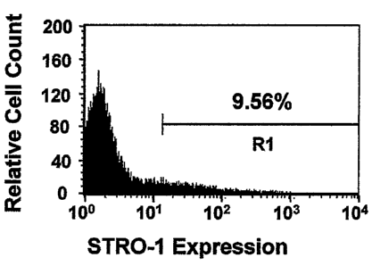
originated from a perivascular microenvironment. A minor proportion (9%) of ex
vivo expanded SHED stained positive for the STRO-1 antibody using FACS
analysis
(Figure 6C). Further immunohistotypic analysis demonstrated that cultured SHED
expressed stromal and vascular related markers ALP, MEPE, bFGF, and endostatin
(Figure 6F-61).
Conditions for the induction of calcium accumulation were as reported
previously (Gronthos et al., Proc. Natl. Acad. Sci. USA, 97: 13625-13630
(2000)),
and recombinant human BMP-4 (R&D systems, Minneapolis, MN) was used to
induce osteogenic differentiation. Calcium accumulation was detected by 2%
Alizarin Red S (pH 4.2) staining. The calcium concentration was measured using
a
commercially available kit (Sigma Calcium Kit #587-A).
To investigate the potential of SHED to differentiate into mineralized tissue,
established secondary SHED cultures were supplemented with L-ascorbate-2-
phosphate, dexamethasone, and inorganic phosphate as previously described
(Gronthos et al., Proc. Natl. Acad. Sci. USA, 97: 13625-13630 (2000)).
Alizarin Red-
positive nodules formed in the SHED cultures following four weeks of induction
(Figure 6J and 6K), indicating calcium accumulation in vitro. Accordingly,
Western
blot analysis revealed that various bone markers CBFA1, ALP, MEPE and BSP were
up-regulated under the induction (Figure 6L). In addition, DSPP was induced by
the
mineralizing induction (Figure 6L). Furthermore, BMP-4 treatment was capable
of
inducing an up-regulated expression of CBFA1, Osterix, and Osteocalcin (OC) by
25
WO 2004/094588 CA 02522669 2005-10-14 PCT/US2003/012276
semi-quantitative RT-PCR (Figure 6M). These data indicated that SHED possessed
the ability to differentiate into functional odontoblast-like cells in vitro.
SHED cells were induced for adipogenesis with procedures used with different
cells (Gimble et al., J. Cell. Biochem., 58:393-402 (1995)). Following five
weeks of
culture with an adipogenic inductive cocktail, around 5% of cultured SHED were
found to possess the potential to develop into Oil red 0-positive lipid-laden
fat cells
(Figure 10A). This correlated with an up-regulation in the expression of two
adipocyte specific transcripts, PPAR1/2 and lipoprotein lipase (LPL), as
detected by
semi-quantitative RT-PCR (Figure 10B).
For neural differentiation, Neurobasal A (Gibco-BRL), B27 supplement
(Gibco-BRL), 1% penicillin, EGF 2Ong/m1 (BD Bioscience), FGF 40 ng/ml (BD
Bioscience) were used to culture cells attached to 0.1% gelatin-coated dishes
(StemCell Technologies Inc, Vancouver, Canada). For sphere-like cell cluster
formation, 3% rat serum and B27 were added.
When cultured either under a neuronal differentiation condition or in 3% rat
serum with B27 supplement, these cells formed sphere-like clusters (Figure 5E)
in
which highly proliferative cells aggregated together in clusters which either
adhered
to the culture dish or floated freely in the culture medium. After separating
the
sphere-like clusters, the cells were able to grow as individual fibroblastic
cells (Figure
5F).
The potential of SHED to develop into neural cells was determined. To
elucidate the neural differentiation potential of SHED, the expression of
neural
markers in SHED was examined. It was determined that cultured SHED expressed a
variety of neural cell markers including Nestin, beta III tubulin, GAD, NeuN,
GFAP,
NFM, and CNPase as measured by immunocytochemical staining (Figure 8A-8H) and
Western blot analysis (Figure 81). After four weeks of neural inductive
culture,
expression levels of neuronal markers including beta III tubulin, GAD, and
NeuN
were increased, while the levels of Nestin, GFAP, NFM, and CNPase remained
unchanged (Figure 81). When cultured under these conditions, SHED lost their
fibroblastic morphology and developed multi-cytoplasmic processes correlating
with
either beta III tubulin/GAD or beta III tubulin/NFM expression (Figure 8J-80).
The
long cellular processes could be best viewed following toluidine blue staining
and
26
CA 02522669 2005-10-14
WO 2004/094588 PCT/US2003/012276
were immunoreactive to MAP2 and Tau antibodies (Figure 8P-8S). Following the
neural inductive culture, SHED continued to express glial cell makers such as
Nestin,
CNPase, GFAP, and NFM (Figure 8T-8W).
Example 7
Implantation of human postnatal deciduous
dental pulp multipotent stem cells
Approximately 2.0x10⁶ SHED were mixed with 40 mg of
hydroxyapatite/tricalcium phosphate (HA/TCP) ceramic powder (Zimmer Inc,
Warsaw, IN) and then implanted subcutaneously into immunocompromised mice
(NIEI-bg-nu-xid, Harlan Sprague Dawley, Indianapolis, IN) as previously
described
(Krebsbach et al., Transplantation, 63: 1059-1069 (1997)).
To validate the capacity of SHED to form odontoblasts, ex vivo expanded
SHED were implanted into immunocompromised mice (Gronthos et al., Proc. Natl.
Acad. Sci. USA, 97: 13625-13630 (2000); Gronthos et al., J. Dent. Res., 81:531-
535
(2002)). The implants yielded human-specific alu-positive odontoblasts
directly
associated with a dentin-like structure (Figure 7A and 7B). The regenerated
dentin
was immunoreactive to dentin-specific DSPP antibody (Figure 7C). These
findings
indicated that human SHED satisfies one important stem cell attribute; the
ability to
differentiate into odontoblasts in vivo. However, SHED were unable to
regenerate a
complete dentin-pulp-like complex as do DPSCs in vivo (Figure 7A and 7E). In
addition, SHED were capable of inducing recipient murine cells to
differentiate into
bone-forming cells as noted by murine-specific pfl in situ hybridization
(Figure 7L),
and lacked DSPP expression (Figure 7D). Skin fibroblasts were never capable of
inducing bone formation upon in vivo implantation. Accordingly, it is thought
that
SHED are distinctively different from DPSC in respect to the odontogenic
differentiation and osteogenic induction.
The characteristics of clonal cell strains, each originating from a single
cell of
deciduous pulp were then determined. When twelve single-colony derived SHED
clones were implanted into immunocompromised mice, only one fourth (3/12) of
the
clones demonstrated a potential to generate ectopic dentin-like tissue on the
HA/TCP
carrier equivalent to that generated by multi-colony derived SHED (Figure 7E
and
27
CA 02522669 2005-10-14
WO 2004/094588 PCT/US2003/012276
7G). SHED either from single-colony or from multi-colony were found to form
dentin-like tissue (Figure 7F) and to survive in the fibrous tissue within the
implants
(Figure 7H) as demonstrated by human-specific alu in situ hybridization. These
results infer that SHED may contain subpopulations of cells, either
differentiating into
odontoblasts or residing in the connective tissue compartments. Surprisingly,
all
implanted single-colony derived SHED clones were capable of inducing bone
formation in immunocompromised mice. About 40 % of the clonal cell strains
(5/12)
induced a significant amount of new bone, while the remaining 60% (7/12)
induced a
limited amount of bone (Figure 71 and 7J). SHED were found to be located on
the
surfaces of HA/TCP but did not participate in bone formation as indicated by
human-
specific alu in situ hybridization (Figure 7K). In contrast, murine host cells
were
found to differentiate into osteoblasts and osteocytes as shown by reactivity
to
murine-specific pfl in situ hybridization (Figure 7L).
SHED were injected into the brain of immunocompromised mice according to
specifications of an approved small animal protocol (NIDCR#01-185).
Coordinates
for the target sites were determined by referencing a murine brain atlas
(Paxinos G et
al, 2nd E, 2001) (see Figure 9A). The anteroposterior (AP), mediolateral (ML),
and
dorsoventral (DV) coordinates were computed relative to Bregma. Ex vivo
expanded
SHED (10,000 cells/ 1) were infused to the dentate gyrus of the hippocampus
(Benedetti et al., Nat. Med., 6:447-450 (2000); Seri et al., Neurosci.,
21:7153-7160
(2001)). Cells (0.5 ill/side) were infused to the coordinates (AP, ML, DV,
respectively: -1.5 mm, +/-0.8 mm, and ¨2.0 mm) using a 1 ,1 Hamilton Syringe.
Neural developmental potential was further studied in vivo by injecting SHED
into the dentate gyrus of the hippocampus of immunocompromised mice (Figure
9A).
Histological examination showed that SHED survived for over 10 days inside the
mouse brain microenvironment as noted by human-specific anti-mitochondria
antibody staining and continued to express neural markers such as NFM (Figure
9B).
Example 8
Reverse Transcriptase-Polymerase Chain Reaction used to characterize
human postnatal deciduous dental pulp multipotent stem cells
28
CA 02522669 2005-10-14
WO 2004/094588 PCT/US2003/012276
The PCR primers included: PPARy2 sense, 5'-
CTCCTATTGACCCAGAAAGC-3' (SEQ ID NO: 1)(114-133), antisense, 5'-
GTAGAGCTGAGTCTTCTCAG-3' (SEQ ID NO: 2)(441-460, Genbank accession
number: XM_003059); LPL sense, 5'-ATGGAGAGCAAAGCCCTGCTC-3' (SEQ ID
NO: 3)(175-195), antisense, 5'-GTTAGGTCCAGCTGGATCGAG-3' (SEQ ID NO:
4)(718-738, Genbank accession number: XM_044682); Core-binding factor, runt
domain, alpha subunit 1 (CBFA1) sense, 5'-CAGTTCCCAAGCATTTCATCC-3'
(SEQ ID NO: 5)(880-900), antisense, 5'-TCAATATGGTCGCCAAACAG-3' (SEQ
ID NO: 6)(1304-1323, Genbank accession number: L40992); Osterix sense, 5'-
GCAGCTAGAAGGGAGTGGTG-3' (SEQ ID NO: 7)(821-840), antisense, 5'-
GCAGGCAGGTGAACTTCTTC-3' (SEQ ID NO: 8)(1160-1179, Genbank accession
number: XM_062600); Osteocalcin sense, 5'-CATGAGAGCCCTCACA-3' (SEQ II)
NO: 9)(18-33), antisense, 5'-AGAGCGACACCCTAGAC-3' (SEQ ID NO: 10)(316-
332, Genbank accession number: X53698); GAPDH sense, 5'-
AGCCGCATCTTCTTTTGCGTC-3' (SEQ ID NO: 11)(12-32), antisense, 5'-
TCATATTTGGCAGGTTTTTCT-3' (SEQ ID NO: 12)(807-827, Genbank accession
number: M33197). Total RNA isolation, first-strand cDNA synthesis and PCR
processes were as previously described (Gronthos et al., J. Dent. Res., 81:531-
535
(2002)).
Example 9
In situ hybridization used to characterize human postnatal
Deciduous dental pulp multipotent stem cells
Human-specific alu and murine-specific pfl sequences labeled with
digoxigenin were used as probes for in situ hybridization as previously
described
(Gronthos et al., Proc. Natl. Acad. Sci. USA, 97: 13625-13630 (2000)). Primers
included: human alu, sense, 5'-TGGCTCACGCCTGTAATCC-3' (SEQ ID NO:
13)(90-108), antisense, 5'-TTTTTTGAGACGGAGTCTCGC-3' (SEQ ID NO:
14)(344-364, Genbank accession number: AC004024); and murine pfl, sense, 5'-
CCGGGCAGTGGTGGCGCATGCCTTTAAATCCC-3' (SEQ ID NO: 15)(170-201),
antisense, 5'-GTTTGGTTTTTGAGCAGGGTTCTCTGTGTAGC-3' (SEQ ID NO:
16)(275-306, Genbank accession number: X78319).
29
CA 02522669 2005-10-14
WO 2004/094588
PCT/US2003/012276
Example 10
Immunohistochemistry used to characterize human postnatal
deciduous dental pulp multipotent stern cells
SHED were sub-cultured into 8-chamber slides (2 x 10⁴ cells/well) (NUNC
Inc, Naperville, IL). The cells were fixed in 4% formaldehyde for 15 minutes
and
then blocked and incubated with primary antibodies (1:200 to 1:500 dilution)
for 1
hour, respectively. The samples were subsequently incubated with goat
secondary
antibodies of either IgG-Rhodamine Red or IgG-CyTM2 (Jackson ImmunoResearch,
West Grove, PA), for 45 minutes. For enzymatic immunohistochemical staining,
the
Zymed broad spectrum immunoperoxidase AEC kit (Zymed Laboratories Inc. South
San Francisco, CA) was used according to the manufacturer's protocol.
Western Blot analysis used to characterize human postnatal Example 11
deciduous dental pulp multipotent stem cells
Primary antibodies were the same as those used in immunohistochemical
staining at dilutions ranging from 1:200 to 1:1000. Western blot was as
previously
reported (Shi et al., Bone, 29:532-539 (2001)).
Example 12
Fluorescence activated Cell Sorting (FACS) used to characterize human
postnatal deciduous dental pulp multipotent stern cells
SHED were collected from culture and incubated with STRO-1 (IgM)
antibodies or isotype-matched negative control antibodies for one hour on ice.
FACS
analysis was the same as previously described (Gronthos et al., J. Dent. Res.,
81:531-
535 (2002)).
Documents
About I, Bottero MJ, de Denato P, Camps J, Franquin JC, Mitsiadis TA (2000).
Human dentin production in vitro. Exp Cell Res 258(1):33-41.
30
WO 2004/094588 CA 02522669 2005-10-14PCT/US2003/012276
About I, Murray PE, Franquin JC, Remusat M, Smith AJ (2001). Pulpal
inflammatory
responses following non-carious class V restorations. Oper Dent 26(4):336-42.
Azizi, S. A., Stokes, D., Augelli, B. J., DiGirolamo, C. & Prockop, D. J.
(1998) Proc
Natl Acad Sci U S A 95, 3908-13.
Bacigalupo A, Frassoni F, Van Lint MT (2000). Bone marrow or peripheral blood
as
a source of stem cells for allogeneic transplants. Curr Opin Hematol 7(6):343-
7.
Baum BJ, Mooney DJ (2000). The impact of tissue engineering on dentistry. J Am
Dent Assoc 131(3):309-18.
Benedetti, S., Pirola, B., Pollo, B., Magrassi, L., Bruzzone, M. G.,
Rigamonti, D.,
Galli, R., Selleri, S., Di Meco, F., De Fraja, C., Vescovi, A., Cattaneo, E. &
Finocchiaro, G. (2000) Nat Med 6, 447-50.
Bjornson, C. R., Rietze, R. L., Reynolds, B. A., Magli, M. C. & Vescovi, A. L.
(1999)
Science 283, 534-7.
Bouma-ter Steege JC, Mayo KH, Griffioen AW (2001). Angiostatic proteins and
peptides. Crit Rev Eukaryot Gene Expr 11(4):319-34.
Brazelton, T. R., Rossi, F. M., Keshet, G. I. & Blau, H. M. (2000) Science
290, 1775-
9.
Brock DP, Marty-Roix R, Spector M (2002). Alpha-smooth-muscle actin in and
contraction of porcine dental pulp cells. J Dent Res 81(3):203-8.
Buchaille R, Couble ML, Magloire H, Bleicher F (2000). A substractive PCR-
based
cDNA library from human odontoblast cells: identification of novel genes
expressed
in tooth forming cells. Matrix Biol 19(5):421-30.
31
WO 2004/094588 CA 02522669 2005-10-14 PCT/US2003/012276
Butler WT, Ritchie HH, Bronckers AL (1997). Extracellular matrix proteins of
dentine. Ciba Found Symp 205(107-15).
Buunma B, Gu K, Rutherford RB (1999). Transplantation of human pulpal and
gingival fibroblasts attached to synthetic scaffolds. Eur J Oral Sci
107(4):282-9.
Chai, Y., Jiang, X., Ito, Y., Bringas, P., Jr., Han, J., Rowitch, D. H.,
Soriano, P.,
McMahon, A. P. & Sucov, H. M. (2000) Development 127:1671-9.
D'Amour, K. A. & Gage, F. H. (2002) Nat Med 8: 213-4.
Decup F, Six N, Palmier B, Buch D, Lasfargues JJ, Salih E, Goldberg M (2000).
Bone {
sialoprotein-induced reparative dentinogenesis in the pulp of rat's molar.
Clin Oral
Investig 4(2):110-9.
Ferrari, G., Cusella-De Angelis, G., Coletta, M., Paolucci, E., Stornaiuolo,
A., Cossu,
G. & Mavilio, F. (1998) Science 279: 1528-30.
Gage, F. H. (2000) Science 287: 1433-8.
Galli, R., Borello, U., Gritti, A., Minasi, M. G., Bjornson, C., Coletta, M.,
Mora, M.,
De Angelis, M. G., Fiocco, R., Cossu, G. & Vescovi, A. L. (2000) Nat Neurosci
3:
986-91.
Gimble, J. M., Morgan, C., Kelly, K., Wu, X., Dandapani, V., Wang, C. S. &
Rosen,
V. (1995) J Cell Biochem 58: 393-402.
Gronthos S, Brahim J, Li W, Fisher LW, Cherman N, Boyde A, DenBesten P, Robey
PG, Shi S (2002). Stem cell properties of human dental pulp stem cells. J Dent
Res
81(8):
531-5.
32
WO 2004/094588 CA 02522669 2005-10-14PCT/US2003/012276
Gronthos S, Mankani M, Brahim J, Robey PG, Shi S (2000). Postnatal human
dental
pulp stem cells (DPSCs) in vitro and in vivo. Proc Natl Acad Sci U S A
97(25):13625-30.
Gronthos, S., Brahim, J., Li, W., Fisher, L. W., Cherman, N., Boyde, A.,
DenBesten,
P., Robey, P. G. & Shi, S. (2002) J Dent Res 81: 531-5.
Gronthos, S., Mankani, M., Brahim, J., Robey, P. G. & Shi, S. (2000) Proc Natl
Acad
Sci US A 97: 13625-30.
Kaigler D, Mooney D (2001). Tissue engineering's impact on dentistry. J Dent
Educ
65(5):456-62.
Kettunen P, Karavanova I, Thesleff 1(1998). Responsiveness of developing
dental
tissues to fibroblast growth factors: expression of splicing alternatives of
FGFR1, -2, -
3, and of FGFR4; and stimulation of cell proliferation by FGF-2, -4, -8, and -
9. Dev
Genet 22(4):374-85.
Krebsbach, P. H., Kuznetsov, S. A., Satomura, K., Emmons, R. V., Rowe, D. W. &
Robey, P. G. (1997) Transplantation 63: 1059-69.
Kuo MY, Lan WH, Lin SK, Tsai KS, Halm LJ (1992). Collagen gene expression in
human dental pulp cell cultures. Arch Oral Biol 37(11):945-52.
LaBonne, C. & Bronner-Fraser, M. (1999) Annu Rev Cell Dev Biol 15: 81-112.
Maim LM, Lennon DP, Caplan AT (1996). Cultured rat pulp cells have the
potential to
form bone, cartilage, and dentin in vivo. In Biological Mechanisms of Tooth
Movement and Cranio facial Adaptation. Edited by Davidovitch Z and Norton LA.
Boston, MA. Harvard Society for the Advancement of Orthodontics, pp. 7-16.
33
WO 2004/094588 CA 02522669 2005-10-14PCT/US2003/012276
Matsushita K, Motani R, Sakuta T, Yamaguchi N, Koga T, Matsuo K, Nagaoka S,
Abeyama K, Maruyama I, Toni M (2000). The role of vascular endothelial growth
factor in human dental pulp cells: induction of chemotaxis, proliferation, and
differentiation and activation of the AP-1-dependent signaling pathway. J Dent
Res
79(8):1596-603.
Mezey, E., Chandross, K. J., Harta, G., Maki, R. A. & McKercher, S. R. (2000)
Science 290: 1779-82.
Moorman, M. A., Simonetti, D. W., Craig, S. & Marshak, D. R. (1999) Science
284:
143-7.
Murray PE, About I, Lumley PJ, Smith G, Franquin JC, Smith AJ (2000).
Postoperative pulpal and repair responses. J Am Dent Assoc 131(3):321-9.
Nakashima M, Nagasawa H, Yamada Y, Reddi AH (1994). Regulatory role of
transforming growth factor-beta, bone morpho genetic protein-2, and protein-4
on
gene expression of extracellular matrix proteins and differentiation of dental
pulp
cells. Dev Biol 162(1):18-28.
Nosrat, I. V., Widenfalk, J., Olson, L. & Nosrat, C. A. (2001) Dev Biol 238:
120-32.
Onishi T, Kinoshita S, Shintani S, Sobue S, Ooshima T (1999). Stimulation of
proliferation and differentiation of dog dental pulp cells in serum-free
culture medium
by insulin-like growth factor. Arch Oral Biol 44(4):361-71.
Ornitz DM, Marie PJ (2002). FGF signaling pathways in endochondral and
intramembranous bone development and human genetic disease. Genes Dev
16(12):1446-65.
Parner, E. T., Heidmann, J. M., Kjaer, I., Vaeth, M. & Poulsen, S. (2002) J
Dent Res
81: 451-4.
34
WO 2004/094588 CA 02522669 2005-10-14 PCT/US2003/012276
Petersen, B. E., Bowen, W. C., Patrene, K. D., Mars, W. M., Sullivan, A. K.,
Murase,
N., Boggs, S. S., Greenberger, J. S. & Goff, J. P. (1999) Science 284: 1168-
70.
Pittenger, M. F., Mackay, A. M., Beck, S. C., Jaiswal, R. K., Douglas, R.,
Mosca, J.
D.,
Prockop, D. J. (1997) Science 276: 71-4.
Qin C, Brunn JC, Cadena E, Ridall A, Tsujigiwa H, Nagatsuka H, Nagai N, Butler
WT (2002). The expression of dentin sialophosphoprotein gene in bone. J Dent
Res
81(6):392-4.
Seri, B., Garcia-Verdugo, J. M., McEwen, B. S. & Alvarez-Buylla, A. (2001) J
Neurosci 21: 7153-60.
Shi S, Robey PG, Gronthos S (2001). Comparison of human dental pulp and bone
marrow stromal stem cells by cDNA microarray analysis. Bone 29(6):532-9.
Shiba H, Fujita T, Doi N, Nakamura S, Nakanishi K, Takemoto T, Hino T, Noshiro
M, Kawamoto T, Kurihara H, Kato Y (1998). Differential effects of various
growth
factors and cytokines on the syntheses of DNA, type I collagen, laminin,
fibronectin,
osteonectin/secreted protein, acidic and rich in cysteine (SPARC), and
alkaline
phosphatase by human pulp cells in culture. J Cell Physiol 174(2):194-205.
Shiba H, Nakamura S, Shirakawa M, Nakanishi K, Okamoto H, Satakeda H, Noshiro
M, Kamihagi K, Katayama M, Kato Y (1995). Effects of basic fibroblast growth
factor on proliferation, the expression of osteonectin (SPARC) and alkaline
phosphatase, and calcification in cultures of human pulp cells. Dev Biol
170(2):457-
66.
Sloan AJ, Rutherford RB, Smith AJ (2000). Stimulation of the rat dentine-pulp
complex by bone morphogenetic protein-7 in vitro. Arch Oral Biol 45(2):173-7.
35
WO 2004/094588 CA 02522669 2005-10-14PCT/US2003/012276
Sloan AJ, Smith AJ (1999). Stimulation of the dentine-pulp complex of rat
incisor
teeth by transforming growth factor-beta isoforms 1-3 in vitro. Arch Oral Biol
44(2):149-56.
Smith AJ, Lesot H (2001). Induction and regulation of crown dentinogenesis:
embryonic events as a template for dental tissue repair? Crit Rev Oral Biol
Med
12(5):425-37.
Spradling, A., Drummond-Barbosa, D. & Kai, T. (2001) Nature 414: 98-104.
Terada, N., Hamazald, T., Oka, M., Hold, M., Mastalerz, D. M., Nakano, Y.,
Meyer,
E. M., Morel, L., Petersen, B. E. & Scott, E. W. (2002) Nature 416: 542-5.
Toma, J. G., Akhavan, M., Fernandes, K. J., Barnabe-Heider, F., Sadikot, A.,
Kaplan,
D. R. & Miller, F. D. (2001) Nat Cell Biol 3: 778-84.
Traver D, Zon LI (2002). Walking the walk: migration and other common themes
in
blood and vascular development. Cell 108(6):731-4.
Tsukamoto Y, Fukutani S, Shin-Ike T, Kubota T, Sato S, Suzuki Y, Mori M
(1992).
Mineralized nodule formation by cultures of human dental pulp-derived
fibroblasts.
Arch Oral Biol 37(12):1045-55.
Weissman, I. L. (2000) Science 287: 1442-6.
Ying, Q. L., Nichols, J., Evans, E. P. & Smith, A. G. (2002) Nature 416: 545-
8.
Yokose S, Kadokura H, Tajima Y, Fujieda K, Katayama I, Matsuoka T, Katayama T
(2000). Establishment and characterization of a culture system for
enzymatically
released rat dental pulp cells. Calcif Tissue hit 66(2):139-44.
36
CA 02522669 2011-02-15
The foregoing specification has been described in relation to
certain embodiments thereof, and many details have been set forth for purposes
of
illustration, however, it will be apparent to those skilled in the art that
the invention is
susceptible to additional embodiments and that certain of the details
described herein
may be varied considerably without departing from the basic principles of the
invention.
37