Note: Descriptions are shown in the official language in which they were submitted.
CA 02522709 2005-10-17
WO 2004/094460 PCT/US2004/012067
POLYPEPTIDES RELATED TO NATRIURETIC PEPTIDES
AND METHODS OF THEIR IDENTIFICATION AND USE
CA 02522709 2005-10-17
WO 2004/094460 PCT/US2004/012067
CROSS-REFERENCE TO RELATED APPLICATIONS
[0001] The present application is a continuation-in-part of USSN 10/419,059
filed April
17, 2003, incorporated by reference in its entirety for all purposes. The
present application
also is a nonprovisional of and claims the benefit of provisional application
60/466,358 filed
April 28, 2003, incorporated by reference in its entirety for all purposes.
FIELD OF THE INVENTION
[0002] The present invention relates to the identification and use of
polypeptides that are
derived from biological active peptides, the peptides generated when the
biological peptide is
generated and the precursors of the,aforementioned peptides.
BACKGROUND OF THE INVENTION
[0003] The following discussion of the background of the invention is merely
provided to
aid the reader in understanding the invention and is not admitted to describe
or constitute
prior art to the present invention.
[0004] Natriuretic peptides are a group of naturally occurnng substances that
act in the
body to oppose the activity of the renin-angiotensin system. There are three
major natriuretic
peptides: atrial natriuretic peptide (ANP), which is synthesized in the atria;
brain-type
natriuretic peptide (BNP), which is synthesized in the ventricles; and C-type
natriuretic
peptide (CNP), which is synthesized in the brain.
(0005] Mature A-type natriuretic peptide (ANP) (also referred to as atrial
natriuretic
peptide) is a 28 amino acid peptide that is synthesized, stored, and released
by atrial
myocytes in response to atrial distension, angiotensin II stimulation,
endothelin, and
sympathetic stimulation (beta-adrenoceptor mediated). Mature ANP is
synthesized as a
precursor molecule (pro-ANP) that is converted to an active form by
proteolytic cleavage. In
addition to atrial natriuretic peptide (ANP99-126) itself, linear peptide
fragments from its N-
terminal prohormone segment have also been reported to have biological
activity.
[0006] Mature B-type natriuretic peptide (BNP) (also called brain-type
natriuretic
peptide) is a 32 amino acid, 4 kDa peptide that is involved in the natriuresis
system to
2
CA 02522709 2005-10-17
WO 2004/094460 PCT/US2004/012067
regulate blood pressure and fluid balance (Bonow, R.O., Circulation 93:1946-
1950, 1996).
The precursor to BNP is synthesized as a 108-amino acid molecule, referred to
herein as
"pro-BNP" that is proteolytically processed into a 76-amino acid N-terminal
peptide (amino
acids 1-76), referred to as "NT pro BNP" and the 32-amino acid mature hormone,
referred to
as BNP or BNP 32 (amino acids 77-108). It has been suggested that each of
these species -
NT pro-BNP, BNP-32, and the pre-pro-BNP - can circulate in human plasma
(Tateyama et
al., Biochem. Biophys. Res. Commun. 185:760-7, 1992; Hunt et al., Biochem.
Biophys. Res.
Commun. 214:1175-83, 1995).
[0007] Mature C-type natriuretic peptide (CNP) a 22-amino acid peptide that is
the
primary active natriuretic peptide in the human brain; CNP is also considered
to be an
endothelium-derived relaxant factor, which acts in the same way as nitric
oxide (NO)
(Davidson et al., Circulation 93:1155-9, 1996). CNP is structurally related to
A-type
natriuretic peptide (ANP) and B-type natriuretic peptide (BNP); however, while
ANP and
BNP are synthesized predominantly in the myocardium, CNP is synthesized in the
vascular
endothelium as a precursor (pro-CNP) (Prickett et al., Biochem. Biophys. Res.
Commun.
286:513-7, 2001). CNP is thought to possess vasodilator effects on both
arteries and veins
and has been reported to act mainly on the vein by increasing the
intracellular cGMP
concentration in vascular smooth muscle cells.
[0008] ANP and BNP are released in response to atrial and ventricular stretch,
respectively, and will cause vasorelaxation, inhibition of aldosterone
secretion in the adrenal
cortex, and inhibition of renin secretion in the kidney. Both ANP and BNP will
cause
natriuresis and a reduction in intravascular volume, effects amplified by the
antagonism of
antidiuretic hormone (ADH). The physiologic effects of CNP differ from those
of ANP and
BNP; CNP has a hypotensive effect, but no significant diuretic or natriuretic
actions.
Increased blood levels of natriuretic peptides have been found in certain
disease states,
suggesting a role in the pathophysiology of those diseases, including stroke,
congestive heart
failure (CHF), cardiac ischemia, systemic hypertension, and acute myocardial
infarction. See,
e.g., WO 02/089657; WO 02/083913; and WO 03/016910, each of which is hereby
incorporated in its entirety, including all tables, figures, and claims.
[0009] The natriuretic peptides, alone, collectively, and/or together with
additional
proteins, can also serve as disease markers and indicators of prognosis in
various
cardiovascular conditions. For example, BNP, which is synthesized in the
cardiac ventricles
3
CA 02522709 2005-10-17
WO 2004/094460 PCT/US2004/012067
and correlates with left ventricular pressure, amount of dyspnea, and the
state of
neurohormonal modulation, makes this peptide the first potential marker for
heart failure.
Measurement of plasma BNP concentration is evolving as a very efficient and
cost effective
mass screening technique for identifying patients with various cardiac
abnormalities
regardless of etiology and degree of LV systolic dysfunction that can
potentially develop into
obvious heart failure and carry a high risk of a cardiovascular event. Finding
a simple blood
test that would aid in the diagnosis and management of patients with CHF
clearly would have
a favorable impact on the staggering costs associated with the disease.
[0010] Removal of the natriuretic peptides from the circulation is affected
mainly by
binding to clearance receptors and enzymatic degradation in the circulation.
See, e.g., Cho et
al., Heart Dis. 1: 305-28, 1999; Smith et al., J. Endocrinol. 167: 239-46,
2000. Additionally,
human pro-BNP is reported to be processed in serum such that circulating pre-
pro-BNP is
unlikely to be the intact 108 amino acid form. Hunt et al., Peptides 18: 1475-
81, 1997. But
some confusion over the stability of the natriuretic peptides, particularly in
blood-derived
samples (e.g., serum, plasma, whole blood) has been reported. For example,
while Norman et
al. (Biochem. Biophys. Res. Commun. 28: 175: 22-30, 1991) report that neutral
endopeptidase
can cleave human BNP between residues 2 and 3, between residues 4 and 5, and
between
residues 17 and 18, Smith et al. (J. Endocrinol. 167: 239-46, 2000) report
that human BNP is
not significantly degraded by purified neutral endopeptidase. Similarly,
Shimizu et al. (Clip.
Chem. Acta 305: 181-6, 2001), Gobinet-Georges et al. (Clin. Chem. Lab. Med.
38: 519-23,
2000) and Murdoch et al. (Heart 78: 594-7, 1997) report that BNP is stable in
certain blood-
derived samples or when blood is collected under certain conditions. A more
recent report by
Shimizu et al. (Clip. Chem. Acta 316: 129-35, 2002) indicates that 94% of BNP
in whole
blood was a digested form in which 2 amino terminal residues had been removed;
and that
BNP in plasma was degraded to a number of unidentified forms.
SUMMARY OF THE INVENTION
The invention provides a purified BNP fragment selected from the group
consisting of BNP79-108, BNP77-106, BNP39-86, BNP53-85, BNP66-98, BNP30-106,
BNP11-107, BNP9-106, BNP69-100, BNP76-107, BNP69-108, BNP80-108, BNP81-108,
BNP83-108, BNP30-103, BNP3-108 and BNP79-106. Optionally, one or more
methionine
residues of the fragment are oxidized.
4
CA 02522709 2005-10-17
WO 2004/094460 PCT/US2004/012067
[0011] In various embodiments, the present invention relates to any purified,
and
preferably substantially purified, BNP polypeptide(s) other than pre-pro-BNP,
BNP1-108,
BNP1-76, and BNP77-108. In preferred embodiments, the present invention
relates to one or
more substantially purified BNP polypeptides selected from the group
consisting of BNP79-
108, BNP77-106, BNP39-86, BNP53-85, BNP66-98, BNP30-106, BNP11-107, BNP9-106,
BNP69-100, BNP76-107, BNP69-108, BNP80-108, BNP81-108, BNP83-108, BNP30-103,
BNP3-108 and BNP79-106. Optionally, BNP80-108, BNP30-106, BNP86-108, BNP77-
107,
BNP77-106, BNP77-103, BNPI-13, and BNP62-76 are excluded in their individually
purified forms.
[0012] The present invention also relates to one or more purified, and
preferably
substantially purified, natriuretic peptide fragments other than mature ANP,
BNP, and CNP,
their precursor molecules, and the fragments generated by cleavage of the
precursor
molecules into the mature ANP, BNP, and CNP peptides.
[0013] The invention further provides a method of assaying BNP. The method
entails
capturing one or more BNP polypeptides from a subject sample; and specifically
measuring a
presence or an amount of at least one captured BNP polypeptide selected from
the group
consisting of BNP79-108, BNP77-106, BNP39-86, BNP53-85, BNP66-98, BNP30-106,
BNP11-107, BNP9-106, BNP69-100, BNP76-107, BNP69-108, BNP80-108, BNP81-108,
BNP83-108, BNP30-103, BNP3-108 and BNP79-106. Preferred BNP polypeptides
include
BNP77-106, BNP39-86, BNP53-85, BNP66-98, BNP30-106, BNP11-107, BNP9-106,
BNP69-100, and BNP76-107.Optionally, the one or more BNP polypeptides are from
a
clinical sample, and the method further comprising correlating the presence or
amount of at
least one captured BNP polypeptide with a clinical parameter. Optionally, the
method further
comprises specifically measuring at least one BNP polypeptide selected from
the group
consisting of BNP1-76, BNP77-108, BNP1-108 and pre-proBNP and correlating the
measurements) with the clinical parameter. Optionally, the specific measuring
step is
performed by mass spectrometry. Optionally, the capturing step comprises
providing a
SELDI probe comprising an antibody attached to a surface of a support;
contacting the
antibody with a sample, whereby the antibody captures the BNP polypeptides
from the
sample; and the specifically measuring step comprises specifically measuring
the presence or
amount of the at least one captured BNP polypeptide by SELDI. Optionally, the
capturing
CA 02522709 2005-10-17
WO 2004/094460 PCT/US2004/012067
captures a plurality of BNP polypeptides selected from the group and the
specifically
measuring specifically measures a plurality of BNP polypeptides selected from
the group.
[0014] The invention further provides a method of classifying the pathology of
a test
sample. The method entails specifically measuring the presence or amount of
one or more
BNP polypeptides selected from each of a plurality of samples of a first class
characterized
by a BNP-related pathology. A presence or amount of said one or more BNP
polypeptides
from a plurality of samples of a second class is specifically measured,
wherein the second
class is characterized by absence of a BNP-related pathology. A classification
model based
on the measurements that classifies a test sample into the first class or the
second class. At
least one of the BNP polypeptides is selected from the group consisting of
BNP79-108,
BNP77-106, BNP39-86, BNP53-85, BNP66-98, BNP30-106, BNP11-107, BNP9-106,
BNP69-100, BNP76-107, BNP69-108, BNP80-108, BNP81-108, BNP83-108, BNP30-103,
BNP3-108 and BNP79-106.
[0015] The invention further comprises a method for specifically measuring pre-
pro-
BNP, BNP1-76, BNP77-108, or BNP1-108 in a sample containing at least one other
BNP
polypeptide. The method entails capturing BNP polypeptides from a sample,
wherein the
polypeptides comprise at least one BNP polypeptide selected from a first group
consisting of
BNP1-76, BNP77-108, BNP1-108 and pre-pro-BNP, and at least one BNP polypeptide
selected from a second group consisting of BNP79-108, BNP77-106, BNP39-86,
BNP53-85,
BNP66-98, BNP30-106, BNP11-107, BNP9-106, BNP69-100, BNP76-107, BNP69-108,
BNP80-108, BNP81-108, BNP83-108, BNP30-103, BNP3-108 and BNP79-106; and (b)
specifically measuring a captured BNP polypeptide from the first group.
Optionally, the
specifically measuring step specifically measures an amount of at least one
captured BNP
polypeptide from the first group and an amount of at least one captured BNP
polypeptide
selected from the second group and the method further comprises determining
relative ratio
of the amounts of each specifically measured BNP polypeptide.
[0016] The invention further provides a method for discovering polypeptides
that interact
with a BNP fragment. The method entails capturing a BNP fragment selected from
the group
consisting of BNP79-108, BNP77-106, BNP39-86, BNP53-85, BNP66-98, BNP30-106,
BNP11-107, BNP9-106, BNP69-100, BNP76-107, BNP69-108, BNP80-108, BNP81-108,
BNP83-108, BNP30-103, BNP3-108 and BNP79-106 with a biospecific capture
reagent.
Molecules that are not bound to the biospecific capture reagent or BNP
fragment are
6
CA 02522709 2005-10-17
WO 2004/094460 PCT/US2004/012067
removed. Molecules bound to the captured BNP fragment are measured.
Optionally, the
molecules are measured by affinity mass spectrometry.
[0017] The invention provides methods of determining a correlation between at
least one
specific measurement and a clinical parameter. The methods entails providing a
learning set
comprising a plurality of data objects representing subjects, wherein each
data object
comprises data representing a specific measurement of a BNP polypeptide
selected from the
group consisting of BNP79-108, BNP77-106, BNP39-86, BNP53-85, BNP66-98, BNP30-
106, BNP11-107, BNP9-106, BNP69-100, BNP76-107, BNP69-108, BNP80-108, BNP81-
108, BNP83-108, BNP30-103, BNP3-108 and BNP79-10; and (b) determining a
correlation
between at least one specific measurement and a clinical parameter.
Optionally, providing
the learning set comprises: capturing BNP polypeptides from a sample with an
antibody, and
specifically measuring one or more of the BNP polypeptides including the BNP
fragment
selected from the group.
[0018] The invention provides methods of classifying a data object according
to clinical
parameter. The methods entail providing a learning set comprising a plurality
of data objects
representing subjects, wherein each subject is classified into at least one of
a plurality of
different clinical parameters and wherein each data object comprises data
representing
specific measurement of a plurality of BNP polypeptides from a subject sample,
and at least
one BNP polypeptide is a BNP fragment selected from the group consisting of
BNP79-108,
BNP77-106, BNP39-86, BNP53-85, BNP66-98, BNP30-106, BNP11-107, BNP9-106,
BNP69-100, BNP76-107, BNP69-108, BNP80-108, BNP81-108, BNP83-108, BNP30-103,
BNP3-108 and BNP79-106; and training a learning algorithm with the learning
set, thereby
generating a classification model, wherein the classification model classifies
a data object
according to clinical parameter.
[0019] Optionally, the clinical parameters are selected from presence or
absence of
disease; risk of disease, stage of disease; response to treatment of disease;
and class of
disease. Optionally, the learning set further comprises data representing
specific
measurement of a polypeptide interactor of a BNP polypeptide. Optionally, the
learning
algorithm is unsupervised. Optionally, the learning algorithm is supervised
and each data
object further comprises data representing the clinical parameter of the
subject. Optionally,
the classification model on subject data from a subject of unknown clinical
parameter to
classify the subject according to a clinical parameter. Optionally, the
clinical parameter is
7
CA 02522709 2005-10-17
WO 2004/094460 PCT/US2004/012067
presence or absence of acute coronary syndrome. Optionally, the supervised
learning
algorithm is selected from linear regression processes, binary decision trees,
artificial neural
networks, discriminant analyses, logistic classifiers, recursive partitioning
processes, and
support vector classifiers. Optionally, the supervised learning algorithm is a
recursive
partitioning process.
[0020] The invention further provides a method for qualifying an immunoassay
calibrator
for a BNP immunoassay. The method entails providing an immunoassay calibrator
for a
BNP immunoassay, wherein the calibrator comprises a designated concentration
of one or
more BNP polypeptides. Polypeptides from the calibrator are captured with an
antibody to a
BNP polypeptide. An amount of at least one polypeptide selected from the group
consisting
of BNP79-108, BNP77-106, BNP39-86, BNP53-85, BNP66-98, BNP30-106, BNP11-107,
BNP9-106, BNP69-100, BNP76-107, BNP69-108, BNP80-108, BNP81-108, BNP83-108,
BNP30-103, BNP3-108 and BNP79-106 is specifically measured, whereby the
measured
amount provides an indication of the quality of the immunoassay calibrator.
Optionally, the
method further comprises specifically measuring at least one BNP polypeptide
selected from
the group consisting of BNP1-76, BNP77-108, BNP1-108, and pre-proBNP.
Optionally, the
method further comprises determining the amount of the at least one BNP
polypeptide
selected from the group consisting of BNP1-76, BNP77-108, BNP1-108, and pre-
proBNP as
a function of total polypeptide captured by the antibody. Optionally, the
amount is measured
by affinity mass spectrometry.
[0021] The invention further provides a method for qualifying an
immunoglobulin
reagent that specifically binds to a BNP polypeptide. The method entails
analyzing the
immunoglobulin reagent by mass spectrometry; and determining the relative
amounts of
intact immunoglobulin and immunoglobulin fragments in the reagent.
[0022] The invention further provides a method of measuring modified forms of
an
antibody to a BNP polypeptide in an antibody reagent for a BNP immunoassay.
Optionally,
the method further comprises measuring un-modified forms of the antibody in
the reagent
and comparing the measurement of un-modified antibody to the measurement of
modified
forms of the antibody. Optionally, the method further comprises specifically
measuring the
amount of at least one BNP fragment selected from the group consisting of
BNP79-108,
BNP77-106, BNP39-86, BNP53-85, BNP66-98, BNP30-106, BNP11-107, BNP9-106,
8
CA 02522709 2005-10-17
WO 2004/094460 PCT/US2004/012067
BNP69-100, BNP76-107, BNP69-108, BNP80-108, BNP81-108, BNP83-108, BNP30-103,
BNP3-108 and BNP79-106 in the immunoassay calibration sample.
[0023] The invention further provides an antibody that specifically binds to
at least one
but not all of the BNP fragments selected from the group consisting of BNP79-
108, BNP77-
106, BNP39-86, BNP53-85, BNP66-98, BNP30-106, BNP11-107, BNP9-106, BNP69-100,
BNP76-107, BNP69-108, BNP80-108, BNP81-108, BNP83-108, BNP30-103, BNP3-108
and BNP79-106. Optionally, the antibody specifically binds to one and only one
of the BNP
fragments selected from the group. Some antibodies distinguish at least one of
the above
fragments from at least another of the above fragments.
[0024] In one embodiment, an assay may be conducted using an antibody or
antibody
cocktail formulated to detect a plurality of natriuretic peptide (e.g., BNP)
fragments as
defined herein. The presence or amount of this plurality of fragments may
provide a more
accurate prognostic or diagnostic result than simply measuring the mature
natriuretic peptide
(or natriuretic peptide precursor) itself. For example, antibodies that detect
only the mature
natriuretic peptide, but that are not able to detect degradation fragments,
may provide an
aberrantly low assay result (e.g., indicating that no BNP or low BNP
concentrations are
present in the sample, when the BNP was present, but has been degraded).
[0025] In an alternative embodiment, individual antibodies that distinguish
amongst a
plurality of natriuretic peptide (e.g., BNP) fragments may be individually
employed to
separately detect the presence or amount of different fragments. The results
of this individual
detection may provide a more accurate prognostic or diagnostic result than
detecting the
plurality of fragments in a single assay. For example, different weighting
factors may be
applied to the various fragment measurements to provide a more accurate
estimate of the
amount of natriuretic peptide originally present in the sample. Additionally,
the relative
amounts of the various fragments may be used to estimate the length of time
since the onset
of an event since, as discussed above, production of such fragments may be a
function of,
inter alia, the elapsed time between onset of an event triggering natriuretic
peptide release
into the tissues and the time the sample is obtained or analyzed.
[0026] In related aspects, the purified natriuretic peptide fragments of the
present
invention may be employed in methods to generate antibodies that recognize one
or a group
of fragments. In various embodiments, a polypeptide may be selected that
comprises a
9
CA 02522709 2005-10-17
WO 2004/094460 PCT/US2004/012067
sequence that is common to a number of natriuretic peptide fragments, and used
to generate
antibodies that recognize this common sequence; such antibodies would
recognize each of the
fragments in which the sequence is in common and expressed such that binding
is sterically
possible. In alternative embodiments, a fragment may be selected that
comprises a sequence
that is distinctive to a specific fragment or set of fragments, and used to
generate antibodies
that recognize only that particular fragment or set of fragments. Such an
antibody is said to
"distinguish" the selected fragments from those fragments that are
unrecognized by the
antibody. Thus, the present invention also relates to antibodies selected to
bind one or more
preselected natriuretic peptide fragments, and methods for their generation
and selection.
[0027] In various embodiments, the present invention relates to antibodies
selected to
bind to a plurality of BNP polypeptides selected from the group consisting of
BNP77-108,
BNP1-76, BNP1-108, pre-proBNP and/or the group consisting of BNP79-108, BNP77-
106,
BNP39-86, BNP53-85, BNP66-98, BNP30-106, BNP11-107, BNP9-106, BNP69-100,
BNP76-107, BNP69-108, BNP80-108, BNP81-108, BNP83-108, BNP30-103, BNP3-108
and BNP79-106. The present invention also relates to methods for the selection
of such
antibodies. Preferably, such antibodies are selected to bind to a plurality of
BNP peptides
generated from BNP77-108, more preferably to bind a plurality of BNP77-108,
BNP77-106,
BNP79-106, BNP76-107, BNP79-108, BNP80-108, BNP81-108, BNP83-108, and most
preferably to each of BNP77-108, BNP77-106, BNP79-106, BNP76-107, BNP79-108,
BNP80-108, BNP81-108, BNP83-108. In other preferred embodiments, antibodies
are also
selected to bind to BNP polypeptides regardless of methionine oxidation state.
[0028] In various embodiments, the present invention relates to antibodies
selected to
specifically bind to a plurality of BNP polypeptides selected from the group
consisting of
BNP77-108, BNP1-76, BNP1-108, pre-proBNP and/or the group consisting of BNP79-
108,
BNP77-106, BNP39-86, BNP53-85, BNP66-98, BNP30-106, BNP11-107, BNP9-106,
BNP69-100, BNP76-107, BNP69-108, BNP80-108, BNP81-108, BNP83-108, BNP30-103,
BNP3-108 and BNP79-106. The present invention also relates to methods for the
selection of
such antibodies. Preferably, such antibodies are selected to bind specifically
to a plurality of
BNP peptides generated from BNP77-108, more preferably to bind a plurality of
BNP77-108,
BNP77-106, BNP79-106, BNP76-107, BNP79-108, BNP80-108, BNP81-108, BNP83-108,
and most preferably to each of BNP77-108, BNP77-106, BNP79-106, BNP76-107,
BNP79-
108, BNP80-108, BNP81-108, BNP83-108. In other preferred embodiments,
antibodies are
CA 02522709 2005-10-17
WO 2004/094460 PCT/US2004/012067
also selected to bind specifically to BNP polypeptides regardless of
methionine oxidation
state.
[0029] In various alternative embodiments, the present invention relates to
antibodies
selected to distinguish between a first group comprising one or more BNP
polypeptides
selected from the group BNP77-108, BNP1-76, BNP1-108, pre-proBNP, BNP79-108,
BNP77-106, BNP39-86, BNP53-85, BNP66-98, BNP30-106, BNP11-107, BNP9-106,
BNP69-100, BNP76-107, BNP69-108, BNP80-108, BNP81-108, BNP83-108, BNP30-103,
BNP3-108 and BNP79-106 and a second group comprising one or more different BNP
polypeptides selected from the group consisting of BNP77-108, BNP1-76, BNPl-
108, pre-
proBNP, BNP79-108, BNP77-106, BNP39-86, BNP53-85, BNP66-98, BNP30-106, BNP11-
107, BNP9-106, BNP69-100, BNP76-107, BNP69-108, BNP80-108, BNP81-108, BNP83-
108, BNP30-103, BNP3-108 and BNP79-106. The present invention also relates to
methods
for the selection of such antibodies. Preferably, members of the first and/or
second groups
comprise BNP peptides generated from BNP77-108, and most preferably members of
the
first and/or second groups comprise BNP77-108, BNP77-106, BNP79-106, BNP76-
107,
BNP79-108, BNP80-108, BNP81-108, BNP83-108. In other preferred embodiments,
antibodies are also selected to distinguish BNP polypeptides on the basis of a
methionine
oxidation state.
[0030] In various embodiments, antibodies are selected, based not upon a
particular
affinity for one or more natriuretic peptide fragments, but instead based upon
a signal that is
obtainable in a binding assay such as an immunoassay. Various binding assay
formats are
known in the art, and it is often the use of antibodies to formulate an
appropriate assay that is
more important than a particular affinity of an antibody for one or more
target molecules. For
example, competitive binding assays may comprise a receptor (e.g., an
antibody) bound to a
solid surface. An analyte of interest in a test sample competes for binding
with a labeled
molecule that also binds to the receptor. The amount of labeled molecule bound
to the
receptor (and hence assay signal) is inversely proportional to the amount of
analyte of interest
in the test sample. In this case, a single antibody attached to the solid
phase is used.
Alternatively, in a sandwich immunoassay, a first antibody, typically bound to
a solid
surface, and a second antibody, typically conjugated to a detectable label,
each bind to an
analyte of interest in a test sample. The amount of labeled molecule bound to
the receptor
11
CA 02522709 2005-10-17
WO 2004/094460 PCT/US2004/012067
(and hence assay signal) is directly proportional to the amount of analyte of
interest in the test
sample.
[0031] In yet another alternative, a sample may be mixed with one or more
compounds
that inhibit the production of natriuretic peptide (e.g., BNP) fragments. In
such embodiments,
one or more proteolytic inhibitors and/or chelators may be added to a
biological sample to
prevent degradation of the natriuretic peptides) fragments that may not be
accurately
detected by an assay.
[0032] The invention further provides a method of assaying BNP polypeptides.
The
method entails capturing one or more BNP polypeptides from a subject sample;
and
specifically measuring a presence or an amount of at least one captured BNP
polypeptide
from among those captured. Optionally, at least 3, 4, 5 or 10 BNP polypeptides
are captured
and specifically measured.
[0033] The invention further provides a method of classifying test samples.
The method
entails specifically measuring the presence or amount of one or more BNP
polypeptides from
each of a plurality of samples of a first class characterized by a BNP-related
pathology. A
presence or amount of said one or more BNP polypeptides is specifically
measured from a
plurality of samples of a second class, wherein the second class is
characterized by absence of
a BNP-related pathology. A classification model is developed based on the
measurements
that classify a test sample into the first class or the second class. At least
one of the BNP
polypeptides is other than BNP1-76, BNP77-108, BNP1-108, pre-pro-BNP.
[0034] The invention further provides a method for discovering polypeptides
that interact
with a BNP polypeptide. The method entails capturing a BNP polypeptide from a
sample
with a biospecific capture reagent; removing molecules that are not bound to
the biospecific
capture reagent or BNP polypeptide; and measuring molecules bound to the
captured BNP
polypeptide.
[0035] The invention further provides a method of correlating specific
measurement of
BNP polypeptides and the clinical parameters. The method entails providing a
learning set
comprising a plurality of data objects representing subjects, in which each
data object
comprises data representing a specific measurement of a BNP polypeptide from a
subject
sample and a clinical parameter of the subject. A correlation is determined
between specific
12
CA 02522709 2005-10-17
WO 2004/094460 PCT/US2004/012067
measurement of the BNP polypeptide and the clinical parameter(s). At least one
of the BNP
polypeptides is other than BNP1-76, BNP77-108, BNP1-108, pre-pro-BNP.
[0036] The invention further provides a method of specifically measuring a BNP
polypeptide selected from the group consisting of BNP1-76, BNP77-108, BNP1-108
and pre-
pro-BNP in a subject sample; and correlating the measurement with a clinical
parameter of
the subject. Optionally, the method further comprises specifically measuring
at least one
BNP fragment selected from the group consisting of BNP79-108, BNP77-106, BNP39-
86,
BNP53-85, BNP66-98, BNP30-106, BNP11-107, BNP9-106, BNP69-100, BNP76-107,
BNP69-108, BNP80-108, BNP81-108, BNP83-108, BNP30-103, BNP3-108 and BNP79-106
and correlating the measurements with the clinical parameter. Optionally, the
method further
comprises specifically measuring at least one biomolecular interactor of a BNP
polypeptide
or antibody to a BNP polypeptide, or a BNP fragment selected from the group
consisting of
BNP79-108, BNP77-106, BNP39-86, BNP53-85, BNP66-98, BNP30-106, BNP11-107,
BNP9-106, BNP69-100, BNP76-107, BNP69-108, BNP80-108, BNP81-108, BNP83-108,
BNP30-103, BNP3-108 and BNP79-106; and correlating the measurement with the
clinical
parameter.
[0037] The invention further provides a method for qualifying an immunoassay
calibrator
for a BNP immunoassay. The method comprises providing an immunoassay
calibrator for a
BNP immunoassay, wherein the calibrator comprises a designated concentration
of one or
more BNP polypeptides; capturing polypeptides from the calibrator with an
antibody to a
BNP polypeptide; and (c) specifically measuring an amount of at least one BNP
polypeptides
whereby the measured amount provides an indication of the quality of the
immunoassay
calibrator.
[0038] The invention further provides biomolecular interactors with BNP or
isolated
biomolecular interactors of anti-BNP antibodies that can be found in
biological samples.
These biomolecular interactors were discovered through affinity mass
spectrometry in which
analytes from a biological sample were captured on a mass spectrometry probe
with an anti-
BNP antibody, and specifically detected and distinguished by laser
desorption/ionization
mass spectrometry from the capture surface. The interactors can be
characterized by
molecular weight.
13
CA 02522709 2005-10-17
WO 2004/094460 PCT/US2004/012067
[0039] In various embodiments, the present invention relates to immunoassays
configured to provide a single signal that relates to the presence or amount
of a plurality of
BNP polypeptides selected from the group consisting of the group consisting of
BNP77-108,
BNP1-76, BNP1-108, pre-proBNP, BNP79-108, BNP77-106, BNP39-86, BNP53-85,
BNP66-98, BNP30-106, BNP11-107, BNP9-106, BNP69-100, BNP76-107, BNP69-108,
BNP80-108, BNP81-108, BNP83-108, BNP30-103, BNP3-108 and BNP79-106.
Preferably,
such immunoassays configured to provide a single signal that is related to the
presence or
amount of a plurality of BNP peptides generated from BNP77-108, more
preferably to a
plurality of BNP77-108, BNP77-106, BNP79-106, BNP76-107, BNP79-108, BNP80-108,
BNP81-108, BNP83-108, and most preferably to each of BNP77-108, BNP77-106,
BNP79-
106, BNP76-107, BNP79-108, BNP80-108, BNP81-108, BNP83-108. In other preferred
embodiments, immunoassays are also configured to provide a single signal that
relates to the
presence or amount of BNP polypeptides regardless of methionine oxidation
state.
[0040] In preferred embodiments, an immunoassay provides a signal that is
within a
factor of S, and most preferably within a factor of two, from an equal number
of molecules of
a plurality of natriuretic peptide fragments, and most preferably a plurality
of the foregoing
BNP polypeptides.
[0041] In various alternative embodiments, the present invention relates to
immunoassays
configured to provide a signal that distinguishes between a first group
comprising one or
more BNP polypeptides selected from the group consisting of BNP77-108, BNP1-
76, BNP1-
108, pre-proBNP, BNP79-108, BNP77-106, BNP39-86, BNP53-85, BNP66-98, BNP30-
106, BNP11-107, BNP9-106, BNP69-100, BNP76-107, BNP69-108, BNP80-108, BNP81-
108, BNP83-108, BNP30-103, BNP3-108 and BNP79-106, and a second group
comprising
one or more different BNP polypeptides selected from the group consisting of
BNP77-108,
BNP1-76, BNP1-108, pre-proBNP, BNP79-108, BNP77-106, BNP39-86, BNP53-85,
BNP66-98, BNP30-106, BNP11-107, BNP9-106, BNP69-100, BNP76-107, BNP69-108,
BNP80-108, BNP81-108, BNP83-108, BNP30-103, BNP3-108 and BNP79-106.
Preferably,
members of the first and/or second groups comprise BNP peptides generated from
BNP77-
108, and most preferably members of the first and/or second groups comprise
BNP77-108,
BNP77-106, BNP79-106, BNP76-107, BNP79-108, BNP80-108, BNP81-108, BNP83-108.
In other preferred embodiments, immunoassays are also configured to
distinguish BNP
polypeptides depending upon methionine oxidation state.
14
CA 02522709 2005-10-17
WO 2004/094460 PCT/US2004/012067
[0042] In yet another aspect, the present invention relates to standard
solutions
comprising a known amount of one or more purified, and preferably
substantially purified,
natriuretic peptide fragments other than mature ANP, BNP, and CNP, their
precursor
molecules, and the fragments generated by cleavage of the precursor molecules
into the
mature ANP, BNP, and CNP peptides. Such standard solutions may find use as
positive
and/or negative control samples in the various assays described herein. In
various
embodiments, the present invention relates to any purified, and preferably
substantially
purified, BNP polypeptide(s) other than pre-proBNP, BNP1-108, BNP1-76, and
BNP77-108.
In preferred embodiments, the present invention relates to one or more
standard solutions
comprising a known amount of one or more purified, and preferably
substantially purified -
related polypeptides selected from the group consisting of BNP79-108, BNP77-
106, BNP39-
86, BNP53-85, BNP66-98, BNP30-106, BNP11-107, BNP9-106, BNP69-100, BNP76-107,
BNP69-108, BNP80-108, BNP81-108, BNP83-108, BNP30-103, BNP3-108 and BNP79-
106.
(0043] In certain aspects, it may be advantageous to formulate such standard
solutions or
calibrants using a composition that is substantially equivalent to the test
sample; for example,
the solution may comprise blood, serum, plasma, etc., as a solvent for the
natriuretic peptide
fragments) of interest. In such a case, it may also be advantageous to include
one or more
protease inhibitors or chelators in order to prevent degradation of the added
natriuretic
peptide fragment(s).
[0044] In another aspect, one or more antibodies, antibody conjugates, and/or
standard
solutions of the present invention may be provided as kits for determining the
presence or
amount of natriuretic peptide fragments. These kits preferably comprise
devices and reagents
for performing at least one assay as described herein on a test sample. Such
kits preferably
contain sufficient reagents to perform one or more such determinations, and/or
Food and
Drug Administration (FDA)-approved labeling.
j0045] In still another aspect, the invention relates to methods for
determining a treatment
regimen for use in a patient. The methods preferably comprise determining the
presence or
amount of one or more natriuretic peptide fragments other than mature ANP,
BNP, and CNP,
their precursor molecules, and the fragments generated by cleavage of the
precursor
molecules into the mature ANP, BNP, and CNP peptides, and relating this
presence or
amount to a disease or prognostic state. As discussed herein, diagnosis and
differentiation of
CA 02522709 2005-10-17
WO 2004/094460 PCT/US2004/012067
various cardiovascular and cerebrovascular diseases, including stroke,
congestive heart
failure (CHF), cardiac ischemia, systemic hypertension, and/or acute
myocardial infarction
may be related to ANP, BNP, and/or CNP levels. Once a diagnosis is obtained, a
treatment
regimen is selected to be consistent with that diagnosis.
[0046] In yet another aspect, the present invention relates to methods of
identifying novel
polypeptides present in biological samples, preferably blood, serum, or plasma
samples, that
are related to known polypeptides. In these methods, an antibody having an
affinity for one or
more known polypeptides (e.g., BNP) is used as an affinity probe for binding
additional
polypeptides that are sufficiently related in structure so as to share binding
affinity to the
antibody, but that are previously unpredicted as being present in the sample.
The sequence of
the polypeptide(s) is(are) then obtained by the methods described herein. Once
obtained, the
sequence may be used in the other aspects described herein; e.g., to select
antibodies that can
differentiate the known polypeptide(s) and the previously unknown
polypeptides, again
according to the methods described herein; to determine if the previously
unknown
polypeptides are useful as diagnostic or prognostic markers; and/or to provide
standard
solutions or isolated peptides.
[0047] In one aspect, a method is described which qualifies an antibody in an
antibody
reagent for tagged immunoassay by mass spectroscopy methods such as SELDI. In
a further
aspect, the method is used to qualify the antibody by determining the amount
of antibody as a
function of total protein of a sample. In a detailed aspect, the method
further includes
preparing an antibody reagent in which the amount of antibody in the reagent
comprises the
same amount reflected in the amount of antibody from the sample as determined
by SELDI.
[0048] In another aspect, a method is described which qualifies peptides in a
calibrator
for tagged immunoassay by SELDI. In a further aspect, the method is used to
qualify
peptides by determining the amount of one or more particular peptides as a
function of total
protein in a sample. In a detailed aspect, the method further includes
preparing a peptide
reagent in which the amount of peptide in the reagent comprises an amount
reflected in the
amount of peptide from the sample as determined by SELDI.
[0049] In a further aspect, the method includes qualifying an antibody in an
antibody
reagent for a tagged immunoassay using a SELDI immunoassay. In a detailed
aspect, the
16
CA 02522709 2005-10-17
WO 2004/094460 PCT/US2004/012067
tagged immunoassay is a BNP immunoassay. In a further detailed aspect, SELDI
is SEAC.
In a further detailed aspect, SELDI is SEND.
[0050] In another aspect, a method is described which includes the steps of
qualifying the
polypeptides captured by an antibody reagent in a tagged immunoassay by
providing a
SELDI probe comprising the antibody reagent attached to a surface of the
probe, contacting
the antibody reagent with a sample, whereby the antibody reagent captures
polypeptides from
the sample, and detecting the captured polypeptides by SELDI. In a detailed
aspect, the
tagged immunoassay is a BNP immunoassay. In a further detailed aspect, SELDI
is SEAC.
In a further detailed aspect, SELDI is SEND.
BRIEF DESCRIPTION OF THE DRAWINGS
[0051] FIG. 1. Predicted amino acid sequence of B-type Natriuretic Peptide
(BNP)
Precursor and fragments thereof is shown. Fragment Arg77- His108 (indicated on
the figure
as "77-108") is one isoform sought to be detected by immunoassay.
[0052] FIG. 2A and B. Mass spectra of proteins in a BNP immunoassay calibrator
solution. SELDI analysis of a calibrator used for BNP immunoassays
demonstrates that the
calibrator contains many polypeptides besides full length BNP (BNP77-108). The
peak at
3464 corresponds to BNP77-108. The peak at 66283.6 presumably corresponds to
bovine
serum albumin.
[0053] Fig. 3. Mass spectrum of antibody reagent comprising anti-BNP
monoclonal also
contains peaks corresponding to many proteins besides the antibody.
[0054] Fig. 4A, B, C, and D. Mass spectra of proteins from a BNP calibrator
solution
captured by SELDI immunoassay. Proteins from the calibrator were spiked into
human
plasma. Anti-BNP was used to capture the proteins. Besides the 77-108 isoform
at 6461,
peaks are detected whose molecular weights correspond to BNP peptide
fragments: A BNP
isoform that weighs about 3170.8 Da and corresponds to amino acids 77 to 106
of proBNP; a
BNP isoform that weighs about 3280 Da and corresponds to amino acid 79 to 108
of
proBNP; a BNP isoform that weighs about 3671 Da and corresponds to amino acid
53-85
(3669) or 66-98 (3674.4) of proBNP; a BNP isoform that weighs about 8215.5 Da
and
corresponds to amino acids 30 to 103 of proBNP; a BNP isoform that weighs
about 10875.3
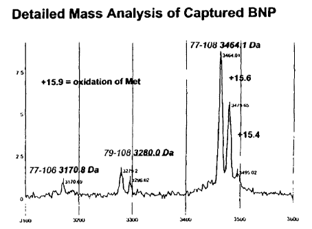
and corresponds to 11-107 (108755.) or 9-106 (10874.4)of proBNP.
17
CA 02522709 2005-10-17
WO 2004/094460 PCT/US2004/012067
[0055] Fig. 5A and B. Mass spectra and standard curve of BNP calibrator at
various
levels of concentration. Spectra show that the calibrator contains as much
BNP79-108
isoform as BNP77-108 isoform.
[0056] Fig. 6A, B and C. Mass spectra and standard curve of BNP calibrator at
various
levels of concentration. BNP77-108 is hardly visible. When the standard is
calibrated to the
amount of protein corresponding to BNP79-106, BNP79-108 and a peak
corresponding to
either BNP69-100 or BNP76-107 the standard curve is skewed to the right,
implying that a
test measurement contains more BNP that the original calibrator key indicated.
[0057] Figs. 7A and B. Mass spectra of subject samples. Peaks corresponding to
BNP77-
109 are difficult to detect. However, degraded forms of BNP appear to be
present - about
3152 (BNP77-106) and about 3282 (BNP79-108).
DEFINITIONS
[0058] Human BNP is derived by proteolysis of a 108 amino acid precursor
molecule,
referred to hereinafter as BNP1-108. Mature BNP, or "the BNP natriuretic
peptide," is a 32
amino acid molecule representing amino acids 77-108 of this precursor, and is
referred to
hereinafter as BNP77-108. The remaining residues 1-76 are referred to
hereinafter as BNP1-
76.
[0059] The sequence of the 108 amino acid BNP precursor pro-BNP (BNP1-108) is
as
follows, with mature BNP (BNP77-108) underlined:
HPLGSPGSAS DLETSGLQEQ RNHLQGKLSE LQVEQTSLEP LQESPRPTGV 50
WKSREVATEG IRGHRKMVLY TLRAPRSPKM VQGSGCFGRK MDRISSSSGL 100
GCKVLRRH 108
(SEQ ID NO: 1).
[0060] BNP1-108 is synthesized as a larger precursor pre-pro-BNP having the
following
sequence (with the "pre" sequence shown in bold):
1~PQTAPSRA LLLLLFLHLA FLGGRSHPLG SPGSASDLET SGLQEQRNHL 50
QGKLSELQVE QTSLEPLQES PRPTGVWKSR EVATEGIRGH RKMVLYTLRA 100
PRSPKMVQGS GCFGRKMDRI SSSSGLGCKV LRRH 134
18
CA 02522709 2005-10-17
WO 2004/094460 PCT/US2004/012067
(SEQ ID NO: 2).
[0061] The sequence of the 126 amino acid ANP precursor pro-ANP (ANP1-126) is
as
follows, with mature ANP (ANP99-126) underlined:
NPMYNAVSNA DLMDFKNLLD HLEEKMPLED EWPPQVLSD PNEEAGAALS 50
PLPEVPPWTG EVSPAQRDGG ALGRGPWDSS DRSALLKSKL RALLTAPRSL 100
RRSSCFGGRM DRIGAQSGLG CNSFRY 126
(SEQ ID NO: 3).
[0062] ANP1-126 is synthesized as a larger precursor pre-pro-ANP having the
following
sequence (with the "pre" sequence shown in bold):
MSSFSTTTVS FLLLLAFQLL GQTR.ANPMYN AVSNADLMDF KNLLDHLEEK 50
MPLEDEWPP QVLSDPNEEA GAALSPLPEV PPWTGEVSPA QRDGGALGRG 100
PWDSSDRSAL LKSKLRALLT APRSLRRSSC FGGRMDRIGA QSGLGCNSFR 150
y 151
(SEQ ID NO: 4).
[0063] The sequence of the 126 amino acid CNP precursor pro-CNP (CNP1-126) is
as
follows, with the mature CNP forms CNP-53 (CNP74-126) in italics, and CNP-22
(CNP105-
126) underlined:
MHLSQLLACA LLLTLLSLRP SEAKPGAPPK VPRTPPAEEL AEPQAAGGGQ 50
KKGDKAPGGG GANLKGDRSR LLRDLRVDTK SRAAWARLLQ EHPNARKYKG 100
ANKKGLSKGC FGLKLDRIGS MSGLGC 126
(SEQ m NO: 5).
[0064] The term "BNP polypeptide" refers to any of BNP1-76, BNP77-108, BNP1-
108,
pre-proBNP, and fragments thereof, includingBNP79-108, BNP77-106, BNP39-86,
BNP53-
85, BNP66-98, BNP30-106, BNP11-107, BNP9-106, BNP69-100, BNP76-107, BNP69-108,
BNP80-108, BNP81-108, BNP83-108, BNP30-103, BNP3-108 and BNP79-106.
[0065] The term "fragment" as used herein refers to a polypeptide that
comprises at least
six contiguous amino acids of a polypeptide from which the fragment is
derived. Thus, a
fragment of BNP1-108 (pro-BNP) refers to a polypeptide that comprises at least
six
19
CA 02522709 2005-10-17
WO 2004/094460 PCT/US2004/012067
contiguous amino acids of BNP1-108; a fragment of mature BNP refers to a
polypeptide that
comprises at least six contiguous amino acids of BNP77-108; a fragment of the
polypeptide
generated by cleavage of pro-BNP into mature BNP refers to a polypeptide that
comprises at
least six contiguous amino acids of BNP1-76. A "BNP" fragment means a fragment
of any of
BNP77-108, BNP1-76, BNP1-108 and pre-pro-BNP. Similarly, a fragment of ANP1-
126
(pro-ANP) refers to a polypeptide that comprises at least six contiguous amino
acids of
ANP 1-126; a fragment of mature ANP refers to a polypeptide that comprises at
least six
contiguous amino acids of ANP99-126; a fragment of the polypeptide generated
by cleavage
of pro-ANP into mature ANP refers to a polypeptide that comprises at least six
contiguous
amino acids of BNP1-98; and a fragment of CNP1-126 (pro-CNP) refers to a
polypeptide that
comprises at least six contiguous amino acids of CNP1-126; a fragment of
mature CNP refers
to a polypeptide that comprises at least six contiguous amino acids of CNP74-
126 or
CNP 105-126; a fragment of the polypeptide generated by cleavage of pro-CNP
into mature
CNP refers to a polypeptide that comprises at least six contiguous amino acids
of CNP1-73 or
CNP1-104. In preferred embodiments, a fragment refers to a polypeptide that
comprises at
least 10 contiguous amino acids of a polypeptide from which the fragment is
derived; at least
15 contiguous amino acids of a polypeptide from which the fragment is derived;
or at least 20
contiguous amino acids of a polypeptide from which the fragment is derived.
[0066] The term "natriuretic peptide fragment" as used herein refers to a
fragment, as
described above, of any natriuretic peptide selected from the group consisting
of mature
ANP, BNP, or CNP, the biosynthetic precursors pre-pro-ANP, pre-pro-BNP, pre-
pro-CNP,
pro-ANP, pro-BNP, or pro-CNP, or the polypeptide remaining after removal of
mature ANP,
BNP, or CNP from the pro-form of the peptide.
[0067] Unless otherwise apparent from the context, reference to natriuretic
polypeptides
includes modified forms of polypeptides bearing post-translational
modification including,
for example, phosphorylation (adds 80 D per phosphate group), glycosylation,
lipidation,
methylation (adds 14 D per methyl group), cysteinylation (adds 199 D per
cysteinyl group),
sulphonation, glutathionylation (adds 305 D per glutathione group), and
acetylation (adds 42
D per acetyl group). Natriuretic peptide fragments, including BNP polypeptide
can comprise
one or more oxidizable methionines, the oxidation of which to methionine
sulfoxide or
methionine sulfone. Changes in the oxidation state of one or more methionines
may alter the
ability of assays to detect such fragments. Thus, in addition to the reduced
forms of the
CA 02522709 2005-10-17
WO 2004/094460 PCT/US2004/012067
substantially purified natriuretic peptide fragments discussed above, the
present invention
also relates to one or more purified, and preferably substantially purified,
natriuretic peptide
fragments other than mature ANP, BNP, and CNP, their precursor molecules, and
the
fragments generated by cleavage of the precursor molecules into the mature
ANP, BNP, and
CNP peptides, in which one or more methionines are oxidized. Preferred are one
or more
substantially purified BNP polypeptides selected from the group consisting of
BNP77-108,
BNP1-76, BNP1-108, pre-proBNP and the group consisting of BNP79-108, BNP77-
106,
BNP39-86, BNP53-85, BNP66-98, BNP30-106, BNP11-107, BNP9-106, BNP69-100,
BNP76-107, BNP69-108, BNP80-108, BNP81-108, BNP83-108, BNP30-103, BNP3-108
and BNP79-106 in which one or more methionines are oxidized. The presence or
absence of
natriuretic peptide fragments in which one or more of these peptides may be
measured by
immunoassay, mass spectrometry, high pressure liquid chromatography and gas
chromatography, as described hereinafter.
[0068] Most preferably, a fragment is "naturally present" in a biological
sample (e.g., a
blood, serum or plasma sample, and most preferably human blood, serum, or
plasma). This
means that the fragment may be obtained from an unsupplemented biological
sample
obtained from a human or animal. "Unsupplemented" refers to a sample in which
the
fragment or its precursor has not been exogenously added once the sample is
obtained.
Examples of fragments naturally present in blood, serum or plasma are
described hereinafter.
Other preferred fragments are said to be "generated from" blood, serum or
plasma if the
fragment is present as a result of supplementing such a sample with pro-ANP,
pro-BNP, pro-
CNP, and/or a fragment thereof, and allowing endogenous factors (e.g.,
proteases) in the
sample to generate additional fragments. Examples of fragments generated from
human
blood, serum or plasma are also described hereinafter. A fragment is "present"
in blood,
serum or plasma if the fragment is either naturally present or generated from
such a sample.
[0069] As used herein, the term "purified" in reference to polypeptides does
not require
absolute purity. Instead, it represents an indication that the polypeptide(s)
of interest is(are) in
a discrete environment in which abundance (on a mass basis) relative to other
proteins is
greater than in a biological sample. By "discrete environment" is meant a
single medium,
such as a single solution, a single gel, a single precipitate, etc. Purified
polypeptides may be
obtained by a number of methods including, for example, laboratory synthesis,
chromatography, preparative electrophoresis, centrifugation, precipitation,
affinity
21
CA 02522709 2005-10-17
WO 2004/094460 PCT/US2004/012067
purification, etc. One or more "purified" polypeptides of interest are
preferably at least 10%
of the protein content of the discrete environment. One or more "substantially
purified"
polypeptides are at least 50% of the protein content of the discrete
environment, more
preferably at least 75% of the protein content of the discrete environment,
and most
preferably at least 95% of the protein content of the discrete environment.
Protein content is
determined using a modification of the method of Lowry et al., J. Biol. Chem.
193: 265,
1951, described by Hartree, Anal Biochem 48: 422-427 (1972), using bovine
serum albumin
as a protein standard.
(0070] The term "antibody" as used herein refers to a peptide or polypeptide
derived
from, modeled after or substantially encoded by an immunoglobulin gene or
immunoglobulin
genes, or fragments thereof, capable of specifically binding an antigen or
epitope. See,.e.g.
Fundamentallmmunology, 3rd Edition, W.E. Paul, ed., Raven Press, N.Y. (1993);
Wilson
(1994) J. Immunol. Methods 175:267-273; Yarmush (1992) J. Biochem. Biophys.
Methods
25:85-97. Natural immunoglobulins are encoded by immunoglobulin genes. These
include
the kappa and lambda light chain constant region genes, the alpha, gamma,
delta, epsilon and
mu heavy chain constant region genes, and the myriad immunoglobulin variable
region
genes. The term antibody includes antigen-binding portions, i.e., "antigen
binding sites,"
(e.g., fragments, subsequences, complementarity determining regions (CDRs))
that retain
capacity to bind antigen, including (i) a Fab fragment, a monovalent fragment
consisting of
the VL, VH, CL and CHl domains; (ii) a F(ab')2 fragment, a bivalent fragment
comprising ,
two Fab fragments linked by a disulfide bridge at the hinge region; (iii) a Fd
fragment
consisting of the VH and CH1 domains; (iv) a Fv fragment consisting of the VL
and VH
domains of a single arm of an antibody, (v) a dAb fragment (Ward et al.,
(1989) Nature
341:544-546), which consists of a VH domain; an scFv protein, which is a
fusion protein in
which a light chain variable region and a heavy chain variable region bound by
a linker; and
(vii) an isolated complementarity determining region (CDR). The "Fc" portion
of an
antibody refers to that portion of an immunoglobulin heavy chain that
comprises one or more
heavy chain constant region domains, CH1, CH2 and CH3, but does not include
the heavy
chain variable region. Single chain antibodies, monoclonal antibodies,
polyclonal antibodies,
chimeric antibodies, humanized antibodies, and antibodies produced by
immunization, from
hybridomas, or recombinantly using molecular biological techniques (e.g., by
phage display
methods) are also included by reference in the term "antibody."
22
CA 02522709 2005-10-17
WO 2004/094460 PCT/US2004/012067
[0071] Individual antibodies (e.g., obtained by phage display or monoclonal
antibody
technology) may be obtained that bind to a plurality of fragments having a
common epitope
to which the antibody may bind. In the alternative, individual antibodies may
be pooled to
provide the desired spectrum of binding affinities. The term "antibody" may
refer to both a
composition in which each antibody molecule present is identical (referred to
specifically as
an "individual antibody"), or a composition in which antibody molecules
present may differ
(e.g., in a pooled or polyclonal composition). Preferred antibodies are
"Omniclonal"
antibodies. Omniclonal antibodies are a mixture of different antibody
molecules selected
from a phage display library, where each antibody specifically binds to a
target antigen with a
minimum affinity of 109 M-1 to 10'° M-1.
[0072] "Epitope" or "antigenic determinant" refers to a site on an antigen to
which B
and/or T cells respond. Epitopes can be formed both from contiguous amino
acids or
noncontiguous amino acids juxtaposed by tertiary folding of a protein.
Epitopes formed from
contiguous amino acids are typically retained on exposure to denaturing
solvents whereas
epitopes formed by tertiary folding are typically lost on treatment with
denaturing solvents.
An epitope typically includes at least 3, and more usually, at least S or 8-10
amino acids in a
unique spatial conformation. Methods of determining spatial conformation of
epitopes
include, for example, x-ray crystallography and 2-dimensional nuclear magnetic
resonance.
See, e.g., Epitope Mapping Protocols in METHODS IN MOLECULAR BIOLOGY, Vol. 66,
Glenn E. Morris, ed (1996).
[0073] The term "specifically binds" does not necessarily require that an
antibody binds
exclusively to its intended target. Rather, an antibody specifically binds if
its affinity for its
intended target is about 2-fold greater when compared to its affinity for a
non-target
molecule. Preferably the affinity of the antibody will be at least about five
fold, preferably 10
fold, more preferably 25-fold, even more preferably SO-fold, and most
preferably 100-fold or
more, greater for a target molecule than its affinity for a non-target
molecule. In preferred
embodiments, specific binding between an antibody or other binding agent and
an antigen
means a binding affinity of at least 106 M-~. Preferred antibodies bind with
affinities of at
least about 107 M~1, and preferably 10g M-~ to 109 M-~ or 10'° M-1. A
ligand or a receptor that
"specifically binds" to a compound analyte can be used to determine the
presence or amount
of the analyte in a sample of unrelated heterogeneous compounds. Thus, the
ligand or
receptor binds preferentially to a particular analyte and does not bind in a
significant amount
23
CA 02522709 2005-10-17
WO 2004/094460 PCT/US2004/012067
to the other compounds present in the sample. For example, an antibody
specifically binds
under immunoassay conditions to an antigen analyte bearing an epitope against
which the
antibody was raised.
[0074] An immunoassay is said to "distinguish" between a first group of
polypeptides and
a second group of polypeptides if the immunoassay provides a signal related to
binding of the
first group of polypeptides that is at least a factor of 10 greater than a
signal obtained from an
equal number of molecules of the second group of polypeptides under the same
assay
conditions. More preferably, the signal is at least a factor of 20 greater,
even more preferably
at least a factor of 50 greater, and most preferably at least a factor of 100
greater or more.
[0075] An antibody is said to "distinguish" between a first group of
polypeptides and a
second group of polypeptides if its affinity for the members of the first
group of polypeptides
is about 2-fold greater when compared to its affinity for members of the
second group.
Preferably the affinity of the antibody will be at least about five fold,
preferably 10 fold, more
preferably 25-fold, even more preferably SO-fold, and most preferably 100-fold
or more,
greater for members of the first group of polypeptides than its affinity for
members of the
second group.
[0076] A molecule is "specifically measured" when its presence and/or amount
is
detected in a sample to the exclusion of other molecules that are structurally
related. One
BNP polypeptide selected from the group consisting of BNP79-108, BNP77-106,
BNP39-86,
BNP53-85, BNP66-98, BNP30-106, BNP11-107, BNP9-106, BNP69-100, BNP76-107,
BNP69-108, BNP80-108, BNP81-108, BNP83-108, BNP30-103, BNP3-108 and BNP79-106
is specifically measured, when the measurement detects that polypeptide in a
manner
distinguishable from measurement of any other BNP polypeptide in the group,
and
distinguishable from any measurement of BNP polypeptides BNP1-76, BNP77-108,
BNP1-
108, and pre-proBNP. BNP77-106 fragment is specifically measured when its
presence
and/or amount are detected or quantified, wherein the presence and/or amount
of other BNP
fragments such as BNP77-108 do not contribute to a signal that constitutes a
specific
measurement of BNP77-106.
[0077] A signal from an immunoassay is said to "depend upon binding to an
antibody" if
the antibody participates in formation of a complex necessary to generate the
signal. For
example, in a sandwich immunoassay formulated using a solid phase antibody and
a second
24
CA 02522709 2005-10-17
WO 2004/094460 PCT/US2004/012067
antibody conjugate, each of which must bind to an analyte to form the
sandwich, each of the
solid phase antibody and second antibody participate in formation of the
complex necessary
to generate the signal. In a competitive immunoassay where a single antibody
is used, and an
analyte competes with an analyte conjugate for binding, the single antibody
participates in
formation of the complex necessary to generate the signal. Numerous additional
immunoassay formulations may be provided.
[0078] The term "plurality" as used herein in reference to natriuretic peptide
fragments
and BNP polypeptides refers to 2 or more molecular species that differ in
amino acid
sequence.
[0079] An "interactor" is a molecule that specifically binds to another
molecule.
[0080] "Immunoassay" refers to a method of detecting an analyte in a sample in
which
specificity for the analyte is conferred by the specific binding between an
antibody and a
ligand such as a natriuretic peptide fragment. This includes detecting an
antibody analyte
through specific binding between the antibody and a ligand. See Harlow and
Lane (1988)
ANTIBODIES, A LABORATORY MANUAL, Cold Spring Harbor Publications, New York,
for a description of immunoassay formats and conditions that can be used to
determine
specific immunoreactivity. A "tagged immunoassay" is an immunoassay in which
the
analyte is not detected directly, but rather through detection of a tag or
label. Generally, the
analyte is itself tagged, or the immunoassay involves binding of the analyte
with a tagged
antibody which is, itself, tagged. The techniques of immunoassay using labeled
reagents for
detecting antigens and antibodies are sensitive. Solid-phase assays for
antibodies employing
ligands labeled with radioisotopes or enzymes (radioimmunoassay; RIA and
enzyme-linked
immunosorbent assay; ELISA) are widely used because large numbers can be
performed in a
relatively short time. RIA and ELISA are direct binding assays for antibody
(or antigen) and
both work on the same principle, but the means of detecting specific binding
is different. For
both methods, a pure preparation of a known antigen or antibody, or both, is
needed in order
to standardize the assay. In RIA for an antigen, pure antibody against that
antigen is
radioactively labeled, usually with l2sl; for the ELISA, an enzyme is linked
chemically to the
antibody. The unlabeled component, which in this case is the antigen, is
attached to a solid
support, such as the wells of a plastic multiwell plate, which will adsorb a
certain amount of
any protein. The labeled antibody is allowed to bind to the unlabeled antigen,
under
conditions where nonspecific adsorption is blocked, arid any unbound antibody
and other
CA 02522709 2005-10-17
WO 2004/094460 PCT/US2004/012067
proteins are washed way. Antibody binding in RIA is measured directly in terms
of the
amount of radioactivity retained by the coated wells, whereas in ELISA,
binding is detected
by a reaction that converts a colorless substrate into a colored reaction
product. Labeled anti-
immunoglobulin antibodies can also be used with RIA or ELISA to detect binding
of
unlabeled antibody to unlabeled antigen-coated plates. Alternatively, the
immunoassay may
be a SELDI MS immunoassay. An immunoassay based on mass spectrometry
automatically
provides discrimination of the various captured polypeptides based on mass.
[0081] A modification of ELISA known as a "capture" or "sandwich ELISA" (or
more
generally referred to as an "antigen-capture assay") can be used to detect
secreted products
such as cytokines. Rather than the antigen being directly attached to a
plastic plate, antigen-
specific antibodies are bound to the plate. These are able to bind antigen
with high affinity,
and thus concentrate it on the surface of the plate, even with antigens that
are present in very
low concentrations in the initial mixture. A separate labeled antibody that
recognizes a
different epitope to the immobilized first antibody is then used to detect the
bound antigen.
[0082] RIA and ELISA do not allow one to measure directly the amount of
antigen or
antibody in a sample of unknown composition, as both depend on the binding of
a pure
labeled antigen or antibody. In a "competitive inhibition assay," the presence
and amount of a
particular antigen in an unknown sample is determined by its ability to
compete with a
labeled reference antigen for binding to an antibody typically attached to a
plastic well. A
standard curve is first constructed by adding varying amounts of a known,
unlabeled standard
preparation; the assay can then measure the amount of antigen in unknown
samples by
comparison with the standard. The competitive binding assay can also be used
for measuring
antibody in a sample of unknown composition by attaching the appropriate
antigen to the
plate and measuring the ability of the test sample to inhibit the binding of a
labeled specific
antibody.
[0083] A molecule such as an antibody can be "qualified" in terms of the
amount of the
molecule, its binding specificity, and/or its quality, e.g., its state of
degradation. For
example, methods of qualifying the peptides in an immunoassay calibrator,
e.g., a BNP
immunoassay calibrator, can be performed by mass spectrometry, in particular
by SELDI.
SELDI allows more precise discrimination of those peptides, as they can be
both
discriminated according to mass and quantified based on the area under a mass
spectrum
26
CA 02522709 2005-10-17
WO 2004/094460 PCT/US2004/012067
peak. Because mass spectrometry qualifies molecules by mass, polypeptides
comprising the
same epitope, but differing in mass may be detected, differentiated and
measured.
[0084] "Detectable moiety" or a "label" or a "tag" refers to a composition
detectable by
spectroscopic, photochemical, biochemical, immunochemical, or chemical means.
For
example, useful labels include 32p, 35S, fluorescent dyes, electron-dense
reagents, enzymes
(e.g., as commonly used in an ELISA), biotin-streptavadin, digoxigenin,
haptens and proteins
for which antisera or monoclonal antibodies are available, or nucleic acid
molecules with a
sequence complementary to a target. The detectable moiety often generates a
measurable
signal, such as a radioactive, chromogenic, or fluorescent signal, that can be
used to quantify
the amount of bound detectable moiety in a sample. The detectable moiety can
be
incorporated in or attached to a primer or probe either covalently, or through
ionic, van der
Waals or hydrogen bonds, e.g., incorporation of radioactive nucleotides, or
biotinylated
nucleotides that are recognized by streptavadin. The detectable moiety can be
directly or
indirectly detectable. Indirect detection can involve the binding of a second
directly or
indirectly detectable moiety to the detectable moiety. For example, the
detectable moiety can
be the ligand of a binding partner, such as biotin, which is a binding partner
for streptavadin,
or a nucleotide sequence, which is the binding partner for a complementary
sequence, to
which it can specifically hybridize. The binding partner can itself be
directly detectable, for
example, an antibody can be itself labeled with a fluorescent molecule. The
binding partner
also can be indirectly detectable, for example, a nucleic acid having a
complementary
nucleotide sequence can be a part of a branched DNA molecule that is in turn
detectable
through hybridization with other labeled nucleic acid molecules. (See, e.g.,
P. D. Fahrlander
and A. Klausner, Bio/Technology 6:1165 (1988)). Quantitation of the signal is
achieved by,
e.g., scintillation counting, densitometry, or flow cytometry.
[0085] Devices for performing the assays described herein preferably contain a
plurality
of discrete, independently addressable locations, or "diagnostic zones," each
of which is
related to a particular peptide or set of peptides of interest. For example,
each of a plurality
of discrete zones may comprise a receptor (e.g., an antibody) for binding a
different peptide.
Alternatively, one or more zones may each comprise a receptor (e.g., an
antibody) for binding
a plurality of peptides. Following reaction of a sample with the devices, a
signal is generated
from the diagnostic zone(s), which may then be correlated to the presence or
amount of the
peptide of interest. In some instances "diagnostic zones" are also referred to
as "addressable
locations."
27
CA 02522709 2005-10-17
WO 2004/094460 PCT/US2004/012067
(0086] The term "discrete" as used herein refers to areas of a surface that
are non-
contiguous. That is, two areas are discrete from one another if a border that
is not part of
either area completely surrounds each of the two areas. The term
"independently addressable"
as used herein refers to discrete areas of a surface from which a specific
signal may be
obtained. Antibody zones can also be independent of each other, but can be in
contact with
each other on a surface. For example, antibodies that recognize different
epitopes of a single
antigen can each be attached to the surface of a biochip that comprises a
plurality of
addressable locations, each of which location has an antibody attached there
[0087] The team "sample" refers to a quantity of biological molecules that are
to be tested
for the presence or absence of one or more molecules.
[0088] The term "test sample" as used herein refers to a sample in which the
presence or
amount of one or more analytes of interest are unknown and to be determined in
an assay,
preferably an immunoassay. Preferably, a test sample is a bodily fluid
obtained for the
purpose of diagnosis, prognosis, or evaluation of a subject, such as a
patient. In certain
embodiments, such a sample may be obtained for the purpose of determining the
outcome of
an ongoing condition or the effect of a treatment regimen on a condition.
Preferred test
samples include blood, serum, plasma, cerebrospinal fluid, urine and saliva.
Some test
samples are more readily analyzed following a fractionation or purification
procedure, for
example, separation of whole blood into serum or plasma components. Preferred
samples
may be obtained from bacteria, viruses and animals, such as dogs and cats.
Particularly
preferred samples are obtained from humans. By way of contrast, a "standard
sample" refers
to a sample in which the presence or amount of one or more analytes of
interest are known
prior to assay for the one or more analytes. Some test samples obtained from
patients are
referred to as "test samples."
[0089] The term "disease sample" as used herein refers to a tissue sample
obtained from a
subject that has been determined to suffer from a given disease. Methods for
clinical
diagnosis are well known to those of skill in the art. See, e.g., Kelley's
Textbook of Internal
Medicine, 4'h Ed., Lippincott Williams & Wilkins, Philadelphia, PA, 2000; The
Merck
Manual of Diagnosis and Therapy, 17'h Ed., Merck Research Laboratories,
Whitehouse
Station, N.J., 1999. "Disease" includes events generally accepted in the
medical field as
adverse outcomes related to a disease, such as stroke, myocardial infarction,
and other
adverse health events.
28
CA 02522709 2005-10-17
WO 2004/094460 PCT/US2004/012067
[0090] A pathological level of BNP refers to a statistically significant
variation (p<_0.05),
usually an increase, of BNP polypeptide(s) in a patient relative to mean
levels in a population
of undiseased individuals. A BNP related-pathology means a disease due to or
otherwise
associated with a pathological level of at least one BNP polypeptide or a
mixture thereof.
Such diseases include cardiovascular diseases, for example, stroke, congestive
heart failure
(CHF), cardiac ischemia, systemic hypertension, acute myocardial infarction,
and acute
coronary syndrome.
[0091] The presence or amount of one or more natriuretic peptide fragments of
interest
may be related to the presence or absence of a disease, or the likelihood of a
future adverse
outcome related to a disease. However, the signal obtained from an assay need
not be related
to the presence or amount of one or more natriuretic peptide fragments;
rather, the signal may
be directly related to the presence or absence of a disease, or the likelihood
of a fixture
adverse outcome related to a disease. For example, a level of signal x may
indicate that y
pg/mL of a fragment is present in the sample. A table may then indicate that y
pg/mL of that
fragment indicates congestive heart failure. It may be equally valid to simply
relate a level of
signal x directly to congestive heart failure, without determining how much of
the fragment is
present. Such a signal is preferably obtained from an immunoassay using the
antibodies of
the present invention, although other methods are well known.
[0092] The term "unpredicted polypeptides" as used herein refers to a
polypeptide that, in
the particular type of biological sample being analyzed, has not previously
been demonstrated
to be naturally present. A polypeptide is preferably unpredicted in a blood,
serum, or plasma
sample, and most preferably a human blood, serum, or plasma sample.
[0093] The term "determining the amino acid sequence" as used herein refers to
methods
by which the amino acid sequence of a particular polypeptide is obtained. Such
methods may
include direct sequencing (e.g., by Edman degradation); identification by mass
spectrometry,
which may comprise comparison of observed m/z to a predicted or known
polypeptide
sequence (see, e.g., Cagney and Emili, Nature Biotechnol. 20: 163-170 (2002));
peptide
mapping; etc.
[0094] The terms "mass spectrometry" or "MS" as used herein refer to methods
of
filtering, detecting, and measuring ions based on their mass-to-charge ratio,
or "m/z." In
general, one or more molecules of interest are ionized, and the ions are
subsequently
29
CA 02522709 2005-10-17
WO 2004/094460 PCT/US2004/012067
introduced into a mass spectrographic instrument where, due to a combination
of magnetic
and electric fields, the ions follow a path in space that is dependent upon
mass ("m") and
charge ("z"). See, e.g., U.S. Patent Nos. 6,204,500, entitled "Mass
Spectrometry From
Surfaces;" 6,107,623, entitled "Methods and Apparatus for Tandem Mass
Spectrometry;"
6,268,144, entitled "DNA Diagnostics Based On Mass Spectrometry;" 6,124,137,
entitled
"Surface-Enhanced Photolabile Attachment And Release For Desorption And
Detection Of
Analytes;" Wright et al., "Proteinchip surface enhanced laser
desorption/ionization (SELDI)
mass spectrometry: a novel protein biochip technology for detection of
prostate cancer
biomarkers in complex protein mixtures," Prostate Cancer and Prostatic
Diseases 2: 264-76
(1999); and Merchant and Weinberger, "Recent advancements in surface-enhanced
laser
desorption/ionization-time of flight-mass spectrometry," Electrophoresis 21:
1164-67 (2000),
each of which is hereby incorporated by reference in its entirety, including
all tables, figures,
and claims.
[0095] For example, in a "quadrupole" or "quadrupole ion trap" instrument,
ions in an
oscillating radio frequency field experience a force proportional to the DC
potential applied
between electrodes, the amplitude of the RF signal, and m/z. The voltage and
amplitude can
be selected so that only ions having a particular m/z travel the length of the
quadrupole, while
all other ions are deflected. Thus, quadrupole instruments can act as both a
"mass filter" and
as a "mass detector" for the ions injected into the instrument.
[0096] Moreover, one can often enhance the resolution of the MS technique by
employing "tandem mass spectrometry," or "MS/MS." In this technique, a
precursor ion or
group of ions generated from a molecule (or molecules) of interest may be
filtered in an MS
instrument, and these precursor ions subsequently fragmented to yield one or
more fragment
ions that are then analyzed in a second MS procedure. By careful selection of
precursor ions,
only ions produced by certain analytes of interest are passed to the
fragmentation chamber,
where collision with atoms of an inert gas occurs to produce the fragment
ions. Because both
the precursor and fragment ions are produced in a reproducible fashion under a
given set of
ionization/fragmentation conditions, the MS/MS technique can provide an
extremely
powerful analytical tool. For example, the combination of
filtration/fragmentation can be
used to eliminate interfering substances, and can be particularly useful in
complex samples,
such as biological samples.
CA 02522709 2005-10-17
WO 2004/094460 PCT/US2004/012067
[0097] Additionally, recent advances in technology, such as matrix-assisted
laser
desorption ionization coupled with time-of flight analyzers ("MALDI-TOF"), or
surface-
enhanced laser desorption ionization coupled with time-of flight analyzers
("SELDI-TOF"),
permit the analysis of analytes at femtomole levels in very short ion pulses.
Mass
spectrometers that combine time-of flight analyzers with tandem MS are also
well known to
the artisan. Additionally, multiple mass spectrometry steps can be combined in
methods
known as "MS/MS" and "MS/MS-TOF," including MS/MS-MALDI-TOF and MS/MS-
SELDI-TOF. Preferred apparatuses and methods for characterization and
identification of
proteins are disclosed in U.S. Patent Application Publication No. US
2002/0182649; U.S.
Patent No. 6,225,047; Issaq et al., Biochem. Biophys. Res. Commun. 292: 587-92
(2002); and
Issaq et al., Anal. Chem. 75: 149A-155A (2003), each of which is hereby
incorporated by
reference in its entirety.
[0098] Ions can be produced using a variety of methods including, but not
limited to,
electron ionization, chemical ionization, fast atom bombardment, field
desorption, and
matrix-assisted laser desorption ionization ("MALDI"), surface enhanced laser
desorption
ionization ("SELDI"), photon ionization, electrospray ionization, and
inductively coupled
plasma.
[0099] "Probe" in the context of this invention refers to a device adapted to
engage a
probe interface of a gas phase ion spectrometer (e.g., a mass spectrometer)
and to present an
analyte to ionizing energy for ionization and introduction into a gas phase
ion spectrometer,
such as a mass spectrometer. A "probe" will generally comprise a solid
substrate (either
flexible or rigid) comprising a sample presenting surface on which an analyte
is presented to
the source of ionizing energy. In general, a probe with an adsorbent surface
is contacted with
a sample for a period of time sufficient to allow biomarker or biomarkers that
may be present
in the sample to bind to the adsorbent. After an incubation period, the
substrate is washed to
remove unbound material. Any suitable washing solutions can be used;
preferably, aqueous
solutions are employed. The extent to which molecules remain bound can be
manipulated by
adjusting the stringency of the wash. The elution characteristics of a wash
solution can
depend, for example, on pH, ionic strength, hydrophobicity, degree of
chaotropism, detergent
strength, and temperature. Unless the probe has both SEAC and SEND properties
(as
described herein), an energy absorbing molecule then is applied to the
substrate with the
bound biomarkers.
31
CA 02522709 2005-10-17
WO 2004/094460 PCT/US2004/012067
[00100] "Surface-enhanced laser desorption/ionization" or "SELDI" refers to a
method of
desorption/ionization gas phase ion spectrometry (e.g., mass spectrometry) in
which the
analyte is captured on the surface of a SELDI probe that engages the probe
interface of the
gas phase ion spectrometer. In "SELDI MS," the gas phase ion spectrometer is a
mass
spectrometer. SELDI technology is described in, e.g., U.S. patent 5,719,060
(Hutchens and
Yip) and U.S. patent 6,225,047 (Hutchens and Yip).
(00101] One version of SELDI is called "affinity mass spectrometry." It also
is called
"Surface-Enhanced Affinity Capture" or "SEAC". This version involves the use
of probes
that have a material on the probe surface that captures analytes through a non-
covalent
affinity interaction (adsorption) between the material and the analyte. The
material is
variously called an "adsorbent," a "capture reagent," an "affinity reagent" or
a "binding
moiety." Such probes can be referred to as "affinity capture probes" and as
having an
"adsorbent surface." The capture reagent can be any material capable of
binding an analyte.
The capture reagent may be attached directly to the substrate of the selective
surface, or the
substrate may have a reactive surface that carries a reactive moiety that is
capable of binding
the capture reagent, e.g., through a reaction forming a covalent or coordinate
covalent bond.
Epoxide and carbodiimidizole are useful reactive moieties to covalently bind
polypeptide
capture reagents such as antibodies or cellular receptors. Nitriloacetic acid
and iminodiacetic
acid are useful reactive moieties that function as chelating agents to bind
metal ions that
interact non-covalently with histidine containing peptides. Adsorbents are
generally
classified as chromatographic adsorbents and biospecific adsorbents.
[00102] "Chromatographic adsorbents" include those adsorbent materials
typically used in
chromatography. Chromatographic adsorbents include, for example, ion exchange
materials,
metal chelators (e.g., nitriloacetic acid or iminodiacetic acid), immobilized
metal chelates,
hydrophobic interaction adsorbents, hydrophilic interaction adsorbents, dyes,
simple
biomolecules (e.g., nucleotides, amino acids, simple sugars and fatty acids)
and mixed mode
adsorbents (e.g., hydrophobic attraction/electrostatic repulsion adsorbents).
[00103] "Biospecific adsorbents" include those molecules that specifically
bind to a
biomolecule. Typically they comprise a biomolecule, e.g., a nucleic acid
molecule (e.g., an
aptamer), a polypeptide, a polysaccharide, a lipid, a steroid or a conjugate
of these (e.g., a
glycoprotein, a lipoprotein, a glycolipid, a nucleic acid (e.g., DNA)-protein
conjugate). In
certain instances, the biospecific adsorbent can be a macromolecular structure
such as a
32
CA 02522709 2005-10-17
WO 2004/094460 PCT/US2004/012067
multiprotein complex, a biological membrane or a virus. Examples of
biospecific adsorbents
are antibodies, receptor proteins and nucleic acids. Biospecific adsorbents
typically have
higher specificity for a target analyte than chromatographic adsorbents.
Further examples of
adsorbents for use in SELDI can be found in U.S. Patent No. 6,225,047. A
"bioselective
adsorbent" refers to an adsorbent that binds to an analyte with an affinity of
at least 10-8 M.
[00104] In some embodiments, a SEAC probe is provided as a pre-activated
surface which
can be modified to provide an adsorbent of choice. For example, certain probes
are provided
with a reactive moiety that is capable of binding a biological molecule
through a covalent
bond. Epoxide and carbodiimidizole are useful reactive moieties to covalently
bind
biospecific adsorbents such as antibodies or cellular receptors.
[00105] "Adsorption" refers to detectable non-covalent binding of an analyte
to an
adsorbent or capture reagent.
[00106] Another version of SELDI is Surface-Enhanced Neat Desorption (SEND),
which
involves the use of probes comprising energy absorbing molecules that are
chemically bound
to the probe surface ("SEND probe"). The phrase "energy absorbing molecules"
(EAM)
denotes molecules that are capable of absorbing energy from a laser
desorption/ionization
~ source and, therea8er, contribute to desorption and ionization of analyte
molecules in contact
therewith. The EAM category includes molecules used in MALDI, frequently
referred to as
"matrix," and is exemplified by cinnamic acid derivatives, sinapinic acid
(SPA), cyano-
hydroxy-cinnamic acid (CHCA) and dihydroxybenzoic acid, ferulic acid, and
hydroxyaceto-
phenone derivatives. In certain embodiments, the energy absorbing molecule is
incorporated
into a linear or cross-linked polymer, e.g., a polymethacrylate. For example,
the composition
can be a co-polymer of a-cyano-4-methacryloyloxycinnamic acid and acrylate. In
another
embodiment, the composition is a co-polymer of a-cyano-4-
methacryloyloxycinnamic acid,
acrylate and 3-(tri-ethoxy)silyl propyl methacrylate. In another embodiment,
the composition
is a co-polymer of a-cyano-4-methacryloyloxycinnamic acid and
octadecylmethacrylate
("C18 SEND"). SEND is further described in U.S. Patent No. 6,124,137 and PCT
International Publication No. WO 03/64594 (Kitagawa, "Monomers And Polymers
Having
Energy Absorbing Moieties Of Use In Desorption/Ionization Of Analytes," August
7, 2003).
[00107] SEAC/SEND is a version of SELDI in which both a capture reagent and an
energy
absorbing molecule are attached to the sample presenting surface. SEAC/SEND
probes
33
CA 02522709 2005-10-17
WO 2004/094460 PCT/US2004/012067
therefore allow the capture of analytes through affinity capture and
ionization/desorption
without the need to apply external matrix. The C 18 SEND biochip is a version
of
SEAC/SEND, comprising a C18 moiety which functions as a capture reagent, and a
CHCA
moiety which functions as an energy absorbing moiety.
[00108] Another version of SELDI, called Surface-Enhanced Photolabile
Attachment and
Release (SEPAR), involves the use of probes having moieties attached to the
surface that can
covalently bind an analyte, and then release the analyte through breaking a
photolabile bond
in the moiety after exposure to light, e.g., to laser light (see, U.S. Patent
No. 5,719,060).
SEPAR and other forms of SELDI are readily adapted to detecting a biomarker or
biomarker
profile, pursuant to the present invention.
[00109] In another mass spectrometry method, the biomarkers can be first
captured on a
chromatographic resin that binds the target molecules. For example, the resin
can be
derivatized with an anti-BNP antibody. Alternatively, this method could be
preceded by
fractionating the sample on an anion exchange resin before application to the
cation exchange
resin. After elution from the resin, the sample can be analyzed by MALDI,
electrospray, or
another ionization method for mass spectrometry. In another alternative, one
could
fractionate on an anion exchange resin and detect by MALDI or electrospray
mass
spectrometry directly. In yet another method, one could capture the biomarkers
on an
immuno-chromatographic resin that comprises antibodies that bind the
biomarkers, wash the
resin to remove unbound material, elute the biomarkers from the resin and
detect the eluted
biomarkers by MALDI, SELDI, electrospray mass spectrometry or another
ionization mass
spectrometry method.
[00110] "Eluant" or "wash solution" refers to an agent, typically a solution,
which is used
to affect or modify adsorption of an analyte to an adsorbent surface and/or
remove unbound
materials from the surface. The elution characteristics of an eluant can
depend, for example,
on pH, ionic strength, hydrophobicity, degree of chaotropism, detergent
strength and
temperature.
[00111] "Analyte" refers to any component of a sample that is desired to be
detected. The
term can refer to a single component or a plurality of components in the
sample.
[00112] The "complexity" of a sample adsorbed to an adsorption surface of an
affinity
capture probe means the number of different protein species that are adsorbed.
34
CA 02522709 2005-10-17
WO 2004/094460 PCT/US2004/012067
[00113] "Molecular binding partners" and "specific binding partners" refer to
pairs of
molecules, typically pairs of biomolecules that exhibit specific binding.
Molecular binding
partners include, without limitation, receptor and ligand, antibody and
antigen, biotin and
avidin, and biotin and streptavidin.
"Monitoring" refers to recording changes in a parameter at multiple time
points. Optionally,
the parameter is continuously varying.
"Solid support" refers to a solid material which can be derivatized with, or
otherwise attached
to, a chemical moiety, such as a capture reagent, a reactive moiety or an
energy absorbing
species. Exemplary solid supports include chips (e.g., probes), microtiter
plates and
chromatographic resins.
[00114] "Chip" refers to a solid support having a generally planar surface to
which a
chemical moiety can be attached. Chips that are adapted to engage a probe
interface are also
called "probes."
[00115] "Biochip" refers to a chip to which a chemical moiety is attached.
Frequently, the
surface of the biochip comprises a plurality of addressable locations, each of
which location
has the chemical moiety attached there.
[00116] "Protein biochip" refers to a biochip adapted for the capture of
polypeptides.
Protein biochips produced by Ciphergen Biosystems, Inc. comprise surfaces
having
chromatographic or biospecific adsorbents attached thereto at addressable
locations.
Ciphergen ProteinChip~ arrays include NP20 (hydrophilic); H4 and H50
(hydrophobic);
SAX-2, Q-10 and LSAX-30 (anion exchange); WCX-2, CM-10 and LWCX-30 (cation
exchange); IMAC-3, IMAC-30 and IMAC 40 (metal chelate); and PS-10, PS-20
(reactive
surface with carboimidizole, expoxide) and PG-20 (protein G coupled through
carboimidizole). These protein biochips comprise an aluminum substrate in the
form of a
strip. The surface of the strip is coated with silicon dioxide. In the case of
the NP-20
biochip, silicon oxide functions as a hydrophilic adsorbent to capture
hydrophilic proteins.
Hydrophobic ProteinChip arrays have isopropyl or nonylphenoxy-polyethylene
glycol)methacrylate functionalities. Anion exchange ProteinChip arrays have
quaternary
ammonium functionalities. Cation exchange ProteinChip arrays have carboxylate
functionalities. Immobilized metal chelate ProteinChip arrays have
nitriloacetic acid
functionalities that adsorb transition metal ions, such as copper, nickel,
zinc, and gallium, by
CA 02522709 2005-10-17
WO 2004/094460 PCT/US2004/012067
chelation. Preactivated ProteinChip arrays have carboimidizole or epoxide
functional groups
that can react with groups on proteins for covalent binding.
[00117] H4, H50, SAX-2, Q-10, WCX-2, CM-10, IMAC-3, IMAC-30, PS-10 and PS-20
biochips further comprise a functionalized, cross-linked polymer in the form
of a hydrogel
physically attached to the surface of the biochip or covalently attached
through a silane to the
surface of the biochip. The H4 biochip has isopropyl functionalities for
hydrophobic binding.
The H50 biochip has nonylphenoxy-polyethylene glycol)methacrylate for
hydrophobic
binding. The SAX-2 and Q-10 biochips have quaternary ammonium functionalities
for anion
exchange. The WCX-2 and CM-10 biochips have carboxylate functionalities for
cation
exchange. The IMAC-3 and IMAC-30 biochips have nitriloacetic acid
functionalities that
adsorb transition metal ions, such as Cu++ and Ni++, by chelation. These
immobilized metal
ions allow adsorption of peptide and proteins by coordinate bonding. The PS-10
biochip has
carboimidizole functional groups that can react with groups on proteins for
covalent binding.
The PS-20 biochip has epoxide functional groups for covalent binding with
proteins. The
PS-series biochips are useful for binding biospecific adsorbents, such as
antibodies, receptors,
lectins, heparin, Protein A, biotin/streptavidin and the like, to chip
surfaces where they
function to specifically capture analytes from a sample. The PG-20 biochip is
a PS-20 chip
to which Protein G is attached. The LSAX-30 (anion exchange), LWCX-30 (cation
exchange) and IMAC-40 (metal chelate) biochips have functionalized latex beads
on their
surfaces. Such biochips are further described in: WO 00/66265 (Rich et al.,
"Probes for a
Gas Phase Ion Spectrometer," November 9, 2000); WO 00/67293 (Beecher et al.,
"Sample
Holder with Hydrophobic Coating for Gas Phase Mass Spectrometer," November 9,
2000);
U.S. patent application US 2003 0032043 A1 (Pohl and Papanu, "Latex Based
Adsorbent
Chip," July 16, 2002) and U.S. patent application 60/350,110 (Um et al.,
"Hydrophobic
Surface Chip," November 8, 2001); U.S. patent application 60/367,837,
(Boschetti et al.,
"Biochips With Surfaces Coated With Polysaccharide-Based Hydrogels," May 5,
2002) and
U.S. patent application entitled "Photocrosslinked Hydrogel Surface Coatings"
(Huang et al.,
filed February 21, 2003).
[00118] Such biochips are further described in: U.S. Patent No. 6,579,719
(Hutchens and
Yip, "Retentate Chromatography," June 17, 2003); PCT International Publication
No. WO
00/66265 (Rich et al., "Probes for a Gas Phase Ion Spectrometer," November 9,
2000); U.S.
Patent No. 6,555,813 (Beecher et al., "Sample Holder with Hydrophobic Coating
for Gas
36
CA 02522709 2005-10-17
WO 2004/094460 PCT/US2004/012067
Phase Mass Spectrometer," April 29, 2003); U.S. Patent Application No. U.5.
2003
0032043 A1 (Pohl and Papanu, "Latex Based Adsorbent Chip," July 16, 2002); and
PCT
International Publication No. WO 03/040700 (Um et al., "Hydrophobic Surface
Chip," May
1 S, 2003); U.S. Provisional Patent Application No. 60/367,837 (Boschetti et
al., "Biochips
With Surfaces Coated With Polysaccharide-Based Hydrogels," May 5, 2002) and
U.S. Patent
Application No. 60/448,467, entitled "Photocrosslinked Hydrogel Surface
Coatings" (Huang
et al., filed February 21, 2003).
[00119] Many protein biochips, adapted for the capture of polypeptides, are
described in
the art. These include, for example, protein biochips produced by Ciphergen
Biosystems
(Fremont, CA), Packard BioScience Company (Meriden CT), Zyomyx (Hayward, CA),
Phylos (Lexington, MA) and Procognia (Sense Proteomic Limited) (Maidenhead,
Berkshire,
UK). Examples of such protein biochips are described in the following patents
or patent
applications: U.S. patent 6,225,047 (Hutchens and Yip, "Use of retentate
chromatography to
generate difference maps," May 1, 2001); International publication WO 99/51773
(Kuimelis
and Wagner, "Addressable protein arrays," October 14, 1999); U.S. patent
6,329,209
(Wagner et al., "Arrays of protein-capture agents and methods of use thereof,"
December 11,
2001), International publication WO 00/56934 (Englert et al., "Continuous
porous matrix
arrays," September 28, 2000), United States patent publication US 2003/0180957
A1
(Koopman et al., "Target and method," September 25, 2003) and United States
patent
publication US 2003/0173513 A1 (Koopman et al., "Probe for mass spectrometry,"
September 18, 2003).
[00120] Upon capture on a biochip, analytes can be detected by a variety of
detection
methods selected from, for example, a gas phase ion spectrometry method, an
optical method,
an electrochemical method, atomic force microscopy and a radio frequency
method. Gas
phase ion spectrometry methods are described herein. Of particular interest is
the use of mass
spectrometry and, in particular, SELDI. Optical methods include, for example,
detection of
fluorescence, luminescence, chemiluminescence, absorbance, reflectance,
transmittance,
birefringence or refractive index (e.g., surface plasmon resonance,
ellipsometry, a resonant
mirror method, a grating coupler waveguide method or interferometry). Optical
methods
include microscopy (both confocal and non-confocal), imaging methods and non-
imaging
methods. Immunoassays in various formats (e.g., ELISA) are popular methods for
detection
37
CA 02522709 2005-10-17
WO 2004/094460 PCT/US2004/012067
of analytes captured on a solid phase. Electrochemical methods include
voltametry and
amperometry methods. Radio frequency methods include multipolar resonance
spectroscopy.
[00121] The summary of the invention described above is non-limiting and other
features
and advantages of the invention will be apparent from the following detailed
description of
the invention, and from the claims.
DETAILED DESCRIPTION OF THE INVENTION
I General Use of Natriuretic Peptide Fragments as Prognostic and Diagnostic
Markers
and Suecific Fragments of BNP
[00122] Increased blood levels of natriuretic peptides have been found in
certain disease
states, suggesting a role in the pathophysiology of those diseases, including
stroke,
congestive heart failure (CHF), cardiac ischemia, systemic hypertension, and
acute
myocardial infarction. See, e.g., WO 02/089657; WO 02/083913; WO 03/016910;
Hunt et
al., Biochem. Biophys. Res. Comm. 214: 1175-83 (1995); Venugopal, J. Clin.
Pharm. Ther.
26: 15-31, 2001; and Kalra et al., Circulation 107: 571-3, 2003; each of which
is hereby
incorporated in its entirety, including all tables, figures, and claims. The
natriuretic peptides,
alone, collectively, and/or together with additional proteins, can also serve
as disease markers
and indicators of prognosis in various cardiovascular conditions.
[00123] It has been reported that removal of natriuretic peptides from the
circulation
involves degradation pathways. Indeed, inhibitors of neutral endopeptidase,
which cleaves
natriuretic peptides under certain circumstances, have been suggested to hold
promise in
treatment of certain cardiovascular diseases. See, e.g., Trindade and Rouleau,
Heart Fail.
Monit. 2: 2-7, 2001. However, the measurement of the natriuretic peptides in
clinical samples
has focused generally upon measurement of the mature BNP, ANP, and/or CNP;
their
precursor molecules (i.e., pro-BNP, pro-ANP, and pro-CNP); and the fragments
resulting
from cleavage of the pro-form to provide the mature natriuretic peptides. The
present
invention describes for the first time a number of fragments produced by
degradation of these
molecules in biological samples. Although described hereinafter mainly with
reference to
BNP fragments, the general concepts described herein apply equally to ANP- and
CNP-
related fragments.
38
CA 02522709 2005-10-17
WO 2004/094460 PCT/US2004/012067
[00124] The failure to consider the degradation fragments that may be present
in a clinical
sample when measuring one or more of the natriuretic peptides may have serious
consequences for the accuracy of any diagnostic or prognostic method. Consider
for example
a simple case, where a sandwich immunoassay is provided for BNP, and all of
the BNP
present has been degraded into two fragments, one of which contains the
epitope
corresponding to the solid phase antibody, the other of which contains the
epitope
corresponding to the antibody conjugate used for signal generation in the
immunoassay.
Because no BNP fragments present contain both epitopes, no signal will be
obtained from the
immunoassay, thus leading to the incorrect assumption that no BNP was
originally present in
the sample.
[00125] Similarly, another simple case may be considered. In a competitive
assay, in
which BNP present in solution competes with labeled BNP for binding to a solid
phase
antibody, consider that the solid phase is configured with a polyclonal
antibody that would
recognize both of the foregoing fragments. Each would bind to the antibody
solid phase, and
compete with the labeled BNP for binding. Such a situation may lead to the
incorrect
assumption that twice the BNP concentration actually present in the sample is
detected. As
described herein, the situation may actually be much more complicated than
these simple
situations. Because production of such fragments is an ongoing process that
may be a
function of, inter alia, the elapsed time between onset of an event triggering
natriuretic
peptide release into the tissues and the time the sample is obtained or
analyzed; the elapsed
time between sample acquisition and the time the sample is analyzed; the type
of tissue
sample at issue; the storage conditions; the quantity of proteolytic enzymes
present; etc. may
affect the extent of the errors in measurement.
[00126] The previously known BNP polypeptides, pre-pro-BNP, pro-BNP (BNP1-108
),
pro-fragment ( BNP1-76), and mature BNP (BNP77-108), contains multiple sites
for possible
amino acid modifications and endoproteolytic cleavage. The following new
fragments have
been observed BNP79-108, BNP77-106, BNP39-86, BNP53-85, BNP66-98, BNP30-106,
BNP11-107, BNP9-106, BNP69-100, BNP76-107, BNP69-108, BNP80-108, BNP81-108,
BNP83-108, BNP30-103, BNP3-108 and BNP79-106. Fig. 1 shows BNP77-108.
Preferred
degradation fragments identified in human serum or plasma include: BNP77-106,
BNP79-
106, BNP76-107, BNP69-108, BNP79-108, BNP80-108, BNP81-108, BNP83-108, BNP39-
86, BNP53-85, BNP66-98, BNP30-103, BNP11-107, BNP9-106, and BNP3-108. BNP80-
39
CA 02522709 2005-10-17
WO 2004/094460 PCT/US2004/012067
108, BNP30-106, BNP86-108, BNP77-107, BNP77-106, BNP77-103, BNP1-13, and
BNP62-76 are excluded in their individually purified forms in certain
embodiments of the
invention. Methionine residues in fragments containing such amino acids may
become
oxidized, further complicating the degradation pattern.
[00127] The mass-to-charge ratios of certain BNP fragments. are as follows
M3464 Da -
BNP77-108, M3280 Da -- BNP79-108, M3170.8 Da -- BNP77-106, M5377.3 Da - BNP39-
86, M3660 Da - BNP53-85, M3674.4 Da - BNP66-98, M8215.5 Da - BNP30-103,
M10875.5 Da-BNP11-107, M10877.4 Da-BNP9-106. The mass-to-charge ratios were
determined from mass spectra generated on a Ciphergen Biosystems, Inc. PBS II
mass
spectrometer. This instrument has a mass accuracy of about +/- 0.15 percent.
Additionally,
the instrument has a mass resolution of about 400 to 1000 m/dm, where m is
mass and dm is
the mass spectral peak width at 0.5 peak height. The mass-to-charge ratio of
the biomarkers
was determined using Biomarker Wizards"' software (Ciphergen Biosystems,
Inc.).
Biomarker Wizard assigns a mass-to-charge ratio to a biomarker by clustering
the mass-to-
charge ratios of the same peaks from all the spectra analyzed, as determined
by the PBSII,
taking the maximum and minimum mass-to-charge-ratio in the cluster, and
dividing by two.
Accordingly, the masses provided reflect these specifications.
[00128] Failure to consider the above-disclosed BNP fragments, including forms
in which
methionine residues are oxidized, can results in an incorrect estimate of the
amount of BNP
present and may be discarding useful information for use in diagnosis or
prognosis. As
discussed above, production of such fragments is an ongoing process that may
be a function
of, inter alia, the elapsed time between onset of an event triggering
natriuretic peptide release
into the tissues and the time the sample is obtained or analyzed; the elapsed
time between
sample acquisition and the time the sample is analyzed; the type of tissue
sample at issue; the
storage conditions; the quantity of proteolytic enzymes present; etc.
Determination of the
relative pattern of degradation may be indicative of time of adverse event;
the success (or
lack thereof) in treatment with protease inhibitors; whether sample storage
has been adequate,
etc. Moreover, the individual fragments may also find use as markers in marker
panels, with
or without additional markers unrelated to natriuretic peptides. Additional
unrelated markers
include those in WO 02/089657; WO 02/083913; and WO 03/016910, each of which
is
hereby incorporated in their entirety, including all tables figured and
claims.
CA 02522709 2005-10-17
WO 2004/094460 PCT/US2004/012067
[00129] The methods described herein are applicable generally to polypeptides,
and the
analysis of the natriuretic peptides described in detail herein is merely
exemplary. Other
suitable polypeptides that may be the subject of similar analysis include
angiotensin I,
angiotensin II, vasopressin, calcitonin, calcitonin gene related peptide,
urodilatin, urotensin
II, free cardiac troponin I, free cardiac troponin T, cardiac troponin I in a
complex comprising
one or both of troponin T and troponin C, cardiac troponin T in a complex
comprising one or
both of troponin I and troponin C, total cardiac troponin I, total cardiac
troponin T,
pulmonary surfactant protein D, D-dimer, annexin V, enolase, creatine kinase,
glycogen
phosphorylase, heart-type fatty acid binding protein, phosphoglyceric acid
mutase, S-100, S-
100ao, plasmin-a2-antiplasmin complex, (3-thromboglobulin, platelet factor 4,
fibrinopeptide
A, platelet-derived growth factor, prothrombin fragment 1+2, P-selectin,
thrombin-
antithrombin III complex; von Willebrand factor, tissue factor, thrombus
precursor protein,
human neutrophil elastase, inducible nitric oxide synthase, lysophosphatidic
acid,
malondialdehyde-modified low density lipoprotein, matrix metalloproteinase-1,
matrix
metalloproteinase-2, matrix metalloproteinase-3, matrix metalloproteinase-9,
TIMP1, TIMP2,
TIMP3, C-reactive protein, interleukin-1 (3, interleukin-1 receptor
antagonist, interleukin-6,
tumor necrosis factor a, soluble intercellular adhesion molecule-1, vascular
cell adhesion
molecule, monocyte chemotactic protein-1, caspase-3, human lipocalin-type
prostaglandin D
synthase, mast cell tryptase, eosinophil cationic protein, KL-6,
procalcitonin, haptoglobin, s-
CD40 ligand, S-FAS ligand, alpha 2 actin, basic calponin 1, CSRP2 elastin,
LTBP4, smooth
muscle myosin, smooth muscle myosin heavy chain, transgelin, aldosterone,
angiotensin III,
bradykinin, endothelin 1, endotehlin 2, endothelin 3, renin, APO B48,
pancreatic elastase 1,
pancreatic lipase, sPLA2, trypsinogen activation peptide, alpha enolase,
LAMP3,
phospholipase D, PLA2G5, protein D, SFTPC, defensin HBD1, defensin HBD2, CXCL-
1,
CXCL-2, CXCL-3, CCL2, CCL3, CCL4, CCLB, procalcitonin, protein C, serum
amyloid A,
s-glutathione, s-TNF P55, s-TNF P75, TAFI, TGF beta, MMP-1 l, brain fatty acid
binding
protein, CA11, CABP1, CACNAIA, CBLN1, CHN2, cleaved Tau, CRHR1, DRPLA, EGF,
GPM6B, GPR7, GPRB, GRIN2C, GRM7, HAPIP, HIF 1 alpha, HIP2 KCNK4, KCNK9,
KCNQS, MAPK10, n-acetyl aspartate, NEUROD2, NRG2, PACE4, phosphoglycerate
mutase, PKC gamma, prostaglandin E2, PTEN, PTPRZ1, RGS9, SCA7, secretagogin,
SLC1A3, SORL1, SREB3, STAC, STX1A, STXBP1, BDNF, cystatin C, neurokinin A,
substance P, interleukin-1, interleukin-1 l, interleukin-13, interleukin-18,
interleukin-4, and
interleukin-10.
41
CA 02522709 2005-10-17
WO 2004/094460 PCT/US2004/012067
[00130] The methods described herein are also applicable generally to
identifying
polypeptides, whether or not they are proteolytic fragments of another,
larger, polypeptide,
that share the ability to bind to an antibody of interest. Taking a known
example, the
polypeptide hormone cardiodilatin has a sequence that is identical to a
portion of pro-ANP.
Antibodies that bind to pro-ANP may, therefore, crossreact with cardiodilatin.
If cardiodilatin
was unknown in blood samples, this crossreactivity can be exploited to
identify its presence
by identifying those additional polypeptides that bind to the antibody.
[00131] Once unpredicted polypeptides that share the ability to bind to an
antibody of
interest are identified, their presence in serum may be characterized for use
as disease
markers as described hereinafter. In addition, antibodies may be selected to
distinguish the
various polypeptides. Returning to the caridodilatin/pro-ANP example above, if
assays for
pro-ANP had been shown to be related to a particular disease state, it may be
that
cardiodilatin was contributing to that relationship, or, in the alternative,
confounding that
relationship. Further characterization would now be possible, based on the
knowledge that the
antibody of interest was binding to more than the expected pro-ANP
polypeptide.
II Selection of Antibodies to Natriuretic Peptide Fragments
[00132] The generation and selection of antibodies that recognize one or more
natriuretic
peptide fragments may be accomplished several ways. For example, one way is to
purify the
fragments of interest or to synthesize the fragments of interest using, e.g.,
solid phase peptide
synthesis methods well known in the art. See, e.g., Guide to Protein
Purification, Murray P.
Deutcher, ed., Meth. Enzymol. Vol 182 (1990); Solid Phase Peptide Synthesis,
Greg B. Fields
ed., Meth. Enzymol. Vol 289 (1997). Regions that are common to a set of
peptides may be
used, rather than the entire fragments) of interest, to generate and/or
identify antibodies that
recognize the set of fragments containing that common region. Similarly,
regions that are not
in common between one or a set of fragments) may be used to generate and/or
identify
antibodies that distinguish between sets of fragments.
[00133] The selected polypeptides may then be injected, for example, into mice
or rabbits,
to generate polyclonal or monoclonal antibodies. Many procedures are available
for the
production of antibodies, for example, as described in Antibodies, A
Laboratory Manual, Ed
Harlow and David Lane, Cold Spring Harbor Laboratory (1988), Cold Spring
Harbor, N.Y.
Binding fragments or Fab fragments which mimic antibodies can also be prepared
from
42
CA 02522709 2005-10-17
WO 2004/094460 PCT/US2004/012067
genetic information by various procedures (Antibody Engineering: A Practical
Approach
(Borrebaeck, C., ed.), 1995, Oxford University Press, Oxford; J. Immunol. 149,
3914-3920
(1992)).
[00134] In addition, numerous publications have reported the use of phage
display
technology to produce and screen libraries of polypeptides for binding to a
selected target.
See, e.g, Cwirla et al., Proc. Natl. Acad. Sci. USA 87, 6378-82, 1990; Devlin
et al., Science
249, 404-6, 1990, Scott and Smith, Science 249, 386-88, 1990; and Ladner et
al., U.S. Pat.
No. 5,571,698. A basic concept of phage display methods is the establishment
of a physical
association between DNA encoding a polypeptide to be screened and the
polypeptide. This
physical association is provided by the phage particle, which displays a
polypeptide as part of
a capsid enclosing the phage genome which encodes the polypeptide. The
establishment of a
physical association between polypeptides and their genetic material allows
simultaneous
mass screening of very large numbers of phage bearing different polypeptides.
Phage
displaying a polypeptide with affinity to a target bind to the target and
these phage are
enriched by affinity screening to the target. The identity of polypeptides
displayed from these
phage can be determined from their respective genomes. Using these methods a
polypeptide
identified as having a binding affinity for a desired target can then be
synthesized in bulk by
conventional means. See, e.g., U.S. Patent No. 6,057,098, which is hereby
incorporated in its
entirety, including all tables, figures, and claims.
[00135] The antibodies that are generated by these methods may then be
selected by first
screening for affinity and specificity with the purified natriuretic fragments
of interest and, if
required, comparing the results to the affinity and specificity of the
antibodies with natriuretic
fragments that are desired to be excluded from binding. The screening
procedure can involve
immobilization of the purified natriuretic fragments in separate wells of
microtiter plates. The
solution containing a potential antibody or groups of antibodies is then
placed into the
respective microtiter wells and incubated for about 30 min to 2 h. If an
antibody to the
fragments) of interest is present in the solution, it will bind to the
immobilized natriuretic
fragment(s). The microtiter wells are then washed and a labeled secondary
antibody (for
example, an anti-mouse antibody conjugated to alkaline phosphatase if the
raised antibodies
are mouse antibodies) is added to the wells and incubated for about 30 min and
then washed.
Substrate is added to the wells and a color reaction will appear where
antibody to the
immobilized natriuretic fragments) is present.
43
CA 02522709 2005-10-17
WO 2004/094460 PCT/US2004/012067
[00136] The antibodies so identified may then be further analyzed for affinity
and
specificity to the natriuretic fragments) of interest in the assay design
selected. In the
development of immunoassays for a target protein, the purified target protein
acts as a
standard with which to judge the sensitivity and specificity of the
immunoassay using the
antibodies that have been selected. Because the binding affinity of various
antibodies for the
various fragments may differ; certain antibody pairs (e.g., in sandwich
assays) may interfere
with one another sterically, etc., assay performance of an antibody may be a
more important
measure than absolute affinity and specificity of an antibody.
[00137] In another preferred embodiment, antibodies or binding fragments are
directed to
epitopes which are not changed by oxidation of methionine residues, or that
can distinguish
oxidized from reduced forms. The various oxidized and reduced forms of the
polypeptides
can be for generating and/or identifying antibodies as discussed above.
[00138] Once antibodies to various regions of the natriuretic peptides have
been obtained,
these antibodies can be used to capture fragments from test samples for
further
characterization in order to identify the sequence of the various peptides
present. Individual
peptides may be obtained and sequenced using microsequencing methods known to
the
skilled artisan. See, e.g., A Practical Guide to Protein and Peptide
Purification for
Microsequencing, Paul T. Matsudaira, ed., Academic Press, San Diego, 1989.
Peptide mass
fingerprinting and amino acid analysis using mass spectrometry techniques are
particularly
well suited to identifying peptides so obtained. See, e.g., Westermeier and
Naven, Proteomics
in Practice: A Laboratory Manual of Proteome Analysis, Wiley-VCH Verlag-GmbH,
Weinheim, 2002.
[00139] Many approaches can be taken in producing antibodies or binding
fragments and
screening and selecting for affinity and specificity for the various
natriuretic peptide
fragments, but these approaches do not change the scope of the invention.
III Qualifyin~ Reagents for Immunoassays
A. Qualifying Antibodies
[00140] Immunoassays typically involve the use an immunoassay reagent that
comprises
an antibody directed against the target analyte. The accuracy of such assays
depends upon
the integrity and purity of the antibody in the immunoassay reagent. The
presence of
44
CA 02522709 2005-10-17
WO 2004/094460 PCT/US2004/012067
contaminants in an antibody reagent can interfere with an accurate measurement
of the
amount of antibody in the antibody reagent. Accordingly, the present invention
provides
methods for determining the quality of an anti-BNP antibody used in an
immunoassay
reagent by specifically detecting modified forms of the antibody, e.g.,
degraded forms, in the
reagent.
[00141] In one version of the method, an anti-BNP antibody used in an
immunoassay, in
particular a commercial immunoassay, is examined by mass spectrometry. This
analysis can
indicate what portion of the antibody reagent is whole and what part is
degraded. For
example, the immunoglobulin may be degraded into heavy chains and light
chains. Also, the
immunoglobulin may be degraded into fragments of the heavy and light chains.
Because
mass spectrometry can distinguish intact immunoglobulin and degraded versions
of it based
on mass differences, the immunoglobulin reagent can thereby be qualified. An
exemplary
mass spectrometry analysis of an antibody is shown in Fig. 3.
[00142] In another version of the method, the antibody is coupled to the
surface of a
SELDI probe and used to capture BNP from a sample or from a BNP calibrant for
an
immunoassay. This method can detect the absolute amount of intact BNP
captured, as well
as the relative amount of intact BNP to other molecules. The absolute quantity
of an analyte
as measured by an immunoassay is dependent on the quality of the reagents used
to measure
the analyte, as well as the quality of the reagents used to generate the
standard curve (i.e. the
calibrators). If the antibody is not specific for the intended analyte, it may
give false elevated
levels. If the calibrator is impure, the calibration curve will be inaccurate.
The inaccurate
quantitation of an analyte can lead to the generation of incorrect conclusions
regarding the
optimal cutoffs for making medical decisions and can lead to the incorrect
quantitation in
individuals, leading to suboptimal management.
[00143] In one aspect, this invention provides methods for characterizing and
providing
quality control for the antibody reagent used in an immunoassay, e.g., a
tagged immunoassay.
It has been found that antibody reagents used in immunoassay kits can contain
contaminating
proteins. These contaminants can interfere with measurements intended to
quantify the
actual amount of antibodies provided in an antibody reagent kit. The methods
are useful for
quality control in the preparation and use of antibody reagents. The methods
involve
measuring the amount of antibody and/or the amounts of other proteins in an
antibody
reagent for use in an immunoassay, e.g., in a tagged immunoassay kit. The
antibody can be
CA 02522709 2005-10-17
WO 2004/094460 PCT/US2004/012067
qualified both in terms the amount of the antibody and its quality, e.g., its
state of
degradation. Reagents that do not pass quality control standards for any
qualifier of interest
can be discarded or modified to come into compliance. Instructions for use of
the reagent can
take into consideration the quality of the reagent and the impact of this
quality on the
immunoassay. For example, one generally wants to use enough antibody reagent
to capture
all the target protein of interest in a sample, Therefore, the amount of
antibody included in an
antibody reagent can be determined with reference to the amount measured by
mass
spectrometry, e.g., SELDI compared with, e.g., total protein.
[00144] In immunoassays, the antibody reagent may recognize an epitope that
exists not
only in the target protein, but in degradation fragments of the target protein
as well. For
example, anti-BNP antibodies can recognize not only BNP77-108 but degradation
fragments
as well. Traditional tagged immunoassays that employ such antibody reagents
cannot
distinguish between the various forms of the target protein.
[00145] The antibody reagent in an immunoassay may not distinguish between a
target
polypeptide and degraded forms of a target polypeptide. Insofar as only one or
some of these
detected polypeptides may be responsible for the sensitivity and specificity
of a diagnostic or
other assay based on this detection, the detection of other polypeptides can
impair sensitivity
and specificity. Therefore, one may improve the assay by determining what
other
polypeptides are captured by the antibody reagent, and directing the assay to
the detection, or
use of specific polypeptides. In one embodiment, this may involve performing
the assay as a
sandwich assay in which the labeled antibody detects the isoform specified.
Alternatively,
the immunoassay may be a SELDI MS immunoassay. An immunoassay based on mass
spectrometry automatically provides discrimination of the various captured
polypeptides
based on mass.
[00146] Kits can include instructional materials containing directions (i.e.,
protocols) for
the practice of the methods of this invention. While the instructional
materials typically
comprise written or printed materials they are not limited to such. Any medium
capable of
storing such instructions and communicating them to an end user is
contemplated by this
invention. Such media include, but are not limited to electronic storage media
(e.g., magnetic
discs, tapes, cartridges, chips), optical media (e.g., CD ROM), and the like.
Such media can
include addresses to Internet sites that provide such instructional materials.
46
CA 02522709 2005-10-17
WO 2004/094460 PCT/US2004/012067
B. Calibrators
[00147] Calibration of an immunoassay is important for ensuring the quality of
results
generated in the immunoassay. Calibration generally involves the use of an
immunoassay
calibrator that contains the target analyte in a prescribed amount or
concentration. The signal
produced by the calibrator in an immunoassay is correlated to the amount of
target analyte in
the calibrator. This calibration, in turn, is used to correlate the amount of
signal measured in
a test sample with an amount of target analyte in the test sample. However,
the signal
generated by the calibrator may not represent the true amount of analyte in
the calibrator if,
for example, the target analyte in the calibrator is degraded or otherwise
modified so as to
corrupt the signal.
[00148] Furthermore, calibrators used in standard immunoassays may comprise
not only
full length calibrator protein, but degradation products, as well. This means
that the
calibrator may lead to mis-measurement of the amount of target in a sample. In
fact,
examination of a calibrant used for BNP immunoassays demonstrated that the
calibrant
contained not only full length BNP, but various degradation fragments of BNP,
identifiable
because their molecular weight corresponded to the molecular weight of
identifiable sub-
sequences of the BNP amino acid sequence.
[00149] Accordingly, this invention provides methods for determining the
quality of a
BNP immunoassay calibrator. The method involves capturing molecules from a
immunoassay calibrator used in an immunoassay against BNP with an antibody
that captures
BNP and specifically measuring the amount of BNP polypeptide(s) captured by
the antibody.
Alternatively, the immunoassay could be directed to measuring a particular
fragment of BNP
and involve the use of antibodies against this form and a calibrator that
included this form.
[00150] The relative or absolute quantities of cardiac biomarkers and protein
interactors
with said biomarkers, in addition to clinical parameters such as patient signs
and symptoms
and electrocardiogram results, can be used for diagnosis, prognosis, and
patient management
purposes. For example, these results can diagnose the absence or presence of
acute coronary
syndrome as well as the specific class of acute coronary syndrome (e.g.
unstable angina
versus recent myocardial infarction); determine the likely outcome of the
patient in the
47
CA 02522709 2005-10-17
WO 2004/094460 PCT/US2004/012067
absence of therapy (i.e. determine prognosis), and determine whether the
patient is likely to
benefit from a course of specific medical therapy (e.g. clotting inhibitors
versus statins).
[00151] Accordingly, in one aspect this invention provides a method for
providing quality
control in the manufacture and use of immunoassay calibrators in general and
BNP
immunoassay calibrators in particular. In one embodiment, the method involves
qualifying
the peptides in an immunoassay calibrator, e.g., a BNP immunoassay calibrator,
by mass
spectrometry, in particular by SELDI. This method allows more precise
discrimination of
those peptides, as they can be both discriminated according to mass and
quantified based on
the area under a mass spectrum peak. According to the method, an immunoassay
calibrator
solution is characterized by mass spectrometry, in particular by SELDI. The
differentiation
and quantitation of the peptides is performed by mass spectrometry. In one
version, the
peptides are captured on an SELDI MS probe, such as a probe with a hydrophobic
surface or
a reactive probe derivatized with an antibody that specifically recognizes
polypeptides with
an epitope of the calibrator polypeptide. In particular the polypeptides in
the calibrator can
be captured on a probe derivatized with antibody reagent used in the
immunoassay kit. One
may then calibrate the assay based on one or more of the peaks of interest.
For example, the
polypeptide can be measured as function of total protein in the calibrator.
Examples SELDI
analyses of calibrators are shown in Figs. 4A, B, C and D, Figs. 5A, and B,
and Figs. 6A, B
and C.
[00152] For example, in a BNP assay, the assay can be calibrated against BNP77-
108. In
the case of a BNP assay, mass spectra showed that the calibrator in plasma
contained many
degraded forms of BNP. This implies the presence of proteases. Accordingly,
one can
stabilize the BNP polypeptide in the calibrator by adding one or more protease
inhibitors.
[00153] In the case of BNP, while such immunoassays are directed to full
length BNP,
they detect other forms of BNP also. However, the general target of these
immunoassays is
BNP77-108. Accordingly, on can perform a SELDI immunoassay in which the amount
of
BNP77-108 is measured. Other fragments may be specifically detected if
desired.
Alternatively, one can develop an antibody that is specific for BNP77-108, and
employ this in
a sandwich tagged immunoassay.
[00154] Accordingly, in one embodiment, this invention provides methods for
qualifying
at least one form of a BNP polypeptide in a sample. The method comprises first
providing a
48
CA 02522709 2005-10-17
WO 2004/094460 PCT/US2004/012067
SELDI probe whose surface has been derivatized with antibodies that
specifically bind to an
epitope of BNP, preferably mature BNP. The probe can be a probe with a
reactive surface,
such as those described above. Such a probe is capable of specifically
capturing the forms of
BNP that comprise this epitope. Then, a sample for testing, such as a subject
sample in a
diagnostic test, is contacted with the bound antibodies. Polypeptides that
possess the epitope
are captured by the bound antibodies and unbound material is washed away. An
energy
absorbing molecule is then associated with the bound material. This may
involve application
of a traditional matrix. Alternatively, if the probe is a SEND probe on which
energy
absorbing molecules are already bound, no external matrix is necessary. The
captured
molecules are then detected by mass spectrometry. Because mass spectrometry
qualifies
analytes by mass, polypeptides comprising the same epitope, but differing in
mass may be
detected, differentiated and measured. For example, the amount of BNP77-108
can be
differentiated from other forms of the molecule and quantified by this SELDI
immunoassay.
Indeed, examination of subject samples demonstrated that the antibody reagent
used in BNP
tagged immunoasays bound to many other BNP fragments other than BNP77-108 as
well.
The present invention allows the differentiation of these species.
IV Use of Natriuretic Peptide Degradation Products in Marker Panels
[00155] A principle of diagnostic testing is the correlation of the results of
a procedure
(e.g. blood test, urine test, CSF, test, sputum test, tissue biopsy,
radiologic examination,
measurement of one or more biomarkers, and the like) with particular clinical
parameters.
The correlation necessarily involves a comparison between two or more groups
distinguished
by the clinical parameter. A clinical parameter could be, for example,
presence or absence of
disease, risk of disease, stage of disease, severity of disease, class of
disease or response to
treatment of disease. Accordingly, the diagnostician uses this correlation to
determine the
status of a subject with respect to the clinical parameter. That is, the
diagnostician uses the
results of a procedure on a subject to classify or diagnose a subject status
with respect to a
clinical parameter, the confidence of the diagnosis/classification being
related to the
classifying or splitting power of the signs or symptoms used in the test.
[00156] Biomarkers having the most diagnostic utility, such as those of this
invention,
show a statistical difference in different clinical parameters of at least p
X0.05, p <_10~2, p <_
10-3, p <_10~ or p <_10-5. Diagnostic tests that use these biomarkers alone or
in combination
49
CA 02522709 2005-10-17
WO 2004/094460 PCT/US2004/012067
show a sensitivity and specificity of at least 75%, at least 80%, at least
85%, at least 90%, at
east 95%, at least 98% and about 100%
[00157] Methods and systems for the identification of a one or more markers
for the
diagnosis, and in particular for the differential diagnosis, of disease have
been described
previously. Suitable methods for identifying markers useful for the diagnosis
of disease states
are described in detail in U.S. Patent Application No. 10/331,127, entitled
METHOD AND
SYSTEM FOR DISEASE DETECTION USING MARKER COMBINATIONS (attorney
docket no. 071949-6802), filed December 27, 2002, which is hereby incorporated
by
reference in its entirety, including all tables, figures, and claims.
Univariate analysis of
markers can also be performed and the data from the univariate analyses of
multiple markers
can be combined to form panels of markers to differentiate different disease
conditions.
[00158] In developing a panel of markers useful in diagnosis, data for a
number of
potential markers may be obtained from a group of subjects by testing for the
presence or
level of certain markers. The group of subjects is divided into two sets, and
preferably the
first set and the second set each have an approximately equal number of
subjects. The first set
includes subjects who have been confirmed as having a disease or, more
generally, being in a
first condition state or exhibiting a first clinical parameter. For example,
this first set of
patients may be those that have recently had a disease incidence, or may be
those having a
specific type of disease. The confirmation of the condition state may be made
through a
more rigorous and/or expensive testing such as MRI or CT. Hereinafter,
subjects in this first
set will be referred to as "diseased".
[00159] The second set of subjects are simply those who do not fall within the
first set and,
therefore, exhibit a second clinical parameter. Subjects in this second set
may be "non-
diseased;" that is, normal subjects. Alternatively, subjects in this second
set may be selected
to exhibit one symptom or a constellation of symptoms that mimic those
symptoms exhibited
by the "diseased" subjects. In still another alternative, this second set may
represent those at a
different time point from disease incidence.
[00160] The data obtained from subjects in these sets includes levels of a
plurality of
markers, including for purposes of the present invention, one or more
fragments of natriuretic
peptides either measured individually or as a group. Preferably, data for the
same set of
markers is available for each patient. This set of markers may include all
candidate markers
CA 02522709 2005-10-17
WO 2004/094460 PCT/US2004/012067
which may be suspected as being relevant to the detection of a particular
disease or condition.
Actual known relevance is not required. Embodiments of the methods and systems
described
herein may be used to determine which of the candidate markers are most
relevant to the
diagnosis of the disease or condition. The levels of each marker in the two
sets of subjects
may be distributed across a broad range, e.g., as a Gaussian distribution.
However, no
distribution fit is required.
[00161] A marker often is incapable of definitively identifying a patient as
either diseased
or non-diseased. For example, if a patient is measured as having a marker
level that falls
within the overlapping region, the results of the test will be useless in
diagnosing the patient.
An artificial cutoff may be used to distinguish between a positive and a
negative test result
for the detection of the disease or condition. Regardless of where the cutoff
is selected, the
effectiveness of the single marker as a diagnosis tool is unaffected. Changing
the cutoff
merely trades off between the number of false positives and the number of
false negatives
resulting from the use of the single marker. The effectiveness of a test
having such an overlap
is often expressed using a ROC (Receiver Operating Characteristic) curve. ROC
curves are
well known in the art.
[00162] The power of a diagnostic test to correctly predict status is commonly
measured as
the sensitivity of the assay, the specificity of the assay or the area under a
receiver operated
characteristic ("ROC") curve. Sensitivity is the percentage of true positives
that are predicted
by a test to be positive, while specificity is the percentage of true
negatives that are predicted
by a test to be negative. An ROC curve provides the sensitivity of a test as a
function of 1-
specificity. The greater the area under the ROC curve, the more powerful the
predictive
value of the test. Other useful measures of the utility of a test are positive
predictive value
and negative predictive value. Positive predictive value is the percentage of
actual positives
that test as positive. Negative predictive value is the percentage of actual
negatives that test
as negative.
[00163] The horizontal axis of the ROC curve represents (1- specificity),
which increases
with the rate of false positives. The vertical axis of the curve represents
sensitivity, which
increases with the rate of true positives. Thus, for a particular cutoff
selected, the value of (1-
specificity) may be determined, and a corresponding sensitivity may be
obtained. The area
under the ROC curve is a measure of the probability that the measured marker
level will
51
CA 02522709 2005-10-17
WO 2004/094460 PCT/US2004/012067
allow correct identification of a disease or condition. Thus, the area under
the ROC curve
can be used to determine the effectiveness of the test.
[00164] As discussed above, the measurement of the level of a single marker
may have
limited usefulness. The measurement of additional markers provides additional
information,
but the difficulty lies in properly combining the levels of two potentially
unrelated
measurements. In the methods and systems according to embodiments of the
present
invention, data relating to levels of various markers for the sets of diseased
and non-diseased
patients may be used to develop a panel of markers to provide a useful panel
response. The
data may be provided in a database such as Microsoft Access, Oracle, other SQL
databases or
simply in a data file. The database or data file may contain, for example, a
patient identifier
such as a name or number, the levels of the various markers present, and
whether the patient
is diseased or non-diseased.
[00165] Next, an artificial cutoff region may be initially selected for each
marker. The
location of the cutoff region may initially be selected at any point, but the
selection may
affect the optimization process described below. In this regard, selection
near a suspected
optimal location may facilitate faster convergence of the optimizer. In a
preferred method,
the cutoff region is initially centered about the center of the overlap region
of the two sets of
patients. In one embodiment, the cutoff region may simply be a cutoff point.
In other
embodiments, the cutoff region may have a length of greater than zero. In this
regard, the
cutoff region may be defined by a center value and a magnitude of length. In
practice, the
initial selection of the limits of the cutoff region may be determined
according to a pre-
selected percentile of each set of subjects. For example, a point above which
a pre-selected
percentile of diseased patients are measured may be used as the right (upper)
end of the cutoff
range.
[00166] Each marker value for each patient may then be mapped to an indicator.
The
indicator is assigned one value below the cutoff region and another value
above the cutoff
region. For example, if a marker generally has a lower value for non-diseased
patients and a
higher value for diseased patients, a zero indicator will be assigned to a low
value for a
particular marker, indicating a potentially low likelihood of a positive
diagnosis. In other
embodiments, the indicator may be calculated based on a polynomial. The
coefficients of the
polynomial may be determined based on the distributions of the marker values
among the
diseased and non-diseased subjects.
52
CA 02522709 2005-10-17
WO 2004/094460 PCT/US2004/012067
[00167) The relative importance of the various markers may be indicated by a
weighting
factor. The weighting factor may initially be assigned as a coefficient for
each marker. As
with the cutoff region, the initial selection of the weighting factor may be
selected at any
acceptable value, but the selection may affect the optimization process. In
this regard,
selection near a suspected optimal location may facilitate faster convergence
of the optimizer.
In a preferred method, acceptable weighting coefficients may range between
zero and one,
and an initial weighting coefficient for each marker may be assigned as 0.5.
In a preferred
embodiment, the initial weighting coefficient for each marker may be
associated with the
effectiveness of that marker by itself. For example, a ROC curve may be
generated for the
single marker, and the area under the ROC curve may be used as the initial
weighting
coefficient for that marker.
[00168] Next, a panel response may be calculated for each subject in each of
the two sets.
The panel response is a function of the indicators to which each marker level
is mapped and
the weighting coefficients for each marker. In a preferred embodiment, the
panel response
(R) for a each subject (j) is expressed as:
i>
where i is the marker index, j is the subject index, w; is the weighting
coefficient for marker i,
I is the indicator value to which the marker level for marker i is mapped for
subject j, and ~ is
the summation over all candidate markers i.
[00169] One advantage of using an indicator value rather than the marker value
is that an
extraordinarily high or low marker levels do not change the probability of a
diagnosis of
diseased or non-diseased for that particular marker. Typically, a marker value
above a certain
level generally indicates a certain condition state. Marker values above that
level indicate the
condition state with the same certainty. Thus, an extraordinarily high marker
value may not
indicate an extraordinarily high probability of that condition state. The use
of an indicator
which is constant on one side of the cutoff region eliminates this concern.
[00170] The panel response may also be a general function of several
parameters including
the marker levels and other factors including, for example, race and gender of
the patient.
Other factors contributing to the panel response may include the slope of the
value of a
particular marker over time. For example, a patient may be measured when first
arriving at
53
CA 02522709 2005-10-17
WO 2004/094460 PCT/US2004/012067
the hospital for a particular marker. The same marker may be measured again an
hour later,
and the level of change may be reflected in the panel response. Further,
additional markers
may be derived from other markers and may contribute to the value of the panel
response.
For example, the ratio of values of two markers may be a factor in calculating
the panel
response.
[00171] Having obtained panel responses for each subject in each set of
subjects, the
distribution of the panel responses for each set may now be analyzed. An
objective function
may be defined to facilitate the selection of an effective panel. The
objective function should
generally be indicative of the effectiveness of the panel, as may be expressed
by, for example,
overlap of the panel responses of the diseased set of subjects and the panel
responses of the
non-diseased set of subjects. In this manner, the objective function may be
optimized to
maximize the effectiveness of the panel by, for example, minimizing the
overlap.
[00172] In a preferred embodiment, the ROC curve representing the panel
responses of the
two sets of subjects may be used to define the objective function. For
example, the objective
function may reflect the area under the ROC curve. By maximizing the area
under the curve,
one may maximize the effectiveness of the panel of markers. In other
embodiments, other
features of the ROC curve may be used to define the objective function. For
example, the
point at which the slope of the ROC curve is equal to one may be a useful
feature. In other
embodiments, the point at which the product of sensitivity and specificity is
a maximum,
sometimes referred to as the "knee," may be used. In an embodiment, the
sensitivity at the
knee may be maximized. In further embodiments, the sensitivity at a
predetermined
specificity level may be used to define the objective function. Other
embodiments may use
the specificity at a predetermined sensitivity level may be used. In still
other embodiments,
combinations of two or more of these ROC-curve features may be used.
[00173] It is possible that one of the markers in the panel is specific to the
disease or
condition being diagnosed. When such markers are present at above or below a
certain
threshold, the panel response may be set to return a "positive" test result.
When the threshold
is not satisfied, however, the levels of the marker may nevertheless be used
as possible
contributors to the objective function.
[00174] An optimization algorithm may be used to maximize or minimize the
objective
function. Optimization algorithms are well-known in the art and include
several commonly
54
CA 02522709 2005-10-17
WO 2004/094460 PCT/US2004/012067
available minimizing or maximizing functions including the Simplex method and
other
constrained optimization techniques. Some minimization functions are better
than others at
searching for global minimums, rather than local minimums. In the optimization
process, the
location and size of the cutoff region for each marker may be allowed to vary
to provide at
least two degrees of freedom per marker. Such variable parameters are referred
to herein as
independent variables. In a preferred embodiment, the weighting coefficient
for each marker
is also allowed to vary across iterations of the optimization algorithm. In
various
embodiments, any permutation of these parameters may be used as independent
variables.
[00175] In addition to the above-described parameters, the sense of each
marker may also
be used as an independent variable. For example, in many cases, it may not be
known
whether a higher level for a certain marker is generally indicative of a
diseased state or a non-
diseased state. In such a case, it may be useful to allow the optimization
process to search on
both sides. In practice, this may be implemented in several ways. For example,
in one
embodiment, the sense may be a truly separate independent variable which may
be flipped
between positive and negative by the optimization process. Alternatively, the
sense may be
implemented by allowing the weighting coefficient to be negative.
[00176] The optimization algorithm may be provided with certain constraints as
well. For
example, the resulting ROC curve may be constrained to provide an area-under-
curve of
greater than a particular value. ROC curves having an area under the curve of
0.5 indicate
complete randomness, while an area under the curve of 1.0 reflects perfect
separation of the
two sets. Thus, a minimum acceptable value, such as 0.75, may be used as a
constraint,
particularly if the objective function does not incorporate the area under the
curve. Other
constraints may include limitations on the weighting coefficients of
particular markers.
Additional constraints may limit the sum of all the weighting coefficients to
a particular
value, such as 1Ø
[00177] The iterations of the optimization algorithm generally vary the
independent
parameters to satisfy the constraints while minimizing or maximizing the
objective function.
The number of iterations may be limited in the optimization process. Further,
the
optimization process may be terminated when the difference in the objective
function
between two consecutive iterations is below a predetermined threshold, thereby
indicating
that the optimization algorithm has reached a region of a local minimum or a
maximum.
CA 02522709 2005-10-17
WO 2004/094460 PCT/US2004/012067
[00178] Thus, the optimization process may provide a panel of markers
including
weighting coefficients for each marker and cutoff regions for the mapping of
marker values
to indicators. In order to develop lower-cost panels which require the
measurement of fewer
marker levels, certain markers may be eliminated from the panel. In this
regard, the effective
contribution of each marker in the panel may be determined to identify the
relative
importance of the markers. In one embodiment, the weighting coefficients
resulting from the
optimization process may be used to determine the relative importance of each
marker. The
markers with the lowest coefficients may be eliminated.
[00179] In certain cases, the lower weighting coefficients may not be
indicative of a low
importance. Similarly, a higher weighting coefficient may not be indicative of
a high
importance. For example, the optimization process may result in a high
coefficient if the
associated marker is irrelevant to the diagnosis. In this instance, there may
not be any
advantage that will drive the coefficient lower. Varying this coefficient may
not affect the
value of the objective function.
V Use of BNP and its Fragments for Determining a Clinical Status of
Patients and a Treatment Regimen
[00180] A useful diagnostic or prognostic indicator, such as the natriuretic
peptide
fragments described herein, can help clinicians select between alternative
therapeutic
regimens. For example, patients with elevation in cardiac troponin T or I
following an acute
coronary syndrome appear to derive specific benefit from an early aggressive
strategy that
includes potent antiplatelet and antithrombotic therapy, and early
revascularization. Hamm et
al., N. Engl. J. Med. 340: 1623-9 (1999); Morrow et al., J. Am. Coll. Cardiol.
36: 1812-7
(2000); Cannon et al., Am. J. Cardiol. 82: 731-6 (1998). Additionally,
patients with elevation
in C-reactive protein following myocardial infarction appear to derive
particular benefit from
HMG-CoA Reductase Inhibitor therapy. Ridker et al., Circulation 98: 839-44
(1998).
Among patients with congestive heart failure, pilot studies suggest that ACE
inhibitors may
reduce BNP levels in a dose dependent manner. Van Veldhuisen et al., J. Am.
Coll. Cardiol.
32: 1811-8 (1998).
[00181] In the present case, elevated levels of BNP correlate with heart
disease, more
particularly cardiac tissue damage and acute cardiac syndrome. The
diagnostician can use a
measurement of BNP to determine the heart disease status of a subject. For
example, a
56
CA 02522709 2005-10-17
WO 2004/094460 PCT/US2004/012067
doctor can use the amount of BNP in a patient blood sample to diagnose the
presence or
absence of acute coronary syndrome. The phrase "acute coronary syndrome
status" includes
distinguishing, inter alia, acute coronary syndrome v. non-acute coronary
syndrome.
[00182] A typical BNP immunoassay does not distinguish between BNP and
fragments of
BNP captured by the antibody, and also does not detect protein interactors.
Therefore, the
typical immunoassay results in the correlation of all BNP forms together with
the clinical
parameter of interest, e.g., acute coronary syndrome. However, by specifically
distinguishing
the measurements of BNP, its various forms and interactors, this invention
allows the specific
correlation of these analytes with the clinical parameter. Specific
correlation of particular
analytes in a sample provides greater specificity and sensitivity in
diagnosis.
[00183] The following are recommended for meaningful BNP assays: They should
use
antibodies that recognize epitopes not affected by proteolysis; should react
with post-
translationally modified BNPs; should be standardized between manufacturers
using
internationally accepted standards when they become available; should be free
of HAMA,
RF, fibrin and other interferences.
[00184] Accordingly, in one aspect this invention provides diagnostic,
prognostic and
theranostic methods using the specific measurement of at least one biomarker
selected from
BNP polypeptides, including fragments, or biomolecular interactors of BNP and
anti-BNP
antibodies with these molecules. The methods involve first providing a
specific measurement
of the target form of BNP by any method, and then correlating the measurement
with the
clinical parameter of interest, e.g., acute coronary syndrome. By correlating
the
measurement, one is able to determine the subject status with respect to the
particular clinical
parameter in question. Based on this correlation, further procedures may be
indicated,
including additional diagnostic tests or therapeutic procedures or regimens.
Each of the
biomarkers of this invention can be individually correlated with disease.
[00185] Any form of BNP or protein interactor, individually, is useful in
aiding in the
determination of acute coronary syndrome status. First, the selected biomarker
is specifically
measured in a subject sample using the methods described herein, e.g., capture
on a SELDI
biochip followed by detection by mass spectrometry. Then, the measurement is
compared
with a diagnostic amount or cutoff that distinguishes one diagnostic parameter
from another,
e.g., a positive acute coronary syndrome status from a negative acute coronary
syndrome
57
CA 02522709 2005-10-17
WO 2004/094460 PCT/US2004/012067
status. The diagnostic amount represents a measured amount of a biomarker
above which or
below which a subject is classified as having a particular clinical parameter.
For example, if
the biomarker is up-regulated compared to normal in clinical parameter, then a
measured
amount above the diagnostic cutoff provides a diagnosis of clinical parameter.
Alternatively,
if the biomarker is down-regulated in acute coronary syndrome, then a measured
amount
below the diagnostic cutoff provides a diagnosis of acute coronary syndrome.
As is well
understood in the art, by adjusting the particular diagnostic cutoff used in
an assay one can
increase sensitivity or specificity of the diagnostic assay depending on the
preference of the
diagnostician.
[00186] In some embodiments, the mere presence or absence of a biomarker,
without
quantifying the amount of the biomarker, is useful and can be correlated with
a probable
diagnosis of acute coronary syndrome. Thus, a detected presence or absence,
respectively, of
these markers in a subject being tested indicates that the subject has a
higher probability of
having acute coronary syndrome.
[00187] While individual biomarkers are useful diagnostic markers, it has been
found that
a combination of biomarkers can provide greater predictive value of a
particular status than
single markers alone. Specifically, the detection of a plurality of markers in
a sample can
increase the percentage of true positive and true negative diagnoses and
decreases the
percentage of false positive or false negative diagnoses. Thus, in one
embodiment, one
measures the relative ratio of various forms of BNP polypeptides, including
fragments, or
BNP interactors.
[00188] In certain embodiments of the methods of determining acute coronary
syndrome
status, the methods further comprise managing subject treatment based on the
status. Such
management describes the actions of the physician or clinician subsequent to
determining
acute coronary syndrome status. For example, if a physician makes a diagnosis
of acute
coronary syndrome, then a certain regime of treatment, such as medical
intervention (e.g.
statins, beta blocker, glycoprotein IIb/IIIa inhibitor) or invasive
intervention (e.g.
revascularization) might follow. The specific complement of biomarkers and
their interactors
can predict the optimal course of treatment. Alternatively, a diagnosis of non-
acute coronary
syndrome might be followed with no treatment. If the diagnostic test gives an
inconclusive
result on acute coronary syndrome status, further tests may be called for.
58
CA 02522709 2005-10-17
WO 2004/094460 PCT/US2004/012067
[00189] Similarly, "tailoring" diuretic and vasodilator therapy based on the
level of the
various natriuretic peptide fragments may improve outcomes. See, e.g.,
Troughton et al.,
Lancet 355: 1126-30 (2000). Finally, in a single pilot study of 16 patients
found that
randomization to an ACE inhibitor rather than placebo following Q-wave MI was
associated
with reduced BNP levels over the subsequent 6-month period. Motwani et al.,
Lancet 341:
1109-13 (1993). Because BNP is a counter-regulatory hormone with beneficial
cardiac and
renal effects, it is likely that a change in BNP concentration reflects
improved ventricular
function and reduced ventricular wall stress. A recent article demonstrates
the correlation of
NT pro-BNP and BNP assays (Fischer et al., Clin. Chem. 47: 591-594 (2001). It
is a further
objective of this invention that the concentration of natriuretic peptide
fragments, either
individually or considered in groups, can be used to guide diuretic and
vasodilator therapy to
improve patient outcome. Additionally, the measurement of natriuretic peptide
fragments,
either individually or considered in groups, for use as a prognostic indicator
for patients
suffering from acute coronary syndromes, is within the scope of the present
invention.
[00190] Recent studies in patients hospitalized with congestive heart failure
suggest that
serial BNP measurements may provide incremental prognostic information as
compared to a
single measurement; that is, assays can demonstrate an improving prognosis
when BNP falls
after therapy than when it remains persistently elevated. Cheng et al., J. Am.
Coll. Cardiol.
37: 386-91 (2001). Thus, serial measurements of natriuretic peptide fragments
may increase
the prognostic and/or diagnostic value of a marker in patients, and is thus
within the scope of
the present invention.
VI Assay Measurement Strategies
[00191] The methods involve capturing one or more BNP polypeptides, including
fragments, and/or biomolecular interactors of BNP and anti-BNP antibodies onto
a solid
substrate. Typically they will be captured using an antibody or other
biospecific capture
reagent specifically binding to a BNP polypeptide, and, in particular, an
antibody used in an
immunoassay. These molecules also can be captured with non-specific methods,
such as
chromatographic materials. The captured molecules are then specifically
detected and
distinguished from one another by any appropriate detection means.
(00192] The biomarkers of this invention can be detected by any suitable
method.
Detection paradigms that can be employed to this end include optical methods,
59
CA 02522709 2005-10-17
WO 2004/094460 PCT/US2004/012067
electrochemical methods (voltametry and amperometry techniques), atomic force
microscopy, and radio frequency methods, e.g., multipolar resonance
spectroscopy.
Illustrative of optical methods, in addition to microscopy, both confocal and
non-confocal,
are detection of fluorescence, luminescence, chemiluminescence, absorbance,
reflectance,
transmittance, and birefringence or refractive index (e.g., surface plasmon
resonance,
ellipsometry, a resonant mirror method, a grating coupler waveguide method or
interferometry), use of biosensors or natural receptors.
[00193] Numerous methods and devices are well known for the detection and
analysis of
polypeptides or proteins in test samples. In preferred embodiments,
immunoassay devices
and methods are often used. See, e.g., U.S. Patents 6,143,576; 6,113,855;
6,019,944;
5,985,579; 5,947,124; 5,939,272; 5,922,615; 5,885,527; 5,851,776; 5,824,799;
5,679,526;
5,525,524; and 5,480,792, each of which is hereby incorporated by reference in
its entirety,
including all tables, figures and claims. These devices and methods can
utilize labeled
molecules in various sandwich, competitive, or non-competitive assay formats,
to generate a
signal that is related to the presence or amount of an analyte of interest.
Additionally, certain
methods and devices, such as biosensors and optical immunoassays, may be
employed to
determine the presence or amount of analytes without the need for a labeled
molecule. See,
e.g., U.S. Patents 5,631,171; and 5,955,377, each of which is hereby
incorporated by
reference in its entirety, including all tables, figures and claims. Robotic
instrumentation
including but not limited to Beckman Access, Abbott AxSym, Roche ElecSys, Dade
Behring
Stratus systems are among the immunoassay analyzers that are capable of
performing the
immunoassays taught herein. Specific immunological binding of the antibody to
the marker
can be detected directly or indirectly. Direct labels include fluorescent or
luminescent tags,
metals, dyes, radionuclides, and the like, attached to the antibody. Indirect
labels include
various enzymes well known in the art, such as alkaline phosphatase,
horseradish peroxidase
and the like. Any suitable immunoassay may be utilized, for example, enzyme-
linked
immunoassays (ELISA), radioimmunoassays (RIAs), competitive binding assays,
sandwich
immunoassays, mass spectroscopy immunoassays, and other types of immunoassays.
Specific immunological binding of the antibody to the one or more natriuretic
peptide
fragments can be detected directly or indirectly. Antibodies attached to a
second molecule,
such as a detectable label, are referred to herein as "antibody conjugates."
Natural receptors
for the natriuretic peptides exist, and that these receptors may also be used
in a manner akin
to antibodies in providing binding assays.
CA 02522709 2005-10-17
WO 2004/094460 PCT/US2004/012067
[00194] The use of immobilized antibodies specific for the one or more
polypeptides is
also contemplated by the present invention. The antibodies can be immobilized
onto a
variety of solid supports, such as magnetic or chromatographic matrix
particles, the surface of
an assay place (such as microtiter wells), pieces of a solid substrate
material or membrane
(such as plastic, nylon, paper), and the like. An assay strip can be prepared
by coating the
antibody or a plurality of antibodies in an array on solid support. This strip
can then be
dipped into the test sample and then processed quickly through washes and
detection steps to
generate a measurable signal, such as a colored spot. Alternatively, the
antibodies can be
immobilized onto biochips that contain probes that can be used in mass
spectroscopy
methods such as SELDI.
[00195] The analysis of a plurality of polypeptides may be carned out
separately or
simultaneously with one test sample. For separate or sequential assay,
suitable apparatuses
include clinical laboratory analyzers such as the ElecSys (Roche), the AxSym
(Abbott), the
Access (Beckman), the ADVIA~ CENTAUR~ (Bayer) immunoassay systems, and the
NICHOLS ADVANTAGE~ (Nichols Institute) immunoassay system. Preferred
apparatuses
or protein chips perform simultaneous assays of a plurality of polypeptides on
a single
surface. Particularly useful physical formats comprise surfaces having a
plurality of discrete,
addressable locations for the detection of a plurality of different analytes.
Such formats
include protein microarrays, or "protein chips" (see, e.g., Ng and Ilag, J.
Cell Mol. Med. 6:
329-340 (2002)) and certain capillary devices (see, e.g., U.S. Patent No.
6,019,944), and
protein biochips as defined herein. In these embodiments, each discrete
surface location may
comprise antibodies to immobilize one or more analyte(s) (e.g., one or more
polypeptides of
the invention) for detection at each location. Surfaces may alternatively
comprise one or more
discrete particles (e.g., microparticles or nanoparticles) immobilized at
discrete locations of a
surface, where the microparticles comprise antibodies to immobilize one
analyte (e.g., one or
more polypeptides of the invention) for detection. Alternatively, polypeptides
can be
analyzed using a mass spectrometer such as a PBSII mass spectrometer
(Ciphergen).
[00196] Often multiple samples (for example, at successive time points) are
tested from
the same individual. Such testing of serial samples will allow the
identification of changes in
polypeptide levels over time. Increases or decreases in polypeptide levels, as
well as the
absence of change in such levels, provide useful information about the disease
status that
includes, but is not limited to identifying the approximate time from onset of
the event, the
61
CA 02522709 2005-10-17
WO 2004/094460 PCT/US2004/012067
presence and amount of salvageable tissue, the appropriateness of drug
therapies, the
effectiveness of various therapies as indicated by reperfusion or resolution
of symptoms,
differentiation of the various types of disease having similar symptoms,
identification of the
severity of the event, identification of the disease severity, and
identification of the patient's
outcome, including risk of future events.
[00197] A panel consisting of the polypeptides referenced above, and
optionally including
other protein markers useful in diagnosis, prognosis, or differentiation of
disease, may be
constructed to provide relevant information related to differential diagnosis.
Such a panel
may be constructed to detect 1, 2, 3, 4, 5, 6, 7, 8, 9, 10, 1 S, 20, or more
or individual analytes,
including one or more polypeptides of the present invention. The analysis of a
single analyte
or subsets of analytes can be carned out to optimize clinical sensitivity or
specificity in
various clinical settings. These include, but are not limited to ambulatory,
urgent care,
critical care, intensive care, monitoring unit, inpatient, outpatient,
physician office, medical
clinic, and health screening settings. Furthermore, a single analyte or a
subset of analytes can
be used in combination with an adjustment of the diagnostic threshold in each
of the
aforementioned settings to optimize clinical sensitivity and specificity. The
clinical
sensitivity of an assay is defined as the percentage of those with the disease
that the assay
correctly predicts, and the specificity of an assay is defined as the
percentage of those without
the disease that the assay correctly predicts (Tietz Textbook of Clinical
Chemistry, 2"a
edition, Carl Burtis and Edward Ashwood eds., W.B. Saunders and Company, p.
496).
[00198] The analysis of analytes can be carried out in a variety of physical
formats as
well. For example, the use of microtiter plates or automation can be used to
facilitate the
processing of large numbers of test samples. Alternatively, single sample
formats can be
developed to facilitate immediate treatment and diagnosis in a timely fashion,
for example, in
ambulatory transport or emergency room settings.
[00199] As discussed above, samples may continue to degrade the natriuretic
peptides
or fragments thereof, even once the sample is obtained. Thus, it may be
advantageous to add
one or more protease inhibitors to samples prior to assay. Numerous protease
inhibitors are
known to those of skill in the art, and exemplary inhibitors may be found in,
e.g., The
Complete Guide for Protease Inhibition, Roche Molecular Biochemicals, updated
June 3,
1999 at http://www.roche-applied science.com/fst/products.htm?/prod
inf/manuals/protease/
prot toc.htm, which is hereby incorporated in its entirety. Because various
metalloproteases
62
CA 02522709 2005-10-17
WO 2004/094460 PCT/US2004/012067
and calcium-dependent proteases are known to exist in blood-derived samples,
chelators such
as EGTA and/or EDTA, also act as protease inhibitors.
VII Natriuretic Peptide Fragment and Interactor Detection Using Mass
Spectrometry
A. General
[00200] Mass spectrometry provides a means to specifically detect different
forms of a
protein and protein interactors in a sample. In mass spectrometry analytes are
separated by
mass and can be distinguished based on their mass signature. Thus, fragments
of a protein
can be distinguished from a full-length protein. Furthermore, the mass also
can indicate the
particular location of the fragment within the protein. Other forms of protein
decoration,
such as phosphorylation, also provide specific mass signatures that can be
identified.
[00201] The use of affinity mass spectrometry provides an immunoassay in which
a
target analyte, its modified forms, and biomolecules that interact with these
proteins or the
antibody all can be specifically distinguished and measured. Affinity mass
spectrometry is a
method in which analytes are captured onto a solid surface with an affinity
reagent, such as
an antibody, another biospecific capture reagent or a chromatographic
adsorbent, and
detected by mass spectrometry through, e.g., laser desorption/ionization from
the surface with
subsequent detection and differentiation by mass spectrometry.
[00202] In a preferred embodiment, the biomarkers of this invention are
detected by
mass spectrometry, a method that employs a mass spectrometer to detect gas
phase ions.
Examples of mass spectrometers are time-of flight, magnetic sector, quadrupole
filter, ion
trap, ion cyclotron resonance, electrostatic sector analyzer and hybrids of
these.
[00203] In a further preferred method, the mass spectrometer is a laser
desorption/ionization mass spectrometer. In laser desorption/ionization mass
spectrometry,
the analytes are placed on the surface of a mass spectrometry probe, a device
adapted to
engage a probe interface of the mass spectrometer and to present an analyte to
ionizing
energy for ionization and introduction into a mass spectrometer. A laser
desorption mass
spectrometer employs laser energy, typically from an ultraviolet laser, but
also from an
infrared laser, to desorb analytes from a surface, to volatilize and ionize
them and make them
available to the ion optics of the mass spectrometer.
63
CA 02522709 2005-10-17
WO 2004/094460 PCT/US2004/012067
[00204] Biospecific adsorbents include those molecules that bind a target
analyte with
an affinity of at least 10-9 M, 10'1° M, 10'11 M or 10'12 M.
Biospecific capture reagents
include antibodies, binding fragments of antibodies (e.g., single chain
antibodies, Fab'
fragments, F(ab)'2 fragments, and scFv proteins and affibodies (Affibody,
Teknikringen 30,
floor 6, Box 700 04, Stockholm SE-10044, Sweden, U.S. Patent No.: 5,831,012))
and any
other molecule that specifically binds to a BNP polypeptide. Depending on
intended use,
they also may include receptors and other proteins that specifically bind
another biomolecule.
In the SELDI-based immunoassay, a biospecific capture reagent for the
biomarker is attached
to the surface of an MS probe, such as a pre-activated ProteinChip array. The
biomarker is
then specifically captured on the biochip through this reagent, and the
captured biomarker is
detected by mass spectrometry.
[00205] The biomarkers bound to the substrates are detected in a gas phase ion
spectrometer such as a time-of flight mass spectrometer. The biomarkers are
ionized by an
ionization source such as a laser, the generated ions are collected by an ion
optic assembly,
and then a mass analyzer disperses and analyzes the passing ions. The detector
then
translates information of the detected ions into mass-to-charge ratios.
Detection of a
biomarker typically will involve detection of signal intensity. Thus, both the
quantity and
mass of the biomarker can be determined.
B. SELDI
1. Sample Preparation
[00206] A preferred protocol for the detection of the biomarkers of this
invention is as
follows. The biological sample to be tested as used herein is a sample of
biological tissue or
fluid and includes human and animal body fluid such as whole blood, plasma,
white blood
cells, cerebrospinal fluid, urine, semen, vaginal secretions, lymphatic fluid,
and various
external secretions of the respiratory, intestinal and genitourinary tracts,
tears, saliva, milk,
ductal lavage, seminal plasma, tissue biopsy, fixed tissue specimens, fixed
cell specimens,
cell extracts and cell culture supernatents and derivatives of these, e.g.,
blood or a blood
derivative such as serum, preferably is subject to pre-fractionation before
SELDI analysis.
This simplifies the sample and improves sensitivity. A preferred method of pre-
fractionation
involves contacting the sample with an anion exchange chromatographic
material, such as Q
64
CA 02522709 2005-10-17
WO 2004/094460 PCT/US2004/012067
HyperD (BioSepra, SA). The bound materials are then subject to stepwise pH
elution using
buffers at pH 9, pH 7, pH 5 and pH 4Various fractions containing the biomarker
are
collected.
[00207] The sample to be tested (preferably pre-fractionated) is then
contacted with an
affinity capture probe comprising an anti-BNP antibody, e.g., a pre-activated
PS10 or PS20
ProteinChip array (Ciphergen Biosystems, Inc.). The probe is washed with a
buffer that will
retain BNP polypeptides, BNP fragments and/or biomolecular interactors of BNP
and anti-
BNP antibodies while washing away unbound molecules. A suitable wash for these
molecules is the buffer identified in the Example. The analytes are detected
by laser
desorption/ionization mass spectrometry.
2. SELDI Data Analysis
[00208] Analysis of analytes by time-of flight mass spectrometry generates a
time-of
flight spectrum. The time-of flight spectrum ultimately analyzed typically
does not represent
the signal from a single pulse of ionizing energy against a sample, but rather
the sum of
signals from a number of pulses. This reduces noise and increases dynamic
range. This
time-of flight data is then subject to data processing. In Ciphergen's
ProteinChip~ software,
data processing typically includes TOF-to-M/Z transformation to generate a
mass spectrum,
baseline subtraction to eliminate instrument offsets and high frequency noise
filtering to
reduce high frequency noise.
[00209] Data generated by desorption and detection of biomarkers can be
analyzed
with the use of a programmable digital computer. The computer program analyzes
the data to
indicate the number of biomarkers detected, and optionally the strength of the
signal and the
determined molecular mass for each biomarker detected. Data analysis can
include steps of
determining signal strength of a biomarker and removing data deviating from a
predetermined statistical distribution. For example, the observed peaks can be
normalized,
by calculating the height of each peak relative to some reference. The
reference can be
background noise generated by the instrument and chemicals such as the energy
absorbing
molecule which is set at zero in the scale.
[00210] The computer can transform the resulting data into various formats for
display.
The standard spectrum can be displayed, but in one useful format only the peak
height and
mass information are retained from the spectrum view, yielding a cleaner image
and enabling
CA 02522709 2005-10-17
WO 2004/094460 PCT/US2004/012067
biomarkers with nearly identical molecular weights to be more easily seen. In
another useful
format, two or more spectra are compared, conveniently highlighting unique
biomarkers and
biomarkers that are up- or down-regulated between samples. Using any of these
formats, one
can readily determine whether a particular biomarker is present in a sample.
[00211] Analysis generally involves the identification of peaks in the
spectrum that
represent signal from an analyte. Peak selection can be done visually, but
software is
available, as part of Ciphergen's ProteinChip~ software package, that can
automate the
detection of peaks. In general, this software functions by identifying signals
having a signal-
to-noise ratio above a selected threshold and labeling the mass of the peak at
the centroid of
the peak signal. In one useful application, many spectra are compared to
identify identical
peaks present in some selected percentage of the mass spectra. One version of
this software
clusters all peaks appearing in the various spectra within a defined mass
range, and assigns a
mass (M/Z) to all the peaks that are near the mid-point of the mass (M/Z)
cluster.
[00212] Software used to analyze the data can include code that applies an
algorithm to
the analysis of the signal to determine whether the signal represents a peak
in a signal that
corresponds to a biomarker according to the present invention. The software
also can subject
the data regarding observed biomarker peaks to classification tree or ANN
analysis, to
determine whether a biomarker peak or combination of biomarker peaks is
present that
indicates the status of the particular clinical parameter under examination.
Analysis of the
data may be "keyed" to a variety of parameters that are obtained, either
directly or indirectly,
from the mass spectrometric analysis of the sample. These parameters include,
but are not
limited to, the presence or absence of one or more peaks, the shape of a peak
or group of
peaks, the height of one or more peaks, the log of the height of one or more
peaks, and other
arithmetic manipulations of peak height data.
VIII Discovery of Patterns of BNP Forms Correlated with Clinical Parameters
Using
Learning Sets
[00213] While single target analytes have traditionally been used as
correlates of
clinical parameters, such as presence or absence of disease, scientists and
physicians have
taken increasing interest in the use of multiple makers. This approach has
become possible as
a result of new technologies, such as gene arrays and affinity mass
spectrometry that allow
differential detection of many different molecules in a clinical sample. The
discovery of
patterns of molecules that can be correlated with a clinical parameter
involves the
66
CA 02522709 2005-10-17
WO 2004/094460 PCT/US2004/012067
multivariate analysis of measurements of a plurality of molecules, such as
proteins, in a
sample.
[00214] Accordingly, in one aspect this invention provides a method for
discovering
patterns of proteins including BNP, BNP fragments, e.g., BNP79-10$, BNP77-106,
BNP39-
86, BNP53-85, BNP66-98, BNP30-106, BNP11-107, BNP9-106, BNP69-100, BNP76-107,
BNP69-108, BNP80-108, BNP81-108, BNP83-108, BNP30-103, BNP3-108 and BNP79-
106, or biomolecules that interact with these, which patterns correlate with a
clinical
parameter of interest. This method involves training a learning algorithm with
a learning set
of data that includes measurements of the aforementioned molecules and
generating a
classification algorithm that can classify an unknown sample into a class
represented by
clinical parameter.
[00215] The method involves, first, providing a learning set of data. The
learning set
includes data objects. Each data object represents a subject for which
clinical data has been
developed. The clinical data included in the data object includes the specific
measurements
of BNP, modified forms of BNP and biomolecular interactors of BNP and anti-BNP
antibodies with these. Each subject is classified into one of at least two
different clinical
parameter classes. For example, the clinical parameters could include presence
or absence of
disease, risk of disease, stage of disease, response to treatment of disease
or class of disease.
[00216] In a preferred embodiment, the learning set will be in the form of a
table in
which, for example, each row is data object representing a sample. The columns
contain
information identifying the subject, data providing the specific measurements
of each of the
molecules measured and optionally identifying the clinical parameter
associated with the
subject.
[00217] The learning set is then used to train a classification algorithm.
Classification
models can be formed using any suitable statistical classification (or
"learning") method that
attempts to segregate bodies of data into classes based on objective
parameters present in the
data. Classification methods may be either supervised or unsupervised.
Examples of
supervised and unsupervised classification processes are described in Jain,
"Statistical Pattern
Recognition: A Review", IEEE Transactions on Pattern Analysis and Machine
Intelligence,
Vol. 22, No. 1, January 2000.
67
CA 02522709 2005-10-17
WO 2004/094460 PCT/US2004/012067
[00218] In supervised classification, each data object includes data
indicating the
clinical parameter class to which the subject belongs. Examples of supervised
classification
processes include linear regression processes (e.g., multiple linear
regression (MLR), partial
least squares (PLS) regression and principal components regression (PCR)),
binary decision
trees (e.g., recursive partitioning processes such as CART - classification
and regression
trees), artificial neural networks such as back propagation networks,
discriminant analyses
(e.g., Bayesian classifier or Fischer analysis), logistic classifiers, and
support vector
classifiers (support vector machines). A preferred supervised classification
method is a
recursive partitioning process. Recursive partitioning processes use recursive
partitioning
trees to classify spectra derived from unknown samples.
[00219] In other embodiments, the classification models that are created can
be formed
using unsupervised learning methods. Unsupervised classification attempts to
learn
classifications based on similarities in the training data set. In this case,
the data representing
the class to which the subject belongs is not included in the data object
representing that
subject, or such data is not used in the analysis. Unsupervised learning
methods include
cluster analyses. Clustering techniques include the MacQueen's K-means
algorithm and the
Kohonen's Self Organizing Map algorithm.
[00220] Learning algorithms asserted for use in classifying biological
information are
described, for example, in PCT International Publication No. WO 01/31580
(Barnhill et al.,
"Methods and devices for identifying patterns in biological systems and
methods of use
thereof '), U.S. Patent Application 2002 0193950 A1 (Gavin et al., "Method or
analyzing
mass spectra"), U.S. Patent Application 2003 0004402 A1 (Hitt et al., "Process
for
discriminating between biological states based on hidden patterns from
biological data"), and
U.S. Patent Application 2003 0055615 A1 (Zhang and Zhang, "Systems and methods
for
processing biological expression data").
[00221] Thus trained, learning algorithm will generate a classification model
that
classifies a sample into one of the classification groups. The classification
model usually
involves a subset of all the markers included in the learning set. The classif
cation model can
be used to classify an unknown sample into one of the groups.
Examples
68
CA 02522709 2005-10-17
WO 2004/094460 PCT/US2004/012067
[00222] The following examples serve to illustrate the present invention.
These
examples are in no way intended to limit the scope of the invention.
Example l: Blood Sampling
[00223] Blood is preferably collected by venous puncture using a 20 gauge
multi-
sample needle and evacuated tubes, although fingertip puncture, plantar
surface puncture,
earlobe puncture, etc., may suffice for small volumes. For whole blood
collection, blood
specimens are collected by trained study personnel in EDTA-containing blood
collection
tubes. For serum collection, blood specimens are collected by trained study
personnel in
thrombin-containing blood collection tubes. Blood is allowed to clot for 5-10
minutes, and
serum is separated from insoluble material by centrifugation. For plasma
collection, blood
specimens are collected by trained study personnel in citrate-containing blood
collection
tubes and centrifuged for >_12 minutes. Samples may be kept at 4°C
until use, or frozen at -
20° C or colder for longer term storage. Whole blood is preferably not
frozen.
Example 2: Biochemical Analyses
[00224] BNP is measured using standard immunoassay techniques. These
techniques
involve the use of antibodies to specifically bind the protein targets. An
antibody directed
against BNP is biotinylated using N-hydroxysuccinimide biotin (NHS-biotin) at
a ratio of
about 5 NHS-biotin moieties per antibody. The biotinylated antibody is then
added to wells
of a standard avidin 384 well microtiter plate, and biotinylated antibody not
bound to the
plate is removed. This formed an anti-BNP solid phase in the microtiter plate.
Another anti-
BNP antibody is conjugated to alkaline phosphatase using standard techniques,
using SMCC
and SPDP (Pierce, Rockford, IL). The immunoassays are performed on a TECAN
Genesis
RSP 200/8 Workstation. Test samples (10 ~,L) are pipeted into the microtiter
plate wells, and
incubated for 60 min. The sample is then removed and the wells washed with a
wash buffer,
consisting of 20 mM borate (pH 7.42) containing 150 mM NaCI, 0.1% sodium
azide, and
0.02% Tween-20. The alkaline phosphatase-antibody conjugate is then added to
the wells and
incubated for an additional 60 min, after which time, the antibody conjugate
is removed and
the wells washed with a wash buffer. A substrate, (AttoPhos~, Promega,
Madison, Wn is
added to the wells, and the rate of formation of the fluorescent product is
related to the
concentration of the BNP in the test samples.
69
CA 02522709 2005-10-17
WO 2004/094460 PCT/US2004/012067
Example 3: Identification of BNP peptides in spiked test samples
[00225] Purified BNP (either BNP1-108 or BNP77-108) is added to human blood,
serum and plasma test samples, and allowed to incubate for from 5 minutes to
24 hours
minutes at 22°C. Following this incubation, the samples are subjected
to the following
analysis to identify BNP-derived peptides present in the samples.
[00226] Test samples were analyzed using a chip-based platform (Ciphergen
Biosystems ProteinChip~) coated with anti-BNP antibodies (mouse monoclonal or
recombinant human antibodies). For preparing the surface, Protein A or Protein
G from
Staphylococcus species or Protein D from Haemophilus species is immobilized to
an epoxide
on a PS2 ProteinChip~ surface by incubation for 2 hours in a humid chamber at
room
temperature. Residual epoxide sites are blocked with O.SM ethanolamine in
phosphate
buffered saline (PBS), pH 8.0 for 15 minutes, then the ProteinChip~ is washed
1X with 0.5%
Triton X-100 in PBS and 3x in PBS for 15 minutes each. The ProteinChip~ is air
dried.
About 2 ~L of each desired antibody is applied to individual array locations
at 2-3 mg/mL.
The chip is incubated in a humid environment for 1-10 hours. The ProteinChip~
is washed
1X with 0.5% Triton X-100 in PBS and 3x in PBS for 15 minutes each, air dried,
and is ready
for use.
[00227] The array locations are exposed to sample for from 10 minutes to 24
hours in a
humid environment at room temperature. Unbound material is removed by washing
in one or
more suitable buffers selected to provide a desired level of stringency (that
is, removal of
material bound at lower affinity, such as nonspecific background binding).
Suitable buffers
include PBS; PBS containing 0.05% v/v Tween 20; PBS containing 0.1-3M urea; 20
mM
borate (pH 7.42) containing 1 SO mM NaCI, 0.1 % sodium azide, and 0.02% Tween-
20; and
O.1M urea, 50 mM CHAPS, 150 mM KCI, pH 7-8. This list is not meant to be
limiting, and
additional buffers can readily be selected for use by those of skill in the
art.
[00228] SELDI-TOF-MS is used to determine the identity of polypeptides bound
to the
anti-BNP antibodies by mass analysis. See, e.g., U.S. Patents 5,719,060;
5,894,063;
6,020,208; 6,027,942; and 6,124,137, each of which is hereby incorporated in
its entirety,
including all tables, figures, and claims. Following drying of the surface, a
matrix solution is
applied (e.g., sinapinic acid). Each array location is subsequently
interrogated with a laser
desorption/ionization source, and the ions generated analyzed by SELDI-TOF.
Peptide ID is
CA 02522709 2005-10-17
WO 2004/094460 PCT/US2004/012067
obtained by matching an observed m/z to a predicted molecular weight.
Additional resolution
can be obtained using the MS/MS methods disclosed in U.S. Patent Application
Publication
No. US 2002/0182649, which is incorporated by reference herein.
[00229] The following BNP fragments were identified in spiked plasma samples:
BNP77-106; BNP79-106; BNP79-108; BNP77-108; BNP69-100; BNP76-107; BNP39-86;
BNP53-85; BNP66-98; BNP30-103; BNP11-107; and BNP9-106. In addition,
methionine
oxidation was be observed as a 15-16 Dalton increase from the predicted
molecular weight of
a given fragment. Significant oxidation of one or two methionines was be
observed in those
fragments containing methionine residues. Moreover, a "total BNP" measurement
obtained
by summation of the area under the peaks of observed fragments indicated that
not all of the
BNP added was being detected by the antibodies used. This leads to the
conclusion that BNP
fragments are present in these samples.
Example 4: Identification of BNP peptides in patient test samples
[00230] Plasma, serum, or blood samples obtained from seven human patients
presenting for clinical evaluation of chest pain are subjected to the same
analysis described in
Example 3. Initial patient screening is performed by trained medical
personnel, and a clinical
diagnosis is obtained by conventional medical means. Plasma samples are
obtained from each
patient at clinical presentation, and an "apparent BNP" concentration measured
by
immunoassay, using purified BNP as a standard.
[00231] A summary of results for 10 patients is provided in the following
table:
PatientClinical Diagnosis Apparent BNP (pg/mL)
22085 Unstable angina 39.6
22995 Non-cardiac chest pain 161
21231 Unstable angina 353.5
16221 Acute myocardial infarction654.8
9240 Congestive heart failure, 905.5
diastolic dysfunction
9842 Echo ejection fraction 44%,1588.7
enlarged left atrium/ventricle
21221 Hospitalization for hyperkalemia3561.9
8329 Class N Congestive heart 1207.3
failure
71
CA 02522709 2005-10-17
WO 2004/094460 PCT/US2004/012067
5478 Ischemic stroke 2410.6
10323 Subarrachnoid hemorrhage 591.9
[00232] The following BNP fragments were identified in plasma samples from the
various samples: BNP3-108; BNP77-108; BNP79-108; BNP80-108; BNP81-108; and
BNP83-108. Additional peaks, which have not yet been related to a BNP
sequence, are seen
at the following molecular weights: about 2576; about 2676; about 2792; about
3154; about
3370 (see Figs. 7A and B). Additional unidentified polypeptides were also
captured by the
antibodies.
[00233] In addition, a fragment corresponding to the molecular weight of a
tetrameric
BNP77-108 was also observed in certain samples (m/z about 12,900). While not
wishing to
be bound to a particular mechanism, thiol-disulfide interchanges have been
reported in
proteins including acetylcholinesterase. The disulfide exchange reaction
originates from
nucleophilic attack on a sulfur atom of the disulfide by the free thiol. As
BNP77-108 contains
cysteine residues that ordinarily participate in intramolecular disulfide bond
formation, high
concentrations of mature BNP formation can result in formation of multimeric
forms by
interaction of reduced and oxidized BNP forms.
[00234] In addition, variations in the BNP fragments were observed that were
diagnosis-dependent. For example, patient 21231 exhibited a high level of
observable BNP3-
108 and an intermediate "apparent BNP" concentration, while patient 9240
exhibited little
BNP3-108 despite a much higher "apparent BNP" concentration. Thus, BNP3-108,
either
alone or together with a BNP concentration reflective of a number of
additional fragments
being bound by the antibody may distinguish unstable angina or myocardial
infarction from
congestive heart failure.
Example 5
[00235] Anti-BNP-106.3 (monoclonal), anti-BNP-.5 (Omniclonal) antibodies were
supplied by Biosite. The antibodies were diluted to a final concentration of
0.5 mglml with
O.1M sodium bicarbonate 0.05% TritonX100 pH 9. Aliquots of 3 p1 were added per
spot of
Reactive Surface (RS) ProteinChip~ array (Ciphergen). The coupling was allowed
to
proceed at 4C for 16 hr. The chips were blocked with 1M TrisHCl pH 8 and then
BSA
(lmgfml) in O.SM TrisHCl, 0.1% TritonX100 pH 8. Excess antibodies were washed
away
with 1 % TritonX 100 PBS, followed by 10% PEG 0.1 % TritonX 100 PBS and
finally with
0.1% TritonX100 PBS.
72
CA 02522709 2005-10-17
WO 2004/094460 PCT/US2004/012067
(00236] Purified BNP (Biosite) was diluted into SO% human serum (Intergen), or
50%
human EDTA plasma (Biosite) with/without protease inhibitor cocktail (Roche).
Aliquots of
100 ~1 of each BNP standard were incubated with antibodies immobilized on RS
ProteinChip~ array in a bioprocessor (Ciphergen). BNP calibrators in plasma
(Biosite) were
diluted 1:1 and aliquots of 1 SO p1 were incubated separately with antibodies
on RS
ProteinChip array. Patient EDTA plasma samples were diluted 1:1 and aliquots
of 150 p1
were incubated separately with antibodies on RS ProteinChip~ array. After 16
hr of
incubation at 4C with shaking, the arrays were washed with 125 ~1 of 1M urea
0.1% CHAPS
SOmM TrisHCl pH 7.5 two times. After rinsing with water and air dried, 2 p,1
of sinapinic
acid or cyano hydroxycinnamic acid were added per spot. The retained proteins
were detected
by a PBSII mass spectrometer (Ciphergen). (see Figs. 2A and B).
[00237] While the invention has been described and exemplified in sufficient
detail for
those skilled in this art to make and use it, various alternatives,
modifications, and
improvements should be apparent without departing from the spirit and scope of
the
invention.
[00238] One skilled in the art readily appreciates that the present invention
is well
adapted to carry out the objects and obtain the ends and advantages mentioned,
as well as
those inherent therein. The examples provided herein are representative of
preferred
embodiments, are exemplary, and are not intended as limitations on the scope
of the
invention. Modifications therein and other uses will occur to those skilled in
the art. These
modifications are encompassed within the spirit of the invention and are
defined by the scope
of the claims.
[00239] It will be readily apparent to a person skilled in the art that
varying
substitutions and modifications may be made to the invention disclosed herein
without
departing from the scope and spirit of the invention.
[00240] All patents and publications mentioned in the specification are
indicative of
the levels of those of ordinary skill in the art to which the invention
pertains. All patents and
publications are herein incorporated by reference to the same extent as if
each individual
publication was specifically and individually indicated to be incorporated by
reference.
73
CA 02522709 2005-10-17
WO 2004/094460 PCT/US2004/012067
[00241] The invention illustratively described herein suitably may be
practiced in the
absence of any element or elements, limitation or limitations which is not
specifically
disclosed herein. Thus, for example, in each instance herein any of the terms
"comprising",
"consisting essentially oP' and "consisting of ' may be replaced with either
of the other two
terms. The terms and expressions which have been employed are used as terms of
description
and not of limitation, and there is no intention that in the use of such terms
and expressions of
excluding any equivalents of the features shown and described or portions
thereof, but it is
recognized that various modifications are possible within the scope of the
invention claimed.
Thus, it should be understood that although the present invention has been
specifically
disclosed by preferred embodiments and optional features, modification and
variation of the
concepts herein disclosed may be resorted to by those skilled in the art, and
that such
modifications and variations are considered to be within the scope of this
invention as defined
by the appended claims.
(00242] Other embodiments are set forth within the following claims.
74
CA 02522709 2005-10-17
WO 2004/094460 PCT/US2004/012067
SEQUENCE LISTING
<110> BUECHLER, KENNETH F. FLING, ERIC AND YIP, T.
<120> POLYPEPTIDES RELATED TO NATRIURETIC PEPTIDES AND
METHODS OF THEIR IDENTIFICATION AND USE
<130> 021996-000200US
<140> To Be Assigned
<141> To Be Assigned
<150> 10/419,059
<151> 2003-04-17
<150> 60/466,358
<151> 2003-04-28
<160> 5
<170> PatentIn Ver. 2.1
<210> 1
<211> 108
<212> PRT
<213> Homo Sapiens
<400> 1
His Pro Leu Gly Ser Pro Gly Ser Ala Ser Asp Leu Glu Thr Ser Gly
1 5 10 15
Leu Gln Glu Gln Arg Asn His Leu Gln Gly Lys Leu Ser Glu Leu Gln
20 25 30
Val Glu Gln Thr Ser Leu Glu Pro Leu Gln Glu Ser Pro Arg Pro Thr
35 40 45
Gly Val Trp Lys Ser Arg Glu Val Ala Thr Glu Gly Ile Arg Gly His
50 55 60
Arg Lys Met Val Leu Tyr Thr Leu Arg Ala Pro Arg Ser Pro Lys Met
65 70 75 80
Val Gln Gly Ser Gly Cys Phe Gly Arg Lys Met Asp Arg Ile Ser Ser
85 90 95
Ser Ser Gly Leu Gly Cys Lys Val Leu Arg Arg His
100 105
<210> 2
<211> 134
<212> PRT
<213> Homo sapiens
<400> 2
Met Asp Pro Gln Thr Ala Pro Ser Arg Ala Leu Leu Leu Leu Leu Phe
1 5 10 15
1/3
CA 02522709 2005-10-17
WO 2004/094460 PCT/US2004/012067
Leu His Leu Ala Phe Leu Gly Gly Arg Ser His Pro Leu Gly Ser Pro
20 25 30
Gly Ser Ala Ser Asp Leu Glu Thr Ser Gly Leu Gln Glu Gln Arg Asn
35 40 45
His Leu Gln Gly Lys Leu Ser Glu Leu Gln Val Glu Gln Thr Ser Leu
50 55 60
Glu Pro Leu Gln Glu Ser Pro Arg Pro Thr Gly Val Trp Lys Ser Arg
65 70 75 80
Glu Val Ala Thr Glu Gly Ile Arg Gly His Arg Lys Met Val Leu Tyr
85 90 95
Thr Leu Arg Ala Pro Arg Ser Pro Lys Met Val Gln Gly Ser Gly Cys
100 105 110
Phe Gly Arg Lys Met Asp Arg Ile Ser Ser Ser Ser Gly Leu Gly Cys
115 120 125
Lys Val Leu Arg Arg His
130
<210> 3
<211> 126
<212> PRT
<213> Homo Sapiens
<400> 3
Asn Pro Met Tyr Asn Ala Val Ser Asn Ala Asp Leu Met Asp Phe Lys
1 5 10 15
Asn Leu Leu Asp His Leu Glu Glu Lys Met Pro Leu Glu Asp Glu Val
20 25 30
Val Pro Pro Gln Val Leu Ser Asp Pro Asn Glu Glu Ala Gly Ala Ala
35 40 45
Leu Ser Pro Leu Pro Glu Val Pro Pro Trp Thr Gly Glu Val Ser Pro
50 55 60
Ala Gln Arg Asp Gly Gly Ala Leu Gly Arg Gly Pro Trp Asp Ser Ser
65 70 75 80
Asp Arg Ser Ala Leu Leu Lys Ser Lys Leu Arg Ala Leu Leu Thr Ala
85 90 95
Pro Arg Ser Leu Arg Arg Ser Ser Cys Phe Gly Gly Arg Met Asp Arg
100 105 110
Ile Gly Ala Gln Ser Gly Leu Gly Cys Asn Ser Phe Arg Tyr
115 120 125
<210> 4
<211> 151
<212> PRT
<213> Homo Sapiens
2/3
CA 02522709 2005-10-17
WO 2004/094460 PCT/US2004/012067
<400> 4
Met Ser Ser Phe Ser Thr Thr Thr Val Ser Phe Leu Leu Leu Leu Ala
1 5 10 15
Phe Gln Leu Leu Gly Gln Thr Arg Ala Asn Pro Met Tyr Asn Ala Val
20 25 30
Ser Asn Ala Asp Leu Met Asp Phe Lys Asn Leu Leu Asp His Leu Glu
35 40 45
Glu Lys Met Pro Leu Glu Asp Glu Val Val Pro Pro Gln Val Leu Ser
50 55 60
Asp Pro Asn Glu Glu Ala Gly Ala Ala Leu Ser Pro Leu Pro Glu Val
65 70 75 80
Pro Pro Trp Thr Gly Glu Val Ser Pro Ala Gln Arg Asp Gly Gly Ala
85 90 95
Leu Gly Arg Gly Pro Trp Asp Ser Ser Asp Arg Ser Ala Leu Leu Lys
100 105 110
Ser Lys Leu Arg Ala Leu Leu Thr Ala Pro Arg Ser Leu Arg Arg Ser
115 120 125
Ser Cys Phe Gly Gly Arg Met Asp Arg Ile Gly Ala Gln Ser Gly Leu
130 135 140
Gly Cys Asn Ser Phe Arg Tyr
145 150
<210> 5
<211> 126
<212> PRT
<213> Homo sapiens
<400> 5
Met His Leu Ser Gln Leu Leu Ala Cys Ala Leu Leu Leu Thr Leu Leu
1 5 10 15
Ser Leu Arg Pro Ser Glu Ala Lys Pro Gly Ala Pro Pro Lys Val Pro
20 25 30
Arg Thr Pro Pro Ala Glu Glu Leu Ala Glu Pro Gln Ala Ala Gly Gly
35 40 45
Gly Gln Lys Lys Gly Asp Lys Ala Pro Gly Gly Gly Gly Ala Asn Leu
50 55 60
Lys Gly Asp Arg Ser Arg Leu Leu Arg Asp Leu Arg Val Asp Thr Lys
65 70 75 80
Ser Arg Ala Ala Trp Ala Arg Leu Leu Gln Glu His Pro Asn Ala Arg
85 90 95
Lys Tyr Lys Gly Ala Asn Lys Lys Gly Leu Ser Lys Gly Cys Phe Gly
100 105 110
Leu Lys Leu Asp Arg Ile Gly Ser Met Ser Gly Leu Gly Cys
115 120 125
3/3