Note: Descriptions are shown in the official language in which they were submitted.
CA 02522753 2005-10-14
WO 2004/094456
PCT/US2004/011918
IMMUNO-AMPLIFICATION
FIELD OF THE INVENTION
[0001] The invention relates to the novel application of analyte-specific
binding
components and nucleic acid amplification to provide an ultra-sensitive, high-
throughput
assay to detect and quantify an analyte in solution.
BACKGROUND OF THE INVENTION
[0002] A primary goal in the areas of detection and quantification of
analytes of interest
is to develop a highly specific and sensitive assay system, capable of
detecting minute
quantities of an analyte in a complex milieu, such as blood, serum, plasma,
urine or other
bodily fluids. Because diagnostically significant molecules may constitute or
be present in
extremely minute amounts relative to the other components in a bodily fluid,
an acceptable
assay format must discriminate analytes that may represent a fraction of a
percent of total
biomaterial within a sample. Conventional procedures use analyte-specific
antibodies to
provide the requisite discrimination, but antibodies are limited by their
cross-reactivity with
other non-targeted analytes. Even for antibodies with high specificities, a
small degree of
cross-reactivity could pose insurmountable problems if the analyte is present
at minute
quantities in a milieu rich in an analyte that binds the antibody with a low
affinity.
[0003] Immuno-amplification has been used as a means of increasing the
sensitivity of
immunoassays. In this procedure, an antigen is contacted with an antibody that
is conjugated
to a DNA marker molecule, which can be amplified. Instead of detecting the
presence of the
antibody by conventional procedures, such as labeling the antibody-antigen
complex with a
CA 02522753 2005-10-14
WO 2004/094456 PCT/US2004/011918
detectably labeled anti-antibody, the antigen-antibody-marker conjugate is
detected indirectly
through the amplification of the DNA marker by a polymerase chain reaction
("PCR"). The
amplified DNA then may be detected through conventional methods, such as the
use of dyes
that fluoresce when they intercalate into double-stranded DNA. This method,
known as
"immuno-PCR," has been used to increase the theoretical sensitivity of
immunoassays by
over 10,000-fold relative to conventional assays that use anti-antibodies for
detection;
however, in practice the sensitivity of immuno-PCR is limited by non-specific
binding of the
antibody-nucleic acid conjugate to other analytes or to the surfaces of the
supports used to
house the reaction. Further, samples may become contaminated by residual
amplified labels
("amplicons") left over from previous reactions. This is problematic for
applying this
technique to clinically acceptable, high-throughput assays.
[0004] Several efforts have been made to alleviate these problems. For
instance,
investigators have used an immobilized antibody to capture the antibody-
nucleic acid-antigen
complex to a solid support, which facilitates the removal of non-complexed
antigens and
unbound antibody-nucleic acid conjugates prior to DNA amplification. In
another case, two
antibodies that are specific for different determinants of an antigen can be
brought into
proximity by binding the antigen. Each antibody is modified with a single-
stranded
oligonucleotide moiety that may hybridize with an oligonucleotide of an
adjacent antibody-
oligonucleotide conjugate to form a double-stranded region. The hybridization
of the
oligonucleotide moieties is facilitated by the proximity of the two antibodies
when they are
bound to the same antigen. The double-stranded region of DNA is then targeted
for
amplification to produce a detectable signal that indicates the presence of
the antigen. This
technique advantageously improves the sensitivity of detection because non-
specific binding
of either antibody alone is insufficient to allow the formation of the
amplicon; however, the
- 2 -
CA 02522753 2005-10-14
WO 2004/094456 PCT/US2004/011918
sensitivity of this method may be limited by, among other things, the non-
specific interaction
of the antibody moieties with each other, which leads to spurious, antigen-
independent
amplicon formation.
[0005] Accordingly, there is a continuing need in the art to provide even
more sensitive
methods of analyte detection and quantification. Methods that are useful in a
clinical
environment preferably are extremely selective for the desired analyte and
easily adapted to
high-throughout screening methodologies.
SUMMARY OF THE INVENTION
[0006] The present invention meets these needs by providing a high
sensitivity, low
background assay that offers a streamlined workflow suitable for high-
throughput assays.
The assay of the present invention detects and quantifies analytes by forming
an analyte-
specific amplicon through the interaction of two "analyte-specific binding
entities," such as
antibodies (a "proximity pair"), to different eptitopes of the same analyte or
to epitopes in
analytes in close proximity. Each member of the proximity pair (a "proximity
member")
comprises an analyte-specific binding entity that is conjugated to a single-
stranded-nucleic
acid, preferably DNA (an "oligonucleotide moiety" or "probe"). The
oligonucleotide
moieties form an amplicon, directly or indirectly, when the proximity members
are brought
into close contact through the interaction with a target or analyte(s)
("target" and "analyte"
are used interchangeably throughout). Interaction of the proximity members
with the analyte
brings the oligonucleotide moieties into close proximity, raising their
effective local
concentration relative to the concentration of the oligonucleotide moieties of
proximity
members that are not bound to an analyte. This concentration effect greatly
facilitates the
interaction of the two oligonucleotide moieties to form an amplicon relative
to the
- 3 -
CA 02522753 2005-10-14
WO 2004/094456 PCT/US2004/011918
oligonucleotide moieties of unbound proximity members. The proximity pair-
analyte
complex then is detected by amplification of the amplicon, using DNA
amplification
technologies that are well-known in the art. Arnplicon formation, therefore,
is highly
sensitive to the presence of the target because oligonucleotide moieties that
have not
interacted with other oligonucleotide moieties are incapable of being
amplified, and the
formation of the amplicon is greatly facilitated by the increase in local
concentration of
oligonucleotide moieties in the proximity pair-analyte complex.
[0007] The sensitivity of the assay of the present invention is
advantageously improved
by preventing spurious and unwanted amplicon formation between proximity
members in
solution that are not complexed with an analyte. The present invention
accomplishes this
goal in part by providing one or more hybridization blocker oligonucleotides
(or
"hybridization blockers"), which hybridize to one or both of the
oligonucleotide moieties of
the proximity members. The hybridization blocker advantageously prohibits
amplicon
formation in solution between proximity members that are not complexed with an
analyte. A
method of using hybridization blockers comprises contacting an analyte with a
first and
second proximity member in a reaction mixture, where the oligonucleotide
moiety of at least
one of the proximity members hybridizes to the hybridization blocker. The
mixture is
warmed or the ionic strength is reduced sufficiently to cause the
hybridization blocker to
dissociate, and the mixture is then cooled or the ionic strength of the
mixture is increased,
allowing amplicons to form between analyte-bound proximity members. In one
embodiment,
a majority of the analyte-bound proximity members remain bound to the analyte
during the
warming step. In another embodiment, the hybridization blocker is added in
molar excess
over the oligonucleotide moieties of the proximity members. In yet another
embodiment, the
hybridization blocker hybridizes to a "splint oligonucleotide," making the
splint
- 4 -
CA 02522753 2005-10-14
WO 2004/094456 PCT/US2004/011918
oligonucleotide unable to hybridize to an oligonucleotide moiety of a
proximity member. In
a further embodiment, the hybridization blocker is removed from the
oligonucleotide moiety
of a proximity member by hybridizing with a complementary sequence, also
referred to as a
"deblocker oligonucleotide" (or a "deblocker"). That is, the deblocker, when
added in
excess, sequesters the hybridization blocker in a duplex so that the
hybridization blocker is
not as capable of hybridizing to the oligonucleotide moiety or to a splint
oligonucleotide.
The deblocker, therefore, reduces the presence of a hybrid between the
hybridization blocker
oligonucleotide and its complementary sequences.
[0008] The hybridization blocker may comprise a hairpin loop at one of its
termini, where
the hairpin structure serves as a double-stranded "primer" for DNA polymerase.
For the
purposes of the present invention, a "primer" is defined as a short stretch of
nucleotides,
typically of DNA, that can hybridize to one strand of a template nucleic acid.
The double-
stranded hybrid between the primer and its complementary sequence provides an
initiation
site for the extension of the primer by a DNA polymerase or reverse
transcriptase, or for
synthesis of RNA molecules by RNA polymerase. The hybridization blocker may
hybridize
to the oligonucleotide moiety at a region downstream of the hairpin structure,
so that
extension by DNA polymerase removes the hybridization blocker from the
oligonucleotide
moiety by strand displacement. This embodiment advantageously allows the
hybridization
blocker to be removed from the oligonucleotide moiety or splint
oligonucleotide without the
necessity of warming the reaction mixture, thereby avoiding or reducing
dissociation of the
proximity member with the analyte. In another embodiment, the hybridization
blocker is
added after the formation of a proximity pair-analyte complex and after the
oligonucleotide
moieties of the proximity pair have hybridized with each other. The
hybridization blocker
hybridizes to the oligonucleotide moiety of at least one of the proximity
members still in
- 5 -
CA 02522753 2005-10-14
WO 2004/094456
PCT/US2004/011918
solution, thereby preventing analyte-independent formation of amplicons by
proximity pairs
not bound to an analyte. In this embodiment as well, heating of the reaction
mixture to
reduce background signal is not required. Hairpin structures may also be used
elsewhere.
For example, one or both of the oligonucleotide moieties of the proximity
members may
comprise a hairpin structure that blocks the formation of the amplicon.
Hybridization of
oligonucleotide moieties through unpaired bases in the loop of the hairpin or
adjacent to the
hairpin (or, alternatively, gentle heating) disrupts the hairpin structure,
thereby allowing
amplicon formation and amplification.
[00091 The
background signal may be advantageously further reduced by providing a
solid phase capture oligonucleotide that either prevents amplicon formation
until a specific
release-oligonucleotide is provided or captures the proximity pair/analyte
complex to allow
removal of unbound components.
[00010] Further advantages are provided by using universal reagents that can
be harnessed
to detect any analyte that can be bound by antibodies. For example,
oligonucleotide moieties
can be coupled to anti-Fc antibodies or proteins A or G, which react with the
immunoglobulin
constant regions of the antibody-analyte complex. In some embodiments, one or
both
antibodies are replaced with any suitable specific analyte-targeting entity,
such as an aptamer,
a ligand specific for a receptor analyte, or a receptor that is specific for a
ligand analyte. This
replacement of one or both antibody moieties reduces spurious amplicon
formation that
would otherwise result from non-specific interactions between the antibody
moieties. Among
other suitable specific analyte-targeting entities are functional fragments of
antibodies, such
as Fc, Fv, Fab' or F(ab1)2 fragments. The reduction in the size of the
antibody structure not
involved in antigen binding is believed to reduce the non-specific
interactions of antibodies
with each other without reducing the specific interaction with antigens or
analytes.
- 6 -
CA 02522753 2005-10-14
WO 2004/094456 PCT/US2004/011918
[000111 The advantages provided by the present invention allow a high-
throughout and
extremely sensitive assay that can be used to detect and quantify analytes in
clinically
relevant samples, such as blood and other bodily fluids. Analytes that may be
detected and
quantified by the methods of the present invention may occur in unprecedented
minute
quantities in a complex mixture (e.g., a bodily fluid). In one embodiment, the
present
invention is ,used to detect about 80 fg/ml of an analyte such as a cytokine.
This translates to
an ability to detect a molar concentration of at least about 10 fM of such
small molecular
weight analytes.
[00012] The present invention accordingly provides various methods to detect
'and/or
quantify target analytes, as well as compositions that are useful in carrying
out the methods of
the present invention. For example, any suitable method of amplification may
be used in the
methods of the invention. Such methods include, but are not limited to, PCR
(described in
U.S. Patents No. 4,683,195; 4,683,202; 4,800,159; and 4,965,188), Strand
Displacement
Amplification ("SDA"; see Walker et al., Proc. Nat'l Acad. Sci. USA 89: 392
(1992); Walker
et al., NucL Acids Res. 20: 1691 (1992); and U.S. Patent No. 5,270,184, the
disclosure of
which is hereby incorporated in its entirety by reference), thermophilic
Strand Displacement
Amplification ("tSDA"; see U.S. Patent Nos. 5,648,211 and 5,744,311, the
disclosures of
which are hereby incorporated in their entirety by reference), Self-Sustained
Sequence
Replication ("3SR"; see Guatelli et al., Proc. Nat'l Acad Sci. USA 87: 1874-78
(1990)),
Nucleic Acid Sequence-Based Amplification ("NASBA"; see U.S. Patent No.
5,130,238), QI3
replicase system (see Lizardi et al., BioTechnology 6: 1197 (1988)); Ligase
Chain Reaction
("LCR"; see U.S. Patent No. 5,427,930); transcription-mediated amplification
("TMA";
Hirose et al., Clin. Chem. 44: 2446-2452 (1998)); and transcription-based
amplification (see
-7-
CA 02522753 2005-10-14
WO 2004/094456 PCT/US2004/011918
Kwoh et al., Proc. Nat'l Acad. Sci. USA 86: 1173-77 (1989)). A preferred
method of
amplification is SDA.
[00013] The amplicon itself may be formed by a number of methods, including
the
hybridization of adjoining oligonucleotide moieties of the proximity pair. For
example,
adjoining oligonucleotide moieties may hybridize over all or a segment of
their length. If
adjoining oligonucleotides hybridize at a portion of the respective termini,
then the resulting
duplex may be extended, using a DNA polymerase. When the amplification
reaction
comprises a SDA reaction, restriction endonuclease recognition sites may be
incorporated on
one or both of the oligonucleotide moieties of the proximity members or their
extension
products.
[00014] The amplicon also may be formed by contacting the oligonucleotide
moieties of
the proximity pair with an oligonucleotide "splint" that hybridizes to the
respective termini of
the oligonucleotide moieties. The oligonucleotide splint may further comprise
a restriction
endonuclease recognition site and a first sequence that is complementary to a
first
oligonucleotide probe. The oligonucleotide moiety of a first proximity member
additionally
may comprise a second sequence that is complementary to a second
oligonucleotide probe.
The splint may be used in a method that comprises adding the first and second
probes and
extending the sequence complementary to the oligonucleotide moieties with a
DNA
polymerase. The oligonucleotide moiety of the second proximity member is
displaced,
leaving the amplicon attached to the first proximity member through the
conjugation with the
oligonucleotide moiety of the first proximity member. For the purpose of the
present
invention, a displaced oligonucleotide moiety that is not amplified is
referred to as a "tether
oligonucleotide." "Displacing," for the purpose of the present invention, may
be
accomplished by such methods as strand displacement or hydrolysis of the
displaced strand
- 8 -
CA 02522753 2005-10-14
WO 2004/094456 PCT/US2004/011918
catalyzed by a polymerase having a 3'-5' exonuclease activity. The method
further comprises
amplifying the amplicon through any of the well-known methods of
amplification, such as
SDA.
[00015] In another embodiment, the amplicon advantageously is released from
the
complex of the proximity pair and the analyte, which reduces the background by
eliminating
signal from antibody-oligonucleotide conjugates that are absorbed to the assay
support
surfaces. In this embodiment, two oligonucleotide splints are used to form the
amplicon, and
both of the oligonucleotide moieties of the proximity members are tether
oligonucleotides. A
first bridging probe hybridizes to the 5' end of the oligonucleotide moiety of
a first proximity
member, and a second bridging probe hybridizes to the 5' end of the
oligonucleotide moiety
of a second proximity member. The first and second bridging probes hybridize
with each
other at their respective 3' ends. Upon extension with a polymerase, the
oligonucleotide
moieties of the first and second proximity members are displaced, and the
amplicon is
released from the remaining components of the proximity pair-analyte complex.
The
amplicon is then amplified by any of the well-known methods of amplification.
[00016] In an alternative embodiment, the proximity pair-analyte complex is
immobilized
on a solid support. The amplicon is released from the complex into solution,
using the
method set forth above, while the remaining components of the proximity pair-
analyte remain
bound to the solid support. In this embodiment, the solution containing the
amplicon can be
removed entirely from the remaining components of the complex prior to
amplification,
which reduces background even further.
[00017] The use of two splint oligonucleotides in the manner set forth above
allows a
method of target-mediated probe cycling. This method comprises contacting a
proximity pair
with first and second splint oligonucleotides, extending the complement of the
- 9 -
CA 02522753 2005-10-14
WO 2004/094456 PCT/US2004/011918
oligonucleotide moieties with DNA polymerase, thereby displacing the amplicon
from the
proximity pair, amplifying the amplicon, and contacting the proximity pair
with additional
first and second splint oligonucleotides. The splint oligonucleotides
optionally may hybridize
to the 3' end of the oligonucleotide moiety of a first proximity member and
the 5' end of the
oligonucleotide moiety of a second proximity member. The splint
oligonucleotides
optionally may hybridize to the 3' end of the oligonucleotide moiety of a
first proximity
member and the 3' end of the oligonucleotide moiety of a second proximity
member. Both of
the splint oligonucleotides optionally may hybridize to complementary
sequences of a third
splint oligonucleotide that forms a bridge between the first and second splint
oligonucleotides.
[00018] In a further embodiment, an oligonucleotide splint may comprise a
sequence
encoding a RNA polymerase promoter in a region of the probe that does not
hybridize with
an oligonucleotide moiety and that is upstream, e., located in a 5'
orientation, of a first
sequence that is complementary to a first oligonucleotide probe. The
oligonucleotide moiety
of a first proximity member additionally may comprise a second sequence that
is
complementary to a second oligonucleotide probe. The splint may be used in a
method that
comprises adding the first and second probes and extending the sequence
complementary to
the oligonucleotide moieties with a DNA polymerase. The oligonucleotide moiety
of the
second proximity member is displaced by the extended strand, leaving the
amplicon attached
to the first proximity member, where the amplicon comprises a now intact,
double-stranded
RNA polymerase binding site. The method further comprises transcribing single-
stranded
RNAs by contacting the RNA polymerase binding site with an RNA polymerase. The
RNAs
may be detected by means well-known in the art, including hybridization with
labeled probes.
In addition to strand displacement, the oligonucleotide moiety of the second
proximity
-10-
CA 02522753 2005-10-14
WO 2004/094456 PCT/US2004/011918
member also may be removed by using a DNA polymerase with 5'-3' exonuclease
activity,
such as Taq DNA polymerase.
[00019] Alternatively, the single-stranded RNA transcript is contacted with a
primer that
hybridizes to the RNA at its 3' region, allowing transcription of the RNA by
reverse
transcriptase to generate a DNA-RNA hybrid. Digesting this DNA-RNA hybrid with
RNase
H yields a complementary DNA strand. Contacting this DNA strand with a primer,
which
comprises the complement to the RNA polymerase binding site, regenerates the
intact
double-stranded RNA polymerase binding site. The DNA strand is contacted with
an RNA
polymerase, which catalyzes the synthesis of a single-stranded RNA transcript.
The steps of
contacting the transcript with a primer, contacting the primer-transcript
hybrid with a reverse
transcriptase, digesting the DNA-RNA hybrid, and contacting the resulting
single-stranded
DNA with a primer that reconstitutes the RNA polymerase binding site may be
repeated,
resulting in exponential amplification of the amplicon.
[00020] The amplification method of the present invention may be conducted
entirely in
solution in a "homogeneous format," or it may comprise the immobilization of
components
of the reaction to a solid support in a "heterogeneous format." For a method
of amplification
using the heterogeneous format, a proximity member, an analyte or a complex
between a
proximity member or pair and an analyte is immobilized to a solid support,
such as a particle
or the surface of a reaction vessel. For this purpose, a proximity member or
analyte
comprises an oligonucleotide moiety complementary to an oligonucleotide
conjugated to the
support (a "capture oligonucleotide"). The hybrid formed between the
oligonucleotide
moiety of the proximity member or analyte and the capture oligonucleotide may
comprise a
restriction endonuclease recognition site. The captured proximity member or
analyte is
released from the solid support by a method comprising contacting the
recognition site with
- 11 -
CA 02522753 2005-10-14
WO 2004/094456 PCT/US2004/011918
the appropriate restriction endonuclease. Alternatively, the method to release
the bound
proximity member or analyte comprises denaturing the hybrid between the
capture
oligonucleotide and the oligonucleotide moiety of the proximity member or
analyte by such
means as increasing the temperature, decreasing ionic strength, changing the
pH of the
reaction mixture, or adding chelating agents that promote hybrid denaturation.
In yet another
embodiment, the capture oligonucleotide comprises a scissile linkage that is
particularly
susceptible to cleavage by, for example, physical, enzymatic, chemical or
photochemical
means. In a further embodiment, the capture oligonucleotide or the
oligonucleotide moiety of
the proximity member or analyte comprises a complementary sequence to a
primer. The
primer is capable of hybridizing to the hybrid formed between the capture
oligonucleotide
and the oligonucleotide moiety of the proximity member or analyte. The
oligonucleotide
moiety of the proximity member or analyte then may be displaced from the
hybrid by
polymerase chain extension and strand displacement. In a related embodiment,
the capture
oligonucleotide is capable of forming a hairpin structure that forms a
template for polymerase
extension, causing release of a captured proximity member or analyte by strand
displacement.
[00021] The hybrid between the capture oligonucleotide and the oligonucleotide
moiety of
the proximity member or analyte optionally may comprise an RNA sequence. The
proximity
member or analyte is released from the surface by contacting the hybrid with
an RNase, such
as RNase H. In one embodiment, the oligonucleotide moiety of a proximity
member that
hybridizes to the capture oligonucleotide is the oligonucleotide moiety that
is involved in
forming the amplicon. The oligonucleotide moiety cannot form an amplicon as
long as it
remains hybridized to the capture oligonucleotide, but release of the
oligonucleotide moiety
from the hybrid by strand displacement, for example, allows the amplicon to
form.
- 12 -
CA 02522753 2005-10-14
WO 2004/094456 PCT/US2004/011918
[00022] Amplification using the heterogeneous format may comprise contacting
an analyte
with a first proximity member in a reaction mixture, adding a second proximity
member that
is immobilized to a solid support or is capable of being immobilized to a
solid support under
conditions sufficient to form a proximity pair-analyte complex that comprises
an amplicon,
washing the bound proximity pair-analyte complex to remove proximity members
that are not
immobilized to the solid support, amplifying the amplicon, and detecting the
amplification
product. The second proximity member may be added before, after or
simultaneously with
the first proximity member. Optionally, the second proximity member may be
immobilized
to the solid support by a scissile linkage, which is cleaved after washing but
prior to
amplification. The method of immobilizing the proximity member to a solid
support and
cleaving the proximity member from the solid support that are set forth above
may be used.
Further, any of the methods for forming the amplicon set forth above, such as
the method that
comprises adding a splint oligonucleotide, may be used in the heterogeneous
format.
[00023] The present invention advantageously provides universal components
that can be
used in any of the amplification methods set forth above. In a preferred
embodiment, an
analyte is contacted with a first antibody that binds a first epitope and a
second antibody that
binds a second epitope, where the first and second epitopes and antibodies may
be the same
or different. Optionally, the first and second antibodies may each be labeled
with a different
hapten moiety (e.g., biotin, fluorescein, digoxigenin, trinitrophenol,
dinitrophenol and the
like). The antibodies are contacted with a universal component that comprises
one or more
proximity members that specifically bind the first and/or second antibodies to
form a
proximity pair comprising an amplicon. The universal component may be, for
example,
protein A or protein G, conjugated to a oligonucleotide moiety. Alternatively,
the universal
component may be an anti-immunoglobulin constant region antibody that is
conjugated to an
- 13 -
,
CA 02522753 2005-10-14
WO 2004/094456 PCT/US2004/011918
oligonucleotide. If the first and second antibodies are labeled with hapten
moieties, then the
universal component may be antibodies (or other agents such as streptavidin)
that are specific
for the particular hapten label. The use of universal components
advantageously eliminates
the necessity of modifying each analyte-specific analyte-binding entity with
an
oligonucleotide moiety.
[00024] The proximity members may be antigens that are conjugated to two
different
oligonucleotide moieties. The analyte in this embodiment is an antigen-
specific antibody,
which may be an IgG or any other type of antibody. The binding of the antigen-
oligonucleotide conjugates by the antibody forms a proximity pair that may
comprise an
amplicon, when the bound antigen-oligonucleotide conjugates comprise different
oligonucleotide moieties. This method, therefore, can be used to detect the
presence of
particular antibodies with great sensitivity.
[00025] The invention also provides a kit, which may comprise individual or
combined
components and reagents that are useful for carrying out the method of the
present invention,
such as buffers, chemical reagents, enzymes, oligonucleotides, proximity
members, and
instructions for the use of these components or reagents. For example, the kit
may comprise
oligonucleotide amplification primers that are suitable for carrying out the
amplification and
detection methods described herein. The kit may additionally comprise reagents
and
solutions for detecting amplified nucleic acids, such as radiolabels, enzyme
substrates,
antibodies, and the like. Suitable solutions and reagents are well-known and
are described in
Sambrook et al., MOLECULAR CLONING, A LABORATORY MANUAL (3r1 ed., 2001), for
example. The components of the kit are packaged together in a common
container, typically
including instructions for performing embodiments of the methods disclosed
herein.
-14-
CA 02522753 2005-10-14
WO 2004/094456 PCT/US2004/011918
BRIEF DESCRIPTION OF THE DRAWINGS
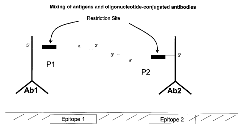
[00026] FIGURE 1A shows mixing of antigens and oligonucleotide-conjugated
antibodies.
[00027] FIGURE 1B shows hybridization of adjacent probes.
[00028] FIGURE 1C shows a polymerase extension and restriction enzyme nicking.
[00029] FIGURE 1D shows extension, displacement and linear amplification.
[00030] FIGURE lE shows hybridization, polymerase extension, nicking and
exponential
amplification.
[00031] FIGURE 1F shows mixing of antigens and oligonucleotide-conjugated
antibodies.
[00032] FIGURE 1G shows hybridization of adjacent probes.
[00033] FIGURE 1H shows extension of probes with a polymerase.
[00034] FIGURE 11 shows denaturation of a probe-extension duplex and the
binding of
SDA primers.
[00035] FIGURE 1J shows amplicon formation from hybridized probes of opposite
sequence orientation.
[00036] FIGURE 2A shows hybridization of a splint oligonucleotide.
[00037] FIGURE 2B shows ligation of adjacent probes.
[00038] FIGURE 2C shows DNA polymerase extension and displacement.
[00039] FIGURE 2D shows the use of two hybridized proximity probes to ligate a
third
probe.
[00040] FIGURE 2E shows the use of two hybridized proximity probes in opposite
sequence orientation to ligate a third probe.
[00041] FIGURE 3A shows a single-tether probe.
[00042] FIGURE 3B shows extension and displacement of a single-tether probe.
[00043] FIGURES 3C and D show nicking, extension, displacement and capture.
- 15 -
CA 02522753 2005-10-14
WO 2004/094456 PCT/US2004/011918
[00044] FIGURE 3E shows splint oligonucleotides having a 373' configuration.
[00045] FIGURE 3F shows extension/displacement of splint oligonucleotides.
[00046] FIGURE 3G shows target-mediated probe cycling.
[00047] FIGURE 3H shows splint oligonucleotides having a 573' configuration.
[00048] FIGURE 31 shows splint oligonucleotides having a 575' configuration.
[00049] FIGURE 3J shows splint oligonucleotides having a 373' configuration.
[00050] FIGURE 3K shows splint oligonucleotides having a 31/3' configuration.
[00051] FIGURE 3L shows displacement of splint oligonucleotides from a
captured
complex.
[00052] FIGURE 4A shows a simple competitive hybridization blocker.
[00053] FIGURE 4B shows a recessed competitive hybridization blocker.
[00054] FIGURE 4C shows a disabling hybridization blocker.
[00055] FIGURE 4D shows a displaceable hybridization blocker.
[00056] FIGURE 4E shows a self-displacing hybridization blocker.
[00057] FIGURE 4EE shows the use of a 3' probe tail to stabilize a probe-
blocker duplex.
[00058] FIGURE 4F shows competitive hybridization blocker in a binary immuno-
SDA
reaction.
[00059] FIGURE 4G shows a disabling hybridization blocker in a binary immuno-
SDA
reaction.
[00060] FIGURE 4H shows step-wise blocking in a binary immuno-SDA reaction.
[00061] FIGURE 41 shows the post-binding addition of hybridization blockers in
a binary
immuno-SDA reaction.
[00062] FIGURE 5A shows 4 splint oligonucleotide hybridization.
[00063] FIGURE 5B shows extension and displacement.
- 16 -
CA 02522753 2005-10-14
WO 2004/094456 PCT/US2004/011918
[00064] FIGURE 5C shows RNA polymerase activity, hybridization and extension.
[00065] FIGURE 5D shows RNase H activity, hybridization and extension.
[00066] FIGURES 6A ¨ C show restriction endonuclease-mediated release of an
attached
conjugate.
[00067] FIGURES 6D and E show polymerase- and restriction endonuclease-
mediated
release.
[00068] FIGURE 6F shows physical release.
[00069] FIGURES 6G and 6GG show scissile linkages and chemical cleavage.
[00070] FIGURE 6H shows oligonucleotide displacement.
[00071] FIGURES 61 and J show oligonucleotide extension.
[00072] FIGURES 6K and L show RNase H release.
[00073] FIGURE 6M shows a self-priming capture/displacement oligonucleotide.
[00074] FIGURE 6N shows the involvement of the displaced probe moiety in the
formation of an amplicon.
[00075] FIGURE 7A shows immobilization of a first proximity member by
hybridization
of an oligonucleotide moiety of the first proximity member with a capture
oligonucleotide.
[00076] FIGURE 7B shows the binding of a target analyte to the immobilized
first
proximity member.
[00077] FIGURE 7C shows the formation of an immobilized two-site "sandwich" by
the
binding of a second proximity member to the immobilized complex between the
target
analyte and the first proximity member.
[00078] FIGURE 7D shows a mechanism by which a target-independent amplicon may
form.
- 17 -
CA 02522753 2005-10-14
WO 2004/094456 PCT/US2004/011918
[00079] FIGURES 7E ¨ H show the use of a hybridization blocker oligonucleotide
to
suppress probe-probe interactions that lead to target-independent amplicon
formation.
[00080] FIGURE 71 shows the release of an immobilized complex between a target
analyte and two proximity members using low ionic strength.
[00081] FIGURE 7J shows the use of a capture oligonucleotide and release in a
heterogeneous assay format.
[00082] FIGURES 8A ¨ C show heterogeneous immuno-amplification.
[00083] FIGURE 8D shows heterogeneous immuno-amplification with a scissile
linkage.
[00084] FIGURE 9 shows heterogeneous immuno-amplification with splint
oligonucleotides.
[00085] FIGURE 10 shows a universal immuno-amplification system.
[00086] FIGURE 11A shows hairpin release probes.
[00087] FIGURE 11B shows hairpin hybridization blocker probes.
[00088] FIGURE 11C shows displacement of hairpin hybridization blocker probes.
[00089] FIGURE 12 shows detection of antigen-specific immunoglobulin.
[00090] FIGURE 13 presents a map of representative probes, primers, and tether
oligonucleotides for binary immuno-SDA.
[00091] FIGURES 14A ¨ E show the use of a 3' capped oligonucleotide moiety to
form
amplicons attached to a first proximity member and to a second proximity
member, but not to
both proximity members simultaneously.
[00092] FIGURE 15A shows a two-color, real-time fluorescence profile for
immuno-SDA
detection of IL-8.
[00093] FIGURE 15B shows a calibration line for quantification of IL-8.
- 18 -
CA 02522753 2005-10-14
WO 2004/094456 PCT/US2004/011918
DETAILED DESCRIPTION OF THE PREFERRED EMBODIMENTS
[00094] Minute quantities of an analyte may be detected with great sensitivity
by the
present invention. The invention provides conjugates of analyte-specific
binding factors,
such as antibodies, conjugated to oligonucleotide moieties that can form an
amplicon. The
conjugation between antibodies and other proteins with oligonucleotides is
known in the art
and taught, for example, in U.S. Patents No. 5,849,878 and No. 5,665,539,
which are
incorporated by reference in their entirety herein. If the analyte-specific
binding factor is a
nucleic acid, for example, an aptamer, then the analyte-specific binding
factor and the
oligonucleotide or probe moiety may be synthesized in one contiguous strand
using chemical
synthesis methods known in the art. The term "conjugate" still applies to such
aptamer-probe
entities. The conditions for establishing an amplicon by adjoining
oligonucleotides that are
each conjugated to an antibody are also known and taught in U.S. Patent No.
6,511,809, for
example. Conditions and methodologies for amplifying amplicons and for
detecting their
presence are also known in the art, as taught in U.S. Patent No. 6,511,809 and
U.S. Patent
Application Publication No. 2002/006779, both incorporated herein by reference
in their
entirety. The use of labeled probes for the detection of amplification
products, for example,
also is taught in U.S. Patents No. 5,928,869, No. 5,919,630; No. 5935,791; No.
6,316,200;
and No. 6,379,888, all incorporated herein by reference in their entirety.
U.S. Patent No.
5,840,487 teaches the use of internal controls for isothermal nucleic acid
amplification
reactions and is also incorporated herein by reference in its entirety.
[00095] According to the present invention, a preferred method of
amplification by SDA is
detailed in FIGURE 1. Abl and Ab2 are antibodies that recognize adjacent
epitopes 1 and 2
and that are conjugated to oligonucleotide probes P1 and P2, respectively
(FIGURE 1A).
The antibodies are representative, but not limiting examples, of the analyte-
specific binding
- 19 -
CA 02522753 2005-10-14
WO 2004/094456 PCT/US2004/011918
components that are useful in the present invention. For instance, useful
analyte-specific
binding components known in the art include functional fragments of
antibodies, such as Fc,
Fv, Fab' and F(ab1)2 fragments. Other examples of analyte-specific binding
components
include aptamers, ligands specific for a receptor analyte, or a receptor that
is specific for a
ligand analyte. Further, it will be understood by the skilled artisan that
various different
types of analyte-specific binding components may be used in combination.
"Oligonucleotide
probes" and "oligonucleotide moieties" are used synonymously for the purposes
of the
present invention. The term "oligonucleotide" should not be understood as
placing an upper
size limit on the nucleic acid moieties for the purpose of this invention;
therefore,
"oligonucleotide" is synonymous with "polynucleotide," as used herein. For the
purposes of
the present invention, an oligonucleotide may be composed in whole or in part
by DNA,
RNA or an analogue or derivative thereof. In this embodiment, P1 and P2
comprise
complementary 3' terminal sequences and upstream SDA nick sites. The use of
nick sites for
SDA and the conditions for SDA in general are described in U.S. Patents No.
5,919,630; No.
5,846,726; and No. 6,054,729, which are incorporated herein by reference in
their entirety.
The 3' ends of P1 and P2 hybridize to one another when the two antibodies to
which they are
linked are held in close proximity by binding to their respective epitopes
(FIGURE 1B).
Conditions conducive to nucleic acid hybridization, including the number of
base pairs or
mismatches in the hybridized portion of a nucleic acid and the temperature and
ionic strength
of the buffer in which the hybridization occurs, are well-known in the art and
are generally
described, for example, in Sambrook et al., MOLECULAR CLONING, A LABORATORY
MANUAL
(31.d ed., 2001). The bulk solution concentration of Abl and Ab2 is relatively
low compared
with that at the surface of the antigen, such that antigen-independent
hybridization of P.1 and
P2 is minimized. DNA polymerase is then used to fill in the recessed 3' ends
of the P1:P2
- 20 -
CA 02522753 2005-10-14
WO 2004/094456 PCT/US2004/011918
hybrid (FIGURE 1C). This serves to generate double-stranded restriction sites
that are
recognized by the SDA nicking enzyme. A nicking enzyme catalyzes the cleavage
of only
one strand of the double-stranded DNA template. Nicking and polymerase
extension from
the site of the nick displaces the downstream DNA strand into solution and
regenerates the
nick site (FIGURE 1D). Repeated cycling of the nicking and
extension/displacement steps
may be used to produce multiple copies of the displaced strand. The displaced
strand is
captured by a complementary SDA primer (FIGURE 1E). Extension from the 3' ends
of the
captured strand and hybridized SDA primer produces a double-stranded DNA
molecule that
may be exponentially amplified through a series of intermediates. In an
alternative
embodiment, only one of the oligonucleotide probes P1 and P2 comprises an SDA
nick site.
[00096] In another embodiment (FIGURE 1F ¨ I), probes P1 and P2 lack SDA nick
sites,
but comprise instead sequences c and d', which also are present on SDA primers
SP2 and
SP1, respectively (see FIGURE 1I). Extension of the 3' ends of P1 and P2
creates a duplex
containing complementary sequence d on the extension product of P1 and
complementary
sequence c' on the extension product of P2 (see FIGURE 114). The strands of
the duplexed
extension products are then separated by, for example, heating, whereupon SDA
primers SP1
and SP2 hybridize to the complement of the newly synthesized sequences d and
c' of the
extended probes. The extended probes optionally may comprise a sequence
located 3' to the
binding sites of the SDA primers, shown as sequence e of extended P1 and
sequence b' of
extended P2. These sequences hybridize with bumper primers SB1 and SB2 (see
FIGURE
1I). During SDA, the SDA primers SP1 and SP2 are extended by polymerase (not
shown).
Extension of the bumper primers, if present, serves to displace the SDA primer
extension
products from the probe strands, and the displaced strands are then amplified
by SDA, as
described in U.S. Patents No. 5,270,184; No. 5,919,630; No. 5,846,726; and No.
6,054,729.
-21-
CA 02522753 2005-10-14
WO 2004/094456 PCT/US2004/011918
In the event that the extended probes do not contain sequences 3' to the SP1
and SP2 binding
sites (not shown), the 3' ends of the probes that are hybridized to SDA
primers are extended
by polymerase, creating nickable restriction sites that allow subsequent
nicking and strand
displacement by SDA as described above.
[00097] In preceding embodiments, oligonucleotide moieties (P1 and P2) were
conjugated
to their respective analyte binding entities (Ab1 and Ab2) through linkages
located at or near
their 5' termini. In an alternative embodiment illustrated in FIGURE 1J,
conjugate Abl-Pi is
formed through a linkage located at or near the 3' terminus of P1, while
conjugate Ab2-P2 is
fowled through a linkage located at or near the 5' terminus of P2. P1
comprises sequence (a
b c d e 1) (read 5' to 3'), and P2 comprises sequence (j' h' g' ' e') (read 5'
to 3'). Ab2-
P2 further comprises an extendible 3' end (i.e., a 3' terminal hydroxyl
group). As shown,
sequence (e 1) of P1, which is capable of hybridizing to sequence (f e') of
P2, is located 5' of
the site at which P1 is conjugated to Abl, whereas (f e') is located 3' of the
site at which P2
is conjugated to Ab2. Probes P1 and P2 are, therefore, said to be linked to
their respective
analyte binding entities (Ab1 or Ab2) in opposite sequence orientations. When
P1 and P2
are brought into close proximity, for example, through binding of their
respective proximity
members to the same target analyte molecule, sequence (e 1) of P1 hybridizes
with (f e') of
P2, as depicted on the left side of FIGURE 1J. Polymerase may then be used to
extend the 3'
end of P2 to create an extension product (i.e., amplicon) P2-ext containing
the new sequence,
as shown. P2-ext may then be detected by methods known in the art, making use
of all or
part of the new sequence (d' c' b' a') to distinguish P2-ext from unconverted
P2. For
example, P2-ext may be amplified by nucleic acid amplification methods
described above.
P2-ext may be separated from P1 by heating the solution, and a primer may
hybridize to the
new sequence at the 3' end of P2-ext and be extended to create a complement of
P2-ext.
- 22 -
CA 02522753 2005-10-14
WO 2004/094456 PCT/US2004/011918
Subsequent rounds of amplification may involve separation of the complement
from P2-ext
and hybridizing to the complement a different primer comprised of a sequence
located near
the 5' end of P2. In a preferred embodiment, sequence b will contain the
single-strand
component of a recognition sequence for an SDA-compatible restriction enzyme.
Formation
of P2-ext then creates a double-stranded recognition sequence that is nicked
by the restriction
enzyme. Extension from the nick creates a new strand that is complementary to
P2-ext,
regenerating the nickable recognition sequence. This product may be amplified
and detected
by SDA methods referred to above. Optionally, sequence i' of P2 may also
comprise the
single-strand component of a recognition sequence for SDA and, if so, the
duplex formed
between P2-ext and its full-length complementary strand will contain two
nickable restriction
enzyme recognition sequences. In another embodiment, sequence b may be a
single-strand
component of an RNA polymerase promoter site. Formation of P2-ext then creates
a double-
stranded RNA polymerase promoter, which may be used to direct the activity of
an RNA
polymerase to synthesize RNA molecules that are complementary to sequence (j'
h' g' F e'
d' c') of P2-ext. These RNA molecules may be detected directly, or they may be
further
amplified by methods such as 3SR, NASBA, TMA, or transcription-based
amplification.
Optionally, sequence i' of P2 may comprise the single-strand component of an
RNA
polymerase promoter. In this case, extension of a primer hybridized to the 3'
end of P2-ext
would create a double-stranded promoter site that can be used to direct the
activity of the
RNA polymerase to synthesize RNA molecules comprising the sequence (h' g' F e'
d' c' b' a'),
which may be detected directly or amplified using the aforementioned methods.
Regardless
of the method of detection or amplification of P2-ext, the embodiments
depicted in Figure 1J
comprise probe moieties P1 and P2 that are linked to their respective analyte-
binding
elements Abl and Ab2 in opposite sequence orientations, and the two probes
hybridize to
- 23 -
CA 02522753 2005-10-14
WO 2004/094456 PCT/US2004/011918
each other in a target-mediated process, creating a duplex with an extendible
3' end that is
subsequently extended to create an amplicon. In the absence of target-analyte,
P1 and P2
will not be brought into close proximity, and P2-ext will not form except
through spurious
(i.e., target-independent) interactions mentioned below, which may be
suppressed by
hybridization blocking oligonucleotides, also described below. P2-ext is,
therefore, produced
as a consequence of the presence of target analyte and in proportion to the
quantity of target
analyte present. Determination of the quantity of P2-ext produced may,
therefore, be used to
determine the quantity of target analyte present in a sample.
[00098] FIGURES 2A - C detail a representative use of a splint
oligonucleotide. Abl and
Ab2 are antibodies that recognize adjacent epitopes 1 and 2 and are conjugated
to
oligonucleotide probes P1 and P2, respectively (FIGURE 2A). P1 is conjugated
to Abl
through a linkage located at or near its 5' terminus, and it comprises a 3'
terminal hydroxyl
group and upstream SDA nick site. Probe P2 is conjugated to Abl at its 3' end,
and it
comprises an SDA primer binding site and 5' terminal phosphate group. The
sequence of the
splint oligonucleotide S is complementary to the 3' end of probe P1 and the 5'
end of probe
P2 such that, when held in close proximity by binding of the antibodies to
their respective
epitopes, oligonucleotides P1 and P2 form a double-stranded hybrid with the
splint S. When
hybridized to the splint oligonucleotide S, the 3'-OH of P1 and 5'-PO4 of P2
are adjacent, and
DNA ligase is used to catalyze the formation of a phosphodiester bond linking
the P1 and P2
sequences (FIGURE 2B). SDA primer SP1 hybridizes to probe P2 upstream of
splint
oligonucleotide S. A strand-displacing DNA polymerase extends from the 3' ends
of primer
SP1 and splint oligonucleotide S. Extension of primer SP1 displaces the
extension product
of splint oligonucleotide S (FIGURE 2C) and creates a double-stranded DNA
molecule with
SDA restriction enzyme nick sites at either end. This molecule is analogous to
that depicted
- 24 -
CA 02522753 2005-10-14
WO 2004/094456 PCT/US2004/011918
in FIGURE 1C. Nicking, polymerase extending from the nick, and displacing the
downstream strand leads to exponential amplification (FIGURES 1D - E). In one
embodiment, the probe P1 does not comprise a SDA nick site. In another
embodiment, the
splint oligonucleotide S comprises a 3' cap to prevent 3' extension of the
splint S.
[00099] FIGURES 2D and 2E illustrate an alternative embodiment for target-
mediated,
ligase-catalyzed amplicon formation using a pair of proximity members. Probes
P1 and P2
are linked, or conjugated, to their respective antibodies (or other analyte
binding entities)
Abl and Ab2. Conjugation may occur through linkages at or near the 5' termini
of both
probes, as shown in FIGURE 2D, or one of the two probes (P1) may be conjugated
through a
linkage located at or near the 3' end of the probe. P1 comprises sequence
(abcdef)
(read 5' to 3'), and P2 comprises sequence (j' h' g' f e') (read 5' to 3').
Conjugate Ab2-P2
further comprises a 3' terminal hydroxyl group. In configurations depicted by
either FIGURE
2D or 2E, sequences (e 1) of P1 and (f' e') of P2 are capable of hybridizing
to each other. A
third probe P3 comprises sequence (d' c' b' x' y') and further comprises a 5'
teiminal
phosphate group. P3 is capable of hybridizing to sequence (b c d) of probe P1
(adjacent to
sequence (el) of P1). In the presence of target analyte, P1 and P2 are brought
into close
proximity and form a duplex through hybridization of sequences (e f) and (f
e'). Probe P3
may be hybridized to P1, as shown, either before or after P1 and P2 hybridize.
In either case,
the 5' nucleotide of P3 is positioned adjacent to the 3' nucleotide of P2 and,
in this
configuration, P2 and P3 may be covalently linked together by DNA ligase (or
other ligation
mechanism) to form the amplicon P2:P3, as shown. P2:P3 may then be detected by
various
methods, including amplification, such as those described above for
embodiments depicted
in FIGURE 1J. In this case, however, sequence x and/or y' will be used as
sites for primer
hybridization. In the absence of target analyte, P1 and P2 will not be brought
into close
- 25 -
CA 02522753 2005-10-14
WO 2004/094456 PCT/US2004/011918
proximity, and P2:P3 will not form, except through spurious (i.e., target-
independent)
interactions between P1 and P2 mentioned below, which may be suppressed by
hybridization
blocking oligonucleotides also described below. P2:P3 is, therefore, produced
as a
consequence of the presence of target analyte and in proportion to the
quantity of target
analyte present. Determination of the quantity of P2:P3 produced may,
therefore, be used to
determine the quantity of target analyte present in a sample. Hybridization
blockers are not
required during amplification of amplicons formed by ligating oligonucleotide
moieties of
proximity members because probes that can be joined by ligation typically do
not form
hybrids with each other and therefore do not have the potential to undergo
spurious probe
conversion during amplification involving 3' extension of oligonucleotides.
[000100] FIGURE 3 shows a representative embodiment of the present
invention that
comprises a splint oligonucleotide designed to bridge the gap between two
oligonucleotide
moieties of proximity members. In one embodiment (FIGURE 3A), one of the
proximity
antibodies Abl is conjugated through a linkage at or near the 3' end of a
tether-
oligonucleotide. Hereafter, a "tether oligonucleotide" denotes an
oligonucleotide moiety that
is displaced from the amplicon but remains conjugated to the analyte-specific
binding moiety.
The tether oligonucleotide TO is complementary to a segment (preferably at or
near the 5'
end) of the splint oligonucleotide Pl. Splint oligonucleotide P1 may further
comprise a
primer sequence to facilitate amplification of the converted probe and a
detector region to
facilitate detection of the converted probe. P1 may also comprise a
restriction recognition
sequence to facilitate amplification by SDA. In addition, the 3' sequence of
the splint
oligonucleotide is complementary to the 3' end of probe P2 that is conjugated
through its 5'
terminus to antibody Ab2. As depicted in FIGURE 3A, the 5' end of P1 is
complementary to
the tether oligonucleotide TO, which is attached to Abl. Optionally, the
tether
-26 -
CA 02522753 2005-10-14
WO 2004/094456 PCT/US2004/011918
oligonucleotide may be complementary to a sequence not on the 5' end of Pl.
When
antibodies Abl and Ab2 are bound to their respective epitopes, splint
oligonucleotide P1 is
able to hybridize to both TO and P2 (FIGURE 3A). Extension from the 3' ends of
probe P2
and the splint oligonucleotide displaces tether oligonucleotide TO and creates
a double-
stranded DNA molecule linked to antibody Ab2 (FIGURE 3B). Nicking of this
double-
stranded product, extending with polymerase, and displacing the downstream
strand
generates a single-stranded oligonucleotide that may form a hybrid with a
complementary
SDA primer (FIGURE 3C). This leads to exponential amplification through a
succession of
intermediate nicking, extending, displacing and priming events (FIGURE 3D).
[000101] FIGURE 3E depicts a second embodiment for bridging the gap between
antibodies. In this configuration, each antibody (Abl and Ab2) is conjugated
with a different
tether oligonucleotide, a' for Abl and j for Ab2. Typically, the tether
oligonucleotide of the
antibody Abl differs from the tether oligonucleotide of the second antibody
Ab2. In this
case, a' and j are not equivalent in sequence. Splint oligonucleotides P1 and
P2 each contain
a sequence (optionally near the 5' end) that is complementary to the
oligonucleotide
sequences a' and j. For example, P1 contains sequence a, and P2 contains
sequence j'.
Sequence a of probe P1 hybridizes to sequence a' of Abl, and sequence j of Ab2
hybridizes
to sequence j' of P2. In this embodiment, P1 and P2 each contain a short 3'
sequence that is
complementary to the other probe; therefore, sequence (e f) of P1 is
complementary to (I" e')
of P2. Appreciable hybridization of these complementary 3' sequences of P1 and
P2 occurs
with high efficiency only when the probes P1 and P2 are brought into spatial
proximity by
consequence of also being hybridized to tether oligonucleotides (a' and j) of
the antibodies
bound to proximate epitopes. Hybridization of the 3' ends of P1 and P2 creates
a short
duplex with recessed 3' ends, which may then be extended by polymerase. In one
-27 -
CA 02522753 2005-10-14
WO 2004/094456 PCT/US2004/011918
embodiment, extension of the 3' ends serves to displace the splint
oligonucleotides P1 and P2
from the tether oligonucleotides (and antibodies), while simultaneously
creating a duplex
comprised of the extension products (P1-ext and P2-ext) of both probes (FIGURE
3F). P1
and P2 extension products may then be detected by a variety of amplification
methods known
in the art, including PCR, SDA, ligase chain reaction, 3 SR, QI3 replicase-
based amplification,
solid phase amplification and NASBA. Sequences contained on the probes, e.g.,
sequences
(b, c, d, e, 1) of P1, and (e', f', g', h', j') of P2, or probe extension
products may be used to
facilitate amplification and detection of the probes. Special sequences that
may be used to
facilitate amplification include primer binding sites, restriction
endonuclease sites, sequences
capable of hybridizing with hybridization blocker oligonucleotides, RNA
promoter sites, and
the like. Detection of amplified products may occur by heterogeneous or
homogeneous
methods well-known in the art. Alternatively, duplex II of FIGURE 3F may be
detected
directly without amplification by methods well-known in the art. If the method
employs a
DNA polymerase that possesses a 5'-3' exonuclease activity, e.g., Taq DNA
polymerase, the
tether oligonucleotide (a' or j) may be degraded during the extension process,
and the
degradation products may be detected as an indication of the presence of
target antigen.
[0001021 While FIGURES 3E and 3F depict antibodies conjugated to the 3'
ends of the
tether oligonucleotides, FIGURE 3H depicts alternative configurations in which
both
antibodies of a proximity pair are conjugated to the 5' ends of the tether
oligonucleotides.
Likewise, FIGURE 31 depicts an embodiment in which one tether oligonucleotide
is
conjugated to an antibody through a 5' linkage and the other oligonucleotide
is conjugated
through a 3' linkage. In each of these latter two configurations, 3' extension
of the probe
sequences P1 and P2 results in displacement of the probes from the tether
oligonucleotides
and creation of a double-stranded duplex identical to that shown in FIGURE 3F.
-28 -
CA 02522753 2005-10-14
WO 2004/094456 PCT/US2004/011918
[000103] If the tether oligonucleotides are not degraded during the
displacement
process, a second set of probe molecules P1 and P2 may hybridize to the
vacated tether
oligonucleotides of the target-bound proximity members (FIGURE 3G). As before,
the 3'
ends of P1 and P2 anneal, and extension again results in displacement of the
probes and
creation of a duplex comprised of P1-ext and P2-ext. The vacated tether
oligonucleotides
again anneal to a new pair of unextended probes (P1 and P2) if present, and
the cycle of 3'
hybridization, extension, displacement, and subsequent binding of unextended
probes
continues as long as Abl and Ab2 remain bound to the proximate epitopes and a
supply of
P1 and P2 exists. As a result of this cycling process, multiple copies of
detectable probe
extension duplexes are formed from each target present.
[000104] In all the examples shown in FIGURES 3A ¨ 3L, initial
hybridization of the
probes to the tether oligonucleotides may occur either before or after the
antibody has bound
to the target molecule, depending on the experimental protocol used. In one
embodiment, at
least one of the antibodies Abl and Ab2 is or may be covalently or non-
covalently linked to a
paramagnetic particle (FIGURE 9) or other solid surface (FIGURE 3L), e.g., the
inner wall of
a microwell. In configurations where at least one of the antibodies is linked
to a bead, solid
surface or other solid matrix, and both probes P1 and P2 (FIGURE 3L) are
attached
indirectly to the antibodies by hybridization to tether oligonucleotides,
extension of the
probes creates a duplex that is displaced from the antibody-target complex,
while the
complex itself remains attached to the bead, solid surface or other solid
matrix. If desired,
the solution containing the displaced duplex then may be removed and analyzed
or amplified
in a separate well or compartment, leaving behind the complex and any material
bound non-
specifically to the matrix surface.
- 29 -
CA 02522753 2005-10-14
WO 2004/094456 PCT/US2004/011918
[000105] In another embodiment of the present invention, a ligation splint
oligonucleotide may be complementary to a portion of both splint
oligonucleotides P1 and P2
as shown, for example, in FIGURE 3J. When hybridized to the ligation splint
oligonucleotide, probes P1 and P2 may be ligated as described in FIGURE 2 so
that the 3'
end of P1 is covalently joined to the 5' end of P2 (FIGURE 3J), which may then
be amplified
as described in FIGURE 2.
[000106] In an alternative embodiment, the splint oligonucleotide
hybridizes to a tether
oligonucleotide (j') and to one probe molecule P2, as exemplified in FIGURE
3K. The 3' end
of the splint-bound P2 may then hybridize with the complementary 3' end of
spatially
proximate Pl. 3' extension of the probes displaces the probes from the splint
and tether
oligonucleotides and forms a full-length, amplifiable duplex analogous to that
produced in
the earlier examples shown in FIGURE 3.
[000107] FIGURE 4 shows hybridization blocker oligonucleotides that are
designed to
reduce the prevalence of hybridization between probe molecules linked to
antibodies that are
not bound to proximate epitopes. Such target-independent hybridization is a
source of
background signal because it results in probe extension products that are
indistinguishable
from those produce by bona fide target binding events, and background signal
reduces the
overall sensitivity of the detection method. The current invention comprises
the use of
hybridization blocker oligonucleotides (or "hybridization blockers") to reduce
spurious,
target-independent probe interactions that lead to background signal. FIGURE
4A depicts
the basic principle underlying the use of hybridization blockers in proximity-
based
amplification methods. Short sequences of mutual complementarity (e f and f'
e') (read 5' to
3') comprise the 3' ends of P1 and P2. These sequences may hybridize to each
other to form
a duplex with 5' overhangs as shown. The number of probe molecules populating
the
- 30
CA 02522753 2005-10-14
WO 2004/094456 PCT/US2004/011918
duplexed versus single-stranded state depends on the total probe concentration
and on the
intrinsic stability of the duplex, which in turn is related to duplex length
and composition.
The interaction between P1 and P2 can be diminished or reversed by the
addition of a
hybridization blocker oligonucleotide, preferably in molar excess over the
probes, which
upon hybridization to P1 competitively blocks the interaction between the two
probes. The
blocker need not be complementary to P1 across the entire subsequence that
interacts with
P2. Partial complementarity across this site also diminishes hybridization of
P1 to P2.
[000108] In one embodiment, the hybridization blocker oligonucleotide
comprises a
first subsequence (e' or f' e') that is identical to part or all of that
portion of P2 that is
complementary with P1 (subsequence e 1). In a second embodiment (FIGURE 4B),
the
hybridization blocker oligonucleotide comprises the first subsequence, defined
above, and a
second subsequence (d' in FIGURE 4B) that is complementary to a segment of P1,
but not
identical to a subsequence of P2. The second subsequence of the hybridization
blocker
stabilizes the blocker-P1 interaction relative to the P1-P2 interaction,
thereby improving
blocking efficiency. The second subsequence also may serve as a site for
nucleation of the
P1-blocker duplex in the event that P2 molecule is already hybridized to Pl.
Following
nucleation, formation of the full P1-blocker duplex then displaces P2 from Pl.
The P1
subsequence d of this embodiment may be directly adjacent to the probe
subsequence e, as
shown in FIGURE 4B, or it may be located some nucleotides away from
subsequence e (not
shown). In the latter case, the hybridization blocker subsequence d' may be
linked indirectly
to hybridization blocker subsequence e' through a spacer, comprising
additional nucleotides
and/or a non-nucleotide linker, such as a tetraethylene glycol (TEG) moiety.
[000109] In a third embodiment (FIGURE 4C), the hybridization blocker
oligonucleotide may comprise a first subsequence (defined above), optionally a
second
- 31 -
CA 02522753 2005-10-14
WO 2004/094456 PCT/US2004/011918
subsequence (defined above), and a third subsequence (t' s' in FIGURE 4C),
which is located
5' of the first subsequence and which may serve as a template for the 3'
extension of P1. The
third subsequence optionally may contain a suitable non-nucleotide moiety m'
(i.e., a "5'
cap"), which are well known in the art, to prevent the addition of non-
templated nucleotides
to the 3' extension product of P1 and to discourage binding of polymerase to
the blunt-ended
duplex formed by extension of P1 (see FIGURE 4C). Preferably, the 3' extension
of P1 on
the blocker-template yields a P1-extension product containing a new sequence
(s t) at its 3'
end that is not complementary to the P2 sequence. Addition of the 3' sequence
(s t),
therefore, serves to disable P1 as a functioning primer for DNA synthesis on a
P2 template.
Addition of the sequence (s t) also serves to stabilize the blocker-Pi
interaction by increasing
the number of complementary base pairs between the two molecules. Optionally,
the new 3'
sequence (s t) produced in this embodiment is entirely or partially
complementary to a
segment of P1, such that, if the extended P1 and hybridization blocker
dissociate, the
extended P1 will fold into a stem-loop (hairpin) structure, diminishing any
interaction with
P2. In this case, the 3' end of the P1 hairpin optionally may be extended to
lengthen the stem
of the hairpin, and this extension optionally may create a nickable or
cleavable restriction
endonuclease site within the P1 molecule.
[000110] In a fourth embodiment, it may be desirable to block reversibly
the interaction
between P1 and P2 during a certain phase of a process. FIGURE 4D depicts a
hybridization
blocker design that allows reversible blocking. In this embodiment, the
hybridization blocker
comprises a subsequence (e' d') that is complementary to the probe to be
blocked (P1 in
FIGURE 4D) and one or more tail sequences (t' and/or k') that are not
complementary to the
probe. Hybridization of the hybridization blocker to P1 precludes P1-P2
interactions. At the
desired time, a deblocking oligonucleotide may be added to displace the
hybridization
- 32 -
CA 02522753 2005-10-14
WO 2004/094456 PCT/US2004/011918
blocker oligonucleotide from Pl, freeing the latter to interact with P2. The
deblocking
oligonucleotide comprises one or more tail sequences (t and/or k) that are
complementary to
the tail sequences of the hybridization blocker. The tail sequences serve as a
site of
nucleation for the blocker-deblocker hybridization. Once hybridization of the
complementary tail sequences is nucleated, additional base-pairs form between
the
hybridization blocker and deblocker until the blocker is displaced from Pl. To
ensure
displacement of the blocker from the probe, the overall thermodynamic
stability of the
blocker-deblocker complex must be higher than the probe-blocker complex. The
deblocking
oligonucleotide need not be perfectly complementary to the hybridization
blocker
oligonucleotide, provided that the thermodynamic stability of the blocker-
deblocker duplex is
higher than the stability of probe-blocker duplex. For instance, it may be
desirable for the
deblocker to contain one or more nucleotides that form mismatches with the
hybridization
blocker oligonucleotide, provided that the resulting blocker-deblocker duplex
is more stable
than the probe-blocker duplex. In particular, it may be desirable for sequence
e of the
deblocker to contain one or more nucleotides that form one or more mismatches
with
sequence e' of the blocker. The primary function of these mismatching
nucleotides of the
deblocker is to destabilize potential interactions between sequence e of the
deblocker and
sequence e' of P2. In a variation of this embodiment, the hybridization
blocker may be
displaced by polymerase-catalyzed extension of sequence k as shown in FIGURE
4E. In this
case, the deblocking sequence is synthesized directly upon the blocking
sequence, and no
separate deblocker oligonucleotide need be added. In yet another embodiment, a
probe may
comprise a 3' stem-loop structure at or near its 3' end that serves to block
interactions
between probe molecules (see FIGURE 11).
-33-
CA 02522753 2005-10-14
WO 2004/094456 PCT/US2004/011918
[0001111 To prevent 3' extension of the blocker by polymerase, all
hybridization blocker
oligonucleotides described above, except the hybridization blocker with the
hairpin structure
depicted in FIGURE 4E, may comprise a cap on the 3' terminal nucleotide.
Hybridization
blocker oligonucleotides with 3' caps are referred to as "capped
oligonucleotides." Such 3'
caps are well-known in the art and include inverted nucleotides, 2'-3'
dideoxyribonucleotides,
and 3' deoxyribonucleotides. Hybridization blocker oligonucleotides may
contain a 3' tail
sequence that does not form complementary base-pairs with the probe
nucleotides when the
hybridization blocker forms a duplex with the probe. The non-base-paired 3'
tail also serves
to prevent 3' extension of the blocker when the hybridization blocker is
duplexed with the
probe and, therefore, serves as a "3' cap" as well.
[0001121 FIGURE 4EE illustrates the use of a 3' tail on Probe 1 (P1) to
facilitate
stabilization of the P1 blocker duplex. The 3' tail of P1 is comprised of
sequence x y and is
located 3' of sequence (e f), which is capable of hybridizing with sequence (f
e') of Probe 2
(P2). The 3' tail of P1 does not hybridize to P2. The hybridization blocker
comprises
sequence (y' f') and optionally e". The hybridization blocker is, therefore,
capable of
hybridizing to P1 to form a duplex covering the 3' tail of P1, as well as all
or part of sequence
e f of Pl. Formation of the blocker:Pl duplex will reduce the prevalence of
P1:P2 hybrids
as described above. Base-pairing between (x y) of P1 and (y' x') of the
hybridization blocker
serves to stabilize the blocker:Pl duplex. The hybridization blocker
optionally comprises
sequence z' located 5' of sequence (y' x'). Sequence z' may serve as a site
for initiating
hybridization of the deblocker oligonucleotides in methods described above.
[000113] FIGURE 4F illustrates the use of hybridization blocker
oligonucleotides in a
binary immuno-amplification reaction. P1 and P2 are conjugated directly to
antibodies Abl
and Ab2, respectively. The same principles apply regardless if the probes are
bound to an
-34-
CA 02522753 2005-10-14
WO 2004/094456 PCT/US2004/011918
antibody indirectly via hybridization to a tether oligonucleotide, or if the
analyte-specific
binding components are aptamers or other analyte-specific binding molecules
other than
antibodies. As depicted, a hybridization blocker oligonucleotide hybridizes to
the 3' end of
P1, precluding its interaction with P2. Hybridization of the hybridization
blocker does not
interfere with binding of the antibody to the target analyte, and initially
the hybridization
blocker is hybridized to P1 strands whether or not Abl is complexed with the
analyte or free
in solution (state I). Typically, the concentration of free Abl and Ab2 (and
the conjugated
probes) in bulk solution is between 1 fM and 10 nM. Because hybridization
blocker
concentrations typically are 10- to100,000-fold higher than probe
concentrations, blocker-Pi
interactions predominate over P1-P2 interactions for probes that are
conjugated to antibodies
that are not target-bound; however, when epitopes 1 and 2 of a target molecule
are bound to
Abl and Ab2, respectively, the effective local concentration of P1 relative to
P2 on the
ternary target-antibody complex becomes much higher (typically 1-100 ptM) than
the
concentration of probes and hybridization blockers in bulk solution. As a
result, the P1:P2
duplex prevails over the P1:blocker duplex for probes linked to target-bound
antibodies (state
II). Polymerase-catalyzed extension of the 3' ends of the P1 :P2 hybrids,
therefore, results in
an amplifiable duplex on the target-Abl-Ab2 complex (state III), while probes
not linked to
target complexes remain blocked, unextended and incapable of being amplified.
While
FIGURE 4F depicts the use of a single hybridization blocker that hybridizes
with only one of
the probes, a second hybridization blocker that hybridizes with the other
probe may be used
in conjunction with the first blocker.
[0001141 FIGURE 4G depicts a similar reaction scheme, employing
hybridization
blockers capable of "disabling" P1 molecules by functioning as a template for
extension (see
FIGURE 4C above). For the reasons set forth above, the blocker:Pl duplex
predominates
- 35 -
CA 02522753 2005-10-14
WO 2004/094456 PCT/US2004/011918
over the P1:P2 duplex for probes in bulk solution, whereas the P1:P2 duplex
prevails on the
ternary, target-antibody complex. Polymerase-catalyzed extension of the
blocker:Pl duplex
results in a "disabled" P1 extension product (see FIGURE 4C), while extension
of the of the
P1:P2 duplex results in an amplifiable duplex as in FIGURE 4F.
[000115] FIGURE 4H depicts a step-wise blocking process, in which a
displaceable
hybridization blocker A hybridizes to P2, forming a blocker A:P2 duplex.
Optionally,
hybridization blocker A in this scheme may have a sufficient length or
concentration to make
the blocker A:P2 duplex more stable than the P1:P2 duplex, both in bulk
solution and on the
ternary, target-Abl-Ab2 complex. A second hybridization blocker B that is
complementary
to a segment of P1 and a deblocker D that is complementary to displaceable
hybridization
blocker A are added to the solution, resulting in displacement of A from P2,
formation of
Pl:blocker B duplexes in bulk solution, and formation of P1:P2 duplexes on the
ternary,
target-antibody complex. Optionally, hybridization blocker B may comprise a
"disabling"
sequence to disable P1 as described above (see FIGURES 4C and 4F). As before,
polymerase-catalyzed extension of the probe-probe hybrid results in an
amplifiable duplex,
while optional extension of the Pl:blocker B duplexes results in a "disabled"
P1 extension
product.
[000116] FIGURE 41 depicts a reaction scheme in which the P1 :P2 duplex is
allowed to
form both in bulk solution and on the ternary, target-antibody complex.
Addition of a
hybridization blocker then preferentially disrupts P1:P2 duplexes in bulk
solution compared
to those on the ternary target complex because of the different effective
probe concentration
in bulk solution versus the ternary complex (see FIGURE 4F).
[000117] In one embodiment, the hybridization blocker oligonucleotide may
be
covalently or non-covalently linked to a paramagnetic particle or other solid
surface and
-36-
CA 02522753 2005-10-14
WO 2004/094456 PCT/US2004/011918
further may be used to reversibly bind proximity members to the surface (see
FIGURE 6). In
another embodiment of the present invention, at least one of the antibodies
Abl and Ab2 is,
or may be, covalently or non-covalently linked to a paramagnetic particle (see
FIGURE 9) or
other solid surface.
[000118] The embodiment of the present invention depicted in FIGURE 5
comprises a
splint oligonucleotide that is designed to bridge the gap between two
proximity members. In
one embodiment, the splint oligonucleotide S comprises an RNA polymerase
promoter
sequence, a downstream primer binding sequence, and a detector region (FIGURE
5A). The
5' sequence of splint S is complementary to the 5' end of P1, which is
conjugated to antibody
Abl at its 3' terminus. In addition, the 3' sequence of splint S is
complementary to the 3' end
of probe P2, which is conjugated to antibody Ab2 at its 5' end. When
antibodies Abl and
Ab2 are bound to their respective epitopes, splint oligonucleotide S is able
to hybridize to
both P1 and P2 (FIGURE 5A). Extension from the 3' ends of probe P2 and splint
S displaces
probe P1 and creates a double-stranded molecule linked to antibody Ab2, which
possesses a
functional RNA polymerase promoter (FIGURE 5B). RNA polymerase produces single-
stranded RNAs, using this double-stranded promoter sequence. In one embodiment
(not
shown), the single-stranded oligonucleotides are detected directly by any
suitable method
known in the art. In a second embodiment, the single-stranded RNA molecules
hybridize to
complementary primers that in turn are extended to generate DNA:RNA hybrids
(FIGURE
5C). Digestion of the RNA strand of these hybrids with an RNase produces
single-stranded
DNA molecules to which primers containing an RNA polymerase promoter may
hybridize
(FIGURE 5D). Extension from the 3' ends of the hybridized primers and their
target strands
generates double-stranded RNA polymerase promoter sequences, leading to
exponential
amplification.
- 37 -
CA 02522753 2005-10-14
WO 2004/094456 PCT/US2004/011918
[000119] Another aspect of the present invention, illustrated in FIGURE 6,
comprises
different methods for attaching antibodies, antigens or antigen-antibody
complexes to solid
surfaces by oligonucleotide hybridization to a capture oligonucleotide that is
attached directly
to a support, which may be a solid surface, polymer, hydrogel, or other
surface. Suitable
supports for the invention, such as a particle or microtiter well surface, are
well-known in the
art. Methods of stably conjugating oligonucleotides to various supports are
well-known in
the art as well. The capture oligonucleotide may interact by hybridization
with an
oligonucleotide moiety or it may interact with another oligonucleotide that is
conjugated to
an analyte-binding moiety of a proximity member. The invention further
comprises methods
of selectively releasing the captured molecules from the surfaces by a variety
of chemical,
physical or enzymatic means.
[000120] As shown in FIGURE 6A, the antibody, antigen or antibody-antigen
complex
C may be conjugated to P1 via its 5' terminus. The conjugated oligonucleotide
comprises a
restriction enzyme recognition sequence and optional flanking sequences and
hybridizes to a
complementary oligonucleotide P2, which is attached by its 5' terminus to a
solid surface.
Release of the complex C from the solid phase occurs through the specific
activity of a
restriction enzyme that cleaves the double-stranded recognition sequence
formed by
hybridization of P1 and P2. P1 in all the panels of FIGURE 6 may be the
amplifiable
oligonucleotide moiety of the proximity member (see FIGURE 6N), or it may be
another
oligonucleotide that is conjugated to the analyte-specific binding moiety.
[000121] As shown in FIGURE 6B, the antibody, antigen or antibody-antigen
complex
C may be conjugated to P1 via its 5' terminus. The conjugated oligonucleotide
comprises a
restriction enzyme recognition sequence and optional flanking sequences and
hybridizes to a
complementary oligonucleotide P2 attached by its 5' terminus to a solid
surface. P2
- 38 -
CA 02522753 2005-10-14
WO 2004/094456 PCT/US2004/011918
comprises the complement of oligonucleotide P1 and a 3' non-complementary
tail. Release
of complex C from the solid phase occurs through the specific activity of a
restriction
enzyme that cleaves the double-stranded recognition sequence formed upon
hybridization of
oligonucleotides P1 and P2. The cleaved P1 oligonucleotide is rendered single-
stranded
through hybridization of the displacement oligonucleotide D.
[000122] As shown in FIGURE 6C, the antibody, antigen or antibody-antigen
complex
C may be conjugated to P1 via its 3' terminus. The conjugated oligonucleotide
comprises a
restriction enzyme recognition sequence and optional flanking sequences and
hybridizes to a
complementary oligonucleotide P2 attached by its 3' terminus to a solid
surface. Release of
the complex C from the solid phase occurs through the specific activity of a
restriction
enzyme that cleaves the double-stranded recognition sequence formed upon
hybridization of
P1 and P2.
[000123] As shown in FIGURE 6D, the antibody, antigen or antibody-antigen
complex
C is conjugated to P1 via its 5' terminus. The conjugated oligonucleotide
comprises a
sequence that is complementary to the 3' end of an oligonucleotide P2 that is
attached
through its 5' terminus to a solid surface. P2 comprises a restriction enzyme
recognition
sequence and optional flanking sequences. DNA polymerase extension from the 3'
end of P1
results in synthesis of the complement of P2 and the formation of a double-
stranded
restriction enzyme recognition sequence that may be cleaved selectively to
release complex
C. In a further embodiment, the 3' end of oligonucleotide P2 may be capped to
prevent
extension.
[000124] As shown in FIGURE 6E, the antibody, antigen or antibody-antigen
complex
C is conjugated to an oligonucleotide P1 via its 5' terminus. The conjugated
oligonucleotide
comprises a restriction enzyme recognition sequence together with optional
flanking DNA
-39-
CA 02522753 2005-10-14
WO 2004/094456 PCT/US2004/011918
and a sequence that is complementary to the 3' end of an oligonucleotide P2,
which is
attached through its 5' terminus to a solid surface. DNA polymerase extension
from the 3'
end of P2 results in synthesis of the complement of oligonucleotide P1 and
formation of a
double-stranded restriction enzyme recognition sequence that may be cleaved
selectively to
release complex C. In a further embodiment, the 3' end of oligonucleotide P1
may be capped
to prevent extension.
[000125] As shown in FIGURE 6F, the antibody, antigen or antibody-antigen
complex
C is conjugated to an oligonucleotide P1 via its 5' terminus. The conjugated
oligonucleotide
comprises a sequence that is complementary to another oligonucleotide P2 that
is attached by
its 5' terminus to a solid surface. Release of the complex C from the solid
phase occurs
through a change in the physical environment such as a reduction in ionic
strength, the
addition of chelating agent(s), a change in pH or an increase in temperature
or a combination
of these factors. Under appropriate conditions, physical release of complex C
is reversible.
[000126] As shown in FIGURE 6G, the antibody, antigen or antibody-antigen
complex
C is conjugated to an oligonucleotide P1 via its 5' terminus. The conjugated
oligonucleotide
comprises a sequence that is complementary to another oligonucleotide P2,
which is attached
by its 5' terminus to a solid surface. The sequence of P2 comprises at least a
partial
complement of P1 and a scissile linkage that may be cleaved by physical,
chemical or
photochemical means to release complex C into solution. Examples of scissile
linkages
include, but are not limited to, disulfide bonds (cleaved, for example, by
DTT) and cis-
hydroxyl groups (cleaved by periodate). In FIGURE 6GG, the probe P1 bearing
antibody,
antigen, antibody-antigen complex is attached to a solid-surface through a
scissile linkage,
e.g., disulfide, cis-glycol, etc. Physical, enzymatic, chemical or
photochemical cleavage of
linkage may be used to liberate the P1-bearing complex from the surface.
- 40 -
CA 02522753 2005-10-14
WO 2004/094456 PCT/US2004/011918
[000127] As shown in FIGURE 6H, the antibody, antigen or antibody-antigen
complex
C is conjugated to an oligonucleotide P1 via its 5' terminus. The conjugated
oligonucleotide
comprises an optional 5' sequence and 3' sequence a'. Oligonucleotide P2 is
attached via its
5' terminus to a solid support and comprises sequence a and upstream sequence
b.
Hybridization of a and a' attaches complex C to the support. Selective release
of complex C
is achieved through the addition of the displacement oligonucleotide D, which
comprises
sequences a' and b'. Hybridization of D to sequences a and b of P2 is
thermodynamically
favored over hybridization of sequences a and a' alone, resulting in
displacement of P1 and
release of complex C into solution. In a second embodiment, the displacement
probe may be
complementary to all or part of Pl. In a third embodiment, the antibody or
antibody-antigen
complex may be linked to the surface-bound oligonucleotide P2 indirectly
through a splint
oligonucleotide (not shown), which comprises sequences complementary to both
surface-
bound oligonucleotide and the Pl. In this latter case, displacement may occur
by
hybridization of oligonucleotide D to either Pl, P2, or the splint
oligonucleotide.
[000128] As shown in FIGURE 61, the antibody, antigen or antibody-antigen
complex
C is conjugated to an oligonucleotide P1 via its 5' terminus. The conjugated
oligonucleotide
comprises an optional 3' sequence a'. Oligonucleotide P2 is attached via its
5' terminus to a
solid support, and P2 comprises sequence a and downstream sequence b.
Hybridization of
sequence a of oligonucleotide P2 and sequence a' of oligonucleotide P1
attaches complex C
to the surface. Selective release of complex C is achieved through the
hybridization of the
displacement oligonucleotide D, comprising sequence b', to sequence b of P2
and extension
of oligonucleotide D from its 3' end using a strand-displacing DNA polymerase.
In a further
embodiment, the polymerase used for the extension reaction may possess 5'-3'
exonuclease
activity, which degrades sequence a' of P1 and releases the hybridized P2
oligonucleotide.
-41 -
CA 02522753 2005-10-14
WO 2004/094456 PCT/US2004/011918
In an alternative embodiment, P2 may comprise a 3' hairpin structure (FIGURE
6M), so that
extension of the 3' end of the hairpin by polymerase results in displacement
of Pl. In this
embodiment, displacement oligonucleotide D is not required. Optionally, a
splint
oligonucleotide may be used in the embodiments depicted by FIGURES 61 and 6M.
[000129] As shown in FIGURE 6J, the antibody, antigen or antibody-antigen
complex
C is conjugated to an oligonucleotide P1 via its 3' terminus. The conjugated
oligonucleotide
comprises 3' sequence b and 5' sequence a. Oligonucleotide P2 is attached via
its 3' terminus
to a solid support and comprises sequence a' and an optional downstream
sequence.
Hybridization of a of oligonucleotide P1 and a' of oligonucleotide P2 attaches
complex C to
the surface. Selective release of complex C is achieved through the
hybridization of the
displacement oligonucleotide D to sequence b of P1 and extension from the 3'
end using a
strand displacing DNA polymerase. In a further embodiment, the polymerase used
for the
extension reaction may possess 5'-3' exonuclease activity, which degrades the
sequence a' of
P2 and releases the hybridized P1 oligonucleotide.
[000130] In FIGURE 6K, the antibody, antigen or antibody-antigen complex C
is
conjugated to an oligonucleotide P1 via its 5' terminus. The conjugated
oligonucleotide
comprises an optional 5' sequence b and a 3' sequence a. Oligonucleotide P2 is
made of
RNA, is attached via its 5' terminus to a solid support and comprises sequence
a'.
Hybridization of sequence a of oligonucleotide Pi and sequence a' of
oligonucleotide P2
attaches complex C to the surface. Release of complex C is achieved through
the addition of
an RNase, such as RNase H, which selectively degrades the RNA strand of a DNA
:RNA
hybrid. In an alternative embodiment, conjugation of P1 to Abl and attachment
of P2 to the
solid support occurs via the 3' terminus of the respective oligonucleotides.
-42
CA 02522753 2005-10-14
WO 2004/094456 PCT/US2004/011918
[000131] In FIGURE 6L, the antibody, antigen or antibody-antigen complex C
is
conjugated to an oligonucleotide P1 via its 5' terminus. The conjugated
oligonucleotide is
made of RNA and comprises an optional 5' sequence b and 3' sequence a.
Oligonucleotide
P2 is attached via its 5' terminus to a solid support and comprises sequence
a'. Hybridization
of sequence a of oligonucleotide P1 and sequence a' of oligonucleotide P2
attaches complex
C to the surface. Release of complex C is achieved through the addition of an
RNase, such
as RNase H, that selectively degrades the RNA strand of the DNA:RNA hybrid. In
an
alternative embodiment, conjugation of P1 to Abl and attachment of P2 to the
solid support
occurs via the 3' terminus of the respective oligonucleotides.
[000132] In the embodiment illustrated by FIGURE 6N, the immobilized
proximity
member is released by cleavage with a restriction endonuclease. Ab1 is
conjugated to an
oligonucleotide P1 that comprises sequence (a b c). Antibody Ab2 is conjugated
to
oligonucleotide P2 that comprises the sequence (a' b' c' d' e'), where the
region d
corresponds to a restriction endonuclease recognition site. Antibody Ab2 is
bound to its
specific epitope, and the antibody-antigen complex is captured to a solid
support by
hybridization of oligonucleotide P2 to a complementary capture oligonucleotide
C, which is
attached to the surface. The support optionally is washed to remove unbound
antigen and
other components of the sample. Antibody Abl is then added, whereupon it binds
epitope 1.
The degree of complementarity between oligonucleotide P2 and capture probe C
is greater
than that between P2 and oligonucleotide P1; therefore, hybridization between
P1 and
capture probe C is thermodynamically favored over hybridization of
oligonucleotides P1 and
P2. The antigen-antibody complex is released upon cleavage of the restriction
endonuclease
recognition site d. The remaining fragment of oligonucleotide P2, attached to
antibody Ab2,
comprises the sequence (a' b' c') and is complementary to probe P1 on antibody
Abl.
-43 -
CA 02522753 2005-10-14
WO 2004/094456 PCT/US2004/011918
Hybridization of (a' b' c') to its complement is thermodynamically favorable
and results in
the linkage of antibodies Abl and Ab2 through an oligonucleotide hybrid with
extendible 3'
ends. This complex may be used in a suitable amplification reaction, such as
that depicted in
FIGURE 1.
[000133] FIGURE 7A shows immobilization of a proximity member through an
interaction of the oligonucleotide moiety with a capture oligonucleotide,
where the
oligonucleotide moiety is capable of forming an amplicon when the
oligonucleotide moiety is
released from the capture oligonucleotide. The capture oligonucleotide,
comprising regions
b, c and y, is shown conjugated to the solid support by its 5' end, although
it also may be
attached via its 3' end. Regions b and c interact by hybridization with
regions b' and c',
respectively, of an oligonucleotide moiety Pl. Region y represents a site that
promotes
release by any of the methods described above, including those shown in
FIGURES 6A ¨ 6N.
Ab2 may be immobilized on the solid support before or after it binds to the
analyte Ag.
FIGURE 7B shows an embodiment in which Ab2 is immobilized before Ab2 binds the
analyte Ag. In one embodiment, the bound proximity member-analyte complex is
washed
prior to amplification to reduce the background signal by removing unbound
molecules. The
complete complex between Ab2, Abl, and the target analyte is shown in FIGURE
7C.
[000134] FIGURE 7D illustrates what is believed to be a source of target-
independent
amplification arising through non-specific interactions between two proximity
members. As
shown, this non-specific interaction takes place between Abl and Ab2, although
other
sources of non-specific interactions are possible. The interaction between Abl
and Ab2
promotes the interaction of P1 and P2 via complementary regions, in this case
regions (d e)
and (d' e'), respectively, which are used to form the amplicon. Formation of
the P1 :P2
interaction likewise may promote the continued association between Abl and
Ab2; therefore,
- 44 -
CA 02522753 2005-10-14
WO 2004/094456
PCT/US2004/011918
destabilization of the P1:P2 interaction may decrease the overall target-
independent signal.
This can be accomplished by providing a hybridization blocker oligonucleotide
that interacts
with region (d e) of P1, for example, to prevent the interaction of this
region with its
complement (d' e') in P2.
[000135] The
use of a hybridization blocker oligonucleotide in a method to detect an
analyte by amplification is illustrated in FIGURES 7E ¨ 7H. Ab2 is bound to
epitope 2 of an
analyte before the addition of Abl or simultaneously with the addition of Abl
to the reaction
mixture, and Ab2 is immobilized to a surface by interaction with a capture
oligonucleotide.
The capture oligonucleotide in this embodiment hybridizes with region (b' c')
of P2.
Optionally, the bound complex is washed before or after complex formation
between Ab2
and the analyte to remove unbound molecules. Abl is added in the presence of a
hybridization blocker oligonucleotide comprising a region (d' e') that is
hybridized with the
region (d e) of P1 to suppress the interaction of Abl and Ab2, as described
above. Unbound
Abl may be washed from the reaction vessel after Abl has formed a complex with
epitope 1
of the analyte. As shown in FIGURES 7E ¨ 7H, the hybridization blocker
oligonucleotide
does not interfere with the formation of a target-specific complex. The
release of P2 from the
capture oligonucleotide allows region (d' e') of P2 to interact with region of
(d e) of P1 to
form a double-stranded initiation site for amplification. P2 may be released
from the capture
oligonucleotide by any of the means described above. For example, as depicted
in FIGURE
71, P2 is released by changing the ionic strength of the buffer (see FIGURE
6F, which shows
physical release from the capture oligonucleotide). As shown in FIGURE 71,
once P2 is
released, it is free to interact with P1 to form a double-stranded sequence
that may be
amplified. Alternatively, a displacement oligonucleotide may be used to
dissociate P2 from
the capture oligonucleotide, along the lines shown in FIGURE 7J (see also
FIGURE 6H).
- 45 -
CA 02522753 2005-10-14
WO 2004/094456 PCT/US2004/011918
[000136] FIGURE 71 illustrates how changes in ionic strength may be used
selectively
to release P2 and promote the subsequent interaction of P1 and P2. In FIGURE
71, the
formation of a complex among Abl-P1, Ab2-P2, and a target analyte is achieved
in a buffer
that may have an ionic strength optimal for the formation of the ternary
complex. Unbound
or non-specifically bound Abl-P1 may be washed away in a high-ionic strength
buffer,
which weakens non-specific interactions but maintains the duplex formed
between sequences
(b' c') of P2 and the capture oligonucleotide, as well as the complex formed
between Abl,
Ab2, and the target analyte. Shifting the ionic strength of the buffer to a
low-ionic strength
has the effect of melting the duplex between P2 and the capture
oligonucleotide. The degree
of destabilization of a nucleic acid duplex by lowering ionic strength can be
calculated for
any sequence of nucleotides using the methods set forth in Sambrook et al.,
MOLECULAR
CLONING, A LABORATORY MANUAL (3rd ed., 2001), for example. Shifting the ionic
strength
back to high ionic strength after the complex is dissociated from the capture
oligonucleotide
allows P1 and P2 to hybridize to form an amplifiable sequence.
[000137] In FIGURE 8A, antibodies Abl and Ab2 recognize adjacent antigenic
epitopes and are conjugated via 5' terminal linkages to oligonucleotides P1
and P2,
respectively. The 3' ends of P1 and P2 are complementary, and antibody Ab2 is
also linked
either covalently or non-covalently to a paramagnetic particle. Antibody Abl
is allowed to
bind to its specific epitope prior to addition of paramagnetic particles that
are coated with
Ab2. Binding of Ab2 adjacent to Abl permits hybridization of the 3' ends of
oligonucleotides P1 and P2. The concentration of Abl and Ab2 in solution is
low relative to
that bound to the surface of the antigen, such that antigen-independent
hybridization of P1
and P2 is minimized. The antibody-antigen complex is captured by application
of a magnetic
-46 -
CA 02522753 2005-10-14
WO 2004/094456 PCT/US2004/011918
field, and unbound Ab1 antibody is removed by washing. The P1:P2 hybrid may
then be
used in a suitable amplification reaction, such as that depicted in FIGURE 1.
[000138] In FIGURE 8B, antibodies Abl and Ab2 recognize adjacent antigenic
epitopes and are conjugated via 5' terminal linkages to oligonucleotides P1
and P2,
respectively. The 3' ends of P1 and P2 are complementary, and antibody Abl is
also linked
either covalently or non-covalently to a paramagnetic particle. Antibody Abl
is allowed to
bind to its specific epitope prior to addition of antibody Ab2. Binding of Ab2
adjacent to
Abl permits hybridization of the 3' ends of oligonucleotides P1 and P2. The
concentration of
Abl and Ab2 in solution is low relative to that bound to the surface of the
antigen, such that
antigen-independent hybridization of P1 and P2 is minimized. The antibody-
antigen
complex is captured by application of a magnetic field, and unbound Ab2
antibody is
removed by washing. The P1:P2 hybrid may then be used in an amplification
reaction.
1000139] In FIGURE 8C, antibodies Abl and Ab2 recognize adjacent antigenic
epitopes and are conjugated via 5' terminal linkages to oligonucleotides P1
and P2,
respectively. The 3' ends of P1 and P2 are complementary, and antibody Abl is
also linked
either covalently or non-covalently to a paramagnetic particle. Both
antibodies Abl and Ab2
are allowed to bind to their specific epitopes simultaneously. Binding of Ab2
adjacent to
Abl permits hybridization of the 3' ends of oligonucleotides P1 and P2. The
concentration of
Abl and Ab2 in solution is low relative to that bound to the surface of the
antigen, such that
antigen-independent hybridization of P1 and P2 is minimized. The antibody-
antigen
complex is captured by application of a magnetic field, and unbound Ab2
antibody is
removed by washing. The P1:P2 hybrid may then be used in an amplification
reaction.
[000140] In FIGURE 8D, antibody Ab2 bearing probe P2 is attached or
attachable to a
surface, e.g., a bead or a microwell wall, through a scissile linkage (see
FIGURE 6GG).
-47 -
CA 02522753 2005-10-14
WO 2004/094456 PCT/US2004/011918
Antibody Abl bearing P1 binds to epitope 1 of the target antigen, and this
complex is
immobilized by binding of epitope 2 to the surface-linked Abl. Unbound Abl is
washed
away, and the scissile linkage is then cleaved, liberating the ternary complex
from the
surface. The solution phase containing the detached ternary complex is then
transferred to a
second reaction well for amplification, leaving behind any Abl that is non-
specifically bound
to original surface.
[000141] Another aspect of the invention is illustrated in FIGURE 9.
Antibodies Abl
and Ab2 recognize adjacent antigenic epitopes. Oligonucleotide P1 is
conjugated by its 3'
end to antibody Abl. Oligonucleotide P2 is conjugated via its 5' terminus to
antibody Ab2.
Antibody Abl is linked either covalently or non-covalently to a paramagnetic
particle. The
two antibodies are mixed with the antigen and allowed to bind to their
respective epitopes.
The resulting antibody-antigen complex is captured by the application of a
magnetic field,
and unbound Ab2 antibodies and other components of the sample matrix are
removed by
washing. Splint oligonucleotide S, which is complementary to the 5' end of
probe P1 and the
3' end of probe P2, is then added. Hybridization between the splint
oligonucleotide S and the
5' end of probe P1 and 3' end of probe P2 bridges the gap between the two
antibodies. The
P1:L:P2 hybrid then may be used in an amplification reaction as depicted in
FIGURE 3.
Other linker configurations depicted in FIGURE 3 may be used as well. In
addition, Abl
may be attached to the paramagnetic or other particle through a cleavable
linkage as
described in FIGURES 6A ¨ 6L.
[000142] Yet another aspect of the invention is set forth in FIGURE 10.
Unlabeled Abl
and Ab2 bind to proximate epitopes on the target antigen. Secondary antibodies
Secl and
Sec2, e.g., anti-Fcl and anti-Fc2, are labeled respectively with
oligonucleotide probes P1 and
P2, which comprise mutually complementary 3' ends. The secondary antibodies
then bind to
-48 -
CA 02522753 2005-10-14
WO 2004/094456 PCT/US2004/011918
the unlabeled primary antibodies Abl and Ab2, bringing the oligonucleotide
probes into
close proximity, whereupon hybridization and extension of the 3' ends converts
the probes
into amplifiable strands. Optionally, Abl and Ab2 may be labeled with hapten
moieties
(e.g., biotin, fluorescein, digoxigenin, trinitrophenol, dinitrophenol and the
like). In this case,
probe-labeled secondary antibodies Secl and Sec 2 possess binding
specificities against the
respective hapten labels of Abl and Ab2.
[000143] In a further embodiment, the secondary antibodies are labeled with
probes that
can be ligated when mutually base-paired to a ligation splint oligonucleotide
(FIGURE 2).
When a pair of secondary antibodies binds to a pair of unlabeled antibodies
that are bound to
proximate epitopes, the probes mutually base-pair with the ligation splint
oligonucleotide and
ligation occurs.
[000144] In a third embodiment, the labeled secondary antibodies are
combined with
(and may bind to) the primary antibodies prior to or during incubation with
the antigen. In a
fourth embodiment, at least one of the secondary antibodies is linked to a
solid surface, e.g., a
microwell wall or a magnetic bead. In a fifth embodiment, the secondary
antibody is
reversibly linked to a solid surface (FIGURES 6A ¨ 6L, 7A ¨ 7D, and 8A ¨ 8D).
In a sixth
embodiment, the surface-linked antibody is released from the solid surface,
and the released
antibody is subjected to an amplification reaction. In a seventh embodiment,
Abl and/or
Ab2 may be an aptamer, receptor, or other epitope binding entity, and Secl and
Sec2 are
probe-labeled recognition molecules that bind to Abl and Ab2. In an eight
embodiment, the
Secl and/or Sec2 may be a Fab' fragment, an aptamer, an antibody against an
antibody, or
any molecule that specifically recognizes Abl or Ab2.
[000145] Protein G or Protein A optionally can be substituted for the Secl
and Sec2, as
illustrated in FIGURE 10. Protein G and Protein A bind to most IgG molecules
and to the Pc
-49 -
CA 02522753 2005-10-14
WO 2004/094456 PCT/US2004/011918
region, with one Protein G or Protein A binding per IgG molecule.
Specifically, Protein G or
Protein A can be modified with "universal oligonucleotide" probes (labeled P1
and P2 in
FIGURE 10). The modified Protein G molecules, for example, can be pre-bound to
the
antibodies that recognizing epitope 1 and epitope 2. In this case, Protein G
or Protein A
modified with P1 would be pre-bound to Abl. Likewise, Protein G or Protein A
modified
with P2 would be pre-bound to Ab2. Alternatively, the probe-modified Protein G
or Protein
A molecules could be mixed together and used as depicted in FIGURE 10.
[000146] This approach has certain advantages. First, only one reagent,
namely Protein
G or Protein A, needs to be modified. Second, almost any primary detector
antibody can be
used to attach to the antigen, e.g., a rat, mouse, or rabbit antibody. Third,
pre-binding the
modified Protein G or Protein A to the primary antibodies are specifically
tagged reagents
available for general use with any Abl or Ab2. The resulting standardization
of the assay
components is expected to improve quantification and reproducibility.
[000147] In some instances it may be advantages to use a Protein A/Protein
G fusion
product in place of Protein A or Protein G. It should also be understood that
"Protein A" or
"Protein G" can refer to either the natural bacterial product or to
genetically engineered or
recombinant versions that have been designed for optimal binding to the IgG
molecules, for
example, by eliminating the albumin binding capability of Protein G.
[000148] Another aspect of the present invention is shown in FIGURE 11, in
which the
present invention comprises oligonucleotide-labeled antibodies for use in
immuno-
amplification reactions that are precluded from participation in non-specific
primer-primer
interactions through the incorporation of hairpin structures. Antibody Abl is
conjugated to
oligonucleotide P1, which comprises an SDA restriction enzyme nicking site and
a
downstream sequence (a b a' c), where a and a' are complementary sequences
that hybridize
- 50 -
CA 02522753 2005-10-14
WO 2004/094456
PCT/US2004/011918
to form a hairpin structure. The T. of the hairpin is sufficiently low so that
a proportion of
the oligonucleotide label exists in an open, relaxed form under the conditions
of the
amplification reaction. The T. of a nucleic acid duplex can be calculated by
methods well-
known in the art for any sequence of nucleotides under a given set of
temperatures and ionic
strengths, using one of the methods described in Sambrook et al., MOLECULAR
CLONING, A
LABORATORY MANUAL (31.1 ed., 2001), for example. The 3' terminal sequence c
does not
form part of the hairpin structure and is designed to prevent self-priming of
DNA polymerase
extension. Antibody Ab2 is conjugated to oligonucleotide P2, which comprises
an SDA
restriction enzyme nicking site and downstream sequence (a b'). When
antibodies Abl and
Ab2 bind to their respective epitopes, breathing of the hairpin of probe P1
permits base
pairing to occur between the two oligonucleotide labels. The T. of the P1:P2
hybrid formed
by pairing of sequences (b a') and (a b') is greater than that of the P1
hairpin; therefore,
hybridization of P1 and P2 is thermodynamically favored. DNA polymerase then
extends the
3' end of P2 to generate a double-stranded restriction site that is capable of
being nicked.
Nicking, extension and strand displacement leads to formation of a double-
stranded DNA
molecule with nickable restriction sites at either end. This construct may be
used in an
exponential SDA reaction.
[000149] In
another embodiment, antibody Abl may be conjugated to oligonucleotide
Pl, which comprises an SDA restriction enzyme nicking site and downstream
sequence (b' a
b a' c), where a and a' are complementary sequences that hybridize to form a
hairpin
structure. Sequences b and b' are also complementary, but they form a less
stable structure
than that formed by hybridization of a and a'. Formation of the a:a' hairpin
is, therefore,
favored. The 3' terminal sequence c does not form part of the hairpin
structure and is
designed to prevent self priming of DNA polymerase extension. Antibody Ab2 is
conjugated
-51 -
CA 02522753 2005-10-14
WO 2004/094456 PCT/US2004/011918
to oligonucleotide P2, which comprises sequence (b' a' b), where b and b' are
complementary and form a hairpin structure. Probe P2 lacks an SDA nicking
site. Thus, if
DNA polymerase extension occurs from the 3' end, a dead-end product is
generated that
cannot undergo linear amplification. The T., of the a:a' and b:b' hairpins is
sufficiently low
so that a proportion of each oligonucleotide exists in an open, relaxed form
under the
conditions of the reaction. When antibodies Abl and Ab2 bind to their
respective epitopes,
breathing of the hairpins of probes P1 and P2 permits base pairing to occur
between the two
oligonucleotide labels. The Tn, of the P1:P2 hybrid formed by pairing of
sequences (b' a b)
and (b a' b') is greater than that of the either hairpin structure; therefore,
hybridization of P1
and P2 is thermodynamically favored. DNA polymerase then extends the 3' end of
P2 to
generate a nickable double-stranded restriction site. Nicking, extension and
strand
displacement leads to formation of a double-stranded DNA molecule that may be
fed into an
exponential SDA reaction.
[000150] A further embodiment is depicted in FIGURE 11C. Probe P1 comprises
hairpin sequences b, d, b', sequence a, which is 5' of the hairpin sequences,
and sequence c,
which is 3' of the hairpin sequences and which optionally contains a non-
extendible 3' cap.
Sequences b and b' are complementary and hybridize to form the stern of a
hairpin structure.
Sequence d forms the loop of the hairpin. Optionally, part of sequence d may
base pair with
itself to form part of the stem structure along with b and b'. Probe P2
comprises sequence d',
which is complementary to sequence d of Pl. The presence of the hairpin
structure precludes
hybridization of d and d'. Addition of a displacement oligonucleotide D opens
the hairpin by
first hybridizing to sequence a of P1 and subsequently displacing the b' arm
of the stem.
Sequence d' of P2 then hybridizes with the unfolded sequence d of Pl, and P2
is extended by
polymerase, displacing oligonucleotide D and creating a nickable double-
stranded restriction
- 52 -
CA 02522753 2005-10-14
WO 2004/094456 PCT/US2004/011918
site on Pl. Amplification then follows in a manner analogous to the embodiment
depicted in
FIGURE 11B.
[000151] FIGURE 12 depicts a method for detecting the presence of antigen-
specific
immunoglobulin antibodies in a test sample. Probes P1 and P2 are conjugated to
an antigen
molecule Ag, such that each Ag molecule is labeled with either P1 or P2, but
not both. The
labeled antigens are mixed with the test sample and bind to the Ag-specific
immunoglobulin
as shown. Complexes that contain both P1 and P2 will be amplifiable,
detectable and
indicative of the presence of an Ag-specific immunoglobulin. In the absence of
an Ag-
specific immunoglobulin, no detectable complex will form. A similar approach
may be used
to detect any ligand-receptor interaction comprising either two or more
identical ligand
binding sites or binding sites to two or more different ligands. In the latter
case, each ligand
is labeled with a different probe sequence. For example, Protein G, which
binds to the Fc
region of IgG, may be labeled with Pl, and Ag may be labeled with P2. Binding
of the
labeled Ag and protein G to the same IgG molecule would create a complex that
is
amplifiable and detectable by the methods of the present invention.
[000152] FIGURE 13 shows representative probes, primers, adapters,
reporters and
tether oligonucleotides that are useful for binary immuno-SDA. The structure
of these
oligonucleotides and a method of their use is set forth in the following
Examples. The
following Examples are in no way intended to limit the scope of the invention.
EXAMPLE 1: Representative sequences of probes, primers, adapters, reporters
useful
for binary immuno-SDA.
[000153] The sequences of some of the probes, primers, adapters, and
reporters shown
below are set forth in FIGURE 13.
- 53 -
CA 02522753 2005-10-14
WO 2004/094456 PCT/US2004/011918
Probes (P1, P2)
[000154] RHP-1 (right hand probe; sequence in bold is common with primer
SRH-1,
below):
5' CCA GTC TTG TCT TGT CTG TTC TCG GGA TGC ATT CAG TGA CGT
GAT GAG CTA GAC AGA TGT ACA GT
[000155] RHP-3 (right hand probe; sequence in bold is common with primer
SRH-1,
below):
5' CCA GTC TTG TCT TGT CTG TTC TCG GGA TGC ATT CAG TGA CGT
GAT GAG CTA GAC AGA TGT AC
[000156] RBD-3v3 (right hand probe; X = biotin-labeled dT; sequence in bold
is
common with primer SRH-1, below):
5' CCA GTC TTG TCT TGT CTG TTC TCG GGA TGC ATT CAG TGA CGT
GAT GAG CTA GAC AGA TGT AC TTT TXT
[000157] LHP-1 (left hand probe; underlined bases are complementary with
the 3' end
of RHP-1):
5' ATT CAC GCT TCC ATT CCA TGT CTC GGG TTT ACT TCA TCT GCA ACT
GTA C
[000158] LHP-2 (left hand probe; underlined bases are complementary with
the 3' end
of RHP-1):
5' ATT CAC GCT TCC ATT CCA TGT CTC GGG TTT ACT TCA TCT GCA ACT
GTA CAT
[000159] LHP-3 (left hand probe; underlined bases are complementary with
the 3' end
of RHP-1):
5' ATT CAC GCT TCC ATT CCA TGT CTC GGG TTT ACT TCA TCT GCA ACT
GTA CAT CTG T
- 54 -
CA 02522753 2005-10-14
WO 2004/094456 PCT/US2004/011918
[000160] LHP-4 (left hand probe; underlined bases are complementary with
the 3' end
of RHP-1):
5' ATT CAC GCT TCC ATT CCA TGT CTC GGG TTT ACT TCA TCT GCA ACT
GTA CAT CTG TCT
[000161] LHP-5 (left hand probe; underlined bases are complementary with
the 3' end
of RHP-1):
5' ATT CAC GCT TCC ATT CCA TGT CTC GGG TTT ACT TCA TCT GCA ACT
GTA CAT CT
Primers
[000162] SRH-1 (right-hand primer; sequence in bold is common with RHP-1):
5' CGA TTC AGC TGC AGA CGA TCT CGG GAT GCA TTC AGT GAC
[000163] SLH-2 (left-hand primer; sequence in bold is common with LHP-1, 2,
3, 4
and 5; underlined bases are complementarity with the 3' end of RHP-1):
5' ACC GCA TCG AAT GAC TGT CTC GGG TTT ACT TCA TCT GCA AC
Adapters
[000164] ADR-2 (underlined bases are identical to the 3' end of TBD10.2
[D/R]):
5' ACG TTA GCC ACC ATA CGG ATA GTG ACG TGA TGA GCT AGA C
[000165] ADR-5 (underlined bases are identical to the 3' end of TBD10.2
[D/R]):
51ACG TTA GCC ACC ATA CGG ATG ATG AGC TAG AC
[000166] ADR-8 (underlined bases are identical to the 3' end of TBD10.2
[D/R]):
5' ACG TTA GCC ACC ATA CGG ATG TGA CGT GAT GAG C
[000167] ADIQS-1 (IQS adapter):
5' ACG TTA GCC ACC ATA CGG ATG ATG AGC ATC TG
-55-
CA 02522753 2005-10-14
WO 2004/094456 PCT/US2004/011918
[000168] ADQS-2 (adapter for IQS-2; underlined bases are identical to 3'
end of
altD6.9(F/D)):
5' AGC TAT CCG CCA TAA GCC AT AC TCA GAG TGA TCA AGT
Reporters
[000169] TBD10.2 (D/R) (underlined bases are identical to the 5' end of ADR-
2 and
ADR-5):
5' (dabcy1)-TAG CGC CCG AGC GCT ACG TT(rox)A GCC ACC ATA CGG AT
[000170] altD6.9 (F/D):
5' (fam)-AGT TGC CCC GAG GCA ACT(dabcyl) AGC TAT CCG CCA TAA GCC
AT
Tether Oligonucleotides
[000171] RCP-1 (tether oligonucleotide; UPPER CASE bases are complementary
to the
5' end of RHP-1):
5' CCG AGA ACA GAC AAG ACA AGA CTG Gat at
[000172] LCP-2 (tether oligonucleotide; UPPER CASE bases are complementary
to the
5' end of LHP 1-5):
5' CGA GAC ATG GAA TGG AAG CGT GAA Ttt tt
[000173] LCP-4 (tether oligonucleotide; UPPER CASE bases are complementary
to the
5' end of LHP 1-5):
5' t tta ttt tat CGA GAC ATG GAA TGG AAG CGT GAA T
Capture and Displacement Oligos
[000174] RCP-13v1 (capture oligonucleotide; UPPER CASE bases are
complementary
to a sequence near the 5' end of RHP-3; underlined bases are complementary to
DO-13v1; X
- 56 -
CA 02522753 2005-10-14
WO 2004/094456 PCT/US2004/011918
= tetra-ethylene glycol; Z = hexa-ethylene glycol; X is linked to Z through a
phosphodiester
moiety; and Z is linked to the 5' end of the oligonucleotide through a
phosphodiester moiety):
5' biotin-X-Z-cct ggt acg agt ttc tat cct AA TGC ATC aCG AGA ACA GAC AAG
ACA AG t
[000175] DO-13v1 (displacement oligonucleotide [cap] = 3' deoxyruidine):
5' CTT GTC TTG TCT GTT CTC GTG ATG CAT TAG GAT AGA AAC TCG TAC
CAG G-[cap] 3'
[000176] RCP-9v2.2 (capture oligonucleotide; UPPERCASE bases are
complementary
to bases near the 5' end of RHP-3; underlined bases are complementary to
displacement oligo
CMPR-9v2; X = tetra-ethylene glycol; Z = hexa-ethylene glycol; X is linked to
Z through a
phosphodiester moiety; and Z is linked to the 5' end of the oligonucleotide
through a
phosphodiester moiety):
5' biotin-X-Z-t tta CAC TGA ATG CAT tCC tAG AAC AGA CAA GAC AAG ACT
ccg tgg cAg cgt
[000177] CMPR-9v2 (capture oligonucleotide; UPPER CASE bases are
complementary to the 5' end of RHP-3; [cap] = 3' deoxyuridine):
5' ACG CTG CCA CGG AGT CTT GTC TTG TCT GTT CTt GGA ATG CAT TCA
GT-[cap] 3'
Blocking Oligonucleotides ([cap] = 2', 3' dideoxycytidine)
[000178] LBK-1 (UPPERCASE bases are complementary to 3' end of LHP-3):
5' ACA GAT GTA CAG Taa ttt-[cap] 3'
[000179] RDB-3p5 (UPPER CASE bases are complementary to 3' end of RHP-1;
underlined bases are complementary to 3' end of RHP-3):
5' cag ttc agc acA CTG TAC ATC TGT CTA GC aa-[cap] 3'
- 57 -
CA 02522753 2005-10-14
WO 2004/094456 PCT/US2004/011918
[000180] RDB-3p8 (UPPER CASE bases are complementary to 3' end of RHP-1;
underlined bases are complementary to 3' end of RIP-3):
5' cag ttc agc acA CTG TAC ATC TGT CTA GCT CA aa-[cap] 3'
[000181] RDB-3p10 (UPPER CASE bases are complementary to 3' end of RHP-1;
underlined bases are complementary to 3' end of RHP-3):
5' cag ttc agc acA CTG TAC ATC TG T CTA GCT CAT Cta-[cap] 3'
[000182] RDB-3z8 (UPPER CASE bases are complementary to 3' end of RHP-3):
5' cag ttc agc ac aa GTA CAT CTG TCT AGC TCA aac-[cap] 3'
[000183] RDB-3z0 (UPPER CASE bases are complementary to 3' end of RHP-3):
5' cag ttc agc ac aa GTA CAT CTG T aac-[cap]3'
Quantification Standards and Quality Control ("QC") Nucleotides
[000184] LTAR-1 (QC oligonucleotide from Epoch Biosciences (Bothell, WA);
underlined bases differ from IQS-1):
5' TTT TAC TTC ATC TGC AAC TGT ACA TCT GTC TAG CTC ATC ACG TCA
CTG AAT GCA T
[000185] IQS-1 (internal quantification standard; underlined bases differ
from LTAR-
1):
5' TT TAC TTC ATC TGC AAC ACA TGA TCT CAG ATG CTC ATC ACG TCA
CTG AAT GCA TC
[000186] IQS-2 (internal quantification standard; lower case bases differ
from target-
derived amplicon):
5' TTA CTT CAT CTG CAA C at ctg tca ctt gat cac tct ga G TCA CTG AAT GCA
TC
-58-
CA 02522753 2005-10-14
WO 2004/094456 PCT/US2004/011918
EXAMPLE 2: Experimental demonstration of homogeneous immuno-SDA.
[000187] In the following series of experiments, the analyte-specific
binding moieties of
the proximity members were biotin moieties, and the chosen test analyte was
streptavidin
("SA"). Biotin was linked to the 5' end of the oligonucleotide moieties P1
(RHP-1) and P2
(LHP-1 or LHP-3). (See EXAMPLE 1, above.) P1 and P2 were each at 1 ,M
concentration
and were mixed with 10 mM Tris-EDTA buffer and bovine serum albumin (BSA) and
optionally SA at 0.25 tM. After 10 minutes at room temperature, the mixtures
were serially
diluted so that the final probe concentrations was in the pM range. The
diluted mixtures were
then mixed with SDA primers (SR}-I1, SLH2), an adapter (ADR-5), and a reporter
probe
(TBD10.2), and the mixtures were heated to 72 C for 10 minutes. The samples
were cooled
to 52 C and added to "amplification wells," containing a dried cocktail of SDA
components
that included dNTPs. Final probe concentrations were either 1 fM or 10 fM, and
final SA
concentration was either zero or one-half the respective probe concentrations.
BsoBI
restriction endonuclease and Bst DNA polymerase (BD Diagnostic Systems,
Baltimore,
Maryland) were then added to the mixtures, and isothermal amplification was
carried out for
1 hour at 52 C. Amplification was monitored by observing the fluorescence
increase
associated with conversion of the fluorescein-labeled reporter probe, TBD10.2,
as described
in U.S. Patent No. 6,316,200.
[000188] MOTA values (a measure of fluorescence intensity integrated over
the course
of the 1 hour reaction) are reported in TABLE 1. When P2 is LHP-3, which forms
a 13 bp
duplex when hybridized to P1 (RHP-1), MOTA values are 100-1000-fold higher for
samples
containing the analyte SA than for the controls that did not contain SA,
demonstrating the
ability of this SDA-based binary probe system to detect the SA protein at sub-
fM
concentration.
- 59 -
0
t..)
=
=
.6.
'a
TABLE 1
,.tD
.6.
.6.
u,
Streptavidin
0 2.5 fM 0 0.25 fM 0 0.025 fM 0
2.5 fM 0 0.25 fM
RHP-1/ LHP-3, each (13 bp menap) RHP-
1/ LHP-1, each (7 bp merlap)
0
fM 10 f11/1 1 fM 1 fM , 0.1 fM 0.1 fM 10 fM
10 fM 1 1M 1 fM 0
I.)
'
I 300 198090 0 161570 10 10 1830
170 80 10 I.)
I.)
-A
320 136430 80 109990 0 32880 8680 0
990 70 in
u.)
MOTA o 166240 630 146010 0
- 86240 20 180 430 140 I.)
0
0
30 171020 890 157530 0 71780 110 120
5680 0 in
1
_
H
0 154760 150 114840 330 350 100
320 , 990 10 0
1
_
150 143390 160 135800 160 0 12870 10
140 10 H
a,
Mean 133 161655 ' 318 137623 83 , 31877
3935 133 1385 40
%C . 111% 14%_ 112% 16% 164% 122% 140%
90% 155% 138%
1-o
n
c)
t..)
o
o
.6.
O-
,-,
,-,
_
o
00
CA 02522753 2005-10-14
WO 2004/094456 PCT/US2004/011918
EXAMPLE 3: Experimental demonstration of heterogeneous immuno-SDA.
[000189] In this experiment, RHP-1, bearing either a 5' biotin or a 5'
aminolinker and no
biotin, served as Pl. LHP-1, bearing either a 5' biotin or a 5' amino linker
and no biotin,
served as P2. 100 nM probes were mixed with SA-coated beads (Promega, Madison,
Wisconsin) and incubated with occasional agitation for 45 minutes at room
temperature. The
beads were then gathered to the sides of the tube, and the solution was
removed. The beads
were resuspended in 0.1 mg/mL BSA before gathering them to the side of the
tube and
discarding the solution phase. These washing steps were repeated four times
before the beads
were finally resuspended in SDA reaction buffer. The resulting suspension was
added to a
mixture containing SDA primers (SLH-2, SRH-1), an adapter (ADR-5), and
reporter
(TBD.10.2). Final concentrations of bead-bound SA in these mixtures was 40 or
400 fM.
SDA was then carried out as described in EXAMPLE 2, above.
[000190] The results are shown in TABLE 2. As expected, strong MOTA values
were
observed for reactions containing biotinylated probes and SA at either 40 or
400 IM,
indicative of conversion of SA-bound probes into amplifiable extension
products. By
contrast, MOTA values were very low for control reactions containing SA and
probes that
were labeled with 5' aminolink groups instead of biotin. As expected for these
control
reactions, the probes lacking biotin were unable to bind to the bead-linked SA
and were
consequently eliminated during the wash steps and, therefore, were not
converted to
amplifiable extension products. The low signal that appears in the control
reactions may
result from non-specific binding of aminolinked probes to the bead surface.
61
0
0
t.J
TABLE 2
Biotin Probes Amino-Linked Probes No Probes No
Probes
SA conc.
400 fM 40 fM 400 fM 40 fM 400 fM
40 fM No SA
103,550 47,240 420 450 120
110 160
115,900 970 0 0 0
310 540
91,580 0 250 40 200
0 250 0
MOTA
105,410 8,890 0 90 120
410 0
-47* 20,040 370 310 -45*
270 50
91,310 49,910 8,510 0 230
380 1,430
0
0
Mean 101,550 21,175 1,592 148 134
247 405
0
c)
6 2
CA 02522753 2005-10-14
WO 2004/094456
PCT/US2004/011918
EXAMPLE 4: Experimental demonstration of immuno-SDA with a single tether
oligonucleotide.
[000191] In
this experiment, un-biotinylated RHP-1 (see above) served as P1, LHP-3
(bearing a 5' biotin) served as P2, and RCP-1 (bearing a 3' biotin) served as
a tether oligo,
TO. Probes P1 and P2 and tether oligo TO were mixed in equimolar ratios and
added to
tubes that either contained or lacked SA. The tubes were incubated briefly at
room
temperature, and the contents of the tube were then serially diluted to give
probe
concentrations in the pM range. The diluted mixtures were then mixed with SDA
primers
(SRH1, SLH), an adapter (ADR-5) and a reporter probe (TBB10.2) for a final
concentration
of SA of either 0 or 0.25 IM and a concentration of 1fM each for P1, P2 and
TO. The
mixtures were then either subjected to a "heat-spike" (72 C for 10 minutes) or
incubated at
52 C for 10 minutes ("no heat spike"). The P1:TO duplex, which has an
estimated Tin of
64 C, is expected to be stable at 52 C and disrupted by incubation at 72 C.
Upon disruption,
the Pl:TO duplex will reform only very slowly (tv, > 100 hours) at the diluted
(1 IM) probe
concentrations. The samples were subjected to SDA by addition of BsoBI
restriction
endonuclease, Bst DNA polymerase and a dried cocktail of dNTPs, followed by
incubation at
52 C in a ProbeTecTm ET instrument. When probes P1 and P2 are bound through TO
or
biotin, respectively, to a common SA molecule, their complementary 3' ends
hybridize and
are extended, creating hybridization sites for the SDA primers (SLH-2 and SRH-
1) and
adapter ADR-5. This enables simultaneous amplification and detection of the
extended P1
and P2 molecules. Amplification was monitored by observing the adapter-
mediated
fluorescence increase associated with conversion of the fluorescein-labeled
reporter probe,
TBD10.2 (see U.S. Patent No. 6,316,200 for details of adapter mediated
reporter probe
conversion).
63
CA 02522753 2005-10-14
WO 2004/094456
PCT/US2004/011918
[000192] The
results are shown in TABLE 3. "No heat spike" samples that contained
the analyte 0.25 fM SA and 1 fM probes showed a strong increase in
fluorescence (average
MOTA = 166,000), while control samples lacking SA but containing 1 fM probes
displayed
average MOTA values of just 3,000, which is comparable to values obtained from
samples in
which the Pl:TO duplex was disrupted by the 72 C heat spike prior to SDA. In
samples
lacking SA, MOTA values remain low because formation of a P1-P2 duplex does
not occur
with appreciable efficiency at 1 fM probe concentration.
TABLE 3
Heat Spike No Heat Spike
No SA SA No SA SA
290 50 4740 177230
22360 250 20 168210
MOTA 30 380 1660 175550
190 210 8240 193620
27730 8100 350 139080
160 60 1500 145330
Mean
10120 1798 3002 166503
%CV 136% 196% 116% 12%
- 64 -
CA 02522753 2005-10-14
WO 2004/094456 PCT/US2004/011918
EXAMPLE 5: Analyte quantification by binary immuno-SDA.
[000193] Levels of target analyte in a sample may be determined
quantitatively by
including an internal standard (e.g., IQS-1 of EXAMPLE 1), which is co-
amplified with
target-mediated probe extension products. The internal standard and target-
dependent probe-
extension products are amplified by common pairs of SDA primers but are
detected by
different and distinguishably labeled reporter probes (e.g., TBD10.2 and
A1tD6.9 of
EXAMPLE 1). By comparing the relative signals of the two reporter probes, one
can deduce
the concentration of the probe-specific extension products relative to the
known quantity of
internal standard. In determining absolute concentrations of analyte, it may
be advantageous
to produce a "standard curve" of the ratio of background-corrected
target/control signals
versus target analyte signals. The ratio of signals observed for the test
sample may then be
compared against the standard curve to produce absolute analyte concentration.
Similar
methods of quantifying nucleic acid target levels are known in the art (see,
e.g., Nadeau et al.,
"Real-time Sequence-specific Detection of Nucleic Acids during Strand
Displacement
Amplification," Anal. Biochem. 276: 177-187 (1999)).
EXAMPLE 6: Experimental demonstration of immuno-SDA with two tether
oligonucleotides.
[000194] In this experiment, unbiotinylated RHP-1 (see above) served as P1,
and
unbiotinylated LHP-3 served as P2, while RCP-1 (bearing a 3' biotin) and LCP-4
(bearing a
5' biotin) served as tether oligonucleotides. The interaction between P1, P2
and the tether
oligonucleotides is shown diagrammatically in FIGURE 3H. Probes P1 and P2 and
tether
oligonucleotides were mixed in equimolar ratios (where the molarity was
determined with
respect to only the tether oligonucleotide moieties of the proximity members)
and added to
tubes that either contained or lacked SA. Reactions were carried out as
described in
- 65 -
CA 02522753 2005-10-14
WO 2004/094456
PCT/US2004/011918
EXAMPLE 4, except no 72 C "heat spike" experiment was performed. The results
are
shown in TABLE 4. Samples containing SA showed strong fluorescence increases
(average
MOTA values = 136,000), while samples lacking SA displayed negligible
increases (MOTA
= 533).
TABLE 4
No SA SA
680 156740
470 138080
MOTA
260 133150
200 137820
1200 127070
390 126840
Mean 533 136617
%CV 69% 8%
- 66 -
CA 02522753 2005-10-14
WO 2004/094456 PCT/US2004/011918
EXAMPLE 7: Experimental demonstration of homogeneous immuno-SDA with IL-8 as
the analyte.
[000195] MAb G265-8 (Abl; BD Bioscience Pharmingen), directed against human
IL-
8, was covalently coupled to SA to yield an anti-IL-8 IgG-SA conjugate (Abl-
SA) containing
one SA moiety per IgG. MAb G265-8 and SA were conjugated using methods well-
known
in the art. A mixture containing 20 nM 5' biotin-labeled probe RHP-3 (P1), 10
nM Abl-SA
conjugate, 10 nM Tris-EDTA buffer, and 0.1 mg/ml BSA was prepared and
incubated
overnight at 4 C to permit the biotinylated oligonucleotide to bind the Abl-SA
conjugate to
form Abl-SA-Pi.
[000196] MAb G265-5 (Ab2; BD Bioscience Pharmingen), which binds an IL-8
epitope
distinct from that of MAb G265-8, was covalently coupled directly to an amino-
modified
form of probe LHP-3 (P2) to produce Ab2-P2 conjugates having an average of 2.5
P2
moieties per Ab2. MAb G265-5 and-LHP-3 were conjugated essentially as
described in U.S.
Patent No. 6,511,809 Bl, where LHP-3 comprised a primary aliphatic amine group
linked the
5' terminus.
[000197] Abl-SA-Pi and Ab2-P2, each with an Ab-probe conjugate
concentration of
1 nM, were mixed with 10 mM Tris-EDTA buffer and BSA and optionally 0.01 ¨ 1
nM IL-8.
After 30 minutes at room temperature, the mixtures were serially diluted so
that the final
concentration of Ab-probe conjugate was in the fM range. The diluted mixtures
were then
mixed with SDA primers SRH-1 and SLH-2, adapter ADR-5, and reporter probe
TBD10.2.
After the mixtures were warmed to 37 C for 10 minutes, a portion of each
sample was added
to amplification wells at 52 C, as described in EXAMPLE 2, where each
amplification
reaction contained BsoBI restriction enzyme and Bst DNA polymerase. The final
concentration of the Ab-probe conjugates was 1 fM, and the final IL-8
concentration was 0,
- 67 -
CA 02522753 2005-10-14
WO 2004/094456 PCT/US2004/011918
0.01, 0.1 or 1 fM. The concentrations of other components were as described in
EXAMPLE
2. The samples were immediately transferred to a ProbeTecTm ET instrument,
where
isothermal amplification was carried out for 1 hour at 52 C. Amplification was
monitored by
observing the fluorescence increase as described in EXAMPLE 2.
[000198] Average MOTA values are reported in TABLE 5. Low MOTA values were
obtained for samples lacking IL-8, while higher levels of IL-8 resulted in
increased MOTA
values, confirming detection of IL-8 by the homogenous immuno-SDA method. In
this
experiment, no hybridization blocker oligonucleotide was employed, but samples
were
diluted about a million-fold after formation the proximity pair-IL-8 complex
to reduce the
occurrence of target-independent probe amplification.
TABLE 5. Detection of IL-8 by
homogeneous immuno-SDA
IL-8 concentration
in binding mixture Average MOTA
(PM) (n=6)
0 492
3,983
100 73,883
1,000 128,847
EXAMPLE 8: Experimental demonstration of background suppression by use of a
hybridization blocker oligonucleotide.
[000199] This experiment illustrates the use of a hybridization blocker
oligonucleotide
to suppress target-independent amplification resulting from base-pairing
between P1 and P2
molecules not associated with target analyte. In this experiment, probe P1 is
5' biotinylated
RHP-3, and probe P2 is 5' biotinylated LHP-3 (see above). The 10 nucleotide
sequences
- 68 -
CA 02522753 2005-10-14
WO 2004/094456 PCT/US2004/011918
comprising the 3' ends of P1 and P2 are complementary to each other. As in
EXAMPLE 2,
the target analyte is SA, which contains four biotin binding sites in its
tetrameric form. The
hybridization blocker oligonucleotide is RDB-3p8 (EXAMPLE 1), which comprises
an 18-
nucleotide sequence that is complementary to the 3' end of RHP-3. A duplex
formed between
P1 and hybridization blocker RDB-3p8, therefore, will include the 10
nucleotides at the 3'
end of P1 that are complementary to P2, as well as an additional eight
nucleotides of P1 that
are not complementary to P2. RDB-3p8 further comprises a 5' tail sequence of
14
nucleotides (the bases 5' of the underlined bases of RDP-3p8 in EXAMPLE 1),
which serve
as a disabling template upon which the 3' end of RHP-3 may be extended
(depicted in
FIGURE 4C). A characteristic feature of the hybridization blockers of the
present invention
is that they do not become covalently attached or ligated to oligonucleotide
moieties of
proximity members. In methods of the present invention that rely on extension
of 3' ends of
oligonucleotide moieties to produce analyte-specific amplicons, it is
necessary for
hybridization blocker-probe duplexes to remain stable during polymerase-
catalyzed
amplification methods (such as SDA and PCR) that require extension of 3' ends
and that
typically occur at elevated temperatures where duplexes become less stable.
The elevated
temperatures typically employed in polymerase-based amplification methods
(e.g. PCR and
SDA) can reduce the prevalence of probe-blocker hybrids to the point where
suppression of
spurious probe conversion becomes ineffective. This difficulty is overcome in
the present
invention by selecting hybridization blockers capable of forming probe-blocker
hybrids that
are more stable than hybrids formed between probes, and by making use of the
disabling
template, which stabilizes probe-blocker templates at elevated temperatures.
[000200] Analysis of SA-containing solutions by immuno-SDA was carried out
as
follows. Solutions were prepared containing 20 pM each of 5' biotin-labeled
probe RHP-3
- 69 -
CA 02522753 2005-10-14
WO 2004/094456 PCT/US2004/011918
(P1) and 5' biotin-labeled LHP-3 (P2), 50 nM RDB-3p8 hybridization blocker
oligonucleotide, 10 mM Tris-EDTA buffer, and 0.1 mg/m1 BSA. Each solution also
contained SA at 0, 0.1, 1, 10, or 100 fM. The solutions were incubated for 2
hours at 37 C,
and the mixtures were diluted 10-fold in immuno-SDA buffer. 100 L of the
diluted samples
were then mixed with 20 !IL of a priming solution containing 1.5 1AM SRH-1 SDA
primer,
3.75 M SLH-2 SDA primer, 2.25 M ADR-8 adapter, 3.75 M TBD10.2 reporter
probe,
and 0.375 M RDB-3p8 hybridization blocker oligonucleotide. The resulting
mixtures were
incubated at 37 C for 10 minutes. The sequences of all oligonucleotides may be
found in
EXAMPLE 1. To initiate an immuno-SDA reaction, 80 L of each mixture were
transferred
to an amplification microwell containing 20 L, of the SDA enzyme solution pre-
equilibrated
at 52 C and comprised of Bst DNA polymerase, BsoBI restriction enzyme and
other SDA
components including potassium phosphate, BSA and dNTPs. The microwells then
were
sealed quickly, placed in a ProbeTecTm ET instrument, and maintained at 52 C
for 1 hour as
the fluorescence of each microwell was monitored. A series of control
reactions that did not
contain the RDB-3p8 hybridization blocker oligonucleotide were prepared, along
with those
described above, and were monitored concurrently in the ProbeTecTm ET
instrument.
[000201] After accounting for dilution of the original binding mixtures,
each immuno-
SDA mixture contained 1.3 pM P1 and P2 and SA concentrations of either 0, 0.6,
6, 66, 666
or 6666 aM. The immuno-SDA reactions also contained 30 mM potassium phosphate
(pH
7.6), 75 mM bicine, 50 mM potassium hydroxide, 3.5% dimethylsulfoxide (DMSO),
5 mM
magnesium acetate, 50 g/m1 BSA, 500 nM SLH-2, 200 nM SRH-1, 50 nM RDB-3p8,
300
nM ADR-8, 500 nM TBD10.2, 0.1 mM dATP, 0.1 mM dGTP, 0.1 mM dTTP, 0.5 mM 2'-
deoxycytidine 5'-0-(1-thiotriphosphate) S-isomer (dCTPocS), approximately 8
units of Bst
DNA polymerase and 18 units of BsoBI restriction enzyme. Amplification of
products
- 70 -
CA 02522753 2005-10-14
WO 2004/094456 PCT/US2004/011918
resulting from mutual hybridization and extension of P1 and P2 (see FIGURE 1)
were
detected by monitoring the increase in ROX fluorescence associated with
amplification of the
TBD10.2 reporter oligonucleotide through the adapter-mediated process
described in U.S.
Patent No. 6,316,200. For each well, one ROX reading was made every minute
during the
course of the reaction. The ROX fluorescence readings for each sample were
plotted over a
time period of 60 minutes.
[000202] MOTA values are reported in TABLE 6. For target-free reactions
(i.e., 0 aM
SA) without the hybridization blocker oligonucleotide, a relatively high
average MOTA
value of 49,382 was obtained. This background signal significantly limits the
sensitivity of
immuno-amplification and is believed to arise from target-independent
hybridization of Pl.
with P2 and subsequent extension of their 3' ends, which converts the probes
into amplifiable
products even in the absence of target analyte. Background signal is
dramatically reduced,
however, when a hybridization blocker oligonucleotide is included in the
reaction mixtures,
as revealed by the low average MOTA value of 376 obtained from the target-free
mixtures
with RDB-3p8. In these reactions, the hybridization blocker oligonucleotide
binds
competitively to the 3' end of P1, thereby preventing P2 from hybridizing to
P1 and
essentially eliminating target-independent conversion of the probes into
amplifiable products.
[000203] Reaction mixtures containing both the target analyte SA and the
RDB-3p8
hybridization blocker oligonucleotide exhibit high MOTA values, indicating
efficient
amplification of target bound probes even in the presence of the hybridization
blocker
oligonucleotide. The inventors estimate that concurrent binding of probes P1
and P2 to the
same molecule of SA increases the local concentration of the two probes by
over 10 million-
fold relative to unbound probes in bulk solution. The estimated effective
local concentration
of the two probes on the SA molecule is greater than 10 p.M, which greatly
exceeds the 50
- 71 -
CA 02522753 2005-10-14
WO 2004/094456
PCT/US2004/011918
nM concentration of hybridization blocker oligonucleotide in bulk solution.
The high local
concentration of target-bound P1 and P2 promotes mutual hybridization of the
probes and
conversion of the probes into amplifiable products, despite the presence of
the competing
hybridization blocker oligonucleotide. By contrast, probes P1 and P2 not bound
to target
have a concentration in bulk solution of 1.3 pM, and mutual hybridization of
these unbound
probes is efficiently suppressed by competitive hybridization of the
hybridization blocker
oligonucleotide with Pl. While some suppression of target-bound probe
conversion appears
to occur, as revealed by reduced MOTA scores for the SDA reactions at 666 aM
SA
containing the hybridization blocker compared with reactions at 666 aM SA
without the
blocker, the ratio of target signal/background signal is nearly 200-fold
greater for reactions
containing the hybridization blocker.
TABLE 6. MOTA values from SDA-based detection of SA with
or without a hybridization blocker oligonucleotide
SA concentration in MOTA with 50 nM MOTA without
SDA reaction (aM) hybridization RDB-3p8
blocker oligo-
nucleotide RDB-3p8
0 376 49,382
0.6 1,037 N.D.
6 6,075 N.D.
66 38,815 N.D.
666 73,175 109,142
6666 98,790 N.D.
[000204] In general, reaction mixtures containing higher concentrations of
unbound
probes P1 and P2 will require increased concentrations of hybridization
blocker
oligonucleotide to provide the same degree of background suppression as
samples containing
lower probe concentrations. The concentration of hybridization blocker
oligonucleotide may
- 72 -
CA 02522753 2005-10-14
WO 2004/094456
PCT/US2004/011918
be adjusted empirically to determine the concentration needed to provide an
adequate degree
of background suppression. Because high concentrations of hybridization
blocker
oligonucleotide also may suppress amplification of target-bound probes to some
degree, the
lowest concentration of a hybridization blocker oligonucleotide found to give
adequate
background suppression will generally be optimal.
[000205] The
hybridization blocker oligonucleotide employed in this example, RDB-
3p8, contains an 18-nucleotide sequence that is complementary to the 3' end of
probe P1,
RHP-3, such that hybridization of RDB-3p8 to RHP-3 creates an 18-base pair
duplex and un-
paired, single-stranded tails on the 5' ends of each oligonucleotide.
Hybridization blocker
oligonucleotides having a complementary sequence either longer or shorter than
RDB-3p8
(e.g., RDB-3p10 or RDB-3p5, respectively) also may be employed. In general,
for a given
concentration, hybridization blocker oligonucleotides with shorter segments of
probe
complementarity will form duplexes with P1 that are of lower stability (lower
Tm) than those
with longer segments of probe complementarity. Hybridization blocker
oligonucleotides that
form less stable duplexes with a given probe generally will need to be
employed at higher
concentrations to provide the same degree of background suppression as
hybridization
blocker oligonucleotides that form more stable duplexes with the probe. The
stability of the
Pl:P2 duplex also will affect the efficiency of a given hybridization blocker
oligonucleotide.
In general, the more stable the P1:P2 duplex, the higher the concentration of
a given
hybridization blocker oligonucleotide that must be employed to impart a
suitable level of
background suppression. Likewise, the more stable the P1:P2 duplex, the more
stable the
probe-blocker duplex must be to impart the same degree of background
suppression for a
fixed concentration of hybridization blocker oligonucleotide. The stability of
the duplex
formed between a hybridization blocker oligonucleotide and probe can be
modulated by
- 73 -
CA 02522753 2005-10-14
WO 2004/094456 PCT/US2004/011918
changing the length or sequence composition of the hybridization blocker
oligonucleotide
sequence that is complementary to the probe. Software for estimating the
duplex stability
from parameters such as oligonucleotide sequence and concentration are well-
known in the
art, such as OLIGO (Cambio, United Kingdom) and Mfold (copyright 1996 Dr. M.
Zuker)
(see http://www.bioinfoxpi.edu/applications/mfold, described in Zuker, Nucl.
Acids. Res.
31:3406-15 (2003), incorporated herein by reference).
[000206] Two hybridization blocker oligonucleotides, one specific for each
probe, may
be employed simultaneously to suppress background signal. In general, lower
concentrations
of hybridization blocker oligonucleotides are required to impart the same
degree of
background suppression obtained with a single hybridization blocker
oligonucleotide.
EXAMPLE 9: Homogeneous detection of sub-picomolar IL-8 concentrations by
immuno-SDA.
[000207] Antibody-probe conjugates Abl-SA-P1 and Ab2-P2 were as described
in
EXAMPLE 7. 50 luL samples containing 10 mM Tris-EDTA buffer, 20 pM Abl-SA-P1,
100
pM Ab2-P2, 1 mg/mL BSA, 0.1 mg/mL mouse gamma globulin, 50 nM hybridization
blocker oligonucleotide RDB-3z8, and IL-8 at 0, 0.005, 0.010, or 0.025 pM were
prepared.
After incubating for 3 hours at room temperature, an 5 pL aliquot of each
sample was diluted
1:10 (v/v) into Tris-EDTA buffer containing 0.1 mg/mL BSA and then further
diluted 1:10
(v/v) into a 100 L solution containing SDA primers SRH-1 (100 nM) and SLH-2
(500 nM),
300 nM adapter primer ADR-8, 500 nM reporter probe TBD10.2(D/R), and 50 nM
hybridization blocker RDB-3z8. Four such diluted mixtures were prepared from
each
original sample. The diluted mixtures were then incubated at 37 C for
approximately 10
minutes before an 801AL aliquot of each mixture was transferred into a
separate microwell
containing 20 lit of SDA enzyme solution that had been pre-warmed to 52 C. The
-74-
CA 02522753 2005-10-14
WO 2004/094456
PCT/US2004/011918
microwells were sealed, placed into a ProbeTecTm ET instrument and incubated
at 52 C for 1
hour. Amplification was monitored by observing the fluorescence increase
associated with
conversion of the fluorescein-labeled reporter probe, TBD10.2, as described in
U.S. Patent
No. 6,316,200, herein incorporated by reference. Resulting MOTA values are
reported in
TABLE 7. Average MOTA values for binding mixtures containing IL-8
concentrations as
low as 0.005 pM are significantly higher than the values from the zero IL-8
samples,
confirming the ability of the current homogeneous method to detect analyte
concentrations in
the low femtomolar range without separating bound from unbound antibodies.
[000208]
Background signals, represented by MOTA scores in the zero IL-8 samples,
are thought to result from spurious amplicon formation arising through weak
interactions
between antibodies not bound to target (see EXAMPLE 16). Background levels are
higher in
this example than in EXAMPLE 7 because antibody concentrations in SDA
reactions of the
current example were at least 200-fold higher than in the earlier example.
TABLE 7. Homogeneous detection of
sub-picomolar IL-8 concentrations by
immuno-SDA
[IL-8] in binding Average MOTA (n=4)
mix standard error
0.000 pM 12,800 3,800
0.005 pM 27,300 4,300
0.010 pM 47,600 12,000
0.025 pM 96,500 2,900
EXAMPLE 10: Experimental demonstration of immuno-SDA employing a tether
oligonucleotide and a bridging probe to detect IL-8.
[000209] 10 nM
of the Abl-SA conjugate of EXAMPLE 9 was mixed with 20 nM 3'-
biotin labeled RCP-1 tether oligonucleotide (TO) in 0.1 M Tris-EDTA buffer
containing 0.1
- 75 -
CA 02522753 2005-10-14
WO 2004/094456 PCT/US2004/011918
mg/mL BSA. This mixture was incubated overnight at 4 C to permit the
biotinylated
oligonucleotide to bind the Abl-SA conjugate, forming Abl-SA-TO.
[000210] Mixtures containing 1 nM Abl-SA-TO, 1 nM RHP-3 with no biotin
label
(P1), 1 nM Ab2-P2 (see EXAMPLE 8), 10 mM Tris-EDTA buffer, 0.1 mg/mL BSA, and
IL-
8 at 0, 10 or 100 pM were prepared. After incubating for 30 minutes at room
temperature,
the mixtures were serially diluted so that the final concentration of Ab-probe
conjugates was
in the fM range. The diluted mixtures then were mixed with SDA primers SRH-1
and SLH-
2, adapter ADR-5, and reporter probe TBD10.2, and the mixtures were warmed to
37 C for
minutes. A portion of each diluted sample was added to dried amplification
wells at 52 C
as described in EXAMPLE 2, which also contained the BsoBI restriction enzyme
and Bst
DNA polymerase. The concentrations of Ab-probe conjugates in resulting SDA
mixture
were 1 fM, and the IL-8 concentration was either 0, 0.01, or 0.1 fM. The
samples were
immediately transferred to a ProbeTecTm ET instrument, where isothermal
amplification was
carried out for 1 hour at 52 C. Amplification was monitored by observing the
fluorescence
increase as described in EXAMPLE 2 above.
[000211] MOTA values are reported in TABLE 8. Low MOTA values were obtained
for samples lacking IL-8, while higher levels of IL-8 resulted in increased
MOTA values,
confirming detection of IL-8 by an immuno-SDA method in which P1 is employed
as a splint
oligonucleotide linked indirectly to analyte binding moiety Abl through
hybridization with a
tether oligonucleotide TO, as depicted in FIGURE 3A. In this experiment, no
hybridization
blocker oligonucleotide was employed, and samples were diluted about a million-
fold after
formation the proximity pair-IL-8 complex to reduce the occurrence of target-
independent
probe amplification.
- 76 -
CA 02522753 2005-10-14
WO 2004/094456 PCT/US2004/011918
TABLE 8. Detection of IL-8 by
homogeneous immuno-SDA with tether
oligonucleotide
IL-8 concentration
in binding mixture Average MOTA
(PM) (n=6)
0 345
505
100 32,227
EXAMPLE 11: Experimental demonstration of immuno-SDA employing a Fab'
fragment as analyte binding moiety in the detection of IL-8.
[000212] MAb G265-8 (see EXAMPLE 9) was digested with pepsin to yield
F(abD2
fragments and fragments of the Fe region. F(ab1)2 was purified and further
treated with
dithiothreitol (DTT) to reduce the disulfide bridges linking the Fab'
fragments. The resulting
Fab' fragment (Abl) was coupled to two RHP-3 oligonucleotides (P1) to form an
Abl-Pi
conjugate.
[000213] Mixtures containing 0.1 nM Abl-P1, 0.1 nM Ab2-P2 (see EXAMPLE 8),
10
mM Tris-EDTA buffer, 0.1 mg/mL BSA, 10 nM hybridization blocker
oligonucleotide RDB-
3p8 (see EXAMPLE 1), and IL-8 at 0, 0.1 or 1 pM were prepared. After
incubating 3 hours
at 37 C, the mixtures were serially diluted so that the resulting
concentration of Ab-probe
conjugates was in the fM range. The diluted mixtures were then mixed with SDA
primers
SRI-1 and SLH-2, adapter ADR-5, additional hybridization blocker RDB-3p8 to a
final
concentration of 10 nM, and reporter probe TBD10.2. The resulting mixtures
were
maintained at 37 C for 10 minutes. A portion of each sample was then added to
dried
amplification wells at 52 C as described in EXAMPLE 2, which also contained
the BsoBI
restriction enzyme and Bst DNA polymerase. In the resulting SDA mixtures, the
- 77 -
CA 02522753 2005-10-14
WO 2004/094456 PCT/US2004/011918
concentrations of the Ab-probe conjugates were 100 fM and the IL-8
concentration was
either 0, 0.1 or 1 fM (TABLE 9). The samples were immediately transferred to a
ProbeTecTm
ET instrument, where isothermal amplification was carried out for 1 hour at 52
C.
Amplification was monitored by observing the fluorescence increase, as
described in
EXAMPLE 2.
TABLE 9. Detection of IL-8 by
homogeneous immuno-SDA with
Fab'-P1 conjugate
IL-8 concentration
in binding mixture Average MOTA
(PM) (n=6)
0 1,920
0.1 4,713
1 19,710
[000214] Average MOTA values for four replicates are reported in TABLE 9.
Low
MOTA values were obtained for samples lacking IL-8, while higher levels of IL-
8 resulted in
increased MOTA values, confirming detection of IL-8 by the immuno-SDA method
in which
a Fab' is employed as the analyte binding moiety of Abl-Pi.
EXAMPLE 12: Target-mediated amplicon formation using reversibly immobilized
proximity member, combined with background suppression using a hybridization
blocker oligonucleotide.
[000215] The buffers used in this example are as follows:
= TBS: 25 mM Tris (pH 7.6), 150 mM NaCl;
= Diluent A: Diluent B plus 0.01% Tween-20, 800 i.tM D-biotin and 5 mM
EDTA;
= Diluent B: TBS, 0.5% Skim Milk Powder (Oxoid Ltd., United Kingdom), 0.1
mg/mL
molecular biology grade DNA (Roche Molecular Systems, Pleasanton, California);
- 78 -
CA 02522753 2005-10-14
WO 2004/094456 PCT/US2004/011918
= Blocking Solution: TBS, 4.5% Skim Milk Powder, 1 mg/mL molecular biology
grade
DNA, 2 mg/mL sodium azide, 5 mM EDTA;
= Wash Buffer: TBS, 5 mM EDTA, 0.05% Tween-20;
= SDA Reaction Buffer (Concentrated): 90 mM bicine, 60 mM KOH, 12 mM
potassium
phosphate, 6.57% glycerol, 4.23% DMSO;
= SDA Primer Mix: 7.5 M SRH-1, 37.5 M SLH-2, 300 AM ADR-5, 37.5 M
TBD10.2 in water; and
= SDA Enzyme Mix: 18 units BsoBI restriction endonuclease and 8 units Bst
polymerase (BD Diagnostic Systems) in 75 mM Bicine, 50 mM potassium hydroxide,
mM potassium phosphate (pH 7.6).
[000216] The chosen target analyte is IL-8, and MAbs G265-5 and G265-8 are
the
analyte-binding moieties. MAb G265-5 was conjugated to probe LHP-3 to produce
Abl-Pi.
MAb G265-8 was conjugated with SA, and this conjugate was mixed with the 5'
biotinylated
probe RHP-3 at a ratio of two probes per Ab molecule to produce Ab2-P2.
[000217] A capture oligonucleotide was immobilized to a solid support
according to the
following procedure. SA-coated 96-microwell plates (Pierce Cat. No. 15121)
were rinsed
three times in TBS and incubated overnight in Blocking Solution before being
washed four
times with Wash Buffer. A 100 L solution containing 80 nM of 5'-biotinylated
RCP-9v2.2
capture oligonucleotide was added to each well and incubated for 1 hour at
room
temperature. The plates were then washed four times with Wash Buffer
containing 800 M
D-biotin.
[000218] Hybridization of the Ab2-P2 conjugate to the immobilized capture
oligonucleotide was performed as follows: 100 I, of 0.1 nM Ab2-P2 in Diluent
A was added
to each microwell and incubated at room temperature for 1 hour. The microwells
were then
- 79 -
CA 02522753 2005-10-14
WO 2004/094456 PCT/US2004/011918
washed four times with Wash Buffer. 100 pL of a sample solution containing
either 0 or 50
pM IL-8 in Diluent B was then added to each microwell and incubated at room
temperature
for 1 hour. The microwells were then washed four times with Wash Buffer. This
step
resulted in a complex formed between IL-8 and the immobilized Ab2-P2.
[000219] Binding of Abl-Pi to the complex between IL-8 and the immobilized
Ab2-P2
was performed as follows: a 100 ,L, solution of 0.1 nM Abl-Pi conjugate in
Diluent A,
containing either 1 tiM LBK-1 hybridization blocker oligonucleotide or no
hybridization
blocker oligonucleotide, was added to the microwells containing the complex
between IL-8
and Ab2-P2 and incubated at room temperature for 1 hour. Microwells containing
the LBK-
1 hybridization blocker oligonucleotide were then washed five times in Wash
Buffer
containing 111M LBK-1, followed by two washes with Wash Buffer devoid of LBK-
1.
Microwells not exposed to the hybridization blocker oligonucleotide were
washed seven
times with Wash Buffer. For both sets of wells, two final washes were carried
out with TBS.
[000220] The captured complexes prepared as described above were eluted
from the
support by addition of 120 I, of SDA Reaction Buffer (Concentrated) and
incubated at room
temperature for 20 minutes. A 100 ILIL volume containing the eluted complexes
was
transferred from each microwell to a new microwell containing 20 [IL of the
SDA Primer
Mix. The microwells were incubated for 20 minutes at room temperature and then
placed on
a 37 C heat block for 10 minutes. To initiate amplification by SDA, 80 IA, was
removed
from each 37 C microwell and transferred to a separate microwell containing 20
tiL of SDA
Enzyme Mix that had been pre-heated to 52 C. The microwells then were quickly
placed
into a BD ProbeTecTm ET instrument and maintained at 52 C for 1 hour while
fluorescence
intensity was monitored during the course of amplification. The MOTA value for
each
- 80 -
,
CA 02522753 2005-10-14
WO 2004/094456 PCT/US2004/011918
amplification reaction was determined from the kinetic fluorescence profile
obtained during
the course of the reaction.
[000221] As depicted in FIGURE 7D, base-pairing between the probe moieties
P1 and
P2 of antibody-probe conjugates Abl-P1 and Ab2-P2 promotes formation of target-
free
binary complexes between the two antibody-probe conjugates. Inclusion of
hybridization
blocker oligonucleotide LBK-1 in binding mixtures suppresses formation of the
target-free
complexes by precluding base-pairing between P1 and P2. The presence of target-
free binary
complexes in the absence of the hybridization blocker oligonucleotide resulted
in high levels
of background signal during immuno-amplification reactions, as revealed in
TABLE 10 by
the high average MOTA value associated with binding mixtures that contained no
IL-8 and
no LBK-1 hybridization blocker oligonucleotide. By contrast, the presence of 1
jiM LBK-1
hybridization blocker oligonucleotide reduces the average MOTA value for the
"no IL-8"
binding mixtures by 20-fold, indicating substantial reduction in the formation
of the target-
free complexes. The presence of the hybridization blocker oligonucleotide
reduces the
intensity of the IL-8 specific signal slightly (compare MOTA values of the 50
pM IL-8
mixtures with and without LBK-1); however, the ratio of specific signal to
background signal
is 12-fold higher for binding mixtures that contained the hybridization
blocker
oligonucleotide than for those that did not.
[000222] The results further demonstrate the use of an immobilized
proximity member
(Ab2-P2) to capture or immobilize a target antigen (IL-8) and to form an
immobilized ternary
complex comprising the target antigen and both members of a proximity pair
(Abl-Pi and
Ab2-P2), as depicted in FIGURES 7A-7G. This example also reveals that a
detectable
number of the immobilized ternary complexes become detached from the solid-
phase during
the 20-minute room temperature elution period following addition of the
concentrated SDA
- 81 -
CA 02522753 2005-10-14
WO 2004/094456 PCT/US2004/011918
buffer, even though the estimated half-life of the immobilizing hybrid (the
duplex formed
between capture oligonucleotide and probe moiety P2) is much longer (many
days) than the
20-minute elution time.
TABLE 10. Average MOTA values (n=6) from immuno-
SDA detection of IL-8 using immobilize proximity
member with or without a hybridization blocker
oligonucleotide
IL-8 With 1 p.M
Without hybrid-
concentration in LBK-1 hybrid-
Binding Mixture ization blocker ization blocker
(PM) oligonucleotide oligonucleotide
0 1,533 31,829
50 66,404 112,451
EXAMPLE 13: Target-mediated amplicon formation using reversibly immobilized
proximity member: Release of immobilized complex by application of low-ionic
strength
solution.
[000223] The MAbs, analyte and buffers used in this example are the same as
those
described in EXAMPLE 12. Biotinylated RCP-9v2.2 capture oligonucleotide was
immobilized on a support in the same manner as described in EXAMPLE 12.
Hybridization
of the Ab2-P2 conjugate to the immobilized capture oligonucleotide was
performed as
described in EXAMPLE 12. 100 !AL Diluent B containing either 0 or 10 pM IL-8
then was
added to each microwell, which were incubated at room temperature for 1 hour.
The
microwells then were washed four times with Wash Buffer. Diluent A containing
0.1 nM of
the Abl-Pi conjugate and 1 p.M of the LBK-1 hybridization blocker
oligonucleotide was
then added to each microwell, and the microwells were incubated at room
temperature for 1
hour. Microwells then were washed as described in EXAMPLE 12, except that the
final two
wash steps contained 10 mM NaC1 rather than TBS.
- 82 -
CA 02522753 2005-10-14
WO 2004/094456 PCT/US2004/011918
[000224] To release the resulting immobilized complex between IL-8 and the
Abl-Pi
and Ab2-P2 conjugates, each microwell was treated with either 75 pi, water or
non-
concentrated SDA Buffer. After incubating for 20 minutes at room temperature,
70 piL of
this solution was removed and analyzed by SDA as described in EXAMPLE 12.
[000225] This example demonstrates the use of a low-ionic strength solution
to release
intact the immobilized ternary complex comprised of an IL-8 molecule bound
simultaneously
to the proximity pairs Abl-Pi and Ab2-P2. The results of this example are
shown in
TABLE 11. The average MOTA value obtained for samples containing 10 pM IL-8
that
were eluted with water (low ionic strength) is nearly 10-fold higher than the
average MOTA
value for samples eluted with SDA buffer (moderate ionic strength), confirming
the release of
the ternary complex by application of a low-ionic strength solution as
depicted in FIGURE
71.
TABLE 11. Average MOTA values (n=6) from
immuno-SDA detection of IL-8 using an immobilized
proximity member: Elution at low ionic strength
or at moderate ionic strength
IL-8 Elution with Elution with SDA
concentration in water (low ionic
buffer (moderate
Binding Mixture strength) ionic strength)
(PM)
0 148 220
46,584 4,888
EXAMPLE 14: Target-mediated amplicon formation using reversibly immobilized
proximity member: Release of immobilized complex by application of a
displacement
oligonucleotide.
[000226] The MAbs, analyte and buffers are the same as those described in
EXAMPLE
12. Biotinylated RCP-9v2.2 capture oligonucleotide was immobilized on a
support in the
same manner as described in EXAMPLE 12. Hybridization of the Ab2-P2 conjugate
to the
- 83 -
CA 02522753 2005-10-14
WO 2004/094456 PCT/US2004/011918
immobilized capture oligonucleotide was performed as described in EXAMPLE 12.
100
Diluent B containing either 0 or 10 pM IL-8 then was added to each microwell,
which were
incubated at room temperature for 1 hour. The microwells then were washed four
times with
Wash Buffer. Diluent A containing 0.1 nM of the Abl-Pi conjugate and 1 gM of
the LBK-1
hybridization blocker oligonucleotide was then added to each microwell, and
the microwells
are incubated at room temperature for 1 hour. Microwells then were washed as
described in
EXAMPLE 12.
[000227] To release the resulting immobilized complexes between IL-8 and
the Ab1-P1
and Ab2-P2 conjugates, each microwell was treated with 120 pt,L of SDA Buffer
(Concentrated) that either contained 0.1 [I,M of the CMPR-9v2 displacement
oligonucleotide
or no displacement oligonucleotide. After incubating for 20 minutes at room
temperature,
this solution was analyzed by SDA as described in EXAMPLE 12.
[000228] This example demonstrates the use of a displacement
oligonucleotide to
release intact the immobilized ternary complex comprised of an IL-8 molecule
bound
simultaneously to proximity pairs Abl-Pi and Ab2-P2. The results of the
current example
are shown in TABLE 12. The average MOTA value obtained for samples containing
10 pM
IL-8 and treated with the displacement oligonucleotide is 10-fold higher than
the MOTA
value for 10 pM IL-8 samples not treated with the displacement
oligonucleotide, confirming
the release mechanism depicted in FIGURE 7J.
- 84-
CA 02522753 2005-10-14
WO 2004/094456 PCT/US2004/011918
TABLE 12. Average MOTA values (n=6) from
immuno-SDA detection of IL-8 using an immobilized
proximity member with or without a displacement
oligonucleotide
IL-8 With 0.1 M Without
concentration in CMPR-9v2 displacement
Binding Mixture displacement oligonucleotide
(PM) oligonucleotide
0 pM 97 220
pM 54,702 4,888
EXAMPLE 15: Experimental demonstration of immuno-SDA using a 3'-capped
proximity probe.
[000229] This example provides an experimental demonstration of the process
depicted
in FIGURE 14, namely the use of a 3'-capped, non-extendible proximity probe
for detection
of an analyte by immuno-amplification. The target analyte in this example is
SA. The 3'-
capped proximity probe P1 is LHP-3 [cap] (shown in EXAMPLE 1). LHP-3 [cap]
comprises
a 3' dexoyuridine moiety that prevents extension of the probe when LHP-3 [cap]
is hybridized
to the complementary template strand P2. In this example, the analyte binding
moiety is a
biotin moiety attached to the 5' end of LHP-3 [cap]. The second probe of the
proximity pair,
P2, is RHP-3 (shown in EXAMPLE 1), which comprises a 5' biotin moiety and an
extendible
3' end. An uncapped control probe, LHP-3, comprises a 5' biotin moiety and an
extendible 3'
end. Amplification primers, adapter oligonucleotide, reporter probe,
hybridization blocker
oligonucleotide and other reaction components are the same as EXAMPLE 8.
[000230] Solutions were prepared containing 20 pM 5' biotin RHP-3, 20 pM 5'
biotin
LHP-3 [cap], 10 mM Tris-EDTA buffer, 5 g.g/mL BSA, and either 0 or 10 fM SA.
The
binding mixtures optionally contained 100 nM RDB-3p5 hybridization blocker
oligonucleotide (see TABLE 13). The binding mixtures were incubated at 37 C
for 2 hours
- 85 -
CA 02522753 2005-10-14
WO 2004/094456 PCT/US2004/011918
and then diluted 10-fold and subjected to SDA as described in EXAMPLE 8. A
control
mixture, in which LHP-3 [cap] was replaced by uncapped LHP-3, was also
prepared and
subjected to SDA as described above. Average MOTA values from the various SDA
reactions are shown in TABLE 13.
TABLE 13. Average MOTA values (n = 4) from immuno-SDA detection
of SA using 3'-capped or uncapped P1 probe
SA concen- Concentration of RDB-3p5 hybridization blocker
tration in oligonucleotide
Binding Mix 0 100 nM 0 100 nM
(fM)
P1 = LPH-3 [cap] " = LP14-3 (no
cap)
0 36,355 2,985 55,588 4,683
N/D 133,325 N/D 115,143
[000231] As indicated by MOTA values for samples without SA, reactions in
which the
proximity probe P1 contained a 3'-extension cap (LHP-3 [cap]) exhibited
significantly lower
background signal than reactions containing the uncapped probe (LHP-3). For
both capped
and uncapped probes, the presence of a hybridization blocker oligonucleotide
suppressed
background signal by about 12-fold relative to the same reaction mixtures
devoid of the
hybridization blocker oligonucleotide. While the background signal was lower
with the
capped probe, signal in the presence of 10 fM SA was slightly higher for the
capped probe
versus the uncapped probe, indicating efficient conversion of the capped P1
and uncapped P2
probes into amplifiable products in the presence of the target analyte. This
example further
demonstrates that analyte-specific amplicon formation can occur when only one
of the
overlapping 3' ends formed by a probe-probe hybrid comprises a 3' OH group.
- 86 -
CA 02522753 2005-10-14
WO 2004/094456 PCT/US2004/011918
EXAMPLE 16: Experiment revealing antibody-antibody interactions as a source of
target-independent amplicon formation.
[000232] This example demonstrates that the interaction between the Ab
moieties of the
proximity members contributes to target-independent amplification. Four test
solutions were
prepared containing the components listed below, as described in EXAMPLE 9, in
a solution
of 10 mM Tris-EDTA buffer and 0.1 mg/mL BSA:
= Test Solution 1: 1 nM Abl-Pi and 1 nM Ab2-SA-P2;
= Test Solution 2: 1 nM Abl-P1, 1 nM unconjugated Ab2 and 2 nM unconjugated
P2;
= Test Solution 3: 1 nM unconjugated Abl, 2 nM unconjugated P1 and 1 nM Ab2-
P2;
and
= Test Solution 4: 2 nM unconjugated-Pi and 2 nM unconjugated P2.
[000233] The test solutions were incubated for 30 minutes at 37 C and then
serially
diluted so that the resulting concentrations of antibodies, probes and
conjugates were in the
pM range. The diluted mixtures were then mixed with SDA primers and enzymes
and
subjected to SDA as described in EXAMPLE 9, except that the SDA reaction
mixtures
optionally contained 50 nM RDB-3p8 hybridization blocker oligonucleotide.
Further, the
unconjugated probes were used at twice the molar ratio of antibody-probe
conjugates to
reflect the known probe:antibody ratio of 2:1 in the conjugates. No target
analyte was present
in the reactions, so MOTA values produced are attributable solely to target-
independent
probe conversion.
[000234] The average MOTA values from the various test solutions are
reported in
TABLE 14. In reaction mixtures without hybridization blocker oligonucleotides,
average
MOTA values exceeded 100,000 for all test solutions. In Test Solution 4,
containing 50 nM
RDB-3p8 hybridization blocker oligonucleotide, average MOTA values were
reduced to
- 87 -
CA 02522753 2005-10-14
WO 2004/094456 PCT/US2004/011918
below 20,000, indicating a greater than 5-fold suppression of background
signal. By contrast,
the MOTA values for Test Solution 1 were reduced only about 2-fold to ¨59,000
by the
presence of the hybridization blocker oligonucleotide, indicating that
blocking efficiency
provided by RDB-3p8 is lower in the presence of two intact antibody probe
conjugates than
in the presence of the unconjugated probes P1 and P2. The higher MOTA values
of Test
Solution 1 compared with Test Solution 4 implies the occurrence of antibody-
mediated
amplicon formation in Test Solution 1 and further suggests that target-
independent adherence
of Abl and Ab2 to each other brings the attached probe moieties into
sufficiently close
proximity to facilitate spurious amplicon formation. Apparently, because the
local probe
concentration in mutually adhering antibody pairs is much higher than the
overall probe
concentration in bulk solution, hybridization blocker oligonucleotides cannot
suppress target-
independent probe conversion in Test Solution 1 as effectively as in Test
Solution 4, where
adhering antibody pairs cannot form. This is consistent with the results of
Test Solutions 2
and 3, which exhibit MOTA values comparable to those of Test Solution 4,
indicating that
both probe moieties of a proximity pair must be antibody-conjugated to produce
the high
MOTA values attributed to the antibody-mediated probe conversion seen in Test
Solution 1.
TABLE 14. Background signal (average MOTA values, where n = 6) produced
by various combinations of proximity components
With 50 nM
Test Proximity components RDB-3p8 hybrid- Without hybridization
Solution blocker oligo-
ization blocker
oligonucleotide nucleotide
1 Abl-Pl + Ab2-P2 59,309 133,521
2 Abl-Pl + Ab2 + P2 14,444 110,704
3 Abl + P1 + Ab2-P2 17,185 121,298
4 P1 + P2 19,674 103,114
- 88 -
CA 02522753 2005-10-14
WO 2004/094456 PCT/US2004/011918
EXAMPLE 17: Detection of IL-8 by immuno-SDA employing a probe with reversed
opposite sequence orientation.
[000235] This example provides an experimental demonstration of the concept
depicted
in FIGURE 1J. Antibody conjugates Abl-SA and Ab2-P2 were as described in
EXAMPLE
7. Abl-SA was incubated overnight at 4 C with probe RBD-3v3 at a
probe:antibody ratio of
2:1 to form Abl-SA-P1. As noted in EXAMPLE 1, RBD-3v3 contains a biotin-moiety
near
its 3' terminus. Samples containing 10 mM Tris-EDTA buffer, 20 pM Abl-SA-P1,
100 pM
Ab2-P2, 1 mg/mL BSA, 50 nM hybridization blocker oligonucleotide RDB-3z8, and
IL-8 at
0, 0.1, 0.25, 0.5 and 1.0 pM were prepared. After incubating for 3 hours at
room temperature,
an aliquot of each standard sample was diluted 1:10 (v/v) into Tris-EDTA
buffer containing
0.1 mg/mL BSA and further diluted 1:10 (v/v) into a solution containing SDA
primers (SRH-
1 and SLH-2), adapter primer (adr-8), reporter probe (TBD10.2(D/R)), and 50 nM
hybridization blocker (RDB-3z8). In the SDA reactions, primer, adapter and
reporter
concentrations were as described in EXAMPLE 18, and hybridization blocker RDB-
3z8
concentration was 50 nM. Four replicates of these diluted mixtures were
prepared from each
original sample. The diluted mixtures were then incubated at 37 C for
approximately 10
minutes before an 80 1.iL aliquot of each mixture was transferred into a
separate microwell
containing 20 !IL of SDA enzyme solution that had been pre-warmed to 52 C. The
microwells were sealed, placed into a ProbeTecTm ET instrument and incubated
at 52 C for 1
hour. Amplification was monitored by observing the fluorescence increase as
described in
EXAMPLE 9.
[000236] Average MOTA values are reported in TABLE 15. Low MOTA values were
obtained for samples lacking IL-8, while increasing levels of IL-8 resulted in
progressively
higher MOTA values, confirming detection of IL-8 by an immtmo-SDA method in
which one
- 89 -
,
CA 02522753 2005-10-14
WO 2004/094456 PCT/US2004/011918
of the probes is joined to an antibody through a linkage near its 3' end as
depicted in
FIGURE 1J. The results further confirm that a probe-probe (P1-P2) duplex
comprising only
one extendible 3' OH group can produce an analyte-specific amplicon.
TABLE 15. Detection of IL-8 by
homogeneous immuno-SDA with
reversed probe (rbd-3v3)
IL-8 concentration Average MOTA
in binding mixture (n=4)
(PM)
0 3,069
0.1 23,098
0.25 81,641
0.5 118,338
1.0 128,724
EXAMPLE 18: Quantification of IL-8 by immuno-SDA employing an internal nucleic
acid control and proximity pair.
[000237] This example illustrates absolute quantification of a target
analyte (in this case
IL-8) in a test sample using the ratio of two fluorescence signals resulting
from co-
amplification of a nucleic acid control and a target amplicon produced from
analyte-bound
proximity members, respectively. According to the present invention, a
plurality of standard
samples and at least one test sample are initially formed. The plurality of
standard samples
each contain a known starting quantity of a nucleic acid control sequence, a
known starting
quantity of target analyte, and a quantity of proximity pairs of the
invention. Typically,
different members of the plurality of the standard samples will have different
known
quantities of target analyte. The test sample contains a known starting
quantity of the nucleic
- 90 -
CA 02522753 2005-10-14
WO 2004/094456 PCT/US2004/011918
acid control sequence, an unknown quantity of a non-nucleic acid target
analyte, and a
quantity of the proximity pair. It is this unknown quantity of the target
analyte that is to be
determined by the absolute quantification method.
[000238] In standard and test samples, the oligonucleotide moieties of
proximity
members that are bound concurrently to the same target analyte molecule are
converted into
amplicons by any of the methods of the invention described above. The
resulting amplicons
and nucleic acid control sequences in each standard and test sample are then
co-amplified.
Within each sample, amplification of amplicons and control sequences may
produce
separately detectable fluorescence emissions, so that the amplification of the
amplicons and
control nucleic acid within the same sample may be monitored independently at
different
fluorescence emission wavelengths during the course of amplification.
[000239] The fluorescence values obtained during amplification may be
displayed as a
two-dimensional graph, termed a "real-time fluorescence profile," with
measurement time
points assigned to the abscissa and the fluorescence values assigned to the
ordinate. FIGURE
15A shows an example of a two-color, real-time fluorescence profile of a
sample resulting
from co-amplification of target and control amplicons in the experiments
described below.
The detection wavelengths used in the current example were those
characteristic for the
fluorescent dyes rhodamine and fluorescein, which were used to label the
reporter
oligonucleotides that are specific for the target amplicon and control nucleic
acid,
respectively.
[000240] For each of the standard and test samples, fluorescence
intensities were
measured at the two independent detection wavelengths over a plurality n of
time-points,
which comprise the amplification interval. For a given sample i, each time-
point (I) has two
associated fluorescence values, one corresponding to amplified target amplicon
(FT(tp)1) and
- 91 -
,
CA 02522753 2005-10-14
WO 2004/094456 PCT/US2004/011918
the other to amplified control nucleic acid (FC(tp)i). These two readings,
gathered from the
same sample at the same time interval, are referred to as a "matched pair" of
fluorescence
values.
[000241] For purposes of analysis, real-time fluorescence profiles of two
or more
different samples are assumed to be temporally coherent; that is, the same
time-point from
two or more different samples corresponds to the same measure of elapsed time
following
initiation of amplification in the respective samples. These equivalent time-
points from
different samples are said to be "coincident." In the event that raw
fluorescence profiles of
different samples are not temporally coherent, methods known in the art may be
employed to
construct temporally coherent "normalized" profiles from the raw data (see,
e.g., U.S. Patents
No. 5,863,736 and No. 6,066,458, the disclosures of which are incorporated
herein by
reference in their entirety).
[000242] For each time-point (0) within the real-time fluorescence profile
of a given
sample i, each matched pair of fluorescence values may be used to compute a
signal ratio,
SR(tyy)i according to the relationship (Equation 1):
sR(t),= [FT(tp)i ¨ FT (base)i]l[FC(tp),¨ FC(base) i] Equation 1,
in which the baseline fluorescence measurements, FT(base) i and FC(base) ,
correspond to
the respective fluorescence intensities prior to detectable amplification of
target and control
amplicons. In practice, FT(base)i is taken as the average value of the target
amplicon
fluorescence measured over the first several time-points during amplification
of sample i, and
FC(base)i is taken as the average nucleic acid control fluorescence measured
over those same
time-points, although other approximations of baseline fluorescence may also
be employed.
- 92 -
CA 02522753 2005-10-14
WO 2004/094456 PCT/US2004/011918
[000243] Each pair of real-time target and control fluorescence profiles
resulting from
an amplified sample will, therefore, give rise to n signal ratios, where n is
the number of
time-points in the profile. Likewise, each time-point that is coincident
across a plurality of k
samples will have k "coincident" signal ratios, SR(0)i, associated with it,
where each signal
ratio corresponds to a sample i at the coincident time-point O.
[000244] To correlate between the signal ratios produced by a sample and
the quantity
of analyte (IL-8) contained in the sample, signal ratios determined for a
plurality of k
standard samples containing various known quantities of IL-8 were analyzed as
follows.
Each set of coincident signal ratios e., signal ratios derived from the same
time-point, 41,
across all k standard samples) was first subjected to linear regression
against the known
analyte concentrations according to Equation 2, which defines a "calibration"
line relating the
quantities log (SR(Ip)i) and log([IL-8],) and possessing slope, m(0), and
intercept, b(0),
values determined by the regression routine:
log (SR(0)i) = {m(0) x logUIL-8101 + b(0) Equation 2.
This operation is repeated for each of the n sets of coincident signal ratios,
producing,,
calibration lines defined by n pairs of slope and intercept values, each pair
corresponding to a
different coincident time-point across the plurality of k standard samples.
One of the n
calibration lines obtained from this analysis (0 = 8 minutes) is shown in
Figure 15B.
[000245] A "best" measurement time-point (Obese), corresponding to "best"
pair of slope
and intercept values, is then selected based on a goodness-of-fit criterion,
and the signal ratio
for the test sample is computed according to Equation 1 from fluorescence
measurements
obtained at the time-point coincident with the selected "best" time point. The
quantity of
analyte IL-8 in a test sample] can then be calculated from the signal ratio of
the test sample
- 93 -
CA 02522753 2005-10-14
WO 2004/094456 PCT/US2004/011918
at best measurement time-point, SROn
best,j and "best" pair of slope, m(tpbest), and intercept,
b(tPbest), values by means of Equation 3:
log(RL-8] j) = {log (SR(tbest)) ¨ b(4'best)} M((t best) Equation 3.
[000246] Various statistical criteria may be employed to determine a "best"
calibration
line, or a corresponding "best" measurement time, cnbest A number of these
statistical criteria
have been described in U.S. Patents No. 5,863,736 and No. 6,066,458. Other
statistical
methods for selecting a best time also may be employed.
[000247] Experimental procedures were performed as follows. Antibody-probe
conjugates Ab1-SA-P1 and Ab2-P2 were as described in EXAMPLE 7. Standard
samples
containing 10 mM Tris-EDTA buffer, 20 pM Ab1-SA-P1, 100 pM Ab2-P2, 1 mg/mL
BSA,
25 nM hybridization blocker oligonucleotide RDB-3p8, and IL-8 at 0.01, 0.1,
1.0, 10.0 and
100 pM were prepared. After incubating for 3 hours at room temperature, an
aliquot of each
standard sample was diluted 1:10 (v/v) into Tris-EDTA buffer containing 0.1
mg/mL BSA
and then further diluted 1:100 (v/v) into a solution containing SDA primers
(SRH-1 and
SLH-2), adapter primers (adr-8 and adqs-2), reporter probes (TBD10.2(D/R) and
ALTD6.9(F/D)), 50 nM hybridization blocker (RDB-3p8), and 100,000 copies of
control
nucleic acid (IQS-2). Two such diluted mixtures were prepared from each
original standard
sample. The diluted standard mixtures were then incubated at 37 C for
approximately 10
minutes before an 80 piL aliquot of each mixture was transferred into a
separate microwell
containing 20 pL of SDA enzyme solution that had been pre-warmed to 52 C. The
microwells were then sealed, placed into a ProbeTecTm ET instrument and
incubated at 52 C
for 1 hour. During this 1-hour incubation, the fluorescence of each microwell
was recorded
through two optical channels, one specific for rhodamine fluorescence and the
other specific
for fluorescein fluorescence. A pair of fluorescence readings (one fluorescein
and one
- 94-
CA 02522753 2005-10-14
WO 2004/094456
PCT/US2004/011918
rhodamine) was recorded at each 1-minute interval during the 1-hour course of
the reaction,
resulting in 60 pairs of fluorescence readings for each SDA reaction.
[000248] A set of test samples containing IL-8 concentrations of 0.01, 0.1,
1.0, 10.0,
and 100.0 pM were prepared and subjected to competitive two-color SDA, as
described
above for the standard samples. The quantity of control oligonucleotide (IQS-
2) was
equivalent to those in the standard samples.
[000249] In the present example, two-color fluorescence data from a total
of 10
duplicate SDA reactions for each of the five IL-8 standard samples were used
to construct a
calibration equation as follows. For each of the 10 amplified standard samples
(1), signal
ratios, SR(tp)i, were calculated according to Equation 1 for each of the 60
time points (tp).
Each set of coincident signal ratios from the standard samples was subjected
to linear
regression against the known IL-8 concentration as described in Equation 2
above, yielding
slope (n(tp)) and intercept (b (tp)) values corresponding to a different
"calibration" line for
each of the 60 time-points. A goodness-of-fit criterion was applied to the
calibration lines to
determine that the best measurement time for this plurality of standard
samples was 4) = 8
minutes. A plot of log (SR(tp=8min)) versus log([IL-8]) and the corresponding
calibration
line are shown in FIGURE 15B, which reveals a linear relationship between
signal ratio and
IL-8 concentration over a 10,000-fold range of analyte concentration.
[000250] Signal ratios were computed from fluorescence data (tp = 8
minutes) for the
various test samples noted above. Equation 3 was then used to calculate 1L-8
concentrations
of the test samples, using the best slope and intercept values, corresponding
to the tp = 8
minutes calibration line derived from the standard curves.
- 95 -
CA 02522753 2005-10-14
WO 2004/094456 PCT/US2004/011918
[000251] The results shown in TABLE 16 reveal close agreement between
calculated
and actual IL-8 concentrations, confirming the accurate quantification of
target analyte by
methods of the present invention.
TABLE 16. Quantification of IL-8 in Test samples by immuno-SDA
Actual IL-8 Calculated IL-8
Test Sample Concentration Concentration
1 0.01 pM 0.02 pM
2 0.1pM 0.09 pM
3 1.0 pM 0.9 pM
4 10 pM 6.8 pM
100 pM 92.0 pM
[000252] Having now fully described the invention with reference to certain
representative embodiments and details, it will be apparent to one of ordinary
skill in the art
that changes and modifications can be made thereto without departing from the
spirit or scope
of the invention as set forth herein. All the methods and procedures set forth
herein are
readily practicable by the artisan of ordinary skill in this field.
- 96 -
CA 02522753 2006-10-24
SEQUENCE LISTING
<110> Becton, Dickinson and Company
<120> IMMUNO-AMPLIFICATION
<130> 08904277CA
<140> 2,522,753
<141> 2004-04-19
<150> 60/463,712
<151> 2003-04-18
<160> 33
<170> PatentIn Ver. 3.2
<210> 1
<211> 65
<212> DNA
<213> Artificial Sequence
<220>
<223> Description of Artificial Sequence: Synthetic
polynucleotide sequence
<400> 1
ccagtcttgt cttgtctgtt ctcgggatgc attcagtgac gtgatgagct agacagatgt 60
acagt 65
<210> 2
<211> 62
<212> DNA
<213> Artificial Sequence
<220>
<223> Description of Artificial Sequence: Synthetic
polynucleotide sequence
<400> 2
ccagtcttgt cttgtctgtt ctcgggatgc attcagtgac gtgatgagct agacagatgt 60
ac 62
<210> 3
<211> 68
<212> DNA
<213> Artificial Sequence
<220>
<223> Description of Artificial Sequence: Synthetic
polynucleotide sequence
<220>
<221> misc_feature
<222> (67)
<223> biotin labeled
<400> 3
ccagtcttgt cttgtctgtt ctcgggatgc attcagtgac gtgatgagct agacagatgt 60
actttttt 68
96/1
CA 02522753 2006-10-24
<210> 4
<211> 49
<212> DNA
<213> Artificial Sequence
<220>
<223> Description of Artificial Sequence: Synthetic
polynucleotide sequence
<400> 4
attcacgctt ccattccatg tctcgggttt acttcatctg caactgtac 49
<210> 5
<211> 51
<212> DNA
<213> Artificial Sequence
<220>
<223> Description of Artificial Sequence: Synthetic
polynucleotide sequence
<400> 5
attcacgctt ccattccatg tctcgggttt acttcatctg caactgtaca t 51
<210> 6
<211> 55
<212> DNA
<213> Artificial Sequence
<220>
<223> Description of Artificial Sequence: Synthetic
polynucleotide sequence
<400> 6
attcacgctt ccattccatg tctcgggttt acttcatctg caactgtaca tctgt 55
<210> 7
<211> 57
<212> DNA
<213> Artificial Sequence
<220>
<223> Description of Artificial Sequence: Synthetic
polynucleotide sequence
<400> 7
attcacgctt ccattccatg tctcgggttt acttcatctg caactgtaca tctgtct 57
<210> 8
<211> 53
<212> DNA
<213> Artificial Sequence
<220>
<223> Description of Artificial Sequence: Synthetic
polynucleotide sequence
96/2
CA 02522753 2006-10-24
<400> 8
attcacgctt ccattccatg tctcgggttt acttcatctg caactgtaca tct 53
<210> 9
<211> 39
<212> DNA
<213> Artificial Sequence
<220>
<223> Description of Artificial Sequence: Synthetic
polynucleotide sequence
<400> 9
cgattcagct gcagacgatc tcgggatgca ttcagtgac 39
<210> 10
<211> 41
<212> DNA
<213> Artificial Sequence
<220>
<223> Description of Artificial Sequence: Synthetic
polynucleotide sequence
<400> 10
accgcatcga atgactgtct cgggtttact tcatctgcaa c 41
<210> 11
<211> 40
<212> DNA
<213> Artificial Sequence
<220>
<223> Description of Artificial Sequence: Synthetic
polynucleotide sequence
<400> 11
acgttagcca ccatacggat agtgacgtga tgagctagac 40
<210> 12
<211> 32
<212> DNA
<213> Artificial Sequence
<220>
<223> Description of Artificial Sequence: Synthetic
polynucleotide sequence
<400> 12
acgttagcca ccatacggat gatgagctag ac 32
<210> 13
<211> 34
<212> DNA
<213> Artificial Sequence
<220>
<223> Description of Artificial Sequence: Synthetic
96/3
CA 02522753 2006-10-24
polynucleotide sequence
<400> 13
acgttagcca ccatacggat gtgacgtgat gagc 34
<210> 14
<211> 32
<212> DNA
<213> Artificial Sequence
<220>
<223> Description of Artificial Sequence: Synthetic
polynucleotide sequence
<400> 14
acgttagcca ccatacggat gatgagcatc tg 32
<210> 15
<211> 37
<212> DNA
<213> Artificial Sequence
<220>
<223> Description of Artificial Sequence: Synthetic
polynucleotide sequence
<400> 15
agctatccgc cataagccat actcagagtg atcaagt 37
<210> 16
<211> 35
<212> DNA
<213> Artificial Sequence
<220>
<223> Description of Artificial Sequence: Synthetic
polynucleotide sequence
<220>
<223> see specification as filed for detailed description of labels
and preferred embodiments
<400> 16
tagcgcccga gcgctacgtt agccaccata cggat 35
<210> 17
<211> 38
<212> DNA
<213> Artificial Sequence
<220>
<223> Description of Artificial Sequence: Synthetic
polynucleotide sequence
<220>
<223> see specification as filed for detailed description of labels
and preferred embodiments
<400> 17
96/4
CA 02522753 2006-10-24
agttgccccg aggcaactag ctatccgcca taagccat 38
<210> 18
<211> 29
<212> DNA
<213> Artificial Sequence
<220>
<223> Description of Artificial Sequence: Synthetic
polynucleotide sequence
<400> 18
ccgagaacag acaagacaag actggatat 29
<210> 19
<211> 29
<212> DNA
<213> Artificial Sequence
<220>
<223> Description of Artificial Sequence: Synthetic
polynucleotide sequence
<400> 19
cgagacatgg aatggaagcg tgaattttt 29
<210> 20
<211> 35
<212> DNA
<213> Artificial Sequence
<220>
<223> Description of Artificial Sequence: Synthetic
polynucleotide sequence
<400> 20
tttattttat cgagacatgg aatggaagcg tgaat 35
<210> 21
<211> 50
<212> DNA
<213> Artificial Sequence
<220>
<223> Description of Artificial Sequence: Synthetic
polynucleotide sequence
<220>
<223> see specification as filed for detailed description of labels
and preferred embodiments
<400> 21
cctggtacga gtttctatcc taatgcatca cgagaacaga caagacaagt 50
<210> 22
<211> 50
<212> DNA
<213> Artificial Sequence
96/5
CA 02522753 2006-10-24
<220>
<223> Description of Artificial Sequence: Synthetic
polynucleotide sequence
<220>
<221> misc feature
<222> (50)
<223> 3'deoxyuridine
<400> 22
cttgtcttgt ctgttctcgt gatgcattag gatagaaact cgtaccaggn 50
<210> 23
<211> 52
<212> DNA
<213> Artificial Sequence
<220>
<223> Description of Artificial Sequence: Synthetic
polynucleotide sequence
<400> 23
tttacactga atgcattcct agaacagaca agacaagact ccgtggcagc gt 52
<210> 24
<211> 48
<212> DNA
<213> Artificial Sequence
<220>
<223> Description of Artificial Sequence: Synthetic
polynucleotide sequence
<220>
<221> misc feature
<222> (48)
<223> 3'deoxyuridine
<400> 24
acgctgccac ggagtcttgt cttgtctgtt cttggaatgc attcagtn 48
<210> 25
<211> 19
<212> DNA
<213> Artificial Sequence
<220>
<223> Description of Artificial Sequence: Synthetic
polynucleotide sequence
<220>
<221> misc feature
<222> (19)
<223> 2',3'deoxycytidine
<400> 25
acagatgtac agtaatttn 19
96/6
CA 02522753 2006-10-24
<210> 26
<211> 32
<212> DNA
<213> Artificial Sequence
<220>
<223> Description of Artificial Sequence: Synthetic
polynucleotide sequence
<220>
<221> misc_feature
<222> (32)
<223> 2',3'deoxycytidine
<400> 26
cagttcagca cactgtacat ctgtctagca an 32
<210> 27
<211> 35
<212> DNA
<213> Artificial Sequence
<220>
<223> Description of Artificial Sequence: Synthetic
polynucleotide sequence
<220>
<221> misc_feature
<222> (35)
<223> 2',3'deoxycytidine
<400> 27
cagttcagca cactgtacat ctgtctagct caaan 35
<210> 28
<211> 37
<212> DNA
<213> Artificial Sequence
<220>
<223> Description of Artificial Sequence: Synthetic
polynucleotide sequence
<220>
<221> misc_feature
<222> (37)
<223> 2',3'deoxycytidine
<400> 28
cagttcagca cactgtacat ctgtctagct catctan 37
<210> 29
<211> 35
<212> DNA
<213> Artificial Sequence
<220>
<223> Description of Artificial Sequence: Synthetic
polynucleotide sequence
96/7
CA 02522753 2006-10-24
<220>
<221> misc_feature
<222> (35)
<223> 2',3'deoxycytidine
<400> 29
cagttcagca caagtacatc tgtctagctc aaacn 35
<210> 30
<211> 27
<212> DNA
<213> Artificial Sequence
<220>
<223> Description of Artificial Sequence: Synthetic
polynucleotide sequence
<220>
<221> misc_feature
<222> (27)
<223> 2',3'deoxycytidine
<400> 30
cagttcagca caagtacatc tgtaacn 27
<210> 31
<211> 55
<212> DNA
<213> Artificial Sequence
<220>
<223> Description of Artificial Sequence: Synthetic
polynucleotide sequence
<400> 31
ttttacttca tctgcaactg tacatctgtc tagctcatca cgtcactgaa tgcat 55
<210> 32
<211> 55
<212> DNA
<213> Artificial Sequence
<220>
<223> Description of Artificial Sequence: Synthetic
polynucleotide sequence
<400> 32
tttacttcat ctgcaacaca tgatctcaga tgctcatcac gtcactgaat gcatc 55
<210> 33
<211> 53
<212> DNA
<213> Artificial Sequence
<220>
<223> Description of Artificial Sequence: Synthetic
polynucleotide sequence
<400> 33
ttacttcatc tgcaacatct gtcacttgat cactctgagt cactgaatgc atc 53
96/8