Note: Descriptions are shown in the official language in which they were submitted.
CA 02522865 2012-05-22
SYSTEM, APPARATUS, AND METHOD FOR VIEWING
A VISUALLY OBSCURED PORTION OF A CAVITY
BACKGROUND OF '1'.012, INVENTION
2. Field of the Invention
[0002] The invention relates to exploratory instruments and, more
particularly!, to
endoscopic type instruments.
3. Description of the Related Art
[0003] Endoscopic type instruments have been developed to allow physicians and
surgeons to view within a visually obscured portion of a body cavity.
Physicians and
surgeons in particular use endoscopic type instruments in a body to perform
certain
surgical procedures with limited trauma, disfiguration, expense, and hazards
usually
associated with conventional types of surgery performed through relatively
large
incisions.
[0004] Endoscopic type instruments may be constructed as rigid, semi-rigid, or
flexible. Before the 1980's, segments of the urinary system such as the
urethra,
prostate and bladder were the anatomical areas that could only be examined and
operated upon using substantially rigid endoscopes and/or conventional
surgical
procedure requiring large incisions. During the early 1980's, the introduction
of new
slimmer and longer endoscopes presented the field of urology with a major
revolution
by allowing the exploration of the ureter (the hollow tubular structure that
leads the
urine from the kidney to the urinary bladder) and upper urinary system within
the
kidney. These revolutionary instruments negated the need for a surgical
procedure
CA 02522865 2005-10-19
WO 2005/048827
PCT/US2004/012364
requiring a large incision. These new devices were named ureteroscopes and
nephroscopes. Thus, the era of minimally invasive surgery had begun. These
instruments were particularly helpful in removing kidney stones. Also, the
advent of
Extracorporeal Shock Wave Lithotripsy (stone fragmentation from outside of the
body) made it necessary to dislodge and remove stone fragments from the kidney
using an endoscopic type device in the ureter. During this time, the
endoscopes were
substantially rigid and their diameters were rather large, which had several
limitations
when entering and exploring a soft and curved conduit, such as the ureter.
[0005] Subsequently and towards the end of the 1980's, the incorporation
of fiber
optics into endoscopes permitted the reduction of the instrument's diameter
and
rendered the instrument's shaft some flexibility, thus overcoming "some" of
the
limitations of rigid endoscopes. This new generation of instruments were named
"semi-rigid" miniscopes and made rigid scopes obsolete for most surgical
procedures
in the ureter and upper urinary tract, with the exception of percutaneous
procedures in
which rigid scopes are still used. These endoscopic type instruments, however,
had
many design and functionality limitations that do not facilitate diagnosis and
surgery
of body cavities, such as those in the upper urinary system. For example,
rigid and
semi-rigid endoscopes could not explore the upper urinary system within the
kidney,
thus, there was a risk of missing some pathology during diagnosis that might
not be
apparent by other imaging techniques such as x-rays, MRI, and CT scans, etc.
Also,
rigid and semi-rigid endoscopes were further inherently limited when
performing
surgical procedures that require flexibility.
[0006] By the end of the 1980's, "flexible" endoscopes were created to
provide an
opportunity to examine and operate on the upper urinary system. Currently, the
semi-
rigid and flexible endoscopes are the devices most commonly utilized for the
ureter
and upper urinary tract. The advantages of the flexible endoscopes are
adaptability
and finesse, or control of the device. The rigid, or even the semi-rigid,
endoscopes do
not permit exploration and intervention of the upper urinary tract within the
kidney
due to their inherent lack of adaptability and flexibility. For example, use
of a rigid
endoscope required penetration of the kidney during examination, or if
entrance
through the natural channels, then excessive rotation and maneuvering of the
device is
required, whereas the flexible endoscope has the versatility to maneuver
through the
2
CA 02522865 2005-10-19
WO 2005/048827
PCT/US2004/012364
urinary tract and directly into the kidney. Specifically, the rigid and semi-
rigid
endoscopes cannot properly explore the upper urinary system within the kidney;
thus
a diagnosis might be missed. Use of nonflexible endoscopes requires the
surgeon to
rotate the instrument to negotiate the passageways. The rigid and semi-rigid
endoscopes are typically made of a hard material that can injure or lacerate
the urinary
system, if not used property, especially during the rotating maneuver.
Therefore,
examination within the natural anatomical curves of the urinary system ideally
requires use of flexible instruments that can adapt and maneuver through, and
to, the
ureter passage instead of forcing the ureter passage to adapt to the shape of
the
instrument.
[0007] Flexible endoscopes, however, are typically very difficult to use
since their
flexibility makes insertion of the instrument difficult, and proper use in
such
anatomical sections such as the upper urinary system is beyond the typical
user's
experience level. Most urologists do not have the skill, sensitivity,
dexterity, or
expertise to operate with fully flexible endoscopes. Fully flexible endoscopic
type
instruments are difficult to insert due to their lower consistency and
firmness because
they are typically made from a soft plastic, or polymer like material formed
around
the fiber optics, which material bends easily. Also, in addition to where the
insertion
end of the instrument bends, the flexible instruments are easily breakable at
the union
of the handle area and the instrument's shaft. The instrument's excessive
flexibility
makes handling difficult and often results in the need for an extra pair of
trained
hands, such as those of a nurse or physician, for its introduction. Because of
its
difficulty of use, most urologists prefer the rigid or semi-rigid endoscopes
in spite of
their limitations. Therefore, urologists have sought an endoscopic type
instrument
that: is surgically friendly; has reduced difficulty of use; should increase
the
probability of success, while minimizing the risks during surgery. Therefore,
the art
has also sought a new generation of endoscopes that will improve and
facilitate
surgery upon the urinary system (including the upper urinary tract within the
kidney,
bladder, prostate and urethra) and minimize the risk of laceration and injury
during
surgical procedures. More specifically, the art has sought an instrument
design that:
can facilitate the insertion of the instrument into, and through, a delicate
non-linear
cavity such as a urinary tract; facilitate the exploration of the upper
urinary tract in
3
CA 02522865 2005-10-19
WO 2005/048827
PCT/US2004/012364
order to diagnose and surgically intervene anywhere in the urinary system;
provides
stability to avoid breakage during the procedure; provides easier mobility
within the
upper urinary system; and facilitates the introduction of different
accessories with
more precision.
[0008] The design and elements of a traditional face tip of an
endoscopic type
instrument, either rigid, semi-rigid or flexible, has changed very little
since the first
one was introduced. Basically they all include one or more of the following
input/output ports: a working channel port to introduce operating accessories
to
perform a procedure; an optical image collector-conductor port, for example, a
telescope port for viewing; a luminous conductor port, for example, an
illumination
fiberoptics port; and sometimes an irrigation & suction channel port. It is
believed that
with conventional endoscopes, the accessories are introduced before they can
actually
be observed within the urinary system. The conventional operating accessories
exit
port is located behind the optics created a "blind spot"; thus they enter the
urinary
system before the surgeon has visual control. In the medical setting, the exit
of the
accessories on the instrument's side is typically very close to the urinary
tract wall.
The surgeon's lack of view of the natural curves of the ureter, caused by the
blind
spot, can produce an inadvertent tear or perforation of the ureteral wall.
Also, by
exiting the operating tools on the side of the instrument, it obligates the
surgeon to
rotate the instrument in order to appropriately target the lesion, or the
foreign body, to
achieve the purpose of the exploration or the intervention. This maneuver, or
"frequent rotation" may increase the risk of perforation and/or the inherent
trauma by
the instrument's insertion or pressure creating inflammation of the structures
under
exploration. Therefore, the art has sought an endoscopic type instrument
wherein the
working tool or accessories exit at the face tip, coincident with or in front
of the
viewing device to reduce the risk of laceration by allowing the surgeon to
view the
instrumental accessories as they exit either in front of the optics, lenses,
or from the
midsection of the instrument face tip
[0009] Endoscopic type instruments have typically ranged in complexity
from
simple viewing scopes which employ a light source and an ocular system, to
relatively
complex instruments having a light source, an image collection system, fluid
channels, and a surgical or working tool channel. The required features
employed in
4
CA 02522865 2005-10-19
WO 2005/048827 PCT/US2004/012364
an endoscopic type instrument are determined in part by the requirements of
the type
of examination or surgery in which the instrument is used.
[0010] The light source for illuminating the site of interest is usually
positioned
outside the cavity. The light is communicated through the instrument by an
illumination, or light conductor, usually formed of a fiber optic bundle. It
is
conceivable that the light conductor could be separate from the instrument
itself. This
would allow for use of an endoscope with a reduced diameter or would allow
additional functions in a scope of a given diameter. No matter what additional
use
endoscopic type instruments have been put to, their examination properties
remain '
their staple use. Conventional lenses for image collection and transmission
generally
require that the instrument be rigid or semi-rigid. Flexible endoscopic type
instruments typically employ coherent optical fiber bundles wherein the
opposite ends
of the fibers are identically ordered. The image quality of lens based image
collection
and transmission is generally superior to image collection and transmission
formed of
fiber optics or fiber optics alone.
[0011] Endoscopic type instruments may be constructed to have fluid
channels
which may serve a variety of different purposes. For example, in certain
procedures
on the lungs, the fluid channel provides an air passage to allow the lung to
breathe. In
other procedures, the fluid channel may be used to insufflate, or inflate, a
cavity in the
body for better access to obtain a better view. In other procedures, a supply
of
cleansing fluid, such as water, may be used to clear away undesirable
contaminant
fluid, such as blood, from a location to facilitate inspection or to clean the
image
collector. A suction line is often used for removing fluids from the site. A
working
tool channel provides for the insertion of various working implements, or
accessories,
through the instrument such as forceps, scissors, punches, electrodes, lasers,
and the
like.
[0012] An endoscopic type instrument may include a typically tubular
shaped shaft
connected to a handle and viewing assembly which typically provide a
mechanical
coupling to which a viewing apparatus is connected. The typical endoscopic
type
instrument may include fluid channels extending through the shaft which
communicate with external fluid connections on the handle and the assembly. A
working tool port on the handle and viewing assembly typically communicates
with a
5
CA 02522865 2005-10-19
WO 2005/048827
PCT/US2004/012364
working tool channel in the shaft and may include a clamp or other support
device to
hold the working tool in place. An illumination port typically communicates
with a
light source. The light is normally transmitted from the viewing end or
proximal end
of the instrument to a light directing lens, or lenses, at the distal end. An
optical
collector including an objective lens is positioned at the distal end and
passes the
image through the image conductor to the handle and viewing apparatus through
which the operator views the section of the cavity of interest. The objective
lens, if
used, is typically fixed and may be oriented along the longitudinal axis of
the shaft or
be angled off-axis for a view to the side. Some endoscopic type instruments
have a
fixed combination of functions, while others may be adapted to allow a
selection of
functions from a variety of working tools and viewing methodologies.
[0013] The handle and viewing apparatus of endoscopic type instruments
usually
accommodate various adapters for connecting various types of video, or other
imaging, devices. In some cases, an image multiplexer is utilized to separate
the
image for simultaneous display on an optical viewer used for direct viewing
and a
video imager to televise or record the procedure.
[0014] An endoscopic type instrument having only a single optical
collector-
optical conductor or single telescope, alone, creates only a two-dimensional,
or
monoscopic, view of the region under inspection. This often results in a lack
of depth
perception for the user of the instrument, making it difficult to perform an
accurate
inspection or surgery. Three dimensional, or 3-D, viewing would allow for more
precise viewing when maneuvering inside such anatomical features as the
urinary
tract, and would allow for better identification and perception of dimensions
and
distances from the instrument tip to the object in question, especially where
the
instrument is being used in a cavity containing a fluid. Although, three-
dimensional,
or stereoscopic, laproscopic type instruments, such as steromicroscopes, have
been
developed for creating a three-dimensional view of the object or region under
inspection these are not suited for use in endoscopics. These instruments are
provided
with a pair of optical pathways or channels for transmitting a plurality of
simultaneously gathered images of the object of interest to a stereoscopic
viewer.
Traditionally, the stereoscopic viewer has had microscope-like eyepieces
through
which the viewer views the respective images. The eyepieces are arranged so
that the
6
CA 02522865 2005-10-19
WO 2005/048827
PCT/US2004/012364
viewer's eyes provide the necessary convergence to combine the images into a
stereoscopic view. Convergence of right eye and left eye images of an object
is done
in normal stereopsis by converging the optical axes with the eyes or
optical/mechanical means to accomplish convergence of the right and left
images so
that the brain receives and perceives the images as sufficiently close
together for the
brain to combine the images as a single three-dimensional image. The
stereomicroscope is an example of such an optical/mechanical device. Although
the
human brain can converge and "fuse" two separate views if the separation
between the
images is not too great, this is not easy or comfortable to achieve in
practice. In
typical stereo-microscopes, the problem is solved by using two converging
optical
systems. However, this is not a practical solution in endoscopic type systems
where
the necessary convergence at very short focal lengths is compounded by the
need to
keep the overall diameter of the system as small as possible so that the
endoscope
tube can be inserted through a single minimum size surgical incision,
minimizing
invasive procedures. Also, traditionally, where a video viewing system is
used, the
two parallel optical systems used in such arrangements do not converge the
images
and provide two separate images or video pictures.
10015] Accordingly, prior to the development of the present invention,
it is
believed that there has been no endoscopic type instrument which: has the
versatility
of a flexible endoscope, while retaining the controllability of a semi-rigid
or rigid
endoscope; has an instrument shaft which is both rigid for a portion of its
length and
flexible for a portion of its length; which avoids, or reduces, the necessity
for rotation
of the instrument when targeting is required and while working inside delicate
cavities; provides three dimensional imaging; does not have a blind spot
associated
with the instrument when working tools or accessories exit the instrument.
Therefore, the art has sought an endoscopic instrument, or endoscope, which
has the
versatility of a flexible endoscope, while retaining the controllability of a
semi-rigid
or rigid endoscope; has an instrument shaft which has a rigid portion and a
flexible
portion; prevents, or reduces, the necessity for rotation of the instrument
when
targeting is required and while working inside delicate cavities; provides
three
dimensional imaging in the viewing system; and does not have a blind spot at
the
point where the working tools or accessories exit the instrument.
7
CA 02522865 2005-10-19
WO 2005/048827
PCT/US2004/012364
SUMMARY OF THE INVENTION
[0016] In
accordance with the invention the foregoing advantages have been
believed to be achieved through the endoscope, endoscope system, and method
for
viewing a portion of a body cavity of the present invention. The endoscope
system
of the present invention for viewing a visually obscured portion of a body
cavity may
include: an endoscope have a face tip assembly, having a plurality of
input/output
ports, associated with a shaft assembly, the shaft assembly being associated
with a
handle and viewing assembly; the shaft assembly including a shaft having a
distal end
and an actively flexible shaft segment disposed at the distal end of the shaft
for
insertion into the cavity; a least one optical image collector adapted to
gather an
image from within the body cavity; at least one optical conductor, associated
with the
at least one optical image collector, and adapted to transmit the image to the
handle
and viewing assembly; at least one luminous conductor adapted to provide
illumination to the body cavity; at least one working channel disposed within
the shaft
assembly adapted to permit a working instrument entry into the body cavity;
and an
imaging apparatus, associated with the at least one optical conductor, and
adapted to
capture the image to send it to a human interface apparatus adapted to permit
viewing
of the image.
[0017] Another
feature of this aspect of the invention is that the endoscope
system may include a working channel extension, associated with the face tip
assembly, which includes at least one protrusion adapted to guide the working
instrument and to prevent impact of foreign matter located within the body
cavity
upon the at least one optical collector. An additional feature of this aspect
of the
present invention is that there may be two optical conductors for producing a
three-
dimensional image. A further feature of this aspect of the present invention
is that
the actively flexible shaft segment may be disposed adjacent a passively
flexible shaft
segment. An additional feature is that the passively flexible shaft segment
may be
disposed adjacent a semi-rigid shaft segment.
[0018] In
accordance with the invention, the foregoing advantages have also been
achieved through the present endoscope for viewing a portion of a body cavity.
This
aspect of the present invention may include: a face tip assembly, have a
plurality of
8
CA 02522865 2005-10-19
WO 2005/048827
PCT/US2004/012364
input/output ports, associated with a shaft assembly, the shaft assembly being
associated with a handle and viewing assembly; the shaft assembly may include
a
longitudinal axis, a shaft having a distal end and an actively flexible shaft
segment
disposed at the distal end of the shaft for insertion into the body cavity; at
least one
optical image collector adapted to gather an image from within the body
cavity; at
least one optical conductor, associated with the at least one optical image
collector,
and adapted to transmit the image to the handle and viewing assembly; at least
one
luminous conductor adapted to provide illumination to the body cavity; and at
least
one working channel disposed within the shaft assembly and adapted to permit a
working instrument entry into the body cavity.
[0019] An additional feature of this aspect of the present invention is
that the
endoscope may include a working channel extension associated with the face tip
assembly, which includes at least one protrusion to guide the working
instrument and
to prevent impact of foreign matter upon the at least one optical image
collector. A
further feature of this aspect of the present invention is that the shaft
assembly may
have a longitudinal axis and the at least one optical image collector may lie
in a first
plane which is disposed substantially perpendicular to the longitudinal axis
of the
shaft assembly; and the at least one protrusion is disposed at the distal end
of the shaft
forward of the first plane in which the at least one optical image collector
lies,
whereby the at least one optical image collector may view an operating tool
passing
forwardly beyond the at least one protrusion.
[0020] Another feature of this aspect of the present invention is that
the plurality of
input/output ports may include at least one operating tool port, and the
operating tool
port lies in a second plane which is disposed substantially parallel with the
first plane
in which the at least one optical collector lies. The first plane and the
second plane
may be substantially coplanar or the second plane be disposed in a spaced
relationship
from the first plane, toward the distal end of the shaft.
BRIEF DESCRIPTION OF THE DRAWINGS
[0021] While some of the features, advantages, and benefits of the present
invention, having been stated, others will become apparent as the description
proceeds
when taken in conjunction with the accompanying drawings in which:
9
CA 02522865 2014-06-09
[0022] FIG. 1 is a side view of an endoscope, such as a ureterscope, in
accordance
with the present invention;
[0023] FIG. 2 is a front view of a face tip assembly for use with the
endoscope of
FIG. 1;
[0024] FIG. 3 is a side view of the face tip assembly of FIG. 2;
[0025] FIG. 4 is a perspective view of the face tip assembly of FIGS. 2.
and 3;
[0026] Fig. 5 is a front view of another embodiment of a face tip assembly for
use
with the endoscope of FIG. 1;
[0027] FIG. 6 is a side view of the face tip assembly of FIG. 5;
[0028] FIG. 7 is a front view of another embodiment of a face tip assembly for
use
with the endoscope of FIG.1;
[0029] FIG. 8 is a front view of another embodiment of a face tip assembly for
use
with the endoscope of FIG.1;
[0030] FIG. 9 is a partial cross-sectional view of the endoscope of FIG.
1, taken
along line 9-9 in FIG. 1;
[0031] FIG. 10 is a partial cross-sectional view of the endoscope of FIG.
1, taken
along line 1040 in. FIG. 1; and
[0032] FIG. 11 is a schematic diagram of a system for viewing a visually
obscured
portion of a cavity.
[0033] While the invention will be described in connection with the
preferred
embodiment it will be understood that it is not intended to limit the
invention to that
embodiment. On the contrary, it is intended to cover all alternatives,
modifications,
and equivalents as may be included within the scope of the invention as
defined by
the appended claims.
CA 02522865 2005-10-19
WO 2005/048827
PCT/US2004/012364
DETAILED DESCRIPTION OF THE PREFERRED EMBODIMENT
[0034] The present invention will now be described more fully
hereinafter with
reference to the accompanying drawings which illustrate embodiments of the
invention. This invention may, however, be embodied in many different forms
and
should not be construed as limited to the illustrated embodiments set forth
herein.
Rather, these embodiments are provided so that this disclosure will be
thorough and
complete, and will fully convey the scope of the invention to those skilled in
the art.
Like reference numbers refer to like elements throughout, and the prime
notation, if
used, indicates similar elements in alternative embodiments. The preferred
embodiment of the present invention implements an endoscopic type instrument,
or
endoscope, which may be in the form of a ureteroscope.
[0035] Referring now to the drawings, a first embodiment of the present
invention
in the form of a ureteroscope 10 is illustrated in FIGS. 1-4. This
ureteroscope 10 is
only one of many variations of endoscopes, or endoscopic type instruments,
that can
be produced using the teachings of the present invention. The preferred
embodiment
of a ureteroscope 10 of the present invention generally comprises: a face tip
assembly
11 connected to a shaft, or shaft assembly 12, the shaft assembly 12 being
connected
to, or associated with, a handle and viewing assembly 13. In conjunction with
the face
tip assembly 11, the shaft assembly 12 provides for a reduced risk of
laceration of a
cavity by allowing the use, or viewing, of conventional, instrument
accessories, or
"operating tools" (not shown), by providing a tool exit, or port 21, in front
of an
optical image collector 61, FIGS. 2 and 5, or an exit, or port 21', in the
center of the
face tip assembly 11, FIG. 7. The shaft assembly 12 also: provides for
simultaneous
usage of both the viewing apparatus and an operating tool; facilitates
exploration in
such cavities as the upper urinary tract; and avoids the necessity for
excessive
rotation of instruments when targeting, or viewing, is required while working
inside
such sensitive cavities such as the ureter.
[0036] Referring now to FIGS. 2-4, an embodiment of face tip assembly 11
includes a plurality of input/output ports. The ports may include: an
operating tool
port, or tool exit, 21 where conventional operating accessories (not shown)
may exit
and are introduced; at least one optical image channel port 22, and at least
one
11
CA 02522865 2005-10-19
WO 2005/048827
PCT/US2004/012364
luminous channel port 23. The face tip assembly 11 may also include at least
one
fluid and/or suction channel port 24. The face tip assembly 11 also includes
at least
one optical image collector 61 interfaced with the at least one optical image
channel
port 22 of face tip assembly 11, for gathering an image from within the
interior body
cavity. The type of optical image collector 61 corresponds with the type of
optical
conductor 62 utilized in ureteroscope 10. For example, selection of an optical
waveguide to implement the optical conductor 62 may result in the requirement
for a
lens, or prism, as an optical image collector 61. If the means for
implementing the
optical conductor 62 utilized is fiber optics, such as a fiber optics bundle
or array, the
face of the fiber-optics array may, in turn, be the only means required to
collect the
optical image for transmission through the optical conductor 62 through to the
handle
and viewing assembly 13, albeit, with reduced visual acuity. The face tip
assembly
design, of FIGS. 2-4, as well as other designs hereinafter described, provide
the
mobility to access the upper urinary system within the kidney and incorporate
improved visibility so as to avoid the "blind spot" inherent in many systems
comprising the state-of-the-art.
[0037] A preferred embodiment of the face tip assembly 11 is best shown in
FIGS.
2-4, as a three-dimensional viewing face tip design. In this embodiment, the
face tip
assembly 11 is a separate unit associated with, or connected to, the distal
end 32 of a
first flexible shaft segment 31 of shaft assembly 12. In this embodiment,
where the
distal end 32 has a substantially circular cross-sectional shape, the face tip
assembly
11 is "face shaped", or appears as having two eyes and a mouth, as shown in
FIG. 2.
The face tip assembly 11 includes a plurality of optical image collectors 61,
which, in
this embodiment take the form of a pair of lenses 55 which provide for a three-
dimensional view. The lenses 55 are preferably positioned in a plane P
disposed
substantially perpendicular to the longitudinal L axis of shaft segment 31 and
shaft
assembly 12 and face assembly 11 (as shown in FIG. 3). Plane P may also be
considered to be disposed substantially parallel to the interface 56 between
face tip
assembly 11 and the distal end 32 of flexible segment 31 of shaft assembly 12.
As
illustrated, the two lenses are preferably spaced upwardly of the longitudinal
axis L.
In this embodiment, face tip assembly 11 also includes an operating tool port
21
which is preferably offset from the center of the face tip assembly 11, or
longitudinal
12
CA 02522865 2014-06-09
axis L, away from the lenses 55, toward the outer perimeter 57, of face tip
assembly
11. In this embodiment, the face tip assembly's 11 outer perimeter 57 may be
partially
congruent with the outer perimeter 58 of the distal end 32 of the first
flexible shaft
segment 31. In this embodiment, luminous conductors 63 for light conduction
and
illumination, in the form of a fiber optics bundle, array, or a single fiber
optic strand is
located in a portion of the spaces between the lenses 55 and operating tool
port 21, as
shown in FIG. 2.
[0038] As shown in FIGS. 3 and 4, face tip assembly 11 may have a working
channel port extension 25 which may be in the form of at least one protrusion
65
which functions as an operating tool guide that tends to restrict movement of
the
operating tool to movement generally along longitudinal axis L. As an
operating tool
(not shown) exits from operating tool, or tool, port 21, the protrusion 65 may
act as a
guide to prevent the operating tool from moving outwardly toward the adjacent
wall
surface of an adjacent body passageway (not shown) until after the end of tool
is
visible to the operator via lenses 55. Additionally, protrusion 65 provides
protection
of the lenses 55 from impact with particulate matter (stone fragments, etc.).
The
protrusion 65 may be formed integrally as a unitary structure with the outer
perimeter
57 of face tip assembly 11, The protrusion 65 may have two spaced apart peaks
60, 70
extending forwardly toward a distal end of the face tip assembly and from a
smooth
rounded outer distal surface 66. The peaks 60, 70 may preferably be disposed
adjacent each side of the tool port 21, as shown in FIGS. 2-4. In this
embodiment,
where the protrusion 65 is formed as a unitary structure, the protrusion 65
has a
concave proximal deflection 67 adjacent outer perimeter 57 in a spaced
relationship
from interface 56. Alternatively, the working channel port extension 25 may be
a
separate structure connected to the front of face tip assembly 11. Although
the
working channel port extension 25 shown in FIGS. 3-4 is a unitary structure
with a
smooth rounded outer distal surface 66 and a smooth inner surface 68 smoothly
contoured and tapering toward operating tool port 21, one of ordinary skill in
the art
would understand there are many variations of positioning the working channel
port
extension 25 within the claimed scope of the present invention. Preferably, as
shown
in FIGS. 3, 4, and 6, the peaks 60, 70 are disposed offset from the center of
the
13
CA 02522865 2005-10-19
WO 2005/048827
PCT/US2004/012364
face tip assemblies 11, 11', or longitudinal axis L, and are disposed with a
substantial
portion of peaks 60, 70 disposed below longitudinal axis L.
[0039] Additionally, although the operating tool port 21, lenses 55, and
the distal
end 32 of first flexible segment 31 are depicted as having generally, circular
cross-
sectional configurations for the preferred embodiment, it is important to note
that in
variations of this embodiment, other geometric shapes as known by those of
ordinary
skill in the art, are within the spirit of the disclosure, such as elliptical,
oval, or other
shapes. Also, still within the spirit of the preferred embodiment, the distal
end 32 of
first flexible segment 31 may have a smaller circumference, or diameter, than
the
main shaft body 34 of first flexible shaft segment 31, whereby the outer
perimeter 57
of face tip assembly 11 may be at least partially received around, and
connected to,
the smaller outer circumference of distal end 32. Still referring to FIGS. 2-
4, face tip
assembly 11, may include variations in the shape of outer perimeter 57,
variations in
the positioning, or location, of lenses 55, operating tool port 21, and
optical conductor
62. Additionally, in other embodiments of the face tip assembly 11 structure
depicted
in FIGS. 2-4, the optical image collector 61 may be in another form such as
prisms or
a substantially flush bundle of fiber optics or other methodologies as known
by those
of ordinary skill in the art. The optical conductor 62 and luminous conductors
63 may
also be in any acceptable form as known by those of ordinary skill in the art
that can
perform substantially the same function as fiber optics.
[0040] Referring now to FIGS. 5-6, a monoptic face-tip assembly embodiment is
illustrated. In this face tip assembly 11', the general shape of the outer
circumference
57 and protrusion 65 are substantially similar to those described in
connection with
FIGS. 2-4. In this embodiment, the face tip assembly 11' is a separate unit
connected
to the distal end 32 of first flexible shaft segment 31. In this embodiment,
when the
distal end 32 has a substantially circular cross-sectional configuration, the
face tip
assembly 11' may have a corresponding generally, circular cross-sectional
configuration shown in FIG. 5. The face tip assembly 11 includes a single
optical
image collector 61, which, in this embodiment takes the form of a single lens
55
which provides for a view to help those users with difficulty in adapting to
three-
dimensional viewing. The lens 55 is positioned, or disposed, on plane P and
spaced
from longitudinal axis L, and offset toward the outer perimeter 57. In this
14
CA 02522865 2005-10-19
WO 2005/048827
PCT/US2004/012364
embodiment, the face tip assembly 11' also includes an operating tool port 21
which
is offset from the center, or longitudinal axis L, of the face tip assembly
11', away
from the lens 55, toward the outer perimeter 57 opposite that of lens 55. In
this
embodiment, outer perimeter 57 of face top assembly 11', is partially
congruent with
the outer perimeter 58 of the distal end 32 of the first flexible segment 31.
Luminous
conductors 63 for light conduction and illumination in the form of fiber
optics may be
located on opposite sides of the lens 55. In this embodiment, face tip
assembly 11'
also has a working channel port extension 25 in the form of the protrusion 65,
previously described, which may function as an operating tool guide as
previously
described, and provides protection of the lens 55 from impact with particulate
matter
(stone fragments, etc.). In this embodiment, the protrusion 65 is again formed
integrally as a unitary structure with the outer perimeter 57 of face tip
assembly 11'.
The protrusion 65 has two peaks 69, 70, formed by smooth rounded outer distal
surface 66. In this embodiment, when the protrusion 65 is formed of a unitary
structure, the protrusion 65 also may have a concave proximal deflection 67,
spaced
from interface 56. The working channel port extension 25 may be a separate
structure
connected to the front of face tip assembly 11'.
[0041] Although the working channel port extension 25 is shown in FIGS. 5-6 as
a
unitary structure extending forwardly from a smooth rounded outer distal
surface 66
and a smooth inner surface 68 smoothly contoured and tapering toward operating
tool
port 21. One of ordinary skill in the art would understand there are many
variations
of positioning the working channel port extension 25 within the spirit of the
disclosure. Additionally, although the operating tool port 21, lens 55, and
the distal
end 32 of first flexible shaft segment 31 are depicted as having generally
circular
shapes for this embodiment, in variations of this embodiment, other geometric
designs, or shapes, as known by those of ordinary skill in the art, are within
the scope
of the present invention. Again, the distal end 32 of first flexible segment
31 may
have a smaller circumference than the main shaft body 34 of first flexible
shaft
segment 31, whereby the outer perimeter 57 of face tip assembly 11 may be at
least
partially received around, and connected to the smaller outer circumference of
distal
end 32. Still referring to FIGS. 5-6, there may be variations in the shape of
outer
perimeter 57, variations in the positioning of lens 55, operating tool port
21, and
CA 02522865 2005-10-19
WO 2005/048827
PCT/US2004/012364
optical conductors 62. Again, the optical image collector 61 may be in another
form
such as prisms or a substantially flush bundle of fiber optics or other
methodologies as
known by those of ordinary skill in the art. The optical conductor 62 and
luminous
conductors 63 may also be in any acceptable form as known by those of ordinary
skill
in the art that can perform substantially the same function as fiber optics.
The
protrusion 65 in the embodiments discussed regarding FIGS. 2-4 and FIGS. 5-6
may
be in more of a form similar to a semicircular hollow cylinder of a more equal
distal
height as opposed to a form similar to peaks and valleys as described above,
as well
as may have the other shapes which provide the desired tool guiding functions.
[0042] With reference to FIG. 7, an alternative three-dimensional viewing
face-tip
assembly 11" is shown. The face tip assembly 11", generally has the shape of
the
face of face tip assemblies 11 and 11', but is generally flatter in
appearance, and lacks
substantial protrusions 65. In other words, the front face 78 of face tip
assembly 11"
generally lies in a plane substantially parallel with plane P previously
described. The
face tip assembly 11" may also include a generally elliptical shaped operating
tool
port 21' which is substantially centered between the lenses 55. Disposed on
either
side of port 21' plurality of optical image collectors 61, which, in this
embodiment
may take the form of a pair of lenses 55 which provide for a three-dimensional
view.
The lenses 55 are generally positioned on a plane substantially parallel with
plane P
previously described.
[0043] Still with reference to FIG. 7, the outer perimeter 57 of face
tip assembly
11" is generally congruent with the outer perimeter 58 of the distal end 32 of
the first
flexible shaft segment 31. In this embodiment, luminous conductors 63 for
light
conduction and illumination in the form of fiber optics as previously
described, may
be positioned above and below the elliptical shaped port 21'. Although the
operating
tool port 21' is depicted as generally elliptical, and lenses 55 and the
distal end 32 of
first flexible segment 31 are depicted as circular for this embodiment, it is
important
to note that in variations of this embodiment, other geometric designs and
shapes, as
known by those of ordinary skill in the art, are within the scope of the
present
invention. Also, still within the spirit of this embodiment, the distal end 32
of first
flexible shaft segment 31 may have a smaller circumference than the main shaft
body
34 of first flexible shaft segment 31, whereby the outer perimeter 57 of face
tip
16
CA 02522865 2005-10-19
WO 2005/048827
PCT/US2004/012364
assembly 11 may be at least partially received around, and connected to, the
smaller
outer circumference of distal end 32. Still referring to FIG. 7, another
embodiment
may include variations in the shape of outer perimeter 57, variations in the
positioning
of lenses 55, operating tool port 21', and optical conductor 63. Additionally,
in other
embodiments of the face tip assembly 11' structure depicted in FIG. 7, the
optical
image collector 61 may be in another form such as prisms or a substantially
flush
bundle of fiber optics or other methodologies as known by those of ordinary
skill in
the art. The optical conductor 62 and luminous conductors 63 may also be in
any
acceptable form as known by those of ordinary skill in the art that can
perform
substantially the same function as fiber optics.
[0044] Referring now to FIG. 8, another alternative three-dimensional
viewing
face-tip assembly 11" is shown. In this face tip assembly 111", the general
shape of
the face 78' of face tip assembly 11" is flatter in appearance than those
described in
FIGS. 2-4 and FIGS. 5-6 and thus lacks a substantial protrusion 25 formed by
peaks
69, 70. Front face 78' also generally lies in a plane substantially parallel
with plane P
previously described. The face tip assembly 111" may include a pair of optical
image collectors 61', which, in this embodiment take the form of a plurality
of
hexagonal shaped lenses 55' which provide for a three-dimensional view. The
lenses
55' are positioned in plane P, as previously described and are offset toward
the outer
perimeter 57. In this embodiment, the face tip assembly 11" also includes an
operating tool port 21" which is offset from the center, or longitudinal axis
L, of the
face tip assembly 11", away from the lenses 55', toward the outer perimeter
57. In
this embodiment, luminous conductors 63 for light conduction and illumination
in the
form of fiber optics are disposed in luminous channel ports 23" located
circumferentially between the lenses 55' and between lenses 55' and operating
tool
port 21" and form a generally triangular shaped array wherein each port 23" is
located at the tips of the triangle. In this embodiment, face tip assembly 11
has a
hexagonal shape and connects with, or alternatively is a part of, the first
flexible shaft
segment 31. Although the operating tool port 21" and lenses 55' are hexagonal
shaped, luminous channel ports 23" is diamond shaped, and the distal end 32 of
first
flexible shaft segment 31 is depicted as circular, it is important to note
that variations
of this embodiment, would permit other geometric designs as known by those of
17
CA 02522865 2005-10-19
WO 2005/048827
PCT/US2004/012364
ordinary skill in the art, within the scope of the invention. Also, the distal
end 32 of
first flexible shaft segment 31 may be in the shape of a hexagon and have a
smaller
circumference than the main body 34 of first flexible shaft segment 31 whereby
the
outer perimeter 57 of face tip assembly 11" may be at least partially received
around,
and connected to, the smaller outer perimeter of distal end 32. Still
referring to FIG.
8, another embodiment may include variations in the shape of outer perimeter
57,
variations in the positioning of lenses 55", operating tool port 21", and
fiber-optic
optical conductor 62. Additionally, in other embodiments of the face tip
assembly
11" depicted in FIG. 8, the optical image collector 61' may be in another form
such
as a prism or a substantially flush bundle of fiber optics or other
methodologies as
known by those of ordinary skill in the art. The optical conductor 62 and
luminous
conductors 63 may also be in any acceptable form as known by those of ordinary
skill
in the art that can perform substantially the same function as fiber optics.
[0045] Referring to FIGS. 1 and 9, the shaft assembly 12 of
ureteroscope, or
endoscope, 10 includes a shaft 27 having at least one longitudinally extending
passageway 28 and handle and viewing assembly interface 29. Preferably, there
is a
passageway 28 which corresponds to, and is in communication with, each
operating
tool port 21-21", optical image channel port 22, 22', and luminous channel
port 22-
23". The shaft 27 is preferably constructed of a suitable nontoxic material,
such as a
plastic or polymer material and includes a first flexible shaft segment 31
having distal
end 32 adapted for insertion into the cavity and interfaced with the face tip
assembly
11 at interface 56; a second flexible shaft segment 41 having a distal end 42
connected to a proximal end 33 of the first flexible shaft segment 31; and a
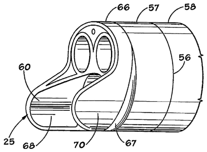
third shaft
_ segment 51 having a distal end 52 connected to a proximal end 43 of the
second
flexible shaft segment 41.
[0046] Preferably, the shaft 27 is constructed so that it has a
substantially smooth,
continuous outer surface, and its preferred cross-sectional configuration is
circular.
Preferably the length of the third shaft segment 51 is approximately 50 cm
long. The
first flexible shaft section 31 is preferably approximately 4 cm long, and the
second
flexible shaft section 41 is preferably approximately 20 cm long. The first
and second
flexible shaft sections 31, 41 preferably have cross-sectional configurations
that are
substantially uniform along their lengths, but they may taper downwardly
toward the
18
CA 02522865 2005-10-19
WO 2005/048827
PCT/US2004/012364
face tip assembly 11. The third section 51 of shaft 27 is constructed so that
it has
sufficient strength and rigidity to permit use within the bladder and to
support the
entry of the first and second flexible sections 31, 41 into the ureter and may
be
described as rigid or semi-rigid in construction. The first and second
flexible shaft
sections 31, 41 are constructed in order to follow the contours of the ureter.
Also, as
is known by those of ordinary skill in the art of endoscopes, the lengths of
the first
segment 31, second segment 41, and third segment 51 of the shaft 27 may vary
according to the intended use of the endoscope 10.
[0047] The third shaft segment 51 is dimensioned to be received in a human
body
so that it extends through the urethra and substantially through the bladder,
The distal
end 52 of segment 51 is tapered to receive the proximal end 43 of the second
flexible
segment 41 and is formed to provide a smooth, gradual transition between the
second
flexible segment 41 and the third segment 51, to permit the non-traumatic
passage of
the shaft 27 through the urethra and into the bladder. Preferably, the third
section 51,
preferably, has sufficient strength and rigidity to enable both axial and
rotational
translation with the maneuvering of the handle and viewing assembly 13,
without
excessive twisting of the shaft 27. Additionally, the connection 14 between
shaft
segment 51 and the handle and viewing assembly 13 has sufficient strength and
rigidity to avoid breaking during use and handling of endoscope 10. Thus, the
user is
able to insert the shaft 27, leading with face tip assembly 11, into the
urethra and
maneuver the instrument through the bladder in order to position the first
flexible
section 31 and thus the face tip assembly 11 into the opening of the ureter.
The first
flexible segment 31 having distal end 32 adapted for insertion into the cavity
is
dimensioned to be received in the ureter of a patient.
[0048] The second flexible shaft segment 41 having a distal end 42, like
first
flexible shaft t 31 is correspondingly also dimensioned to be received in the
ureter of
a patient and is sufficiently flexible along its length to follow various
canals of the
human body, such as the ureter. In order to optimize the versatility of a
flexible
endoscope while retaining the controllability of a rigid endoscope, the second
flexible
segment 41 is "passively flexible". The term "passively flexible" is intended
to mean
that shaft segment 41 may be moved, flexed, or bent, to assume a curved
configuration, in response to forces exerted upon the shaft 27 as it passes
through a
19
CA 02522865 2005-10-19
WO 2005/048827
PCT/US2004/012364
cavity or body passageway, but the movement, flexing, or bending is not
substantially
controllable by the operator of the instrument. While the third shaft segment
51
provides the user with sufficient feel and control of the instrument 10, the
second
flexible segment 41 has the ability to readily flex and follow the contours of
a cavity
or passageway, such as the ureter, without excessive deformation of its cavity
or
passageway, in order to minimize any traumatic effects.
[0049] In contrast, the first flexible shaft segment 31 is "actively
flexible". The
term "actively flexible" is intended to mean that shaft segment 31 may be
moved,
flexed, or bent to assume a curved configuration, such as shown in phantom
lines 15
in FIG. 1, or an angular disposition with respect to longitudinal axis L, and
such
movement, flexing or bending is substantially controlled by the operator, who
can
cause and control the desired movement, flexing, and/or bending. The
deflection of
face tip assembly 11 upon operator, or user, command, or control, aids the
user in the
detection and penetration of the opening of the ureter. Additionally, the
relatively
small diameters of face tip assembly 11 and first flexible shaft segment 31
allow the
user to insert the shaft 27 into the narrow opening of the ureter to gain
access to the
ureter and kidney. The active flexibility of the first flexible shaft segment
31 also
provides for non-traumatic use of the instrument 10 and precise positioning of
the
face tip assembly 11 adjacent to items of interest such as a lesion or kidney
stone.
Most significantly, the actively flexible first flexible segment 31 enables
the user to
view and, along with other features of the present invention, non-
traumatically deliver
a working tool via the working channel 71 and operating tool port 21 to the
item of
interest. The flexibility of the first flexible segment 31 generally negates
the need for
rotating the instrument when targeting or advancing the instrument as
required.
[0050] The first flexible shaft segment 31 may be made actively flexible
using
various methodologies. In the preferred embodiment, the first flexible segment
31 is
made actively flexible through use of operating, or guide, wires 30 guided
through
individual conduits which pass longitudinally through shaft segment 31 or a
through
passageway 28 within shaft 27 toward the distal end 32, which wire, or wires,
may be
manipulated, or pulled, so as to bend, move, or flex, the shaft segment 31 in
a desired
direction. The distal ends of the wires 30 may be suitably anchored adjacent
he distal
end 32 of shaft segment 31, whereby upon pulling on the wire, or wires 30, the
CA 02522865 2005-10-19
WO 2005/048827
PCT/US2004/012364
desired controlled flexing, moving, or bending will occur. Alternatively, the
first
flexible shaft segment 31 may be comprised of a connected string of body
members
consisting of semicircular disc-like ring elements forming selectively
controllable
expandable bodies, whereby upon controlled expansion of selected ring
elements, the
shaft segment 31 moves or flexes in the desired direction, similar to the
manner in
which a snake moves. Other methodologies for providing the requisite
flexibility
could include the use of springs, separate wire guides, or the working tool
itself,
among others. If desired, the cross-sectional shape of first flexible shaft
segment 31
could be varied in order to provide varying inherent flexibility
characteristics. In other
words, one or more portions, or sides, of the first flexible shaft segment 31
can be
made to be more pliable, or flexible, than other portions, or sides, of the
same first
flexible shaft segment in order to make a shaft segment that more readily
flexes in a
first direction and is more rigid in a second direction.
[0051] Alternatively, the first flexible shaft segment 31 can be made
from a
composite material that has differing properties that will result in having a
first
flexible segment 31 predisposed to more readily bend, or flex, in a first
direction, for
example, upwardly and downwardly, rather than from side to side.
Alternatively, the
active desired flexibility of the first flexible shaft segment 31 could be
obtained by
use of a longitudinally disposed tension cable with a distal spring deflection
recovery
member, whereby increased tension or compression on the tension cable
initiated
through a suitable control causes the flexible shaft segment 31 to deflect or
flex in a
desired direction.
[0052] Face tip assemblies 11, 11', 11", 11" may be a separate multi-
port piece
which is connected to the distal end 32 of first flexible shaft segment 31 of
shaft 27 as
previously described. In an alternative embodiment, the face tip assemblies 11-
11"
may be a unitary piece formed integral with first flexible segment 31 as
previously
described. If desired, the same material used to form the first flexible shaft
segment
31 may also be used for the second flexible shaft segment 41. The first
flexible shaft
segment 31 may have approximately the same diameter as the second flexible
shaft
segment 41, and the two segments may be formed integral with each other or
formed
separately and connected by any suitable connection. If desired, as seen in
FIGS. 1
and 9, the first shaft segment 31 could extend along the longitudinal axis L
of the
21
CA 02522865 2005-10-19
WO 2005/048827 PCT/US2004/012364
shaft 27 from its distal end 32 to the handle and viewing assembly, whereby
shaft
segment 31 is concentrically disposed within the second shaft segment 41 and
third
shaft segment 51. In turn, the second shaft segment could also extend along
the =
longitudinal axis L of shaft 27 to the handle and viewing assembly 13, whereby
shaft
segment 41 is concentrically disposed within the third shaft segment 51. Where
the
first shaft segment 31 enters the second shaft segment 41, and where the
second shaft
segment 41 enters the third shaft segment 51, define transition zones, or
transition
locations, 39, 49, and preferably at these zones the larger diameter shaft
segment as
shown at zone 49 in FIG. 1. These tapering transition zones 39, 49 provide
increased
durability of the shaft 27 to bending fatigue and ease the insertion of the
shaft 27 into
the desired body cavity. The second flexible segment 41 may have a different
diameter than the third segment 51, and the second flexible segment 41 may be
disposed inside the third segment 51. In the preferred embodiment, the first
and
second flexible section 31, 41 have a diameter of approximately 7.2 French
equal to
approximately 2.16 millimeters, wherein the third segment 51 has a diameter of
approximately 8.2 French equal to approximately 2.46 millimeters.
[0053] With reference to FIGS. 1, 9, and 10 the handle and viewing assembly 13
has a plurality of passageways, or channels, 88 in communication with
corresponding
passageways, or channels, 28 of shaft 27 and longitudinally extend to the
first flexible
shaft segment 31 to the input/output ports of the face tip assemblies 11-11m.
The
passageways, or channels, between face tip assembly 11 and handle and viewing
assembly 13 may be of equal diameter, or of differing diameter sizes, whereby
they
taper from one end to another to provide a smooth continuation of the
passageways or
channels.
[0054] With reference to FIGS. 1, 9, and 10, the handle and viewing
assembly 13
includes: a distal section 81 which connects, or interfaces, with shaft 27, a
working
channel interface section 82 including a working channel interface assembly 72
which
provides access for various operating tools through the instrument 10; a
luminous
conductor interface assembly 73 which provides for connecting, or interfacing,
a light
source such as a lamp box, for example, with the luminous conductor 63; and a
proximal section 83 including proximal section assembly 84, including optical
channel interface assembly 74, and which provides either an interface, or an
22
CA 02522865 2005-10-19
WO 2005/048827
PCT/US2004/012364
intermediate connection, to a conventional imaging apparatus (not shown). The
handle and viewing assembly 13 may include, if desired, any one or more of the
following connection components: a handhold or pistol-type grip; a telescopic
viewing assembly; an eyepiece adjustment; an optical tap for transmission of
the
optical image to an imaging apparatus; an electronic image
enhancer/transmitter;
and/or valve(s) for irrigation/suction.
[0055] The instrument 10 includes a working channel 71 for providing a pathway
into the internal cavity for a conventional working instrument. Referring now
to
FIGS. 1, 2, 9 and 10, in the preferred embodiment, the working channel 71 is
formed
via passageways 28, 88 and provides working tool access to the interior
cavity, the
channel 71 extending from the working channel interface assembly 72 through to
the
operating tool port 21. In an embodiment, the working channel 71 has a
substantially
smooth interior surface to provide smooth movement of a working tool through
instrument 10. The working channel 71 may have a substantially circular cross-
sectional configuration, and may be coaxially surrounded by shaft segments 31,
41,
and 51 of shaft 27. The interior wall surface 75 of working channel 71 may be
coated
with, or formed of, a material having a reduced coefficient of friction to
facilitate easy
passage and use of working accessories, or tools, in the working channel 71.
[0056] The instrument 10 includes at least one luminous conductor 63 for
providing illumination within the interior cavity. The luminous conductor 63
extends
from the luminous conductor interface assembly 73 of handle and viewing
assembly
13, through shaft 27, to distal end 32 of first flexible shaft segment 31 to
face
assembly 11-11"'. The luminous conductor 63 is in the form of a fiber optic
light
carrying bundle. The luminous conductor interface assembly 73 provides a
connector,
as understood by those skilled in the art, between the luminous conductor 63
(light
guide) and a conventional light source (not shown). Light travels through the
luminous conductor interface assembly 73 and through the handle and viewing
assembly housing 87 and shaft 27 to the interior cavity in a manner depending
upon
the configuration of the face tip assembly 11-11'. For example, in one of the
embodiments described with regard to FIG. 2, the luminous conductor 63 is a
single
fiber-optic bundle and may be interspersed among the optical conductor 62 in
working channel 71. However, in one of the embodiments of FIGS. 3, 4, or 5,
the
23
CA 02522865 2005-10-19
WO 2005/048827
PCT/US2004/012364
implementation may be best had through a plurality of independent fiber-optic
bundles or a single fiber-optic bundle divided prior to or upon reaching
luminous
channel port 23. Also, in an embodiment, the luminous conductor interface
assembly
73 may include an adjustable light valve (not shown) for selectively adjusting
the
intensity of the light. In another embodiment, the handle and viewing assembly
13
may include a plurality of the luminous conductor interface assemblies 73.
[0057] Referring again to FIGS. 1 and 9, an embodiment of the present
invention
also comprises at least one optical conductor 62 optically interfaced with the
optical
collector 61 for transmitting the gathered interior cavity image to the handle
and
viewing assembly 13. In the preferred embodiment, the optical conductor 62 is
in the
form of a fiber-optic bundle 64. In this embodiment, the instrument 10
includes an
optical conductor channel 92 which encloses and receives the optical conductor
62,
64. The optical conductor 62, 64 may be located within the instrument 10, such
as by
disposing it in the working channel 71, or it may be formed as a separate
channel. A
luminous conductor channel 93 may be provided to carry light to the face tip
assembly 11-11' and correspondingly the internal cavity and thus, the area of
interest. A fused fiber optic image bundle 62 would extend through shaft 27 to
the
face tip assembly 11-11" and correspondingly to the optical image collector
61. In
an embodiment, the optical conductor 62 is supported within handle and viewing
assembly housing 87 by means known by those skilled in the art. For example,
the
optical conductor 62 would be supported within the handle and viewing assembly
housing 87. The handle and viewing assembly 13 of endoscope 1'0 may be
equipped
to interface with an imaging apparatus 91 (FIG. 11) having an imaging
processor 93
in order to capture the image gathered by optical image collector 61 in order
to
process the image for transmission to a human interface apparatus 101, such as
a
monitor and/or to video capable glasses. In an alternative embodiment, the
handle and
viewing assembly 13 is used as a form of telescope as known by those skilled
in the
art, whereby an ocular lens and lens support would cooperate with a spring
means to
permit relative movement between the optical conductor 62 and the housing 87
to
provide direct, adjustable, visual imaging. In an embodiment, an optical wedge
(not
shown) is included, the optical wedge can be located near the distal end 32 of
the first
flexible segment 31 to provide a direction of view compensation of about 5-10
24
CA 02522865 2005-10-19
WO 2005/048827
PCT/US2004/012364
degrees when viewed under water, as would be the case if implemented as a
ureteroscope.
[0058] Referring now to FIGS. 1, 2, and 11, a system to view a visually
obscured
portion of a body cavity will be described. The system may include an
endoscope 10
as previously described. The system may further include an imaging apparatus
91
coupled with the at least one optical conductor 62 via the handle and viewing
assembly 13, for capturing the image to send it to a human interface apparatus
101. In
an embodiment, instead of the user strictly viewing the image gathered by the
optical
image collector 61 through a telescope or eye piece portion of a viewing
assembly as
is the case with much of the state-of-the-art, the handle and viewing assembly
13 of
the present invention may include a proximal section assembly 84 which
provides an
interface for the imaging apparatus 91 as known by those skilled in the art.
In an
embodiment, the imaging apparatus 91 is an image transceiver 92 including an
image
processor 93 capable of providing video output to a human interface apparatus
101. In
another embodiment, the imaging apparatus 91 is a pair of cameras optically
coupled
with a plurality of optical conductors .62. The preferred function of the
imaging
apparatus 91 is to render a three-dimensional image of the area of interest as
selected
by the user. Typically this is accomplished using individual "optical feeds."
Additionally, in the preferred embodiment utilizing a pair of optical
conductors 62
and optical image collectors 61, the imaging apparatus 91 captures each half
of the
image to render a complete and broader view of the area of interest.
[0059] The system may include a human-interface apparatus 101, as shown
in
FIG. 11. The human interface apparatus 101 is electrically or optically
coupled with
the imaging apparatus 91. In various embodiments, the human interface
apparatus 101
may include such display/interface devices including a first image display
device 94
such as a CRT, HDTV, for example, and in the preferred embodiment, a second
image display device 94 including a video stereoscopic viewer unit 95 as known
and
understood by those skilled in the art. Although visual clarity is an
important feature
of the human interface apparatus 101, the invention is not limited to, or to
the quality
of, the examples provided above.
[0060] An embodiment of the present invention includes a method of performing
a
procedure in a visually obscured portion of a body cavity while under direct
visual
CA 02522865 2014-06-09
control. Specifically, the method of the present invention comprises the steps
of;
providing an endoscopic type instrument 10, having a face tip assembly, such
as face
tip assembly 11-11"' connected to a shaft assembly 12 having an actively
flexible
shat segment 31 at its distal end, the shaft assembly 12 being connected to a
handle
and viewing assembly 13; providing an illumination source, such as luminous
conductor 62; inserting the distal etui of the shaft assembly into a body
cavity;
manipulating the actively flexible shaft segment to a desired angular
deflection in
order to properly target, or view, the area of interest to allow both
diagnosis and
operative procedures. Another step may be advancing a working tool through the
working channel 71, into the body cavity, while simultaneously monitoring its
exit
through the face tip assembly 11-1V". The user can view the inner portion of a
cavity such as, for example, the ureter or kidneys, and simultaneously view
the
insertion of a working/operating tool. Thus, various procedures can be carried
out
within the cavity while under direct visual control. The method may include
the steps
of irrigating the area of interest and suctioning particulate matter from the
body
cavity. Note, that one skilled in the art would know that some of the above
steps do
not need to be accomplished in the order provided in this embodiment. The
method
may also include the step of viewing during insertion, the relative location
of the face
tip assembly to properly position the assembly with respect to an area of
interest 114;
[0061) In the drawings and specification, there have been disclosed a typical
preferred embodiment of the invention, and although specific terms are
employed, the
terms are used in a descriptive sense only and not for purposes of limitation.
The
invention has been described in considerable detail with specific reference to
these
illustrated embodiments. It will be apparent, however, that various
modifications and
changes can be made within the scope of the invention as described in the
foregoing specification. It is understood that other materials and dimensions
may be
used for the endoscopic type instrument of the present invention keeping in
mind the
dimensions of the affected body parts. Further, the number and dimensions of
the
channels or passageways employed are variable depending on the accessories
(i.e. dye
laser, fiber optics, etc,) used in conjunction with the instrument.
Additionally, the
actively flexible shaft segment could be used with other shaft segments which
are all
rigid, all semi-rigid, all flexible, or combinations thereof. Further, the
face tip
26
CA 02522865 2005-10-19
WO 2005/048827
PCT/US2004/012364
assemblies may be used with any type of endoscopic instrument or shaft
assembly.
Also, other shaped handles and handles of other designs may be used.
Accordingly,
the invention is to be limited only by the scope of the appended claims.
27