Note: Descriptions are shown in the official language in which they were submitted.
CA 02522966 2005-10-20
WO 2004/096089 PCT/US2004/012356
1231
DISTAL PROTECTION DEVICE
BACKGROUND
This application claims priority from provisional application. serial no.
60/466,491, filed
4129103, the entire contents of which is incorporated herein by reference.
Technical Field
This application relates to a vascular device and more particularly to a
vascular device for
capturing embolic material during surgical procedures.
Background of Related Art
During vascular surgical procedures such as stenting, angioplasty,
thrombectomy, and
atherectomy, embolic material such as plaque and blood clots can become
dislodged.
Dislodgement of such embolic material can cause the emboli to flow downstream
to lodge in the
vascular system, thereby occluding flow of oxygenated blood to the brain or
other vital organs.
Such occlusion can compromise peripheral circulation or result in heart
attack, stroke or even
death.
Techniques to cut the debris into smaller sizes, such as by use of lasers,
have had
significant drawbacks, such as the inability to ensure all the debris is cut
into sufficiently small
fragments. If some of the fragments remain too large, then occlusion of the
vessels can occur
causing the problems and risks enumerated above.
Attempts have been made to place a device distal (downstream) of the stenosis,
thrombus, etc. to capture the emboli. Such distal protection devices typically
are collapsible for
insertion and expandable once in the vessel. Some devices are in the form of
an expandable
balloon which is inserted within the vessel inside a sheath. When the sheath
is withdrawn, the
balloon is expanded to block emboli. These balloon devices even in the
collapsed position
increase the profile of the device since they are wrapped on the outside of
the device. In other
distal protection devices, a wire is covered by a membrane. These wires extend
laterally from
the device and may not enable the membrane to block the entire region of the
vessel. Failure to
expand to geometry to block the entire region can result in the unwanted
passage of debris which
can cause vessel occlusion and the aforementioned adverse consequences.
The need therefore exists for an improved distal protection device. Such
device would
have a reduced profile to facilitate insertion and to better enable placement
of the device distal of
CA 02522966 2005-10-20
WO 2004/096089 PCT/US2004/012356
the emboli to block potential downstream flow. The device would also be easy
to manipulate
and sufficiently fill the vessel area to ensure all passage is blocked.
SUMMARY
The present invention overcomes the problems and deficiencies of the prior
art. The
present invention provides a distal protection device comprising a catheter
and a flexible member
movable from a first retracted position to a second looped position extending
laterally with
respect to the catheter such that a first loop opening extends substantially
in a direction of blood
flow as the first loop opening lies in a plane substantially parallel to a
transverse axis of the
catheter. Filtering material is movable from a collapsed position to a
deployed position in
response to movement of the flexible member.
In one embodiment, the flexible member is contained within the catheter in the
first
position so the cross sectional dimension of the catheter at a portion
containing the flexible
member does not exceed other cross-sectional dimensions of the catheter. In
one embodiment,
the flexible member extends through a sidewall in the catheter.
In one embodiment, a second loop is spaced from the first loop and movable
from a first
position to a second looped position extending laterally from the sidewall of
the catheter.
Preferably the loops extend in opposite directions with respect to the
catheter so in the second
looped position the loops are approximately 180 degrees apart. In one
embodiment of the
multiple loop configuration, the loops are axially offset.
The present invention also provides a distal protection device comprising a
catheter
having an opening in a sidewall, a flexible wire positioned within the
catheter and movable from
a first position having a lower profile for insertion of the catheter to a
second position extending
laterally from the catheter. In the second position, the wire forms a loop
extending laterally such
that a first end of the wire extends in a proximal direction and a second end
of the wire extends
in a distal direction, with the loop therebetween having an opening in a
proximal to distal
direction. Filtering material is disposed over at least a portion of the wire
and movable from a
collapsed position to a deployed position in response to movement of the wire.
Preferably on opening of the wire loop is positioned at an angle to a
transverse axis and a
longitudinal axis of the catheter. In one embodiment the wire forms a second
loop in the second
position.
2
CA 02522966 2005-10-20
WO 2004/096089 PCT/US2004/012356
The distal protection device may also include an actuating member for moving
the wire
between the first and second looped positions.
The present invention also provides a distal protection device comprising a
catheter and a
flexible member movable from a first position to a second looped position
extending laterally
with respect the catheter such that in the second looped position a loop
opening is formed lying
in a plane that is non-aligned with a longitudinal axis of the catheter. The
flexible member is
movable between the first and second positions by user control. Filtering
material is provided
and movable from a collapsed position to a deployed position in response to
movement of the
flexible member, wherein the filtering material automatically moves from the
deployed position
to the collapsed position upon movement of the flexible member back to the
first position.
The present invention also provides a distal protection device comprising an
outer tube,
an inner core, a first inner filter having a series of openings of a first
dimension and a second
outer filter having a series of openings of a second dimension smaller than
the first dimension.
At least a portion of the outer filter is positioned external of at least a
portion of the inner filter.
The device may include a ring positioned on a proximal end of the outer
filter.
BRIEF DESCRIPTION OF THE DRAWINGS
Preferred embodiments) of the present disclosure are described herein with
reference to
the drawings wherein:
Figures 1-5 illustrate a first embodiment of the distal protection device of
the present
invention, wherein
Figure 1 is a perspective view;
Figure 2 is a cross-sectional view taken along lines 2-2 of Figure l;
Figure 3 is a perspective view showing the outer tube withdrawn to enable
expansion of the balloons;
Figure 4 is a perspective view illustrating the inner and outer balloons in
the
expanded configuration; and
Figure 5 is a side view in partial cross section showing the balloons
expanded;
Figure 6 is a perspective view of a second embodiment of the distal protection
device of
the present invention having a self expanding umbrella device;
3
CA 02522966 2005-10-20
WO 2004/096089 PCT/US2004/012356
Figure 7 is a perspective view of the device of Figure 6 showing the umbrella
device
expanded by blood flow;
Figures 8 and 9 are perspective and longitudinal cross-sectional views,
respectively, of a
third embodiment of the distal protection device of the present invention
having a membrane
formed over a coiled wire, the membrane and wire shown in the retracted
insertion position;
Figure 9A is an enlarged longitudinal cross-sectional view of a portion of the
catheter of
Figure 9;
Figure 10 is a view similar to Figure 9A except showing the wire and membrane
in the
expanded position;
Figure 11 is a perspective view of the device in the expanded position of
Figure 10;
Figures 12 and 13 are perspective views of a fourth embodiment of the distal
protection
device having a membrane fused to a shape memory wire, wherein Figure 12 shows
the shape
memory wire and membrane contained within the catheter and Figure 13 shows the
shape
memory wire advanced from the catheter to open the membrane to an expanded
position;
Figure 13A is an enlarged view of the area of detail of Figure 13;
Figures 14-17 illustrate three alternate embodiments of the device of Figure
12, showing
varying shape memory wire configurations fused to a membrane wherein;
Figures 14 and 15 are perspective views showing looped wires, with Figure 14
showing the attachment to the membrane ring and Figure 1 S showing an exploded
view;
Figure 16 is an exploded view showing looped wires with pointed bend points;
and
Figure 17 is an exploded view showing individual wire segments terminating in
rings;
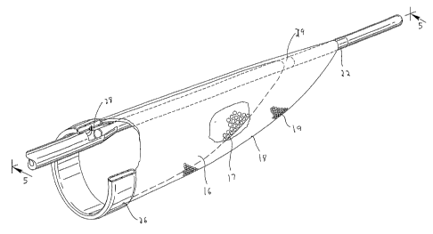
Figures 18 and 19 are perspective and side views, respectively, of another
alternate
embodiment of the distal protection device having a membrane attached to a
coiled wire and
shown in the expanded position;
Figure 20 is a side view of an alternate embodiment of the device of Figure 18
wherein
the coiled wire for deploying the membrane extends on one side of the
guidewire;
Figures 21 and 22 are respectively perspective and longitudinal cross-
sectional views
(taken along lines 22-22) of another alternate embodiment of the distal
protection device having
4
CA 02522966 2005-10-20
WO 2004/096089 PCT/US2004/012356
a single wire loop for deploying the membrane, the membrane and wire shown in
the non-
expanded (collapsed) position;
Figures 23 and 24 are respectively perspective and longitudinal cross-
sectional views
(taken along lines 24-24) similar to Figures 21 and 22 except illustrating the
wire in the looped
position and membrane in the deployed position;
Figure 24A is an end view of the device of Figure 23;
Figure 25 and 26 are perspective views of yet another alternate embodiment of
the distal
protection device of Figure 21 having a double looped wire for deploying the
membrane, the
membrane shown in the non-expanded position in Fig. 25 and the deployed
position in Fig. 26;
Figure 27 is a perspective view illustrating another alternate embodiment of
the distal
protection device of Figure 21 having multiple wire loops, the membrane shown
in the deployed
position;
Figure 28 is a perspective view of an alternate embodiment of the distal
protection device
of Figure 26 wherein the wire loops are axially offset; and
Figures 29 and 29A illustrate placement of the device of Figure 26, wherein
Figure 29
shows the catheter advanced through the femoral to the carotid artery and
Figure 29A shows the
device deployed in the carotid artery to block distal flow of emboli.
DETAILED DESCRIPTION OF PREFERRED EMBODIMENTS
Referring now in detail to the drawings where like reference numerals identify
similar or
like components throughout the several views, several different embodiments
for capturing
embolic material during surgical procedures.
Turning first to the embodiment of Figures 1-5, a catheter 10 has an outer
tube 12, a
coaxial inner core 14 disposed in the outer tube 12, an inner balloon filter
16 and an outer
balloon filter 18. The tube 12 and core 14 are preferably composed of Nitinol,
with the core
preferably having a platinum wind therearound, however other materials such as
stainless steel
are also contemplated. The inner balloon filter 16, preferably made of
polyurethane, is attached
to the catheter 10 at a distal end via ring 29. Balloon 16 has small holes 17
dimensioned for
filtering embolic material. The holes 17 are preferably 180 microns, although
other dimensions
axe contemplated. Outer filter balloon 18, also preferably made of PET, is
attached at a distal
end to catheter 10 via ring 22. As shown, outer filter balloon 18 is external
of inner filter balloon
20. Balloon 18 also has a series of holes 19 for filtering embolic material.
The holes of the outer
CA 02522966 2005-10-20
WO 2004/096089 PCT/US2004/012356
balloon 18 are preferably smaller than those of the inner balloon 16 ~o
capture embolic material
filtering through the inner balloon 16. In one embodiment, the holes 19 of the
outer balloon are
120 microns, although other dimensions are contemplated. Mounting ring 26
supports proximal
ends of balloons 16 and 18 and has an inflation port 28 communicating with the
space between
the balloons.
In use, the space between the balloons 16 and 18 is inflated through inflation
port 28, so
they are moved to assume the expanded configuration of Figures 4 and 5. The
inner and outer
balloons 16, 18 are preferably attached along a surface so fluid injection to
expand the space
between the balloons does not enter the balloon and exit through the holes 17,
19.
Thus, embolic material exceeding a certain size carried by the blood through
the proximal
opening in the balloons is captured in the balloon filters 16, 18, with the
blood and smaller
particles flowing through the holes 17, 19 in the balloons. As can be
appreciated, instead of
balloon filters, other inner/outer filtering material with appropriate size
holes can be utilized.
Figures 8-10 illustrate an alternate embodiment of the distal protection
device, designated
generally by reference numeral 30. In this embodiment, the blocking membrane
is deployed by
mechanical actuation of a wire or shaft. More specifically, catheter 31 has a
coiled wire 32
attached at a distal end to end wall 37 of tip 34 and at its proximal end to
wall 36 by means such
as welding. An actuation shaft 38, or alternatively a wire, is slidably
positioned within a bore in
the catheter 31 and is attached at a distal end 39 to proximal extension 35 of
tip 34. A porous
membrane 40 is positioned over the coiled wire 32. The membrane 40 can be
attached at a
proximal end to wall 36 and at a distal end to wall 37 of tip 34. Membrane 40
may also be
attached to coiled wire 32. As shown, the membrane 40 fully covers the distal
portion of the
wire 32 and has enlarged open regions or windows, defined between elongated
strips 42, to allow
entry of blood.
To deploy the membrane 40 of distal protection device 30 from a low profile
insertion
position of Figures 8 and 9 to an expanded configuration of Figure 10 to block
particles,
actuation shaft 38 is pulled proximally (in the direction of the arrow of
Figure 10), thereby
pulling tip 34 proximally. Such retraction of tip 34, forces the coiled wire
32 to compress and
extend radially outwardly as shown, thereby forcing the membrane 40 radially
to a stretched or
expanded configuration to block and capture flow of embolic material. The
pores in the
extended membrane 40 enable blood flow therethrough while capturing embolic
material
6
CA 02522966 2005-10-20
WO 2004/096089 PCT/US2004/012356
exceeding a predetermined size, i.e. the size of the pores in the membrane. To
remove the device
30, actuation shaft 38 is pushed distally to retract the wire 32 and collapse
membrane 40.
It should be appreciated that instead of a coiled wire, a tubular braid could
be provided
with a membrane, e.g. of urethane material, over the braid. The braid would be
attached to the
catheter and moved between retracted and expanded positions in a similar
manner as wire 32.
The braided version could alternately be obtained by providing a braided
catheter and etching a
section of the outer plastic to expose the braid.
Figures 6 and 7 illustrate a self expanding distal protection device 50. The
self expansion
occurs as a result of blood flow. The distal protection device 50 is an
umbrella type device
attached to a distal region 51 of guidewire 52. The g~uidewire 52 is shown
with a reduced
diameter distal portion 53. The umbrella is in the form of a porous balloon
54, preferably
composed of polyurethane, although other materials are also contemplated. A
suture loop or ring
56 is attached to a proximal end 51 of the balloon 54 and a suture 58 extends
proximally from
the suture loop 56. In use, the device is inserted with the balloon 54 in the
collapsed low profile
position of Figure 6. When the device 50 is exposed from the catheter or
sheath, either by
advance of device 50 or retraction of the catheter or sheath, blood flow will
expand the balloon
54 to the position shown in Figure 7 with the mouth 59 open in a proximal
direction. The blood
will flow through the holes (pores) 55 in the balloon 54, with the embolic
material exceeding the
size of the pores being captured within the balloon 54. At the end of the
procedure, the suture 58
is pulled proximally to flatten and close the mouth 59 of the balloon 54, thus
capturing the
embolic material inside. The reduced profile of the flattened balloon enables
withdrawal of the
device through the catheter or sheath.
Being part of a guidewire, in use, the device 50 of Figure 6 could be placed
within the
catheter after the guidewire for introducing the catheter is withdrawn. The
catheter can then be
withdrawn and another catheter, such as a stmt delivery catheter could be
inserted over the
guidewire 52.
Alternate embodiments of a guidewire containing a self expanding distal
protection
device are illustrated in Figures 12-17. However, rather than expansion by
blood flow, the
membrane automatically expands when deployed from the catheter as a result of
the shape
memory or springiness characteristics of the wire underlying the membrane. A
.Ol 8 inch
7
CA 02522966 2005-10-20
WO 2004/096089 PCT/US2004/012356
diameter wire can be utilized by way of example, it being understood that wire
of other
dimensions could be used.
Turning first to the embodiment of Figures 12 and 13, distal protection device
70
comprises a membrane or bag 80 and a guidewire 71 having a guidewire extension
72,
illustratively of a larger diameter, extending from its distal end. A membrane
80, preferably
made of PET, is welded to region 74 of the extension 72. A series of wires 76,
preferably
composed of shape memory material such as Nitinol, a nickel titanium alloy,
extends past the
distal end 75 of the guidewire 70 and are welded thereto. Alternately, other
materials such as
stainless steel with sufficient springiness could be utilized. Four wires are
shown but a different
number to expand the membrane could be provided. The distal end of the wires
76 are
connected to a proximal end 73 of extension 72. Each of the wires curves in
the expanded
condition as shown. Guidewire 71 is shown by way of example comprising a wound
coil around
the four wires 76. However, alternately the wires can extend only from the
distal end of the
guidewire. A flexible mounting ring or band 82 is attached to the proximal end
of the membrane
80, at the mouth, and is attached, e.g. welded, to the wires 76. The ring 82
can also be composed
of shape memory material to automatically expand when deployed or
alternatively of other
flexible material to expand when the shape memory wires move to their expanded
memorized
position. The wires 76 and membrane 80 are retained in a collapsed position
within catheter 79
for delivery as illustrated in Figure 12.
When catheter 79 is pulled proximally in the direction of the arrow, the wires
76 are
exposed from the catheter 79, and automatically expand to the memorized
position shown in
Figure 13. As they expand they move the membrane 80 from a contracted position
to the
expanded position of Figure 13 aided by expansion of band 82. Embolic material
flowing
through the mouth 84 of the membrane 80 will be captured in the membrane 80,
with the blood
flowing through the membrane pores. At the end of the procedure, the wires 76
can be fully or
partially withdrawn into the catheter 79, or the catheter advanced partially
or fully over the wires
76, thus collapsing at least the mouth of the membrane to contain the embolic
material therein as
the device is withdrawn. A 5-7 French catheter can be used by way of example.
In Figures 14-17, three alternate embodiments of wires for attaching the
membrane are
illustrated. In these embodiments, the wires are also preferably made of shape
memory material
and form part of the guidewire. They can extend the length of the guidewire as
in Figure 13A or
8
CA 02522966 2005-10-20
WO 2004/096089 PCT/US2004/012356
alternatively only extend from the distal end. In these embodiments, the
porous membrane is
attached only at the end portions of the wires 96, thereby reducing the
presence of wires in the
flow path. In Figures 14 and 15, guidewire 93 of distal protection device 90
has four looped
wires 96 extending from region 95 and connected to band or ring 97 of membrane
91 at bend
points 98. Membrane 91 is attached at its distal end 94 to guidewire extension
92. Upon
deployment from catheter 99, wires 96 move to their memorized position to
expand membrane
91. In Figure 16, shape memory looped wires 96' have more pointed bend points
98' fox
attachment to the band 97' of membrane 91'. Otherwise, the device 90' is
identical to distal
protection device 90 of Figure 15. In the Figure 17 embodiment, each wire 96"
has a curved
wire section 92" and a looped section 93" extending from a distal end of the
wire section 92"
forming a bend point 95" for attachment to the band 97" of membrane 91 ".
These looped
wires function in the same manner as in the Figure 12 embodiment as they are
preferably
composed of shape memory material so that membrane expansion occurs upon
release of the
wires from the catheter and collapse of the wires by the catheter closes the
membrane to
withdraw the device.
Figures 18 and 19 illustrate an alternate embodiment of the distal protection
device of the
present invention. Wire 102 of distal protection device 100 expands to a
coiled shape as shown
to expand a porous membrane 104 into a substantially spherical shape. Wire 102
wraps around
the outer surface of the guidewire 105 and forms several loops when expanded.
More than one
wire could optionally be used. In Figure 20, wire 102' is offset for
positioning on one side of the
guidewire 105' such that expansion of the membrane 104' provides a larger
region for blood
flow and a wider opening in membrane 104' for capture of material. Wire 102'
loops around the
outer surface of guidewire 105, forming a plurality of loops of progressively
increasing diameter
toward the proximal end and proximal opening 107 in the membrane 104' . The
wires 102, 102'
are preferably composed of shape memory material (or springy material) so they
expand to the
coiled (looped) position when deployed from catheter 110. The wires 102, 102'
assume a
substantially linear configuration within catheter 110 to maintain a low
profile for delivery.
When exposed from catheter 110, the wire assumes its coiled shape to expand
the membrane
104, 104' to a radially deployed position as shown. Besides a membrane, as
with each of the
embodiments described herein, other filtering material can be utilized.
9
CA 02522966 2005-10-20
WO 2004/096089 PCT/US2004/012356
In Figures 21-24, an alternate embodiment of the distal protection device of
the present
invention is shown and represented generally by reference numeral 110. A
flexible member such
as a wire 112 is seated within slot 120 formed in the sidewall of tube 122 and
is attached at its
distal end 114 to the tube 122 and at its proximal end 116 to slidable tube or
shaft 118. A
filtering material such as porous membrane 126 covers a region of the tube and
the slot 120. In
the collapsed position, the wire 112 is preferably fully contained within the
tube 122 to reduce
the overall insertion profile. In this collapsed position it is in alignment
with the slot 120 and the
membrane 126 is collapsed around the tube 122. A .005 inch diameter wire can
be utilized
although wires of other dimensions could also be used.
To deploy the device, slidable member such as shaft or tube 118 is advanced in
a distal
direction (see arrow of Figure 24) to deploy the wire 112 laterally to bend
into a loop extending
transversely to a longitudinal axis of the tube 122. End 117 extends
proximally and end 119
extends distally. The expanded loop thus lies in a plane at an angle to both
the longitudinal axis
and transverse axis of the catheter. In other words, the plane of the loop
opening would be at an
angle (preferably at a slight angle) to the longitudinal and transverse axis
of tube 122. The wire
112 would thus extend such that the loop opening is slightly offset from the
direction of the
longitudinal axis of tube 122 but still open generally in the direction of
blood flow. That is, a
central longitudinal axis extending through the loop opening would be at an
angle with respect to
the longitudinal axis of the tube 122.
Consequently, in one embodiment, the plane of the loop opening is
perpendicular to the
longitudinal axis of the catheter (parallel to the transverse axis) and
perpendicular to the direction
of blood flow. In other embodiments, rather than perpendicular, the plane of
the loop opening is
at an angle less than 90 degrees, but preferably greater than about 45 degrees
to the longitudinal
aXIS.
The formation of the wire loop stretches the membrane 126 on one side of the
catheter to
the illustrated expanded configuration of Figures 23 and 24 to block the flow
of material. This
provides additional coverage of the vessel lumen as the catheter can be placed
adjacent the
internal wall of the vessel with the membrane 126 filling the space above. The
windows 127 of
membrane 126 provide enlarged openings for blood flow, with the membrane 126
blocking flow
of materials exceeding the pore size.
CA 02522966 2005-10-20
WO 2004/096089 PCT/US2004/012356
To withdraw the device, the shaft 118 is moved proximally to retract the loop
and
membrane to the initial low profile insertion position. In a preferred
embodiment, the membrane
126 is made of a material that would return automatically from its stretched
position to the
original collapsed position when the wire is retracted. This passive self
contraction would avoid
the need for insertion of a separate device over the membrane to cover it for
removal, thus
reducing the overall profile of the instrumentation necessary for the
procedure. That is, in the
preferred embodiment the wire is expanded by active control while the membrane
would
automatically retract without other assistance.
In another embodiment, the membrane can be attached to wire 112 and move with
the
wire 112.
Other materials for the embodiments of Figures 21-28 can be utilized which as
in
membrane 126 would be movable between collapsed and deployed positions.
In the embodiment of Figures 25 and 26, a flexible member in the form of a
wire 131 of
distal protection device 130 forms two looped wire regions 132, 134 when
expanded so the
filtering material such as membrane 136 stretches in two directions. When
slidable actuating
member such as tube or shaft 138 is advanced distally in the direction of the
arrow, the wire 131
bends to extend further through slot 144 in the sidewall of the catheter 142,
forming the first
looped wire region 132 on one side of catheter 142 and the second looped wire
region 134 on the
other side of the catheter 142, preferably about 180 degrees apart. This
double looped
configuration causes membrane 136 to be stretched on opposing sides of the
tube 142 to filter
materials. As in the embodiment of Figure 23, the loops are open generally in
a direction of
blood flow (the plane of the loop opening is substantially transverse to the
direction of blood
flow and substantially transverse to the longitudinal axis of the device) with
blood flowing
through windows 137 of membrane 136.
Although shown in axial alignment in Fig. 26, alternatively the wire can be
configured so
the two looped sections are axially offset as shown in Fig. 28. That is, the
loop sections 162, 164
of wire 161 of distal protection device 160 are axially displaced so that loop
162 is positioned
distal of loop section 164. As in the previous embodiment, advancement of tube
or shaft 172
deploys wire 161 through the slot in the sidewall of tube 174 to assume the
looped configuration
and stretch porous membrane 176 to the deployed configuration on both sides of
tube 174.
11
CA 02522966 2005-10-20
WO 2004/096089 PCT/US2004/012356
In Fig. 27, a double loop configuration of distal protection device 180 is
achieved on each
side of the tube 192. The flexible member in the form of wire 181 extends
through slot 194 of
tube 192, forming two loops on each side of tube 192 to stretch membrane
(filtering material)
186 when tube or shaft 188 is moved distally. That is, in the expanded
configuration, wire 181
extends out of slot 194 to form first loop 185 on a first side of the tube
192, then extends to form
second loop 183 on a second opposing side of tube 192, extends upwardly (as
viewed in the
orientation of Fig. 27) to form third loop 182 on the first side, and then
extends to form fourth
loop 184 on the second side, after which it extends back through slot 194. As
in the other
embodiments the loop openings are generally in the direction of blood flow
with the plane of the
loop openings substantially transverse to the direction of the blood flow.
Porous membrane 186
has windows 189.
As noted above, in these embodiments of Figures 21-27, the loop opening can be
in a
plane perpendicular or at an angle less than 90 degrees to the longitudinal
axis, but preferably
greater than about 45 degrees.
Figure 29 shows the positioning of the distal protection device of the present
invention.
By way of example, device 130 of Figure 26 is shown deployed in the carotid
artery "c", it being
understood that the other devices described herein can be placed in the same
location. The
catheter 200 is inserted through the femoral vein "f' as shown in Figure 24
and advanced to the
carotid artery "c". Once positioned at the desired site, catheter 200 is
retracted to expose the
device 130, or alternately the device 130 is advanced from the catheter 200.
Once exposed at the
site, the tube is advanced as described above to deploy the wire to the looped
configuration to
expand membrane 136 to block emboli in the artery.
While the above description contains many specifics, those specifics should
not be
construed as limitations on the scope of the disclosure, but merely as
exemplifications of
preferred embodiments thereof. For example, the wire can include radiopaque
material for
imaging. Those skilled in the art will envision many other possible variations
that are within the
scope and spirit of the disclosure as defined by the claims appended hereto.
12