Note: Descriptions are shown in the official language in which they were submitted.
CA 02523579 2005-10-25
WO 2004/096290 PCT/US2004/011232
GENETIC MODIFICATION OF TARGETED REGIONS OF THE CARDIAC
CONDUCTION SYSTEM
The present invention relates to compositions, apparatus, and methods for
providing curative therapy for cardiac dysfunction, and more particularly to
biological
systems and methods relating to implementing curative therapeutic agents and
systems for
arrhythmias and cardiac pacing dysfunction.
In a normal, healthy heart, cardiac contraction is initiated by the
spontaneous
excitation of the sinoatrial ("SA") node, located in the right atrium. The
electrical impulse
generated by the SA node travels to the atrioventricular ("AV") node where it
is
transmitted to the bundle of His and Purkinje network, which branches in many
directions
to facilitate simultaneous contraction of the left and right ventricles.
In certain disease states, the heart's ability to pace properly is
compromised.
Currently, such dysfunction is commonly rectified by the implantation of
implantable
pacemakers. While improving the lives of many patients, implantable pacemakers
have a
limited lifetime and hence, may expose a patient to multiple surgeries to
replace the
implantable pacemaker. Moreover, implantable pacemakers may not be capable of
directly responding to the body's endogenous signaling that interacts with the
SA node to
increase or decrease its pacing rate.
Recently, biological methods of influencing the pacing rate of cardiac cells
have
been developed, including the use of various drugs and pharmaceutical
compositions.
Developments in genetic engineering have resulted in methods for genetically
modifying
cardiac cells to influence their intrinsic pacing rate. For example, U.S.
Patent No.
6,214,620 describes a method for suppressing excitability of ventricular cells
by
overexpressing (e.g. K+ channels) or underexpressing certain ion channels
(e.g. Na+and
Ca2+ channels). PCT Publication No. WO 021087419 describes methods and systems
for
modulating electrical behavior of cardiac cells by genetic modification of
inwardly
rectifying K+ channels (IKl) in quiescent ventricular cells. PCT Publication
No. WO
02/098286 describes methods for regulating pacemaker function of cardiac cells
with
HCN molecules (HCN l, 2, 3, or 4 isofonns of the pacemaker current If).
CA 02523579 2005-10-25
WO 2004/096290 PCT/US2004/011232
2
A need remains, however, to implement a system of genetic modification therapy
(biopacing) in cooperation with an implantable medical device (IMD) to insure
successful
curative therapy for cardiac dysfunction.
The present invention provides a biological pacemaker ("bio-pacemaker") that
is
capable of responding to physiological signals as well as facilitating and
restoring
synchronous contractions of the ventricles to thus mimic the function of a
healthy heart.
The bio-pacemaker is generated through the genetic modification of myocardial
cells in a
targeted region of the cardiac conduction system, through use of a bio-
pacemaker
composition.
In one aspect of the invention, a bio-pacemaker composition includes at least
two
coding sequences that encode one or more molecules in myocardial cells of the
cardiac
conduction system to increase the pacemaking rate of the cells. The coding
sequences
include a coding sequence that encodes a channel or subunit thereof that
produces funny
current, a coding sequence that encodes a T-type Caz+ channel or subunit
thereof, and a
coding sequence that encodes one or more molecules that suppresses the
expression of the
wild type potassium channel.
Preferably, cells of the conduction system are genetically modified using the
bio-
pacemaker composition to increase their pacing rate to a level resembling the
intrinsic
pacing rate of the SA nodal cells in a normal heart.
Preferably, the bio-pacemaker composition of the invention generates a bio-
pacemaker in
the cardiac conduction system cells by altering two or more characteristics of
the cell to
obtain the following: 1) increased inward Ca2+ current, 2) increased inward
funny current
(If), and/or 3) decreased outward K+ current.
Increased inward Ca2+ current may be obtained by genetically modifying the
target
cells to overexpress T-type Ca2+ chaimels or subunits thereof, and in one
embodiment, the
a1H subunits of the T-type Ca2+channels are overexpressed.
Increased fumly current (If) may be obtained by increasing the expression of
funny current
channels or subunits thereof. Preferably, the channels expressed are an
isoform of the
hyperpolarization-activated canon channel gene (HCN~. The isoform chosen will
be
related to the mammalian species of cells being modified.
CA 02523579 2005-10-25
WO 2004/096290 PCT/US2004/011232
Decreased outward K+ current may be obtained by delivering a bio-pacemaker
composition to the target cells including a coding sequence designed to encode
a molecule
or protein that will suppress the expression of the wildtype potassium
channels responsible
for producing rapid potassium current (IK,.). In one embodiment, the protein
expressed is a
dominant-negative form of the potassium channel protein.
In one further embodiment of the invention, a bio-pacemaker of the invention
is
used in combination with an implantable pacemaker. Specifically, the
implantable
pacemaker is programmed to work in cooperation with the genetically engineered
bio-
pacemaker to prevent cardiac dysfunction or to sense and monitor the
pacemaking action
of the genetically engineered bio-pacemaker. Further, the implantable
pacemaker operates
to pace the heart when the pacemaking action of the bio-pacemaker is not as
expected.
For example, two possible triggers for resorting to the implantable pacemalcer
are 1) a bio-
pacemaker pacing rate less than a certain predetermined threshold value and 2)
an
intermittent but presumably normal function of the bio-pacemaker. Implantable
pacemaker can be switched to the role of a primary pacemaker if one or more
attempts to
engineer a biological pacemaker fail in a patient.
In case the bio-pacemaker location is the AV node, the top portions of the SA
node
may be ablated to isolate the atria from the AV node. When the bio-pacemaker
is located
in the Purkinje network, the entire AV node may be ablated.
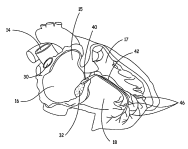
Figure 1 is a diagram of a human heart.
Figure 2 is a schematic diagram of a right side of a heart, similar to Figure
l, in
which a guiding catheter is positioned for delivery of the genetic construct
of the
invention.
Figures 3A and 3B are schematics illustrating how an embodiment of the
invention
operates.
Figures 4A and 4B show the action potential (AP) characteristics of the AV
nodal
cells (one location of the bio-pacemaker) before and after genetic
modification in
accordance with a method of this invention.
Figure SA illustrates the use of a small implantable backup pacemaker working
in
cooperation with the bio-pacemaker of the invention based on transforming the
cells of the
AV node in the conduction system.
CA 02523579 2005-10-25
WO 2004/096290 PCT/US2004/011232
4
Figure SB is a logic flow diagram depicting the operational logic of the
invention.
Figure 6 is a schematic of the tripartite rAAV producer plasmid, pTP-
D6deltaNot.
The present invention relates to biological methods of increasing the
intrinsic
pacemaking rate of cells of the cardiac conduction system, such as the AV node
of the
heart by genetic modification of the cells.
Figure 1 is a schematic diagram of a right side of a heart having an anterior-
lateral
wall peeled back to expose a portion of a heart's intrinsic conduction system
and chambers
of a right atrium 16 and a right ventricle ("RV") 18. Pertinent elements of
the heart's
intrinsic conduction system, illustrated, in Figure 1, include a SA node 30,
an AV node 32,
a bundle of His 40, a right bundle branch 42, and Purkinje fibers 46. SA node
30 is shown
at a junction between a superior vena cava 14 and right atrium ("RA") 16. An
electrical
impulse initiated at SA node 30 travels rapidly through RA 16 and a left
atrium (not
shown) to AV node 32. At AV node 32, the impulse slows to create a delay
before
passing on through a bundle of His 40, which branches, in an interventricular
septum 17,
into a right bundle branch 42 and a left bundle branch (not shown) and then,
apically, into
Purkinje fibers 46. Following the delay, the impulse travels rapidly
throughout RV 18 and
a left ventricle (not shown). Flow of the electrical impulse described herein
creates an
orderly sequence of atrial and ventricular contraction to efficiently pump
blood through
the heart. When a portion of the heart's intrinsic conduction system becomes
dysfunctional, efficient pumping is compromised.
Typically, a patient, whose SA node 30 has become dysfunctional, may have an
implantable pacemaker system implanted wherein lead electrodes are placed in
an atrial
appendage 15. The lead electrodes stimulate RA 16 downstream of dysfunctional
SA
node 30 and the stimulating pulse travels on to AV node 32, bundle of His 40,
and
Purlcinje fibers 46 to restore physiological contraction of the heart.
However, if a patient
has a dysfunctional AV node 32, pacing in atrial appendage 15 will not be
effective, since
it is upstream of a block caused by the damage.
Pacing at the bundle of His 40 provides the advantage of utilizing the normal
conduction system of the heart to carry out ventricular depolarizations. In
other words,
stimulation provided at the bundle of His will propagate rapidly to the entire
heart via the
right bundle 42, the left bundle (not shown), and the Purkinje fibers. This
provides
CA 02523579 2005-10-25
WO 2004/096290 PCT/US2004/011232
synchronized and efficient ventricular contraction, unlike pacing from the
apex of the right
ventricle where the electrical activity propagates at a slower rate because
myocardial
tissue is a slow conductor compared to the rapidly conducting Purkinje
network.
Like cells of other excitable tissue in the body, cardiac cells allow a
controlled
flow of ions across the membranes. This ion movement across the cell membrane
results
in changes in transmembrane potential, which is a trigger for cell
contraction. The heart
cells can be categorized into several cell types (e.g. atrial, ventricular,
etc.) and each cell
type has its own characteristic variation in membrane potential. For example,
ventricular
cells have a resting potential of ~-85mV. In response to an incoming
depolarization wave
front, these cells fire an action potential with a peak value of ~20mV and
then begin to
repolarize, which takes 350 ms to complete. In contrast, SA nodal cells do not
have a
stable resting potential and instead begin to spontaneously depolarize when
their
membrane potential reaches ~-SOmV. Cells, such as SA nodal cells, that do not
have a
stable resting transmembrane potential, but instead increase spontaneously to
the threshold
value, causing regenerative, repetitive depolarization, are said to have
automacity.
Cardiac muscle cells are structurally connected to each other via small pore-
like
structures known as gap junctions, so that when a few cardiac cells
depolarize, they act as
a current source to adjacent cells causing them to depolarize as well; and
these cells in turn
relay the electrical charge to adjacent cells. Once depolarization begins
within a mass of
cardiac cells, it spreads rapidly by cell-to-cell conduction until the entire
mass is
depolarized causing a mass of cardiac cells to contract as a unit.
The cells in the SA node are specialized pacemaker cells and have the highest
firing rate. Depolarization from these cells spreads across the atria. Since
atrial muscle
cells are not connected intimately with ventricular muscle cells, conduction
does not
spread directly to the ventricle. Instead, atrial depolarization enters the AV
node, and after
a brief delay, is passed on to the ventricles via the bundle of His and
Purkinje network,
initiating cellular depolarization along the endocardiuim. Depolarization then
spreads by
cell-to-cell conduction throughout the entire ventricular mass.
The SA node's unique cells include a combination of ion channels that endow it
with its automacity. A review of the features of cardiac electrical function
and description
of the current understanding of the ionic and molecular basis, thereof, can be
found in
CA 02523579 2005-10-25
WO 2004/096290 PCT/US2004/011232
6
Schram et al., Circulation Research, May 17, 2002, pages 939-950, the
teachings of which
are herein incorporated by reference.
Some of the unique features of the SA node cells include the absence of Na and
inwardly rectifying K+ (IK~) channels. In the absence of sodium current, the
upstroke of
SA node action potential is primarily mediated by L-type Ca2+ channels (ICaL).
SA node
cells do not have a stable resting potential because of the lack of the IKI
and begin to
depolarize immediately after the repolarization phase is complete. The maximum
diastolic
potential for SA node cells is approximately -50 mV compared to -78 mV and -85
mV for
atrial and ventricular cells, respectively. The slow depolarization phase is
mediated by
activation of "fumiy current" (If) and T-type Ca2+ channels and deactivation
of slow and
rapid potassium (IBS and IK,., respectively). The rate of pacemaker discharge
in the SA
node in a normally functioning heart is approximately in the range of about 60
to 100
beats per minute.
In the diseased state, the ability of the SA node to properly pace the heart
can be
severely compromised. A method of the present invention includes genetically
modifying
the cells of the AV node to modify the electrophysiohogy and pacing rate to
resemble more
closely the electrophysiology and pacing rate of the specialized pacemaker
cells of the SA
node.
Figure 2 is a schematic diagram of the right side of a heart; similar to that
shown in
Figure l, wherein a guide catheter 90 is positioned for delivery of the
genetic construct of
the invention. A venous access site (not shown) for catheter 90 may be in a
cephalic or
subclavian vein and means used for venous access are well known in the art,
including the
Seldinger technique performed with a standard percutaneous introducer kit.
Guide
catheter 90 includes a lumen (not shown) extending from a proximal end (not
shown) to a
distal end 92 that slideably receives delivery system 80. Guide catheter 90
may have an
outer diameter between approximately 0.115 inches and 0.170 inches and is of a
construction well known in the art. Distal end 92 of guide catheter 80 may
include an
electrode (not shown) for mapping electrical activity in order to direct
distal end 92 to an
implant site near bundle of His 40. Alternatively a separate mapping catheter
may be used
within lumen of guide catheter 90 to direct distal end 92 to an implant site
near bundle of
His 40, a method well known in the art.
CA 02523579 2005-10-25
WO 2004/096290 PCT/US2004/011232
7
The schematics of Figures 3A and 3B illustrate an embodiment of the invention.
Figure 3A illustrates a heart with normal pacemaker function in the SA node 30
wherein
the pacemalcer function of the SA node is impaired. In a heart with
dysfunctional SA node
pacemaker function, the other structures in the heart with intrinsic
pacemaking activity can
take over the pacing function, but the heart rate generated will not be
sufficient to support
the normal circulation. Figure 3B illustrates the delivery of a bio-pacemaker
composition
including a coding sequence in a genetic construct or vector 38 to the AV node
portion of
the conduction system. After the composition has been delivered to the host
cell and
modified gene expression has occurred, the AV node's electrophysiology will be
restored
to more closely resemble that of a normally functioning SA node.
In one embodiment of the invention, the top portions of the AV node may be
ablated to isolate the atria from the AV node. This will serve three purposes:
1) enhance
the firing rate of the AV node for a given expression of the exogenous
channels; 2)
prevent the AV node from being invaded by rapid atrial activity as can occur
during atrial
fibrillation and flutter; and 3) prevent the patient from experiencing
uncomfortable
functional beats wherein atria and ventricles beat almost simultaneously.
An aspect of the present invention is to genetically modify the cells of the
conduction system of a mammalian heart to increase the intrinsic pacing rate
of such cells
to resemble more closely the pacing rate of the SA node. In an embodiment of
the
invention, the intrinsic pacemaking rate of the cells is increased by
delivering a bio-
pacemaker composition of the invention to AV nodal cells to: 1) increase the
inward Ca2+
current, 2) increase the inward funny current (If), and/or 3) decrease the
outward I~+
current in the modified cells.
The cells of the conduction system can be modified to maximize the
transformation of these cells into the primary pacemaker and to increase their
intrinsic
pacing rate to a level resembling that of the SA node. Desirably, the
intrinsic pacing rate
of the modified cells is increased to a level substantially identical to that
of the SA node.
As used herein, "resembling" or "resembles" means that the pacing rate of the
modified
cells is increased to a level of at least about 85% of the pacing rate of the
SA node cells for
a particular patient when the heart is functioning normally and "substantially
identical"
means that the pacing rate of the modifted cells is increased to a level of at
least about
CA 02523579 2005-10-25
WO 2004/096290 PCT/US2004/011232
95% of the pacing rate of the SA node cells for the patient when the SA node
of the heart
is functioning normally.
The terms "encodes", "encoding", "coding sequence", and similar ternis as used
herein, refer to a nucleic acid sequence that is transcribed (in the case of
DNA) and
translated (in the case of mRNA) into a polypeptide in vitro or in vivo when
place under
control of the appropriate regulatory sequences.
In one embodiment of the invention, the cells of the conduction system may be
genetically modified to increase the inward Ca2+ current by delivering a
genetic construct
including one or more coding sequences to these cells. As a specific example,
for the AV
node the genetic construct includes a coding sequence encoding a T-type Ca2+
channel
resulting in increased expression (overexpression) of the T-type Ca2+channels
thereby
facilitating the depolarization of AV nodal cells and increasing their
intrinsic pacing rate.
In another embodiment, the genetic construct includes a coding sequence of a
subunit of
the T-type Ca2+ channel and in one embodiment; the subunit is the aIH subunit
of the T-
type Ca2+ channel.
According to another embodiment, the cells of the conduction system are
genetically modified to increase the funny current (If) by delivering a
genetic construct
including a coding sequence that encodes a channel producing the fumly
current. One
such coding sequence is the hyperpolarization-activated cation channel gene
(HCN) or a
portion thereof. One or more isoforms of HCN may be used in the method of the
invention. Four isoforms of the HCN family, HCN1, HCN2, HCN3, and HCN4 have
been
identified. Recent studies suggest that the HCN4 isoform is the predominant
subunit
encoding for the cardiac funny current channel in the SA node. (See, e.g.,
"Molecular
Characterization of the Hyperpolarization-activated Cation Chaimel in Rabbit
Heart
Sinoatrial Node," J. Biol. Clzem. 274:12835-12839 (1999)).
In yet another embodiment of the invention, the outward K~ current of cardiac
cells
of the cardiac conduction system is decreased by suppressing the expression of
the
outward rectifying rapid potassium channel (I~,.). Deactivation of IK,. during
late phase
repolarization facilitates depolarization of the SA node cells. SA node cells
express both
the rapid and slow K+ channel with the rapid form predominating. The
expression of K+
chamiels varies in the conduction system. As a specific example, AV node cells
express
significantly higher levels of IK,.. Without being bound by theory, it is
predicted that these
CA 02523579 2005-10-25
WO 2004/096290 PCT/US2004/011232
9
increased levels retard the subsequent depolarization that gives rise to an
action potential
thereby slowing the pacemaking rate of the AV node. Therefore, in accordance
with
another aspect of the invention, AV node cells are modified so that they
express lower
amounts of IK,., similar to the SA node or the conduction of each channel is
lowered using
genetic manipulation. The IK,. channel is comprised of subunits that
coassemble to form
IK,.. One or more mutations of the pore forming 0-subunit encoded by HERG or
the
channel modulating subunit encoded by MiRPl can potentially lower chamlel
conductance.
According to another embodiment of the invention, the cells of the conduction
system (e.g. AV node) are subject to one or more of the following
modifications 1)
overexpress T-type Ca2+ channels 2) overexpress channels producing funny
current (If)
and 3) suppress wildtype potassium channel current. These channel
modifications are
preferably performed to an extent that the resulting electrophysiology of the
AV node
closely resembles that of the SA node. The modifications could be performed
simultaneously or sequentially.
Figures 4A and 4B illustrate the effect of genetic alteration of the pacing
rate of the
AV node in the conduction system obtained with modification of these
electrophysiological characteristics. As shown in Figure 4A, in the wild type
AV node,
the L-type Ca2+ channel mediates depolarization. However, as shown in Figure
4B, after
genetic modification using the method of the present invention relating to the
delivery of
one or more genetic constructs including a coding sequence that encodes the
fiimzy current
channel and a T-type Ca2+ channels, depolarization is mediated by both the L-
type Ca2+
and T-type Ca2+ channels and the Ering rate of the AV node is increased to the
level of the
SA node.
Referring to Figure SA, an implantable pacemaker 50 is implemented with the
bio-
pacemaker 52 of the invention. In this embodiment, an implantable pacemaker
50, is
implanted by methods well known in the art. The implantable pacemaker 50 may
be
adapted or programmed to serve several purposes. First, because cardiac
disease onset is
often sudden, the patient may require immediate pacemaker treatment. As is
well known,
the effects of gene or polynucleotide transfer may not be appreciated or
effective for as
long as several days. Thus, the implantable pacemaker may act as a bridge in
the days
CA 02523579 2005-10-25
WO 2004/096290 PCT/US2004/011232
following the genetic treatment of the present invention before full
expression or
suppression of channels is accomplished, as is depicted in the flow chart of
Figure SB.
Referring to Figure SB, one aspect of the operational logic between the
implantable
pacemaker 50 and the bio-pacemaker 52 is shown. Computer implemented software
logic
5 system 60 includes logic step 62 where a gene vector is delivered to a
targeted region of
the cardiac conduction system and a pacemaker is implanted under logic step
62. Under
logic step 64, the pacemaker is used to pace the patient's heart while
intermittently
monitoring the maturation of the biological pacemaker or the number of therapy
occasions
at which the gene vector has been delivered. Under decision step 66, when a
targeted or
10 programmable heart rate is reached by the biological pacemaker, the
implantable medical
device is switched to a monitoring mode under logic step 68. However, if the
targeted
heart rate has not been reached by the biological pacemaker, then under
decision logic step
70, the time of the biological pacemaker maturation is checked whether it has
expired. If
the time has expired, then the logic proceeds to enable implantable pacemaker
as a
primary pacemaker under logic step 82. If, on the other hand, the threshold
time for the
biological pacemaker has not expired, the system reverts back to logic step 64
where
pacing is done by the device while intermittently monitoring maturation of the
biological
pacemaker. Referring now to logic step 66, if the targeted heart rate is
reached by the
biological pacemaker, then under logic step 68, the implantable pacemaker is
switched to
only the monitoring operation of the biological pacemaker. Subsequently, under
logic step
72, the biological pacemaker is checked to see whether it is maintaining the
appropriate
rate. If the appropriate pacing rate is maintained by the biological
pacemaker, the
implantable pacemaker is maintained in a monitoring mode, and in the
alternative if the
biological pacemaker is not keeping the appropriate rate, a patient alert is
triggered to
make the patient aware for a follow-up visit. Typically, the alert is
communicated via
device patient alarm, or other equivalent perceptible means. Further, under
logic step 78,
the system looks to see whether another dose of gene vector should be
administered based
upon a physician's opinion. If such a dose is confirmed, another dose of gene
vector
under logic step 80 is achninistered and the logic reverts back to logic step
64 to pace using
the device while intermittently monitoring the maturation of the biological
pacemaker. In
the alternate, if the administration of another dose of gene vector is not
advisable, the
system reverts to logic step 82 where it would enable the implantable
pacemaker to
CA 02523579 2005-10-25
WO 2004/096290 PCT/US2004/011232
11
operate as the primary pacer. Further, the implantable pacemaker may act as
backup to the
bio-pacemaker of the present invention. In the event the bio-pacemaker fails,
malfunctions, or a slowing in the pacing rate is sensed, the implantable
pacemalcer may be
activated to take over the pacing function. Specifically, the implantable
pacemaker may
supplement the activity of the bio-pacemaker in the event the bio-pacemaker
fails to
produce sufficient stimulation. Finally, the implantable pacemaker alerts the
patient to
visit his/her physician if the pacemaking rate is not adequately keeping up
with the patient
activity. The data retrieved from the device can be used by the physician to
asses and
make decision as to whether the patient should be administered another dose of
gene
vector or genetic therapy should be abandoned and device itself should be used
as the
main pacer. Other purposes for employing an implantable pacemaker to
supplement or to
be used with the genetic modification of the AV node includes chronic data
management
for diagnostic purposes and tracking and monitoring long term performance of
the genetic
pacemaker.
Modified cells may also be delivered to the AV node to genetically modify the
myocardial cells to increase the intrinsic pacing rate of the cells. The
modified cells may
be the cells that can provide increased pacing rate and have been
differentiated from stem
cells such as embryonic or bone marrow stem cells.
Delivery of the bio-pacemaker composition of the invention can be carried out
according to any method known in the art. It is only necessary that the
composition reach
a small portion of the cells that are targeted for gene manipulation (e.g.
cells of the AV
node). For example, a therapeutically effective amount of the bio-pacemaker
composition
may be injected into an artery that specifically perfuses the AV node.
Alternatively the
bio-pacemaker composition may be injected directly into the myocardium as
described by
R.J. Guzman et al., Cir°c. Res., 73:1202-1207 (1993). The delivery step
may further
include increasing microvascular permeability using routine procedures,
including
delivering at least one permeability agent prior to or during delivery of the
bio-pacernalcer
composition including one or more genetic construct. Perfusion protocols
useful with the
methods of the invention are generally sufficient to deliver the genetic
construct to at least
about 10% of cardiac myocytes in the mammal. Infusion volumes from about 0.5
to about
500 ml are useful. Methods for targeting non-viral vector genetic constructs
to solid
CA 02523579 2005-10-25
WO 2004/096290 PCT/US2004/011232
12
organs, for example, the heart, have been developed such as those described in
U.S. Pat.
No. 6,376,471, the teachings of which are hereby incorporated by reference.
Therapeutic methods of the invention comprise delivery of an effective amount
of
a genetic construct of the invention to the cells of the conduction system to
increase the
intrinsic pacing rate of these cells to resemble the pacing rate of the SA
node cells when
functioning normally. The delivery or administration may be accomplished by
injection,
catheter and other delivering means known in the art. A delivery system for
delivering
genetic material in a targeted area of the heart is described in PCT
Publication No. WO
98102150, assigned to the assignee of the present application, the teachings
of which are
herein incorporated by reference.
The genetic construct can be delivered into a cell by, for example,
transfection or
transduction procedures. Transfection and transduction refer to the
acquisition by a cell of
new genetic material by incorporation of added nucleic acid molecules.
Transfection can
occur by physical or chemical methods. Any transfection techniques are know to
those of
ordinary skill in the art including, without limitation, calcium phosphate DNA
co-
precipitation, DEAE-dextrin DNA transfection, electroporation; naked plasmid
adsorption,
and cationic liposome-mediated transfection. Transduction refers to the
process of
transferring nucleic acid into a cell using a DNA or RNA virus. Suitable viral
vectors for
use as transducing agents include, but are not limited to, retroviral vectors,
adeno
associated viral vectors, vaccinia viruses, an Semliki Foret virus vectors.
In the context of the present invention, methods for detecting modulation of
the
cells of the conduction system of the heart by electrophysiological assay
methods relates
to any conventional test used to determine the cardiac action potential
characteristics, such
as action potential duration (APD). An example of such a method related to
performing
such tests is disclosed by Josephson ME, Clinical Cardiac Electrophysiolo~y:
Techniques
and Interpretations, Lea & Febiger. (1993), pp 22:70, the teachings of which
are herein
incorporated by reference. Briefly, a standard electrophysiological assay
includes the
following steps: providing a mammalian heart (in. vivo o~ ex vivo), delivering
to the heart
a bio-pacemaker of the invention including a genetic construct or modified
cells,
transferring the genetic construct and/or modified cells into the heart under
conditions
which can allow expression of an encoded amino acid sequence; and detecting
increase of
at least one electrical property in the cells of the heart to which the
genetic construct
CA 02523579 2005-10-25
WO 2004/096290 PCT/US2004/011232
13
and/or modified cells were delivered, wherein at least one property is the
pacing rate of the
cells, relative to a baseline value. Baseline values will vary with respect to
the particular
target region chosen in the conduction system. Additionally, modulation of
cardiac
electrical properties obtained with the methods of the invention may be
observed by
performing a conventional electrocardiogram (ECG) before and after
administration of the
genetic construct of the invention and inspecting the ECG results. ECG
patterns from a
heart's electrical excitation have been well studied. Various methods are
known for
analyzing ECG records to measure changes in the electrical potential in the
heart
associated with the spread of depolarization and repolarization through the
heart muscle.
In the invention, a genetic construct that includes a polynucleotide capable
of
increasing the expression of a particular ion channel or suppressing, in whole
or in part,
the expression or function of an ion channel may be made. Polynucleotides
encoding the
ion channel of choice can be made by traditional PCR-based amplification and
known
cloning techniques. Alternatively, a polynucleotide of the invention can be
made by
automated procedures that are well known in the art. A polynucleotide of the
invention
should include a start codon to initiate transcription and a stop codon to
terminate
translation.
Suitable polynucleotides for use with the invention can be obtained from a
variety
of public sources including, without limitation, GenBank (National Center for
Biotechnology Information (NCBI)), EMBL data library, SWISS-PROT (University
of
Geneva, Switzerland), the PIR-International database; and the American Type
Culture
Collection (ATCC)(10801 University Boulevard, Manassas, VA 20110-2209). See
generally, Benson, D.A. et al, Nucl. Acids. Res., 25:1 (1997) for a
description of GenBank.
The particular polynucleotides useful with the present invention are readily
obtained by
accessing public information from GenBank.
Any DNA vector or delivery vehicle can be utilized to transfer the desired
nucleotide sequence to the cells of the AV node. For example, a1H cDNA, HCN
cDNA, or
both may be cloned into a viral vector such as an adenoviral associated vector
(AAV).
Alternatively, other viral vectors such as, herpes vectors, and retroviral
vectors such as
lentiviral vectors may be employed. The type of viral vector selected is
dependent on the
target tissue and the length of the sequence to be delivered. For a discussion
of viral
vectors see Gene Transfer and Expression Protocols, Murray ed., pp. 109-206
(1991).
CA 02523579 2005-10-25
WO 2004/096290 PCT/US2004/011232
14
Alternatively, non-viral delivery systems may be utilized. For example,
liposome:DNA
complexes, plasmid:liposome complexes, naked DNA, DNA-coated particles, or
polymer
based systems may be used to deliver the desired sequence to the cells. The
above-
mentioned delivery systems and protocols therefore can be found in Gene Tar
egg
Protocols, Kmeic 2ed., pp. 1-35 (2002) and Gene Transfer and Expression
Protocols, Vol.
7, Murray ed. P. pp. 81-89 (1991).
AAV vectors can be constructed using techniques well known in the art.
Typically, the vector is constructed so as to provide operatively linked
components of
control elements. For example, a typical vector includes a transcriptional
initiation region,
a nucleotide sequence of the protein to be expressed, and a transcriptional
termination
region. Typically, such an operatively linked construct will be flanked at its
5~ and 3~
regions with AAV ITR sequences, which are required viral cis elements. The
control
sequences can often be provided from promoters derived from viruses such as,
polyoma,
Adenovirus 2, cytomegalovirus, and Simian Virus 40. Viral regulatory sequences
can be
chosen to achieve a high level of expression in a variety of cells.
Alternatively,
ubiquitously expressing promoters, such as the early cytomegalovirus promoter
can be
utilized to accomplish expression in any cell type. A third alternative is the
use of
promoters that drive tissue speciftc expression. This approach is particularly
useful where
expression of the desired protein in non-target tissue may have deleterious
effects. Thus,
according to another preferred embodiment, the vector contains the proximal
human brain
natriuretic brain (hBNP) promoter that functions as a cardiac-speciftc
promoter. For
details on construction of such a vector see LaPointe et al., "Left
Ventricular Targeting of
Reporter Gene Expression In Vivo by Human BNP Promoter in an Adenoviral
Vector,"
Am. J. P7aysiol. Heart Circ. Playsiol., 283:H1439-45 (2002).
Vectors may also contain cardiac enhancers to increase the expression of the
transgene in the targeted regions of the cardiac conduction system. Such
enhancer
elements may include the cardiac specific enhancer elements derived from
Csx/Nlcx2.5
regulatory regions disclosed in the published U.S. Patent Application
20020022259, the
teachings of which are herein incorporated by reference.
Introducing the AAV vector into a suitable host, such as yeast, bacteria, or
mammalian
cells, using methods well known in the art, can produce AAV viral particles
carrying the
sequence of choice.
CA 02523579 2005-10-25
WO 2004/096290 PCT/US2004/011232
Thus, in the practice of the present invention, a construct can be produced
that
includes the coding sequence of the a1H subunit of the T-type Ca2+ channel or
the HCN
subunit of the funny current channel. When practicing the embodiment that
calls for the
introduction of both subunits, the sequences can be delivered simultaneously
on a
5 compound construct or may be co-delivered utilizing two separate constructs.
The latter
would allow for differential expression of the channels relative to each other
by the
selection of different promoters or administration of differing dosages.
A number of different constructs may be generated. For example, constructs for
embodiments calling for expression of a single channel can be generated by
cloning cDNA
10 for a specific channel into a cloning plasmid. The constructs including
coding sequence
for a single channel are referred to as single gene constructs. Additionally,
the single gene
constructs can be used to titrate expression of the channels. For example, the
level of
expression of any particular introduced channel can be increased or decreased,
relative to
the expression level of another introduced channel by generating single gene
constl-ucts
15 with differing promoters or administering differing dosages.
Targeted gene suppression can be accomplished by a number of techniques. In
general, polynucleotides that interfere with expression of I~,. at the
transcription or
translation level may be administered to cells of the AV node. For example, a
polynucleotide that encodes for a dominant negative form of the IK,., channel,
may function
as a decoy, or may sterically block transcription by triplex formation.
Alternatively,
antisense approaches may be employed.
A polynucleotide encoding a dominant negative form of the III,. may be
administered to cells of the AV node by techniques already described herein.
Multimeric
proteins are particularly emendable to this technique. Dominant negatives act
to decrease
levels of a particular protein by interfering with the assembly or function of
the wild type
protein. Preferably, the dominant negative is specific to targeted gene so
that the function
of other proteins is not altered.
Dominant negative gene suppression is achieved by introducing mutations in the
gene and expressing the gene in a cell expressing wild type protein. The
mutations may be
introduced by site-directed mutagenesis. Effective dominant negative mutations
of the IK,.
may include those directed to the pore region such that the channel's
conductance is
reduced. Alternatively, mutations can be introduced that inhibit the
trafficking of the
CA 02523579 2005-10-25
WO 2004/096290 PCT/US2004/011232
16
channels to the cell surface and thereby decrease the number of functional
channels and
effective chamlel (macro) conductance at the cell membrane. Any such mutations
are
designed not alter ionic specificity of the channel. Additional dominant
mutations include
the introduction of hydrophilic amino acids in hydrophobic transmembrane
regions. Such
alterations prevent the effective assembly of the channel into the cell
membrane. Other
mutations that result in protein misfolding may also be utilized.
A particular construct for use in the present invention is an IK,. construct
with the
LQT2 A516V mutation. This mutation has been shown to have a dominant negative
effect
early when mutant subunits assemble with wild type subunits. See Kagan et al.,
"The
Dominant Negative LQT2 Mutation A516V Reduces Wildtype HERG Expression," J.
Biol. Clzem., 275:11241-11248 (2000). Thus, a vector including the mutated
form may be
introduced into the cells of the AV node by techniques already described.
Suppression of IK,. in the cells of the cardiac conduction system through a
method
of this invention can also be accomplished by the administration of
oligonucleotides that
act as a decoy for transcription factors for at least one of the subunits of
the channel.
Decoys function to suppress the expression of a gene by competing with native
regulatory
sequences. The oligonucleotide may be administered to the cells of the AV node
by
techniques well known in the art. The oligonucleotide should be specific for
transcription
factors that regulate genes encoding at least one the subunits of the channel.
The invention may also be practiced employing triple helix technology to
suppress
Ix,. expression. Thus, a single strand oligonucleotide may be introduced to
the cells of the
targeted region of the cardiac conduction system (e.g. AV node). Suppression
of a
targeted gene is accomplished by inhibition of transcription via the formation
of a triple
helix structure comprised of the targeted double strand DNA sequence and the
oligonucleotide. Potential triple helix sites may be identified using computer
software to
search targeted gene sequence with a minimum of 80% purine over a 15 basepair
stretch.
The oligonucleotide may be synthesized with 3' propanolamine to protect
against 3 °
exonucleases present in cells. For a discussion of triple helix techniques see
Vasquez et al.
Triplex-directed site-specific genome modification. Gene Tar~etin~ Protocols,
Kmiec
2ed., pp. 182-200 (2000).
In accordance with the invention, IK,. expression may also be suppressed using
antisense techniques. Antisense therapeutics is based on the ability of an
antisense
CA 02523579 2005-10-25
WO 2004/096290 PCT/US2004/011232
17
sequence to bind to mRNA and block translation. Antisense oligonucleotides
must have
high specificity for the target gene to avoid disruption of other non-targeted
gene
expression. More preferably, antisense oligodeoxynucleotides directed against
IK,. subunit
genes are employed. Artificial antisense oligodeoxyribonucleotides are favored
because
they can be synthesized easily, are readily transferred to the cytoplasm of
cardiac
conduction system cells using liposomes, and resist nuclease activity.
The pacing rate of any cardiac cell type is the product of the composition of
channels expressed by the cell as well as electrotonic influences exerted by
neighboring
cells. For example, evidence suggests that the atria exerts electrotonic
influences on the
AV node, thereby inhibiting its pacing rate. Thus, to be effective, proposed
genetic
modifications must take into account the wild type channel expression as well
as
influences exerted by neighboring cells.
In accordance with the above described aspect of the present invention, in
case AV
node is the targeted region of the conduction system, ablation of the upper
region of the
AV node may be carried out in conjunction with the genetic treatment and
implantable
pacemalcer implantation. Ablation will serve three purposes: 1) Enhance the
efficiency of
the bio-pacemaker since it is believed that the atria exert electrotonic
influences on the AV
node; 2) Prevent functional beats that while being benign can cause
significant discomfort
to the patient 3) Uncouple atria from the AV node in patients suffering from
atrial
fibrillation.
In accordance with still another aspect of the present invention, the genetic
manipulations described here may be practiced on stem cells. The genetically
modified
stem cells can then be administered to the cells of the cardiac conduction
system to elicit
pacemaking activity. For example, cardiac myocardial cells derived from stem
cells may
be treated with the genetic procedures described herein and implanted into a
region of the
conduction system (e.g. AV node) with a catheter or by direct injection to the
AV nodal
tissue.
The invention will be further described with reference to the following non-
limiting Examples. It will be apparent to those skilled in the art that many
changes can be
made in the embodiments described in the Examples without departing from the
scope of
the present invention. Thus, the scope of the present invention should not be
limited to the
CA 02523579 2005-10-25
WO 2004/096290 PCT/US2004/011232
18
embodiments described in this application, but only by the embodiments
described by the
language of the claims and the equivalents of those embodiments.
EXAMPLE l: Increased Intrinsic Pacemaking Rate of Genetically Modified AV
Node:
CONSTRUCTION OF rAAV CLONING PLASMIDS
CONSTRUCT GENERATION
Genetic constructs (vectors) useful with the instant invention can be
generated
using traditional techniques as described by Schnepp and Clark in Gene Therauy
Protocols, Morgan 2ed., pp. 490-510 (2002). The T-type Caz+ channel is
comprised of an
a1H subunit that has been cloned and its location mapped to human chromosome
16p13.3
(Cribbs et al., "Cloning and Characterization of a1H From Human Heart, a
Member of the
T-type Calcium Channel Gene Family," Ci~°. Res., 83:103-109 (1998). The
sequence is
deposited at GenBank accession No. AF051946. The role HCN4 plays in encoding
the
funny current channel is described, for example, in "Molecular
Characterization of the
Hyperpolarization-activated Cation Charnlel in Rabbit Heart Sinoatrial Node,"
J. Biol.
CIZena., 274:12835-12839 (1999). The human HCN4 sequence is deposited at
GenbBank
accession No. NM005477.
cDNA of the alH subunit of the T-type Ca2+ channel and HCN4 is cloned into the
rAAV
producer plasmid, pTP-D6deltaNot. This tripartite plasmid, shown in Figure 6,
includes
AAV rep and cap genes, a neomycin resistance gene flanked by the SV40 promoter
and
thymidine lunase polyadenylation signal, and a gene expression cassette
flanked by AAV
inverted terminal repeats (ITRs) and includes the CMV promoter, SV40 large T-
antigen
intron, and polyadenylation signal, and beta galactocidase gene flanked by two
unique
NotI restriction sites. The cDNA replaces the beta galactocidase gene by
excising the
gene using NotI restriction enzymes and cloning in the above-mentioned cDNA.
The
resulting producer plasmid is used to produce rAAV particles. A person of
ordinary skill
in the art will know how similar constructs may be generated using different
promoters.
For example, a rAAV producer plasmid containing alternate promoters may be
utilized.
The producer plasmid containing the coding sequence of the a1H subunit of the
T-
type Ca2+ channel and HCN4 is amplified by transformation of DHS-alpha E. coli
and
produces colonies that are screened by neomycin resistance. Producer plasmid
is then
isolated from resistant colonies and co-transfected with wild type adenovirus
5 (E1
CA 02523579 2005-10-25
WO 2004/096290 PCT/US2004/011232
19
deleted) into HeLA host cells. (for a discussion of the use of HeLA cells to
produce rAAV
particles see Clark et al., "Cell Lines for the Production of Recombinant
Adeno-
Associated Virus," Human Gene Ther. 6:1329-1341 (1995). Host cells containing
the
vector are purified using ammonium sulfate followed by double cesium banding.
The
bands containing the viral particle are isolated from the cesium chloride
preparation and
dialysis into Tris buffer.
AV nodal cells are modified by suppressing the expression of the rapid
potassium
channel using the dominant negative LQTR A516V of HERG. The dominant negative
sequence is produced by synthesizing a synthetic oligonucleotide including the
A516V
substitution, using any known method such as the site directed rnutagenesis
system
available in the Altered Sites~ II Systems (Prornega, Madison WI). This
oligonucleotide
is used as a primer to produce a plasmid containing the hybrid gene sequence.
E. coli are
transformed with the hybrid plasmid for amplification of the mutagenic gene.
In vivo Vector Administration
Adult guinea pigs are infected by perfusing a solution of saline with a viral
concentration range of approximately 3X101° to 3X10¹⁴ plaque forming
units (PFU)
directly into the AV nodal artery. Such a delivery method ensures that the
vector reaches
the cells of the AV node. After 4 days to allow for expression of the T-type
Ca2+ channels
and the funny current chamiel, the modified AV mode activity is confirmed by
transiently
suppressing the interconnection between the atria and AV node using a
cryoablation
catheter to temporarily ablate the AV node and monitoring the ventricular rate
using ECG
procedures.
All patents and publications referenced herein are hereby incorporated by
reference
in their entireties. It will be understood that certain of the above-described
structures,
functions and operations of the above-described preferred embodiments are not
necessary
to practice the present invention and are included in the description simply
for
completeness of an exemplary embodiment or embodiments. In addition, it will
be
understood that specifically structures, functions and operations set forth in
the above-
referenced patents can be practiced in conjunction with the present invention,
but they are
not essential to its practice. It is therefore to be understood that within
the scope of the
appended claims, the invention may be practiced otherwise than as specifically
described
without actually departing from the spirit and scope of the present invention.