Note: Descriptions are shown in the official language in which they were submitted.
CA 02523661 2005-10-25
WO 2004/098381 PCT/US2004/013085
-1-
METHOD AND APPARATUS FOR ASSESSING
VENTRICULAR CONTRACTILE STATUS
The invention relates to cardiac health and, more particularly, to devices and
techniques for improving myocardial~calcium regulation and/or ventricular
contractile
status.
Congestive heart failure (CHF) is a widespread and seriously debilitating
condition
1 o in which the heart fails to pump sufficient blood to meet the body's
demand. Heart failure
often results in reduced exercise tolerance, higher incidents of ventricular
amhytlunia, and
shortened life expectancy. It is believed that about ftve million Americans
presently suffer
from heart failure, and it is known that heart failure is the most frequent
cause for
hospitalization among the elderly. Heart failure costs the U.S. healthcare
system
15 approximately $38 billion annually, and this figure continues to grow as
the population
ages.
By tracking the contractile status of the patient's heart, the early onset of
CHF can
be identif ed and/or the progression of CHF can be monitored. Each heartbeat
in a patient
is triggered by a change in the calcium levels of the heart's muscle cells
(called
20 "myocytes"). More particularly, contraction and relaxation of the heart are
controlled by
regulation of intracellular calcium in the myocardium. As the heart ages, it
generally
becomes less efftciently able to pump blood, particularly during periods of
exertion or
exercise. This phenomenon results in part from impairment of calcium release
and/or
calcium uptake by the sarcoplasmic reticulum in each myocyte. Calcium
regulation is
25 therefore directly related to the contractile ability of the heart, and is
a good indicator of
ventricular contractile status.
Monitoring a patient's intracellular calcium regulation is therefore
beneficial in
diagnosing cardiac health, but tools to provide a diagnostic have not been
available.
Although several techniques have attempted to observe intracellular calcium
regulation in
3o the left ventricle, difficulties have arisen in practice in assessing
intracellular calcium
regulation fox the entire heart. Further, although techniques for gauging
intracellular
calcium regulation have existed for some time, these techniques have been
performed
while the patient is undergoing an electrophysiological procedure and is not
currently
CA 02523661 2005-10-25
WO 2004/098381 PCT/US2004/013085
-2-
available for use in the ambulatory setting. As a result, patients are
typically unaware of
issues with the regulation of intracellular calcium in their heart. Even
following admission
to a emergency room, the patient is not likely to have a procedure which would
provide
insight into the state of intracellular calcium regulation.
Accordingly, it is desirable to create a device and/or technique that is
capable of
gauging intracellular calcium regulation and contractile status of the heart
so that any
issues can be quickly and appropriately treated. Further, it is desirable to
monitor
contractile status within an implantable or other device that can remain with
the patient at
all times.
1o Moreover, in a further embodiment it may be desirable to use contractile
status to
administer a therapy, or to provide another appropriate response to the
patient or
physician. Such information may also be desirably used to create a technique
for
optimizing the performance of a pacemaker or other implantable device.
Furthermore, other desirable features and characteristics of the present
invention will
become apparent from the subsequent detailed description and the appended
claims, taken
in conjunction with the accompanying drawings and this background of the
invention.
According to various exemplary embodiments, ventricular contractile status of
a
patient may be determined in an implanted medical device (IMD) by observing a
2o perturbation of the patient's heart rate, measuring the resulting
potentiation resulting from
the perturbation, and quantifying the potentiation to determine the patient's
contractile
status. This information may be stoxed within the device and retrieved by a
health care
provider at a later time to further diagnose and/or monitor the patient's
health. In a further
embodiment, the patient's ventricular intracellular calcium regulation status
may also be
used to provide a response to the patient, such as providing an alarm and/or
administering
a therapy. Potentiation may be further used to tune and/or optimize a pacing
parameter
such as AV or W timing intervals.
Various exemplary embodiments will hereinafter be described in conjunction
with
so the following drawing figures, wherein like numerals denote like elements,
and:
CA 02523661 2005-10-25
WO 2004/098381 PCT/US2004/013085
-3-
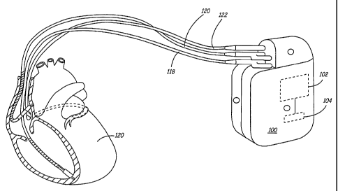
FIG. 1 is a diagram illustrating an exemplary implantable medical device in
association with a patient's heart;
FIG. 2 is a conceptual block diagram showing exemplary processing modules for
an implantable medical device;
FIG. 3A is a flowchart of an exemplary process for gauging a patient's
contractile
status that may be executed within an implantable medical device;
FIG. 3B is an exemplary plot of observed values for recirculation fraction in
the
left and right ventricles for various heart xates;
FIG. 3C is an exemplary plot of observed values for potentiation ratios
observed in
left and xight ventricles;
FIG. 4 is a flowchart of an exemplary process for optimizing timing parameters
and/or therapy application as a function of ventricular force interval; and
FIG. 5 is a flowchart of an exemplary process for tuning a response generated
by
an implanted medical device as a function of ventricular force interval.
The following detailed description is exemplary in nature and is not intended
to
Iimit the invention or the application and uses of the invention. Furthermore,
there is no
intention to be bound by any theory presented in the preceding background of
the
invention or the following detailed description of the drawings.
2o As mentioned above, it has been known for some time that contraction and
relaxation of the myocardium is controlled by the uptake and release of
calcium from the
sarcoplasmic reticulum (SR). More recently, several observers have noted that
alterations
in intracellular handling of calcium (Ca+Z) is associated with CHF. Changes in
intracellular calcium regulation, then, can be directly correlated to the
contractile status of
a patient's heart, and may be indicative of the onset and/or progression of
CHF and other
conditions. By monitoring changes in intracellular calcium regulation, cardiac
health
issues can therefore be identified, monitored and treated more effectively.
One technique fox evaluating SR calcium regulation involves monitoring the
force-
interval property of the myocardium using the contractile parameter, dP/dtmaX.
This
3o quantity represents the time derivative (i.e. the rate of change) of
pressure in the heart
(typically in the left ventricle, although also measured in the right
ventricle and
elsewhere), and is known to be a good index of the force of myocardial
contraction. More
CA 02523661 2005-10-25
WO 2004/098381 PCT/US2004/013085
-4-
particularly, potentiation in dP/dt observed following a cardiac perturbation
(e.g. an extra-
systole, pre-mature ventricular contraction (PVC), or the like) can be
quantified and
tracked over time to identify changes in intracellular calcium regulation. It
has been
observed that premature ventricular depolarizations typically produce a
relatively weak
first contraction due to impairment in intracellular release of calcium.
Subsequent beats,
however, typically exhibit increased contractile force (i.e. potentiation)
that can be
measured With a ventricular pressure monitor or the like. Factors that may be
monitored
include the degree of systolic potentiation, as well as the time to recover
from potentiation,
and the like. Accordingly, the amount of potentiation following a heart beat
perturbation
1o can be a good indicator of the intracellular calcium regulation, and may
provide insight
into the overall hemodynamic status of the patient. In particular, measuring
potentiation
following a heart rate perturbation is believed to be useful in identifying
patients at risk ,
CHF decompensation or sudden cardiac death.
The relationship between myocardial force interval and calcium regulation can
be
~5 beneficially exploited in an implantable medical device (IMD) such as a
pacemaker,
implantable cardioverter defibrillator (ICD), or heart monitor to assess the
patient's overall
cardiac health. According to various .embodiments, an implantable medical
device (IMD)
monitors potentiation resulting from a heart rate perturbation (e.g. a PVC or
extrasystole)
and provide information regarding the state of the patient's regulation of
intracellular
20 calcium and/or contractile status. The perturbation may be naturally
occurring in the
patient, or may be produced by the IMD or another appropriate device.
Data obtained at the IMD could be used for enhanced monitoring, diagnosis
and/or
therapeutic functions. The IMD may store diagnostic data in a memory, for
example, or
may activate an alarm to the patient if immediate medical attention is
required, or may
25 take other action as appropriate. In further embodiments, the IMD
administers or adjusts
an appropriate therapy or other response when such treatment or adjustment to
the
treatment is warranted. As used herein, the term "response" is intended to
broadly
encompass any type of medical response, alarm, report or the like (including
storage of
data within the IMD), as well as any of the various therapies that may be
provided by the
3o IMD to the patient. In a further embodiment, potentiation may be used to
determine
optimal settings for a pacing device, or for optimal delivery of a
pharmaceutical or other
therapy. In practice, potentiation following a cardiac perturbation can be
effectively
CA 02523661 2005-10-25
WO 2004/098381 PCT/US2004/013085
-5-
manipulated and monitored by mechanisms present in many conventional IMDs,
thus
making potentiation a very effective parameter for monitoring or improving a
patient's
cardiac health.
With reference now to FIG. 1, an exemplary implantable medical device (IMD)
100 is connected to monitor a patient's heart 120. IMD 100 may be further
configured to
integrate both monitoring and therapy features, as will be described below.
IMD 100
suitably collects and processes data about heart 120 from one or more sources
(e.g. heart
rate monitor, blood pressure monitor, electrocardiogram (ECG) waveform,
electrogram
waveform (EGM), or more generally PQRST waveform, etc.). IMD 100 may further
1o provide therapy or other response to the patient as appropriate, and as
described more fully
below. As shown in FIG. 1, IMD 100 may be generally flat and thin to permit
subcutaneous implantation within a human body, e.g.,.within upper thoracic
regions or the
lower abdominal region. IMD 100 may include a hermetically-sealed housing that
encloses a processor 102, a digital memory 104, and other components as
appropriate to
~ 5 produce the desired functionalities of the device. W various embodiments,
IMD 100 is
implemented as any implanted medical device capable of measuring the heart
rate of a
patient, including, but not limited to a pacemaker, defibrillator,
electrocardiogram monitor,
blood pressure monitor, drug pump, insulin monitor, or neurostimulator. An
example of a
suitable IMD device that may be used in various exemplary embodiments is the
2o CHRONICLE monitoring device available from Medtronic, Inc. of Minneapolis,
Minnesota, which includes a mechanical sensor capable of detecting changes in
ventricular pressure (dP/dt). In a further embodiment, IMD 100 is any device
that is
capable of sensing ventricular pressure and of providing pacing and/or
defibrillation to the
heart. Another example of an IMD capable of sensing dP/dt and other pressure-
related
25 parameters is described in commonly assigned United States Patent No.
6,438,408B l,
which issued to Mulligan et al. on August 20, 2002.
Processor 102 may be implemented with any type of microprocessor, digital
signal
processor, application specific integrated circuit (ASIC), field programmable
gate array
(FPGA) or other integrated or discrete logic circuitry programmed or otherwise
configured
3o to provide functionality as described herein. Processor 102 executes
instructions stored in
digital memory 104 to provide functionality as described below. Instructions
provided to
processor 102 may be executed in any manner, using any data structures,
architect<ire,
CA 02523661 2005-10-25
WO 2004/098381 PCT/US2004/013085
programming language and/or other techniques. Digital memory 104 is any
storage
medium capable of maintaining digital data and instructions provided to
processor 102
such as a static or dynamic random access memory (RAM), or any other
electronic,
magnetic, optical or other storage medium.
As further shown in FIG. 1, IMD 100 may receive one or more cardiac leads for
connection to circuitry enclosed within the housing. In the example of FIG. 1,
IMD 100
receives a right ventricular endocardial lead 118, a left ventricular coronary
sinus
endocardial lead 122, and a right atrial endocardial lead 120, although the
particular
cardiac Ieads used will vary widely from embodiment to embodiment. In
addition, the
housing of IMD 100 may function as an electrode, along with other electrodes
that may be
provided at various locations on the housing of IMD 100. In alternate
embodiments, other
data inputs, leads, electrodes and the like may be provided. Ventricular leads
118 and 122
may include, for example, pacing electrodes and defibrillation coil electrodes
(not shown)
in the event IMD 100 is configured to provide pacing, cardioversion and/or
defibrillation.
~ 5 In addition, ventricular leads 118 and 122 may deliver pacing stimuli in a
coordinated
fashion to provide biventricular pacing, cardiac resynchronization, post-
extrasystolic
potentiation (PESP) therapy or other benefits. Exemplary PESP therapy includes
those
described in U.S. Pat. No. 6,213,098 to Bennett et al. and non-provisional
U.S. patent
application serial number 10/xxx,xxx to Deno et al. filed on 28 August 2002,
the contents
20 of both disclosures are hereby incorporated by reference herein. IMD 100
may also obtain
input data from other internal or external sources (not shown) such as a
ventricular
pressure monitor, pH monitor, arterial pressure monitor, accelerometer or the
like.
In operation, IMD 100 suitably obtains data about heart 120 via leads 118,
120, 122,
and/or other sources. This data is provided to processor 102, which suitably
analyzes the
25 data, stores appropriate data about the episode in memory 104, and/or
provides a response
or report as appropriate. Any identiried cardiac episodes (e.g. an arrhytlnnia
or heart
failure decompensation) can be treated by intervention of a physician or in an
automated
manner. In various embodiments, IMD 100 activates an alarm upon detection of a
cardiac
episode. Alternatively or in addition to alarm activation, IMD 100 selects or
adjusts a
30 therapy and coordinates the delivery of the therapy by.IMD 100 or another
appropriate
device. Optional therapies that may be applied in various embodiments may
include drug
CA 02523661 2005-10-25
WO 2004/098381 PCT/US2004/013085
delivery, electrical stimulation, neurostimulation, modifications in pacing
rate, and/or the
like.
With reference now to Figure 2, an exemplary data processing layout for an IMD
100 suitably includes a data collection module 206, a data processing module
202, a
response module 218 and/or a reporting module 220. Each of the various modules
may be
implemented with computer-executable instructions stored in memory 104 and
executing
on processor 102 (FIG. 1), or in any other manner. The exemplary modules and
blocks
shown in FIG. 2 are intended to illustrate one logical model for implementing
an IMD
100, and should not be construed as limiting. Indeed, the various practical
embodiments
1 o may have widely varying software modules, data structures, applications,
processes and
the like. As such, the various functions of each module may in practice be
combined,
augmented, optimized or otherwise differently-organized in any fashion.
Data collection module 206 suitably interacts with one or more data sources
207 to
obtain data about the patient. Data sources 207 include any source of
information about
~ 5 the patient's heart, blood, temperature andlor the like. In various
embodiments, data
sources 207 include an ECG or EGM~ source 208 that provides electrical
impulses or other
observed signals that can be used to model the patient's electrocardiogram
(ECG)
waveform. Other data sources 207 may include a heart rate sensor 210, a
ventricular
pressure monitor 21.4, an accelerometer 212, a sensor 216 for determining
cardiac
2o conduction time andlor the like. The various data sources 207 may be
provided alone or in
any combination with each other, and may vary widely from embodiment to
embodiment.
Sensors for cardiac conduction time 216 and heart waveform 208 data could be
combined
into a single pair of electrodes, for example. Moreover, other data sources
207 such as
temperature sensors, blood pH sensors or the like could additionally or
alternatively be
25 provided. One example of a pressure sensor 214 is described in commonly
assigned
United States Patent No. 5,564,434.
Data collection module 206 suitably receives data from each of the data
sources
207 by polling each of the sources 207, by responding to interrupts or other
signals
generated by the sources 207, by receiving data at regular time intervals, or
according to
3o any other temporal scheme. Data may be received at data collection module
206 in digital
or analog format according to any protocol. If any of the data sources
generate analog
data, data collection module 206 suitably translates the analog signals to
digital
CA 02523661 2005-10-25
WO 2004/098381 PCT/US2004/013085
_g_
equivalents using any form of analog-to-digital conversion scheme presently
known or
subsequently developed. Data collection module may also convert data from
protocols
used by data sources 207 to data formats acceptable to data processing module
202, as
appropriate.
Data processing module 202 is any cixcuit, programming routine, application or
other hardware/software module that is capable of processing data received
from data
collection module 206. In various embodiments, data processing module 202 is a
software
application executing on processor 102 (FIG. 1) to implement the process
described below
in conjunction with FIG. 3. Accordingly, data processing module 202 suitably
interprets
1o received ventricular pressure (i.e. dP/dt) or other data to quantify
potentiation or other
effects in the patient's cardiac status and to produce an appropriate
response, as described
more fully below.
Issues in the patient's cardiac health can be detected, for example, when the
amount of potentiation in dP/dtmaX deviates from a baseline reading by more
than a
~5 threshold amount, or according to any other criteria. The baseline amount
of potentiation
may be a static value, or may be updated over time. In various embodiments,
the baseline
data represents a mean or median value observed over any appropriate number of
preceding samples. Threshold values may be any nominal values derived from a
typical
population of patients, or from any other source. Alternatively, the threshold
values may
2o be independently adjusted and set for~a given patient as desired by the
attending physician.
In various embodiments, the more recent values of potentiation, as well as
other
information, may be stored in a memory 204 to facilitate diagnosis of the
patient. In
another embodiment, data values observed during a particular time period or
near a
cardiac event deemed important by algorithms in the IMD (e.g. preceding an
observed
25 arrhythmia) may be stored in a memory 204 to facilitate diagnosis of the
patient.
In an exemplary embodiment, processing module 202 receives ventricular
pressure data
214 and/or other appropriate information from data collection module 206 and
interprets
the data using conventional digital signal processing techniques. If a heart
beat
perturbation occurs, data about the episode (e.g. the duration and/or
magnitude of
3o potentiation, time and date of the episode, and/or the like) may be stored
in memory 204,
which may correspond to hardware memory 104 shown in FIG. 1, or may be
implemented
with any other available digital storage device.
CA 02523661 2005-10-25
WO 2004/098381 PCT/US2004/013085
_9_
When a perturbation is identified, processing module 202 may trigger an
appropriate response if warranted by the data resulting from the perturbation.
Responses
may be activated by sending a digital message in the form of a signal, passed
parameter or
the Iike to response module 2 ~ & and/or reporting module 220.
Reporting module 220 is any circuit or routine capable of producing
appropriate
feedback from the IMD to the patient or to a physician. In various
embodiments, suitable
reports might include storing data in memory 204, generating an audible or
visible alarm
228, producing a wireless message transmitted from a telemetry circuit 230 via
an antenna
234, or providing other data that may be downloaded from a serial, parallel or
other
interface 232. Reports may include information about the potentiation duration
and/or
magnitude, time and date of episode occurrence, or any other appropriate data.
In a
further embodiment, the particular response provided by reporting module 220
may vary
depending upon the severity of the episode. Minor episodes may result in no
alarm at all,
for example, or a relatively non-obtrusive visual or audible alarm. More
severe episodes .
might result in a more noticeable alarm, in addition to an automatic response
as described
below.
Telemetry circuitry 230 communicates data from IMD 100 to an external device
via antenna 234. The external device receiving the wireless message may be a
programmer/output device that advises the patient, a physician or other
attendant o~
2o serious conditions, e.g., via a display or a visible or audible alarm.
Information stored in
memory 204 may be provided to an external device via antenna 234 for example,
to aid in
diagnosis or treatment of the patient. Alternatively, the external device may
be an
interface to a telephone network such that IMD 100 is able to automatically
notify
emergency personnel if an extreme episode occurs.
Interface 232 is any serial, parallel or other interface to an external
computing
device. Interface 232 and/or telemetry circuit 230 may be used to provide
information
from IMD 100 to an external device. Information stored in memory 204 may be
provided
to an external digital computer or other device, for example, to aid in
diagnosis or
treatment of the patient.
3o Response module 218 is any circuit, software application or other component
that
interacts with any type of therapy-providing system 264, which may include any
type of
therapy deliver mechanisms such as a drug delivery system 222,
neurostimulation 226
CA 02523661 2005-10-25
WO 2004/098381 PCT/US2004/013085
- 10-
and/or cardiac stimulation 224. In some embodiments, response module 218 may
alternatively or additionally interact with an electrical stimulation therapy
device
integrated with IMD 100 to deliver pacing, post-extrasystolic potentiation,
cardioversion,
defibrillation and/or any other therapy. Accordingly, the various responses
that may be
provided by IMD 100 vary from simple storage of data to actual provision of
therapy in
various embodiments. Any therapy provided may be titrated or otherwise
adjusted in
response to potentiation observed, as described more fully below. Drug dosage
may be
adjusted according to episode severity, for example, or pacing parameters can
be adjusted
in response to observed potentiation.
1 o The various components and processing modules of IMD 100 may be housed in
a
common housing such as that shown in FIG. 1. Alternatively, portions of IMD
100 may
be housed separately. For example, portions of the therapy delivery system 264
could be
integrated with IMD 100 or provided in a separate housing, particularly where
the therapy
delivery system includes drug delivery capabilities. In this case, response
module 218
~ 5 may interact with therapy delivery system 264 via an electrical cable or
wireless link, or
via interface 232.
With reference now to FIG. 3, an exemplary process 300 for gauging the
contractile status of a patient suitably includes the broad steps of
generating and/or
observing a heart rate perturbation (step 304), measuring the associated
potentiation
2o generated by the perturbation (step 306), and processing or quantifying the
data to
correlate the potentiation with the patient's intracellular calcium
regulation, ventricular
contractile status and/or cardiac health (step 308). In various embodiments,
the various
steps of process 300 may be implemented with computer-executable instructions
that are
stored in a digital memory 104 and that are appropriately executed by
processor 102 (FIG.
25 1), or by any other processor associated with the IMD.
Process 300 suitably begins by setting appropriate pacing intervals by IMD
and/or
otherwise initializing the IMD for the gauging process (step 302). An
exemplary
technique for determining optimum pacing intervals is set forth below in
conjunction with
FIG. 5, although any steady state pacing routine could be used in alternate
embodiments.
3o Initialization rnay also include setting or resetting any counters, timers
or other variables
Within processor 102 as appropriate. After pacing intervals are set, it may be
desirable to
maintain the pacing state for a short period of time (e.g. on the order of
thirty seconds or
CA 02523661 2005-10-25
WO 2004/098381 PCT/US2004/013085
-11-
so) to allow the patient's hemodynamics to settle into a relatively steady
state. In a further
embodiment, process 300 may be performed when the patient is asleep or at rest
to further
minimize transient effects upon the heart. Periods of sleep or rest may be
identified by a
clock in IMD 100, by a manual activation, by accelerometer data (e.g.
accelerometer 212
in FIG. 2), or by any other technique. Likewise, process 300 may be withheld
when the
patient is active or extremely active, or otherwise has a high heart rate, as
appropriate.
Analysis of potentiation suitably begins by identifying a perturbation to the
patient's heart
such as a PVC or other change the heartbeat that results in a change in the
patient's
hemodynamics. Various forms of cardiac perturbations may include any
ventricular beat
originating from a different source than a baseline beat, or that produces a
smaller or
larger output from the heart. Perturbations may be naturally-occurring, or may
be initiated
by IMD 100 as described more fully below. A perturbation may be generated, for
example, by inducing a premature beat in either ventricle, and/or by adjusting
the rate at
which either the left and/or right ventricle are paced. In the context of
baseline ventricular
pacing, for example, changes in hemodynamic pressure can be induced in the
patient by
pacing a single ventricle for one or more beats.
In an exemplary embodiment, naturally-occurring perturbations (e.g. PVCs) in
the
patient are identified by monitoring electrocardiogram (ECG) data such as a
PQRST
waveform or the like within IMD 100. Data may be collected according to any
scheme,
2o but in an exemplary embodiment data measurements are taken at regular time
intervals
with a sufficiently high frequency to identify any natural perturbations of
the patient's
heart rate. Although an exemplary process 300 discussed herein emphasizes
monitoring
dP/dt for purposes of simplicity and illustration, other equivalent data
factors such as atrial
and/or ventricular pressure may be used in addition to or in place of dP/dt
data in various
alternate but equivalent embodiments. In a further exemplary embodiment,
perturbations
following unusual conditions may be ignored or differently processed by IMD
100, as
discussed below, so that the patient's condition can be monitored over time
under
relatively constant conditions.
In an alternate embodiment, IMD 100 induces extrasystolic beats (atrial or
3o ventricular), PVCs and/or other cardiac perturbations so that the patient's
reactions can be
appropriately monitored and/or tested. In such embodiments, IMD 100 suitably
provides
pacing to the heart prior to the premature beat to place the heart into a
steady rhythmic
CA 02523661 2005-10-25
WO 2004/098381 PCT/US2004/013085
- I2-
state, as described above. Further, IMD 100 may provide a string of
extrastimuli
entrainment beats (e.g. S1 beats) immediately prior to the premature beat to
further place
the heart into a known state. In an exemplary embodiment, a train of S 1 beats
having a
pacing rate roughly equal to the intrinsic rate may be provided by IMD 100,
followed by a
premature S2 beat at a rate of about forty percent to about sixty percent of
the S 1 rate,
followed in turn by a train of S3 beats having approximately the same rate as
the S 1 beats
preceding the premature beat. Of course, any combination of S1, S2, S3, S4
and/or other
beats at any pacing rate or prematurity could be used in alternate
embodiments, and the
particular rates used for each pulse could be adjusted accordingly. The S1 and
S3 beats,
1o for example, could be provided at a rate that is slightly (e.g. about ten
percent) faster than
the intrinsic rate in an alternate embodiment. Again, cardiac perturbations
observed
within step 304 may be naturally-occurring and/or induced by IMD 100 or
another device
in any manner.
When a perturbation is identified, the patient's reaction to the perturbation
is
observed and/or recorded (step 306). The reaction may be observed by
monitoring data
from a pressure sensor 214 (FIG. 2) to determine the magnitude and/or duration
of any
resulting potentiation. In an exemplary embodiment, dP/dt",~ data is obtained
for either or
both ventricles. Data may be gathered for any interval of time or for any
number of beats,
or for any other duration. In an exemplary embodiment, data is gathered for
about twenty
2o beats following the perturbation, or until the heart returns to its
original pre-perturbed
state. Data gathered is stored in memory 104 (FIG. 1) or another appropriate
location for
processing by IMD 100. Data gathered prior to the perturbation rnay also be
stored within
memory 104, or elsewhere on IMD 100.
In various embodiments, it may be desirable.to analyze the patient's condition
under relatively constant conditions over time. Variations in the perturbation
may
therefore create inconsistent data that may be of reduced benef t. To avoid
this situation,
in certain embodiments IMD monitors the patient's heart beat cycle length,
coupling
intervals and/or other parameters prior to the perturbation so that
perturbations resulting
from unusual baseline conditions may be flagged or otherwise differently
processed. If a
3o patient experiences PVCs following coupling intervals of 500 ms, 550 ms and
800 ms, for
example, analysis of the 800 ms PVC may be ignored or separated from the
analysis of the
other PVCs in various embodiments. Accordingly, certain embodiments may ignore
or
CA 02523661 2005-10-25
WO 2004/098381 PCT/US2004/013085
-13-
otherwise differently-process perturbations that occur following unusual or
non-standard
conditions.
After data is gathered, the stored data is processed to quantify the
potentiation
experienced by the patient and to correlate the potentiation to the patient's
intracellular
calcium regulation status and/or ventricular contractile status. Potentiation
may be
quantified according to any process or technique, including evaluation of
recirculation
fraction, potentiation ratio, and/or any other parameter related to changes in
dP/dt
following a perturbation to the heart.
Recirculation fraction (RF) is considered to be the ratio of calcium (Ca+2)
released
from the sarcoplasmic reticulum that is re-sequestered back on the SR on each
beat.
Because calcium re-uptake is known to be linearly related to the force-
interval propeuty of
the myocardium, however, RF may be derived from the recovery of potentiated
beats
folhowing the perturbation, although other techniques could be used in
alternate
embodiments. FIG. 3B shows an exemplary plot of RF measurements in both the
left and
right ventricles following an extra-systole at 320 msec for several different
heart rates.
As can be seen in FIG. 3B, no significant difference was observed between the
left and
right ventricular RF for each respective heart rate. Similarly, RF is believed
to be only
minimally influenced by the extra-systolic interval, making RF a convenient
and effective
parameter for evaluating calcium regulation in the heart.
2o While recirculation fraction focuses primarily on systolic recover,
however,
additional or alternative parameters may be measured to describe both systolic
and
diastolic function. The potentiation ratio (PR), for example, conventionally
provides a
ratio of the force at the greatest level of potentiation to the force in
response to the last
priming beat. Stated another way, PR may be determined by comparing the
potentiation
from one or more beats following the perturbation to the mean of the control
beats prior to
the perturbation. PR may also be evaluated following an abrupt decrease in
heart rate, as
shown in FIG. 3C.
Equivalent time-based teclmiques for quantifying potentiation include
measuring
the time for the dP/dtmaX to return to a normal level following the
perturbation, or
3o measuring the time from the perturbation to a minimum or maximum dP/dt
observed in a
window of time following the perturbation. Accordingly, PR may be used alone
or in
CA 02523661 2005-10-25
WO 2004/098381 PCT/US2004/013085
-14-
conjunction with RF and/or other parameters to monitor intracellular calcium
regulation,
which in turn correlates to ventricular contractile status.
Potentiation data (e.g. RF, PR andlor the like) may be correlated to a
patient's
hemodynamic condition or overall cardiac health in any mamier. Generally
speaking,
greater amounts of potentiation following a perturbation are considered to be
more
favorable than lesser values, since the greater amount generally indicates
better
intracellular calcium regulation. As discussed more fully below, data may be
stored
within IMD 100 to track changes over time. Extremely low amounts of
potentiation may
provoke IMD 100 to issue an alarm or warning for the patient to seek medical
attention,
1o and/or IMD 100 may use potentiation to process an additional response (step
310) such as
administering a drug, neurological or other therapy, or to adjust pacing rates
or other
parameters. Process 300 may be executed repetitively (step 312) to maintain
data over
time, or to iteratively adjust a therapy or other parameter. In such
embodiments, therapies
may be applied in a "closed loop" manner, whereby continuous monitoring of the
patient's
condition is provided as feedback to drive application andlor adjustment of
one or more
therapies. Neurostimulation or other treatments, for example, may be applied
in such
magnitudes and durations as appropriate to bring the patient's cardiac
condition back to
normal, or to improve the condition. In such embodiments, potentiation or
other
parameters can be monitored and/or titrated in a "closed loop" mamier using
conventional
2o control techniques until the parameter reaches a desired value.
Potentiation observations following a perturbation may be used to optimize
therapy
parameters within a pacemaker or other implantable device 100 capable of
delivering
therapy. With reference now to FIG. 4, an exemplary process 400 for optimizing
pacing
parameters suitably includes the broad steps of setting initial pacing
parameters to be
evaluated (step 402), adjusting one parameter (e.g. the atrial-ventricular (A-
V) interval)
(step 404), adjusting a second parameter (e.g. the cross-ventricular (V-V)
interval) (step
406), optionally re-visiting the first parameter (e.g. A-V interval) (step
408), and storing
the optimal settings (step 410) for continued operation of IMD 100. The
various steps of
process 400 may be implemented with computer-executable instructions stored in
a digital
3o memory 104 and that are appropriately executed by processor 102 (FIG. 1),
or by any
other processor associated with IMD 100.
CA 02523661 2005-10-25
WO 2004/098381 PCT/US2004/013085
-15-
Initial pacing parameters (step 402) may be set to any convenient initial
value as
determined from statistical models, historical data, patient history,
physician input or any
other source. In an exemplary embodiment, initial pacing intervals may be
about 100 ms
for A-V interval and about 0 ms for V-V interval, although any other intervals
could be
used. Optimization of pacing intervals takes place using any suitable
technique, such as
the iterative technique described below in conjunction with FIG. 5. Generally
spealcing,
IMD 100 gradually modifies the pacing parameters while monitoring potentiation
resulting from the changes. Because high potentiation generally correlates to
better
calcium regulation, the parameter that produces the highest amount of
potentiation may be
deemed to be optimal for continued pacing. After an optimal parameter for one
type of
pacing (e.g. A-V pacing) is identified; that setting can be used during
optimization of
another pacing parameter. After both parameters have been optimized, various
embodiments include cross-checking of the first parameter (step 408) so that
the optimal
pacing parameters for both types of pacing are evaluated together. Although
FIG. 4 shows
A-V interval evaluation (step 404) as taking place prior to V-V interval
evaluation (step
406), the respective order may be altered such that V-V intervals are
optimized prior to A-
V intervals, with any follow-up V-V optimization taking place after an optimal
A-V
interval is determined.
With reference now to FIG. 5, an exemplary process 500 for optimizing a
response
2o from an IMD 100 suitably includes iteratively providing a response (step
502),
determining the potentiation produced by the response (step 504), and
adjusting the
response (step 508) until an optimal (e.g. a maximum) potentiation is
identified.
Responses that may be optimized in various embodiments include pacing
parameters,
administration of drug or neuro-therapies, or the like. As with the processes
described
above, the various steps of process 500 may be implemented with computer-
executable
instructions stored in a digital memory 104 or other storage medium and
executed by any
processor 102 associated with IMD 100.
To begin the optimization process, a baseline response is initially provided
from
IMD 100 (step 502). Baseline responses may be obtained from historical data,
patient
3o history, physician input, or any other source. For example, to optimize A-V
intervals, the
baseline AV interval may be initially set at about 100 ms with no V-V delay.
Once the
AV interval is optimized, the V-V interval optimization may begin with an
interval of
CA 02523661 2005-10-25
WO 2004/098381 PCT/US2004/013085
- 1G -
about 0 ms, with the A-V interval set at the optimal level previously
determined, for
example, in step 404 of FIG. 4. Baseline levels of drug or neurostimulation
therapy could
alternatively be provided.
As the initial response from IMD 100 is applied, the patient's potentiation is
observed (step 504) using the techniques described above in conjunction with
FIG. 3 as
appropriate. Potentiation may be quantified using PR, andlor any other
parameter, for
example, to determine the patient's reaction to the initial therapy. After the
initial
response is processed, IMD 100 suitably varies the response provided (step
508) to obtain
additional data points for comparison (step 506). As mentioned above,
increased
1o potentiation generally correlates to improved hemodynamic condition, at a
given extra-
systolic interval, so process 500 generally seeks to maximize the level of
potentiation in
the patient (step 506). The observed value for each iteration is suitably
maintained in IMD
100 for comparison against subsequent observations. In an embodiment that
seeks to
optimize A-V intervals, for example, potentiation observations may be obtained
for AV
intervals of 80 ms, 100 ms, 120 ms or the like. If the maximum potentiation is
produced
at 120 ms, further data may be collected at 130 ms or so until a maximum value
is
identifted. If the maximum poter~tiation is produced at 100 ms, the response
may be
adjusted to, say, 90 ms and/or 110 ms to isolate a maximum value. Further
iterations may
provide improved resolution, thus resulting in a more accurate optimal value
produced. Of
2o course other embodiments will use widely varying values, and the particular
parameters
used in this illustrative example are not intended to be limiting in any way.
When an optimal parameter value is identified by the iterative process (steps
504,
506 and 508 of FIG. 5), that parameter may be set (step 510) within the IMD
100 for
continued application, or the value may be processed in other ways. In an
equivalent
embodiment of process 500, application of the response (i.e. steps 502 and
508) may be
manually provided by a health care clinician or another source external to IMD
100, While
monitoring functions (step 504) continue to be provided by IMD 100.
Accordingly, various methods and apparatus for diagnosing and gauging cardiac
condition using potentiation are provided. While exemplary embodiments have
been
3o presented in the foregoing detailed description of the invention, it should
be appreciated
that a vast number of variations exist. It should also be appreciated that
these exemplary
embodiments are only examples, and are not intended to limit the scope,
applicability, or
CA 02523661 2005-10-25
WO 2004/098381 PCT/US2004/013085
- 17-
configuration of the invention in any way. Rather, the foregoing detailed
description will
provide a convenient road map for implementing an exemplary embodiment of the
invention. Various changes may be made in the function and arrangement of
elements
described in an exemplary embodiment without departing from the scope of the
invention
as set forth in the appended claims and their legal equivalents.