Note: Descriptions are shown in the official language in which they were submitted.
CA 02524000 2005-11-02
WO 2004/098398 PCT/CA2004/000683
REAL-TIME CONTEMPORANEOUS MULTIMODAL IMAGING AND
SPECTROSCOPY USES THEREOF
FIELD OF INVENTION
Various optical apparati such as microscopes, endoscopes, telescopes, cameras
etc. support viewing or analyzing the interaction of light with objects such
as planets,
plants, rocks, animals, cells, tissue, proteins, DNA; semiconductors, etc.
Some mufti-
band spectral images provide morphological image data whereas other mufti-band
spectral images provide information related to the chemical make-up, sub-
structure
and/or other target object characteristics which may be . measured from mufti-
band
spectral images of reflected or emitted light. These light emission images,
such as
htminescence or fluorescence, may indicate and provide means to assess
endogenous
chemicals or exogenous substances such as dyes employed to enhance
visualization,
drugs, therapeutic intermediaries, or other agents.
In the field of medical imaging and more particularly endoscopy, reflected
white light, native tissue autofluorescence, luminescence, chemical emissions,
near-IR
reflectance, and other spectra provide a means to visualize tissue and gather
diagnostic information. In addition to visualization of tissue morphology the
interaction of light in various parts of the electromagnetic spectrum has been
used to
collect chemical information. Three general real-time imaging modalities for
endoscopy that are of interest include white-light reflectance imaging,
fluorescence
emission and near infrared reflectance imaging modalities.
CA 02524000 2005-11-02
WO 2004/098398 PCT/CA2004/000683
In endoscopy, conventional white light imaging is typically used to view
surface morphology, establish landmarks, and assess the internal organs based
on
appearance. Applications for viewing the respiratory and gastro-intestinal
tracts are
well established. Fluorescence imaging has evolved more recently and using the
properties of tissue autofluorescence has been applied to the detection of
early cancer.
Similarly, observations of various native and induced chemical interactions,
such as
labeling tissue with proteins, for example, have been accomplished using
fluorescence
imaging. Near infrared light may be used to measure tissue oxygenation and
hypoxia
in healthy and diseased tissue. Alternatively, fluorescently-tagged monoclonal
antibodies may be used to label specific cellular proteins, which in turn may
be
detected and/or be measured optically.
Presently, methods and device configurations exist which use each of these
imaging modalities to gather data in real-time, at video-rate. However, for
imaging,
this real-time information from different modalities has been available
sequentially or
in part, but not simultaneously.
As used herein, "multimodal" means at least two imaging modes which differ
in their spectral bands of illumination or their spectral bands of detection,
or both.
"Optical modulator" as used herein means a device or combination of optical
and/or electro-optical devices to alter the wavelength(s), and/or to alter the
intensity,
and/or to time-gate various spectra of electromagnetic radiation. Various
filters, filter
wheels, lenses, mirrors, micro-mirror arrays, liquid crystals, or other
devices under
mechanical or electrical control may be employed alone or in combination to
comprise such an optical modulator. Certain embodiments of the present
invention
utilize two optical modulators, one associated with modulating light source
spectrum
-2-
CA 02524000 2005-11-02
WO 2004/098398 PCT/CA2004/000683
that will be used to interrogate or interact with an object. Modulation of
source
illumination therefore could be as simple as switching (gating on) one or more
illumination sources in a controlled manner, or accomplishing optical
modulation
with the devices as described. A second modulator is used to process the light
returned after interacting with the object. The second optical modulator could
be
serve to split imaging light segments to direct them to various detectors, and
be
comprised of; for example.a moving mirror, a rotating mirror as part of
a.filter wheel,
or a digital mufti-mirror device (DMD). The detectors may be imaging devices
such
as cameras with CCD sensors or these sensors may comprise spectrometers. In
some
cases, such as in vivo endoscopic use, interaction of source illumination may
be with
lung tissue and returned light may include various reflected and re-emitted
spectra.
Control and synchronization as used herein means to provide control over the
optical modulators andlor the electromagnetic radiation source and/or the
detectors,
for example at real-time video rates, and to further synchronize the operation
of these
components to provide a means to generate the desired source spectrum for the
desired time periods, and to process (e.g. amplify, attenuate, divide, gate)
and detect
image ,signals of various spectrum, contemporaneously. In some embodiments
relatively tight control and synchronization are required, in other
embodiments, these
returned signals may themselves be used for co-ordination, for example, their
intensity or wavelength may be used to provide information for control and
synchronization.
In addition to viewing and analysis, at the same time, selected.spectra of
light
may be directed to stimulate certain photosensitive chemicals so that
treatments such
as photodynamic therapy (PDT) may be delivered and monitored. .
-3-
CA 02524000 2005-11-02
WO 2004/098398 PCT/CA2004/000683
While prior art discusses means to sequentially provide white-light imaging
(typical spectral range 400 nm to 700 nm), fluorescence imaging (e.g. tissue
autofluorescence stimulated with blue light from 400 nm to 450 nm and
re=emitted in
the 470 nm to 700 nm range) and near-infrared images with an approximate
spectral
range of 700 nm to 800nm or beyond, and/or particular spectra in these ranges,
and/or
an imaging modality combined with a spectral signal, there remains a need for
apparatus and methods to provide these various imaging modes,
contemporaneously,
at video rates. The present invention meets this need.
BRIEF DISCUSSION OF ART
United States Patent No. 6,364,829, to Fulghum, entitled, "Autofluorescence
imaging system for endoscopy", discusses a broad-band light source to provide
both
visible light (which induces minimal autofluorescence) and ultraviolet light
(capable
of inducing tissue autofluorescence). Images are detected, for example, by a
single
imaging detector at the distal tip of an endoscope and provisions are . made
for
electronically switching between these source illumination spectrum. Various
light
sources, filter wheels, shutters, mirrors, dichroic mirrors, spectrum, light
sources,
intensities and timing diagrams are provided and therefore this prior art is
included by
reference.
United States Patent .No. 6,148,227, to Wagnieres, entitled, "Diagnosis
apparatus for the picture providing recording of fluorescing biological tissue
regions ", discusses illumination spectrum and components for fluorescence
imaging.
In one embodiment red and green components are directed to separate portions
of a
CCD with independent signal processing.
-4-
CA 02524000 2005-11-02
WO 2004/098398 PCT/CA2004/000683
United States Patent No. 6,061,591, to Freitag, entitled, "Arrangement and
rnethod for diagnosing malignant tissue by fluorescence observation';
discusses a
strobed white-light illumination source and laser to stimulate fluorescence.
Alternatively, a desired fluorescence spectrum may be isolated and provided
from a
single lamp, for example, a Mercury-Xenon arc lamp. Filter wheels (with red,
green
and blue filters as well as filters to divide fluorescence . into red and
green
components) and timing requirements are also discussed.' Measurements of white
light images and fluorescence are performed in sequence, although both may be
displayed on the monitor. Various Figures describe light sources which are
similar to
those contemplated for the present invention.
The system described in Fulghum has the ability to switch back and forth
between white light and fluorescence visualization methods electronically with
display rates up to 10 Hz, or higher. Unlike other prior art (e.g. U.S. Patent
No.
5,647,368 which will be discussed), switching between normal visible light
imaging,
in full color, and fluorescence imaging is accomplished by an electronic
switch rather
than by physical modulation (switching) by the operator. This prior art also
discusses
a fluorescence excitation light at ultraviolet to deep violet wavelengths
placed at the
end of an endoscope, as well as gallium nitride laser diodes and mercury arc
lamps for
UV which are also contemplated as illumination sources for various embodiments
of
the present invention. Also of interest, Fulghum discusses limitations of
endoscopes
and more particularly limitations related to the UV-transmissive properties of
optical
fibers. Some of these limitations are addressed by co-pending. United States
Application No. 10/226,406 to Ferguson/Zeng, filed approximately August 23,
2002,
entitled "Non-coherent fiber optic apparatus and imaging methods ".
-5-
CA 02524000 2005-11-02
WO 2004/098398 PCT/CA2004/000683
United States Patent No. 6,019,719, to Schulz, entitled, "Fully auotclavable
electronic endoscope ", discusses an objective lens, crystal filter, IR filter
and CCD
chip arranged at the distal end of an endoscope for imaging.
United States Patent No. 5,930,424 to Heimberger, entitled, "Device for
connecting a fiber optic cable to tlae fiber optic connection of an endoscope
",
discusses various aspects of coupling devices such as light sources to an
endoscope.
United States Patent No. 5,926,213 to Hafele, entitled, "Device for correcting
the tone of color pictures recorded by a video tames°a ' ; such as an
endoscope camera,
is discussed along with a rotary transducer to activate tone correction. Color
correction, calibration or normalization is useful for quantization from image
data or
comparison of images and is considered for various embodiments of the present
invention.
United States Patent No. 5,827,190, to Palcic, entitled, "Endoscope having an
integrated CCD sensor ", discusses illumination light sources and sensors to
measure
various signals associated with tissue and tissue disease.
United States Patent No. 5,647,368, to Zeng, entitled, "Imaging system for'
detecting diseased tissue using native fluorescence in the gastrointestinal
and
respiratory tract ", among other things discusses use of a mercury arc lamp to
provide
for white light and fluorescence imaging with an endoscope to detect and
differentiate
effects in abnormal or diseased tissue.
United States Patent No. 5,590,660, to MacAulay, entitled, "Apparatus' and
method for imaging diseased tissue using integrated autofluorescence"
discusses light
source requirements, optical sensors, and means to provide a background image
to
normalize the autofluorescence image, for uses such as imaging diseased
tissue.
-6-
CA 02524000 2005-11-02
WO 2004/098398 PCT/CA2004/000683
United States Patent No. 5,769,792, to Palcic, entitled, "Endoscopic imaging
system for' diseased tissue'; further discusses light sources and means to
extract
information from the spectral intensity bands of autofluorescence, which
differ in
normal and diseased tissue.
Also co-pending United States Patent Application No. 09/741,731, to Zeng,
filed approximately December 19, 2000 and entitled, "Methods and apparatus for
fluorescence and reflectance imaging and spectroscopy and for contemporaneous
measurements of electromagnetic radiation with multiple naeasuring devices';
(a
continuation-in-part of U.S. Publication No. 200210103439) discusses
contemporaneous methods of providing one mode of imaging and spectroscopy
contemporaneously, but multiple imaging and associated spectroscopy modalities
is
sequential. In the present invention, methods are described to perfornl
multimodal
imaging contemporaneously at various desired wavelengths. Unlike Zeng's prior
art,
Zeng's present invention does not seek to provide images and measurements of
wavelength spectrum, instead it seeks to provide contemporaneous multimodal
imaging, where entire images in defined spectrum are detected and utilized for
display
or analysis.
United States Patent No. 5,999,844, to Gombrich, entitled, "Method and
appaf-atus for imaging and sampling diseased tissue using autofluorescefzce ",
discusses a plurality of image detectors that receive excitation light as well
as
depositing biopsies in separate compartments or captive units.
United States Patent No. 6,212,425, to Irion, entitled, "Apparatus for
photodynamic diagnosis'; discusses endoscopic imaging using a light-induced
.. -7-
CA 02524000 2005-11-02
WO 2004/098398 PCT/CA2004/000683
reaction or intrinsic fluorescence to detect diseased tissue and delivery
light for
therapeutic use or to stimulate compounds that in turn provide therapy, for
example.
United States Patent No. 4,884,133, to Kanno, entitled "Ehdoscope light
source apparatus'; discusses light sources, light guides and control of these
elements
for endoscopic use.
United States Patent No. 5,749,830 to Kaneko entitled . "Fluorescent
endoscope apparatus" discusses use of two light sources, a first (e.g. lamp)
for white
light and a second (e.g: helium-cadmium laser) for fluorescence to provide
interrogating spectrum. Kaneko '830 also employs a filter wheel placed in the
pathway of a single detector. For multimodal imaging the filter wheel has a
plurality
of filters (e.g. three in Fig. 4a and 5 in Fig. 4b). While they illustrate the
display of
two imaging modalities (110 of Fig 7.), they do not discuss simultaneous real-
time
multimodal imaging. As this prior art discusses a wide range of issues
utilized within
the present invention, such as combining light sources, synchronization and
filter
wheels, '830 is included by reference herein.
Endoscopes and imaging applications are further discussed in co-pending
United,States Application No. 10/226,406 to Ferguson/Zeng, entitled "Non-
coherent
fiber optic apparatus and i~aagihg methods'; which among other. things,
discusses
apparatus to overcome some existing limitations of fiber optic devices, such
as
endoscopes.
SUMMARY AND OBJECTIVES OF THE INVENTION
The present invention solves the problems described above by providing
simultaneous multimodal spectral images of a target object. Targeting
radiation or
_g_
CA 02524000 2005-11-02
WO 2004/098398 PCT/CA2004/000683
illumination is modulated to provide segments of radiation of different
wavelengths,
for example, alternating segments of white, green, blue, red, and near-
infrared light.
The target object returns reflected and re-emitted (for example, fluoresced)
light,
which is further modulated to separate the returned light into segments
corresponding
to different wavelengths. The returned radiation can be processed, displayed,
and
analyzed.
BRIEF DISCUSSION OF DRAWINGS
FIGURE 1 (prior art) shows a series of typical desired spectra utilized for
endoscopic imaging.
FIGUREs 2a and 2b (prior art) illustrate the spectra from a typical
fluorescence endoscopy system.
FIGURE 3 (prior art) illustrates a typical spectra from the fluorescence mode
of a sequential white light and fluorescence endoscopy system.
FIGURE 4 shows an illumination source placed for example at the distal end
of an endoscope.
FIGURE 5 is a perspective view of an embodiment of the present invention.
FIGURE 6a is a perspective view of the simultaneous white light and
fluorescence imaging with a single detector comprising multiple sensors.
FIGURE 6b is a perspective view of the detector configuration associated with
FIGURE 6a.
FIGURE 6c is a perspective view of another detector configuration associated
with FIGURE 6a, which can be placed at the distal tip of an endoscope.
-9-
CA 02524000 2005-11-02
WO 2004/098398 PCT/CA2004/000683
FIGURE 6d is a block diagram of the control and synchronization for
contemporaneous imaging modes described in FIGURES 6a, 6b and 6c.
DETAILED DISCUSSION OF DRAWINGS AND PREFERRED EMBODIMENTS
While the invention may be susceptible to embodiments in different forms,
there is shown in the drawings, and herein will be described in detail,
specific
embodiments with the understanding that the present disclosure is to be
considered an
exemplification of the principles of the invention, and is not intended to
limit the
invention to that as illustrated and described herein.
Endoscopy and endoscopic apparatus may be described and differentiated in
terms of tissue illumination and generated signals which include reflected
light and/or
emission spectrum.
FIGURE 1 (prior art) illustrates typical spectra utilized for white light and
fluorescence assessment. Spectrum 0 100 shows the broad range of illumination
typically utilized. Such illumination may be provided by a single source or
multiple
combined sources as discussed in prior art and further in this application.
Spectrum 1 101 shows a typical white .light (broad-band) illumination
spectrum. Various illumination sources (lamps etc.) are available to produce
broad-
band illumination, for example U.S. Patent No. 6,364,829 to. Fulghum discusses
desired illumination. Illumination as shown in Spectrum 1 101 may interact
with a
target tissue providing reflected light, such as typical white light signal
(reflectance),
illustrated in Spectrum 2 102, in substantially the same spectral range as the
source,
but attenuated relative to the incident illumination. Such attenuation may be
-10-
CA 02524000 2005-11-02
WO 2004/098398 PCT/CA2004/000683
preferential based on tissue absorption, presence of blood and other factors
as
observed in Spectrum 2 102.
Spectrum 3 103 represents typical short wavelength light, for example, blue
light, intended to excite tissue fluorescence. A typical returned signal
Spectrum 4 104
has two components, a tissue reflectance component 1048, which is typically
not
utilized, and a tissue fluorescence emission signal 104E. The reflectance
component
is often blocked or filtered out so that it does not interfere with
fluorescence detection.
Accordingly, to excite tissue fluorescence, narrow illumination bands may be
preferred. The narrow bands may be isolated from broad-band illumination or
they
may be provided by a narrow band source such as an LED or laser. Typical UV
illumination as illustrated in Spectrum 5 105, may be used to excite tissue
autofluorescence producing a spectrum such as is shown in Spectrum 6 106.
Again,
the reflectance component 1068 is usually not used. Typical illumination
illustrated
in Spectrum 7 107 in the red/near IR provides a reflectance component as shown
in
Spectrum 8 108.
In addition, illumination spectrum may be combined and used to advantage.
For example, typical illumination shown in Spectrum 9 109, blue light. plus
red/near
IR light, produces a signal spectrum such as shown in Spectrum 10 110. These
spectra (0 to 10) will be referred to during the discussion of various
Figures.
FIGURES 2a and 2b (prior art) describe and represent endoscopic imaging
principles encompassing United States patent No. 5,413,108 to Alfano entitled,
"Method arad appaf-atus fof~ mapping a tissue sample for and distinguishing
different
regions thereof based on luminescence measu~enaefzts of cancer-indicative
native
fluorophoY" and United States Patent No. 6,091,985 to Alfano, entitled,
"Detection of
-11-
CA 02524000 2005-11-02
WO 2004/098398 PCT/CA2004/000683
cancer and precancerous conditions in tissues af~dlor cells using native
fluorescence
excitation spectroscopy ", both of which are included herein by reference. As
was
introduced, these principals may be applied to other optical systems such as
microscopes, cameras, telescopes etc. and are described in United States
Patent No.
6,080,584 to Alfano, entitled "Method afzd apparatus for detecting the
presefzce of
cancerous afZd precancerous cells in a smear using native fluorescence
spectf°oscopy." This prior art to Alfano is included by reference.
Accordingly, FIGURE 2a illustrates white light, reflectance and emission
endoscopy, generically, in terms of input spectra 212 (illumination) and
output signal
spectra 214, with input and output delineated by indicator line 210. A first
illumination 201, ~,1-I, is selected in the UV range to stimulate tissue
autofluorescence (e.g. Spectrum 5 as discussed in association with FIGURE 1).
The
resulting tissue emission spectra 251 occur in the blue/green region, which is
further
identified as ~,l-E (e.g. 106E of Spectrum 6 in FIGURE 1). Using the
interr~gating
illumination 201, the emission signal intensities of normal and diseased
tissue are
similar. This is further shown by the characteristic curve for normal tissue
221 and
diseased tissue, 226. A first representative (reference) image of tissue
emission
(autofluorescence) is typically acquired during time interval T1.
FIGURE 2b shows input spectra 216 and signal spectra 218. During time
interval T2, a second interrogating illumination 202, ~,2-I in the UV/blue
region,
illuminates tissue to excite autofluorescence (e.g. Spectrum 3 discussed in
association
with FIGURE 1 ). The resulting tissue emission spectra 252, further identified
as ~,2-E
(emission) again occurs in blue/green region. Under these conditions, a
measurable
difference is observed between the characteristic curves for normal tissue 222
and
-12
CA 02524000 2005-11-02
WO 2004/098398 PCT/CA2004/000683
diseased tissue 227. A tissue image is acquired during this interval, T2. ~
Ratios
andlor differences between the first (reference) image acquired during T1 and
a
second image acquired during T2 provides a basis to normalize, process and
extract
diagnostic information. One advantage of such a configuration is that, since
the
images are acquired sequentially, this may be accomplished using a single
image
sensor. Additionally, because the two tissue autofluorescence images are
produced in
the same general spectral region (251, 252 are both blue/green), they cannot
be
separated in space by optical means and are therefore separated in time domain
(T1
and T2) as indicated. Various limitations result, for example, it becomes more
difficult to.register (pixel align) the two images which may be shifted due to
breathing
or motion of the organ or target tissue (e.g. lung).
FIGURE 3 (prior art) illustrates the fluorescence mode used for sequential
white light and fluorescence endoscopy as discussed in United States patent
No.
5,647,368, to Zeng, entitled "Imaging system fof- detecting diseased tissue
using
faative fluorescence in the gastrointestinal a~zd respiratory ts~act" and
further discussed
in United States patent No. 6,462,770 to Cline entitled; "Imaging system with
automatic gain control for reflectance atad fluorescefzce endoscopy". As will
be
further described, Zeng '368 typically employs two illumination sources to
provide
sequential illumination spectra such as Spectrum 1 and Spectrum 3 as discussed
in
association with FIGURE 1.
FIGURE 3 shows input spectra 312 above line 310 and output spectra' 314
below line 310 for the fluorescence imaging mode. An input spectra 321,
further
labeled ~,1-I provides blue light such as Spectrum 3 discussed with FIGURE 1
to
excite tissue fluorescence. Tissue emission 351, further identified as ~,1-E,
occurs in
-13-
CA 02524000 2005-11-02
WO 2004/098398 PCT/CA2004/000683
the green region and typical tissue .characteristic curves for normal tissue
301 and
diseased issue 307 are also indicated. In Zeng '36~ optical modulation is
accomplished, for example by turning off a broad-band white light source and
turning .
on the blue light source as described above. And as will be described with
FIGURE 5
for the present invention, a second form of optical modulation is provided by
inserting
or displacing a mirror that directs either white light reflectance or
fluorescence
emissions to the desired detector(s). Accordingly, it is one objective of the
present
invention to provide a means to switch illumination spectra at video-rates,
and
coordinate the direction and capture of images. While it may be possible to
physically accomplish this switching at a high rate, maintaining this
switching,
reproducibly, over an extended period is beyond the scope of the prior art,
and is
required to accomplish multimodal contemporaneous imaging as contemplated
herein.
These principals are further described in Cline '770 with FIGURE 1
illustrating a
combined light source (36) modulated by switching mode 106 and operator
control
switches 65. As this prior art also discusses, among other things, desired
illumination
it is included by reference.
FIGURE 4 shows a means of providing and modulating illumination for
contemporaneous white light and fluorescence endoscopy for exploitation by the
present invention. Endoscope 400 is provided with one or more illumination
sources
at the distal end 410. One advantage of such a configuration is that it
eliminates
transmission losses associated with the endoscope, which for certain
wavelengths may
be substantial. In addition, the fast switching of these devices provides a
simple
means to modulate the desired illumination(s). As depicted, three LEDs provide
illumination and via electrical connections, may be synchronized for
illumination and
-14-
CA 02524000 2005-11-02
WO 2004/098398 PCT/CA2004/000683
image detection. LED 451 for example, could provide a broad spectrum such as
Spectrum 0 as discussed in association with FIGURE 1. Typically this broad
spectrum would be further modulated as will be discussed in association with
FIGUREs 3 and 6. LED 451 could also provide a narrower spectrum such as
Spectrum 1 as discussed with FIGURE 1. A second LED 452 could be provided with
output such as Spectrum 3 or Spectrum 5 (as per FIGURE 1) thereby supporting
simultaneous white light and fluorescence endoscopy. Similarly, a third LED
453
having an illumination such as Spectrum 7 (as per FIGURE 1) could extend
imaging
into the red and near-IR wavelength ranges. Various imaging modes and
synchronization requirements will now be further described.
FIGURE 5 illustrates an embodiment . of the present invention providing
simultaneous white light and fluorescence imaging. Light source 580 delivers
broadband illumination (such as Spectrum 0 discussed in association with
FIGURE
1).. The light source may be a single unit or be comprised of a combination of
light
sources to deliver the desired illumination. New higher powered LEDs provide
useful
spectra at intensity levels appropriate for use at the tip of an endoscope as
described
or as part of the light source, for example blue LEDs of over 200 mW.
Accordingly,
these light sources may be electronically switched at high rates (under 1
,sec) to
provide modulation illumination spectra as described.
The emerging light beam 581 interacts with an optical modulator, which in
this instance is rotating filter wheel 550, which consists of a white light or
color
balance filter ,552 to provide an output spectrum (such as Spectrum 1
discussed in
association with FIGURE 1) for white light imaging, and a fluorescence
excitation
filter 554 to provide excitation light spectrum (such as Spectra 3, 5, 'or 9
as discussed
-15-
CA 02524000 2005-11-02
WO 2004/098398 PCT/CA2004/000683
in association with FIGURE 1) for fluorescence imaging. The two optical
filters 552
and 554 may further include a light blocking strip 553 to separate the
spectral beams.
Accordingly, light beam 581 is modulated into white light illumination
segments 582
and fluorescence excitation segments 592 which may be spaced by unlighted
segments 555. The modulated light beam contacts and interacts with a target
object
such as tissue 540 which may produce reflected white light segments 583 (with
spectral content such as Spectrum 2 discussed in association with FIGURE 1)
and
fluorescence emission segments such as 593 (with spectral components such as
Spectra 4,6, or 10 discussed in association with FIGURE 1). The imaging beam
of
spaced, alternative segments is then further processed by optical modulator
520,
which in this instance is a second rotating filter wheel positioned at 45
degrees to the
incident light generating imaging segments, 90 degrees apart from each other.
The
second optical modulator in this instance consists of an opening or a color
balance
filter 522 to pass the white light imaging segments 585, and filter 524, which
could be
a .reflection mirror (approximating 100 percent reflectivity) to direct
fluorescence
imaging beam segments 595. The white light imaging segments arrive at detector
500
which could be an RGB video color camera outputting standard RGB and
synchronization video signals 502 for processing and/or display. The
fluorescence
imaging segments arrive at detector 530 which could be a fluorescence imaging
camera, outputting standard RGB and synchronization video signals . 532, again
for
further processing and/or display.
Optical encoders 510, 560, function as frame sensors associated with optical
modulators (rotating filter wheels) 550 and 520, respectively, and interface
with
synchronization device 570 via cables 571 and 572 to provide means to
coordinate
-16-
CA 02524000 2005-11-02
WO 2004/098398 PCT/CA2004/000683
and synchronize the two optical modulators along with providing frame sync
signals
to control and synchronize white light detector 500 and fluorescence detector
530 via
cables 574 and 573.
White light images from detector 500 and fluorescence images from detector
530 may be displayed on separate monitors or on different partitions of the
same
viewing monitor to be viewed simultaneously. Alternatively, because the two
images
are synchronized, they may be overlaid, processed, pseudo-colored or combined
as
required or desired.
Another useful image display mode would be to display the R (red) channel of
the fluorescence imaging mode (alone or in combination with other display
modes) as
this R signal is generated by the near infrared reflectance signal 11082
(Spectrum 10
of FIGURE 1) which is less affected by blood absorption and thus may permit
the
physician to observe tissue structures through blood, for example to verify
that a
biopsy was performed at the desired location.
Various options such as spatial light modulators (SLMs) comprised of liquid
crystals, digital micro-mirror devices (DMD), or other optical/electrical
apparati
incorporating gratings, prisms etc., may accomplish the same ends as the
optical
modulators discussed above. In general, solid-state devices with no moving
parts may
improve use factors such as reliability, and under electronic control may also
simplify
design by eliminating components such as the associated optical encoders.
In the illustrated embodiment, white light and fluorescence are having
approximately a 50 percent duty cycle. Various other ratios, such as 25
percent for
white light and 75 percent for fluorescence may be implemented as required or
-17-
CA 02524000 2005-11-02
WO 2004/098398 PCT/CA2004/000683
desired by changing the filter area or timing if another form of optical
modulator is
utilized.
FIGURE 6a shows another embodiment of the present invention which
reduces the number of components required to realize simultaneous multi-mode
imaging. Illumination source 630 provides the broad-band illumination (such as
Spectrum 0 discussed in association with FIGURE 1 ). The emerging illumination
681
is further processed by optical modulator 650 which in this instance is a
rotating filter
wheel comprised of a white light or color balance filter 652 which passes
modulated
white illumination (such as Spectrum 1 discussed in association with FIGURE 1)
and
fluorescence imaging filter 654 (which provides illumination such as spectra
3, 5, and
9 as discussed in association with FIGURE 1). Filter wheel 650 may also
utilize
beam blocker 653. Accordingly, interleaved white light and fluorescence
illumination
segments such as 682 and 692 are produced with unlighted spacing segments 655,
if
desired. Illumination segments interact with a target object such .as tissue
640.
Reflected white light imaging segments such as 685 (with corresponding
properties
such as Spectrum 2 discussed in association with FIGURE 1) and fluorescence
imaging segments (with components such as those of Spectra 4, 6, 10 discussed
in
association with FIGURE 1) are directed to detector 600. Frame sensor (optical
encoder) 660 generates Frame Sync signals as a means to indicate the position
of the
filter wheel 650, with synchronization information interfaced to detector 600
via
communication cable 661. For example, a negative pulse on the Frame Sync
signal
could be used to indicate timing for fluorescence detection while a positive
pulse may
indicate white light synchronization information. A detector 600 (detailed in
FIGURE 6b) receives the imaging segments and generates fluorescence imaging
-18-
CA 02524000 2005-11-02
WO 2004/098398 PCT/CA2004/000683
signal and white light imaging signal simultaneously via image processing
electronics
(shown and discussed with FIGURE 6d). In a simple configuration, filter wheel
650
consists of two equal proportion filters 652 and 654 for white light
illumination and
fluorescence excitation, respectively. The wheel 650 rotates at 900 rpm or 15
rotations
per second providing for 15 frames/second each for white light and
fluorescence
detection at similar light sensitivity. The filter areas may be provided in
another ratio,
for example to increase fluorescence sensitivity, which is typically lower
than the
intensity of reflected white light. U.S. patent Application No. 09/741,731 by
Zeng,
entitled "Methods and apparatus for fluorescence and reflectance imaging and
spectroscopy and for contemporaneous measurements of electf°omagnetic
radiation
with. multiple measuring devices" (and continuation filing No. 10/028,568,
Publication
No. 2002/0103439) discusses these principals and is therefore included herein
by
reference.
FIGURE 6b shows a detector configuration for multimodal contemporaneous
acquisition of white light reflectance and fluorescence emission imaging
utilizing a
detector with multiple sensors (e.g. CCDs), thus reducing or eliminating
mechanical
switching mechanisms as used in prior art such as (368). Accordingly, detector
600 is
comprised of at least three sensors such as sensor 615, sensor 625 and 645
which
could be for blue, green and red light, for example. Typically it is
advantageous to
configure sensors with comparable path lengths, for example, from the surface
of
dichroic mirror 621, the distance to sensor 645 is substantially equivalent to
the
distance from that point to sensor 615. An additional sensor such as 635 may
be
provided for another imaging mode such as near-IR imaging.
-19-
CA 02524000 2005-11-02
WO 2004/098398 PCT/CA2004/000683
Alternating imaging light segments 610 enter the detector 600 in the direction
indicated by arrow 688. When a fluorescence imaging segment (such as 695,
discussed in association with FIGURE 6a) enters the detector (typical examples
are
spectra 104E, 106E or 110E and 11082 as discussed in association with FIGURE
1),
some of this light 610 interacts (passes through) dichroic mirror 621, which
has a cut-
off wavelength of approximately 500 nm, for example, reflecting light below
500 nm
(611) and transmitting light above 500 nm (612). The imaging segment.then
further
interacts with dichroic mirror 622 having a cut-off wavelength around 600 nm,
reflecting fluorescence components 613 in the 500 nm to 600 nm towards sensor
625
(for green light), while transmitting imaging spectral components 614.
Similarly,
dichroic. mirror 623 (optional with fourth sensor 645) divides the now
substantially
red spectral components into red and near infrared. This reflected
fluorescence
component 655 is further optically processed with band pass filter 636 (e.g.
having
out of band rejection > O.D. 5) and then focused by lens 637 to form an image
on
sensor 635. The transmitted reference imaging spectral component 656 is
further
filtered by band pass filter 646 (e.g. having out of band rejection > O.D. 5)
which is
then focused by lens 647 to form an image on sensor 645. These multispectral
images
and signals as well as synchronization signals are fed to the electronics
(discussed
with FIGURE 6d) for further processing, control, and display.
Similarly; when a white light imaging segment, such as 685 discussed in
FIGURE 6a, enters the detector, its blue spectral component in the 400 nm to
500 nm
range is reflected by dichroic mirror 621, this light 611 is then filtered by
band pass
filter 616, and then focused by lens 617 to form the blue image on blue CCD
sensor
615. The green (500 - 600 nm) and red (600 - 700 nm) spectral components 612
-20-
CA 02524000 2005-11-02
WO 2004/098398 PCT/CA2004/000683
transmit through dichroic mirror 621 and are incident on dichroic mirror 622,
which
reflects the green spectral components 613 onto band pass filter 626 and this
light is
then focused by lens 627 to form the green image on the sensor 625, while red
spectral components to pass through the dichroic mirrors and are filtered and
focused
to form the red images) on the red sensor 645, and, if provided, the near-IR
components to sensor 635. These multispectral images (R, G, B and perhaps near-
IR)
as well as synchronization signals are fed to the electronics discussed in
FIGURE 6d
for further processing and generating standard video signal outputs for
display and/or
analysis.
Alternatively, if a near-IR image is desired (in additional to the red image)
the
dichroic mirror may be selected to pass the near-IR and reflect red light thus
changing
the position where these two images are sensed.
The gain and/or shuttle speed of each sensor will be changed between different
imaging modalities to assure the optimal signal output for all imaging
modalities
which could have quite different optical signal intensities. While these gains
and/or
shuttle speeds vary dynamically, there are always fixed amplification
relationships
between different sensors and that relationship is different for different
imaging
modalities.
The multimodal images are viewed on any type of video image display
device(s), such as a standard CRT monitor, an LCD flat panel display, or a
projector.
Because the images are available contemporaneously, but in multiple bands, the
user
can display the images in any variety of formats: The user can mix and match
white,
red, green, and blue color images separately or together with fluorescence,
infrared,
and near infrared images, separately or together, on the same or separate
monitors.
-21-
CA 02524000 2005-11-02
WO 2004/098398 PCT/CA2004/000683
FIGURE 6c shows a different detector configuration for multimodal
contemporaneous acquisition of white light reflectance, NIR reflectance, and
fluorescence emission imaging utilizing a miniaturized single CCD sensor with
patterned filter coating at the distal tip of an endoscope. A microlens 642
focuses the
image onto CCD sensor 643, both mounted at the distal end of endoscope 641,
which
has either illumination fiber bundle to conduct illumination .from a outside
light
source to illuminate the tissue or LEDs located at the same distal tip to
provide tissue
illumination. The different adjacent pixels on CCD sensor 643 are designed to
capture
images at different spectral bands, for example, pixel 646 (B) is designated
to capture
image in the blue band with corresponding high quality band pass filter
coating to
pass only light from 400 nm to 500 nm; pixel 647 (G) captures image in the
green
band with corresponding high quality band pass filter coating to pass only
light from
500 nm to 600 nm; pixel 648 (R) captures image in the red band with
corresponding
high quality band pass filter coating to pass only light from 600 nxri to .700
nm; while
pixel 649 (NIR) captuies image in the NIR band with corresponding high quality
band
pass filter coating to pass only light from 70Q nm to 900 nm. This CCD sensor
output
R, G, B, NIR signals as well as synchronization signals similar to camera 600
as
shown in FIGURE 6b and these signals are fed to the electronics discussed in
FIGURE 6d for further processing and generating standard video signal outputs
for
display and/or analysis.
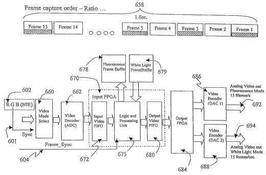
FIGURE 6d shows the block diagram for synchronization and control of
imaging as described for FIGUREs 6a and 6b to realize simultaneous white light
and
fluorescence imaging. Imaging signals 602 from detector 600 provide
alternating
fluorescence and white light images (frames) into the Video Mode Select switch
660,
-22-
CA 02524000 2005-11-02
WO 2004/098398 PCT/CA2004/000683
which assigns these signals to independent analog to digital converters (ADCs)
in
Video Decoder 662 to digitize images. Video synchronization is provided in
this
instance by the green channel 601. Digitized images are fed to Input FPGA
(field
programmable gate array) 670 for processing. Inside the Input FPGA 670, the
digitized images are directed to Input FIFO (first in first out) video buffer
672 and
then into the programmable.processing unit 675 which splits the images into
white
light imaging frames and fluorescence frames as determined by the Frame Sync
signal 604 connected to the processing unit 675. Two memory buffers
communicate
with FPGA 670: Frame Buffer 678 for temporary fluorescence image storage and
Frame Buffer 679 for temporary white light image storage. Various imaging
processing functions may be implemented within FPGA 670, for example, x-y
pixel
shifting for R, G, and B images for alignment and registration. X-y pixel
shifting
means to shift the digital image (image frame) in the horizontal . direction
(x) and/or
vertical direction (y), one or more pixels. Such processing eliminates the
need for
more complicated or mechanical mechanisms, thus simplifying alignment of
sensors
such as 615, 625, 635 and 645 discussed with FIGURE 6b. Another programmable
image processing function may take ratios of corresponding pixels in two or
more
images. The processed digital images are output by video FIFO 680 to the
Output
FPGA 684, which splits the fluorescence image frames and white light image
frames
into video encoder (DAC 1) 686 and video encoder (DAC 2)' 688 respectively.
Video
encoders 686 and 688 with digital to analog converters (DAC) to transform the
digital
image signals, for example, to standard analog video signals 692 and 694 to be
displayed on standard analog video monitors. In addition to providing for
synchronization of optical modulation, the Frame Sync signal 604 may be
utilized by
-23-
CA 02524000 2005-11-02
WO 2004/098398 PCT/CA2004/000683
the detector, for example as a means to switch between fixed gain settings
employed
by different imaging modalities.
In the embodiment described with FIGURES 6a, 6b, 6c and 6d, 15
frames/second of digital fluorescence images and 15 frames/second of digital
white
light images are generated to preserve the same light sensitivity (for
fluorescence
mode) as if the camera shown in FIGURE 6b is acquiring fluorescence images and
white light images in sequential (a imaging modality as outlined in U.S.
Application
Number 09/741,731 by Zeng et al.. titled "Methods and apparatus for
Fluof°escence
and Reflectance imaging and spectroscopy and for contemporaneous measurements
of electromagnetic. radiation with multiple measuring devices", along with
continuation application number 10/028,568, U.S. Publication No.
2002/0103439).
The video encoders 686 and 688 still output standard video signals, i.e., 30
frameslsecond by repeating (duplicating) each of the 15 frames digital images
once .
per second. If a higher frame rate, for example 30 frames/second digital
fluorescence
images and white light images are desired (proportionately decreasing the
light
sensitivity), this. may be realized by rotating the filter wheel 650
(discussed with
FIGURE 6a) at the appropriate rate, in this instance, 1800 rpm (30 rotations
per
second).
While preferred embodiments of the present invention have been shown and
described, it is envisioned that those skilled in the art may devise various
modifications of the present invention without departing from the spirit and
scope of
the appended claims.
-24-