Note: Descriptions are shown in the official language in which they were submitted.
CA 02524496 2005-11-02
WO 2004/101743 PCT/US2004/014092
THREE DIMENSIONAL CELL CULTURES 1N A MICROSCALE FLUID HANDLING
SYSTEM
CROSS-REFERENCE TO RELATED APPLICATIONS
[0001] This application claims the benefit of U.S. Provisional Patent
Application
Serial Number 601468,358, filed on May 6, 2003, which is incorporated by
reference herein
in its entirety.
STATEMENT REGARDING FEDERALLY SPONSORED
RESEARCH OR DEVELOPMENT
[0002] Not applicable.
TECHNICAL FIELD
[0003] The present invention relates to spheroids in a microscale fluid
handling
system. The present invention provides a device and methods for initiating,
culturing, and
manipulating three dimensional (3D) multicellular surrogate tissue assemblies,
such as
spheroids, culture media, extra cellular matrix components, soluble signaling
molecules and
cell-to-cell interactions. The present invention further provides high
throughput screening
(HTS) methods to study agents capable of intervening in diseases modeled by
spheroids in a
microscale fluid handling system.
BACKGROUND OF INVENTION
[0004] Mammalian cell culture has been traditionally used as a model for
studying
disease processes, especially cancer, and testing of potential therapeutic
agents used in
treatment thereof. Generally, cells used for mammalian cell cultures are grown
in
monolayers on plastic plates covered with liquid medium, which supplies
essential nutrients
and growth factors for the cells. However, for most cell types this method of
culturing does
not adequately mimic the in vivo environment from which the cells were
originally isolated
(1). This is because disease pathogenesis occurs in the context of 3D tissue
structures, and
involves interactions between different cell types in the stromal and
epithelial compartments and
with the extracellular matrix (ECM) (1). Not surprisingly, cells grown in
monolayers often do
not exhibit the same biological responses and behaviors that they would
otherwise in an in
CA 02524496 2005-11-02
WO 2004/101743 PCT/US2004/014092
vivo environment. In contrast, cells grown in spheroid structures and in the
presence of extra
cellular components that simulates their normal environment generally
represent an in vivo
biological environment much more faithfully (1). Accordingly, three
dimensional
multicellular aggregate, such as: spheroids, mammospheres, organoids, and
organotypic
cultures offer great potential for improved in vitro disease models that may
be used to screen
and develop novel therapeutic agents.
[0005] A spheroid is a 3D aggregate of living mammalian cells cultured in
vitro from
tissue explants, established cell cultures or a mixture of both. Spheroid
research, initially,
focused largely on monoculture of cells as 3D aggregates. However, recently
heterologous
spheroids with more than one cell type have been used to investigate the
interactions of
different cell types in both normal tissue and tumor development (1). The
internal
environment of a spheroid is dictated by the metabolism and adaptive responses
of cells with
a well-defined morphological and physiological geometry. Beyond a critical
size (> 500 uM)
most monotypic spheroids develop concentric layers of heterogeneous cell
populations with
proliferating cells at the periphery and a layer of quiescent cells close to
the necrotic core (1).
This heterogeneous arrangement of cells in a spheroid mimics initial avascular
stages of early
tumors. Another type of monotypic spheroid forms well organized acini-like
structures with
a central lumen when epithelial cells are cultured over reconstituted basement
membrane (2).
These monotypic spheroids are able to mimic important ih vivo morphology,
although much
of the biological complexity is lost. With the co-culture of more than one
cell type in a
spheroid, tumor cell interactions with other cell types can be studied under
standardized
conditions. Both the normal tissue and tumor micro milieu can be better
defined as the
epithelium, its underlying basement membrane and the sub-adjacent stroma. Co-
culture of
tumor cells and normal endothelial cells, as spheroids, has been useful for
studying
angiogenesis. The co-culture of tumor cells with stromal elements including
stromal
fibroblasts has demonstrated the importance of the complex micro-environment
in neoplastic
progression. Normal stroma was shown to inhibit tumor cells, while stroma from
tumor
biopsies was shown to have a mitogenic effect on tumor cells (2).
[0006] Spheroids have already made contributions to the general understanding
of
tumor biology and normal tissue development. In fact, the importance of
soluble signaling
molecules, cell-to-cell signaling and the influence of ECM on tumor
progression has been
elucidated using spheroid tumor models (1, 3). Compared to homogeneous
monolayer cell
culture methodologies, these in vitro tumor models preserve many biochemical
and
morphological characteristics, which correspond to in vivo tumors (3).
Furthermore, in vitro
2
CA 02524496 2005-11-02
WO 2004/101743 PCT/US2004/014092
tumor models provide a useful method of testing the influence of etiological
and potential
therapeutic agents. For example, it has been demonstrated that ih vit~~o
breast tumor models
respond to estrogen stimulation, a key factor in the etiology of the disease
in vivo (3).
[0007] Furthermore, a vast body of oncology research has shown the importance
of
mutations that activate dominant oncogenes and inactivate tumor suppressor
genes (4).
However, these studies have focused on the cancer cell alone, while
overlooking the
complexity and heterogeneity of the whole tumor. This cell autonomous
perspective
describes cancer as a progressive set of genetic alterations that drive the
transformation of
normal cells to highly malignant tumor cells. However, it is equally important
to understand
neoplastic progression as increasingly abnormal signaling between the
different cell types
and between the cells and the ECM in the tumor microenvironment (2).
Accordingly,
spheroids may provide a more realistic in vitro tumor model than traditional
monolayer
cultures where these signaling abnormalities may be analyzed and the effects
of anti-cancer
agents may be readily evaluated. The importance of modeling tumorigenesis
using 3D cell
culture methods is well illustrated in the case of breast cancer.
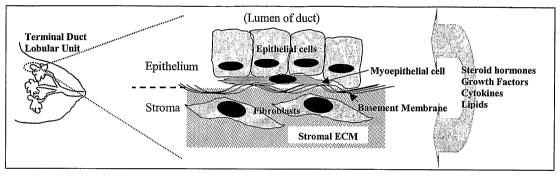
[0008] Mammary glands are composed of a network of epithelial ducts supported
by
a dense stroma, which accounts for snore than 80% of breast volume. The ducts
are formed
by an inner layer of polarized epithelial cells, an outer discontinuous layer
of myoepithelial
cells and a sheathing of specialized ECM called basement membrane as
illustrated in FIG. 1.
The stroma contains fibroblasts, endothelial cells, inflammatory and other
specialized cells
imbedded in a macromolecular network of ECM. The basement membrane and stromal
ECM
are composed of different combinations of collagen, laminin and other
glycoproteins and
proteoglycans that mediate binding and signaling to epithelial cells via
transmembrane
integrin proteins. The ECM provides both architectural support to cells and
contextual
information that influences their response to external stimuli for growth,
differentiation, and
motility.
[0009] Breast tumors originate in the epithelial cells of terminal duct
lobular units,
and it is well established that the accumulation of mutations and chromosomal
aberrations
within these cells are central to tumorigenesis. It is the tissue
microenvironment (FIG. 2),
however, that defines and controls , the cell-ECM interactions that define
mammary tissue
architecture - polarized epithelial cells bounded within the confines of a
basement membrane
- are subverted, allowing the tumor cells to invade the stromal compartment,
grow, and
metastasize. Much of the cellular signaling controlling this process occurs
via cell surface
receptors known as integrins, which bind to components of the ECM, and whose
expression
3
CA 02524496 2005-11-02
WO 2004/101743 PCT/US2004/014092
and distribution is frequently altered in malignant cells (5). Integrins
modulate intracellular
signaling pathways that control cell proliferation and apoptosis and they
regulate the activity
of extracellular proteases involved in invasion and metastasis. Signaling
between different
cells - known as paxacrine signaling also plays a key role in mammary
tumorigenesis.
Aberrant paracrine signaling by steroid hormones and polypeptide growth
factors - both
intraepithelial and stromal-epithelial are responsible for many aspects of
malignancy in the
breast (6, 7), and the effects of these hormones are integrated with cell-ECM
interactions (8).
Furthermore, recent data on' stromal mutations in mammary tumors suggests that
the genetic
underpinnings of carcinogenesis may be stromal, as well as epithelial in
nature (9, 10).
[00010] To overcome the lack of phenotypic differentiation observed in
monolayer
cultures, 3D cell culture methods incorporating a basement membrane have been
developed (11).
For example, when grown in the presence of a reconstituted basement membrane,
normal and
malignant human mammary epithelial cells form 3D structures with clear
morphological and
biochemical differences that reflect their in vivo phenotypes (12). Normal
cells form organoids
similar in their overall organization to mammary acini: polarized epithelial
cells surrounding a
central lumen. Normal cells also deposit surrounding basal lamina (even in the
presence of the
reconstituted basement membrane) and cease growing after they reach a diameter
of 40-SO~,m
(12). In contrast, malignant cells form solid, disordered masses similar to
tumors that continue to
grow to much larger sizes and do not secrete a basement membrane. The
differences between the
growth and differentiation patterns of normal and malignant cells are
distinguishable only when
cells are grown in the presence of a matrix rich in collagen and laminin or
MatrigelTM that
provide the ECM components necessary to direct tissue architecture. Such 3D-
reconstituted
basement membrane (3D-rBM) culture methods are an improvement over monolayer
cell culture
because they incorporate cell-ECM interactions important to tumorigeneis.
However, still these
methods do not account for stromal cells, and thus, do not reflect stromal-
epithelial signaling that
occurs in native mammary tissue.
[00011] Recently, mammary epithelial cells and various types of stromal cells
have been
incorporated into heterotypic spheroids (13, 14) and used to study aspects of
tumorigenesis
involving paracrine signaling. In one model, tumor cell and fibroblast
spheroids were grown
separately, then combined and allowed to merge; the tumor cells eventually
enveloped and
invaded the fibroblast spheroid (15). In another experimental model, tumor
cells (epithelial) were
cocultured with fibroblasts and/or endothelial cells in the presence of
reconstituted basement
membrane (16), an extension of the 3D-rBM culture methods described above. In
this system, a
mutual interdependence between tumor cells and endothelial cells for estrogen
dependent ductal
4
CA 02524496 2005-11-02
WO 2004/101743 PCT/US2004/014092
morphogenesis and neovascularization was observed (16). In similar
experiments, fibroblasts
isolated from tumor fibroblasts were shown to be necessary and sufficient to
induce
morphogenesis of both normal and malignant epithelial cells, and these effects
were fiarther
enhanced by the addition of endothelial cells (17).
[00012] Another approach that has been used to recapitulate the dynamics of
tissues,
specifically mammary tissue has been to coculture epithelial and stromal cells
in adjacent ECM
layers (18, 19). A layer of fibroblasts in collagen is overlaid with
epithelial cells in collagen or
reconstituted basement membrane. This provides a two compartment system for
studying
interactions between the lower "stromal" layer and the upper epithelial layer.
This model has
been used to study how estrogen dependent proliferation of epithelial
organoids is mediated via
growth factors produced by fibroblasts (18), and to study the role of
fibroblast-produced growth
factors and proteases in epithelial organoid branching (19).
[00013] Although, both of the models described above establish the feasibility
of
recapitulating paracrine signaling between stromal and epithelial cells in
vitro, both systems also
have shortcomings. Heterotypic spheroids are basically disordered masses of
cells and thus, bear
little resemblance to the ordered structure of mammary tissue. In addition,
there is no clear
separation between epithelial and stromal compartments with these models. The
two
compartment coculture methods are an improvement on this approach in that they
incorporate
separate epithelial and stromal compartments. However, the "bulk" nature of
classical tissue
culture methods used for both models limits the ability to control
experimental variables and to
monitor the activities of the system at the scale of the tissue
microenvironment. Once the intact
cocultures are established, any changes made to the system are global. Test
agents must be added
directly to the liquid medium that overlays the coculture, exposing the entire
system to the agents
with no control of how their effects are changed by different cell types in
either compartment.
Also, it is not possible to specifically stimulate the epithelial or stromal
compartment with a test
agent. In addition, exposure of the system to a constant concentration of test
agent for a defined
period of time is not practical with these models either, as it would require
frequent aspiration and
replacement of the liquid overlay which is both cumbersome and stressful to
cells. These
physical constraints limit the ability to experimentally probe the system and
to mimic the
paracrine signaling and compartmental control systems that operate in vivo.
[00014] More generally, current methods for the initiation and analysis of
spheroids
involve labor-intensive processes and are not easily amenable to the high
degree of
standardization and automation that are required for routine drug screening.
Furthermore,
current methods such as static cell culture and flow through cell culture
generally subject the
s
CA 02524496 2005-11-02
WO 2004/101743 PCT/US2004/014092
spheroids to mechanical stresses and it is difficult to control the
microenvironment around the
cell mass. Static culture methods fail to allow for the gradually changing
milieu in the
normal tissue or tumor microenvironment. Flow through culture methods use
large fluid
volumes and the medium is replenished so quickly that important growth factors
and other
biological signaling molecules are washed away.
[00015] Furthermore, it has been established that at the micro-scale different
forces
become dominant over those experienced at larger scale (20) these include
laminar flow,
diffusion, fluidic resistance, surface area to volume ratio, and surface
tension. Laminar flow
is the definitive characteristic of microfluidics. Fluids flowing in channels
with dimensions
up to several hundred microns in width and at readily achievable flow speeds
are
characterized by low Reynolds number, (Re). Flows in this regime are laminar,
not turbulent.
The surfaces of constant flow speed axe smooth over the typical dimension of
the system, and
random fluctuations of the flow in time are absent. In the long, narrow
geometries of
microchannels, flows are also predominantly uniaxial. The entire fluid moves
parallel to the
local orientation of the walls. A suitable feature of mliaxial laminar flow is
that all transport
of momentum, mass, and heat in the direction normal to the flow is left to
molecular
mechanisms: molecular viscosity, molecular diffusivity, and thermal
conductivity.
[00016] Microfluidics allows for precise and unique control of the local fluid
environment
as well as the ability to work with smaller reagent volumes and shorter
reaction times.
Microscale phenomena enable techniques and experiments not possible on the
macroscale. For
instance, the laminar flow properties of microchannels are such that the
mixing between two
streams flowing in contact is diffusion dependent - i. e., not affected by
turbulence mixing factors.
This makes it possible to generate concentration gradients and discrete
packets of reagents for use
as stimuli to biological systems.
[00017] The first microfluidic devices were fabricated in silicon and glass by
conventional, planar fabrication techniques - photolithography and etching -
adapted from the
microelectronics industry. These methods are precise, but expensive,
inflexible, and poorly
suited to exploratory work. Recently new techniques such as soft lithography,
in situ
construction, micro-molding and laser ablation have been applied to the
fabrication of
microfluidic devices (20). These nonphotolithographic microfabrication methods
are based
on printing and molding .organic materials, and are much more straightforward
than
photolithography for making both prototype devices and special-purpose devices
for physical
investigations. These methods have also made it practical to build 3D networks
of channels
and components (21). Thus, they may offer access to new types of fluidic
elements, such as
6
CA 02524496 2005-11-02
WO 2004/101743 PCT/US2004/014092
valves and pumps fabricated of elastomeric materials (22). In addition, they
offer the high
level of control over the molecular structure of the channel surfaces that is
required in many
applications. To date use of spheroid cell cultures in a drug discovery
setting has been
limited because existing methodologies used for their growth and manipulation
have not
allowed accurate reconstruction of tissue morphology, specifically in the
signaling between
stromal and epithelial cells. Accordingly, it would be desirable to provide
alternative
approaches using microfluidics to precisely manipulate the micro-environment
of a 3D cell
culture.
SUMMARY OF THE INVENTION
[00018] The present invention is summarized as a microscale fluid handling
system
comprised of a microfluidic device and a three dimensional (3D) multicellular
assembly of
living cells, wherein the device is used for initiating, culturing,
manipulating, and assaying
the multicellular assembly, preferably at least one spheroid.
[00019] One aspect of the present invention provides a microfluidic device for
initiating, culturing, manipulating, and assaying multicellular surrogate
tissue assemblies
including at least one microfluidic channel; at least one chamber; and at
least one spheroid,
wherein the walls of the chamber are lined with a cell layer; and wherein
fluid medium flows
through each of the channels and chambers.
[00020] In another aspect the invention provides a microfluidic device for
initiating,
culturing, manipulating, and assaying multicellular surrogate tissue
assemblies having two
adjacent chambers are lined with a cell layer, wherein each chamber contains a
spheroid
representing a different tissue, and wherein each chamber contains a fluid
medium specific
for a tissue.
[00021] In another aspect the invention provides a method of performing high
throughput screening of test agents using surrogate tissue assemblies by
making a
microfluidic device including fluid flow channels and chambers; making
surrogate tissue
assemblies of multiple cell types of mammalian cells; placing surrogate tissue
assemblies,
preferably spheroids into chambers in the device; introducing test agents
through the fluid
flow channels to the surrogate tissue assemblies; and observing the responses
of the surrogate
tissue assemblies.
[00022] In another aspect the invention provides a high throughput screening
system
for mimicking the reaction of multicellular tissues to test agents. The system
includes a
microfluidic device having a plurality of fluid flow channels and a plurality
of chambers; and
7
CA 02524496 2005-11-02
WO 2004/101743 PCT/US2004/014092
a plurality of surrogate tissue assemblies formed of living mammalian cells,
each surrogate
tissue assembly located in one of the chambers.
[00023] Still another aspect of the invention encompasses providing kits
having a
microfluidic device of the invention and spheroids used to study agents
capable of
intervening a variety of medical conditions.
[00024] These and other aspects of the present invention would be better
appreciated
upon an examination of the' following drawings, description, taken in
conjunction with the
appended claims.
BRIEF DESCRIPTION OF THE DRAWINGS
[00025] FIG. 1 shows the structure of human mammary tissue. Mammary ducts are
composed of polarized epithelial cells surrounded by a discontinuous layer of
myoepithelial cells
encased in a specialized cell.layer (ECM) layer called the basement membrane.
The ducts are
imbedded in a dense layer of stroma composed of ECM proteoglycans,
glycoproteins and
specialized cell types including fibroblasts, macrophages, adiopocytes, and
endothelial cells.
[00026] FIGS. 2A-B show flow patterns inside the microfluidic channels. Two
streams flowing in contact will not mix except by diffusion. As the time of
contact between
two streams increases, the amount of diffusion between the two streams
increases. The top
and bottom portions of spheroid 1 are exposed to different test agents with a
sharp boundary
across the equator and spheroid 2 is exposed to a gradient from top to bottom
(A). Fluid flow
in direction 1 with minimal leakage into the perpendicular channel. Fluid is
then allow to
flow in direction 2 to move a packet of fluid out of stream 1 and down the
channel exposing
the spheroid to a short pulse of soluble factors in the fluid packet (B).
[00027] FIG. 3 shows the top view of a narrow section of the channel that
allows the
fluid, but not the spheroid to pass.
[00028] FIG. 4 shows the cross-sectional view of an obstacle in the bottom of
the
channel that prevents the spheroid from traveling airy further.
[00029] FIG. 5 shows spheroids attached to a fibroblast seeded microchamber.
[00030] FIGS. 6A-B show spheroid behavior in culture flasks and microchannels.
In
culture flasks spheroids lie at the bottom of a layer of media. Any
established micro-
environment will diffuse away because of the large volume of media (A). In
microchannels,
there is much less media surrounding spheroids, thus reducing the effects of
diffusion on the
micro-environment (B).
[00031] FIGS. 7A-C show spheroid development and organization. Cross sectional
view of spheroid where concentric growth pattern mimics early avascular tumor
(A).
s
CA 02524496 2005-11-02
WO 2004/101743 PCT/US2004/014092
Spheroid comprised of mammary epithelial cells grown over reconstituted
basement
membrane to induce mammary acini lilce structure development (B). Heterotypic
spheroid
showing formation of ducts and vascular elements with epithelial cells and
basement
membrane surrounding the stromal core (C).
(00032] FIG. ~ shows a portion of a microscale device with multiple channels
and
chambers providing identical microenviromnents for many spheroids and
individual testing
and sampling capabilities.
[00033] FIG. 9 shows latitudinal cross sectional views of chambers showing
different
ways of distributing equal numbers of cells or spheroids of a uniform size.
[00034] FIG. 10 shows longitudinal cross-sectional view of channels and
chambers
showing how basement membrane and spheroids can be introduced to chambers.
[00035] FIGS. 11A-B show a schematic of a microfluidic device for coculture of
epithelial
organoids and stromal fibroblasts in adjacent compartments (A) and T junction
that is used to
allow delivery of discrete pulses of reagents (B). Arrows indicate the
direction of fluid flow
through the channels of the device. The two compartments are formed
sequentially by flowing
cell/collagen suspensions into the incubation chamber via the upper channel
and increasing the
temperature to allow collagen to gel. The stromal layer is supported by a
microporous filter
(dashed line). The upper and lower channels provide the ability to separately
control the
exposure of each compartment to factors of interest.
[00036] FIGS. 12A-B show a detailed drawing of a microfluidic device for
coculture of
epithelial organoids and stromal fibroblasts in adjacent compartments,
specifically (A) side view
and (B) top view. The three layers of material are indicated by brackets; the
numbers indicate the
sequence of fabrication. Each layer is approximately 300~.m thick. The
epithelial and stromal
fluid channels and the incubation chamber with a filter in the bottom is
fabricated using liquid
phase photopolymerization of PEG diacrylate. The filter supports both layers
of cells embedded
in ECM (collagen) and allows the free passage of soluble molecules. The layers
of cells
suspended in ECM are introduced via the epithelial fluid channel and allowed
to gel in the device.
Arrows indicate direction of fluid flow in channels.
[00037] Before an embodiment of the invention is explained in detail, it is to
be
understood that the invention is not limited in its application to the details
set forth in the
following description. The invention is capable of other embodiments and of
being practiced
or being carned out in a variety of ways. Also, it is to be understood that
the phrases and
terminology used herein is for the purpose of description should not be
regarded as limiting
in any way.
9
CA 02524496 2005-11-02
WO 2004/101743 PCT/US2004/014092
DETAILED DESCRIPTION OF THE INVENTION
[00038] We have developed a novel microscale fluid handling system which
combines
a microfluidic device to a three dimensional (3D) multicellular assembly of
living cells. The
device is used for modeling a medical condition by initiating, culturing,
manipulating, and
assaying the 3D multicellular assembly of living cells, preferably a spheroid.
The invention
also provides methods for using such a device to model disease progression by
assaying test
agents for stimulation, inhibition or prevention of neoplastic progression in
a spheroid. The
method includes high throughput screening of the agents capable of intervening
in diseases
modeled by spheroids in a microscale fluid handling system; detecting an
acquired
spheroid characteristic; and assaying acquired spheroid characteristic to
select agents for
stimulation, inhibition or prevention of neoplastic progression, suitably
mammary cancer.
[00039] As used herein, the term "three dimensional" or "3D" cell culture
refers to any
method used to effect the growth of cells into a 3D multicellular r surrogate
tissue assembly
or spheroid; includes organotypic cell culture methods that are used to effect
the growth of
cells into their native tissue morphology.
[00040] Also, as used herein, the term "spheroid" refers to an aggregate or
assembly of
cells cultured to allow 3D growth as opposed to growth as a monolayer. It is
noted that the
term "spheroid" does not imply that the aggregate is a geometric sphere. The
aggregate may
be highly organized with a well defined morphology or it may be an unorganized
mass; it
may include a single cell type or more than one cell type. The cells may be
primary isolates,
or a permanent cell line, or a combination of the two. Included in this
definition are
mammospheres, organoids, and organotypic cultures; and more specifically, the
well ordered
acini-like organoids formed by mammary epithelial cells in certain culture
conditions.
[00041] In general in vivo tissue development and homeostasis rely on
carefully
orchestrated "cues" from soluble signaling molecules, attachment factors in
the ECM, and
cell-to-cell signals. The term "ECM" or "extracellular matrix" refers to a
cell layer
composed of different combinations of collagen, laminin and other
glycoproteins and
proteoglycans that mediate cell binding and signaling. The ECM provides both
architectural
support to cells and contextual information that influences their response to
external stimuli for
growth, differentiation, and motility. Includes native ECM, plain collagen,
synthetic mixtures,
and natural isolates (i.e., MatrigelT~.
[00042] Both the temporal and spatial coordination of these cues is critical
for creating
an irz vitro model of the normal tissue. Compared to other mammalian cell
culture systems,
to
CA 02524496 2005-11-02
WO 2004/101743 PCT/US2004/014092
one embodiment of the present invention provides a microscale fluid handling
system, which
provides an environment which more faithfully models ih vivo conditions for
growth and
differentiation by allowing precise control of soluble signaling factors in
the fluid medium,
cell attachment surfaces, pressure, pH, and cell-to-cell communication.
[00043] In this embodiment, the invention also provides a means for
manipulating the
micro-environment of a 3D multicellular surrogate tissue assembly. As used
herein, the term
"manipulating" or "manipulate" refers to the ability to precisely control the
micro-
environment of a 3D cell culture, including the pH, hydrostatic pressure,
gradients, flow rate,
introduction of soluble factors representing endocrine and paracrine signals,
cell-to-cell
interactions, and specific ECM components.
[00044] Specifically, the invention provides for gradual changing of the fluid
medium
in a precise manner representative of an ih vivo environment. Current methods
for culturing
suspended spheroids (23) require transferring the spheroid in a pipette.
Pipette transfer can
shock the spheroid with sudden changes in local environment and expose the
spheroid to
mechanical stresses that are not representative of the in vivo environment.
Another method
of changing media for both suspended and attached spheroids is flow through
culture.
Standard flow through culture dilutes and washes away exogenous signaling
molecules that
are important to normal tissue differentiation and development.
[00045] Also, it is envisioned that the laminar flow properties of the
channels used in
the present invention enable unique assays, not possible using macro-scale
tissue culture
methods. Two or more laminar flow streams can be joined into a single mufti
component
laminar flow stream. This allows researchers to expose different portions of a
single spheroid
cell culture to different soluble factors simultaneously or to establish
gradients across the
spheroid cell culture. Test compounds, signaling molecules, or enzymes can be
delivered in
discrete packets within the laminar flow stream allowing precisely timed
exposure of the
spheroid cell culture. The channel geometry and flow rate define the temporal
and spatial
aspects of the exposure of the packet to the spheroid. The physiology and
state of
differentiation of cells at different depths within the spheroid plays an
important role in tissue
differentiation (24). The ability to peel layers off the spheroid cell culture
allowing assays for
morphological characteristics and surface markers of cells at different depths
within the cell
mass may provide insights into their state of differentiation (23). Further
the introduction of
discrete packets of enzymes associated with both normal processes of tissue
differentiation
and tissue invasion or metastasis may allow assays to further elucidate the
role of these
enzymes (2) in tumor progression. It is also possible to combine chemical
treatments (e.g.
11
CA 02524496 2005-11-02
WO 2004/101743 PCT/US2004/014092
fluid packets) with mechanical manipulation. For example, suction through
small ports has
been used to vacuum the cumulus cells off of bovine oocytes. Similar
manipulations could
be used to selectively remove layers of cells from spheroids.
[00046] Furthermore, it is envisioned that obstacles within the channels can
hold a
suspended spheroid in place while maintaining a continuous or pulsed flow of
medium across
the spheroid (25). The spheroid can easily be moved out of these holding
places by reversing
the fluid stream (26). A series of obstacles within the channels can also
serve to sort the
spheroids by size. The first obstacle in the series prevents spheroids larger
than the opening
around the obstacle from entering the culture channel. At the downstream end
of the channel
a second obstacle holds the desired spheroids in place while allowing smaller
spheroids to
exit the culture channel.
(00047] In another embodiment of this invention, the microfluidic device is
capable of
supporting spheroid cultures with basement membrane and ECM components. The
ECM
consists of macromolecules secreted by cells into their immediate
microenvironment. These
macromolecules interact to form an insoluble matrix. The ECM can serve as the
scaffolding
on which cells migrate, or it may induce differentiation in certain cell
types. Some
macromolecules forming the ECM include for example, collagens, proteoglycans,
and
substrate adhesion molecules. The basement membrane is a thin layer of
insoluble
macromolecules interposed between cells and the adjacent connective tissue. In
the
capillaries, basement membrane forms a boundary between the endothelial lining
of the blood
vessel and the adjacent mesenchyme. Macromolecules forming the basement
membrane
include for example, collagen IV, laminin, and proteoglycans. The basement
membrane has
a supportive function in some tissue and may also act as a passive selective
filter (27). It is
envisioned that microfluidic channels used for the initiation of spheroid cell
culture may be
coated with biopolymers that are important components of the ECM and basement
membrane. These biopolymers may include, but are not limited to laminin,
fibronectin,
gelatin and collagens. MatrigelTM (Collaborative Biomedical Products, Catalog
No. 40234) a
synthetic basement membrane preparation may also be used to prepare the
culture channels.
[00048] In addition to the ECM, it has become clear that cell to cell
signaling is a vital
part of both normal and neoplastic differentiation and development (8).
Diffusible factors
like growth factors, hormones, and morphogens are secreted by one cell type to
change the
behavior of other cell types. Cells can also selectively recognize other cells
based on cell
surface properties causing some cells to adhere and others to migrate past
each other based on
affinity. These affinities can be for the surfaces of other cells or for
components of the ECM.
12
CA 02524496 2005-11-02
WO 2004/101743 PCT/US2004/014092
The dominant paradigm of morphogenesis is differential cell affinities to
localize cells
appropriately within tissues, organs and tumors (27).
[00049] Furthermore, the progression from normal tissue to malignancy can be
characterized by increasingly abnormal communication between cells that
comprise the
tumor and the tumor microenvironment. In this context, tumor formation can be
considered a
developmental process where a complex organ forms in response to signaling
between
different cell types and the ECM. It has been demonstrated that targeted
expression of
stromelysin-l, a matrix metallo-protease, produced spontaneous acquisition of
tissue features
characteristic of neoplastic states (2). Increasingly abnormal communication
between
fibroblasts, endothelial, and epithelial cells has been shown to induce
processes such as tumor
angiogenesis using ih vitro tumor models (3). As used herein, the term "tumor
model" refers
to an ih vitro cell culture system used to mimic the behavior of a malignant
tumor.
[00050] Additionally, the present invention provides methods for seeding
microfluidic
channels with different cells types to initiate spheroid cultures.
Specifically, it is envisioned
that the present invention provides methods to seed biopolymer coated channels
with
fibroblasts, which are the predominant cell type found in the stromal
compartment. It is
further envisioned that the growing fibroblasts will condition the channel by
adding their own
ECM components to the biopolymer coating. The fibroblast-conditioned channels
will then
be seeded with spheroids containing epithelial cells, and possibly additional
cell types.
[00051] The present invention also provides a microfluidic device designed to
incorporate stromal and epithelial cells in adjacent comparhnents as shown in
FIG. 11A and FIG.
12 A, mimicking the structure of a mammary tissue ih vivo (FIG. 1). An example
of such a
device is shown in FIG. 11, which illustrates how two tissue compartments may
be separately
addressed by two different solvent streams. Specifically, FIGS. 11A-B show a
schematic of a
microfluidic device for coculture of epithelial organoids and stromal
fibroblasts in adjacent
compartments (A) and T junction that is used to allow delivery of discrete
pulses of reagents (B).
Arrows indicate the direction of fluid flow through the channels of the
device. The two
comparfrnents are formed sequentially by flowing cell/collagen suspensions
into the incubation
chamber via the upper channel and increasing the temperature to allow collagen
to gel. The
stromal layer is supported by a microporous filter (dashed line). The upper
and lower channels
provide the ability to separately control the exposure of each compartment to
factors of interest.
[00052] It is believed that the ability to selectively probe either tissue
compartment is a
significant improvement over other two compartment models described earlier,
because it will
13
CA 02524496 2005-11-02
WO 2004/101743 PCT/US2004/014092
enable simulation of stromal-epithelial signaling more accurately. For
instance, it will be possible
to selectively initiate signals in one compartment and monitor the response of
the other.
[00053] It is envisioned that polymers sensitive to stimuli including pH,
light,
temperature, biological signals, and electrical current (22) may be
incorporated into the
microfluidic channels. Subtle changes in external stimuli can cause the
hydrophilic polymer
to expand or contract exerting or relieving pressure on spheroids growing in
the microfluidic
device. Furthermore pressure may be applied to an elastomeric membrane
adjacent to
spheroid cell cultures. The rate and extent of deformation can be measured and
controlled
using particle imaging (2~). , Development of organs and tumors in vivo is
often responsive to
pressure exerted by the surrounding tissues. Therefore, in accordance with the
invention, we
envision that pressure sensitive polymers and elastomeric membranes may be
used in the
device of the invention to control pressure on the ih vitro tumor.
[00054] In each case, parallel control channels within the microfluidic device
may be
used to compare spheroids exposed to test agents with control spheroids
exposed to the same
growth conditions but without the test agents. The control channel has a
similar structure and
is part of the same fluid control system used for the test channels so that
flow rates and cell
culture media are identical during the course of the experiment.
[00055] In another embodiment, the present invention provides methods for
measuring
growth, proliferation, differentiation, and development of spheroids.
Observation of spheroid
size, cell shape, and developmental features such as angiogenisis, and duct
formation can
often give information on its state of differentiation. Morphological analysis
may be carried
out using an inverted microscope to analyze spheroids in place or by standard
histological
techniques, as well as image analysis of specific cell markers (23).
Fluorescence labeling of
cells, organelles, or macromolecules using exogenous fluors or expressed
fluorescent
proteins, such as green fluorescent protein, may be useful for detecting
changes in spheroid
properties. Proliferation may be measured directly using a number of methods
including but
not limited to the MTS colorimetric method (Promega Corporation, Madison, WI).
[00056] An attached spheroid culture may be dissociated from the channel using
a
trypsin or pronase solution and the suspended spheroid may be extracted for
fiuther analysis
outside of the microfluidic device. The fluid media may also be assayed for
soluble factors.
The microfluidic channels do not dilute soluble factors to the extent seen in
standard culture
techniques and the ability to precisely control a pulse of fluid across the
cell culture allows
factors such as enzymes, hormones, and growth factors to be washed out of
culture in a more
concentrated form (20). Some of these soluble factors would include growth
factors and
14
CA 02524496 2005-11-02
WO 2004/101743 PCT/US2004/014092
proteases. Enzyme linked immunosorbent assays (ELISA) may be used to determine
the
presence or quantity of growth factors. Metallo-proteases are often an
indicator of tissue
differentiation or tissue invasion and Zymogram gels (Invitrogen, Carlsbad,
CA) are useful in
measuring this activity.
[00057] In the search .for new therapies pharmaceutical companies screen vast
libraries
of compounds for their ability to increase or inhibit specific enzyme
activities or binding to
specific nuclear receptors. Many of these assays involve measuring a change in
absorbance,
fluorescence or nuclear magnetic resonance (NMR) properties of reporter
molecules in a high
throughput screening mode in parallel arrays, where the characteristics of
spheroids between
channels are constant so that many agents may be tested for effects on
essentially identical
spheroids. Accordingly, in another embodiment, the invention provides a means
to establish
spheroid cell cultures in channels that are compatible with 24, 48, or 96 well
format currently
used for drug candidate screening. It is envisioned that biochemical assay
reporter
molecules can be introduced into the microfluidic culture channels or produced
by cells in the
spheroid and direct measurements of change in the reporter molecule could be
taken directly
from the microfluidic device (29). This may provide a rapid method for
verifying that
compounds showing desired biochemical properties during initial screening and
a
corresponding inhibition or promotion of spheroid development are actually
functioning as
predicted in the spheroid.
EXAMPLES
[00058] Producing a~ plurality of multicellular surrogate tissue assemblies
(e.g.,
spheroids) of a uniform size and overall structural properties using a
microscale device is an
important capability because it allows one to test diverse chemicals for their
effects on
essentially identical spheroids. The prophetic examples below describe methods
used to
produce multiple spheroids of a similar size and overall set of
characteristics, isolated from
each other in separate addressable chambers, and thus can be subject to
different
experimental variables. There are two approaches that can be used: initiation
and growth of
spheroids in the microscale (MS) device, or use of the MS device to sort and
distribute
spheroids from bulk cultures grown outside the device.
[00059] Example 1. Initiation and growth of similar sized spheroids in a MS
device
[00060] In this prophetic example, cells are grown initially in plates as
monolayers,
then enzymatically detached, collected and introduced to a microscale (MS)
device with
multiple channels leading to chambers where the spheroids will form. This is
done in such a
is
CA 02524496 2005-11-02
WO 2004/101743 PCT/US2004/014092
way that equal numbers of cells are distributed to each chamber, insuring that
the size of the
spheroids that form will be uniform if maintained under identical culture
conditions.
Distributing cells to multiple chambers can be achieved in several ways,
including
introducing a uniform cell suspension through a single opening that branches
to multiple
channels each leading to a chamber, using concurrent flow through each channel
or using a
valve that opens onto each channel separately.
[00061] Alternatively, an equal volume of a uniform cell suspension is
introduced
through separate openings for each channel, or through separate openings
directly into the
chambers. The cells can be introduced via a manual syringe, or a pneumatically
operated
syringe with single or multiple outlets, or by an automated liquid handling
device such as is
commonly used for dispensing biological reagents in an HTS setting. Spheroid
culture
chambers will be coated with a stationary non-adherent layer of agarose (23)
or other
biopolymer. Distributing an equal or nearly equal, number of cells to each
chamber can be
achieved in several ways, including maintaining constant flow rates for the
same period of
time for introduction of cells to each channel and then using valves to shut
the chamber off
from fluid flow while excess cells are washed out of the channels.
Alternatively distribution
of equal number of cells to chambers is achieved by filling a depression or
catch basin in the
chamber. In this latter case, cells are introduced at a low flow rate such
that laminar flow
properties occur until the chamber is filled, then the channels are cleared of
excess cells by
flushing out the channels with media or a wash solution at an increased flow
rate, such that
the fluid path is uniformly horizontal and does not enter the catch basin as
illustrated in FIGS.
9 and 10. In a variation of.this approach, the catch basin can include a
filter in the bottom
that retains cells such that laminar flow is out the bottom of the catch
basin.
[00062] Example 2. Distributing spheroids of a uniform size to chambers for
further growth and analysis
[00063] As an alternative to initiating spheroids in the MS device, it is also
envisioned
that spheroids may be grown by standard cell culture methods - on agar plates
or in spinner
flasks - and distribute them to the chambers of the microscale device using
fluid flow.
Spheroids of a uniform size can be attained by the use of physical structures
such as filters,
funnels or barriers in the fluid path that act as sieves. For example, FIGS. 3
and 4 illustrate
the presence of such physical structures or obstacles in a channel of the
microfluidic device,
enabling the fluid, but not the spheroid to pass. Also, FIG. 9 shows
latitudinal cross sectional
views of chambers showing different ways of distributing equal numbers of
cells or spheroids
of a uniform size. Thus, if a funnel is used, all spheroids larger than the
desired size pass
16
CA 02524496 2005-11-02
WO 2004/101743 PCT/US2004/014092
through the chamber because they are unable fit in the large opening of the
funnel, and those
that are smaller than desired.pass through the small end of the funnel.
[00064] Example 3. Culturing spheroids
[00065] Also, prophetically exemplified here are a variety of techniques for
culturing
spheroids. Once spheroids or surrogate tissue assemblies are in chambers, they
are cultured
by flowing media through the chamber, such that nutrients are replenished, and
waste
products are removed. The flow rate is slow enough to allow growth factors and
other
soluble signaling molecules in the spheroid microenvironment to carry out
their functions
before they are washed away from the spheroid. In this way, an essentially
identical
microenvironment is maintained in each chamber, allowing spheroids to form and
grow at the
same rate and develop similarly. In some cases, it may be desirable to change
the type of
media or some components at some point in the development of the spheroid. If
desired,
some chambers can be subject to different growth conditions or soluble factors
in order to test
their effects of spheroid formation. It may also be desirable also to culture
spheroids in the
presence of basement membrane components and/or stromal cells such as
fibroblasts or
immune cells. If this is the case, basement membrane and stromal cells are
flowed into the
chambers prior to introduction of the intact spheroid or spheroid cells.
[00066] Example 4. Analysis of spheroids
[00067] In accordance with this invention, it is envisioned that the cultured
spheroids
described above may be analyzed either for size, or gross morphological
properties is
generally done using microscopy, and for this purpose, spheroids can be
observed while still
in the MS device.
[00068] Alternatively, if a soft polymer matrix such as PDMS is used for
casting MF
device, the spheroids in their individual chambers can be excised from the
device using a
boring or sectioning device and subject to detailed morphological and
histological analyses
either in whole, or following thin sectioning, fixing, enzymatic digestion,
staining, andlor
other tissue and cell preparation methods. Alternatively, the spheroids can be
released
individually from their chambers using enzymatic digestion of basement
membrane and ECM
matrix and their contents collected for analysis. Inhibition or stimulation of
the spheroid cell
proliferation may be measured directly using the MTS colorimetric method
(Promega
Corporation, Madison, WI). The MTS reagents may be introduced through the
common fluid
handling system and the change in absorbance measured directly from in the MS
device. In
addition, many other experimental outputs can be measured, either in the MS
device, or
17
CA 02524496 2005-11-02
WO 2004/101743 PCT/US2004/014092
following release of the spheroid. These include outputs that give an
indication of the status
of tumorigenesis, such as gene expression patterns, presence of specific
enzyme activities or
cell surface receptors, secretion of soluble factors, etc. Methods for
measuring these outputs
are well established and include immunochemical, DNA or RNA hybridization, the
use of
reporter proteins, and the use of reporter substrates, and involve mostly
colorimetric,
luminescent and fluorescence detection methods.
[00069] Example 5. Spheroids as a model for cancer
[00070] There is a developing body of literature describing the use of
spheroids as in
vitro tumor models (2, 3). Both monotypic and heterotypic spheroids have
proven useful as
tumor models. Heterotypic spheroids offer the ability to investigate
interactions between
different cell types in the tumor microenvironment (3). Monotypic spheroids
comprised of
malignant cells offer the advantage of simplicity and they can effectively
represent initial
avascular stages of early tumors. Monotypic spheroids may be prepared by
seeding single
cell suspensions of tumor cells on a stationary nonadherent layer of agarose.
After 3 to 8
days the spheroids reach a size of 300 to 400 nm.
[00071] It is prophetically contemplated that in experiments to test new
drug,therapies
it is important that the spheroids are all of the same size because small
differences in spheroid
diameter have a dramatic effect on the volume and morphological
characteristics. Thus, the
spheroids of the invention will be sorted by size using a series of obstacles
before the
spheroids analyzed.
[00072] Example 6. High throughput screening of test agents
[00073] Also, prophetically exemplified here are methods for high throughput
screening (HTS) of etiological agents that stimulate tumors and potential
drugs to suppress
tumors. It is envisioned that a microscale fluid handling device with will be
used to analyze
spheroid cultures, and this device will be compatible with existing
instrumentation for
measuring absorbance, fluorescence, luminescence or other signals used to
quantify
biological responses in an HTS setting. The analysis device may be the same as
the device
used to initiate and/or grow the spheroids, or it may be a separate device
that spheroids are
transferred prior to analysis. Spheroids for testing and analysis will be
present in chambers in
the MS device in a pattern that is consistent with existing multiwell plates,
including but not
limited to 24, 96, or 384-well plates.
[00074] Alternatively, new instrumentation might be developed that is more
suitable
for analysis of spheroids in microfluidic devices. The chambers will be
depressions or wells
in the channels or alternatively the chambers will be sequestered using
barriers or walls. The
is
CA 02524496 2005-11-02
WO 2004/101743 PCT/US2004/014092
depressions will be coated with reconstituted basement membrane, and possibly
also seeded
with fibroblasts and/or other stromal cell types creating an extra cellular
matrix to mimic in
vivo conditions. Monotypic spheroids comprised of a tumor cell line will be
sorted for size
and spheroids within a specific size range will be deposited in chambers
within the channels.
The spheroids will attach to the basement membrane and their growth and
morphology will
be monitored by microscope. When the spheroids reach an optimum size for
testing, test
agents will be introduced to the non-control spheroids. All of the chambers
share a common
fluid handling system and each chamber can be addressed separately. One way to
achieve
this is by the use of separate ports and channels leading to each chamber,
another is by the
use of valves. The non-control spheroids will be exposed to test agents while
both control
and non-control spheroids are exposed to the same flow rates and biological
media.
Inhibition or stimulation of the spheroid cell proliferation may be measured
directly using the
MTS colorimetric method (Promega Corporation, Madison, Wl~. The MTS reagents
may be
introduced through the cormnon fluid handling system and the change in
absorbance
measured directly from the analysis stations using the 24, 4~ or 96 well
format currently used
for drug candidate screening.
[00075] In addition, many other experimental outputs can be measured that give
an
indication of the status of tumorigenesis, including but not limited to gene
expression
patterns, presence of specific enzyme activities or cell surface receptors,
and secretion of
soluble factors. Alternatively, if a soft polymer matrix such as PDMS is used
for casting MF
device, the spheroids in their individual chambers can be excised from the
device using a
boring or sectioning device and subject to detailed morphological and
histological analyses
either in whole, or following thin sectioning, fixing, enzymatic digestion,
staining, andlor
other tissue and cell preparation methods. Alternatively, the spheroids can be
released
individually from their chambers using enzymatic digestion of basement
membrane and ECM
matrix and their contents collected for analysis.
[00076] Example 7. Fabrication of a two compartment device for reconstructing
mammary tissue using liquid phase photopolymerization
[00077] With the recent development of liquid phase photopolymerization, the
entire
design and fabrication cycle for a microfluidic device has been reduced to
minutes, making it
possible to produce and test many devices during the prototype development
process (30). Such
an iterative approach is particularly well suited for building a device that
will house complex
biological systems as described by the present invention.
19
CA 02524496 2005-11-02
WO 2004/101743 PCT/US2004/014092
[00078] It is envisioned that to construct a microfluidics device, a
prepolymer solution is
flowed into a chamber and exposed to UV light through a mask that prevents
photopolymerization where channels or other openings are desired. Uncured
prepolymer is
subsequently flushed from the channel. The sides and bottom of the chamber are
formed by an
adhesive gasket adhered to a microscope slide and the top is a polycarbonate
film (FIG. 6). The
adhesive gasket maintains the cavity height, (increments of 125 ~,m), and the
flexibility of the
polycarbonate top accommodates the inherent shrinkage of the polymer solution.
Small holes are
prepunched in the polycarbonate layers for access to fluid channels.
Multilayered devices are
constructed by repeating this fabrication process - with whatever channel
configuration is desired
- on top of the preceding layer. Interconnections between the horizontal
channel layers are
achieved by through holes in the photopolymer that align with prepunched holes
in the
polycarbonate tops; these holes become the sites for input and output ports on
the top layer. In
this manner, any number of layers can be fabricated, one on top of the next.
[00079] It is envisioned that porous polycarbonate filters will be integrated
in the device
shown in FIG. 12. The filters in the device is used to physically support the
two ECMlcell layers
while allowing free diffusion ~of soluble molecules. The first layer is
punched with a through hole
in the center and a channel network is formed underneath. Before making the
second layer, the
filter is placed on top of the through hole and secured with glue to the
surface. The upper channel
is then built around the filter using the same layering technique as described
earlier.
[00080) Furthermore, it is envisioned that liquid phase photopolymerization
will be used
to fabricate the three-layered.design shown in FIG. 12, and these are used
assembly of stromal-
epithelial cocultures. Polyethylene glycol diacrylate is used as a prepolymer
and 4-(2-
hydroxyethoxy) phenyl-(2-hydroxy-2-propyl) ketone (Irgacure 2959, Ciba, Inc.)
as a
photoinitiator for the photopolymerization process; both of these components
are have been
validated for biocompatibility with mammalian cells (31, 32). Briefly, the
three layers of the
device, comprised of polymerized PEG and a flexible polycarbonate top, are
fabricated in
sequence starting with the bottom layer on a glass slide (FIG. 8). For each
layer, the liquid
prepolymer is introduced by syringe into the chamber formed by the
polycarbonate top resting on
a perimeter gasket (Hybriwell, Grace BioLabs, Bend, OR). A mask with the
desired pattern for
the channel blacked out is placed over the chamber, the exposed prepolymer is
irradiated with
UV light (360nm, 2-10 s at 20mW/cm2), and excess prepolymer is flushed from
the channels
with distilled water. A S~,m pore size polycarbonate filter (Osmonics) is
incorporated into the
bottom of the incubation chamber in the second layer with glue. All three
layers of the device are
CA 02524496 2005-11-02
WO 2004/101743 PCT/US2004/014092
fabricated before introduction of ECM or cell/ECM mixtures. The collagen
matrices used are
identical to those used for actual cell culture, including Vitrogen-100
(Cohesion Corp, Palo Alto,
CA) and MatrigelTM (Collaborative Research, Inc., Waltham, MA).
[00081] Example 8. Coculture of mammary epithelial cell organoids and stromal
cells in separately addressable compartments of the microfluidic device.
(00082] Another prophetic example, envisioned by the applicants is the ability
to coculture
mammary epithelial cell organoids and stromal cells in separately addressable
compartments of
the microfluidic device. It is believed that MCF10A and its sublines can be
used as the mammary
epithelial cell line because it has been used extensively for 3D- culture
(11), and is relatively
simple to maintain. MCF10A is a spontaneously immortalized cell line isolated
from a woman
with fibrocystic breast disease, and is one of only three human mammary
epithelial cell lines
considered to be non-malignant (33). A number of MCFlOA sublines that have
been made
malignant by the introduction of oncogenes such as H-ras and Erb-B family
members (21, 33, 34,
35). Use of these sublines allows comparison of organoid behavior using
genetically matched
normal and malignant cells. There are no commercially available human mammary
fibroblasts
cell lines of stromal origin, and most of the basic research in this area is
done using primary
isolates. If obtaining and processing human tissue and maintaining primary
cultures is too
difficult, N1H-3T3 cells - a very well characterized mouse fibroblast cell
line - can be used.
Also, a human fibroblast line that was derived from normal breast skin (CCD-
1086Sk, ATCC
No. CRL2103) is a reasonable replacement for primary fibroblast cultures. The
CCD-1086Sk
cells are not immortalized, but are capable of at least 23 doublings. NIH3T3
cells are maintained
as monolayer cultures on 100mm plates in DMEM with 10% bovine calf serum, and
passaged
once per week. MCF10A cells are maintained as monolayers in DMEM/Fl2 media
supplemented with fetal bovine serum, growth factors, and antibiotics (33,
36), and passaged
twice per week.
[00083] The formation of separate stromal and epithelial layers containing
mixtures of
ECM and cells with proper size into the incubation channels is achieved by
using a T junction
(Fig. 11B). Briefly, a cooled aqueous collagenlcell mixture (collagen remains
liquid at 4°C) is
flowed into the latitudinal channel and pressure is applied to the
longitudinal channel to push and
separate a collagen/cell packet with the size determined by the design of the
T junction. The
packet is then directed (by creating a flow between the upper inlet and lower
outlet ports) into the
incubation chamber that is formed at the connection between the upper and
lower channels where
the packet is allowed to gel at 37°C. Prior to gelling, the collagen is
prevented from flowing
through the filter by prefilling the lower chamber with liquid to the bottom
of the filter. In typical
21
CA 02524496 2005-11-02
WO 2004/101743 PCT/US2004/014092
3D culture constructs, 1-2 ml of collagen solution will gel in about 15 rains
at 37°C. However, in
microchannels the volumes used are smaller by several orders of magnitude
(0.1~0.5~1), thus
faster gelation due to improved heat transfer can occur. Other control
parameters are the
concentration, pH, temperature, and dimensions of microchannels. If necessary,
to provide more
precise control of the gelation process, additional heat exchange channels can
be included in the
design to provide precise spatial and temporal control of the thermal
conditions.
[00084] Furthermore, the desired final format for the two compartment device
described
above is a layer of ECM containing stromal cells such as fibroblasts adjacent
to a layer of ECM
containing epithelial organoids, or acini, which mimic the morphology of
mammary terminal
lobular ducts (see FIGS. 1 and 11).
[00085] To recapitulate the in vivo structure of the mammary gland, two gel
compartments
are required. Thus, the process described above is performed first to form the
stromal
compartment, and then repeated to form a second epithelial compartment on top
of stromal
compartment. Because the gelation of collagen is controlled by the pH,
temperature, and
concentration, it is feasible to introduce another package of epithelia-
collagen without disturbing
the first compartment. The second packet (epithelial cells mixed with
collagen) is introduced via
the T- junction as described above and brought into contact with the stromal
compartment formed
previously and allowed to gel. Because fluid flow through the first collagen
layer is restricted,
one relies primarily on density sedimentation for deposition of the top layer
of collagen.
Different collagen concentrations are tested to find the optimal combination
of viscosity and
density. After the two compartments are formed they each can be fed from
separate channels: the
epithelia culture media and reagents can be introduced via the upper channel
and fluids for the
stromal compartment can be introduced via the bottom channel. Thus, the
overall structure of the
system allows for the independent exposure of each compartment via the two
channels.
[00086] For establishing the sixomal compartment, different approaches -
including
allowing fibroblasts to adhere first and overlaying with ECM or adding cells
in an ECM
suspension can be optimized for a particular stromal cell line. Filters of
different materials and
pore sizes can also be tested for cell attachment. For establishing 3D
organoids of MCF-l0A
cells in the device, well described methods for 3D culture of mammary
epithelial cells (33, 11,
36) in conventional tissue culture apparatus are adapted. The basic protocol
involves dissociating
monolayer cell cultures and resuspending them in a commercially available ECM
material called
MatrigelTM (Collaborative Research, Inc., Waltham, MA), which is liquid at low
temperatures,
but gels at 37°C. Confluent monolayer cultures of MCF10A are
dissociated with trypsin-EDTA
22
CA 02524496 2005-11-02
WO 2004/101743 PCT/US2004/014092
for several minutes at 37°C, pelleted by centrifugation, and
resuspended in DMEM/F12 media
containing soybean trypsin inhibitor. Resuspended cells are counted by
hemocytometer and then
pelleted and kept on ice for seeding 3D-rBM cultures. Cells are resuspended
with ice cold
MatrigelTM to the desired density and introduced into the rMTS device by
syringe as described
above. The microfluidic device is incubated at 37°C to solidify the
MatrigelTM, then liquid media
is added to the wells over the 3D cell-ECM layer. The microfluidic devices is
incubated in a
standard humidified incubator in 5% COZ in air. Liquid media is replenished as
frequently as
necessary to maintain cell viability and allow organoid formation. The MCF-l0A
organoids are
generally fully formed and growth arrest 6-8 days after seeding (12).
[00087] Some of the 'key parameters that are important in adapting
conventional cell
culture methods for the microfluidic device include for example, the number of
cells in seed
inoculum and the volumeltluckness of ECM layers, and frequency of media
changes. The
number of cells needed to generate fibroblast monolayers and epithelial
organoids in the
microfluidic chamber is determined empirically; as a guideline it is useful to
scale down from the
seeding cell densities used for 24 well plates (11). The minimal thickness
(<100,uM) is most
desirable for microscopic examination, and well structured epithelial
organoids will form even
when only partially imbedded in ECM (3, 12). However, the ability to
separately address the
stromal and epithelial compartments may be compromised if the layers are too
thin. Also, the
surface area to volume ratio is much higher in a microfluidic device than in
conventional cell
culture, so media andlor oxygen can be exhausted more quickly, requiring more
frequent changes
or even a constant flow.
(00088] While the present invention has now been described and exemplified
with
some specificity, those skilled in the art will appreciate the various
modifications, including
variations, additions, and omissions that may be made in what has been
described.
Accordingly, it is intended that these modifications also be encompassed by
the present
invention and that the scope of the present invention be limited solely by the
broadest
interpretation that lawfully can be accorded the appended claims.
23
CA 02524496 2005-11-02
WO 2004/101743 PCT/US2004/014092
CITED PUBLICATIONS
1. Leoni A. Kunz-Shughart, Marina Kreutz, and Ruth Knuechel, Int. J. Exp.
Path.
(1998), Vol. 79: pp 1-23.
2. Derek Radisky, Carmen Hagios, and Mina J. Bissel, Seminars in Cancer
Biology,
(2001), Vol. 12: pp 97-104.
3. Malathy P. V. Shekhar, Jill Werdell, Steve J. Santer, Robert J. Pauley, and
Larry Tait,
Cancer Research (2001), Vol. 61: pp 1320-1326.
4. Douglas Hanahan, and Robert A. Weinnberg, Cell, (2000), Vol. 100: pp 57-70.
5. Hood, J.D. and D.A. Cheresh, Nat Rev Cancer, (2002), 2(2): p. 91-100.
6. Yu, J.L. and J.W. Rak, Breast Cancer Res, (2003), 5(2): p. 83-8.
7. Haslam, S.Z. and T.L. Woodward, Breast Cancer Res, (2003), 5(4): p. 208-15.
8. Hansen, R.K. and M.J. Bissell, Endocr Relat Cancer, (2000), 7(2): p. 95-
113.
9. Wernert, N., et al., Anticancer Res, (2001), 21(4A): p. 2259-64.
10. Moinfar, F., et al., Cancer Res, (2000), 60(9): p. 2562-6.
11. Blaschke, R.J., et al.,.Methods Enzymol, (1994), 245: p. 535-56.
12. Petersen, O.W., et al., Proc Natl Acad Sci USA, (1992), 89(19): p. 9064-8.
13. Kunz-Schughart, L.A., et al., Exp Cell Res, (2001), 266(1): p. 74-86.
14. Kunz-Schughart, L.A., M. Kreutz, and R. Knuechel, Int JExp Pathol, (1998),
79(1):
p. 1-23.
15. Seidl, P., et al., Int J Cancer, (2002), 102(2): p. 129-36.
16. Shekhar, M.P., J. Werdell, and L. Tait, Cancer Res, (2000), 60(2): p. 439-
49.
17. Shekhar, M.P., et al., CancerRes, (2001), 61(4): p. 1320-6.
18. Zhang, H.Z., et al., Endocrinology, (2002), 143(9): p. 3427-34.
19. Simian, M., et al., Development, (2001), 128(16): p. 3117-31.
20. David J. Beebe, Glannys A. Mensing, and Glenn M. Walker. Annu. Rev.
Biomed.
Eng. (2002), Vol. 4: pp 261-286.
21. Christopher Khoury, Glennys A. Mensing, and David J. Beebe, Lab Chip,
(2002),
Vol. 2: pp 50-55.
22. David J. Beebe, Jeffrey S. Moore, Quing Yu, Robin H. Liu, Mary L. Kraft,
Byung-Ho
Jo, and Chelladurai Devadoss, PNAS, (2000), Vol. 97: pp 13488-13493.
23. Rolf Bjerkvig, 1992,.SplZeroid Culture in Cancer research, CRC Press.
24. David R. Blatchford, Lynda H. Quarrie, Elizabeth Tonner, Corina McCarthy,
David J.
Flint, Colin J. Wilde, .Iournal of Cellular Physiology, (1999), Vol. 181: pp
304-311.
24
CA 02524496 2005-11-02
WO 2004/101743 PCT/US2004/014092
25. Ian K. Glasgow, Henry Chris Zeringue, David J. Beebe, Seong-Jun Choi,
Joseph T.
Lyman, Natalie G. Chan, Mathew B. Wheeler, IEEE Transactions on Biomedical
Engineering (2001), Vol. 48: pp 570- 578.
26. H. C. Zeringue, D. J. Beebe, and M. B. Wheeler, Biomedical Microdevices
(2001),
Vol. 3: pp 219-224.
27. Scott F. Gilbert, 1997, Developyraental Biology, Sinauer Associates, Inc.
28. Michael G. Olsen, Joseph M. Bauer, David J. Beebe, Applied Physics Letters
(2000),
Vol. 76: pp 3310 - 3313.
29. J. D. Trumball, I. K. Glasgow, D. J. Beebe, R. L. Magin, IEEE Transactions
in
Biomedical Engineering, (2000), Vol 47: pp 3-7.
30. Khoury, C., G.A. Mensing, and D.J. Beebe, Lab on a Chip, (2002), 2(1): p.
50-55.
31. Drumheller, P.D.a.J.A.H., Journal of Biomedical Materials Research,
(1995), 29(2):
p. 207-15.
32. Bryant, S.J., C.R. Nuttelman, and K.S. Anseth, Journal of Biornaterials
;Science-
PolymerEdition, (2000, 2000), 11(5): p. 439-457.
33. Debnath, J., S.K. Muthuswamy, and J.S. Brugge, Methods, (2003), 30(3): p.
256-68.
34. Muthuswamy, S.K., et al., Nat Cell Biol, (2001), 3(9): p. 785-92.
35. Zantek, N.D., et al., Clin Cancer Res, (2001), 7(11): p. 3640-8~.
36. Wang, F., et al., JNatl Cancer Inst, (2002), 94(19): p. 1494-503.
2s