Note: Descriptions are shown in the official language in which they were submitted.
CA 02525193 2005-11-08
WO 2005/000206 PCT/US2004/016186
SYSTEM AND METHOD FOR DETECTING, DIAGNOSING, AND
TREATING CARDIOVASCULAR DISEASE
Background of the Invention
Field of the Invention
This invention relates generally to systems and methods for detecting,
diagnosing and
treating cardiovascular disease in a medical patient.
Description of the Related Art
The optimum management of patients with chronic diseases requires that therapy
be
adjusted in response to changes in the patient's condition. Ideally, these
changes are
measured by daily patient self-monitoring prior to the development of
symptoms. Self-
monitoring and self-administration of therapy forms a closed therapeutic loop,
creating a
dynamic management system for maintaining homeostasis. Such a system can, in
the short
term, benefit day-to-day symptoms and quality-of-life, and in the long term,
prevent
progressive deterioration and complications.
In some cases, timely administration of a single dose of a therapy can prevent
serious
acute changes in the patient's condition. One example of such a short-term
disease
management strategy is commonly used in patients with asthma. The patient
acutely self-
administers an inhaled broncho dilator when daily readings from a hand-held
spirometer or
flowmeter exceed a normal range. This has been effective for preventing or
aborting acute
asthmatic attacks that could lead to hospitalization or death
In another chronic disease, diabetes mellitus, current self-management
strategies
impact both the short and long term sequelae of the illness. Diabetic patients
self-monitor
blood glucose levels from one to three times daily and correspondingly adjust
their self-
administered injectable insulin or oral hypoglycemic medications according to
their
physician's prescription (known as a "sliding scale"). More "brittle"
patients, usually those
with juvenile-onset diabetes, may require more frequent monitoring (e.g., 4 to
6 times daily),
and the readings may be used to adjust an external insulin pump to more
precisely control
glucose homeostasis. These frequent "parameter-driven" changes in diabetes
management
prevent hospitalization due to symptoms caused by under-treatment (e.g.,
hyperglycemia with
-1-
CA 02525193 2005-11-08
WO 2005/000206 PCT/US2004/016186
increased hunger, thirst, urination, blurred vision), and over-treatment
(e.g., hypoglycemia
with sweating, palpitations, and weakness). Moreover, these aggressive
management
strategies have been shown to prevent or delay the onset of long-term
complications,
including blindness, kidney failure, and cardiovascular disease.
There are approximately 60 million people in the U.S. with risk factors for
developing
chronic cardiovascular diseases, including high blood pressure, diabetes,
coronary artery
disease, valvular heart disease, congenital heart disease, cardiomyopathy, and
other disorders.
Another 10 million patients have already suffered quantifiable structural
heart damage but are
presently asymptomatic. Still yet, there are 5 million patients with symptoms
relating to
underlying heart damage defining a clinical condition known as congestive
heart failure
(CHF). Although survival rates have improved, the mortality associated with
CHF remains
worse than many common cancers. The number of CHF patients is expected to grow
to 10
million within the coming decade as the population ages and more people with
damaged
hearts are surviving.
CHF is a condition in which a patient's heart works less efficiently than it
should, and
a condition in which the heart fails to supply the body sufficiently with the
oxygen-rich blood
it requires, either during exercise or at rest. To compensate for this
condition and to maintain
blood flow (cardiac output), the body retains sodium and water such that there
is a build-up
of fluid hydrostatic pressure in the pulmonary blood vessels that drain the
lungs. As this
hydrostatic pressure overwhelms oncotic pressure and lymph flow, fluid
transudates from the
pulmonary veins into the pulmonary interstitial spaces, and eventually into
the alveolar air
spaces. This complication of CHF is called pulmonary edema, which can cause
shortness of
breath, hypoxemia, acidosis, respiratory arrest, and death. Although CHF is a
chronic
condition, the disease often requires acute hospital care. Patients are
commonly admitted for
acute pulmonary congestion accompanied by serious or severe shortness of
breath. Acute
care for congestive heart failure accounts for the use of more hospital days
than any other
cardiac diagnosis, and consumes in excess of 20 billion dollars in the United
States annually.
Summary of the Invention
In one embodiment of the present invention, an apparatus for treating
cardiovascular
disease in a medical patient is provided. The apparatus includes a sensor, an
implantable
-2-
CA 02525193 2005-11-08
WO 2005/000206 PCT/US2004/016186
cardiac rhythm management apparatus, an implantable lead, a signal processor,
and a
signaling device. The sensor is operable to generate a sensor signal
indicative of a fluid
pressure within a left atrium of a heart. The cardiac rhythm management
apparatus includes a
housing and an electrode, where the electrode is operable to deliver an
electrical stimulus to a
location in the heart, and where, in one embodiment, the electrical stimulus
is based at least
in part on the sensor signal. In another embodiment, the electrical stimulus
is not based on
the sensor signal. The implantable lead is coupled to the implantable housing
and to the
electrode. The signal processor is operable to generate a processor output
indicative of a
treatment, where the processor output is based at least in part on the sensor
signal. The
signaling device is operable to generate at least two treatment signals
distinguishable from
one another by the patient, where each signal is indicative of a therapeutic
treatment and
where the treatment signals are based at least in part on the processor
output.
In another embodiment of the invention, an apparatus for treating
cardiovascular
disease in a medical patient that includes a first sensor and a second sensor
is provided. The
first sensor is operable to generate a first sensor signal indicative of a
fluid pressure within
the heart. The apparatus also includes a cardiac rhythm management apparatus
to deliver at
least one electrical stimulus to a location in the heart, where the electrical
stimulus may or
may not be based at least in part on the sensor signal. The apparatus also has
at least one
implantable lead that is coupled to the cardiac rhythm management apparatus.
The apparatus
further includes a signal processor, operable to generate a processor output
indicative of a
treatment, wherein the processor output is based at least in part on the first
sensor signal. The
apparatus also has a signaling device, operable to generate at least two
treatment signals
distinguishable from one another by the patient, each signal indicative of a
therapeutic
treatment, and where the treatment signals are based at least in part on the
processor output.
The apparatus, in one embodiment, may include an electrode as part of the
cardiac rhythm
management apparatus.
In a further embodiment of the invention, an apparatus for treating
cardiovascular
disease is provided. The apparatus includes an implantable sensor module,
operable to
generate a sensor signal indicative of a fluid pressure within the left atrium
of a heart. The
apparatus also has an implantable flexible lead connecting the sensor module
to an
-3-
CA 02525193 2005-11-08
WO 2005/000206 PCT/US2004/016186
implantable housing, where the housing has a telemetry apparatus configured to
communicate
the sensor signal through the patient's skin. The apparatus also includes an
external
telemetry device configured to communicate with the implantable apparatus. The
apparatus
further includes a signal processing apparatus operable to generate a signal
indicative of an
appropriate therapeutic treatment based at least in part on the sensor signal
and a patient
signaling device operable to generate at least two treatment signals
distinguishable from one
another by the patient, each treatment signal indicative of a therapeutic
treatment.
In yet another embodiment, an apparatus for treating cardiovascular disease
that
includes a sensor, a cardiac rhythm management apparatus, a telemetry
apparatus, at least one
implantable lead, a signal processor, and a signaling device is provided. The
sensor is
operable to generate a pressure signal indicative of a fluid pressure within a
left atrium of a
heart. The cardiac rhythm management apparatus, the cardiac rhythm management
apparatus
includes an electrode which is operable to deliver at least one electrical
stimulus to a location
in the heart. The electrical stimulus may or may not be based at least in part
on the pressure
signal. The telemetry apparatus is operable to transmit the pressure signal to
a location
outside of the patient. The implantable lead is coupled to the electrode. The
signal processor
is operable to generate a processor output indicative of a therapeutic
treatment, where the
processor output is based at least in part on the pressure signal. The
signaling device is
operable to communicate the processor output to the medical patient.
In one embodiment of the invention, an apparatus for treating cardiovascular
disease
in a medical patient is provided. The apparatus includes a sensor operable to
generate a
pressure signal indicative of one or more pressures, or pressure parameters
within the heart, a
telemetry apparatus operable to communicate the pressure signal to a location
outside of the
medical patient, and a signal processor operable to generate a treatment
signal indicative of a
therapeutic treatment. The treatment signal is based at least in part on the
pressure signal.
The apparatus also includes a signaling device operable to communicate the
treatment signal
to a user.
In yet another embodiment of the invention, an apparatus for treating or
preventing
cardiovascular disease is provided. The apparatus includes a sensing means for
generating a
signal indicative of one or more cardiac pressures, a means to deliver an
electrical stimulus to
-4-
CA 02525193 2005-11-08
WO 2005/000206 PCT/US2004/016186
the heart, a signal processor for generating a treatment signal indicative of
a treatment, where
the treatment signal is based at least in part on the pressure signal, at
least one implantable
lead coupled to the means to deliver an electrical stimulus, and a signaling
means for
communicating the treatment signal a user. In one embodiment, the sensing
means includes
a pressure transducer. In one embodiment, the means to deliver an electrical
stimulus
includes a pacemaker. In one embodiment, the means to deliver an electrical
stimulus
includes a defibrillator. In one embodiment, the signaling means includes a
personal digital
assistant.
In another embodiment of the invention, an apparatus for treating
cardiovascular
disease in a medical patient is provided. The apparatus includes a sensor to
generate a sensor
signal indicative of a fluid pressure within the left atrium and a cardiac
rhythm management
apparatus to deliver an electrical stimulus to the patient. The apparatus also
includes a signal
processor to generate a processor output indicative of a treatment, where the
processor output
is based at least in part on the sensor signal, and a signaling device to
generate at least two
treatment signals distinguishable from one another by the patient. Each signal
indicates a
therapeutic treatment and is based at least in part on the processor output.
In one embodiment of the present invention, a method of treating
cardiovascular
disease in a medical patient is provided. The method includes the steps of
generating a
sensor signal indicative of a fluid pressure within a left atrium of a heart,
delivering an
electrical stimulus to the heart, generating a processor output indicative of
a treatment to a
signaling device, and providing at least two treatment signals to the medical
patient. The
electrical stimulus may or may not be based at least in part on the sensor
signal. The
processor output is based at least in part on the sensor signal. Each
treatment signal is
distinguishable from one another by the patient, and is indicative of a
therapeutic treatment.
At least one signal is based at least in part on the processor output. In one
embodiment, the
step of delivering an electrical stimulus includes using a pacemaker or a
defibrillator.
In another embodiment, a method of treating cardiovascular disease is
provided. The
method includes generating a sensor signal indicative of a fluid pressure
within the heart and
delivering an electrical stimulus to the patient, such as, for example, to a
location in the heart.
The method further includes providing a processor output indicative of a
treatment, and
-5-
CA 02525193 2005-11-08
WO 2005/000206 PCT/US2004/016186
providing at least two treatment signals to the medical patient. The
electrical stimulus may
be based at least in part on the sensor signal. The processor output is based
at least in part on
the sensor signal. The treatment signals are distinguishable from one another
by the patient
and are based at least in part on the processor output.
In a further embodiment of the current invention, a method of treating
cardiovascular
disease that includes a telemetry device is provided. The method includes the
steps of
generating a sensor signal indicative of a fluid pressure within a left atrium
of a heart, and
transmitting the sensor signal using an internal telemetry apparatus to an
external telemetry
device. The method further includes providing the sensor signal from the
external telemetry
device to a signal processor, processing the sensor signal to generate a
treatment signal, and
communicating the treatment signal to a user by providing at least two signals
to the user.
In yet another embodiment of the invention, a method of determining fluid
pressure
within the left atrium of a medical patient's heart is provided. The method
includes the steps
of obtaining a sensor signal from the one or more implanted sensors in a
medical patient by
telemetry through the patient's skin, obtaining the atmospheric pressure, and
determining an
adjusted pressure signal. The adjusted pressure signal is based at least in
part upon the sensor
signal and the obtained atmospheric pressure and substantially indicates the
fluid pressure
within the left atrium of the heart relative to the atmospheric pressure.
In another embodiment of the present invention, a method of treating or
preventing
cardiovascular disease in a medical patient using at least two sensors is
provided. The
method includes generating a first sensor signal indicative of a cardiac fluid
pressure within
the patient, and generating a second signal indicative of a physiological
parameter. The
method further includes delivering an electrical stimulus to the patient,
where the electrical
stimulus may or may not be based at least in part on the first sensor signal.
The method also
includes generating a processor output indicative of a treatment to a
signaling device, where
the processor output is based at least in part on the first sensor signal, and
providing at least
two treatment signals to the patient. The treatment signals are
distinguishable from one
another by the patient, are indicative of different therapeutic treatments,
and are based at least
in part on the processor output.
-6-
CA 02525193 2005-11-08
WO 2005/000206 PCT/US2004/016186
In another embodiment of the present invention, a method of treating
cardiovascular
disease using electrical pulses is provided. The method includes generating a
sensor signal
indicative of a fluid pressure within a heart and delivering at least one
electrical pulse to the
patient, where the pulse delivery may or may not be based at least in part on
the sensor signal.
The method also includes providing a processor output to a signaling device,
where the
processor output is indicative of a therapeutic treatment, and where the
processor output is
based at least in part on the sensor signal. The method further includes
providing a treatment
signal to the medical patient, where the treatment signal is based at least in
part on the
processor output.
In one embodiment of the invention, a method for treating cardiovascular
disease is
provided. The method includes generating a pressure signal indicative of a
fluid pressure
within a heart and controlling the delivery of an electrical pulse from a
pacemaker to the
heart. The controlling step may or may not be based at least in part on the
pressure signal.
The method further includes communicating the pressure signal to a patient
signaling
apparatus located at least partially external to the medical patient. The
method also includes
processing the pressure signal with the patient signaling apparatus to
determine a processor
output indicative of a therapeutic treatment, the therapeutic treatment based
at least in part on
the fluid pressure within the heart, and signaling the patient with the
processor output.
In yet another embodiment of the invention, a method for treating
cardiovascular
disease in a medical patient that includes the following steps is provided:
generating a
pressure signal indicative of a fluid pressure within a heart, communicating
the pressure
signal to location outside of the medical patient, generating a processor
output indicative of a
therapeutic treatment, where the processor output is based at least in part on
the pressure
signal, and communicating the processor output to the medical patient.
In an alternative embodiment of the present invention, a method for treating
cardiovascular disease in a medical patient includes generating a sensor
signal indicative of a
fluid pressure within the left atrium, communicating the sensor signal to an
external telemetry
apparatus, and generating a processor output indicative of an appropriate
therapeutic
treatment based at least in part on the sensor signal. The method further
includes signaling a
patient with a patient signaling device. The signaling device is operable to
generate at least
-7-
CA 02525193 2005-11-08
WO 2005/000206 PCT/US2004/016186
two treatment signals distinguishable from one another by the patient, each
treatment signal
indicative of a therapeutic treatment, wherein each treatment signal is based
at least in part on
the processor output.
In several embodiments of the current invention, the apparatus and/or method
for
treating cardiovascular disease includes a cardiac rhythm management
apparatus. In one
embodiment, the cardiac rhythm management apparatus includes a pacemaker. In
another
embodiment, the cardiac rhythm management apparatus includes a defibrillator.
In one
embodiment, the cardiac rhythm management apparatus is controlled at least in
part by one or
more sensor signals, including, but not limited to, one or more pressure
signals.
In one embodiment, the apparatus and/or method for treating cardiovascular
disease
includes an external patient advisory module. In one embodiment, the external
patient
advisory module includes an external telemetry device, a signal processor, and
a signaling
device. In one embodiment, the external patient advisory module includes a
barometer
configured to sense atmospheric pressure.
In several embodiments of the current invention, the apparatus and/or method
for
treating cardiovascular disease includes one or more sensors. In one
embodiment, the sensor
includes a pressure transducer. In another embodiment, the sensor is in
pressure
communication with the left atrium. In one embodiment, the sensor is located
in the atrial
septum or the left atrium. In one embodiment, the sensor is placed in one or
more of the
following locations: a right atrial appendage, a left atrial appendage, a
pulmonary artery, a
pulmonary vein, a pulmonary capillary wedge position, a right ventricle, a
left ventricle, a
right atrium, an intrathoracic space, and a central vein. In one embodiment,
the sensor
includes a low compliance titanium foil. In one embodiment, the sensor
includes at least one
silicon strain gauge.
In several embodiments of the current invention, the apparatus and/or method
for
treating cardiovascular disease includes one or more sensor signals. In one
embodiment, the
sensor signal includes at least one pressure signal. In one embodiment, the
pressure signal
includes a central venous blood pressure, a peripheral arterial blood pressure
and/or a left
atrial pressure. In another embodiment, the pressure signal includes a
parameter of a left
atrial pressure. In one embodiment, the parameter is selected from the group
including, but
-8-
CA 02525193 2005-11-08
WO 2005/000206 PCT/US2004/016186
not limited to one or more of the following: mean left atrial pressure,
temporally filtered left
atrial pressure, heart rate, respiratory variation of left atrial pressure,
and respiration rate. In
another embodiment, the parameter is determined based upon at least one wave
selected from
the group including, but not limited to one or more of the following: an a
wave, a v wave, and
a c wave. In yet another embodiment, the parameter is determined based upon a
parameter
signal selected from the group including, but not limited to one or more of
the following: a
wave amplitude, a waveform rate of ascent, a waveform rate of descent, timing
of a wave
feature with respect to a cardiac cycle, timing of a wave feature with respect
to another wave
feature, time difference between an a wave and a c wave, time difference
between an a wave
and a v wave, and time difference between a v wave and a c wave. In one
embodiment, the
parameter is determined based upon at least one descent selected from the
group including,
but not limited to one or more of the following: an x descent, an x' descent,
and a y descent.
In another embodiment, the parameter is determined based upon a parameter
signal selected
from the group including, but not limited to one or more of the following: a
descent
amplitude, a descent rate of ascent, a descent rate of descent, timing of a
descent feature with
respect to a cardiac cycle, timing of a descent feature with respect to
another wave feature,
time difference between an x descent and an x' descent, time difference
between an x descent
and a y descent, and time difference between an x' descent and a y descent. In
one
embodiment, the parameter is independent of ambient atmospheric pressure.
In one embodiment, the sensor signal is measured during an interval. In
another
embodiment, the sensor signal is sampled in response to an event, including
but not limited to
a detected event, a symptom, and/or an instruction.
In one embodiment, the apparatus and/or method for treating cardiovascular
disease
further includes a sensor module. The sensor module includes at least one
sensor. In one
embodiment, the sensor module has a cylindrical shape. In one embodiment, the
sensor
module has a length of about 8 mm, and a diameter of about 3 mm. In one
embodiment, the
sensor module has a length in a range between about 5 and 15 mm, and a
diameter in a range
between about 1 and 5 mm. In one embodiment, the sensor module is connected to
at least
one implantable lead. In another embodiment, the sensor module is coupled to
an
implantable housing with an additional lead. In one embodiment, the sensor is
connected to
-9-
CA 02525193 2005-11-08
WO 2005/000206 PCT/US2004/016186
the implantable housing. In yet another embodiment, the sensor module further
includes
electronics. In one embodiment, the electronics comprise an application-
specific integrated
circuit (ASIC) and/or an analog-to-digital converter. In a further embodiment,
the electronics
include circuitry for communicating a digital signal.
In one embodiment of the invention, the apparatus and/or method for treating
cardiovascular disease further includes a housing that is a flat oval shape.
In one
embodiment, the housing includes a first dimension and a second dimension,
where the first
dimension is about 30 mm and the second dimension is about 20 mm. In one
embodiment,
the housing is implanted near a shoulder in the medical patient or in an
abdominal site. In
another embodiment, the housing further includes an antenna or coil. In one
embodiment, the
housing further includes a power source.
In one embodiment of the invention, the apparatus and/or method for treating
cardiovascular disease has a signaling device that is at least partially
located in the housing.
In another embodiment, the apparatus further includes a telemetry apparatus.
In one
embodiment, the telemetry apparatus is at least partially located within the
housing. In one
embodiment, the housing further includes a data memory.
In one embodiment of the invention, the apparatus and/or method for treating
cardiovascular disease has a signal processor that is located in an external
apparatus outside
of the patient's body. In one embodiment, the external apparatus includes an
external
telemetry apparatus. In one embodiment, the external telemetry apparatus
includes, but is not
limited to, a personal digital assistant, a computer, a radio frequency
telemetry hardware
module, and a coil antenna. In one embodiment, the telemetry apparatus is
operable to
communicate by reflected impedance of radio frequency energy. In a further
embodiment,
the telemetry apparatus is operable to communicate by frequency or amplitude
shifting of
radio frequency energy.
In one embodiment of the invention, the apparatus and/or method for treating
cardiovascular disease includes an external power source. In one embodiment,
the power
source provides power through radio frequency coupling. In one embodiment, the
radio
frequency includes, but is not limited to, frequencies of about 125 kHz, about
8192 Hz, about
10.9 kHz, and about 30 kHz.
-10-
CA 02525193 2005-11-08
WO 2005/000206 PCT/US2004/016186
In one embodiment of the invention, the apparatus and/or method for treating
cardiovascular disease includes a signal processor. The signal processor can
be located inside
the patient, on the patient, completely outside the patient, or partially in
or on the patient. In
one embodiment, the signal processor includes a personal digital assistant.
In one embodiment of the invention, the apparatus and/or method for treating
cardiovascular disease includes at least one implantable lead. In one
embodiment, two leads
are provided. In another embodiment, three leads are provided. In another
embodiment,
more than three leads are provided. In one embodiment, the lead includes a
pacemaker lead.
In one embodiment, the lead includes a defibrillator lead. In one embodiment,
the lead
carries a lead signal. In one embodiment, the lead signal includes, but is not
limited to, an
electrical signal, a hydraulic signal, an optical signal, and/or an ultrasonic
signal, or some
combination thereof. In one embodiment, the lead communicates the sensor
signal to the
implantable housing. In one embodiment, the sensor signal and the electrical
stimulus are
provided by the implantable lead. In another embodiment, the implantable lead
provides one
or more power pulses between the implantable housing and the sensor. In one
embodiment,
the implantable lead provides a data signal between the implantable housing
and the sensor.
In one embodiment, the data signal includes, but is not limited to one or more
of the
following: a pressure signal, a non-pressure sensing signal, a pacing signal
and a
programming signal.
In one embodiment, the implantable flexible lead is upgradable. In one
embodiment,
the implantable flexible lead is configured to operate in a plurality of
configurations. In one
embodiment, the lead is configured to operate in a telemetry configuration. In
another
embodiment, the lead is configured to operate in a telemetry configuration and
a cardiac
management configuration. In a further embodiment, the implantable flexible
lead is
configured to operate in a telemetry configuration and a therapy
configuration. In one
embodiment, the implantable flexible lead includes electronics that
automatically senses the
appropriate configuration.
In one embodiment of the invention, the apparatus and/or method for treating
cardiovascular disease includes a signaling device. In one embodiment, the
signaling device
includes a personal digital assistant. In one embodiment, the signaling device
includes, but is
-11-
CA 02525193 2005-11-08
WO 2005/000206 PCT/US2004/016186
not limited to an electrical buzzer, an alarm, and/or a telephone. In one
embodiment, the
signaling device provides an audible signal. In one embodiment, the signaling
device
provides a visible signal.
In one embodiment of the invention, the apparatus and/or method for treating
cardiovascular disease includes processor output. In one embodiment, the
processor output
comprises a signal output from the signal processor. In one embodiment, the
processor
output comprises a signal output to the signaling device. In one embodiment,
the processor
output includes, but is not limited to text, numerical, and/or graphics
display. In one
embodiment, the processor output includes, but is not limited to codes and
data.
In one embodiment of the invention, the apparatus and/or method for treating
cardiovascular disease includes at least one anchor. In one embodiment, the
sensor package,
or module, has anchoring mechanisms configured to anchor the sensor package
'within the
atrial septum of a patient's heart. In another embodiment, one or more anchors
are used to
position or hold one or more of the components described herein to a site
within the patient.
In one embodiment of the invention, the apparatus and/or method for treating
cardiovascular disease further includes an automated therapy device. In one
embodiment, the
automated therapy device includes, but is not limited to, a dynamic
prescription, a drug
delivery unit, and/or a cardiac rhythm management apparatus. In one
embodiment, the
automated therapy device controls the AV interval of a dual chamber pacemaker.
In one
embodiment, the automated therapy device is at least partially controlled
based upon
parameters indicative of congestive heart failure. In another embodiment, the
automated
therapy device is at least partially controlled based upon parameters
indicative of atrial
fibrillation.
In one embodiment of the invention, the apparatus and/or method for treating
cardiovascular disease includes a signal processor that generates the
processor output based
in part on a physician's dynamic prescription. In one embodiment, the dynamic
prescription
includes at least two treatment instructions corresponding to at least two
physiological
conditions. In one embodiment, a physician workstation is provided that is
configured to
receive and store the dynamic prescription. In another embodiment, an
interface for
-12-
CA 02525193 2005-11-08
WO 2005/000206 PCT/US2004/016186
communicating the stored dynamic prescription from the physician workstation
to the signal
processor is provided.
In one embodiment of the invention, the apparatus and/or method for treating
cardiovascular disease includes the generation of at least one treatment
signal. In one
embodiment, the treatment signal includes a patient instruction. In one
embodiment, the
treatment signal is a numerical designation. In one embodiment, two treatment
signals are
provided. In one embodiment, both treatment signals are numerical
designations. In one
embodiment, the numerical designation is indicative of a pressure
'measurement. In one
embodiment, the treatment signal is based at least in part on two or more
physician
instructions. In one embodiment, the treatment signal is provided to a user.
In one
embodiment, the user is a medical practitioner. In one embodiment the, the
user is a patient.
In one embodiment, the treatment signal is provided substantially
simultaneously two or
more users.
In one embodiment of the invention, the apparatus and/or method for treating
cardiovascular disease is configured to treat or prevent congestive heart
failure.
In one embodiment of the invention, the apparatus and/or method for treating
cardiovascular disease includes one or more additional sensors in addition to
a first sensor.
In one embodiment, a range of about three sensors to about twenty sensors is
provided. In
one embodiment, more than twenty sensors are provided. In one embodiment, a
first sensor
and a second sensor is provided. In one embodiment, the first sensor and the
second sensor
are located within a sensor module. In one embodiment, the first sensor is
implanted within
the patient and the second sensor is located externally to the patient, either
on the patient or
completely independent of the patient. In one embodiment, the second sensor
measures a
physical dimension. The physical dimension includes, but is not limited to, a
left atrial
dimension, a left atrial cross-sectional area, a left atrial volume, a left
ventricular dimension,
a left ventricular cross-sectional area, and a left ventricular volume. In one
embodiment, at
least one of the sensors measures a parameter that includes, but is not
limited to, one or more
of the following: electrical activity of the heart, a temperature, an atrial
septum position, a
velocity of a cardiac structure, an acceleration of a cardiac structure, an
electrical resistance, a
thoracic electrical impedance, a respiratory tidal volume, a respiratory rate,
a respiratory
-13-
CA 02525193 2005-11-08
WO 2005/000206 PCT/US2004/016186
minute volume, a total body weight, oxygen saturation, oxygen partial
pressure, oxygen
partial pressure in a left chamber of a heart, oxygen partial pressure in a
right chamber of a
heart, and cardiac output. In one embodiment, a single sensor measure two or
more
parameters and is multi-functional. In one embodiment, a second sensor
includes an
automated arterial pressure cuff or a weight scale.
In one embodiment, a kit, assembly, compilation, or system is configured to be
used
for a common purpose is provided. In one embodiment, the kit, assembly,
compilation, or
system includes a lead, a housing, and a patient advisory module. In one
embodiment, the
lead is coupled to a sensor, and the sensor measures a physiological parameter
of the medical
patient. In another embodiment, the housing is operable to couple to the lead
and the housing
includes at least one communication device operable to communicate a signal
indicative of
the physiological parameter. In another embodiment, the patient advisory
module includes a
signal processor and telemetry hardware adapted to send or receive data with
the
communication device.
In another embodiment, the communication device includes a coil suitable for
radio:
frequency communication. In another embodiment, the communication device
includes a
cardiac rhythm management apparatus, which in one embodiment includes a
defibrillator. In
another embodiment, the kit, assembly, compilation, or system also includes
software for
implementing a dynamic prescription and for processing data generated by the
sensor. In yet
another embodiment, the kit, assembly, compilation, or system also includes a
cradle,
wherein the cradle facilitates communication between the patient advisory
module and a
computer.
The embodiments summarized above and described in greater detail below are
useful
for the treatment of cardiovascular disease, including congestive heart
failure (CHF). CHF is
an important example of a medical ailment currently not treated with timely,
parameter-
driven adjustments of therapy, but one that the inventors believe could
potentially benefit
greatly from such a strategy. Patients with chronic CHF are typically placed
on fixed doses
of four or five drugs to manage the disease. The drug regimen commonly
includes but is not
limited to diuretics, vasodilators such as ACE inhibitors or A2 receptor
inhibitors, beta-
-14-
CA 02525193 2005-11-08
WO 2005/000206 PCT/US2004/016186
blockers such as Carvedilol, neurohonnonal agents such as spironolactone, and
inotropic
agents usually in the form of cardiac glycosides such as, for example,
Digoxin.
The inventors believe that it would be far more cost effective, and much
better for the
patient's health, if chronic CHF could be managed and controlled by the
routine
administration of appropriate outpatient oral drug therapy rather than by
hospital treatment
upon the manifestation of acute symptoms. As with all drugs, these agents are
to be taken in
doses sufficient to ensure their effectiveness. Problematically, however, over-
treatment can
lead to bradycardia, hypotension, renal impairment, hyponatremia, hypokalemia,
worsening
CHF, impaired mental functioning, and other adverse conditions. Adding to the
challenge of
maintaining proper drug dosage is the fact that the optimal dosage will depend
on diet,
particularly salt and fluid intake, level of exertion, and other variable
factors. Adding further
to the problem of managing this condition is the fact that patients frequently
miss scheduled
doses by forgetting to take pills on time, running out of medications, or
deciding to stop
medications without consulting their physician. It is important, therefore,
that the patient's
condition be monitored regularly and thoroughly, so that optimal or near
optimal drug
therapy can be maintained. Easily obtained measures of a patient's condition
are known,
such as weight, peripheral blood pressure, subcutaneous edema, temperature,
and subjective
measures such as fatigue and shortness of breath. Unfortunately, these
measures either do not
correlate well enough with specific physiological states to serve as a
controlling parameter for
therapy, or do correlate but change too late for adjustment of oral
medications to be effective.
Measures that do change specifically, sensitively, and early in response to
changes in the
patient's condition are known in the art of heart failure management, but
monitoring these
measures is problematic in that such monitoring typically involves inserting a
catheter into
the heart or central blood vessels, therefore requiring frequent visits with a
caregiver, and
resulting in discomfort, inconvenience, expense, and repeated risks.
The inventors believe that it would be advantageous, therefore, if methods and
apparatus could be devised by which an outpatient's cardiovascular status in
general, and
congestive heart failure in particular, could be monitored routinely or
continuously, without
performing an invasive procedure each time, with attendance by a caregiver
only when
actually required. The inventors believe that it would be further advantageous
if such
-15-
,
CA 02525193 2005-11-08
WO 2005/000206 PCT/US2004/016186
methods and apparatus included the ability to communicate diagnostic and
treatment
information promptly to the patient himself. Such feedback would allow the
patient to
continue or modify his medications, as prescribed by his physician or licensed
caregiver, such
that optimal therapeutic doses are achieved, generally without the direct
intervention of his
physician.
For some classes of drugs (e.g., beta blockers, digoxin, calcium antagonists,
amiodorone, etc.), the optimal dose for treating heart failure may be
associated with, or
exaggerate, episodes of excessively lowered resting heart rate (bradycardia)
or an inability to
adequately increase heart rate in response the body's demand for augmented
blood flow
(cardiac output), such as occurs with exercise or stress. The latter condition
is known as
chronotropic incompetence. Inappropriately low heart rate causes fatigue, poor
exercise
tolerance, and in the worst cases, deteriorating kidney function, low blood
pressure and
shock. The risk of these potentially serious complications limits the dose of
these beneficial
drugs that can be safely prescribed.
It would be additionally advantageous, therefore, if the methods and apparatus
for
monitoring a patient's cardiovascular status in general, and congestive heart
failure in
particular, and notifying the patient to continue or modify his medications,
could also provide
electronic pacemaker stimulation of the heart as needed to prevent bradycardia
or
chronotropic incompetence as a side effect of these drugs.
Several embodiments of the present invention provides these advantages, along
with
others that will be further understood and appreciated by reference to the
written disclosure,
figures, and claims included herein.
Brief Description of the Drawings
The structure and operation of the invention will be better understood with
the
following detailed description of embodiments of the invention, along with the
accompanying illustrations, in which:
Figure 1 depicts apparatus suitable for practicing at least one embodiment of
the
invention.
Figure 2 depicts an implantable apparatus suitable for practicing another
embodiment
of the invention.
-16-
CA 02525193 2005-11-08
WO 2005/000206 PCT/US2004/016186
Figure 3 is a schematic of one embodiment of the electronics located within
the
implantable housing of the implantable apparatus illustrated in Figure 2.
Figure 4 is a system for treating cardiovascular disease.
Figure 5 is a block diagram of an external patient advisor/telemetry module
for use in
one embodiment of the present invention.
Figures 6A-6C provide a list of examples by which signals may be interpreted
to
facilitate diagnosis, prevention and treatment of cardiovascular disease.
Figure 7 shows a table of cardiac and non-cardiac diagnostic states derivable
from
measurements at the intra-atrial septum.
Figure 8 shows the flexible lead of Figure 13. The sheath has been withdrawn
to
deploy the proximal distal anchors on the right and left atrial sides of the
atrial septum, and a
pressure sensing transducer is in fluid contact with the patient's left
atrium.
Figure 9 depicts a method for anchoring a flexible electrical lead within the
patient's
heart.
Figure 10 is a schematic sectional view of a patient's heart illustrating an
atrial septal
puncture for implanting one embodiment of the current invention.
Figure 11 shows another method for anchoring a lead within the heart, which
includes
a helical screw for advancement into the patient's atrial septum.
Figure 12 shows the apparatus depicted in Figure 11, with a pressure sensing
transducer in place in the patient's left atrium.
Figure 13 is a schematic sectional view of a patient's heart showing a part of
an
embodiment of the invention positioned therein.
Figure 14 shows the flexible lead of Figure 15 and Figure 16, with a pressure
sensing
transducer in place inside the patient's left atrium.
Figure 15 depicts a flexible lead including deployable anchors carried inside
a
removable sheath and placed through the atrial septum.
Figure 16 shows the flexible lead of Figure 15 with the sheath withdrawn to
deploy
the anchors on opposite sides of the atrial septum.
-17-
CA 02525193 2005-11-08
WO 2005/000206 PCT/US2004/016186
Figure 17 shows the correlation between the pulmonary capillary wedge pressure
(PCW) referenced to atmospheric pressure (abscissa) and the differential
pressure between
the right atrium and PCW (PCW-RA).
Figure 18 illustrates typical normal pressure tracings.
Figure 19 provides a table of normal hemodynamic values.
Figure 20 shows a combination of one embodiment of the present invention with
an
implantable cardiac pacemaker, in which the sensor is a left atrial pressure
sensor implanted
in the intra-atrial septum, and the sensor lead also serves as the atrial lead
of the pacemaker.
A separate ventricular pacing lead is also provided.
Figure 21 shows the relationships between the electrocardiogram and the left
atrial
pressure tracing.
Figure 22 is a sensor package or module in accordance with one embodiment of
the
present invention.
Figure 23 is another sensor package or module in accordance with another
embodiment of the present invention.
Figure 24 is a pulse timing diagram showing one embodiment for sensing one or
more
physiological parameters and performing cardiac pacing using a two-conductor
digital
sensor/pacemaker lead.
Figure 25 is a schematic showing one embodiment of circuitry that provides
both
pacing and physiological monitoring over a two-conductor pacemaker lead.
Figures 26A-D are schematics showing circuitry within a sensor module in
accordance with another embodiment of the present invention.
Figure 27 is a schematic diagram depicting digital circuitry suitable for use
in one
embodiment of the invention.
Figure 28 is an implantable housing in accordance with one "Stand-Alone"
embodiment of the invention.
Figure 29 is an implantable housing in accordance with one "CRM Combination"
embodiment of the invention.
-18-
CA 02525193 2005-11-08
WO 2005/000206 PCT/US2004/016186
Detailed Description of the Preferred Embodiments
In one embodiment of the present invention an apparatus for treating
cardiovascular
disease in a medical patient is provided. The apparatus includes a sensor, an
implantable
housing, at least one implantable lead, a signal processor, and a signaling
device. In one
embodiment, the apparatus is a physiologically optimized dosimeterTM (PODTm),
such as the
HeartPODO device developed by the Applicant. Cardiovascular disease, as used
herein,
shall be given its ordinary meaning, and shall also include conditions that
create or are the
result of heart disease, such as high blood pressure, coronary artery disease,
valvular heart
disease, congenital heart disease, arrhythmia, myocarditis, pericarditis,
cardiomyopathy
including dilated, hypertrophic, obliterative, and restrictive/infiltrative
types, cardiac
transplant rejection, and congestive heart failure (CHF). Additionally,
cardiovascular disease
shall also include, but not be limited, disease states that affect the
circulatory system,
including but not limited to peripheral arterial atherosclerosis, Berger's
disease, cerebral
vascular atherosclerosis, aortic or other great vessel aneurysm, aortic or
other great vessel
dissection, vasculitis, venous thrombophlebitis, and their sequelae.
In one embodiment of the present invention, a method of treating
cardiovascular
disease in a medical patient is provided. The method includes the steps of
generating a
sensor signal indicative of a fluid pressure within a left atrium of a heart,
delivering an
electrical stimulus to the heart, generating a processor output indicative of
a treatment to a
signaling device, and providing at least two treatment signals to the medical
patient. The
electrical stimulus may be based at least in part on the sensor signal. The
processor output is
based at least in part on the sensor signal. Each treatment signal is
distinguishable from one
another by the patient, and is indicative of a therapeutic treatment. At least
one signal is
based at least in part on the processor output. In one embodiment, the step of
delivering an
electrical stimulus includes using a pacemaker or a defibrillator.
In several embodiments of the current invention, the apparatus and/or method
for
treating cardiovascular disease includes a cardiac rhythm management
apparatus. In one
embodiment, the cardiac rhythm management apparatus includes a pacemaker. The
term
pacemaker includes antibradycardia and antitachycardia types. The term
pacemaker also
includes single chamber, dual chamber, and cardiac resynchronization therapy
types the latter
-19-
CA 02525193 2005-11-08
WO 2005/000206 PCT/US2004/016186
also called a biventricular pacemaker. In another embodiment, the cardiac
rhythm
management apparatus includes a defibrillator. The term defibrillator, as used
herein shall be
given its ordinary meaning and shall include atrial and ventricular
defibrillators with or
without combination with any of the pacemaker types listed above, or other
devices. In
another embodiment, the cardiac rhythm management apparatus includes related
devices
which do not electrically depolarize all or some portion of the heart muscle
to manage a
cardiac rhythm or the synchrony of depolarization but are used to perform some
other
function. For example, delivering electrical stimuli to cardiac muscle during
the refractory
period after depolarization may increase the strength of cardiac contraction,
also known as
`ionotropic' effect. This may be helpful in generating more cardiac output in
CHF patients
with low cardiac output. In another example, many CHF patients have a
condition known as
sleep apnea where they momentarily stop breathing during sleep. This condition
is
potentially dangerous because lack of oxygen can induce fatal cardiac
arrhythmias or
worsening heart failure due to ischemia. In one embodiment of the current
invention, the
CRM is a rhythm management system that paces the diaphragm muscle or the
phrenic nerves
to benefit such patients.
In one embodiment of the invention, the apparatus and/or method for treating
cardiovascular disease includes one or more sensors. In one embodiment, the
sensor is
designed to generate a sensor signal that is indicative of a fluid pressure
within the left atrium
of the patient's heart. As described herein, fluid pressure within the left
atrium of a patient's
heart is an excellent indicator for quantifying the severity of congestive
heart failure, and for
assessing the efficacy of drug therapy for treating congestive heart failure.
A measurement of
the fluid pressure within the left atrium of a patient's heart can be used for
other clinical
purposes as well, as described in greater detail below.
In one embodiment of the invention, the apparatus and/or method for treating
cardiovascular disease includes one more housing units. In one embodiment, the
implantable
housing of the apparatus includes a cardiac rhythm management (CRM) apparatus,
such as,
for example, a pacemaker, or a defibrillator. The implantable housing
generally includes
various subassemblies for control, operation, processing, and communication.
However, in
some embodiments, any one or more of control, operation, processing, and
communication
-20-
CA 02525193 2005-11-08
WO 2005/000206 PCT/US2004/016186
may be performed by an assembly, or module that is not included with the
implantable
housing. In one embodiment, when implanted in the patient, the implantable
housing
contains a coil antenna and electronics to provide reflected impedance
communications with
an external device. However, as the patient's medical condition changes and
indications for
CRM develop, the implantable housing may be subsequently accessed, and the
coil antenna
may be removed and replaced with a CRM system. The implantable housing and
electronics
may include an interface that permits such interchangeability of components
within the
implantable housing without requiring explantation of the remaining components
of the
implanted congestive heart failure treatment apparatus. This feature of one
embodiment of
the invention is herein included within the term "upgradeabilitiy," or
"upgradeable." These
and additional embodiments of the implantable housing, as well as the
apparatus, are
provided in greater detail below.
In one embodiment, the lead couples the sensor to the implantable housing, and
provides an electrical conduit for the transmission and/or communication of
the sensor signal
from the sensor to the housing. In other embodiments, however, as described in
greater detail
below, the lead provides an electrical stimulus, such as, for example, an
electrical pulse, to a
location in the heart, as determined by the CRM apparatus. In some
embodiments, the
electrical stimulus and the sensor signal are transmitted through the same
lead. In one
embodiment, the electrical stimulus and the sensor signal are transmitted
through the same
conductor. This embodiment is particularly advantageous because the use of one
conductor
allows for a lead that is thinner, more flexible and/or sturdier. In another
embodiment,
separate conductors are provided within the lead for sensor signal
communication and the
CRM therapy. In yet another embodiment, energy or power is transmitted from
the
implantable housing through the lead to a distal module that may contain
portions of a CRM
apparatus, sensors, and portions of the signal processing necessary to control
the
cardiovascular disease treatment. These and other embodiments are described in
greater
detail below.
In one embodiment of the invention, the apparatus and/or method for treating
cardiovascular disease includes one or more signal processors. In one
embodiment, the
signal processor determines a processor output that is indicative of an
appropriate therapeutic
-21-
CA 02525193 2005-11-08
WO 2005/000206 PCT/US2004/016186
treatment in response to the pressure-indicative signal provided by the
sensor. The processor
output is provided to a signaling device, which provides an appropriate
treatment signal to
the medical patient. The term "processor output" as used herein shall be given
its ordinary
meaning and shall also mean output from a signal processor and/or input to a
signaling
device, and shall include, but not be limited to, signals, including analog,
digital, and/or
optical signals, data, code, and/or text. The treatment signal may be provided
by, for
example, vibrating a signaling device located within the implantable housing.
Alternatively,
the treatment signal may be generated within the implantable housing and
transmitted to a
signaling device located external to the patient, such as a personal digital
assistant, or
recorder (PDA). In another embodiment, the sensor signal is transmitted to an
external
device, such as, for example, a PDA, which includes a processor and signaling
device to
generate a processor output and provide a treatment signal to the patient.
These and other
embodiments are described in greater detail below.
In one embodiment of the invention, the apparatus and/or method for treating
cardiovascular disease includes one or more signaling devices. In one
embodiment, the
signaling device includes a buzzer, an alarm, a display, a computer, a
telephone, or a PDA,
such as a PALM PLLOTTm (Palm Computing, Inc.), a HANDSPRING VISOR
(Handspring,
Inc.), or a combination cellular telephone/PDA. The signaling device may be
operable to
generate at least two treatment signals distinguishable from one another by
the patient. In
one embodiment, each signal is indicative of a specific therapeutic treatment.
The treatment
signal may be an electrical pulse, a vibration, a noise, audio or visual data,
including, but not
limited to, instructions on a display screen or light emitting diodes. In one
embodiment, the
at least two treatment signals may include two numerical values or
designations, a numerical
value and an electrical pulse or vibration, multiple vibrations of varying
amplitudes,
durations, or frequencies, or any combination of two or more of any of the
treatment signals
described herein. In one embodiment, the signaling device is a PDA that
displays an
instruction, such as "take medication," "rest," or "call Doctor". These and
other
embodiments are described in greater detail below.
-22-
CA 02525193 2013-03-05
I. THE SYSTEM
A. Stand-Alone system
FIG. 1 shows an apparatus for treating cardiovascular disease, such as
congestive
heart failure, which includes an implantable module 5 in accordance with one
embodiment
of the invention. The implantable module 5 includes a housing 7 and a
flexible, electrically
conductive lead 10. The lead 10 is connectable to the housing 7 through a
connector 12 that
may be located on the exterior of the housing. In one embodiment, the housing
7 is
outwardly similar to the housing of an implantable electronic defibrillator
and/or pacemaker
system. Defibrillator and pacemaker systems are implanted routinely in medical
patients for
the detection and control of tachy and bradyarrhythmias. The flexible lead 10
is also
generally similar to leads used in defibrillator and pacemaker systems, except
that a compact
sensor package 15 is disposed at or near the distal end 17 of the lead 10, the
opposite end
from the connector 12 on the housing 7. The sensor package 15 contains sensors
to measure
one or more physical parameters. An electrical signal or another form of
signal indicative
of these physical parameters is then communicated or transmitted along the
lead 10 through
the connector 12 and to the housing 7. The housing 7 may include a signal
processor (not
shown) to process the signal received from the sensor package 15 via the lead
10. In
addition, the housing 7 may include telemetry or signaling devices (not
shown), to either
communicate with an external device, or signal the patient, or both. The
elements inside the
housing 7 may be configured in various ways, as described below, to
communicate to the
patient a signal, such as a treatment signal, indicative of an appropriate
therapy or treatment
based at least in part on one or more of the measured physical parameters.
One skilled in the art will appreciate that the lead can be of any length
appropriate to
connect the sensor package located at a first location with the housing
located at a second
location. In another embodiment, the lead length is zero, such that the sensor
package are
configured to occupy substantially the same location. Thus, in one embodiment,
a leadless
implantable system using telemetry between the heart and an external device is
provided.
In one embodiment, reflected impedance, rather than transmitted energy, is
used to
communicate with the implanted device, as described by US Pat. No. 6,409,674
to Brockway
et al.
-23-
CA 02525193 2005-11-08
WO 2005/000206 PCT/US2004/016186
FIG. 2 shows another embodiment in which the sensor package or module 15 has
distal 68 and proximal 70 anchoring mechanisms configured to anchor the sensor
package 15
within the atrial septum of a patient's heart. FIG. 2 shows one embodiment of
the implanted
internal module 5, in which the implanted internal module 5 includes a
physiologic sensor
package or module 15. The physiologic sensor package 15 includes one or more
sensors 155
and their accompanying electronics (not shown). The implanted module 5 also
includes a
flexible lead 10. The flexible lead 10 has a distal end 17 that comprises the
sensor module
15, the metallic housing, which also functions as an electrode for sensing the
intracardiac
electrogram (IEGM), and an indifferent electrode 14. A header or connector 12
connects the
flexible lead 10 and housing 7 of the implanted module 5. The housing 7
contains electronics
(not shown) and other components (not shown) for communicating with an
external module
(not shown). One embodiment showing the contents of the housing 7 is
illustrated in FIG. 3.
= As shown in FIG. 3, in one embodiment housing 7 includes a power supply,
a CRM
system 159, and a signal processing 157 and patient signaling modules. The CRM
system
159 is configured to provide an electrical stimulus, such as a pacing signal,
to the patient's,
heart, and receive a sensor signal from implanted sensors (not shown). In one
embodiment,
the CRM system 159 is configured to include a defibrillator. The signal
processing module is
coupled to at least one sensor that provides a signal indicative of the fluid
pressure within the
left atrium of the heart. The signal processing module 157 may also be
configured to control
a distally implanted CRM components, or sensor package or module, as described
in greater
detail herein.
In one embodiment of the invention, the apparatus (and method thereof) for
treating
cardiovascular disease comprises at least one housing. In one embodiment, the
housing
includes a shape that is flat and oval. In another embodiment, the shape is
cylindrical,
rectangular, elliptical, or spherical. One of skill in the art will understand
that a variety of
other shapes suitable for implantation can also be used. In one embodiment,
the housing is
about 20 mm by about 30 mm, about 10 mm by about 20 mm, or about 5 mm by about
10
mm. In one embodiment, the housing is about 5 nun thick. In one embodiment,
the housing
is implanted in the medical patient near the shoulder. In another embodiment,
the housing
has dimensions suitable for containing at least some components for
controlling, powering
-24-
CA 02525193 2005-11-08
WO 2005/000206 PCT/US2004/016186
and/or communicating with a sensor, and suitable for implantation inside of
the body, as is
well known to those of skill in the art. In another embodiment, the housing
includes: an
antenna, or a coil; a power source, including but not limited to a battery or
a capacitor; a
signal processor; a telemetry apparatus; a data memory; or a signaling device.
In one
embodiment, the apparatus is powered by an external power source through
inductive,
acoustical, or radio frequency coupling. In one embodiment, power is provided
using
electromagnetic emissions emitted from an electrical coil located outside the
body. In one
embodiment, power and data telemetry are provided by the same energy signal.
In another
embodiment, an electrical coil is implanted inside the body at a location
under the skin near
the patient's collarbone. In another embodiment, an electrical coil is
implanted inside the
patient's body at other locations. For example, in one embodiment, the coil is
implanted
under the skin in the lower abdomen, near the groin. One of skill in the art
will understand
that the device can be implanted in a variety of other suitable locations.
As described above and in other embodiments herein, a system for treating
cardiovascular disease in a medical patient may include at least one
physiological sensor used
to generate a signal indicative of a physiological parameter on or in the
patient's body. The
system includes signal processing apparatus operable to generate a signal,
such as a processor
output, indicative of an appropriate therapeutic treatment, which in one
embodiment is based
at least in part upon the signal generated by the physiological sensor. hi one
embodiment, the
system also includes a patient signaling device, which is used to communicate
the signal
indicative of the appropriate therapeutic treatment, such as a treatment
signal, to the patient.
In one embodiment, the physiological sensor is a pressure transducer that is
positioned to measure pressures within the patient's left atrium. Signals from
the pressure
sensor are monitored continuously or at appropriate intervals. Information is
then
communicated to the patient conesponding to appropriate physician-prescribed
drug
therapies. In one embodiment, the information is the treatment signal. In many
cases, the
patient may administer the drug therapies to him or herself without further
diagnostic
intervention from a physician.
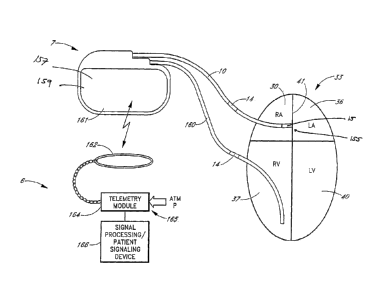
FIG. 4 shows one embodiment of a system for treating cardiovascular disease 9.
The
system 9 includes a first component comprising an implantable module 5, such
as that
-25-
CA 02525193 2005-11-08
WO 2005/000206 PCT/US2004/016186
described with reference to FIG. 2, and a second component comprising-an
external patient
advisory module 6, such as that described below with reference to FIG. 5.
During system 9
operation, radio frequency signals are canied by a lead 10 between a pressure
sensor package
15 located near the distal end 17 of the lead 10, and a housing 7 of an
implantable module 5.
The sensor package 15 includes at least one sensor 155. The lead 10 includes a
sensing/pacing electrode which is part of the sensor module 15 and an
indifferent electrode
14. The circuitry inside the housing 7 includes an antenna coil (not shown).
In this
embodiment, signals are communicated between the implantable module 5 and an
external
device, such as a patient advisory module 6, via the antenna coil of the
housing 7 and a
second external coil (not shown) coupled to the external device 6.
In one embodiment, the housing 7 contains a battery (not shown) that powers
the
implantable device 5. In another embodiment, the implanted device 5 receives
power and
programming instructions from the external device 6 via radio frequency
transmission
between the external and internal coils. The external device 6 receives
signals indicative of
one or more physiological parameters from the implanted device 5 via the coils
as well. One
advantage of such externally powered implantable device 5 is that the patient
will not require
subsequent surgery to replace a battery. In one embodiment of the present
invention, power
is required only when the patient or the patient's caregiver initiates a
reading. In other
situations, where it is desired to obtain physiological information
continuously, or where it is
desired that the implanted device 5 also perform functions with higher or more
continuous
power requirements, the housing 7 may also contain one or more batteries. As
described
below, the housing 7 may also contain circuitry to perform additional
functions that may be
desirable.
FIG. 5 shows one embodiment of the second component of the system, a patient
advisory module 6. In one embodiment, the patient advisory module 6 includes a
palm-type
computer with added hardware and software. Referring to FIG. 5, a patient
advisory module
6 includes a radio frequency telemetry module 164 with an associated coil
antenna 162,
which is coupled to a processing unit 166. In one embodiment, the processing
unit 166
includes a palm-type computer, or personal digital assistant (PDA), as is well
known to those
of skill in the art. In one embodiment, the patient advisory module 6 powers
the implanted
-26-
CA 02525193 2005-11-08
WO 2005/000206 PCT/US2004/016186
apparatus (not shown) with the telemetry hardware module 164 and coil antenna
162. In
another embodiment, the patient advisory module 6 receives physiological
signals from the
implanted first component of the system by wireless telemetry through the
patient's skin.
The patient advisory module 6 may include an RF unit 168 and a barometer 112
for
measuring the reference atmospheric pressure. In one embodiment, the RF unit
168 and
barometer are located within the telemetry module 164, although they can be
integrated with
the processing unit 166 as well. The signal processing unit can be used to
analyze
physiologic signals and to determine physiologic parameters. The patient
advisory module
166 may also include data storage, and a sub-module that contains the
physician's
instructions to the patient for therapy and how to alter therapy based on
changes in
physiologic parameters. The parameter-based physician's instructions are
referred to as "the
dynamic prescription," or DynamicRx (Savacor, Inc.). The instructions are
communicated
to the patient via the signaling module 166, or another module. The patient
advisory module
166 is located externally and used by the patient or his direct caregiver. It
may be part of
system integrated with a personal digital assistant, a cell phone, or a
personal computer, or as
a "Stand-Alone" device (e.g., in one embodiment, the HeartPODTM diagnostic and
therapeutic drug management system) without coinbination with CRM apparatus.
In one
embodiment, the external patient advisory module comprises an external
telemetry device, a
signal processing apparatus, and a patient signaling device. In one
embodiment, the patient
advisory module is operable to obtain the sensor signal from the implantable
sensor by
telemetry through the patient's skin; obtain the atmospheric pressure from the
barometer; and
adjust the sensor signal indicative of a fluid pressure based at least in part
upon the
atmospheric pressure obtained by the barometer so that the adjusted sensor
signal indicates
the fluid pressure within the left atrium of the heart relative to the
atmospheric pressure. In
one embodiment, the patient advisory module communicates with a remote site
such as a
doctor's office, clinic, hospital, pharmacy, or database. Revised patient
instructions
including the parameter-based dynamic prescription can be communicated back to
the patient
advisory module. This can be performed remotely via hard-wired telephone or
fiberoptic
cable networks, or wirelessly using a host of communication technologies
currently available.
-27-
CA 02525193 2005-11-08
WO 2005/000206 PCT/US2004/016186
Data may be communicated in either direction and the Internet may be in part
the conduit for
such communication.
In one embodiment, the physiologic signals are analyzed and used to determine
adjustable prescriptive treatment instructions that have been placed in the
patient advisory
module 6 by the patient's personal physician. Communication of the
prescriptive treatment
instructions to the patient may appear as written or graphic instructions on a
display of the
patient advisory module 6. These treatment instructions may include what
medications to
take, dosage of each medication, and reminders to take the medications at the
appropriate
times. In one embodiment, the patient advisory module 6 displays other
physician-specified'
instructions, such as "Call M.D." or "Call 911" if monitored values become
critical.
In an alternative embodiment, the treatment signal may be the numerical
representation of the mean left atrial pressure in mm Hg, or the numerical
representation of
some other parameter indicative of fluid pressure in the left atrium.
Physician specified
treatments would be supplied to the patient in the form of a decoding
reference providing
different treatment instructions for specified ranges of left atrial pressure.
Such a decoding
reference could be written or printed instructions on a card that the patient
keeps for
reference. For example, a mean left atrial pressure (LAP) of 15 mm Hg would
could indicate
the same treatment as a mean LAP of 16 mm Hg, both values being in a range
indicating that
the patient's heart failure is well compensated. An LAP of 25 mm Hg however
would
indicated decompensated CHF and would decode as different therapeutic
instructions aimed
at recompensating the state of CHF.
A third component of this system is designed for physician use. The third
component
is used to program the dynamic prescription and communicate it or load it into
the patient
advisory module 166. The third component may also contain stored data about
the patient,
including historical records of the physiologic signals and derived parameters
transmitted
from the patient implant and signaling modules. The third component may also
communicate
with external databases. In one embodiment, the third component is a physician
input device,
and includes a personal computer, a PDA, a telephone, or any other such device
as is well
known to those of skill in the art also comprising specific third component
software or
firmware programs.
-28-
CA 02525193 2005-11-08
WO 2005/000206 PCT/US2004/016186
In one embodiment, the second component (e.g., the patient advisory module
166) is
in the form of one or more implants.
In one embodiment of the present invention, the first implant module (such as,
for
example, implantable module 5 of FIG. 1 and FIG. 2) may also contain an
implant therapy
unit, or ITU. The ITU generates an automatic therapy regime based upon the
programmed
dynamic prescription. The therapy may include, but is not limited to, a system
for releasing
bioactive substances from an implanted reservoir, a system for controlling
electrical pacing of
the heart, and controllers for ventricular or other types of cardiac assist
devices. For example,
in one embodiment the sensor package is placed across the infra-atrial septum
and serves as
the atrial lead of a multichamber pacemaker. The physiologic sensor
information is used to
adjust pacing therapy such that pacing is performed only when needed to
prevent worsening
heart failure. One skilled in the art will appreciate that many systems or
devices that control
the function of the cardiovascular system may be used in accordance with
several
embodiments of the current invention.
In one enibodiment of the invention, the advisory module 6 is programmed to
signal
the patient when it is time to perform the next cardiac status measurement and
to take the
next dose of medication. It will be recognized by those skilled in managing
CHF patients
that these signals may help the many patients who have difficulty taking their
medication on
schedule. Although treatment prescriptions may be complex, one embodiment of
the current
invention simplifies them from the patient's perspective by providing clear
instructions. To
assure that information regarding the best treatment is available to
physicians, professional
cardiology organizations such as the American Heart Association and the
American College
of Cardiology periodically publish updated guidelines for CHF therapy.
These
recommendations can serve as templates for the treating physician to modify to
suit
individual patient requirements. In one embodiment, the device routinely
uploads data to the
physician or clinic, so that the efficacy of the prescription and the response
to parameter
driven changes in dose can be monitored. This enables the physician to
optimize the
patient's medication dosage and other important treatments without the
physician's moment-
to-moment intervention.
-29-
CA 02525193 2005-11-08
WO 2005/000206 PCT/US2004/016186
In various embodiment of the invention, a device and method for dynamically
diagnosing and treating cardiovascular illness in a medical patient are
provided. In one
embodiment, at least one physiological sensor is used to generate a signal
indicative of a
physiological parameter. In another embodiment, signal processing apparatus
operable to
generate a signal indicative of an appropriate therapeutic treatment based, at
least in part,
upon the signal generated by the physiological sensor, is also provided. In
another
embodiment a patient signaling device used to communicate the signal
indicative of the
appropriate therapeutic treatment to the patient is provided as well.
In one embodiment, a device and method for continuously or routinely
monitoring the
condition of a patient suffering from chronic cardiovascular disease are
provided. As will be
described in detail below, a system incorporating various embodiments of the
invention
monitors various physiologic parameters, such as the patient's left atrial
pressure. Depending
upon the magnitude of or changes in this pressure, for example, the system
communicates a
signal to the patient indicative of a particular course of therapy appropriate
to manage or
correct, as much as possible, the patient's chronic condition. In some
embodiments,
physician instructions and automated therapy are provided.
In one embodiment, the physiological sensor generates a signal indicative of a
physiological parameter on or in the patient's body. In one embodiment, the
signal
processing apparatus generates a signal indicative of an appropriate
therapeutic treatment
based at least in part upon the signal generated by the physiological sensor.
The patient
signaling device may generate signals indicative of therapeutic treatments or
courses of
action the patient can take to manage or correct, as much as possible, the
patient's condition.
In one embodiment, this method includes the steps of implanting one or more
physiological sensors substantially permanently within the patient, operating
the
physiological sensor to generate a signal indicative of a physiological
parameter, processing
this physiological signal to generate a signal indicative of an appropriate
therapeutic
treatment, and communicating the appropriate therapeutic treatment to a user.
In one
embodiment, the user includes, but is not limited to, the patient, a
caregiver, a medical
practitioner or a data collection center.
-30-
CA 02525193 2005-11-08
WO 2005/000206 PCT/US2004/016186
In another embodiment, the system is combined with or incorporated into a CRM
system, with or without physiologic rate control, and with or without backup
cardioversion/defibrillation therapy capabilities.
In one embodiment, at least one indication of congestive heart failure (CHF)
is
monitored. Elevated pressure within the left atrium of the heart is the
precursor of fluid
accumulation in the lungs, which results in signs and symptoms of acute CHF.
Mean left
atrial pressure in healthy individuals is normally less than or equal to
twelve millimeters of
mercury (mm Hg). Patients with CHF that have been medically treated and
clinically "well
compensated" may generally have mean left atrial pressures in the range from
12 to 20 mm
Hg. Transudation of fluid into the pulmonary interstitial spaces can be
expected to occur
when the left atrial pressure is above about twenty-fivp mm Hg, or at somewhat
more than
about thirty nun Hg in some patients with chronic CHF. Pulmonary edema has
been found to
be very reliably predicted by reference to left atrial pressures and less well
correlated with
conditions in any other chamber of the heart. Thus, the methods and apparatus
of several
embodiments of the invention may prove very useful in treating and preventing
pulmonary
edema and other adverse conditions associated with CHF. Pressure in the
pulmonary veins,
pulmonary capillary wedge position, and left ventricular end diastolic
pressure (LVEDP) are
generally indicative of left atrial pressure and are commonly used as
surrogates of LAP.
There are, however, specific conditions, that are well known to those skilled
in the art,
including cardiologists and physiologists, where these surrogates vary
substantially from LAP
and may be less predictive of impending heart failure. One example of such a
condition is
mitral valve stenosis where pulmonary edema develops despite a normal LVEDP
due to a
significant pressure gradient across the mitral valve. However, one of skill
in the art will
understand that several embodiments of the current invention can be used in
conditions
where the surrogates do not directly correlate with LAP. Other surrogate
pressures that also,
on specific occasion, indicate LAP include, but are not limited to: the
pulmonary artery
diastolic (PAD), mean pulmonary artery pressure, or algorithms that estimate
PAD from the
right ventricular waveform, the right ventricular end diastolic, the right
atrial pressure, and
the central venous pressure, or the response of arterial pressure during
forced expiration
against a closed glottis or other resistance (Valsalva maneuver). Also
included are other
-31-
CA 02525193 2005-11-08
WO 2005/000206 PCT/US2004/016186
pressures, parameters, or algoritluns that are indicative of left atrial
pressure that may be
known to those skilled in the art.
An embodiment of the invention includes a permanently implanted device
designed to
define- the presence of worsening CHF hours to days before the onset of
symptoms and to
provide for early preventative treatment according to the -physician's
individualized
prescription. As such, an embodiment of the invention includes an integrated
patient
therapeutic system that determines therapeutic dosages for an individual
patient based at least
in part on internal physiologic signals. In another embodiment, the system
consists of a small
implantable sensor device and an external patient advisory module comprising a
personal
data assistant (PDA) and a telemetry module. The sensor system may be
implanted into the
patient's left atrial chamber by a transseptal catheterization procedure.
There are already
several thousand physicians in the U.S. and abroad with the experience and
skills required for
such device implantation. The implantation procedure can be performed on an
outpatient
basis in a hospital's cardiac catheterization laboratory. The implant may
alternatively be
placed at the time of open-heart or minimally invasive valve or bypass surgery
where the
surgeon, under direct or laparoscopic or thoroscopic vision, positions the
device in the left
atrium, left atrial appendage, or an adjacent pulmonary vein.
In one embodiment, the sensor system measures a left atrial pressure waveform,
core
body temperature and a cardiac electrogram, such as the intra cardiac
electrogram (IEGM).
Elevated left atrial pressure is the most accurate predictor of impending CHF,
often preceding
clinical symptoms by hours to days. Other embodiments of the left atrial
pressure waveform
may be used to diagnose a number of conditions, as listed in FIGS. 6A-6C. Core
temperature is often depressed in acute CHF, but elevated prior to the
development of fever
in response to an infection, making core temperature a useful parameter for
differentiating
between these common conditions with similar symptoms but which require
different
treatments. The intracardiac electrogram may be useful in diagnosing
arrhythmias and
precipitating causes of worsening CHF.
FIG. 7 shows how left and right atrial pressure measurements may be combined
with
IEGM and core temperature measurement to diagnose a number of cardiac and non-
cardiac
conditions. The list of diagnostic states in FIG. 7 is exemplary, and by no
means exhaustive
-32-
CA 02525193 2005-11-08
WO 2005/000206 PCT/US2004/016186
of all the potential diagnostic states definable by the given parameters.
Multiple states can
exist simultaneously, for example, moderate CHF and rapid atrial fibrillation.
The measured
parameters can be used over large populations to define the probability of any
given
diagnostic state. Each diagnostic state may have a unique treatment. For
example, mild CHF
may be treated by increasing diuretic therapy, whereas rapid atrial
fibrillation is treated with a
drug that blocks AV node conduction. Many of the states listed can contribute
to worsening
CHF. Therefore, one of skill in the art will appreciate that several
embodiments described
herein can be used to treat on only CHF, but to treat cardiovascular disease
in general.
The embodiments summarized above and described in greater detail below are
useful
for the treatment of cardiovascular disease, including congestive heart
failure (CHF). CHF is
an important example of a medical ailment currently not treated with timely,
parameter-
driven adjustments of therapy, but one that the inventors believe could
potentially benefit
greatly from such a strategy. Patients with chronic CHF are typically placed
on fixed doses
of an average of six drugs to manage the disease. The drug regimen commonly
includes but
is not limited to diuretics, vasodilators such as ACE inhibitors or A2
receptor inhibitors, beta-
blockers such as Carvedilol, neurohonnonal agents such as spironolactone, and
inotropic
agents usually in the form of cardiac glycosides such as, for example,
digoxin. In addition,
patients typically are taking other cardiovascular drugs to limit disease
progression,
symptoms or complications. Examples include `statins' to lower cholesterol,
nitrate to
relieve chest pain, and aspirin or warfarin to prevent clotting.
1. Implantation and Anchoring
a. Placement and anchoring in the left atrium
In one embodiment, such as that illustrated in FIG. 8, an implantable device
is
implanted percutaneously in the patient by approaching the left atrium 36
through the right
atrium 30, penetrating the patient's atrial septum 41 and positioning one or
more
physiological sensors 15 in the atrial septum 41, on the septal wall of the
left atrium 36, or
inside the patient's left atrium 36. FIG. 8 shows an embodiment in which a
sensor package
15 is deployed across the atrial septum 41. The sensor lead 10 is coupled to a
physiological
sensor or sensors 15 and anchoring apparatus at the lead 10 distal end. The
anchoring
apparatus includes a distal foldable spring anchor 68 that expands in diameter
upon release
-33-
CA 02525193 2005-11-08
WO 2005/000206 PCT/US2004/016186
and is located at or near the distal tip of the sensor 15, and a proximal
foldable spring anchor
70. The distal and proximal anchors 68, 70 are sufficiently close together
that when deployed
the two anchors 68, 70 sandwich the intra-atrial septum 41 between them, thus
fixing the
'sensor/lead system to the septal wall. The intra-atrial septum 41 is
typically between about 1
and about 10 mm thick. In one embodiment, the anchors 68, 70 are made of a
highly elastic
biocompatible metal alloy such as sup erelastic nitinol. The lead 10 may
contain a lumen that
exits the lead 10 at its proximal end. A stiffening or bending stylet can be
insert in the lumen
to aid in passage of the sensor(s) and lead 15, 10. After a transseptal
catheterization has been
performed, a sheath/dilator system of diameter sufficient to allow passage of
the sensor/lead
system is placed from a percutaneous insertion site over a guidewire until the
distal end of a
sheath 67 is in the left atrium 36. Left atrial position can be confirmed
under fluoroscopy by
contrast injection, or by the pressure waveform obtained when the sheath 67 is
connected to a
pressure transducer. To aid the procedure, the sheath 67 may include a
proximal hemostasis
valve to minimize air entrainment during device insertion. A side port with a
stopcock is
useful to aspirate any remaining air and to inject radiographic contrast
material. Additionally,
later sheath 67 removal may be facilitated by using a "peel-away" type of
sheath. These
features of vascular sheaths are commercially available and well know to those
familiar with
the art. With the spring anchors 68, 70 folded and forming a system with
minimal diameter,
the system is loaded into the sheath 67 and advanced until the distal spring
68 just exits the
sheath 67 in the left atrium 36 and is thus deployed to its sprung diameter.
The sheath 67 is
carefully withdrawn without deploying the proximal anchor 70 and the sheath 67
and
sensor/lead system are withdrawn as a unit while contrast is injected through
the sheath 67
around the sensor lead until contrast is visible in the right atrium 30. The
proximal sheath 67
is further withdrawn, allowing the proximal anchor 70 to spring to its
unloaded larger
diameter, thus fixing the distal portion of the sensor lead to the septum 41.
It will also be apparent that, in several embodiments, a similar sensor/lead
system can
be inserted through an open thoracotomy or a minimally invasive thoracotomy,
with the
anchoring system fixating the sensor/lead to a location such as the free wall
of the left atrium,
the left atrial appendage, or a pulmonary vein, all of which provide access to
pressures
indicative of left atrial pressure.
-34-
CA 02525193 2013-03-05
In one alternative embodiment, a flexible lead 10 is partially advanced into a
pulmonary vein 50 connected to the left atrium 36 such that one or more
physiological
sensors 15 disposed on the flexible lead 10 a predetermined distance from its
distal end 17
are positioned within the left atrium 36 or the pulmonary vein 50, as shown in
FIG. 9. In
another embodiment, the distal portion 17 of the flexible lead 10 is partially
advanced into
the left atrial appendage such that anchoring apparatus will be occlusive of
the appendage,
for example as taught by Lesh et at. in U.S. Patent No. 6,152,144. The
physiologic sensors
15 are positioned on the lead 10 proximal to the occlusive anchors so that
they sense
conditions in the left atrium.
In other embodiments, such as those shown in FIG. 12 and FIG. 14, a first lead
component 53 includes an anchoring apparatus, for example, a helical screw 57,
which is
advanced to the atrial septum 41. The anchoring apparatus is deployed to
anchor the first
lead component 53 into the patient's atrial septum 41.A second lead component
60 includes
a physiological sensor, for example, a pressure transducer 62, which is
advanced along the
first lead component 53 until the second lead component 60 is in a position
such that the
physiological sensor is positioned within the patient's left atrium 36.
b. Implantation in the Left Atrium
Referring to the embodiment depicted in FIG. 8, the system is implanted
through the
left atrial septum 41 such that the pressure sensor 15 is exposed to the
pressure in the left
atrial chamber 36 of the heart. The left atrial septum 41 can be accessed from
the right
atrium 30 through the inferior or superior vena cava 35, 28, as is well known
to those skilled
in the arts of, for example, pacemaker lead placement, catheter ablation for
control of
arrhythmias originating in the left atrium or pulmonary veins, percutaneous
repair of the
mitral valve, and percutaneous closure of an atrial septal defect. In one
embodiment, the
flexible lead 10 and pressure transducer 15 are anchored to the atrial septum
41. This
placement can be achieved using vascular access techniques that are well known
to those
familiar with the performance of invasive cardiovascular procedures, in
particular,
interventional cardiologists, electrocardiologists, and cardiovascular
surgeons. These
procedures are commonly performed with the aid of visualization techniques,
including
-35-
CA 02525193 2005-11-08
WO 2005/000206
PCT/US2004/016186
standard fluoroscopy, cardiac ultrasound, or other appropriate visualization
techniques used
alone or in combination.
Access to the central venous circulation may be achieved by use of the
standard
Seldinger technique through the left or right subclavian vein, the right or
left internal jugular
vein, or the right or left cephalic vein. Alternatively, access may be made
via the Seldinger
technique into the right femoral vein. In either case, a Brockenbrough
catheter and needle are
used to pierce the atrial septum 41 for access to the left atrium 36, as
described below.
i. Superior Venous Access (Subclavian or Internal Jugular
Vein)
FIG. 10 provides a schematic sectional view of the patient's heart 33 and
shows the
apparatus used to access the left atrium 36. FIG. 10 depicts an access
assembly 18
comprising a Brockenbrough catheter 20 inside a sheath 22, with a flexible
guidewire 25
residing within the Brockenbrough catheter 20. As FIG. 10 indicates, the
access assembly
has been placed through the superior vena cava 28 into the right atrium 30 of
the heart 33.
FIG. 10 also shows the inferior vena cava 35, the left atrium 36, the right
ventricle 37, the
left ventricle 40, the atrial septum 41 that divides the two atria 30, 36, and
the valves 42
between the right atrium 30 and right ventricle 37, and the left atrium 36 and
left ventricle 40.
The reader will appreciate that the view of FIG. 10 is simplified and somewhat
schematic,
but that nevertheless FIG. 10 and the other views included herein will suffice
to illustrate
adequately the placement and operation-of an embodiment of the present
invention.
Placement of the Lead
With the access assembly 18 in place within the right atrium 30, the
Brockenbrough
catheter 20 is used to pierce the atrial septum 41 by extending the
Brockenbrough needle (not
shown) through the atrial septum 41 into the left atrium 36. In the figures,
the atrial septum
41 has been pierced by the needle, the catheter 20 has been advanced over the
needle, and the
needle has been withdrawn from the catheter 20, leaving the catheter 20 in
place inside the
left atrium 36. Optionally, a guidewire 25 may be advanced through the needle
into the left
atrium 36 before or after advancing the catheter 20, or it may be placed into
the left atrium 36
through the catheter 20 alone after the needle has been withdrawn. A lead
placement
procedure is described above.
-36-
CA 02525193 2005-11-08
WO 2005/000206 PCT/US2004/016186
As indicated by the arrows 45 in FIG. 10, the sheath 22 may extend into the
left
atrium 36, or it may remain on the proximal side of the atrial septum 41
within the right
atrium 30. FIG. 10 shows the guidewire 25 extended from the end of the
Brockenbrough
catheter 20 to secure continuous access into the left atrium 36. As depicted
therein, the
guidewire 25 has a curled, "pig-tail" style distal tip 48 to better secure the
guidewire 25
within the left atrium 36 and to safeguard against inadvertent withdrawal
through the atrial
septum 41. Alternatively, a "floppy tip" guide wire may be used, which can be
safely
advanced well into one of the pulmonary veins, again to safeguard against
inadvertent
withdrawal through the atrial septum 41. Once the guidewire 25 is securely in
place in the
left atrium 35, the Brockenbrough catheter 20 may be withdrawn so that the
flexible lead 10
may be placed through the peel-away sheath 22.
With the guidewire 25 securely in place with its distal tip 48 inside the left
atrium 36,
the flexible lead 10 may be advanced into the left atrium 36. The flexible
lead 10 might itself
include a central lumen configured to receive the proximal end of the
guidewire 25, thereby
allowing the flexible lead 10 to be advanced down the guidewire 25 toward the
left atrium
36. More commonly, an exchange catheter, which may be in the form of a peel-
away sheath
22, will be advanced down the guidewire 25 and placed into the left atrium 36,
the guidewire
25 may then be withdrawn, after which the flexible lead 10 will be advanced
down the
exchange catheter and into position.
In one embodiment, a peel-away sheath 22 is used to allow the sheath to be
removed
once the distal end of the lead 10 is implanted; The peel-away feature is used
if the proximal
end of the lead 10 is permanently attached to the coil housing assembly
(described above).
Alternatively, a non-peel-away sheath with proximal hemostasis valve and side
port as
described above can be used, for example, if the lead is detachable from the
coil assembly
and if the lead or a stiffening stylet fixed within the central lumen of the
lead is long enough
to remove the sheath wile maintaining control over the proximal end of the
lead . These
configurations of sheaths and methods of sheath removal are well known to
those skilled in
the art.
iii. Anchoring the Sensor and Lead
-37-
CA 02525193 2005-11-08
WO 2005/000206 PCT/US2004/016186
Once the pressure transducer 15 of the flexible lead 10 is positioned within
the left
atrium 36, the lead 10 should be anchored in place to ensure that the pressure
transducer 15
stays reliably and permanently in the desired location.
One method for anchoring the flexible lead 10 in place is depicted in FIG. 9,
which is
a somewhat schematic depiction of the major structures of the heart. FIG. 9
shows the four
pulmonary veins 50 that connect to the left atrium 36. In the particular
apparatus depicted in
FIG. 9, the flexible lead 10 includes a pressure transducer 15 located on the
body of the lead
a predetermined distance proximal of the distal end 17 of the lead 10.
Referring back to FIG. 9, the distal end 17 of the flexible lead 10 in this
embodiment
can be bent by the operator in much the same way as a distal tip such as might
be found on a
steerable angioplasty guidewire or another similar device. This feature
assists the operator in
steering the flexible lead 10 into a selected one of the pulmonary veins 50,
with the pressure
transducer 15 disposed within the interior space of the left atrium 36, or
even within the
pulmonary vein itself. Placement of the pressure transducer 15 within the
pulmonary vein is
effective because pressures within the pulmonary vein are very close to
pressures within the
left atrium. It will be appreciated by those skilled in the art that
visualization markers (not
shown) may be provided at appropriate locations on the flexible lead 10 to
assist the operator
in placing the device as desired. With the flexible lead 10 in place as shown,
the body's own
natural healing mechanism may permanently anchor the flexible lead 10 in place
both at the
penetration site through the atrial septum 41, and where the flexible lead 10
contacts the
interior surface of the pulmonary vein 50 in which the tip of the lead 10
resides. The pressure
transducer 15 might also be placed at locations such as the left atrial
appendage (not shown in
FIG. 9) where the pressure is nearly the same as the left atrium 36, or the
left ventricular
cavity, where at identifiable phases of the cardiac cycle the pressure is
momentarily nearly the
same as that in the left atrium 36.
FIG. 11 and FIG. 12 show alternative methods and devices for anchoring the
pressure transducer 15 in a location appropriate for measuring pressures
within the left atrium
36. The lead in this embodiment includes a helical screw 57 for anchoring the
lead to the
atrial septum 41. Similar configurations are used in some leads for pacemakers
and thus may
be familiar to those skilled in the art.
-38-
CA 02525193 2005-11-08
WO 2005/000206 PCT/US2004/016186
Referring now specifically to FIG. 11, the guidewire 25 is shown positioned
across
the atrial septum 41 between the left atrium 36 and the right atrium 30. A
first lead
component 53 is delivered over the guidewire through an appropriate guiding
catheter 55 or
sheath. This first lead component 53 includes a helical screw 57 on its
exterior surface. The
helical screw 57 is advanced into the tissue of the atrial septum 41 by
applying torque to the
shaft of the first lead component 53. The helical screw 57 could also be
coupled to a hollow
or solid cylindrical mandrel (not shown), or to a spirally wound mandrel (also
not shown)
disposed along substantially the entire length of the first lead component.
When the helical
screw 57 has been turned and advanced sufficiently into the atrial septum 41,
the guidewire
25 and guiding catheter may then be withdrawn leaving the first lead component
53 anchored
securely in place.
iv. Two-component Lead with optional Second Pressure
Transducer
In one embodiment, a second lead component 60 is advanced as shown in FIG. 12
through a central lumen in the first lead component 53. The first and second
lead
components 53, 60 are sized and configured so that when the second lead
component 60 is
fully advanced with respect to the first lead component 53, a left atrial
pressure transducer 62
at the end of the second lead component 60 protrudes by an appropriate
predetermined
amount into the left atrium 36. In one embodiment, the second lead component
60 is then
securely fixed with respect to the first lead component 53.
It should be noted that the embodiments depicted in FIG. 11 and FIG. 12
includes a
second pressure transducer 65 on the exterior of the first lead component 53
that may be
exposed to pressure within the right atrium 30. This illustrates, in a
simplified way, the
general principle, in which a pressure transducer is used to measure fluid
pressure within the
left atrium, but in which one or more additional transducers or sensors may
also be used to
detect a physiologic condition other than left atrial pressure. These
physiologic conditions
may include pressures in locations other than the left atrium 36, and physical
parameters
other than pressure.
v. Alternative Anchoring Systems and Methods
-39-
CA 02525193 2005-11-08
WO 2005/000206 PCT/US2004/016186
FIG. 8 and FIG. 13 through FIG. 16 show embodiments of the flexible lead 10,
in
which folding spring-like fins or anchors deploy to anchor the lead in place
in the atrial
septum 41. Referring specifically to FIG. 13, a first lead component 53 is
advanced through
a sheath 67, the sheath 67 having been advanced across the atrial septum 41.
In this
embodiment, the first lead component 53 includes folding distal anchors 68 and
proximal
anchors 70 that lie folded and are held in place inside the interior lumen of
the sheath 67.
When the first lead component 53 and sheath 67 are properly positioned, which
will generally
involve the use of fluoroscopy or an alternative technique for imaging, the
operator may
carefully withdraw the sheath 67 from around the first lead component 53. As
the distal and
proximal anchors exit the sheath 67, they deploy themselves (as depicted in
FIG. 8) on either
side of the atrial septum 41, thereby anchoring the first lead component 53
securely in place.
Similar anchors are sometimes used with leads for pacemakers and other medical
devices
where permanent anchoring is desired, and the operation of these anchors thus
will not be
entirely unfamiliar to the knowledgeable reader.
Referring now to FIG. 14, a second lead component 60 is advanced through a
central
lumen of the first lead component 53 after the guidewire 25 (see FIG. 15 and
FIG. 16) and
sheath 67 are removed. As in the previous embodiment, a left atrial pressure
transducer 62 is
carried at the distal end of the second lead component 60. Again, the first
and second lead
components 53, 60 are sized and configured with respect to one another so that
the left atrial
pressure transducer 62 protrudes from the first lead component 53 an
appropriate amount into
the left atrium 36. In addition, as in the previous embodiment, a second
pressure transducer
65 on the exterior of the first lead component 53 allows for the measurement
and transmittal
of pressure within the right atrium 37.
Other anchoring methods may be devised by those skilled in the relevant arts.
Moreover, approaches have been described by which the lead is positioned
between the left
atrium and an exit site from the patient's superior venous circulation.
Alternate lead routes
and exit sites may find use as well.
vi. Surgical Methods of Device Implantation
As described above, percutaneous transvenous implantation methods are used in
accordance with several embodiments of the current invention. One skilled in
the art will
-40-
CA 02525193 2013-03-05
understand that alternative lead routes and exit sites from the venous system
may also be
used. One important class of alternative implantation methods consists of
surgical
implantation through the wall of the heart, either directly into the left
atrium through the left
atrial free wall or left atrial appendage, into the left atrium via a
pulmonary vein, into the left
atrium through the intra-atrial septum via the right atrial free wall, or
directly into a
pulmonary vein.
In one embodiment, the pressure transducer is implanted in the atrial free
wall or in
the wall of the atrial appendage. As described above, in one embodiment, at
these locations
the pressure sensing surface of the transducer is exposed to left atrial
pressure, and the body
of the transducer extends through the wall of the atrium or atrial appendage.
A flexible lead
from the implanted transducer provides signal connection to a telemetry
antenna coil that the
surgeon implants near the surface of the skin. In another embodiment, this
coil may be
connected directly to the implanted pressure transducer on the outside surface
of the heart,
without need for a flexible lead. In yet another embodiment, the flexible lead
provides signal
connection to a CRM generator housing located near the surface of the skin.
c. Pulmonary Vascular Implant
Vascular stents are implants that are deployed in blood vessels to support the
size of
the vascular channel and maintain adequate blood flow. A stent may also be
used to anchor
another type of device in a fixed location within the cardiovascular system.
U.S. Patent No.
5,967,986, describes a stent coupled to one or more pressure transducers for
the purpose of
measuring blood flow in a vessel. In one embodiment of the current invention,
a stent is used
to support and anchor the sensor measuring a signal indicative of left atrial
pressure. As
mentioned above, the pressure in the pulmonary veins is substantially
identical to that in the
left atrium. Thus, in one embodiment of this invention, the pressure sensor is
anchored in
a pulmonary vein by means of a stent expanded within the vein.
In one embodiment of the current invention, a method and apparatus for
continuous
ambulatory detection, diagnosis and treatment of acute congestive heart
failure is provided.
It will be understood that the current invention may be implemented using
digital signal
processing methods in which various input signals are sampled and the
described procedures
-41-
CA 02525193 2005-11-08
WO 2005/000206 PCT/US2004/016186
are performed on a set of samples. Hence, a periodic determination of the
physiological
parameter of interest is within the definition of the term continuous. In one
embodiment, a
percutaneously implantable system comprises a hermetically sealed pressure
transducer/communications module mounted on an unexpanded vascular stent-like
member.
In one embodiment, the stent-like member is a cylindrical vascular stent such
as a balloon
expandable or self-expanding metallic stent similar to those used to treat
vascular stenosis
such as atherosclerotic stenosis of a coronary or peripheral artery. The
pressure
transducer/communications module is mechanically coupled to the unexpanded
stent and the
stent/transducer module is mounted on a delivery catheter constituting a
stent/transducer
delivery system. The stent/transducer delivery system is percutaneously
inserted into a
patient's body via the venous or arterial system.
In one embodiment, the delivery system courses over a guide wire that has been
positioned from proximal to distal, starting outside the patient,
percutaneously entering into
the venous system and into the right atrium, through the right ventricle and
into a branch of
the pulmonary artery. The stent/transducer module is then advanced over the
guide wire into
the selected branch of the pulmonary artery that is approximately the diameter
of the ,
expanded stent/transducer module.
In another embodiment, a standard transseptal catheterization procedure is
performed
to place a guide wire that courses from proximal to distal starting outside
the patient
percutaneously into the venous system into the right atrium, across the intra-
atrial septum,
into the left atrium and finally into one of the four pulmonary veins. The
stent/transducer
delivery system is then advanced over the guide wire until the unexpanded
stent/transducer is
positioned in the pulmonary vein that is approximately the diameter of the
expanded
stent/transducer module. The stent is then expanded such that the cylinder
described by the
stent is coaxially in contact with the vessel wall confining the
transducer/communications
module so that its outer surface contacts the vessel wall.
2. Pressure Transducer
a. Pressure Sensor Locations
In one embodiment of the invention, the apparatus and/or method for treating
cardiovascular disease includes one or more sensors, such as pressure sensors.
In one
-42-
CA 02525193 2005-11-08
WO 2005/000206 PCT/US2004/016186
embodiment, the pressure sensor is located in the atrial septum, the left
atrial appendage, the
left atrial free wall, one of the pulmonary veins,. or any other location in
pressure
communication with the left atrium, for example, but not limited to, the right
atrium, the
central veins, or any location as known to those of skill in the art suitable
for measuring a
pressure related to the pressure in the pulmonary veins, the pulmonary
capillary wedge
pressure, the pulmonary artery diastolic pressure, the left ventricular end
diastolic pressure, or
the right ventricular end diastolic pressure. In one embodiment, the pressure
signal includes
a pulmonary vein pressure, a pulmonary capillary wedge pressure, a pulmonary
artery
diastolic pressure, a left ventricular end diastolic pressure, a right
ventricular end diastolic
pressure, right atrial pressure, or the pressure measured in the intrathoracic
space, or the
central veins. In another embodiment, the signal includes algorithms that
estimate pulmonary
artery diastolic pressure from the right ventricular waveform, the right
ventricular end
diastolic pressure, the right atrial pressure, or the response of the arterial
blood pressure to the
Valsalva maneuver. In yet another embodiment, signals indicative of left
atrial pressure
include spatial parameters (e.g., dimension of chambers), septal shape,
position, motion, and
acceleration.
b. Pressure Sensor Design
In one embodiment, the physiological sensor includes a pressure transducer. In
one
embodiment, the pressure transducer is contained within a hermetically sealed
sensor
package, or module. The sensor package may be provided in a wide range of
sizes and
shapes. In one embodiment, the sensor1 package is cylindrical, and is between
about 1 mm
and 5 inm long, and 3 mm in diameter. In another embodiment, the sensor
package is
between about 5 mm and about 15 mm long. In another embodiment the package is
about 8
mm long, and about 3 mm in diameter. In one embodiment the package is less
than about 1
mm in diameter. In another embodiment, the package is less than about 10 mm
long. Micro
electro-mechanical system (MEMS) pressure sensor devices may also be used. In
one
embodiment, the package may be rectangular, square, spherical, oval,
elliptical, or any other
shape suitable for implantation. In one embodiment, the sensor package is
rigid, and in
another embodiment, the sensor package is flexible.
-43-
CA 02525193 2005-11-08
WO 2005/000206 PCT/US2004/016186
In one embodiment, the sensor package includes a titanium cylindrical housing
that is
closed at one end by titanium foil membrane. In one embodiment, the foil
membrane is
between about 0.001 to 0.003 inches, between about 0.003 inches and about
0.005 inches, or
less than 0.001 inches thick. In another embodiment, the foil membrane is
between about 25
microns to about 50 microns thick, and about 0.08 to 0.10 inches (about 2.0 to
2.5 mm) in
diameter. Foil diaphragms of this type have relatively low compliance, meaning
that they
exhibit relatively little strain, or displacement, in response to changes in
pressure. For
example, in one embodiment, a 2.5 mm diameter by 50-micron thick titanium foil
diaphragm
has a displacement at its center of only about 4.3 microns per mm Hg pressure
change.
Higher compliance is a disadvantage for implantable pressure sensors because
tissue
overgrowth can limit the relatively larger motion of a high compliance
diaphragm, causing
errors in the sensed pressure reading.
In one embodiment, resistive strain gauges are bonded to the inside surface of
the foil.
In one embodiment, the titanium cylindrical housing comprises an application
specific
integrated circuit (ASIC or "chip") or "measurement electronics." Measurement
electronics
are contained within the housing, connected to the strain gauges by fine gold
wires. The
other end of the housing is sealed by a ceramic feed-through that is brazed to
a titanium
cylinder.
In one embodiment, the pressure of the gas sealed in the cylinder is slightly
lower
than the lowest external pressure anticipated, so that the net force on the
foil will be inward
under normal conditions of operation, forming a concave membrane shape. The
advantage of
maintaining a concave membrane shape throughout the pressure range of
operation is that it
avoids potential pressure measurement artifacts that are known to sometimes
occur when a
pressure sensing membrane transitions between a concave and a convex shape, a
phenomenon known as "oil-canning." In one embodiment, oil-canning is avoided
by using a
transducer diaphragm that has low compliance, with low compliance as described
above, and
that is nearly flat in the absence of a pressure differential. In one
embodiment, the diaphragm
is about 2.0 to 2.5 mm in diameter and is within about 25 microns of flat in
the absence of a
pressure differential. In another embodiment, the diaphragm thickness is
maximized to
-44-
CA 02525193 2005-11-08
WO 2005/000206 PCT/US2004/016186
maximize flatness and minimize compliance, consistent with the sufficient
compliance to
derive a useable transducer signal.
In one embodiment, the pressure sensor includes temperature compensation so
that
pressure measurements will not be affected by temperature change. This also
provides the
temperature at the site of the sensor. In one embodiment, temperature
compensation or
modulation is achieved by using multiple resistive strain gauges arranged in a
Wheatstone
bridge, such that the electrical voltage output of the bridge is proportional
to the ratio of two
or more resistances, as is well known in the art of electrical measurements.
By selecting
resistive strain gauges with substantially identical temperature coefficients,
the intrinsic
output of the bridge is made to be temperature independent. However, the
overall response
of the pressure transducer may still be temperature dependent because of
imperfect matching
of the resistive strain gauges, or due to other factors, such as the ideal gas
law behavior of the
gas sealed within the chamber, or different thermal expansions of the various
components
and contents of the device. Another embodiment of temperature compensation
utilizes an
internal thermometer consisting of, for example, a resistor whose resistance
depends upon
temperature in a reproducible way, and which is placed in a location isolated
from the
transducer diaphragm so that its resistance does not depend on pressure
variations. Prior to
implanting the device, calibration data is collected consisting of the output
of the transducer
versus pressure as a function of the reading of the internal thermometer.
After implantation,
the signal from the internal thermometer is used together with the transducer
output and the
calibration data to determine the temperature compensated pressure reading. In
one
embodiment, a band gap voltage reference is used to create a current
proportional to absolute
temperature that is then compared to the temperature-independent voltage
reference. Such
methods are well-known in the art of CMOS integrated circuit design.
In one embodiment, the devices described herein are configured similarly to a
cardiac
pacemaker, with a hermetically sealed housing implanted under the patient's
skin and a
flexible lead with a pressure transducer at its distal end. The housing
contains a battery,
microprocessor and other electronic components, including a patient signaling
device and
transcutaneous telemetry means for transmitting programming information into
the device
and for transmitting physiological data out to an external
programmer/interrogator.
-45-
CA 02525193 2013-03-05
One skilled in the art will understand that alternative distributions of the
components
may be constructed in accordance with several embodiments of the present
invention. In one
alternative, the pressure sensing circuitry is incorporated into the pressure
transducer unit
implanted in the heart, reducing the number of conductors needed in the lead
to as low as
two.
In another embodiment, the signal processing, prescription algorithms, and
patient
signaling components are located in a device external to the patient's body in
communication
with the implanted subcutaneous housing via one of various forms of telemetry
well known
in the art, such as two-way radio frequency telemetry.
In another embodiment, the pressure sensor is fabricated by micro electro-
mechanical
systems (MEMS) techniques, as taught by, for example U.S. Patent No.
6,331,163.
c. Sensor-tissue interaction issues
In one embodiment, within several weeks after implantation, the entire device
is
covered with new tissue, including fibrous tissue and endothelium. A covering
of
endothelium is desirable because it prevents the formation of blood clots
that, if formed,
could break loose and cause a blocked artery elsewhere in the body, most
dangerously in the
brain. A covering of fibrous tissue is also a common component of the body's
healing
response to injury and/or foreign bodies. An excessive growth of fibrous
tissue on the left
atrial surface of the pressure sensor may be undesirable because it may
interfere with accurate
transmission of fluid pressure in the left atrium to the pressure sensitive
diaphragm. In
addition, contraction of fibrous tissue over time may cause progressive
changes in the
pressure waveform or mean value, which could confound interpretation of the
data.
i. Low compliance sensor membrane
In one embodiment, the pressure transducer membrane is designed to have very
low
compliance. In one embodiment, a low compliance pressure transducer is
fabricated using
titanium foil as described above. In another embodiment, a low compliance
pressure
transducer is fabricated from, for example, silicon, using micro
electromechanical systems
(MEMS) techniques. In yet another embodiment, a coating is provided on the
left atrial
surface of the pressure sensor.
Coatings, polishin_g, and drug eluting surfaces
In one embodiment, a coating inhibits or minimizes the formation of
undesirable fibrous
-46-
CA 02525193 2013-03-05
tissue, while not preventing the beneficial growth of an endothelial covering.
Coatings with
these properties are well known in the art of implanting medical devices,
particularly
intravascular stents, into the blood stream. Surface coating materials
include, but are not
limited to, paralene, PVP, phosphoryl choline, hydrogels, albumen affinity,
and PEO.
In one embodiment, at least some areas of the sensor package and diaphragm are
electropolished. Electropolished surfaces are known by those skilled in the
art to reduce the
formation of thrombosis prior to endothelialization, which leads to a reduced
burden of
fibrotic tissue upon healing. Metallic intracoronary stents currently approved
for clinical use
are electropolished for this purpose.
Release of antiproliferative substances including radiation and certain drugs
are also
known to be effective in stenting. Such drugs include, but are not limited to,
Sirolimus and
related compounds, Taxol and other paclitaxel derivatives, steroids, other
anti-inflammatory
agents such as CDA, antisense RNA, ribozymes, and other cell cycle inhibitors,
endothelial
promoting agents including estradiol, antiplatelet agents such as platelet
glycoprotein IIb/IIIa
inhibitors (ReoPro), anti-thrombin compounds such as heparin, hirudin, hirulog
etc,
thrombolytics such as tissue plasminogen activator (tPA). These drugs may be
released from
polymeric surface coating or from chemical linkages to the external metal
surface of the
device. Alternatively, a plurality of small indentations or holes can be made
in the surfaces
of the device or its retention anchors that serve as depots for controlled
release of the above
mentioned antiproliferative substances, as described by Shanley et al. in U.S.
Publication No.
2003/0068355, published April 10, 2003.
d. Pressure Signal Detection
In one embodiment, the implanted portion of the device is comprised of a
plurality
of up to n physiologic signal detection sensors S described by the set:
{S1, S2, .
In one embodiment, Sl, the first sensor, detects a parameter that is
indicative of left
atrial pressure or SILAp , thus
ISI.AP, S2, = = = Sd==
Signals indicative of left atrial pressure can be pressure signals measured at
a variety
of sites and may be detected by a variety of pressure transducer types. The
signals may be
obtained from locations in the cardiovascular system or adjacent to the
cardiovascular system
-47-
CA 02525193 2013-03-05
known to be similar to or highly correlated with direct pressure readings from
the left atrium.
Such locations for obtaining pressure signals similar to the left atrium are
well known to
those skilled in the art, such as Cardiologists. Locations for sensing
pressure include, but are
not limited to, the left atrium and its contiguous structures, the pulmonary
veins, the
pulmonary capillary wedge or occlusion pressure, the pulmonary artery
diastolic pressure,
and the left ventricular end diastolic pressures. Other pressures indicative
of left atrial
pressure include differential pressures such as the difference between the
left atria and the
right atria, or the difference between the pulmonary capillary wedge and right
atrial pressures,
as shown by the correlation in FIG. 17. The individual signals comprising the
differential
signal correlate independently with left atrial pressure.
3. Non-Pressure Sensors
a. Left Atrial Dimension
In one embodiment, the system may include one or more additional sensors. In
one
embodiment, a non-pressure sensor is also provided to generate a signal
indicative of
pressure in the left atrium. Hemmingsson (U.S. Patent No. 6,421,565) describes
such an
implantable cardiac monitoring devices as an A-mode ultrasound probe which is
adapted to
be positioned in the right ventricle of a heart, and which emits an ultrasound
signal which is
reflected from one cardiac segment of the left ventricle of the heart, and the
ultrasound probe
receives the resulting echo signal. The delay between the emission of the
ultrasound signal
and the reception of the resulting echo is measured, and from this delay a
position of the
cardiac segment is determined. In one embodiment, an A-mode ultrasound probe
is deployed
in the right atrium of a heart so that an ultrasound signal is reflected from
one or more cardiac
segments of the left atrium, either the atrial septal segment, the lateral
wall segment, or both.
Increased left atrial pressure is known to cause in increase in the volume of
the left atrium
by displacing the walls of the left atrium away from each other. Thus,
measurement of the
positions of one or more left atrial walls provides a signal indicative of
left atrial pressure,
as described below, that can be used to guide therapy for CT-IF.
Kojima (U.S. Patent No. 4,109,644), describes another implantable ultrasound
transducer that could be used in the manner described above to determine left
atrial
dimension and thus derive a signal indicative of left atrial pressure.
In one embodiment, the sensor comprises one pressure sensor, a pressure sensor
-48-
CA 02525193 2013-03-05
package, or module, with pressure sensor and electronics, or a sensor package
containing
electronics, a pressure sensor, and at least one non-pressure sensor. In one
embodiment, the
at least one non-pressure sensor provides a signal indicative of: an internal
electrocardiogram;
a temperature; a physical dimension; an electrical resistance, such as, but
not limited to, a
thoracic electrical impedance; a respiratory tidal volume; a respiratory rate;
lung acoustics;
oxygen saturation; oxygen partial pressure, including oxygen partial pressure
in the left
chamber or the right chamber; or cardiac output. In another embodiment of the
invention,
the non-pressure sensor measures: left atrial dimension, cross-sectional area,
or volume; left
ventricular dimension, cross-sectional area or volume; atrial septum position;
velocity, or
acceleration. In one embodiment, a non-implanted sensor is provided. In one
embodiment,
the non-implanted sensor includes: an arterial pressure cuff, including an
automated arterial
pressure cuff; and a weight scale. In one embodiment, two sensors are
provided, a first
sensor and a second sensor. In one embodiment, the first sensor measure a
pressure in the
heart and the second sensor measures a non-pressure parameter, including, but
not limited
to the parameters listed above. In one embodiment, the second sensor is also a
pressure
sensor. In one embodiment, the first sensor is located internal to the patient
and the second
sensor is located external to the patient. Located "external", as used herein,
shall be given
its ordinary meaning and shall also mean located on the patient, in contact
with the patient,
or located completely independent of the patient.
b. Core Temperature
Other non-pressure physiologic parameters may be used in other embodiments.
Casscells III, et al. (U.S. Patent No. 6,454,707), describe a method and
apparatus for
predicting mortality in congestive heart failure patients by monitoring body
temperature and
determining whether a downward trend in temperature fits
-49-
CA 02525193 2005-11-08
WO 2005/000206 PCT/US2004/016186
any predeteimined criteria. The apparatus described by Casscells et al.
determines when
death is imminent and generates an alarm. In one embodiment of the present
invention, the
trend in body temperature is used daily to adjust the patient's therapy at an
earlier point
before any downward trend in temperature becomes critical. In one embodiment,
core body
temperature is measured at the atrial septum. In another embodiment, core body
temperature
is measured at the site of a measurement module located anywhere within the
heart, heart
chambers, great vessels, or other locations within the thorax known in the
medical arts to
maintain a temperature related in a predictable way to core body temperature.
Regional elevations in temperature are known to those skilled in the art of
temperature physiology to occur in the presence of inflammation. Inflammation
occurs in the
heart in many cardiovascular diseases. Examples of such diseases include
myocarditis due to
infectious causes such as certain viruses, and other infectious agents,
pancarditis associated
with acute rheumatic fever, and the inflammation associated with immunological
rejection of
a transplanted heart. A temperature sensor of sufficient precision residing in
proximity to the
walls of the heart may detect regional elevations in temperature due to local
tissue
inflammation. Inflammatory cardiac conditions may also be associated with a
rise in left
atrial pressure. In one embodiment of the present invention, an implanted
monitoring system
that measures both local tissue temperature with a precision of approximately
0.1 C and a
parameter indicative of left atrial pressure can be used to diagnose active
cardiac
inflammation and concomitant cardiac dysfunction.
4. ,Signals
a. Left atrial pressure signals
In one embodiment, one of the physiological sensors is a pressure transducer
that is
used to generate a signal indicative of pressure in the left atrial chamber of
the patient's heart
(the "left atrial pressure," or LAP). In one embodiment, a LAP versus time
signal is
processed to obtain one or more medically useful parameters. These parameters
include, but
are not limited to, mean LAP, temporally :filtered LAP (including low-pass,
high-pass, or
band-pass filtering), heart rate, respiratory variations of LAP, respiration
rate, and parameters
related to specific features of the LAP waveform such as the so-called a, v,
and c waves, and
the x, x', and y descents. All these parameters are well known to those
skilled in the art.
-50-
CA 02525193 2005-11-08
WO 2005/000206 PCT/US2004/016186
Examples of such features in normal cardiac pressure tracings are illustrated
in FIG. 18.
Examples of parameters derived from specific LAP waveform features include the
mechanical A-V delay interval, as defined below (as distinct from the
electrical A-V interval
derived from the electrocardiogram); the relative peak pressures of the a and
v waves, normal
values of which are given in the table in FIG. 19; and the pressure values at
specific times in
the LAP waveform, as are understood by those skilled in the art.
In another embodiment, the parameter is determined based upon at least one
wave
selected from the group including, but not limited to, one or more of the
following: an a
wave, a v wave, and a c wave. In yet another embodiment, the parameter is
determined based
upon a parameter signal selected from the group including, but not limited to
one or more of
the following: a wave amplitude, a waveform rate of ascent, a waveform rate of
descent,
timing of a wave feature with respect to a cardiac cycle, timing of a wave
feature with respect
to another wave feature, time difference between an a wave and a c wave, time
difference
between an a wave and a v wave, and time difference between a v wave and a c
wave. In one
embodiment, the parameter is determined based upon at least one descent
selected from the
group including, but not limited to one or more of the following: an x
descent, an x' descent,
and a y descent. In another embodiment, the parameter is determined based upon
a parameter
signal selected from the group including, but not limited to one or more of
the following: a
descent amplitude, a descent rate of ascent, a descent rate of descent, timing
of a descent
feature with respect to a cardiac cycle, timing of a descent feature with
respect to another
wave feature, time difference between an x descent and an x' descent, time
difference
between an x descent and a y descent, and time difference between an x'
descent and a y
descent. In another embodiment, the parameter is determined based upon the
width of a
wave feature, such as the width of the a wave or the v wave. In yet another
embodiment, the
parameter is determined based upon the difference between the mean pressure
and the
minimum of the respiratory component of the pressure. It is well known to the
skilled artisan
that several of these parameters are independent of ambient atmospheric
pressure and
independent of pressure transducer calibration.
In one embodiment, signals indicative of left atrial pressure are periodic
signals that
repeat with a period the length of which is equal to the period in between
heartbeats. Any
-51-
CA 02525193 2005-11-08
WO 2005/000206 PCT/US2004/016186
portion of the signal or a summary statistic of that periodic signal may be
indicative of left
atrial pressure and provide diagnostic information about the state of the
heart. For example,
the a, c, v waves and the x, x', and y descents, described above, correlate
with mechanical
events such as heart valves closing and opening. Any one of these elements can
yield useful
information about the heart's condition. Each discrete element represents an
individual
signal indicative of left atrial pressure. A summary statistic such as the
arithmetic mean left
atrial pressure also represents a signal indicative of left atrial pressure.
One skilled in the art
will appreciate that there are additional discrete elements and summary
statistics that are
valuable indicators of left atrial pressure. Advantageously these components
of left atrial
pressure are relative to each other and therefore do not have to be
compensated for
atmospheric pressure and are not subject to offset drift inherent in most
pressure transducers.
In one embodiment, the relative heights and/or shapes of the left atrial a, c,
and v
waves are monitored to detect and diagnose changes in severity of
cardiovascular disease.
This information permits differentiation between worsening symptoms of CHF due
to
volume overload versus impaired left ventricular pump function (such as
decrease left
ventricular compliance, or acute mitral regurgitation), allowing medical
therapy to be
appropriately targeted. For example, pure volume overload is usually manifest
with a
progressive elevation of the mean left atrial pressure and generally responds
to fluid removal
by taking a diuretic medication, natriuretic peptide, or and invasive
technique known as
ultrafiltration of the blood. Decreased left ventricular compliance is the
diagnosis when the a
wave increases without shortening of the atrioventricular (AV) delay or in the
presence of
mitral stenosis. Acutely decreased compliance may be indicative of left
ventricular (LV)
ischemia, while chronically decreased compliance may be indicative of LV wall
thickening
know as hypertrophy. The former may respond to nitrates or coronary artery
interventions,
while the latter may respond to beta or calcium antagonist drugs, or chemical
septal ablation.
Increases in the v wave amplitude and merging with the c wave to produce a cv
wave is
usually indicative of acute mitral valve regurgitation. This may be due to a
sudden
mechanical failure of the valve or its supporting apparatus, or it may be due
to acute ischemia
of the supporting papillary muscles as part of an acute coronary artery
syndrome. Sudden
mechanical failure requires surgical repair or replacement, while ischemia may
require anti-
-52-
CA 02525193 2005-11-08
WO 2005/000206 PCT/US2004/016186
ischemic medications such as nitroglycerin or coronary artery interventions
such as
angioplasty or bypass surgery. FIGS. 6A-6C list these and other parameters
derivable from
cardiac pressure tracings that may be interpreted to facilitate diagnosis of
cardiovascular
disease states.
In another embodiment, atrial fibrillation and atrial flutter are detected by
analysis of
the LAP waveform. In another embodiment, spectral analysis of the LAP versus
time signal
is performed.
i. Measurement of Absolute Pressure
In one embodiment, an apparatus for measuring absolute pressure at a location
within
the body is provided. In one embodiment, the apparatus includes a
transducer/communications module for making measurements, and communicating
the
measurement to another device, as described above. The
transducer/communications module
can include transducers or sensors suitable for measuring pressure, as are
well known to those
of skill in the art, temperature, or other physiological parameters. In one
embodiment, the
transducer/communications module measures an absolute pressure. In another
embodiment,
the transducer/communications module measures the pressure difference between
a location
in the body and a reference pressure within the implanted
transducer/communications
module.
Measurement of Relative Pressure (Gauge Pressure)
In one embodiment, the system contains the necessary components to obtain a
signal
indicative of pressure relative to atmospheric pressure. An implanted
apparatus for
measuring absolute pressure at a location within the body is provided as
above, which further
communicates this information, as either an analog or digital signal, to an
external signal
analyzer/communications device. The external signal analyzer/communications
device
further contains a second pressure transducer configured to measure the
atmospheric
(barometric) pressure. The analyzer/communications device performs a
calculation using the
absolute pressure from the implanted module and the atmospheric pressure to
obtain the
internal pressure relative to atmospheric pressure, that is, difference
between the absolute
pressure at the location within the body and the absolute barometric pressure
outside the
-53-
CA 02525193 2005-11-08
WO 2005/000206 PCT/US2004/016186
body. This pressure, also known as the gauge pressure, is known to those
skilled in the art to
be the most physiologically relevant pressure measure.
In one embodiment, gauge pressure measurements are performed only when the
implanted apparatus is queried by the external analyzer/communications device,
advantageously assuring that the atmospheric pressure at the time and
patient's location is
available and correctly matched with the absolute internal pressure reading.
It will be clear to
those skilled in the art that unmatched internal and barometric pressure
readings would render
the gauge pressure measurement inaccurate or useless. In this embodiment,
internal absolute
measurements are made only when the external analyzer/communications device is
physically
present. In one embodiment, this is accomplished by having the external device
supply
operating power to the implant module to make the measurement. In another
embodiment,
this is accomplished by requiring a proximity RF link to be present between
the external and
implantable modules, either immediately before and/or after and/or during the
measurement.
The implanted module may contain an internal power source such as a battery,
or it can be
powered transcutaneously by induction of radio frequency current in an
implanted wire coil
connected to the module to charge an internal power storage device such as a
capacitor.
Other arrangements of pressure transducers will be apparent to one skilled in
the art.
The transducericonumnications module may contain other types of sensing
apparatus. In one
embodiment, in addition to the implanted pressure sensor, electrocardiographic
and
temperature sensors are provided.
Measurement of Differential Pressure
In another embodiment, an apparatus for measuring differential pressure is
provided.
In one embodiment, the apparatus includes a transducer/communications module
for making
measurements, and communicating the measurement to another device, such as a
processor,
or patient advisory module. The transducer/communications module can include
transducers
or sensors (these terms are used synonymously herein), suitable for measuring
pressure as are
well known to those of skill in the art, temperature, or other physiological
parameters. In one
embodiment, the transducer/communications module measures a differential
pressure that
includes the pressure difference between two locations inside of the body. For
example, the
transducer/communications module measures the difference between the fluid
pressure of the
-54-
CA 02525193 2013-03-05
blood in an artery, and the intrathoracic pressure, detected through the
artery's wall. In
another example, the transducer/communications module measures the difference
between
the fluid pressure in the left atrium and the right atrium of the heart,
detected by a module
In one embodiment, the transducer/communications module includes a plurality
of pressure
sensing membranes, each with an outer surface and an inner surface. In one
embodiment,
there are two pressure sensing membranes in the module so that when the device
is
implanted, for example in the atrial septum, one pressure sensing membrane's
outer surface
is in contact with the blood of the left atrium and the other pressure sensing
membrane's
surface is in contact with the blood of the right atrium. The inner surfaces
of both pressure-
sensing membranes are exposed to the same internal space within the device.
Each
membrane has an associated strain gauge, each strain gauge creating a signal
indicative of the
pressure difference between the outer and the inner surfaces of the respective
membrane.
Since the two membranes share the internal space, the pressures on their inner
surfaces are
equal. Thus, the differential pressure, determined by subtracting the pressure
of one
transducer from the other, is proportional to the left atrial pressure in
reference to the right
atrial pressure. The baseline-offset calibration of the differential
transducer can be
determined by having the patient perform a Valsalva maneuver, taught in U.S.
Patent
Publication No. 20040019285 Al to Eigler etal.
In one embodiment, the module contains the necessary components to obtain from
the transducers a signal indicative of differential pressure, and to
communicate this
information either as an analog or digital signal, indicative of the severity
of a condition, such
as congestive heart failure, to an external signal analyzer/communications
device. The
implanted module may contain an internal power source such as a battery, or it
can be
powered transcutaneously by induction of radio frequency current in an
implanted wire coil
connected to the module to charge an internal power storage device such as a
capacitor.
b. Other Measures Indicative of Left Atrial Pressure
In one embodiment, pulmonary artery diastolic pressure (PADP) is estimated
from
an analysis of the right ventricular pressure waveform, as taught by Carney in
U.S. Pat. No.
5,368,040. In one embodiment, the pressure module is placed in the right
ventricle. In other
embodiments, the pressure module is placed in the right atrium or a pulmonary
artery. It is
known to those skilled in the art that under certain circumstances, PADP
approximates the
-55-
CA 02525193 2013-03-05
pulmonary capillary wedge pressure (PCWP), which is a clinically useful
measure of mean
left atrial pressure. In this case, the right ventricular pressure waveform
provides a signal
indicative of left atrial pressure. Right ventricular end diastolic pressure,
right atrial pressure,
and central venous pressure have been shown to linearly correlate with LAP or
PCWP but
do so with a slope less than 1.0, that is, they generally underestimate
LAP/PCWP. An inverse
linear transformation of any of these right-sided cardiac pressures will
therefore yield a
pressure that is indicative of LAP. Similarly, the arterial blood pressure
response to forced
exhalation against a blocked airway outflow, known as the Valsalva maneuver,
is linearly
correlated with PCWP (as described by Finkelstein in U.S. Patent No.
4,899,758) and is
therefore also indicative of LAP.
In several embodiments, non-pressure physiologic signals are used to indicate
left
atrial pressure. In most cases, these non-pressure physiologic signals
correlate to left atrial
pressure through straightforward mathematical relationships. For example, for
periodic
signals of left atrial pressure and volume, a periodic pressure-volume
relationship may be
used. One well-known example of a pressure-volume relationship occurs during
atrial
diastole, when the ratio AV/AP, known as the diastolic compliance, is
generally stable. Thus,
a given left atrial volume, cross-sectional area or any dimension indicative
of that volume is
also a signal indicative of left atrial pressure, and a sensor capable of
measuring a left atrial
dimension or area may be used to determine left atrial pressure. Thus, in one
embodiment
of the invention, one or more physiological sensors are provided to directly
or indirectly
sense one or more of the following physiological parameters: left atrial
dimension, cross-
sectional area, and/or volume; left ventricular dimension, cross-sectional
area or volume;
atrial septum position; heart chamber wall velocity, and/or acceleration.
Examples of sensors capable of measuring such dimensions or areas include, but
are
not limited to an intracardiac ultrasonic imaging system operating in M-mode,
2-dimensional,
or 3-dimensional modes, as well as paired ultrasonic crystals. It is well
known in the art that
heart chamber dimensions or cross-sectional areas may be measured and volumes
estimated
by the use of ultrasound, as described, for example, by Kojima (U.S. Patent
No. 4,109,644)
and by Hemmingsson (U.S. Patent No. 6,421,565). Such ultrasonic systems may
have
additional diagnostic value in that Doppler analysis can detect changes in
atrial flow patterns
due, for example, to mitral regurgitation.
-56-
CA 02525193 2013-03-05
It is also known in the art that electrical impedance changes may be
indicative of
changes in heart chamber dimensions. An example of a physiological sensor
suitable for use
in one embodiment of the current invention is described by Alt (U.S. Patent
No. 5,003,976).
Alt describes how analyzing the impedance between two intracardiac electrodes
may be used
to determine changes in cardiac chamber volumes, which under certain
circumstances as
described above are indicative of changes in chamber pressures, and thus may
be used to
detect worsening heart failure and guide therapy according to the present
invention.
In accordance with the above description, an embodiment of the present
invention
comprises a physiologic signal detection sensors set which may be alternately
described as:
(15, f p S2, . . . SõI, where SwAl is a sensor indicative of left atrial
volume;
. . S,),
where SILAA is a sensor indicative of left atrial cross-sectional area;
or
{S,LAD, S2, . . . Sd, where S,, A/) is a sensor indicative of left atrial
dimension;
where the first sensor in all sets detects a signal that is indicative of left
atrial pressure.
Additional sensors in the implanted portion of the device may include
detectors for any other
physiologic signal. For example:
iSd.Ap, Sam)? Sil.co Sn-r S12, = = = Sni,
where sensors denoted by subscripts iECG, iCT, i02 are detectors or signals
indicative of the
electrogram, core temperature, and oxygen saturation, respectively. One
skilled in the art will
appreciate that there are numerous sensor configurations and sensor types that
may be used
in accordance with various embodiment of the present invention.
In one embodiment of the invention, multiple physiologic sensors are contained
in
a single package. In another embodiment, a plurality of packages is spatially
distributed.
Some of the packaging may place a particular sensor outside of the body. For
example, in
one embodiment, signal detection sensor packages P,, P2, and 133 may be
located internally
or external to the body and consist of the following sets:
PI = {S,141, S,(7), located in the intra-atrial septum;
P, ={S,,c(;), located in the superior vena cava; and
P3 = {SiAml, where iABP is a signal indicative of arterial blood pressure.
One skilled in the art will appreciate that several embodiments of the current
invention include the detection of various signals indicative of left atrial
pressure. Such
-57-
CA 02525193 2013-03-05
signals include, but are not limited to: a, c, v, x, x', and y of LAP, mean
LAP, the respiratory
portion of LAP, the total cardiac portion of LAP, and filtered LAP between
frequencies. In
several embodiments, non-LAP signals are used. These non-LAP signals include,
but are not
limited to, the detection of left atrial dimension, left atrial cross-
sectional area, left atrial
volume, left ventricular volume, atrial fibrillation, atrial flutter,
respiratory tidal volume,
respiratory rate, weight change, blood pressure or change in blood pressure,
core temperature,
oxygen saturation, oxygen partial pressure, cardiac output, LA to RA
temperature differential,
lung acoustic signal, and EEG.
One skilled in the art will understand that numerous configurations of sensors
and
sensor packaging and locations may be used in accordance with various
embodiments of the
current invention.
c. Other blood pressure signals
In another embodiment, one or more physiological sensors measure central
venous
blood pressure.
In one embodiment of the invention, one or more of the physiological sensors
measure peripheral arterial blood pressure. Analysis of peripheral artery
blood pressure to
obtain a parameter indicative of congestive heart failure status has been
described, by
Finkelstein (U.S. Patent No. 4,899,758). In one such embodiment, the
peripheral artery blood
pressure sensor may be a cuff sphygmomanometer, and the patient's systolic and
diastolic
blood pressures are entered into the signal processing apparatus by the user.
In a further
embodiment, the blood pressures may be sent by direct signal communication to
the signal
processor.
d. Other physiological parameters
In one embodiment, the internal electrocardiogram (known as the IEGM) is
sensed
at one or more locations. In a further embodiment, the IEGM is processed to
obtain one or
more medically useful parameters. These parameters include, but are not
limited to, heart
rate, the timing of atrial and ventricular depolarization, the time interval
between atrial and
ventricular depolarization (known in the art as the A-V interval), the
duration of ventricular
depolarization (known in the art as the Q-T interval), ST segment changes to
detect acute
ischemia, and spectral analysis to detect t-wave alternans (a known harbinger
of life
threatening arrhythmias), all of which are familiar to those skilled in the
art.
-58-
CA 02525193 2013-03-05
In one embodiment, one of the physiological sensors is a thermometer measuring
core
body temperature, as described above.
In one embodiment of the present invention, Doppler ultrasound provides a
signal that
is proportional to the relative velocity of the ultrasound probe and a
structure, such as a heart
chamber wall, producing an ultrasound echo. A velocity signal can be
differentiated to obtain
acceleration, as is well known to those skilled in the art. Conversely,
implantable
accelerometers are sensors known in the art that provide a signal that is
proportional to the
acceleration of the implanted sensor. An acceleration signal can be integrated
to obtain a
velocity plus an arbitrary constant velocity. Because it is known that the
average velocity of
any structure in the body, relative to the body, is necessarily zero, the
arbitrary constant
velocity is determined, and the relative velocity signal can be uniquely
recovered from the
acceleration signal. Thus, velocity and acceleration measurements of
structures in the heart
are essentially equivalent, the one being derivable from the other. As is well
known in the
art, a velocity signal may be integrated to obtain a position or displacement
signal plus an
arbitrary constant displacement. Thus, the motion and displacement of a
structure in the
heart, or the range of variation of the dimension of a chamber of the heart,
may be recovered
from the velocity or acceleration signal of the structure or of the chamber
walls, respectively.
Vallana and Garberoclio (U.S. Patent No. 5,454,838) teach that components of
the
velocity or acceleration signal are indicative of aspects of cardiac activity,
such as opening
of the mitral valve, closure of the mitral valve, opening of the aortic valve,
closure of the
aortic valve, an amount of ventricular ejection, rapid ventricular filling,
delayed ventricular
filling during atrial systole, and cardiac flow rate. As these aspects of
cardiac activity may
be indicative of changes in the patient's condition, and may be responsive to
changes in the
patient's prescription, they are within the scope of parameters contemplated
to be used with
embodiments of the present invention.
In another embodiment of the present invention, a physiological sensor
measures
respiratory tidal volume, respiratory rate, lung acoustic signal, and/or
thoracic electrical
impedance.
In one embodiment of the invention, one of the physiological sensors measures
total
body weight. In one embodiment, the sensor is a scale. In another embodiment,
the patient's
weight is entered into the signal processing apparatus by the user. In another
embodiment,
-59-
CA 02525193 2013-03-05
the weight sensor is a scale that communicates a signal indicative of the
patient's weight to
the signal processing apparatus without requiring a user to enter the value.
Lloyd et al. (U.S.
Patent No. 6,080,106), describe a digital scale suitable for use in one
embodiment of this
invention.
In yet another embodiment of the invention, one or more sensors measure:
oxygen
saturation; oxygen partial pressure in the left, right, both left and right-
sided cardiac
chambers, or adjacent great blood vessels; or cardiac output.
5. Signal processing apparatus
In one embodiment, the signal processing apparatus of the present invention
receives
signals from the one or more sensors, and processes them together with stored
parameters
relevant to the patient's medical management. In one embodiment, the result of
this
processing is a signal indicative of the appropriate therapeutic treatment or
course of action
the patient or an immediate personal care giver can take to manage or correct,
as much as
possible, the patient's condition. In one embodiment, the signal processing
apparatus is
located outside the patient's body. In one embodiment, signals from one or
more
permanently implanted physiological sensors are received by the external
signal processing
apparatus by wireless telemetry. In one embodiment, certain signal processing
is performed
within the one or more individual sensor devices prior to the signal being
sent to the signal
processing apparatus. In one embodiment one signal received by the signal
processing
apparatus is the LAP versus time waveform sampled at over 20 Hz for a duration
of several
respiratory cycles (for example, but not limited to, 10 to 30 seconds). In one
embodiment,
the signal processing apparatus also receives a signal from a temperature
sensor located at
-60-
CA 02525193 2005-11-08
WO 2005/000206 PCT/US2004/016186
substantially the same position as the LAP sensor and uses this temperature to
apply a
temperature compensation correction to the LAP signal using calibration data
stored in the
signal processing apparatus. In one embodiment, the processor also receives
ambient
temperature and atmospheric pressure, performs temperature compensation, and
subtracts the
atmospheric pressure from the LAP to obtain the relative or "gauge" LAP. In
one
embodiment, the signal processing apparatus then computes the mean LAP from
the relative
LAP versus time waveform. In one embodiment the signal processing apparatus
then
compares the mean LAP with patient-specific treatment ranges for mean LAP that
have been
programmed into the signal processing apparatus by the patient's physician. In
one
embodiment, for each patient-specific programmed treatment range the patient's
physician
stores in the signal processing apparatus an indication of the appropriate
therapeutic
treatment or action the patient should take to manage or correct, as much as
possible, the
patient's condition. A signal indicative of the physician-prescribed
therapeutic action
corresponding to the patient-specific range into which the measured
physiologic parameter
falls is then sent to a patient signaling device.
In another embodiment of the invention, the signal processing apparatus is
essentially
permanently implanted within the body, in either the same or a different
location as the one
or more physiological sensors. In one embodiment, the sensors may be in signal
communication with the signal processing apparatus by means of one or more
connective ,
leads that may carry electrical, optical, hydraulic, ultrasonic or other forms
of signaling
energy. The conductive lead(s) may vary in length up to and exceeding about
100 cm. hi
another embodiment, the sensors may be in wireless communication with the
signal
processing apparatus. The lead can be coupled to an antenna for wireless
transmission or to
additional implanted signal processing or storage apparatus.
6. Interpretation of Signals
In one embodiment of the present invention, patients are diagnosed based upon
the
interpretation of signals generated by one or more sensors. For example, a
signal indicating
low mean right atrial pressure may suggest hypovolemia or improper zeroing of
the
transducer. FIGS. 6A-6C provide other examples by which signals may be
interpreted to
-61-
CA 02525193 2005-11-08
WO 2005/000206 PCT/US2004/016186
facilitate diagnosis, prevention and treatment of cardiovascular disease
according to various
embodiments of the present invention.
One skilled in the art will understand that other interpretations may be used
in
accordance with various embodiments of the current invention. Further, one
skilled in the art
will understand that normal ranges of the various physiologic parameters
measured in several
embodiments of the current invention can be found in cardiology textbooks or
reference
books. Additionally, it may be useful to compare patient parameters within the
same patient
by ascertaining initial baseline values and comparing these baseline numbers
to values
generated at some later desired time. This may be particularly useful in
determining
progression of disease and response to treatment.
In several embodiments, sensors in addition to the left atrial pressure sensor
are used.
Additional sensors provide further refined diagnostic modes capable of
distinguishing
between different potential causes o f worsening cardiovascular illness, and
then of signaling
an appropriate therapeutic treatment depending upon the particular cause for
any particular
occurrence.
For example, increased left atrial pressure is commonly caused by improper
administration of medication, patient non-compliance, or dietary indiscretion,
e.g., salt
binging. These causes will be generally well-handled by changes in the
patient's drug
regimen like those described above. However, there are other causes of
increased left atrial
pressure that are less common, but by no means rare, and which require
different therapies
for adequate treatment. For example, one such potential cause is cardiac
arrhythmia, and
especially atrial fibrillation with a rapid ventricular response. Other
arrhythmias may
contribute as well to worsening heart failure. A system including an ECG
electrode in
addition to the left atrial pressure sensor would allow the system to diagnose
arrhythmias and
determine whether the arrhythmia preceded or came after the increase in left
atrial pressure.
Depending on the unit's programming, as specified by the patient's physician,
specific
therapies could be signaled tailored to treat the specific causes and
conditions associated with
particular adverse events.
In another example of the usefulness of additional physiological signals is to
distinguish between pulmonary congestion caused by worsening CHF and that
caused by a
-62-
CA 02525193 2005-11-08
WO 2005/000206 PCT/US2004/016186
respiratory infection. In a further embodiment, core body temperature is used
together with
left atrial pressure to allow the early detection of fever associated with
infection. It is well
known that core body temperature often becomes elevated hours to days prior to
symptomatic
fever associated with infection-related pulmonary congestion. In one
embodiment, increased
core temperature in the presence of stable left atrial pressure would trigger
a message to the
patient not to increase the dosage of oral diuretic despite symptoms of
increasing congestion,
and to consult with the physician.
7. Patient signaling devices
In one embodiment, the signal processing apparatus and the patient signaling
device
are permanently implanted, and the patient is signaled using at least two
distinguishable
stimuli, such as distinguishable sequences of vibrations, acoustic signals, or
mild electrical
shocks, perceptible by the patient.
According to one embodiment of the invention, one or more physiological
sensors is
implanted within the body, the signal processing apparatus and the patient
signaling device
are located outside the body, and the signal indicative of a physiological
parameter is
communicated by wireless telemetry through the patient's skin. In one
embodiment, an
external telemetry system is combined with the signal processing apparatus and
the patient
signaling device. In one embodiment, a hand-held personal data assistant (PDA)
such as the
PALM PILOTTm (Palm Computing, Inc.) and/or HANDSPRING VISOR (Handspring,
Inc.) is used for the signal processing and patient signaling apparatus. In
one embodiment,
patient signaling is accomplished using sound, text, and/or images.
B. Combination with other devices
It will be clear to those skilled in the art that many patients who would
benefit from
several embodiments of the present invention would also benefit from an
implantable CRM
apparatus such as a cardiac pacemaker. In one embodiment, the present
invention is
combined with an implantable CRM. apparatus generator. In one embodiment, the
flexible
lead on which the physiological sensor is disposed also serves as the sensing
or pacing lead of
an implantable rhythm management apparatus. In this case, conductors within
the lead
provide for EKG sensing, powering of the physiological sensor, data
communication for the
physiological sensor, and pacing stimulus.
-63-
CA 02525193 2005-11-08
WO 2005/000206 PCT/US2004/016186
In another embodiment, the present invention is functionally integrated with
another
implantable device, such as, for example, a pacemaker or a defibrillator. In
one embodiment
of this invention, one or more parameters indicative of a physiological
condition produced by
the present invention are used by the integrated device to control its
therapeutic function, as
described below.
In yet another embodiment, the sensor and lead of the Stand-Alone device may
be
connected without modification either to a subcutaneous coil antenna as
described above, or
to a combination CRM generator housing containing a battery power supply and
other
components as described below. In one embodiment the device may be upgraded
after
permanent implantation by replacing the coil antenna assembly with an
implantable CRM
apparatus.
1. Combination with Cardiac Rhythm Management (CRM) Apparatus
Many patients who might benefit from several embodiments of the present
invention
described above would also be likely to benefit from an implantable CRM
apparatus for
therapy of brady- or tachy-arrhythmia in the setting of CHF. Examples of such
CRM devices
include single or multichamber cardiac pacemakers; automatic implanted cardiac
defibrillators; combined pacemaker/defibrillators; biventricular pacemakers;
and three-
chamber pacemakers, all well known to those skilled in the art. In these
patients, it would be
beneficial to combine several embodiments of the present invention with such a
CRM device.
This combination would have the advantage that certain components of both
systems could
be shared, reducing cost, simplifying implantation, minimizing the number of
implanted
devices or leads. As described in detail below, in some embodiments a
combination with a
CRM apparatus includes adding pacing and/or defibrillation to the therapeutic
actions
included in the dynamic prescription of several embodiments of the present
invention.
In one embodiment, a flexible lead serves also as an atrial septal pacing
lead. It will
be recognized by those skilled in the art, such as cardiologists, that pacing
the atrial septum
provides certain advantages for patients with congestive heart failure. These
advantages may
include more direct control over left atrial/left ventricular synchrony,
inhibition of atrial
fibrillation, and it requires one less lead to be inserted in patients that
are in need of a rhythm
-64-
CA 02525193 2005-11-08
WO 2005/000206 PCT/US2004/016186
management device that includes atrial pacing and a hemodynamic
monitoring/therapy
device, etc.
It will also be known to those skilled in the art that pacing multichamber
sites in
appropriate sequence in addition to the atria, such as the right ventricle and
the lateral wall of
the left ventricle in combination, or the lateral wall of the left ventricle
alone, has specific
advantages for some patients with congestive heart failure due to enhanced
synchrony of left
ventricular contraction. FIG. 20 illustrates one embodiment of the present
invention in
which a sensor package 15 at the end of flexible lead 10 is implanted across
the atrial septum
41 of a patient's heart 33. The sensor package 15 includes at least one sensor
155. The
sensor package 15 measures the left atrial pressure and also serves as the
atrial septal pacing
electrode of a CRM device 159, which may be located within an implanted
housing 7. A
second flexible lead 160 is placed via the right atrium 30 into the right
ventricle 37. Each
lead is shown with an indifferent electrode 14 proximal to its respective
distal electrode,
although those skilled in the art will recognize one of these could be
eliminated. The housing
7 contains the CRM device 159, which in one embodiment includes a battery and
electrical
circuitry for pacing the heart 33, and components of physiological monitoring
system. In one
embodiment, the housing 7 also includes a signal processor 157. It will be
clear to the skilled
artisan that a variety of configurations may be used to combine the CRM 159
and
physiological monitoring functions o f such a combined device, examples of
which are
described below.
In one embodiment, the housing 7 includes a coil antenna 161 for communicating
the
one or more physiological signals from sensor package 15 to an external
patient advisory
module 6. In one embodiment, the external patient advisory module 6 includes a
telemetry
module 164 and antenna 162, a barometer 165 for measuring atmospheric
pressure, and a
signal processing/patient signaling device 166, such as described above with
reference to
FIG. 5.
In one embodiment, components are housed within the implantable housing of an
implantable CRM apparatus 159, including but not limited to the power source,
signal
processing apparatus 157, telemetry apparatus, or patient alarm.
Alternatively, in another
embodiment, components of a CRM 159 may be shared with other implantable
devices, such
-65-
CA 02525193 2013-03-05
as the apparatus for treating congestive heart failure described in greater
above. Components
that may be shared include, but are not limited to, a power source, telemetry
module, data
memory, etc. For example, the flexible physiological sensing lead of any of
the apparatus
for treating congestive heart failure described above may be use as a pacing
lead of a CRM
159. In other embodiments, separate pacemaker and sensing leads are provided.
In one embodiment of the present invention, components of the apparatus for
treating
congestive heart failure are shared with the components of a CRM apparatus 159
in such a
way that, while sharing components, the two systems function essentially
independently. In
one embodiment, the implantable CRM apparatus generator has a housing that
also serves
as the housing for at least some components of the apparatus described in
greater detail
above. In a further embodiment, the power supply of the CRM apparatus 159,
typically
comprising a long lifetime battery and power management circuitry, also
supplies power for
one or more components of the apparatus for treating congestive heart failure.
In yet another
embodiment, the flexible lead or leads connecting the sensors of the apparatus
of FIG. 1,
FIG. 2, and FIG. 4, to a shared housing/generator are also coupled to sensing
and/or pacing
electrodes of the CRM apparatus 159.
In one embodiment, one or more separate leads coupled to the physiological
sensor
described above, such as a pressure transducer, is also coupled to the CRM
apparatus 159.
In this embodiment, the CRM apparatus 159 shares its generator housing with
components
of the implantable heart monitor apparatus described above, but the CRM
apparatus 159
leads are separate from the physiological sensor leads. In another embodiment,
the pressure
sensing lead may be combined with a pacing lead, as described for example by
Pohndorf
(U.S. Patent No. 4,967,755) or Lubin (U.S. Patent No. 5,324,326).
a. Integration of sensor and pacing lead
In one embodiment of the present invention, a system and method is provided
for
combining a CRM apparatus, implantable heart monitor, and patient
communication device.
The system provides the following functionality via a single pacing/sensing
lead which in one
embodiment includes only two conductors: (1) provides power to the
physiological
measurement module(s); (2) provides signaling for atrial pacing and sensing;
(3) provides for
-66-
CA 02525193 2005-11-08
WO 2005/000206 PCT/US2004/016186
programming of the physiological sensor package(s); and (4) provides
measurement data
from the physiological sensor package(s) to the monitor/defibrillator housing
for immediate
or delayed use by the patient, doctor or other caregiver via the patient
signaling module.
Additional pacing and/or sensing leads may be added.
In one embodiment, an external telemetry device (such as described----;eiove
with
reference to FIG. 4 and FIG. 5) is used to communicate with and query a
CRM/heart monitor
system. The external device analyzes the data with respect to the doctor's
prescription, and
then indicates to the patient which and what dose of Medications or other
actions he or she
should take. In one embodiment, the data is also provided to the logic within
the CRM
system for improving pacing or defibrillation therapy.
For example, in one embodiment, the pressure waveform from the left atrial
chamber
contains information pertinent to adjusting atrioventricular dual chamber or
atrio-
biventricular triple chamber pacing for optimizing the synchrony between left
atrial and left
ventricular mechanical contraction. FIG. 21 shows why it is difficult for
pacemakers to
automatically control the optimal delay between the left atrium (LA) and left
ventricle (LV).
The electrical atrioventricular delay (AV delay), which a conventional CRM
system can
sense, may be substantially different that the mechanical AV delay, which the
conventional
CRM cannot sense, but which is the relevant interval for optimizing cardiac
function. The
relationship between the electrical AV delay and the mechanical AV delay is
dependent on
several, difficult to measure variables, including intra-atrial conduction
time, sub-AV
node/HIS bundle conduction delays, volume/pressure preloading of the atria and
ventricles
and ventricular contractility, among other things, as is known to
cardiologists and
electrophysiologists. The mechanical AV delay is clinically important because
if the delay is
too long, usually greater than about 250 msec, then atrial contraction does
not have an
effective pressure boosting/volume priming effect on the left ventricle, thus
adversely
effecting LV contractility, stroke volume, and cardiac output. If the
mechanical AV delay is
too short, usually less than about 120 msec, atrial contraction occurs against
a closed or
closing mitral valve, again adversely affecting atrial emptying, and
pressure/volume boosting
of the LV pump. Both a too long and a too short LA-LV mechanical delay can
potentially
worsen heart failure by further raising the LA pressure. These conditions are
potentially
-67-
CA 02525193 2005-11-08
WO 2005/000206 PCT/US2004/016186
extractable from an LA pressure tracing in the following ways. Too long an LA-
LV
mechanical delay will manifest as an increase in the amplitude of the LA
pressure "v" wave
relative to the "a" wave and an exaggeration of the "x" descent. Too short an
LA-LV
mechanical delay will manifest as an increase in the LA pressure "a" wave
relative to the "v"
wave and a reduction in the "x" descent. As illustrated in FIG. 21, the actual
mechanical
LA-LV delay can be directly measured from the LA pressure waveform as the
interval from
the onset of LA contraction represent by the LA pressure "a" wave, to mitral
valve closure
represented by the "c" wave. In one embodiment, the measured mechanical AV
delay is used
to adjust the electrical AV delay by a feedback control system or an algorithm
to achieve a
preset ideal AV delay, or alternatively by minimizing LA mean pressure. In
another
embodiment, the frequency response of the left atrial pressure transducer is
sufficiently high
to detect the acoustic energy or sound of mitral valve closure or of other
cardiovascular
acoustic energy generating phenomena. These frequencies are well known to
those skilled in
the art of phonocardiography. In one embodiment, this allows for even more
precise timing
of the AV mechanical interval or other mechanical intervals that are useful in
regulating
pacing and/or other therapeutic measures. In another embodiment, a sensor is
provided that
includes an intracardiac microphone. In one embodiment, the sensor is operated
with a
sampling rate sufficient to capture the desired acoustic waveforms, including
but not limited
to 200 Hz, or 2000 Hz, etc.
There are other features in the LA pressure waveform that can be used to
modify
pacing parameters such as backup atrial pacing rate and rate-responsive
algorithms that will
be apparent to one skilled in the art. For example, to increase cardiac
output, and potentially
lower the left atrial pressure, the resting heart rate may be raised from the
typical backup
atrial pacing rate in the range of 60 to 70 beats per minute when the patient
is in compensated
heart failure (mean LAP. < 16-20 mm Hg), to a faster backup atrial rate when
the patient is
decompensated with an elevation of LAP. Similarly, the mean left atrial
pressure can be used
to modify rate response algorithms, normally based on activity, minute
ventilation, or other
physiologic parameters, so that the rate response is also specific to the
state of congestive
heart failure.
-68-
CA 02525193 2013-03-05
In another embodiment, the signal processor, dynamic prescription, and patient
signaling device are completely contained within the implanted CRM apparatus
housing.
Several methods of patient signaling from an implanted device are well known
in the art,
including the use of mild electrical stimulation (e.g., U.S. Patent Nos.
4,140,131, 4,619,653
and 5,076,272), or audible sounds (e.g., U.S. Patent Nos. 4,345,603 and
4,488,555), including
intelligible speech (e.g., U.S. Patent No. 6,247,474).
In another embodiment, the measurement of pressure or other physiological
parameters may be multiplexed with the pacing signal (as described in greater
detail below)
so that pressure sensing and telemetry would occur between pacing signals, for
example as
taught by Barcel (U.S. Patent No. 5,275,171) or Weijand etal. (U.S. Patent No.
5,843,135).
In one embodiment, pressure sensor electronics are integrated within a
miniature
hermetically sealed sensor package implanted in the heart, minimizing the
number of
conductors required in the lead between the sensor and the CRM apparatus
generator
housing. In this embodiment, the pressure sensor lead may also be used for
pacing, with the
sensor package, or portion thereof, used to include one of the electrodes of
the CRM
apparatus. In addition, in one embodiment, some of the pacing electronics are
integrated
within the sensor package that is implanted within the heart. This has the
advantage that the
lead conductors are isolated from the pacing electrode, providing immunity
from induced
currents when, for example, the patient is placed in the rapidly changing
strong magnetic
fields of a magnetic resonance imaging machine.
In clinical use, conventional cardiac pacemakers use analog voltages on the
lead
between the pacemaker generator and the heart for pacing, sensing and
physiological
measurements. As such, the sensing signals in particular are subject to noise
due to muscular
activity, radio frequency (RF) interference, and potential cross-talk between
physiological
and electrical sensing signals. Lead conductors carrying analog signals act as
antennas for
RF noise and for induced voltages due to RF energy used in magnetic resonance
imaging
(MRI) scanners. RF noise on a sense conductor may cause erroneous pacing, even
with
sophisticated filtering algorithms that are commonly used in pacemaker sensing
systems.
-69-
CA 02525193 2005-11-08
WO 2005/000206 PCT/US2004/016186
Voltages induced by RF and changing magnetic fields are a primary reason why
MRI
scanning is contraindicated for patients with implantable cardiac pacemakers.
In one embodiment, a pacemaker is provided in which the electronics for
producing
the pacing pulse output and for sensing the ECG are integrated within a sensor
package at the
site of the pacing electrode, which is generally implanted within the heart.
This allows the
lead conductors to be substantially isolated from the pacing electrode,
thereby providing
increased immunity from induced currents when, for example, the patient is
placed in the
rapidly changing, strong magnetic fields of a magnetic resonance imaging
machine. The lead
may incorporate one or more sensors without requiring additional lead
conductors.
In one embodiment, the electronics in the proximal housing, for example, a
housing
implanted near the shoulder, operate at lower voltage than voltages required
for pacing, and
as a result are fabricated using smaller feature size CMOS technology. This
allows for a
smaller package and lower power consumption. The distal pacemaker components,
for
example, those located in the heart, are fabricated using larger feature size
CMOS technology
to handle the higher pacing voltage.
In one embodiment, the system allows sensing signals to be processed within
the
heart, thereby eliminating the risk of picking up noise with lead conductors.
Separate sensing
and pacing electrodes may be provided, with no additional lead conductors.
This allows the
sensing and pacing electrodes to be individually optimized. Pacing electrodes
are optimally
small in area to minimize required voltage for pacing, while sensing
electrodes are optimally
of large area to minimize impedance.
Referring now to FIG. 22 and FIG. 23, two embodiments of a sensor package 200
are
shown in which separate electrodes for sensing 202 and pacing 204 are
included. In the
embodiment of FIG. 22, the sensing electrode 202 is located at the proximal
portion or
segment 208 of the sensor package 200, while the pacing electrode 204 is
located at the
package distal portion or segment 210. The sensing and pacing electrodes 202,
204 are
electrically separated by an insulating segment or ring 206. In one embodiment
the insulating
ring 206 is a cylindrical ceramic segment to which metallic proximal and
distal segments
208, 210 of the sensor package 200 are hermetically fastened. Hermetic
fastening may be
-70-
CA 02525193 2005-11-08
WO 2005/000206 PCT/US2004/016186
achieved by using methods that are well known to those skilled in the art,
such as, for
example, braising.
In one embodiment, the surface area of the pacing electrode 204 is reduced by
coating
selected areas of the metallic distal segment 210 with an insulating material.
In one
embodiment, the insulating material is a tenacious thin coating such as, for
example,
parylene. One or more selected small areas may be masked off prior to coating
to provide for
one or more electrically conducting pacing electrodes 204. In one embodiment,
the pacing
electrode 204 includes an annular region 212. Tn another embodiment, the
pacing electrodes
204 include areas on the distal anchor members 214 such that the pacing
current is applied
preferentially to the left atrial wall of the septum. In one embodiment, the
pacing electrodes
204 include metallic electrodes fastened to tips of one or more of the distal
anchor members
214. In one embodiment, the metallic tip electrodes are made of tantalum,
which has the
desirable property that it can be made as a porous, high surface area
material. It will be
familiar to the skilled artisan that such materials reduce contact impedance
with tissue. Other
materials known in the art to make effective pacing electrodes include
titanium nitride, and a
coating of finely divided platinum called "platinum black." Tantalum has the
additional
property of high x-ray density, which allows the anchor tips to be visualized
under
fluoroscopy for verifying the positioning and deployment of the anchor 214.
Refening now to FIG. 23, in another embodiment, two insulating ceramic
segments
216, 218 are provided, which divide the sensor package housing 200 into
distal, middle, and
proximal metallic segments 220, 222, and 224. In one embodiment, the distal
and proximal
metal segments 220, 224 are substantially uncoated and serve as a sensing
electrode 202,
while the middle metallic segment 222 includes the pacing electrode 204. In a
further
embodiment, portions of the middle segment 204 are coated with a material such
as, for
example, parylene, to produce one or more smaller area pacing electrodes.
In one embodiment, the pacing and sensing electrodes 202, 204 of FIG. 22 and
FIG.
23 are electrically coupled to pacing electronics located within the sensor
package 200. In
another embodiment, the sensor package pacing electronics are configured to
detect a specific
electrical event within the heart, such as the p-wave of the internal
electrogram, as is well
known to those skilled in the art of electrophysiology, cardiology and cardiac
pacing. In one
-71-
CA 02525193 2005-11-08
WO 2005/000206 PCT/US2004/016186
embodiment, the sensor package pacing electronics are further configured to
send a digital
signal indicating a sensed event, such as detection of the p-wave, to the
pacing electronics in
the proximal housing, as described further below.
In one embodiment of the present invention, a defibrillator and an implantable
heart
monitor (such as describe above with reference to FIG. 1 through FIG. 5) are
combined to
provide the following functionality via an essentially standard
pacing/defibrillator lead with
only two conductors: (1) provide power to a physiologically optimized
dosimeter (POD)
measurement module(s); (2) provide signaling for atrial and/or ventricular
pacing and
sensing, (3) provide for atrial and/or ventricular defibrillation through a
third lead attached to
a defibrillation electrode; (4) provide for programming of the physiological
sensor package;
and (5) provide measurement data from the physiological sensor package(s) to
the
monitor/defibrillator housing for storage and recovery by, e.g., a doctor or
the patient via the
patient signaling module.
In one embodiment, digital signaling is used to provide for power, two-way
data
communication, and pacing over a two-wire lead. In one embodiment, digital
signaling
consists of dividing a "frame" of a defined duration into a number of distinct
sub-frame
intervals, each with a defined function, as shown in the pulse timing diagram
in FIG. 24. In
one interval, a power pulse may be provided to charge the power supply of the
sensor/pacing
module. In one embodiment, the power pulse is provided during the first
interval of every
frame, so that the power pulse defines the end of one frame and the beginning
of the next
frame. In one embodiment, power pulses are generated at a precisely timed
frequency within
the generator module and this timing is used within the sensor/pacing
module(s) to adjust an
internal RC or current source clock for better synchronization between the
distal
sensor/pacing module and the generator module at the proximal end of the lead.
Between
one power pulse interval and the next, other intervals may be defined as
needed for the
transmission of data and signals over the lead. In one embodiment, the
amplitude or
magnitude of the power pulse is the same as the amplitude or magnitude of the
data pulses,
such as shown in FIG. 24. However, in other embodiments, the amplitude or
magnitude of
the power pulse is greater than, or less than the amplitude or magnitude of
the data pulses. In
one embodiment, the amplitude of the power pulse does not vary between pulses,
and in
-72-
CA 02525193 2005-11-08
WO 2005/000206 PCT/US2004/016186
another embodiment, the amplitude of the power pulse varies between pulses, or
within
pulses.
In the embodiment described in FIG. 24, the next two intervals are provided
for
signaling from the CRM module to the sensor/pacing module(s). In one
embodiment, these
two intervals are called the "download interval." The first interval is
asserted by the CRM
module to command that a pacing stimulation pulse be applied (e.g., A-pulse
Trigger). The
second interval is asserted by the CRM module to indicate that commands
producing a
change in the mode of operation of the sensor/pacing module are to follow
(e.g.,
Programming Bit set). Following the download interval, an "upload interval"
may be
provided for communication of information from the sensor/pacing module back
to the
generator module. As shown in FIG. 24, this information may include a bit
that, if asserted,
indicates an atrial and/or ventricular sensed event, and/or measurement data,
and/or status
infolination about the cunent mode of operation of the sensor/pacing module.
In one embodiment, the type of data following the A-sense upload interval may
be
either upload or download data depending, for example, on whether a
programming or pacing
command had been asserted. In the embodiment of FIG. 24, if neither of the two
download
bits has been asserted in the current or the previous frame, the time
intervals following the A-
sense interval are used by the sensor/pacing module to upload measured data,
such as
pressure, temperature and electrogram (IEGM) waveform data. In order to
conserve power,
the pressure, temperature and TFGM data could be measured and output at a low
duty cycle.
If the Programming Bit is asserted in the current frame, the sensor/pacing
module is set to
listen for programming command bits sent by the CRM module. If either the A-
pulse trigger
or the Programming Bit was set in the previous frame, the sensor/pacer module
provides
status information indicating whether the command was successful.
In one embodiment, the download and upload intervals are subdivided into data
words, each containing a predefined number of bits, so that multiple pieces of
information are
communicated. For example, the download interval may consist of a pacing
command pulse
followed by one or more programming bits. The upload interval may consist of a
sensing bit
(set if P- or R-wave of internal electrocardiogram is sensed by the
measurement module),
followed by a predetermined number of bits of pressure data, followed by a
second
-73-
CA 02525193 2005-11-08
WO 2005/000206 PCT/US2004/016186
predetermined number of bits of temperature data. All signals, including
pressure, MGM,
and temperature, may be "alternated' in some fashion rather than being
included in any single
frame, to allow for shorter frames and therefore more frequent power supply
support and
synchronization. It will be clear to one skilled in the art that data from
additional sensors
may be appended in the same way. In one embodiment, additional checksum bit(s)
are added
to guard against data transmission errors.
In one embodiment, the power and signaling pulses described above are carried
between the CRM module and the measurement module(s) via a two-conductor lead.
Each
conductor is internally connected within both modules. The first conductor may
also be
attached to the "indifferent" electrode, which defines the baseline potential
for sensing and
pacing. In one embodiment, a low impedance common conductor such as DFT wire
extends
between the indifferent electrode and the measurement module in order to
prevent the
signaling pulses from affecting the sensing of the electrogam. In another
embodiment, the
indifferent electrode is connected to the sensing/pacing module by a third
conductor. The
second conductor is electrically isolated from the body. Advantageously, this
conductor is
physically contained by the outer coaxial first conductor and the housings at
each end. To
ensure electrical isolation at the CRM package, a spring contact without a
setscrew and seal
are provided on the second inner conductor rather than on the outer conductor
as is customary
in CRM devices. The measurement module stores electrical energy from one or
more power
pulses and applies an appropriate pacing pulse to a pacing electrode when a
pacing command
is received from the CRM module during the download interval. Importantly, the
distance
between the pacing electrode and the indifferent is substantially reduced,
thereby greatly
reducing any induced voltages during magnetic resonance imaging (MRI) or
electrocautery
procedures. In one embodiment, the sensor/pacing module stores electrical
energy from one
or more power pulses and applies an appropriate pacing pulse to the pacing
electrode when a
pacing command is received from the CRM module, for example, during the
download
interval. In an alternative embodiment, pacemaker voltage and/or timing are
provided by
circuitry within the sensor/pacing module, autonomous from the CRM module. In
both
embodiments, the sensor/pacing module may generate or store electrical energy
for
application of an appropriate pacing pulse to the pacing electrode at
intervals defined by
-74-
CA 02525193 2013-03-05
either the CRM module or the circuitry within the sensor/pacing/measurement
module itself.
In another embodiment, the pacing interval is modified or synchronized with a
second digital
electrode in another location by the generator module by downloading the
appropriate
command to the sensor/pacing/measurement module.
In one embodiment, the circuitry includes current and voltage limiting
features known
to those skilled in the art to provide protection from defibrillator
discharges, either from an
external or implantable defibrillator. In one embodiment, series-connected
oppositely
oriented zener diodes are provided for defibrillation protection as described,
for example, by
Langer (U.S. Patent No. 4,440,172).
Referring to FIG. 25, three embodiments are described to implement a hybrid
approach for performing pacing and physiological sensing using the same lead.
In the first embodiment, the output voltage during the pacer pulse is provided
by a
CRM device 306. Alternatively, in another embodiment, an output voltage
storage capacitor
and a charge pump are provided by a device 320 contained within the
intracardiac module.
Sensing may be performed according to at least three different embodiments. In
one
embodiment, the circuitry is located in the intracardiac module 320, and a
digital signal is
provided when a p-wave is detected. In the second embodiment, an electrode 328
is switched
by switch 322 onto the lead conductor 324 either before or after the output
capacitor 326, and
the CRM device 306 contains the sensing circuit. In this embodiment, the lead
324 may be
pre-charged to the electrode voltage to avoid generating signals on the
electrode 328. A third
embodiment is a hybrid of the first two. In the third embodiment, an IEGM
signal is
amplified by an amplifier 330 and applied to the lead 324. For all three of
these options,
IEGM sampling is time-multiplexed in the frame sequence.
Either on-chip or back-to-back Zener diodes 332 are provided in the device
320,
thereby keeping the RF path (during MRI) small in order to improve immunity.
2. Upgrade from Stand-Alone to combination system
Referring now to FIG. 26A, in one embodiment, the same sensor and lead 318 can
be used either as part of a Stand-Alone system (such as a heart monitoring
system, pressure
monitoring and feedback system, HeartPOD, POD, or apparatus for treating
congestive heart
failure, as described above) or as part of a combination system that includes
a CRM or
automated therapy system. This flexibility allows for the implantation of a
Stand-Alone
-75-
CA 02525193 2013-03-05
sensor that can be "upgraded" to include pacing and/or defibrillation therapy
if the need arises
without having to implant an additional lead. The combination system also
allows the
communication coil 302 of the apparatus for treating congestive heart failure
(such as that
described above with reference to FIG. 4) to be removed and replaced with a
CRM 306.
Furthermore, in one embodiment, the sensor electronics (which in one
embodiment are
located in a distal sensor package implanted within the patient's heart, as
schematically
illustrated in FIG. 26B) include the pace/sense circuitry that allows it to be
used as a smart
"digital" electrode in conjunction with a CRM device, as described below.
In an alternate embodiment, an additional lead conductor is included to allow
operation with pacing and sensing electronics located within the CRM housing
306 of a CRM
device. In one embodiment, a sensor or sensor module 320 is coupled to the
distal end of a
lead 318, which has a proximal IS1 connector 316, as is familiar to those of
skill in the art.
In one embodiment, an upgrade is performed by surgically opening the
subcutaneous pocket,
unplugging the IS1 connector 316 from the RF coil antenna 302, or pressure
monitoring and
feedback implanted module, and plugging the lead 318 into an IS1 port 317 of a
CRM
housing 306, as described in greater detail below.
In one embodiment, the intracardiac module (ICM) containing the sensor is
powered
either by a pair of tuned coils 320, and 303 (Stand-Alone configuration) at
125 kHz (although
any other suitable frequency could be used) or "power" pulses from the
implanted CRM at
a frame frequency (CRM configuration). In the Stand-Alone configuration, data
from the
sensor is telemetered to a patient advisory module (not shown) using reflected
impedance.
Other telemetry schemes may also be employed, such as disclosed, for example,
in U.S.
Patent Nos. 4,681,111 and 5,058,581 to Silvian. Electronics provided with the
intracardiac
module contain circuitry that detects whether an incoming signal is a 125 kHz
signal (as may
be provided by the external patient advisory module, in one embodiment) or a
frame power
pulse at a frequency between about 50 Hz and 20 kHz (as may be provided from a
CRM
device, in one embodiment). In one embodiment, a frame rate between about 600
and about
800 Hz is used. This autosensing functionality allows the pressure monitoring
and feedback
system described herein to be "upgraded,"
-76-
CA 02525193 2005-11-08
WO 2005/000206 PCT/US2004/016186
whereby the additional functionality of a CRM system, such as a pacemaker or
defibrillator
or other such device, is able to be provided by merely changing, or swapping
one implanted
component, or module, with another. At least two methods are provided for
determining
which configuration (Stand-Alone or combination) is operable, as described
below with
reference to FIGS. 26A-D. One method is based on frequency discrimination and
the other
is based amplitude discrimination. In both cases, the signals are half-wave
rectified by
rectifier 300 to provide power for the sensor (and pace/sense) electronics. As
is recognized
by the skilled artisan, full-wave rectification could be employed as an
alternative. Two
embodiments of rectifier 300 are provided in FIGS. 26C-D.
In one embodiment, in the Stand-Alone configuration (e.g., when a CRM 306 is
not
present), the 125 kHz signal is output from a tuned coil 302 that resides in a
subcutaneous
pocket. The 125 kHz signal is rectified to provide DC power for the sensor
electronics of the
sensor module 320 and a 125 kHz clock for operation and timing. A shorting
FET, which in
one embodiment is located within communications module 304, is placed across
the 125 kHz
input to provide a reflected impedance signal that can be detected by the
external device for
telemetry of the sensor(s) output. The FET is disabled after power up until
the POD has
determined that the Stand-Alone configuration is operable. Although full wave
rectification
could be used, in one embodiment, half wave rectification is employed.
Detection of the
unused half cycle is one of the methods used to differentiate between the two
modes of
operation. In one embodiment, power is turned off to the pacing and sensing
electronics in
the Stand-Alone configuration to conserve power.
In one embodiment, in the CRM configuration, the sensor lead 318 is attached
to a
CRM device 306 that provides a power pulse at a fixed frame rate, a pace
trigger signal, and
apparatus for changing memory registers in the intracardiac module (ICM). The
power pulse
is rectified to provide DC power for the ICM electronics. The reflected
impedance shorting
FET used in the Stand-Alone configuration is disabled at power up and in CRM
mode. As
shown in FIG. 26B, a frame clock detector 308 is employed to obtain the frame
clock that is
input to a DPLL 310 (digital phase lock loop). The DPLL 310, by way of
example, includes
or is coupled to an oscillator 311 with electronic frequency adjustment with
its output used
for operation and timing for the ICM electronics. This clock is fed into a
divide by N
-77-
CA 02525193 2005-11-08
WO 2005/000206 PCT/US2004/016186
counter, or bit counter 312 through a clock select switch 314. The output of
the bit counter
312 is coupled to the other input the DPLL 310 whose output is connected to
the frequency
adjustment of the oscillator 311. This provides for an internal clock, which
is N times the
frame clock and is synchronized to the frame clock. In the Stand-Alone
configuration, the
divide by N counter 312 receives its clock signal from the 125 kHz clock
divider 313. In
another embodiment, an analog PLL is used instead of a digital PLL. The DPLL
310 also
provides a signal to indicate the mode of operation (the frequency
discrimination method). If
the DPLL 310 is locked at its limit (no sync), then Stand-Alone operation is
indicated. In the
CRM mode, the CRM device 306 goes to high impedance between power pulses
during the
upload period, thereby allowing the ICM to send sensor output(s) and a pacing
sense-detect
signal to the CRM device 306. In one embodiment, the output of the frame clock
detector
308 is also used to reset oscillator 311 and divide by N counter 312.
Since the physical connection is different between the two modes of operation,
the
detection mechanisms for mode determination can be optionally latched at power
up and then
disabled to conserve power.
One embodiment of the present invention provides for a novel variation of a
standard
IS1 header 316. In conventional IS1 headers, typically a spring connector is
employed for the,
outer conductor and a setscrew is used for the inner conductor. Both the 125
kHz for the
Stand-Alone device and the digital power/signaling signals of the combination
device are
isolated from the body, and especially the heart, for patient safety.
Advantageously, in one
embodiment of the present invention, the active conductor is the inner
conductor of a coaxial
lead and a spring connector is used for the inner conductor in the IS1 header,
while the
setscrew is used to secure the outer conductor. This assures that, even in a
damaged lead or
leaking setscrew seal, all leakage paths to the body are completely surrounded
by the
common coax outer conductor, and therefore isolated from the body.
In one embodiment, the system is designed to operate in at least two different
configurations, and in at least two modes of operation. A first mode is the
"Stand-Alone
Configuration." A second mode is "the CRM Combination" (or "Combination
Configuration"). One advantage of a multi-configuration system is that it
allows the device
to be implanted as a Stand-Alone system for CHF therapy and later to be
upgraded for use
-78-
CA 02525193 2005-11-08
WO 2005/000206 PCT/US2004/016186
with a CRM device if the patient's condition changes. In the Combination
Configuration, in
one embodiment, the sensor module 320 acts as a pace/sense electrode for the
CRM device.
In one embodiment, there are three modes of operation based on the
configuration: (1)
A "Power-Up Mode" which is used to automatically detect whether the Stand-
Alone
Configuration or the Combination Configuration is present. This mode is
entered into when
the power is applied to the sensor module 320. As described below by way of
example, at
least two alternative Methods are described for detecting the configuration.
Alternative
methods will be apparent to one skilled in the art; (2) A Stand-Alone
Configuration; and (3)
A Combination configuration.
In one embodiment, the CRM module logic includes logic to detect any problems
with the sensor module 320. Should any unrecoverable problem be detected, the
CRM
module stops the power pulses to the sensor module 320 and restarts, thus
allowing for a new
power-up sequence. In another embodiment, restart can be limited to be under
physician
supervision.
Communication block: In one embodiment, a communication block 304 is provided.
In one embodiment, the communication block 304 is responsible for the
bidirectional
communication. The Mode and PwrUp inputs define how the device operates.
Incoming
communication in a preferred embodiment is by FSK on the 125 kHz carrier for
the Stand-
Alone Configuration and by digital command signals between power pulses for
the
Combination Configuration. Outgoing communication in one embodiment is by
reflected
impedance for the Stand-Alone Configuration and by digital signals between
power pulses
for the Combination Configuration. During the power-up mode, all outgoing
communication
is suppressed. The figure for Combination Configuration signals depicts a RZ
code. One
skilled in the art will understand that other encoding methods, such as NRZ,
Manchester, etc.,
can also be used in accordance with several embodiments of the current
invention.
Voltage Detector Block: In one embodiment, a voltage detector block 322 is
provided.
In one embodiment, the voltage detector block 322 detects the operating
configuration after
power is applied (during the power-up mode). In one embodiment, it only needs
to be
powered during this brief time and can be disabled to conserve power. In one
embodiment,
the voltage detector block 322 detects whether or not there are 125 kHz
excursions above
-79-
CA 02525193 2005-11-08
WO 2005/000206 PCT/US2004/016186
Vdd, which will occur in the Stand-Alone Configuration, but not in the
Combination
Configuration.
Clock Detector Block: In one embodiment, a clock detector block 308 is
provided. In
one embodiment, this block 308 is a comparator with two thresholds that
outputs a digital
clock signal from the signal on the lead. In the Stand-Alone Configuration,
the threshold is
set to Vdd and the output is a 125 kHz square wave. In both the Combination
Configuration
and Power-Up Mode, the threshold is set to approximately 0.5 V below Vdd
(although other
thresholds may be used) and the output is used to recover the frame sync which
are the power
pulses in the combination mode and 125 kHz during the power-up mode in the
Stand-Alone
Configuration. One reason for the 0.5 V threshold is to allow signaling pulses
to have lower
amplitude than the power pulses and will not be erroneously detected as clock
pulses (and
will also dissipate less power). Alternatively, the midpoint supply voltage
may be used as a
threshold, with equal amplitude power and signaling pulses, provided that the
DPLL 310 and
related timing provides for a defined gap between the last signaling pulse and
the next power
pulse.
Clock Divider Block: In one embodiment, a clock divider block 314 is provided.
In
one embodiment, this block 314 divides down the 125 kHz to provide a bit clock
in the
Stand-Alone configuration. It is disabled in the CRM configuration.
Oscillator block: In one embodiment, an oscillator block 311 is provided. In
one
embodiment, this block 311 contains a capacitor that is charged up from Vss to
a settable
threshold voltage. A short reset pulse is provided to fully discharge the
capacitor after the
threshold reached and if a reset pulse is provided. The threshold is
determined by the
oscillator control lines that specify to either to increase or to decrease the
threshold by a small
delta V. In an alternative embodiment, the capacitor is arranged in a binary
array and the
DPLL 310 is an up/ down counter.
Clock Select Block: In one embodiment, a clock select block 314 is provided.
In one
embodiment, this block 314 switches the bit clock to the sensor module's
internal oscillator
output for the CRM Combination Con figuration and during the power-up mode.
For the
Stand-Alone Configuration, the bit clock 314 is switched to the output of the
125 kHz clock
divider.
-80-
CA 02525193 2005-11-08
WO 2005/000206 PCT/US2004/016186
Bit Counter Block: In one embodiment, a bit counter block 312 is provided. In
one
embodiment, this block 312 is a divide by N counter that is reset by the Frame
sync in the
CRM configuration and during power-up. It provides the bit timing sequence for
each frame.
During power-up, in the Stand-Alone Configuration, it is substantially held
reset by the 125
kHz "frame sync" pulses.
DPLL Block: In one embodiment, a DPLL block 310 is provided. In one
embodiment, the DPLL 310 provides the feedback to control the internal
oscillator frequency
to be N times the frame sync. In one embodiment, it also determines the
configuration during
power-up mode by detecting that the Bit counter 312 is stuck reset.
Rectifier Block: In one embodiment, a rectifier block 300 is provided. Two
alternative embodiments are shown in greater detail in FIGS. 26C-D. In the
embodiment of
FIG. 26C, Vdd is tied to the outer lead winding which is tied to the
Indifferent Electrode. A
schottky diode is provided to protect the CMOS from the positive swing on the
inner "Lead"
winding in the Stand-Alone configuration. Alternatively, a full wave rectifier
could be used.
A separate charge pump and pacing output voltage storage cap is provided to
generate and
store the pace voltage. In the second rectifier embodiment, which is
illustrated in FIG. 26D,
the charge pump and storage cap are omitted from the sensor module. Instead, a
MOS switch
is provided between Vdd and the Indifferent. This switch is normally ON but is
switched
OFF during a pacer pulse so that the pace voltage is stored in the CRM device
and switched
out to the distal electrode. Additional circuitry is provided to handle start-
up and well
switching issues.
Control Circuit Block: Referring back to FIG. 26B, in one embodiment, a
control
circuit block 324 is provided. In one embodiment, this block 324 provides
substantially all
the memory storage, logic and timing required for operation.
Measurement Circuit Block: in one embodiment, a measurement circuit block 326
is
provided. In one embodiment, this block 326 provides substantially all the
measurement
circuitry to measure pressure, temperature, etc.
Input Amp & Filter Block: In one embodiment, an input amp & filter block 328
is
provided. In one embodiment, this block 328 contains an AC coupled amplifier,
filter and
window comparator for the detection of heart depolarization signals (P-wave
and/or R-wave).
-81-
CA 02525193 2005-11-08
WO 2005/000206 PCT/US2004/016186
The circuits for this function are well known in the art. This block 328 is
shown connected to
a separate sensing electrode. Normally the pacing and sensing electrode are
the same, which
is still possible in this invention by merely shorting these points together.
Advantageously,
one embodiment provides for the possibility of separate pacing and sensing
electrodes
without having to have a separate lead conductor and extra connector pin. This
allows each
electrode to be optimized independently for each electrode. In addition, the
recovery
discharge voltage is eliminated on the sensing electrode, allowing for sensing
of the induced
P or R-wave for capture verification and/or threshold tracking. This advantage
is due to the
inclusion of the pacing & sensing electronics remotely in the sensor module.
If two distinct
electrodes are employed, additional defibrillator protection may be needed for
the sense
amplifier. This protection is relatively easy because the impedances can be
much higher and
the induced currents are easily handled.
Defibrillation Protection Block: In one embodiment, a defibrillation
protection block
330 is provided. In one embodiment, this block 330 is composed of two back-to-
back zener
diodes or other method as is known in the art.
= One embodiment of an upgradeable system illustrated in FIG. 28 and FIG.
29. The
system of FIG. 28 illustrates a "Stand-Alone" embodiment, and includes an
implantable
housing 400 coupled to an implantable lead 402 with a connector 404. In one
embodiment,
the housing 400 is the housing 7 as described above. In another embodiment,
the lead 402 is
the lead 318 or lead 10 as described above. In one embodiment, connector 404
is the IS1
header 316, IS1 port 317, or connector 10, as described above. The connector
404 may be
any connector known to those of skill in the art used to couple an implantable
lead to an
implantable housing.
The lead 402 is connected to a sensor module (not shown) as described in
greater
detail above. The lead 402 is also electrically coupled to an indifferent
electrode 406, as is
well known to those of skill in the art. The implantable housing 400 of the
Stand-Alone
embodiment includes an antenna 408. In one embodiment, the antenna 408 is the
antenna
162 or coil 302 as described in greater detail above. The antenna 408 may be
any coil of wire
as is known to those of skill in the art, which may be used for telemetry
communications with
an external device, such as a patient advisory module (not shown), as
described in greater
-82-
CA 02525193 2005-11-08
WO 2005/000206 PCT/US2004/016186
detail above with reference to FIGS. 4 and 5. In one embodiment, the antenna
408 is coupled
to the lead 402 via the connector 404, and functions as described above.
One embodiment of a "combination" unit is described with reference to FIG. 29.
As
described above, in one embodiment, when the Stand-Alone unit is upgraded to
provide
CRM functionality in addition to left atrial pressure sensing and patient
feedback, the housing
of the Stand-Alone system may be exchanged with the housing of a combination
system
without having to provide an additional lead for cardiac rhythm management.
As illustrated in FIG. 29, in one embodiment, the housing 400 of the
combination unit
is coupled to a lead 402 via a connector 404 as described above. In one
embodiment, the lead
is coupled to an indifferent electrode 406, also as described above. In one
embodiment, the
housing 400 of the combination unit is the same as the housing 400 of a Stand-
Alone unit, or
CRM housing 306, as described in greater detail above.
The housing 400 of the combination unit includes an antenna 408, battery 410,
telemetry module 412, communication and power pulses module 414, programming
module
416, and pacing circuitry 418. The battery 410 provides power to the
components within the
housing 410, as well as those within the sensor module (not shown), as
describe above. The
telemetry module 412 provides communication between the combination unit and
the patient
advisory module (not shown). The communication and power pulses module 414
control
communication between the sensor module (not shown) and the housing 400
components as
well as power distribution to the sensor module from the battery 410.
Programming module
416 provides programming control over the system, including the pacing module
418, which
control the transmission of electrical pulses or stimuli as required by the
CR_M device.
FIG. 29 illustrates one embodiment of a CRM Combination configuration. In this
configuration, the housing 400 contains a battery 410 that powers both the CRM
device and
the sensor module (not shown). The communication and power pulse circuit 414
provides
power to and communicates with the sensor module via the lead conductor 402
using, in one
embodiment, for example, the coding scheme described with respect to FIG. 24.
The
communication circuit 414 also decodes physiological sensor signals, such as
pressure
signals, a-wave and/or p-wave sense signals received from the sensor module
via the lead
-83-
CA 02525193 2005-11-08
WO 2005/000206 PCT/US2004/016186
402. Sense signals received by the communication circuitry 414 are passed to
the pacing
circuitry 418 where they are used to determine if and when to provide a pacing
stimulus.
In one embodiment, the pacing circuitry 418 triggers a pacing stimulus by
sending a
signal to the communication circuitry 414, which sets the appropriate pulse
trigger bit to the
sensor module as described above with respect to FIG. 24. In one embodiment,
the pacing
circuitry 418 delivers the pacing stimulus to the lead 402 a predetermined
interval after
setting the pulse trigger bit, and commanding the sensor module to allow the
pacing stimulus
to pass from the lead 402 through the sensor module electronics to the pacing
electrode. In
another embodiment, the pacing stimulus is applied to the pacing electrode
from a storage
capacitor within the sensor module when a pulse trigger bit is received by the
sensor module
from the communication circuitry 414.
In one embodiment, various operational modes and parameters are programmed
using
an external programming device (not shown) that communicates with the
implanted
pacemaker transcutaneously using telemetry system 412, which decodes
programming
commands from a programmer and passes them to the programming circuitry 416.
In one
embodiment, physiological sensor signals, such as but not limited to pressure,
temperature, or
internal electrocardiogram signals, are passed from the communication
circuitry 414 to the
telemetry circuitry 412 for telemetry to the external patient advisory module,
such as the
patient advisory module illustrated and described above with reference to FIG.
4. In one
embodiment, physiological sensor signals are also communicated from the
communication
circuitry 414 to the programming circuitry 416, where they are used to at
least partially to
control the operation of the pacemaker in response to the patient's condition.
3. Automated therapy
According to one embodiment of the current invention, a method for treating
cardiovascular disease in a medical patient includes implanting a
physiological sensor
package and a therapy delivery unit (e.g., the "treatment system") within the,
patient's body,
operating the physiological sensor package to generate a signal indicative of
a physiological
parameter, communicating the signals indicative of the physiological
parameters to a signal
processing apparatus, operating the signal processing apparatus to generate a
signal indicative
of an appropriate therapeutic treatment, and communicating to the patient the
signal
-84-
CA 02525193 2005-11-08
WO 2005/000206 PCT/US2004/016186
indicative of the appropriate therapeutic treatment. The patient may then
administer to him
or herself the prescribed therapeutic treatment indicated by the signal or
instructions. In
another embodiment, the signal indicative of the appropriate therapeutic
treatment is
communicated to an automated therapy unit to generate an automatic therapy
regime.
a. Dynamic Prescription
In one embodiment, the automatic therapy regime is based upon a programmed
dynamic prescription. "Dynamic prescription," as used herein, shall mean the
information
that is provided to the patient for therapy, including instructions on how to
alter therapy based
on changes in the patient's physiologic parameters. The instructions may be
provided by a
physician, practitioner, pharmacist, caregiver, automated server, database,
etc. The
information communicated to the patient includes authorizing new prescriptions
for the
patient and modifying the patient's medicinal dosage and schedule. The
"dynamic
prescription" inforination also includes cornmunicating information which is
not "prescribed"
in its traditional sense, such as instructions to the patient to take bed
rest, modify fluid intake,
modify physical activity, modify nutrient intake, modify alcohol intake,
perform a "pill
count," measure additional physiological parameters, make a doctor's
appointment, rush to
the emergency room, call the paramedics, etc. One skilled in the art will
understand that
numerous other instructions may be beneficially provided to the patient
predicated at least in
part upon measurement of one or more physiological parameters in accordance
with various
embodiments of the present invention.
b. Therapy delivery units
According to another embodiment, a therapy delivery unit is provided,
including but
not limited to a system for releasing bioactive substances from an implanted
reservoir, a
system for controlling electrical pacing of the heart, and cardiac assist
devices including
pumps, oxygenators, artificial hearts, cardiac restraining devices,
ultrafiltration devices,
intravascular and external counterpulsati on devices, continuous positive
airway pressure
devices, and a host of related devices for treating cardiovascular conditions
where knowledge
of the left atrial pressure would be beneficial for optimal therapy delivery.
Cardiac electrical
pacing may be controlled in response to changes in physiological parameters in
accordance
-85-
CA 02525193 2005-11-08
WO 2005/000206 PCT/US2004/016186
with the present invention by, for example, AV delay optimization or any
number of other
methods, as are well known to one skilled in the art of cardiology.
According to one embodiment of the invention, the therapy delivery unit is
implanted
according to the methods described herein for the pressure transducer.
i. Drug Infusion
In one embodiment of the invention, a drug delivery unit is provided. In this
embodiment, intravenous or subcutaneous, bolus or continuous infusion of drug
from an
implantable drug delivery unit can be triggered or regulated by the signal
processing
apparatus when certain predefined conditions are met. In one embodiment,
automatic drug
delivery or other therapeutic measure is used as a last resort "rescue mode"
when the
monitored physiological parameters indicate the patient's condition requires
urgent
therapeutic response. Typically, in "rescue mode", the patient's condition is
not amenable to
a change in oral medication dose (see "Dynamic Prescription"). Thus, in one
embodiment,
this invention includes both the dynamic prescription with patient signaling,
and automated
therapy via electrical stimulation, drug infusion, or other therapy delivery
unit. Drugs that
may be so administered include but are not limited to natriuretic peptides
(e.g., Natricor),
diuretics (e.g., fiirosimide), and inotropes (e.g., epinephrine,
norepinephrine, dopamine,
dobutamine, milrinone). In one embodiment, rescue mode emergency drug
infusion,
defibrillation, or other therapy is performed automatically based at least in
part on signals
indicative of the patient's condition derived from the one or more sensors of
the invention.
In another embodiment, rescue mode therapy is initiated by the present
invention only after
receiving doctor authorization to deliver the therapy. In one embodiment,
doctor
authorization is given by entering a password into the external patient
signaling/communication module. This permits potentially dangerous emergency
therapy to
be delivered only after consultation with and authorization by a qualified
healthcare
professional.
In one embodiment, dosimetry for multiple drugs or other associated
therapeutic
devices is relayed based on parameter values as input to a parameter-driven
prescription. In
one embodiment, the system essentially replicates, in the home setting, the
way inpatients are
managed based on their doctor's standing orders in the Intensive Care Unit
(ICU) of a
-86-
CA 02525193 2005-11-08
WO 2005/000206 PCT/US2004/016186
hospital. In the ICU, nurses periodically look at real-time physiologic values
from diagnostic
catheters, and administer medications based on predetermined orders by the
patient's
attending physician. One embodiment of the present invention accomplishes the
same thing.
In one embodiment, wireless communications technology is integrated with
diagnostic and
treatment methods that are well established in cardiology. As such, the system
is designed to
be convenient and time-efficient for both the patient and his physician. The
combination of
monitoring key physiologic parameters and the patient's own physician's
prescription drive a
real-time feedback loop control system for maintaining homeostasis. Thus, in
one
embodiment, the system comprises an integrated patient management system
tightly and
directly linking implantable sensor diagnostics with phaimacologic and other
therapies. As a
result, this therapeutic approach enables better, more cost effective care,
improves out-of-
hospital time, and empowers patients to play a larger and more effective role
in their own
healthcare.
In one embodiment, a portable system for continuously or routinely monitoring
one or
more parameters indicative of the condition of a patient is provided.
Depending upon
changes in the indicated condition, the system determines, based on parameter-
driven
instructions from the patient's physician, a particular course of therapy. The
course of
therapy is designed to manage or correct, as much as possible, the patient's
chronic condition.
In one embodiment, the system communicates the course of therapy directly to
the patient or
to someone who assists the patient in the patient's daily care, such as, for
example, but not
limited to, a spouse, an aid, a visiting nurse, etc.
C. Telemetry
In one embodiment of the invention, one or more signals are communicated
between
the permanently implanted components of the system and a component of the
system external
to the patient's body. In one embodiment, signaling from the implanted to the
external
components is achieved by reflected impedance using radio frequency energy
originating
from the external device, and signaling from the external components to the
internal
components is achieved by frequency or amplitude shifting of radio frequency
energy
originating from the external device. Thus, in this embodiment, the current
invention allows
for telemetry of data from within the heart without transmitting radio
frequency energy from
-87-
CA 02525193 2013-03-05
the implanted device, advantageously resulting in significantly reduced power
consumption
compared to implants that perform telemetry by transmitting signals from
within the body.
In another embodiment, signaling from the implanted to the external components
is
achieved through the metal housing of the implanted device using the method of
Silvian
(U.S. Patent No. 6,301,504).
In yet another embodiment, signaling from the implanted housing containing
components of a CRM device is achieved via an antenna embedded within a
dielectric around
the periphery of the housing, as taught, for example, by Amundson et al. in US
Pat. No.
6,614,406.
D. Power
In one embodiment of the invention, the implanted apparatus is powered by a
battery
located within an implanted housing, similar to that of a cardiac pacemaker,
as is well known
in the art of cardiac pacing. In another embodiment, the implanted apparatus
is powered by
an external power source through inductive, acoustical or RF coupling. In one
embodiment,
power is provided to the implanted device using 125 kHz emissions emitted from
an
electrical coil placed outside the body. In one embodiment power and data
telemetry are
provided by the same energy signal. In one embodiment of the system a second
electrical coil
is implanted inside the body at a location under the skin near the patient's
collarbone, similar
to the placement of the generator housing of an implantable pacemaker.
E. Physical Location of System Components
In one embodiment of the present invention, the apparatus for diagnosing and
treating
cardiovascular disease is modular and consists of a plurality of modules. Each
module
contains hardware, and may contain one or more software programs. The
component
modules can be physically located in different places and their functions can
differ dependent
on the particular design of the modules. FIG. 4 shows one embodiment of the
current
invention, in which the first implantable module 5 of the apparatus is
implanted within the
patient. A patient advisory module 6 is located external to the patient's body
and generally
resides with the patient or his direct caregivers. A third module (not shown
in FIG. 4) may
reside with the physician. Each module performs multiple functions and some of
the
-88-
CA 02525193 2005-11-08
WO 2005/000206 PCT/US2004/016186
functions may be performed on multiple modules. In one embodiment, the modules
consist
of component sub-modules that perform a particular function, such as described
above.
1. Leads
Although the pressure transducer in the embodiment produces an electrical
signal
indicative of pressures in its vicinity and, accordingly, an electrical lead
is used to transmit
the signals to the electronic circuitry, other types of pressure transducers
may be used as well.
For example, the pressure transducer and lead might comprise a tube filled
with an
incompressible fluid leading from the site in the body where the pressure is
to be measured
back to a transducer in another location. Signals in the form of pressures in
the
incompressible fluid indicate pressures at the site of interest, and those
pressures are sensed
by the transducer and utilized by the electronic circuitry in generating
signals indicative of
appropriate therapeutic treatments. Signals in other forms may be used as well
and may be
transmitted, for example, by fiber optic means, or by any other suitable
electrical, electro-
mechanical, mechanical, chemical, or other mode of signal transmission.
Moreover, although the signal lead in one embodiment is of an appropriate
length so
that the housing containing the electronic circuitry can be implanted in the
region of the
patient's shoulder, in alternative embodiments the lead may be of virtually
any useful length,
including zero. In one embodiment, an integrated unit is used in which the
pressure
transducer is disposed directly on the housing and the entire device is
implanted inside or
very near to the site at which pressure measurement is desired, for example
the left atrium of
the patient's heart.
II. SYSTEM OPERATION
A. Signal Processing
FIG. 27 is a schematic diagram of operational circuitry that in one embodiment
is
located inside the housing 7 and is suitable for use in accordance with one
embodiment of the,
present invention. The apparatus depicted in FIG. 27 includes digital
processors, but the
same concept could also be implemented with analog circuitry, as is well known
to those of
skill in the art.
As described above, in one embodiment, the system of the invention includes a
pressure transducer 73 permanently implanted to monitor fluid pressure within
the left atrium
-89-
CA 02525193 2005-11-08
WO 2005/000206 PCT/US2004/016186
of the patient's heart. Moreover, the system may include one or more
additional sensors 75
configured to monitor pressure at a location outside the left atrium, or a
different physical
parameter inside the left atrium or elsewhere. For each sensor 73, 75, a
sensor lead 77, 80
conveys signals from the sensor 73, 75 to a monitoring unit 82 disposed inside
the housing of
the unit. Alternatively, several sensors may be located in a compact sensor
package or sensor
module as, for example, illustrated in FIGS. 1, 2, 4, 22 and 23. In this case,
the several
sensors may share a single sensor lead for conveying signals from the sensors
to the
monitoring unit or a telemetry antenna. It should also be noted that the
sensor lead
connecting the pressure transducer to the monitoring apparatus might also be
combined with
or run parallel to another lead such as an electrical EKG sensor lead or a
cardiac pacing lead,
either of which might be placed in or near the left atrium.
In one embodiment, when the signal from the left atrial pressure transducer 73
enters
the monitoring unit 82, the signal is first passed through a low-pass filter
85 to smooth the
signal and reduce noise. The signal is then transmitted to an analog-to-
digital converter 88,
which transforms the signals into a stream of digital data values, which are
in turn stored in
digital memory 90. From the memory 90, the data values are transmitted to a
data bus 92,
along which they are transmitted to other components of the circuitry to be
processed and
archived. The stream of binary digital values may be immediately transmitted
to a telemetry
device external to the patient one bit at a time as they are generated from
the most significant
bit to the least significant bit by a successive approximation analog-to-
digital converter. An
additional filter 95, analog-to-digital converter 97, and digital memory area
100 may be
provided as shown for each optional sensor 75 whenever such a sensor 75 is
present. In
another embodiment, several sensors share one analog-to-digital converter.
In one embodiment, the digital data on the data bus 92, are stored in a non-
volatile
data archive memory area 103. The archive 103 stores the data for later
retrieval, for
example, by a physician at the patient's next regularly scheduled office
visit. The data may
be retrieved, for example, by transcutaneous telemetry through a transceiver
105 incorporated
into the unit. The same transceiver may serve as a route for transmission of
signals into the
unit, for example, for reprogramming the unit without explanting it from the
patient. The
physician may thereby develop, adjust, or refine operation of the unit, for
example, as new
-90-
CA 02525193 2005-11-08
WO 2005/000206 PCT/US2004/016186
therapies are developed or depending on the history and condition of any
individual patient.
By way of an additional example, reprogramming the implanted device could
include
changing the sampling frequency for digitizing the pressure, 1EGM or other
waveforms, or
selecting which sensor data is to be monitored. Devices for transcutaneous
signal
transmission are known in the art in connection with pacemakers and
implantable cardiac
defibrillators (collectively known as cardiac rhythm management apparatus),
and the
transceiver used in the present invention may be generally similar to such
known apparatus.
In one embodiment of the present invention, the digital data indicative of the
pressure
detected in the left atrium, as well as data corresponding to the other
conditions detected by
other sensors, where such are included, are transferred via the data bus 92
into a central
processing unit 107, which processes the data based in part on algorithms and
other data
stored in non-volatile program memory 110. The central processing unit 107
then, based on
the data and the results of the processing, sends an appropriate command to a
patient
signaling device 113, which sends a signal understandable by the patient and
based upon
which the patient may take appropriate action such as maintaining or changing
the patient's
drug regimen or contacting his or her physician.
Circuits for extracting relevant components from a pressure waveform are
familiar to
those skilled in the art. For example, a low pass filter element may be used
to extract the
long-term average, or "DC" component. In one embodiment, the outputs of
overlapping low
pass filters, one designed to include only frequencies lower than respiratory
cycle
frequencies, and the other designed to include respiratory but not cardiac
cycle frequencies,
are sampled at a fixed time in each cardiac cycle and subtracted to derive the
respiratory
component. In general, the respiratory contribution to the waveform is
negative during
inspiration and positive during expiration, with a mean contribution of zero.
Thus, the long-
term average of the pressure waveform is equal to the average of the cardiac
component. The
term of the long-term average is chosen to be long compared to the respiration
rate but short
compared to the rate of mean pressure change due to changes in a change in the
patient's
condition, so that slowly changing physiological information relevant to
managing the
patient's condition is not lost.
-91-
CA 02525193 2005-11-08
WO 2005/000206 PCT/US2004/016186
B. Signal Communication
In several embodiments of the invention, the patient signaling device 113
comprises a
mechanical vibrator housed inside the housing of the system. In one
embodiment, the
vibrator delivers a small, harmless, but readily noticeable electrical shock
to the patient. In
some embodiments, a low power transmitter configured to transmit information
transcutaneously to a remote receiver, which could include a display screen or
other means
for communicating instructions to the patient. In one embodiment, the system
includes
communication devices for communicating information back to a base location.
These
telecommunication devices and methods include, but are not limited to cellular
or land-line
telephone equipment or a device connected to the Internet, for communicating
information
back to a base location. In one embodiment, these telecommunication devices
and methods
are used to transmit information concerning the patient's condition back to a
hospital or
doctor's office, or to transmit information concerning the patient's
prescription usage back to
a pharmacy. In another embodiment, these telecommunication devices and methods
may be
used bidirectionally such that the physician, clinic, hospital, pharmacy,
disease management
service, and/or database, etc., may modify patient instructions and dynamic
prescriptions
based on the information communicated from apparatus coupled to the patient.
In one embodiment, the signal processing and patient signaling components of
the
invention are combined into a patient advisory module, external to the
patient's body. The
patient advisory module further comprises a telemetry module to receive
pressure and other
physiological data from the implanted sensor system via wireless telemetry.
This
configuration has the advantage that the external device may be based in part
on a general
purpose computer such as a personal data assistant (PDA), allowing increased
flexibility and
complexity in signal processing, prescription algorithm processing, as well as
providing
telecommunications or other wire-based or wireless communications capability.
Wireless
communication platforms include, but are not limited to, Bluetooth, IEEE
802.11(a), (b), and
(g) standards, radiofrequency communications, and other platforms as are well
known to
those skilled in the art. An additional advantage is that it provides
essentially unlimited
storage for digital physiological data from the patient, as well as for
information on
-92-
CA 02525193 2005-11-08
WO 2005/000206 PCT/US2004/016186
medications and other relevant information to help the patient and physician
manage
congestive heart failure.
Yet a further advantage of the externalized patient signaling device component
is that
a much richer and easier to use interface with the patient is facilitated
using a display screen
and/or audio communication with the patient. In one embodiment, a reminder
function is
incorporated in the external device such that the patient is prompted to
initiate measurement
just prior to scheduled medications or other therapy. The patient is then
advised of the
appropriate doses of medications and/or other therapies based on the
measurements and his
physician's dynamic prescription.
In one embodiment, the patient advisory module is external and serves as a
treatment
and medications record. In this use, the patient will be asked to verify which
of the
prescribed medications were taken and which were, for whatever reason, were
skipped, thus
creating a record of compliance with the dynamic management program. This
function will
permit the physician to better manage the patient and, additionally, will
improve patient
compliance. Yet another advantage of the externalized patient advisory module
is that it can
be easily integrated with a cellular telephone or PDA/cell phone combination,
allowing
automated telemetry of alerts and/or physiological data to a remote health
care provider such
as the patient's physician, hospital, nursing clinic, or monitoring service.
Apparatus as described herein may also be useful in helping patients comply
with
their medication schedule. In that case, the patient advisory module could be
programmed to
signal the patient each time the patient is to take medication, e.g., four
times daily. This
might be done via an audio or vibratory signal as described above. In versions
of the
apparatus where the patient signaling device includes apparatus for
transmitting messages to
a hand held device, tabletop display, or another remote device, written or
visual instructions
could be provided. In one embodiment, apparatus generates spoken instructions,
for
example, synthesized speech or the actual recorded voice of the physician, to
instruct the
patient regarding exactly what medication is to be taken and when.
Where the system includes apparatus for communicating information back to a
base
location, e.g., the hospital, doctor's office, or a pharmacy, the system in
one embodiment,
-93-
CA 02525193 2005-11-08
WO 2005/000206 PCT/US2004/016186
tracks the doses remaining in each prescription and to reorder automatically
as the remaining
supply of any particular drug becomes low.
In one embodiment of this invention, the external device communicates with a
personal computer (PC) in the doctor's office either directly when the patient
is present for an
office visit, or via electronic communications, including, but not limited to,
a telephone
modem or the internat. During this communication, data is uploaded from the
external
device to the PC, including the records of physiological measurements,
symptoms, and
medication compliance, as well as information regarding the operation and
calibration of the
implanted device. Software on the PC displays the patient infoimation, and the
doctor enters
a new dynamic prescription or edits the existing one. The PC then downloads
the new or
edited dynamic prescription to the external device. Re-calibration of the
pressure transducer
in the external device may be performed relative to a reference manometer in
the physician's
office.
In one embodiment, the physician's PC maintains a database of all the patients
under
medical management by the physician using the device of this invention. The
database
includes the patients identifying, demographic, and medical information, the
implantable
device's unique identification number. For each patient, the database
maintains a record of
all data uploaded from the external device, device calibration records,
patient dynamic
prescription records, and compliance records.
In one embodiment, data stored in the external patient advisory module is
uploaded to
the physician's PC at the time of the patient's regular office visit. The
external device is
placed in a data interface cradle connected to the PC, and the data is
transferred. In one
embodiment of the data transfer, the external device includes a personal data
assistant such as
a PALM PILOT (Palm Computing, Inc.), and the data interface cradle is the
cradle used by
such PDA devices for data synchronization with a personal computer.
In another embodiment, the data from the external device is uploaded to the
physician
PC via the Internet, telephone, or cellular telephone network. In this case,
the data may be
uploaded at regular intervals, or whenever the patient or physician determines
there is a need
for physician review of the patient's management.
-94-
CA 02525193 2005-11-08
WO 2005/000206 PCT/US2004/016186
The prescription editor is a software program on the physician's PC that
allows the
physician to create, view, and modify the dynamic prescription for each
patient. The dynamic
prescription may consist of sets of prescribed treatments depending on the
values of one or
more physiological measurements, and/or patient symptoms, and/or changes
and/or rates of
change of measurements or symptoms (collectively, input parameters). A
prescription editor
allows the physician to define thresholds for each input parameter and to
define the
combination of treatments to be administered for each possible combination of
input
parameters. In one embodiment, the prescription editor has a graphical user
interface that
displays the possible combinations of input parameter ranges and tfie
corresponding
treatments in a way that the physician can clearly see that all possibilities
have been defined
according to his intended management of the patient. In another embodiment,
the
prescription editor provides for the entry and/or editing by the physician of
a set of rules
relating data collected from the patient and treatments to be administered or
instructions to be
followed by the patient.
In one embodiment, the revised dynamic prescription and/or calibration data is
downloaded from the physician's PC to the external device in the same way that
data is
uploaded from the external device to the physician's PC. Such downloading
and/or
uploading may occur, for example, by cormecting the patient's external device
to the
physician's PC by direct hardwire connection (e.g., serial interface, USB,
Firewire, etc.), by
wireless connection, or via the Internet. In one embodiment, a unique
identification number
from the external device is used to verify the correct match between the
prescription and the
patient. This unique identification number is obtained by the external device
from the
implanted device, which has a unique identification number programmed into its
integrated
processor chip at the time of manufacture. In one embodiment, a 27-bit unique
identification
code is permanently programmed into the implanted device at the time of
manufacture. This
identification number is sent along with data communicated from the implanted
device to the
external device to uniquely identify the implanted device to the external
device software.
C. Power Management
In one embodiment, the circuitry of the invention may also include a power
management module 115 configured to power down certain components of the
system
-95-
CA 02525193 2005-11-08
WO 2005/000206 PCT/US2004/016186
between times when those components are in use. Such components include, but
are not
limited to, analog-to-digital converters 88, 97, digital memories 90, 100, and
central
processing unit 107, as shown in FIG. 27. This helps to conserve battery power
and thereby
extend the useful life of the device so that it can remain operational inside
the patient's body
for extended periods between maintenance or replacement. Other circuitry and
signaling
modes may be devised by one skilled in the art.
In one embodiment, the implanted pressure monitor operates on transmitted
power
from outside the body, eliminating the need for an implanted battery. This
approach is
particularly well suited when periodic, as opposed to continuous, monitoring
is required. In
one embodiment, 125-kHz radio-frequency energy is transmitted from an external
coil,
through the patient's skin, and received by an implanted antenna coil
connected to the
electronics package of the implantable pressure monitor, as described above.
The signal in
the antenna coil is rectified and used to charge a capacitor, which in turn
powers the
measurement electronics. Low power telemetry of the measured data is performed
by
varying the impedance of the antenna coil circuit. In still another
embodiment, the coil
antenna is incorporated into or immediately adjacent to the pressure sensor
within the heart.
III. EXAMPLES OF SYSTEM APPLICATION
A. Example 1
Exemplary modes of operation for an embodiment of the system of the invention
are
described as follows. The following Example illustrates various embodiments of
the present
invention and is not intended in any way to limit the invention.
In one embodiment, the system is programmed to power up once per hour to
measure
the left atrial pressure and other conditions as dictated by the configuration
of the particular
system and any other sensors that might be present. Left atrial pressure
measurements are
taken at a 20-Hertz sampling rate for sixty seconds, yielding 1200 data values
reflective of
the fluid pressure within the left atrium. The central processing unit then
computes the mean
left atrial pressure based on the stored values. Then, if the mean pressure is
above a threshold
value predetermined by the patient's physician, the central processing unit
causes an
appropriate communication to be sent to the patient via the patient signaling
device.
-96-
CA 02525193 2005-11-08
WO 2005/000206 PCT/US2004/016186
A set of coded communications to the patient can be devised by the treating
physician
and encoded into the device either at the time of implantation or after
implantation by
transcutaneous programming using data transmission into the non-volatile
program memory
110 via the transceiver 105. For example, assume that the physician has
determined that a
particular patient's mean left atrial pressure can be controlled at between 15
and 20 mm Hg
under optimal drug therapy. This optimal drug therapy might have been found to
comprise a
drug regimen including 5 milligrams (mg) of Lisinopril, 40 mg of Lasix, 20
milliequivalents
(mEq) of potassium chloride, 0.25 mg of Digoxin, and 25 mg of Carvedilol, all
taken once
per day.
The patient is implanted with the device and the device is programmed as
follows.
The device includes a pressure transducer implanted across the atrial septum
such that the
transducer responds to the difference in pressure between the right and left
atria. This
differential pressure is independent of changes in atmospheric pressure, and
in most
circumstances is well correlated with, and thus indicative of, the left atrial
pressure. The
device's programming provides for four possible "alert levels" that are
specified according to
mean differential atrial pressure detected by the transducer and computed in
the central
processing unit, and that the patient signaling device is a mechanical
vibrator capable of
producing pulsed vibrations readily discernable by the patient.
At predetermined intervals, for example, hourly, daily, weekly, monthly, 3-4
times per
day, or in response to a detected event, in response to a symptom, or in
response to an
instruction, the device measures the patient's mean left arterial pressure as
described above,
and determines the appropriate alert level for communication to the patient
according to
programming specified by the physician. For example, a mean left atrial
pressure of less than
15 mm Hg could be indicative of some degree of over-medication and would
correspond to
alert level one. A pressure between 15 and 20 mm Hg would indicate optimal
therapy and
correspond to alert level two. A pressure between 20 and 30 mm Hg would
indicate mild
under-treatment or mild worsening in the patient's condition, and would
correspond to alert
level three. Finally, a mean left atrial pressure above 30 mm Hg would
indicate a severe
worsening in the patient's condition, and would correspond to alert level
four.
-97-
CA 02525193 2005-11-08
WO 2005/000206 PCT/US2004/016186
When the proper alert level is determined, the device sends a two-second
vibrating
pulse to notify the patient that the device is about to communicate an alert
level through a
sequence of further vibrations. A few seconds later, a sequence of one to four
relatively short
(one second) vibratory pulses, the number corresponding to the applicable
alert level, are
made by the device and felt by the patient. The patient can easily count the
pulses to
determine the alert level, then continue or modify his own therapy with
reference to a chart or
other instructions prepared for him by the physician.
For example, two pulses corresponds to alert level two, an optimal or near
optimal
condition for that particular patient. In that case, the doctor's instructions
tell the patient to
continue his or her therapy exactly as before. The signal for alert level two
is given once
every 24 hours, at a fixed time each day. This serves mainly to reassure the
patient that the
device is working and all is well with his therapy, and to encourage the
patient to keep taking
the medication on a regular schedule.
One pulse, in contrast, corresponds to alert level one, and most likely some
degree of
recent over-medication. The doctor's orders then notify the patient to reduce
or omit certain
parts of his therapy until the return of alert level two. For example, the
doctor's instructions
might tell the patient temporarily to stop taking Lasix, and to halve the
dosage of Lisinopril to
2.5 mg per day. The coded signal is given to the patient once every twelve
hours until the
return of the alert level two condition.
Three pulses indicates alert level three, a condition of mild worsening in the
patient's
condition. Accordingly, the doctor's instructions notify the patient to
increase the diuretic
components of his therapy until alert level two returned. For example, the
patient might be
instructed to add to his to his normal doses an additional 80 mg of Lasix,
twice daily, and 30
mEq of potassium chloride, also twice daily. The level three alert signal
would be given
every four hours until the patient's condition returned to alert level two.
Four pulses indicates alert level four, indicating a serious deterioration M
the patient's
condition. In this case, the patient is instructed to contact his physician
and to increase his
doses of diuretics, add a vasodilator, and discontinue the beta-blocker. For
example, the
patient might be instructed to add to his therapy an additional 80 mg of
Lasix, twice daily, an
additional 30 mEq of potassium chloride, twice daily, 60 mg of Imdur, twice
daily, and to
-98-
CA 02525193 2005-11-08
WO 2005/000206 PCT/US2004/016186
stop taking the beta-blocker, Carvedilol. The signal corresponding to alert
level four would
be given every two hours, or until the physician was able to intervene
directly.
B. Example 2
In one embodiment, the system is configured as an externally powered
implantable
device with a sensor implanted in the intra-atrial septum. The pressure
transducer of the
sensor is exposed to the pressure in the left atrium. In one embodiment, the
sensor is
anchored in the septum such that the pressure transducer is substantially
flush with the left
atrial wall in fluid contact with blood in the left atrium. In another
embodiment, the anchor is
designed such that the pressure sensor extends a predetermined distance into
the left atrium.
In both these embodiments, the pressure sensor package is located in the
septum with its
, proximal end extending back into the right atrium. A flexible lead extends
from the proximal
end of the sensor package back through the right atrium, into the superior
vena cava, up to a
subclavian vein, and out through the wall of the subclavian vein, terminating
at an antenna
coil assembly located in a subcutaneous pocket near the patient's clavicle,
similar to a
pacemaker generator housing.
The temperature at the site of the sensor and an internal electrocardiogram
(MGM)
are also detected by the sensor. A digital signal is communicated to an
external telemetry
device via an antenna coil implanted under the patient's skin and connected to
the sensor by a
flexible lead. The sensor is powered by radio frequency energy received by the
implanted
coil from an external coil connected to the external telemetry device. The
external telemetry
device forms part of an external patient advisory module, that also includes a
battery power
source, a signal processor, and a patient signaling device that consists of a
personal data
assistant (PDA) with a display screen and software for communicating with the
patient.
The external patient advisory module is programmed to alert the patient at
times
determined by the physician, preferably at the times the patient is scheduled
to take
prescribed medications, typically one to three times per day. In one
embodiment, the alert
consists of an audible alarm and the appearance of a written message on the
graphical
interface of the patient-signaling device. The message instructs the patient
to perform a
"heart check," that is to obtain physiological measurements from the implanted
device.
Instructions to the patient may include instructions to establish certain
standard conditions,
-99-
CA 02525193 2005-11-08
WO 2005/000206 PCT/US2004/016186
such as sitting quietly in a chair, prior to beginning the measurements. The
patient is
instructed to place the external telemetry/power coil over the implanted
antenna coil, then to
press a button to initiate the measurement sequence. Once the patient presses
the button, the
external device begins emits energy via the external coil to power and
communicate with the
implanted device. In one embodiment the external device emits an audible
signal while
communication is being established, then emits a second audible signal
distinct from the first
when communication has been established and while the measurement is taking
place. Once
the measurement is concluded, typically after 5 to 20 seconds, a third audible
signal, distinct
from the first two, is emitted to signal the patient that the measurement is
complete.
In one embodiment, the external device will further instruct the patient,
using its
graphical interface, to enter additional information relevant to the patient's
condition, such as
weight, peripheral blood pressure, and symptoms. The signal processing
apparatus of the
external device then compares the measured physiological parameters from the
implanted
device, together with information entered by the patient, with ranges and
limits corresponding
to different therapeutic actions as predetermined by the physician and stored
in the external
device as a dynamic prescription, or DynamicRx . The prescribed therapeutic
action will
then be communicated to the patient on the graphic display.
In one embodiment, the patient signaling apparatus will prompt the patient to
confirm
that each prescribed therapy has been performed. For example, if the therapy
is taking a
specific dose of oral medication, the patient will be prompted to press a
button on the
graphical interface when the medication has been taken. In one embodiment of
the invention,
this information is used to keep track of the number of pills remaining since
the last time the
patient's prescription was Filled, so that the patient or caregiver can be
reminded when it is
time to refill the prescription.
As an example of a DynamicRx0 for a congestive heart failure patient, the
level and
rate of change of left atrial blood pressure (LAP) may be used by the
physician to determine
the dosage of diuretic. If the LAP remains in the normal range for that
patient, the patient
signaling device would display the normal dosage of diuretic. As in Example 1
above, if the
LAP falls below the patient's normal range, the doctor may prescribe a
reduction or
withholding of diuretic, and that instruction would appear on the graphical
interface. In
-100-
CA 02525193 2005-11-08
WO 2005/000206 PCT/US2004/016186
another embodiment of DynamicRx the patient may be instructed to take some
other kind
of action, such as calling the physician or caregiver, altering diet or fluid
intake, or getting
additional rest. Thus, the apparatus and methods of the present invention
allow the physician
to conditionally prescribe therapy for the patient, and to communicate the
appropriate therapy
to the patient in response to dynamic changes in the patient's medical
condition.
In one embodiment, the physician enters the therapeutic plan for the patient,
e.g., the
DynamicRx , on a personal computer and the DynamicRx is then loaded from the
PC into
the patient advisory module. In one embodiment, the patient advisory module is
a PDA using
the PALM OS (Palm Computing, Inc.), or like, operating system and the
DynamicRx is
loaded from the physician's PC via the HOTSYNCO (Palm Computing, Inc.), or
like, facility
of PALM OS . Loading of the DynamicRx from the physician's PC could be
performed
in the physician's office, or could be performed over a telephone modem or via
a computer
network, such as the Internet.
In one embodiment, DynamicRx software running on the PC contains treatment
templates that assist the physician in creating a complete DynamicRx , such
that appropriate
therapies/actions are provided for all possible values of the patient's
physiological
parameters.
In one embodiment of the present invention, the DynamicRX includes a patient
instruction. In one embodiment, the patient instruction may includes
directions or
instructions to take medications, instructions to call 911, instructions to
rest; or instructions
to call a physician or medical care provider. In another embodiment of the
present invention,
one or more devices are provided to enable a physician or medical care
provider to provide
instruction to the patient. These devices include, but are not limited to,
workstations,
templates, PC-to-PALM HOTSYNC (Palm Computing, Inc.) operations, uploading
processes, downloading processes, linking devices, wireless connections,
networking, data
cards, memory cards, and interface devices that permit the physician
instruction to be loaded
onto a patient's signal processor. In another embodiment, a user instruction
is provided,
where the user includes a patient, a physician, or a third party.
-101-
CA 02525193 2013-03-05
C. Example 3
Heart failure patients implanted with the embodiments described in the above
two
examples may at the time of such implantation, or subsequently develop a
medical indication
for concurrent implantation of a CRM device. For example, required heart
failure treatment
with beta-blocking medication may slow the heart rate sufficiently to induce
symptoms such
as fatigue, or may prevent the heart rate from increasing appropriately with
exertion, a
condition known as chronotropic incompetence. These conditions are recognized
indications
for atrial pacing or atrial pacing with a rate responsive type of pacemaker.
Normally this
involves the placement of a pacemaker generator and an atrial pacing lead
usually positioned
in the right atrial appendage. In many cases, a dual chamber pacemaker is
placed to
synchronously pace the right atrium via one lead and the right ventricle via a
second pacing
lead. In other cases, such heart failure patients may have an abnormality of
electrical
conduction within the heart such as is known to occur with a condition called
left-bundle
branch block that causes dysynchronous left ventricular contraction thereby
worsening heart
failure. Implantation of a biventricular pacemaker has been shown to improve
many of these
patients. Because severe heart failure also carries an increased risk of
sudden cardiac death
due to a ventricular cardiac tachyarrhythmia, many of these patients are now
being treated
with implantable cardiac defibrillators (ICDs). In some cases combination
rhythm
management devices comprised of a biventricular pacemaker and an ICD are
implanted.
In such cases where a CRM device is needed, it would be beneficial to the
patient if
the rhythm management device were integrated with the heart failure management
devices
described by Eigler, et al., in U.S. Patent No. 6,328,699 and U.S. Patent
Application
Publication Nos. 2003/0055344 and 2003/0055345, to utilize the sensing lead
yielding a
pressure indicative of left atrial pressure additionally as an atrial pacing
lead. It would be
further beneficial if the LAP sensing lead system described in Example 2 could
be upgraded
to combination heart failure management/CRM device by replacing the coil
antenna with an
appropriately integrated CRM generator without removing or changing the LAP
sensing lead.
In one embodiment, the implanted heart failure device of Example 2 above is
modified by replacing the implanted communications coil with an appropriately
integrated
-102-
CA 02525193 2005-11-08
WO 2005/000206
PCT/US2004/016186
CRM generator and additional pacing/ICD leads. The LAP sensing lead is
connected as the
= atrial pacing lead to the generator. The generator has appropriate
circuitry to power the
sensing circuitry of the atrial lead. LAP is read out by telemetry between the
external PDA
and the telemetry coil in the housing of the integrated rhythm management
generator. If
clinically appropriate, right and left ventricular pacing or defibrillation
leads can be placed
and connected to the generator. There are many potential benefits from such a
combined
rhythm and heart failure management system in addition to the clinical
benefits from each
individual system. Fewer leads need to be placed in the heart and a single
venous insertion
site can be used with the combined system. Atrial pacing from the intra-atrial
septum has
been show to inhibit paroxysmal atrial fibrillation, an arrhythmia common in
heart failure
patients. Patients can be titrated to higher or more appropriate beta-blocker
dose levels with
potentially increased survival benefits. Additionally, the LAP sensor can be
used to control
pacing parameters. As described above, the LAP waveform may be helpful in
adjusting
mechanical left-sided AV delay to optimize LV filling. Also, when LAP is
within the desired
normal range and thus the patient is not in acute heart failure, synchronous
ventricular pacing
can be inhibited to prolong battery life. It is understood by those skilled in
the art, such as
cardiologists and cardiac surgeons, that there may be additional clinical
benefits bestowed by
the combination of heart failure and rhythm management devices.
While this invention has been particularly shown and described with references
to
embodiments thereof, it will be understood by those skilled in the art that
various changes in
form and details may be made therein without departing from the scope of the
invention. For
all of the embodiments described above, the steps of the methods need not be
performed
sequentially.
-103-