Note: Descriptions are shown in the official language in which they were submitted.
CA 02525328 2005-11-09
WO 2004/104015 PCT/US2004/015876
NOVEL PHOSPHOTETRAHYDROPYRANS AND METHODS
BACKGROUND OF THE INVENTION
Field of the Invention
[0001] The present invention relates generally to novel phosphotetrahydropyran
compounds,
primarily derivatives of mannose-67phosphate, and their use in treating
diseases or disorders that are
mediated at least in part by T lymphocyte emigration from blood to tissues. In
particular, the
present invention relates to the use of these compounds and pharmaceutical
compositions
comprising them to treat T lymphocyte mediated inflammatory and autoimmune
diseases in animals
and man.
Description of the Background Art
[0002] The adaptive immune response of mammals may be viewed as being divided
into
two arms: antibody (or humoral) and cell-mediated immune responses. Different'
classes of
lymphocytes play key role in these two type of responses. Antibody responses
are generated by
antibody-producing B lymphocytes (or B cells) which differentiate into plasma
cells, while cell-
mediated immune responses are mediated by T cells, such as cytotoxic T
lymphocytes (CTL) which
specifically recognize and kill antigen-bearing target cells, such as infected
cells or tumor cells.
These "effector" T cells commonly recognized their target antigens in the
context of major
histocompatibility complex (MHC) proteins, usually MHC class I proteins. Both
classes of immune
responses usually depend upon the action of another set of T lymphocytes, T
helper cells, which
also recognize antigenic epitopes presented in the context of major
histocompatibility complex
(MHC) proteins, usually MHC class II proteins. The processes involved in the
generation and
manifestation of these responses and the roles played by the various classes
of lymphocytes in
infection are well understood. For a more detailed explication of the
foregoing and other
description in this section, see immunology textbooks such as Abbas, AK et
al., eds., Cellular and
Molecular Immunology (4th Ed.), W.B. Saunders Co., Philadelphia, 2000,
Janeway, CA et al., eds.,
linmunobiology, 5th ed., Garland Publishing Co., New York, 2001; Roitt, I et
al., eds, Immunology,
5th ed., C.V. Mosby Co., St. Louis, MO (2001); Klein, J et al., Immunology,
2"d edition, Blackwell
Scientific Publications, Inc., Cambridge, MA, (1997).
-1-
CA 02525328 2005-11-09
WO 2004/104015 PCT/US2004/015876
[0003] T cells are believed to engage in a process termed "immunological
surveillance"
which they execute by continuously circulating (recirculating) throughout the
body. Recirculation
involves migration of T cells from lymph nodes (LN) into the blood stream via
the efferent
lymphatic ducts and then re-entry into LN from the blood via post capillary
venules. T cells also
exit the circulation by crossing capillary walls and entering tissues, moving
through the tissues, and
entering afferent lymphatic vessels draining these tissues, and finally making
their way via these
lymphatics to local draining LNs which are positioned around the body.
[0004] If, during this sojourn through the tissues, T cells encounter an
antigen that they
recognize specifically, via their clonally expressed T cell receptors (TCR),
and to which these cells
are programmed to respond, the T cells are activated, leading to a state of
cell-mediated immunity.
Thus, when recognizing and responding to an infectious agent or other foreign
antigen, T cells
generate responses that ultimately result in destruction and clearance of the
pathogen. In some
cases, however, the T cell response may not be controlled optimally and
therefore become
excessive, resulting in collateral damage to normal tissues in the vicinity of
the infectious (or other
foreign) agent. In other cases, T cells initiate an inappropriate immune
response directed to normal
tissue components or "self-antigens." Irrespective of the mechanism of these
"normal," aberrant or
dysregulated responses, when they become clinically apparent, the resulting
disease or disorder is
often termed an "autoimmune disease." The cell and tissue damage is commonly
referred to as
"immunopathology." Many pathological disorders of humans have been attributed
to autoreactive
T lymphocytes and the inflammatory responses they induce. See, Gallin, J et
al. (eds),
Inflammation: Basic Principles and Clinical Correlates, 3rd Edition,
Lippincott Williams &
Wilkins, 1999. Included among these immunopathological maladies a number of
well-known
autoimmune diseases (see, for example, A.N. Theofilopoulos et al. (eds), 2nd
edition, The Molecular
Pathology ofAutoimmune Diseases, Taylor & Francis, 2002)). Examples of these
are multiple
sclerosis (MS), rheumatoid arthritis (RA), inflammatory bowel disease (IBD),
acute disseminated
encephalomyelitis (ADE) and insulin-dependent diabetes mellitus (IDDM, also
Type I diabetes).
Psoriasis too is a T cell-mediated inflammatory disease of the skin (Bos, JD
et al., Immunol. Today
20:40-46 (1999)).
[0005] Approaches and agents for treatment or prevention of immunopathology
and
autoimmune diseases, developed over decades, target many and varied facets of
the immune and
-2-
CA 02525328 2005-11-09
WO 2004/104015 PCT/US2004/015876
inflammatory processes described above. Though some agents are specific to
particular antigens,
the vast majority have been nonspecific (see textbook references, supra).
Although current
approaches have met with varying degrees of success, many carry with them
multiple undesirable
side effects and risks.
[0006] Several investigators have targeted various steps in the T cell
migration/ extravas-
ation process as an approach to suppressing some of the autoimmune disorders
noted above.
Several studies by Israeli investigators are described first. Naparstek, Y et
al., Nature 310:241-244
(1984) discussed earlier studies of lines of activated T lymphocytes
specifically sensitized to the
central nervous system (CNS) antigen, myelin basic protein (MBP); upon
intravenous inoculation
into syngeneic rats, these T cells penetrated blood vessels, accumulated in
the CNS parenchyma and
caused the inflammatory/immune sequelae manifested as experimental autoimmune
encephalo-
myelitis (EAE), a well-recognized animal model of human MS. These authors
studied interactions
of activated anti-MBP T lymphocytes with the basement membrane-like
extracellular matrix (ECM)
produced by vascular endothelial cells. They found that activated, but not
resting T lymphocytes,
produced an endoglycosidase (heparanase) enzyme capable of degrading heparan
sulfate side chains
of the proteoglycan scaffold of the ECM and responded to MBP presented by the
ECM by enhanced
elaboration of this enzyme. These results suggested that tissue-specific
antigens on blood vessel
walls could direct lymphocyte "homing" by activating enzymes that facilitate
penetration of the
subendothelial basal lamina. Following up the above study, Lider, 0 et al., J
Clin. Invest. 83:752-
756 (1989), found that administration of low dose heparin to mice inhibited
lymphocyte traffic and
delayed-type hypersensitivity (DTH) reactions ('classic' cell-mediated immune
responses).
Treatment with commercial or chemically modified heparins at relatively low
doses once daily
(e.g., 5 g/mouse; 20 g/rat) led to inhibition of allograft rejection and two
experimental auto-
immune diseases (EAE and adjuvant arthritis). The ability of chemically
modified heparins to
inhibit the migration stages of the immune reaction was associated with their
ability to inhibit
expression of T lymphocyte heparanase. Importantly, there was no relationship
of this T cell
inhibitory effect with the heparins' anticoagulant activity. Thus appropriate
doses of heparins, even
if devoid of anticoagulant activity, could effectively regulate or inhibit
undesired T cell migration
involved in autoimmune diseases.
-3-
CA 02525328 2005-11-09
WO 2004/104015 PCT/US2004/015876
[0007] Subsequently, Lider, 0 et al., Eur. J. linmunol. 20:493-499 (1990),
reported studies
of the effects in vitro and in vivo of the heparanase inhibitor, heparin, on
the expression of T
lymphocyte heparanase and on the ability of T lymphocytes to mediate a DTH
reaction. T cell
heparanase activity could be induced in vivo by immunizing mice with an
antigen or in vitro by
activating T lymphocytes polyclonally with a mitogen. Again, low doses of
heparin inhibited the
expression of heparanase induced either way. The same doses of heparin that
inhibited expression
of heparanase also inhibited the ability of LN T cells to migrate to a site of
antigen and adoptively
produce a DTH reaction. These findings further supported the notion of
modulating cell-mediated
immunity using heparin which would inhibit expression of T lymphocyte
heparanase expression
and cell migration. Vlodavsky, I et al. (Invas. Metas. 12:112-127 (1992),
further discussed the
importance of heparanase in the interactions of T lymphocytes (as well as B
lymphocytes, platelets,
granulocytes, macrophages and mast cells) with the subendothelial ECM, due to
degradation of
heparan sulfate by this enzyme. The enzyme is released from intracellular
compartments (i.e.,
lysosomes, specific granules) in response to various activation signals (e.g.,
antigens, mitogens),
explaining heparanase's role in inflammation and cellular immunity. Of
interest was the fact that
various tumor cells expressed and secreted heparanase in a constitutive
manner, which was
correlated with their metastatic potential. Thus, utilizing a shared
mechanism, T cells and other
normal leukocytic cells on the one hand, and metastatic tumor cells on the
other, which enter the
bloodstream, can travel to distant sites and extravasate to the tissue
parenchyma there by means of
this cellular heparanase enzyme.
[0008] There is clearly a need in the art for new inhibitors of undesired
cellular migration,
particularly T cell migration, that can be exploited in the treatment of
various diseases or disorders
associated with inflammation and immune responses that involve such cellular
migration as a step
in the pathophysiology.
[0009] As is described below, a cell surface receptor for mannose-6-phosphate
(M6P) on T
lymphocytes appears to play a role in their extravasation in vivo. The
background to that
observation is as follows. Recirculating lymphocytes initiate extravasation
from the blood stream
by binding to specialized high endothelial venules (HEV) within peripheral LNs
and other
secondary lymphoid organs. Stoolman, LM et al. (J. Cell Biol. 99:1535-1540
(1984)) reported
selective inhibition of lymphocyte attachment to HEV by M6P and related
carbohydrates.
-4-
CA 02525328 2005-11-09
WO 2004/104015 PCT/US2004/015876
Yednock, TA et al. (J Cell Biol. 104: 713-723, 725-731 (1987)) employed a cell-
surface probe -
fluorescent beads derivatized with `PPME", an MR-rich polysaccharide - to
directly identify a
carbohydrate-binding receptor on lymphocytes surfaces. Lymphocyte attachment
to PPME beads
mimicked the interaction of lymphocytes with LN HEV: both interactions were
selectively
inhibited by the same panel of structurally related carbohydrates, were
calcium-dependent, and were
sensitive to mild trypsin treatment of lymphocytes. Thymocytes (and certain
thymic lymphoma cell
lines) which bind very weakly to HEV, also bound poorly to PPME beads. The
authors concluded
that a carbohydrate-binding receptor on lymphocytes, detected by these PPME
beads, is involved in
lymphocyte attachment to LN HEV.
[0010] The initiation of lymphocyte extravasation employs a family of cell
adhesion
molecules called homing receptors that mediate lymphocyte attachment to HEV
within the lymph-
atic tissues. A putative homing receptor was identified by the monoclonal
antibody (mAb), MEL-
14, which recognized an 80-90kDa glycoprotein on the surface of mouse
lymphocytes and blocked
their attachment to LN HEV. The authors examined the relationship between the
carbohydrate-
binding receptor and the putative homing receptor identified by MEL-14 and
found that: MEL-14
completely and selectively blocked the activity of the lymphocyte carbohydrate-
binding receptor;
the ability of six lymphoma cell lines to bind PPME beads correlated with cell-
surface expression of
the MEL-14 antigen, as well as LN HEV-binding activity; selection of lymphoma
variants that bind
to PPME-beads produced highly correlated and selective changes in MEL-14
antigen expression.
The authors concluded that the carbohydrate-binding receptor on lymphocytes
and the MEL- 14
antigen, which have been independently implicated as receptors involved in LN-
specific HEV
attachment, are very closely related, if not identical, molecules.
[0011] A group of investigators in Canberra, Australia, that included one of
the present
inventors (Cowden) studied the ability of phosphosugars to inhibit CNS
inflammation (Willenborg,
DO et al., FASEB J. 3:1968-1971 (1989)). They found that adoptively
transferred EAE was
inhibited by various phosphosugars, particularly M6P. The authors speculated
that the sugar
specificity may be due to depletion of lymphocyte cell-surface lysosomal
enzymes that are essential
for the passage of lymphocytes across the vascular endothelium and entry into
the CNS
parenchyma. A later study by the same group (Willenborg et al. Immunol. Cell
Biol. 70:369-377
(1992)) showed that development of joint inflammation in a model of adoptively
transferred
-5-
CA 02525328 2005-11-09
WO 2004/104015 PCT/US2004/015876
arthritis in rats was also inhibited by treatment with M6P and by the alkaloid
inhibitor of a-
glucosidase, castanospermine (CS). M6P was effective at a dose of 25 mg/kg per
day delivered via
mini-osmotic pumps implanted either subcutaneously (sc) or intraperitoneally.
CS was given orally
in the drinking water (actual dose -60-65 mg/kg per day), which treatment
greatly reduced inflam-
matory infiltrates in the synovium and surrounding tissue. CS also inhibited
disease progression
when treatment was commenced after the onset of symptoms. The authors
speculated that the
mechanism(s) of action included inhibition of the passage of leucocytes
through vascular
subendothelial basement membranes by inhibiting the function or expression of
leucocyte cell
surface-bound enzymes that are essential for such migration.
[0012] In another study by the same group (Bartlett, MR et al., Immunol. Cell
Biol. 72:367-
374 (1994)), M6P, CS and some sulfated polysaccharides (SPS) were tested in
murine models of
allograft rejection and elicitation of peritoneal exudates. CS, M6P and the
SPS, fucoidin (or
fucoidan), partially inhibited rejection of permanently accepted thyroid
allografts (induced by the
i.p. injection of donor strain allogeneic spleen cells). Elicitation of
inflammatory exudates by
thioglycollate was inhibited by CS, M6P and fucoidin with sustained leukopenia
being induced by
CS. In contrast, CS and fucoidin, but not M6P, inhibited antigen-elicited
peritoneal exudates. The
authors claimed that, while these results suggested that CS, M6P and the
fucoidin exhibited subtle
differences in their anti-inflammatory activity, the mechanism of inhibition
was at the level of
leukocyte extravasation.
[0013] The Canberra group directly tested the hypothesis that heparin, M6P and
CS mediate
their anti-inflammatory effects by inhibiting the passage of leukocytes
through the subendothelial
basement membrane (SBM) (Bartlett et al., J. Leukoc. Biol. 57:207-213 (1995)).
These three
compounds were examined for their ability to prevent the in vitro degradation
of a 35S04-labeled
ECM by neutrophils, lymphocytes, endothelial cells (ECs), and platelets. While
all three
compounds inhibited ECM degradation, M6P and CS were cell-type specific in
their effects.
Heparin inhibited the heparanase activity of all cell types examined,
confirming the results of
previous studies (discussed above). M6P selectively inhibited lymphocyte
heparanase but not that
of platelets, neutrophils, or ECs. CS selectively inhibited induced EC
heparanase and sulfatase
activity but did not affect the constitutive expression of these degradative
enzymes by unstimulated
-6-
CA 02525328 2005-11-09
WO 2004/104015 PCT/US2004/015876
ECs. The results were said to support the view that leukocytes markedly differ
in the mechanisms
by which they degrade SBM/ECM to enable extravasation.
[0014] In a review article (Parish, CR et al. lmmunol. Cell Biol. 76(1):104-13
(1998)), the
Canberra group discussed the inadequacy of current anti-inflammatory drugs in
the treatment of MS
and other inflammatory diseases because (a) disease progression was not
arrested and (b)
undesirable side effects posed problems. They discussed their decade-long (see
studies described
above) development of novel drugs that could interfere with the entry of
leucocytes into
inflammatory sites by inhibiting their passage through the SBM. An important
point emerging from
their research was that breach of the SBM is a cooperative process, involving
activation-induced
and cytokine-induced degradative enzymes contributed by leucocytes,
endothelial cells and
platelets. This document described the properties of three separate classes of
anti-inflammatory
compounds: (1) phosphosugars, (2) sulfated polysaccharides/oligosaccharides
and (3) CS, all of
which inhibit the passage of leukocytes through SBM. Each "drug" type appears
to prevent SBM
degradation by a different mechanism. Sulfated
polysaccharides/oligosaccharides mediate their
anti-inflammatory effect by inhibiting the endoglycosidase, heparanase, which
plays a key role in
the solubilization of SBM by invading leucocytes. Phosphosugars probably
inhibit inflammation by
displacing lysosomal enzymes involved in SBM degradation from cell surface M6P
receptors. This
mechanism - expression of degradative enzymes on the cell surface - was
particularly evident in
activated T lymphocytes. For reasons which were said to be unclear, CS
specifically inhibits SBM
degradation by ECs, which results in a characteristic perivascular arrest of
leucocytes in inflam-
matory sites. The review concluded that inhibitors of SBM degradation
represent viable anti-
inflammatory agents for future development.
[0015] A more recent publication by the Canberra group (Hindmarsh, EJ et al.,
lrnmunol
Cell Biol 79:436-43 (2001)) evaluated the antiinflammatory action of M6P,
notably in the inhibition
of EAE and adjuvant-induced arthritis in rats. It was proposed that M6P
exerted its anti-
inflammatory effect by displacing lysosomal enzymes (which are involved in T
cell extravasation
into inflammatory sites) from the 300 kDa M6P receptor (=MPR-300) on the T
cell surface. The
authors hypothesized that MPR-300 would be selectively expressed on the
surface of activated T
cells, as T cell entry into the CNS in EAE depends on the activated state of
the cells. They
therefore examined (a) correlation between cell surface expression of MPR-300
on T cells and their
-7-
CA 02525328 2005-11-09
WO 2004/104015 PCT/US2004/015876
state of activation, and (b) whether T cells in inflammatory sites expressed
the receptor. Flow
cytometric studies showed MPR-300 was absent from the surface of unstimulated
rat T cells
isolated from peripheral blood and lymphoid tissues, and from T cells resident
within the peritoneal
cavity. In contrast, MPR-300 was expressed on activated T cells derived from
an inflammatory
peritoneal exudate. In vitro studies demonstrated transient expression of MPR-
300 on the surface of
splenic T cells following stimulation with Con A. MPR-300 was also induced on
T cell lines by
antigen stimulation. The authors concluded that T cells in inflammatory sites
express MPR-300 on
their surface and that activation of these cells induces cell surface
expression of this receptor. Such
findings were said to be consistent with the notion that cell surface MPR-300
is required for the
entry of T cells into inflammatory sites.
[0016] A commonly owned PCT application published as WO/0204472, exploited the
foregoing observations by the Canberra group and described various novel M6P
derivative
compounds and their use in treating diseases that are dependent upon T
lymphocyte migration.
[0017] As a next step in the development of effective inhibitors of T cell
migration and
extravasation, the present inventors have discovered yet other, improved
phosphotetrahydropyran
compounds (distinct from those in WO/0204472) that are defined by Formula T,
below. The
compounds of this invention are more resistant to endogenous mannosidase and
phosphatase
enzymes, are effective inhibitors of T lymphocytes migration from the blood
into tissues and are
thus useful additions to our armamentarium of treatments for autoimmune
diseases and, in general,
for any disease or disorder that involves such T lymphocyte migration in its
pathogenesis.
SUMMARY OF THE INVENTION
[0018] The present inventors have discovered a series of derivatives of
mannose-6-
phosphate (M6P) that are resistant to phosphatase and mannosidase enzymes.
[0019] The present invention provides a tetrahydropyran compound of formula
(I):
-8-
CA 02525328 2005-11-09
WO 2004/104015 PCT/US2004/015876
OH
owl
HO ~P O O(CH2)nR
,
HO" OH
OH
(I)
wherein n is an integer from 0 to 3, the -O(CH2)õ R group is in an axial or
equatorial "position", and
R is (i) an optionally substituted aryl or heteroaryl, for example an aralkyl
or heteroaralkyl, or (ii) an
optionally substituted lower alkyl, for example, a di-substituted butyl group
wherein C2 and C4 are
substituted. Also included are salts, derivatives or prodrugs of the above
compound.
[0020] In a preferred embodiment of the compound of claim 1, R is an aryl
group preferably
substituted by one or more substituents selected from the group consisting of
halo, alkyl, alkenyl,
alkynyl, alkoxy, aryl, acyl, acyloxy carboxy, amido and amino groups. In
accordance with the
definitions below, the alkyl, alkenyl, alkynyl, alkoxy, aryl, acyl, etc.,
substituents of the aryl R
group are themselves optionally substituted.
[0021] Examples of preferred substituents are selected from the group
consisting of -CH3,
-CH2CH3, -(CH2).CO2R', -(CI2)mCH2OR2, -(CH2)mCONHR2, -(CH2)mNHR2, -
(CH2)mCONR2R3
and -(CH2)mCONR2R3, wherein in is an integer from 0 to 3; Rl is selected from
the group of
consisting of H, alkyl and aryl; and R2 and R3 are independently selected from
the group consisting
of H, alkyl, aryl and acyl.
[0022] The present invention includes subgenuses of the compound of claim 1
with various
molecules or groups of structures disclaimed. These are listed in the Detailed
Description section
below.
[0023] In another embodiment of the compound of claim 1, R is selected from
the group
consisting of phenyl; 2-methylphenyl; 2,4-dimethylphenyl; 2,4,6-
trimethylphenyl; 2-methyl-4-
chlorophenyl; 2-methyl-4-fluorophenyl; aryloxyalkyl; phenoxymethyl;
phenoxyethyl; benzyl;
phenethyl; 2, 3 or 4-methoxyphenyl; 2, 3 or 4-methylphenyl; 2, 3 or 4-pyridyl;
2, 4 or 5-
pyrimidinyl; 2 or 3-thiophenyl; 2,4, or 5-(1,3)-oxazolyl; 2,4 or 5-(1,3)-
thiazolyl; 2 or 4-imidazolyl;
and 3 or 5-symtriazolyl.
-9-
CA 02525328 2005-11-09
WO 2004/104015 PCT/US2004/015876
[0024] Preferred R groups include 2,4-dimethylphenyl, 2,4,6-tmmethylphenyl, 2-
methyl,4-
chlorophenyl and 2-methyl,4-fluorophenyl, so that the compounds are 1-(2,4-
dimethylphenyl)-6-
phosphono-mannoside, 1-(2,4,6-trimethylphenyl)-6-phosphono-mannoside, 1-(2-
methyl,4-
chlorophenyl)-6-phosphono-mannoside, and 1-(2-methyl,4-fluorphenyl)-6-
phosphono-mannoside.
[0025] A preferred compound in which the R group is a di-substituted lower
alkyl group is
2-methyl,4-trifluoromethyl -6-phosphono-mannoside.
[0026] Also included is a salt, a derivative or a prodrug of any one of the
above
compounds.
[0027] The present invention also provides a pharmaceutical composition
comprising:
(a) any compound as indicated above, including a salt, a derivative or a
prodrug; and
(b) a pharmaceutically acceptable carrier, diluent or excipient.
[0028] The present invention further provides a method of inhibiting T
lymphocyte
migration from blood to a tissue or other extravascular site in a subject,
comprising administering to
the subject an effective amount of (1) a compound as described above, (2) a
pharmaceutically
acceptable salt derivative or prodrug thereof, or (3) a pharmaceutical
composition as described
above.
[0029] The T lymphocyte migration being inhibited is preferably that
associated with a
disease or condition in which migrating T lymphocytes mediate an undesired
inflammatory or
immune response in the tissue or extravascular site.
[0030] Also provided is a method of treating an inflammatory or autoimmune
disease or
condition in a subject in need thereof, comprising administering to the
subject an effective amount
of (1) a compound as described above, (2) a pharmaceutically acceptable salt
derivative or prodrug
thereof, or (2) a pharmaceutical composition as described above, wherein the
compound, salt,
derivative or prodrug results in an inhibition of T lymphocyte migration,
primarily T lymphocyte
extravasation.
[0031] In the above method, the disease or condition may be rheumatoid
arthritis, multiple
sclerosis, acute disseminated encephalomyelitis, psoriasis, Crohn's disease or
other inflammatory
bowel diseases, T cell-mediated dermatitis, stromal keratitis, uveitis,
thyroiditis, sialitis or type I
diabetes.
-10-
CA 02525328 2011-09-22
[0032] The present invention is also directed to the use of (1) a compound as
described above, or (2) a pharmaceutically acceptable salt derivative or
prodrug thereof, in
the manufacture of a medicament for the treatment of a disease or condition
wherein T
lymphocyte migration from blood to a tissue or other extravascular site is a
step in the
development of the disease or condition.
In accordance with an aspect of the present invention, there is provided a
compound of formula I:
OH
o: J
HOB O O(CH2),R
He OH
Formula I
OH
wherein n is an integer from 0 to 3,
the -O(CH2)õR group is in an axial or equatorial position, and
R is optionally substituted heteroaryl or optionally substituted aryl wherein
the
substituent is selected from the group consisting of -Cl, -F, CF3, -CH2CH3, -
OCH3, -OCF3, -
OCH2CH3, -(CH2)rr,CO2R', -(CH2),,,CONHR2, -(CH2)NHR2, and -(CH2),,,CONR2R3,
wherein m is an integer from 0 to 3;
R' is selected from the group of consisting of H, alkyl and aryl; and
R2 and R3 are independently selected from the group consisting of H, alkyl,
aryl and
acyl, wherein when n=0, R is not 4-aminophenyl.
BRIEF DESCRIPTION OF THE DRAWINGS
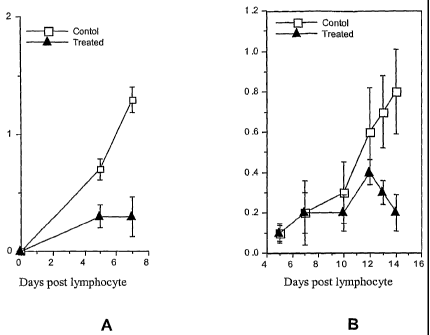
[0033] Figures 1A and 1B are graphs showing the effect of 1-(2, 4-
dimethylphenyl)-
6-phosphono-mannoside delivered at a dose of 25 mg/kg/day (Fig. 1A) or 37
mg/kg/day (Fig.
1B) on passively transferred adjuvant-induced arthritis in rats. The ordinate
shows a disease
score (swelling in affected joints in arbitrary units).
DESCRIPTION OF THE PREFERRED EMBODIMENTS
[0034] The present inventors discovered that certain tetrahydropyrans that are
derivatives of M6P are potent inhibitors of T lymphocyte migration and are
therefore useful
for treating and ameliorating diseases associated with undesired migration of
T cells to
tissues or other extravascular sites where they mediate immune and
inflammatory responses
- 11-
CA 02525328 2011-09-22
associated with autoimmune diseases. These compounds and their uses are
described and
exemplified in detail below.
Chemical Structures
[0035] The central chemical entity upon which the novel compounds of the
present
are based is shown in Formula I, below:
OH
Off
HO~P )
O O(CH2),R
He OH
Formula
OH
In the preferred compounds of this invention, n is an integer, preferably from
0 to 3, the -
O(CH2)õR group is in an axial or equatorial position, and the substituent R is
an optionally
substituted aryl or heteroaryl, for example an aralkyl or heteroaralkyl. This
genus of
compounds is referred to as the
-11a-
CA 02525328 2005-11-09
WO 2004/104015 PCT/US2004/015876
"Formula I compounds". Also included are salts, derivatives and prodrugs of
the above Formula I
compounds.
[0036] The present invention includes various subgenuses of the Formula I
compounds,
which are listed below:
(1) Formula I, wherein, when n=0, R is not 4-aminophenyl;
(2) Formula I, wherein, when n=0, R is not an amino substituted phenyl;
(3) Formula I, wherein, when n=0, R is not an amino substituted aryl;
(4) Formula I, wherein, R is not 4-aminophenyl;
(5) Formula I, wherein, R is not an amino substituted phenyl; and
(6) Formula I, wherein, R is not an amino substituted aryl;
[0037] In another embodiment, the compound of Formula I is one in which R
cannot be a
phenyl that is substituted with one or more reactive substituents that are
characterized by their
ability to participate in a nucleophilic attack on a carbonyl to form an amide
bond.
[0038] In another embodiment, the compound of Formula I is one in which the
substituent
of the C1 oxygen atom is a group that is less susceptible to the catalytic
action of a mannosidase
enzyme than is free mannose or a mannoside.
[0039] In general, the compounds of Formula I (and the above subgenuses of
Formula I) are
designed to be less susceptible to the catalytic action of a phosphatase
enzyme than is free M6P or
another naturally occurring phosphomannoside.
[0040] As used herein the term "alkyl", denotes straight chain, branched or
cyclic fully
saturated hydrocarbon residues. Unless the number of carbon atoms is specified
the term preferably
refers to C1_6 alkyl which is also referred to as "lower alkyl." When "alkyl"
groups are used in a
generic sense, e.g., "propyl," "butyl", "pentyl" and "hexyl," etc., it will be
understood that each
term may include all isomeric forms (straight, branched or cyclic) thereof. A
preferred alkyl is C1.4
alkyl; more preferred is C1_3 alkyl. Examples of straight chain and branched
C1_5 alkyl include
methyl, ethyl, n-propyl, isopropyl, n-butyl, sec-butyl, tert-butyl, n-pentyl,
iso-pentyl, 1,2-dimethyl-
propyl, 1,1-dimethylpropyl. Example of cycloalkyl groups are cyclopropyl,
cyclopropylmethyl,
cyclopropylethyl, cyclobutyl, cyclopentyl, cyclohexyl, etc.
-12-
CA 02525328 2005-11-09
WO 2004/104015 PCT/US2004/015876
[0041] An alkyl group, as defined herein, may be optionally substituted by one
or more
substituents. Suitable substituents may include: halo (fluoro, chloro, bromo
or iodo); haloalkyl
(e.g., trifluoromethyl, trichloromethyl); hydroxy; mercapto; phenyl; benzyl;
amino; alkylamino;
dialkylamino; arylamino; heteroarylamino; alkoxy (e.g., methoxy, ethoxy,
butoxy, propoxy
phenoxy; benzyloxy, etc.); thio; alkylthio (e.g., methyl thio, ethyl thio);
acyl, for example acetyl;
acyloxy, e.g., acetoxy; carboxy (-CO2H); carboxyalkyl; carboxyamide (e.g., -
CONH-alkyl, -
CON(alkyl)2, etc.); carboxyaryl and carboxyamidoaryl (e.g., CONH-aryl, -
CON(aryl)2); cyano; or
keto (where a CH2 group is replaced by C=O).
[0042] The terms "alkoxy" and "acyloxy" refer to alkyl and acyl groups
respectively when
linked by oxygen.
[0043] As used herein the term "alkenyl" denotes groups formed from straight
chain,
branched or cyclic hydrocarbon residues containing at least one C=C double
bond including
ethylenically mono-, di- or poly-unsaturated alkyl or cycloalkyl groups as
previously defined.
Thus, cycloalkenyls are also intended. Unless the number of carbon atoms is
specified, alkenyl
preferably refers to C2.20 alkenyl. More preferred are lower alkenyls (C2.6),
preferably C2.5, more
preferably C2_4 or C2.3. Examples of alkenyl and cycloalkenyl include ethenyl,
propenyl, 1-
methylvinyl, butenyl, iso-butenyl, 3-methyl-2-butenyl, 1-pentenyl,
cyclopentenyl, 1-methyl-
cyclopentenyl, 1-hexenyl, 3-hexenyl, cyclohexenyl, 1-heptenyl, 3-heptenyl, 1-
octenyl, cyclooctenyl,
1-nonenyl, 2-nonenyl, 3-nonenyl, 1-decenyl, 3-decenyl, 1,3-butadienyl, 1,4-
pentadienyl, 1,3-
cyclopentadienyl, 1,3-hexadienyl, 1,4-hexadienyl, 1,3-cyclohexadienyl, 1,4-
cyclohexadienyl, 1,3-
cycloheptadienyl, 1,3,5-cycloheptatrienyl and 1,3,5,7-cyclooctatetraenyl.
Preferred alkenyls are
straight chain or branched. As defined herein, an alkenyl group may optionally
be substituted by
the optional substituents described above for substituted alkyls.
[0044] As used herein the term "alkynyl" denotes groups formed from straight
chain,
branched or cyclic hydrocarbon residues containing at least one C=C triple
bond including
ethynically mono-, di- or poly- unsaturated alkyl or cycloalkyl groups as
previously defined.
Unless the number of carbon atoms is specified, the term refers to C2_20
alkynyl. More preferred are
lower alkynyls (C2.6), preferably C2.5, more preferably C2.4 or C2.3 alkynyl.
Examples include
ethynyl, 1-propynyl, 2-propynyl, butynyl (including isomers), and pentynyl
(including isomers). A
particularly preferred alkynyl is a C2_6 alkynyl. Preferred alkynyls are
straight chain or branched
-13-
CA 02525328 2005-11-09
WO 2004/104015 PCT/US2004/015876
alkynyls. As defined herein, an alkynyl may optionally be substituted by the
optional substituents
described above for alkyl.
[0045] The term "acyl" denotes straight chain or branched alkanoyl
(C(O)alkyl), alkenoyl
(C(O)alkenyl) or alkynoyl (C(O)alkynyl). Preferred alkanoyls are ethanoyl
(=acetyl), propanoyl, n-
butanoyl, 2-methylpropanoyl, pentanoyl, 2,2-dimethylpropanoyl, hexanoyl,
heptanoyl, octanoyl,
nonanoyl, decanoyl, undecanoyl, dodecanoyl, tridecanoyl, tetradecanoyl,
pentadecanoyl,
hexadecanoyl, heptadecanoyl, octadecanoyl, nonadecanoyl, icosanoyl. Examples
of alkenoyls are
propenoyl, butenoyl, pentenoyl, palmitoyl, oleoyl and lineoyl. The hydrocarbon
chain of an acyl
may optionally be further substituted by one or more substituents as described
above, so that "acyl"
is also intended to refer to a substituted acyl.
[0046] The term "aryl" denotes a single, polynuclear, conjugated or fused
residue of an
aromatic hydrocarbon ring system. Examples of aryl are phenyl, biphenyl and
naphthyl. An aryl
group may be optionally substituted by one or more substituents as
herein'defined. Accordingly,
"aryl" as used herein also refers to a substituted aryl.
[0047] The term "heteroaryl" denotes a single, polynuclear, conjugated or
fused aromatic
heterocyclic ring system, wherein one or more carbon atoms of a cyclic
hydrocarbon residue is
substituted with a heteroatom to provide a heterocyclic aromatic residue.
Where two or more
carbon atoms are replaced, the replacing atoms may be two or more of the same
heteroatom or two
different heteroatoms. Suitable heteroatoms include 0, N, S and Se. Examples
of heteroaryls
include pyridyl, 4-phenylpyridyl, 3-phenylpyridyl, thienyl, furyl, pyrrolyl,
indolyl, imidazolyl,
oxazolyl, pyridazinyl, pyrazolyl, pyrazinyl, thiazolyl, pyimidinyl,
quinolinyl, isoquinolinyl,
benzofuranyl, benzothienyl, purinyl, quinazolinyl, phenazinyl, acridinyl,
benoxazolyl,
benzothiazolyl and the like. As defined herein, a heteroaryl group may be
optionally further
substituted by one or more substituents as described above.
[0048] As used herein the term "aralkyl" denotes the group --Ar--R', wherein
Ar is an aryl
group and R' is lower alkyl or substituted lower alkyl group. Aryl groups can
optionally be
substituted at other positions with, e.g., halo, lower alkyl, alkoxy,
alkylthio, lower alkenyl, lower
alkynyl, amino, amido, carboxyl, hydroxyl, aryl, aryloxy, heterocycle,
substituted heterocycle,
heteroaryl, substituted heteroaryl, nitro, cyano, thiol, sulfamido and the
like. Examples of aralkyl
compounds include aromatic compounds having a divalent halomethyl group,
hydroxymethyl
-14-
CA 02525328 2005-11-09
WO 2004/104015 PCT/US2004/015876
group, and alkoxymethyl group. Specific examples of aralkyl compounds include
monosubstituted
2-, 3- or 4-(chloromethyl)phenyl; disubstituted 2,4- or 2,6-
bi(chloromethyl)phenyl; trisubstituted
2,4,6-tri(chloromethyl)phenyl, and other halomethyl and haloalkyl aromatic
compounds. Other
substituents that can be use in place of the chloromethyls listed above
include, for example,
hydroxymethyl, or alkoxymethyls (e.g., methyoxymethyl, ethoxymethyl, etc.).
[0049] In one preferred embodiment, R in formula I is a substituted aryl group
which is
substituted by one or more alkyl, carboxy, amido or amino groups, for example,
-CH3, -CH2CH3, -
(CH2)mCO2R1, -(CH2)mCH2OR2, -(CH2)mCONHR2, -(CH2)mNHR2, -(CH2)mCONR2R3 or -
(CH2)mCON RZR3 wherein m= 0-3, R1 is H, alkyl or aryl, and wherein R2 or R3,
independently, is
H, alkyl, aryl or acyl.
[0050] Other preferred R groups in formula I include: phenyl; 2-methylphenyl;
2,4-dimethylphenyl; 2,4,6-trimethylphenyl; 2-methyl, 4-chlorophenyl;
aryloxyalkyl (e.g.,
phenoxymethyl or phenoxyethyl); benzyl; phenethyl; 2, 3 or 4-methoxyphenyl; 2,
3 or 4-
methylphenyl; 2, 3 or 4-pyridyl; 2, 4 or 5-pyrimidinyl; 2 or 3-thiophenyl;
2,4, or 5-(1,3)-oxazolyl;
2,4 or 5-(1,3)-thiazolyl; 2 or 4-imidazolyl; 3 or 5-symtriazolyl.
[0051] Carboxylic acid groups can be esterified by known means, for example,
treatment
with an appropriate alcohol under acidic conditions or by treatment with a
suitable alkyl halide. A
carboxylic acid (carboxylate) can also be reduced one oxidation level to an
aldehyde which in turn
can be reduced one more oxidation level to an alcohol. Suitable reductive
procedures are known in
the art and may include treatment with hydride reagents, such as LiAlH4,
diisobutylaluminum
hydride (DIBAL-H) , or borohydrides (such as NaBH4). Corresponding alcohols
can be alkylated
or acylated using standard procedures. Suitable alkylating agents include
alkyl halides, e.g., methyl,
ethyl and propyl chloride, bromide or iodide, and dialkyl sulfates such as
dimethyl sulfate and
diethyl sulfate. Suitable acylating agents include carboxylic acids, chlorides
and anhydrides.
[0052] Carboxylic acids may be converted to amides by treatment with a
suitable amine in
the presence of a catalyst or coupling agent such as dicyclohexylcarbodiimide
(DCC). Amides may
also be prepared by treating an acid chloride with a suitable amine. In turn,
an amide (or nitrile)
can be reduced with a suitable reducing agent e.g., LiAlH4a to an amine.
Acylation or alkylation of
an amine can be carried out as described above. Further methods for the
interconversion of these
groups are described in references such as Larock, RC, Comprehensive Organic
Transformations,
-15-
CA 02525328 2010-12-07
VCH Publishers, 1989, Larock, RC, Comprehensive Organic Synthesis: A Guide to
Functional
Group Preparations, Tohn Wiley and Sons Ltd., 1989; and Smith, MB et al.,
March's Advanced
Organic Chemistry: Reactions, Mechanisms, and Structure, 5th Edition, Wiley-
Interscience, 2001.
[0053] As is well-known in the art, a nitrile group is at the same oxidation
level as a
carboxylic acid or amide group and can be converted into these groups by )mown
means, for
example, by treatment with strong aqueous acid or base.
[0054] An alkylene chain can be lengthened, for example, by the Arndt-Eistert
synthesis
wherein an acid chloride is converted to a carboxylic acid with the insertion
of CR2. Thus, a
carboxylic acid group can be converted td its acid chloride derivative, for
example by treatment
with SO2C12= The acid chloride derivative can be reacted with diazomethane to
form the
diazoketone which can then be treated with Ag2/H20 or silver benzoate and
triethylamine. The
process can be repeated to further increase the length of the alkylene chain.
Altersatively, an
aldehyde (or keto) group could be subjected to Wittig-type reaction (using
e.g.,Ph3(P)=CHCO2Me)
to produce the a,[3-unsaturated ester. Hydrogenation of this double bond
yields the alkylenc chain
that has been increased in length by two car-bon atoms. In a similar manner,
other phosphoranes can
be used to generate longer (and optionally substituted, branched or
unsaturated) carbon chains.
[0055] It should be evident that chemical manipulation of a substituent at the
2-position in
the sugar backbone of Formula I may require protection of other potentially
reactive groups, such as
the bydroxy groups, in the molecule. Suitable protective groups for use under
the appropriate
conditions, as well as methods for their introduction and removal are well-
known in the art and are
described in Greene TW at al., Protective Groups in Organic Synthesis, 3'd ed,
John Wiley and Son,
1999. A further aspect of this invention are these protected M6P derivatives.
Salts, Derivatives and Prodrup-s
[0056] The term "salt, derivative or prodrug" includes any pharmaceutically
acceptable salt,
ester, solvate, hydrate or other compound which, upon administration to a
subject, is capable of
generating (either directly or indirectly) a compound as described herein.
However, it will be
appreciated that pharmaceutically "unacceptable" salts also fall within the
scope of the invention
= 16-
CA 02525328 2005-11-09
WO 2004/104015 PCT/US2004/015876
since these may be used to prepare pharmaceutically acceptable salts. Suitable
pharmaceutically
acceptable salts include, but are not limited to, salts of pharmaceutically
acceptable:
(a) inorganic acids such as hydrochloric, sulfuric, phosphoric, nitric,
carbonic, boric, sulfamic, and
hydrobromic, or
(b) organic acids such as acetic, propionic, butyric, tartaric, maleic,
hydroxymaleic, fumaric,
maleic, citric, lactic, mucic, gluconic, benzoic, succinic, oxalic,
phenylacetic, methanesulfonic,
toluenesulfonic, benezenesulfonic, salicylic sulfanilic, aspartic, glutamic,
edetic, stearic,
palmitic, oleic, lauric, pantothenic, tannic, ascorbic and valeric.
[0057] Base salts include, but are not limited to, those formed with
pharmaceutically
acceptable cations, such as sodium, potassium, lithium, calcium, magnesium,
ammonium and
alkylammonium. In particular, cationic salts are within the scope of this
invention, e.g., sodium or
potassium salts; also included are alkyl (e.g., methyl, ethyl) phosphoesters.
[0058] Basic nitrogen-containing groups may be quatemized using: (1) a lower
alkyl halide,
such as methyl, ethyl, propyl or butyl chloride, bromide or iodide; (2)
dialkyl sulfates, e.g., dimethyl
or diethyl sulfate; and others.
[0059] The compounds of the invention may be in crystalline form either as the
free
compounds or as solvates (e.g., hydrates) both of which classes are within the
scope of this
invention. Methods of solvation are routine in the art.
[0060] Any prodrug of a compound of formula I is within the scope and spirit
of the
invention. The term "pro-drug" is used in its broadest sense to encompass
those derivatives that are
converted in vivo to the compounds of the invention. Such derivatives are
readily apparent to those
skilled in the art, and include, for example, compounds in which (1) a free
hydroxy group is
converted into an ester (such as an acetate), or (2) a free amino group is
converted into an amide.
Procedures for acylating the compounds of the invention are well known in the
art and include
reaction with an appropriate carboxylic acid, anhydride or chloride in the
presence of a suitable
catalyst or base.
T Lymphocytes and their Migration
[0061] By the term "T lymphocyte" or "T cell" is intended a cell of the
lymphocyte lineage
which is thymus-derived in origin, as is well known in the art. See any
textbook of immunology,
-17-
CA 02525328 2010-12-07
for example, Abbas et al., supra; Janeway et al., supra; Roitt et al., supra;
Klein, J, supra).
As is known in the art, a variety of defined subsets of T cells exist in the
body, such as CD4+
T helper cells, CD8+ cytotoxic T cells and the like. For the purposes of the
present invention,
methods of inhibiting migration of T lymphocytes are directed to multiple T
cell subsets, with
no established preference for T cells of any given subset.
[0062] The T lymphocytes of the present invention may be derived from an
established T
cell line or clone maintained in cell culture, or way be taken from the blood,
lymph or organized
lymphatic tissue of a cell donor. By the term "organized lymphatic tissue" is
intended any tissue or
organ which contains large collections of lymphocytes, including, but not
limited to, thymus, bone
marrow, spleen, LN, gut-associated lymphatic tissue, bronchial-associated
lymphatic tissue and
skin-associated lymphatic tissue. A_preferred source of T cells for the
present invention is the site
of an ongoing antigen-specific response, such as a draining LN in a subject
immunized so,
in radermally or intracutaneously, or from the circulation of such a subject.
[0063) By the term "antigen-primed" is intended a subject to which has been
administered a
dose of the antigen of interest prior to obtaining lymphocytes. The dose,
route and timing of
antigen administration will be easily determined by one of skill in the art
without undue experimen-
tation, and includes, but is not limited to, the doses, routes and time
intervals disclosed herein.
[0064) By the term "activated T lymphocyte" or "activated T cell," as used
herein, is
intended a T cell which has been exposed to an activating agent. Preferred
activating agents for the
present invention include the specific antigen or a polyclonal activator such
as a niitogen, capable of
inducing a response by that T cell. Such responses include a large number of
intermediary metabol-
ic changes, induction of macromolecular synthesis, such as DNA, RNA or protein
synthesis, cell
division, and the like, as is well-known iti the art. Suitable non-antigen-
specific agents capable of
activating T cells are known in the art and include, but are not limited to,
mitogens (polyclonal T
cell activators) such as concanavalin A and phytoheniagglutinin. Additional
activating agents are
antibodies to T cell-surface structures, including but not limited to,
antibodies to the CD3 cell-
surface -molecule, antibodies to the CD2 cell-surface molecule, antibodies to
the CD28 cell-surface
molecule, and the natural ligands of CD2 or CD28. Other activating agents
include phorbol esters,
such as phorbol myristate acetate, or a combination of a phorbol ester and a
calcium ionophore,
such as ionornycin. Also intended as T cell activating agents are antibodies
to T cell receptor
-18-
CA 02525328 2005-11-09
WO 2004/104015 PCT/US2004/015876
chains, specific for either the constant or the variable portions of those
chains. Any activation of T
lymphocytes in vitro may or may not include the addition of T cell growth
factors or stimulatory
factors, such as ILl, IL2 or IL4, etc., to the culture medium for part or all
of the activation interval.
Activation, among other effects, produces changes in T cell membrane
components, modifies T cell
traffic in the body, and induces expression of enzymes that may affect the way
in which T cells exit
from blood and transit through tissues, as is described in more detail below.
[0065] As noted in the Background section, above, T lymphocytes are known to
migrate
from the blood stream into tissues, including tissues in which an antigen is
present for which
antigen the cells are specific and to which they can respond. One way to
assess T lymphocyte
migration is to label T lymphocytes (or a broader population of lymphoid or
blood cells that
includes T lymphocytes, preferably enriched to at least 80%, more preferably
at least 90% T
lymphocytes). The numbers or percentages of T lymphocytes are assessed using
routine methods,
for example using antibodies specific for T cell markers, preferably CD3) in
serologic
(immunoassay), flow cytometric or other immunofluorescence-based methods, or
immunochemical
techniques.
[0066] The labeled cells are administered by injection or infusion, preferably
intravenously
(iv), into a subject, and their presence in a selected site, tissue or organ
is determined by subjecting
the desired tissue to an appropriate detection method in vivo or ex vivo.
[0067] Many detectable labels are well known for use herein. General classes
of labels
which can be used in evaluating agents that are useful for the present
invention include radioactive
isotopes, paramagnetic isotopes, and compounds which can be imaged by positron
emission
tomography (PET), fluorescent or colored compounds, etc. Suitable detectable
labels include
radioactive, fluorescent, fluorogenic, or chromogenic labels.
[0068] Useful radiolabels (radionuclides), which are detected by measuring
radioactivity in
a gamma counter, scintillation counter, by autoradiography, etc., include 3H,
14C, 35S, 51 Cr, 1251 and
131I. Other useful radionuclides are 99Tc, 111In 97Ru 67Cu 67Ga 68Ga 72As
89Zr, 90Y and 201T1. If
whole tissue counting is to be used, a preferred radionuclide is one that is
not reutilized once it has
been lost from the interior of a cell into which it was originally
incorporated. That property permits
a more direct relationship to be described between the amount of radioactivity
(counts/min. or
CPM) at a selected site or tissue, and the number of cells that have migrated
to that location. 51Cr
-19-
CA 02525328 2005-11-09
WO 2004/104015 PCT/US2004/015876
(in the form of (Na251CrO4) is particularly preferred. In another embodiment,
cells may be labeled
with 1251-iododeoxyuridine which is taken up by cells and may be incorporated
into their DNA.
[0069] Common fluorescent labels include fluorescein, rhodamine, dansyl,
phycoerythrin,
phycocyanin, allophycocyanin, o-phthaldehyde and fluorescamine. The
fluorophore, such as the
dansyl group, must be excited by light of a particular wavelength to
fluoresce. See, for example,
Haugland, RP Handbook of Fluorescent Probes and Research Chemicals, Sixth Ed.
(or later),
Molecular Probes, Eugene, OR., 1996).
[0070] In situ detection of the detectable label may be accomplished by
removing a histo-
logical specimen from a subject and examining it by microscopy under
appropriate conditions to
detect the label. Those of ordinary skill will readily appreciate that any of
a wide variety of histo-
logical methods (such as staining procedures) can be modified in order to
achieve such in situ
detection.
[0071] Thus, in a preferred embodiment, T cell migration is evaluated after iv
injection of
labeled, preferably 51Cr-labeled, antigen-specific T cells into a mammal which
has the antigen
present at one or more discrete sites that can be sampled. For example, it is
possible to assess the
degree to which the injected T cells have accumulated in an antigen-containing
tissue by measuring
the amount of radioactivity present in the tissue or analyzing the tissue
histologically to determine
the number of infiltrating labeled cells (e.g., by autoradiography).
[0072] A migration-inhibitory agent such as a composition of the present
invention, or a
candidate agent, is tested for its ability to inhibit such T cell migration
using any known or yet to be
developed assay.
[0073] In one embodiment, animals are immunized with an antigen to stimulate
antigen-
specific T cells. The antigen is preferably one that is associated with an
immunopathological
condition, such as an autoimmune disease, and may be a self antigen
(autoantigen), a foreign
antigen that cross reacts with or mimics a self antigen (i.e., a case of
antigenic mimicry).
[0074] In a preferred embodiment, this "immunization" is accompanied by the
admin-
istration of a potent immunological adjuvant. For an extensive description of
adjuvants, see, for
example, A Compendium of Vaccine Adjuvants and Excipients (2nd Edition),
Vogel, FR et al.,
available from the NIAID web site
(niaid.nih.gov/daids/vaccine/pdf/compendium.pdf); see also
Gregoriades, G et al., Immunological Adjuvants and Vaccines, Plenum Press, New
York, 1989;
-20-
CA 02525328 2005-11-09
WO 2004/104015 PCT/US2004/015876
Bennett, B et al., J. Immunol. Meth. 153:31-40 (1992). Examples of adjuvants
include Complete
Freund's Adjuvant (CFA), a mineral oil adjuvant employing a water-in-oil
emulsion It contains
paraffin oil, killed mycobacteria and mannide monoosleate. Incomplete Freund's
Adjuvant (IFA) is
a mineral oil adjuvant similar to CFA but without the mycobacteria. Montanide
ISA (incomplete
seppic adjuvant) is a mineral oil adjuvant that uses mannide oleate as the
major surfactant
component. The Ribi Adjuvant SystemTM (RAS) is an oil-in-water emulsion that
contains
detoxified endotoxin and mycobacterial cell wall components in 2% squalene.
Multiple
formulations are commercially available. TiterMax is a water-in-oil emulsion
that combines a
synthetic adjuvant and microparticulate silica with the metabolizable oil
squalene. The copolymer is
the immunomodulator component. Antigen is bound to the copolymer and presented
to the immune
cells in a highly concentrated form. Syntex Adjuvant Formulation (SAFTM) is a
preformed oil
(squalene)-in-water emulsion that uses a block copolymer for a surfactant. A
muramyl dipeptide
derivative is the immunostimulatory component. Aluminum salt adjuvants are
generally weaker
adjuvants than emulsion adjuvants and are therefore used with more strongly
immunogenic
antigens. In a nitrocellulose-adsorbed antigen, the nitrocellulose is
basically inert while slow
degradation of nitrocellulose paper allows prolonged release of antigen.
Encapsulated or entrapped
antigens permit prolonged release of antigen over time. Immune-stimulating
complexes (ISCOMs)
are antigen modified saponin/cholesterol micelles. Stable structures are
formed which rapidly
migrate to draining lymph nodes. Both cell-mediated and humoral immune
responses are induced.
Examples are Quil A and QS-21.
[0075] At an appropriate time after immunization, for example 7- 14 days, T
cells are
harvested from the animal. Any lymphatic organ or tissue, or a body fluid rich
in lymphocytes such
as blood or lymph, may be the source of these T cells. Preferred sources are
the spleen or, more
preferably, draining LNs that drain the immunization site. Lymphocytes are
harvested from these
sources, and T cells may be further isolated or enriched from these lymphocyte
populations using
conventional T cell enrichment methods.
[0076] These T cells (or T cell enriched lymphocyte populations) are then
detectably labeled
(using a label described above or any other appropriate detectable label known
in the art. The
labeled cells are then introduced into a naive recipient animal. Preferably,
inbred mice or rats are
used and the recipient and donor are syngeneic or at least matched at the MHC
so that the infused
-21-
CA 02525328 2005-11-09
WO 2004/104015 PCT/US2004/015876
cells are histocompatible with the recipient. The labeled T cells are
preferably injected or infused
systemically, preferably iv or ip, that they may circulate and migrate into a
target tissue. The target
tissue is one that either naturally expresses the antigen, e.g., a self
antigen, against which these T
cells are specific. Alternatively, a foreign antigen may be provided to the
recipient animals via
local or regional administration. In either case, a proportion of the infused
or injected labeled T
cells will migrate to and accumulate in the site or tissue or organ in which
the antigen is present and
expressed in a form recognizable by the T cells.
[0077] For example, in the case of diseases of the central nervous system
(CNS) involving a
CNS antigen, or their animal models, such as MS and EAE, myelin basic protein
(MBP) is a
disease-associated antigen. In MS or EAE, T cells localize in the brain and
spinal cord, often as
extravasated collections of cells in the form of perivascular cuffs.
[0078] To test inhibitors of T cell migration in the setting of these CNS
diseases, it is
desirable to have a stable reproducible measure of T lymphocyte movement into
and through brains.
The number or proportion of labeled cells at a selected site can be determined
by the appropriate
detection method, as discussed above. This can also be done in a visual form,
e.g., by histochemical
or other histological methods.
[0079] As noted above, when testing a migration inhibitory agent in a system
based upon a
foreign antigen, a depot or site of antigen accumulation is created as part of
the test. Preferred
routes of administering the antigen is s.c., intradermal or topical (as with a
reactive hapten such as
picryl chloride, picryl sulfonic acid, or fluorodinitrobenzene, or
dinitrobenzene sulfonic acid. A
certain number of T cells will migrate to the site of antigen or to a draining
LN if sufficient time has
elapsed for some of the antigen to reach the draining LN.
[0080] One approach, exemplified herein, is induction of a cell-mediated
immune reaction
of the type that was once classified as a "type IV hypersensitivity" reaction.
[0081] The foregoing methods are recognized and well understood by those
skilled in the art
and the underlying principles can be found in any immunology textbook such as
those cited above.
Here donor animals are sensitized to a self protein (in practice, skin is the
easiest tissue to use) by
chemically modifying the protein(s) in this tissue thereby allowing the
chemical moiety to be `seen'
as `foreign' tissue by the immune system. This is done by reacting the skin
("painting") with a
`hapten' which is usually an alkylating or arylating agent that reacts
covalently with and thereby
-22-
CA 02525328 2005-11-09
WO 2004/104015 PCT/US2004/015876
modifies protein(s) in the skin. Seven to ten days following sensitization,
spleens or draining LNs
are taken from the donor animals. T lymphocytes are isolated, radio- or
fluorescently-labeled and
transferred into naive recipient animals by intravenous injection. Prior to
cell transfer the recipient
animals have had a portion of skin, for simplicity's sake usually the ear
pinna, painted with the
same reactive hapten. Within a short time of cell transfer sensitized T cells
begin to accumulate in
the hapten-modified tissue, 8-24 hours later the tissue can be removed and the
cell accumulation
assessed. In the case of fluorescently-labeled cells, their accumulation is
assessed histologically or
histochemically, and in the case of radiolabeled cells, accumulation is
assessed by counting
radioactive decay in a suitable device. Typically, the agents of the present
invention can inhibit T
cell accumulation in this model by between about 20% and 85%. In this model,
it preferred to
utilize a relatively pure or' enriched T lymphocyte population because the
inhibitory compositions of
the present invention are not expected to interfere with B lymphocyte
migration.
[0082] Vascular endothelial cells (VECs) grown in culture will attain
confluence and
deposit an endothelial subcellular matrix. The cells and matrix are akin to
the same components
found in blood vessels in vivo (Jaffe, EA et al.,. J. Clin. Invest. 52:2745-
2756 (1973)). Activated T
cells migrate through this matrix in the same fashion as they do through
tissues in the body. This
migration can be studied and put into practice by growing VECs on special
devices that contain a
fenestrated barrier between two chambers through which cells can move. When
VEC are cultured
in the upper chamber of such a device, they will grow to confluence and
deposit a subcellular
matrix over the fenestrated barrier. If activated T cells are suspended above
the VEC layer in this
chamber, they will migrate through it and degrade the subcellular matrix, and
migrate further
through the fenestrations into the lower chamber where they can be observed,
counted, etc. The
efficacy of agents that can inhibit the ability of T cells to migrate through
this matrix can thus be
determined by placing the agent in either or both chambers during the culture
period when the T
cells are present. Efficacy of the agent is quantified by comparing the number
of T cells in the
lower chambers (i.e., migrated cells) of the agent-treated group versus the
number of T cells in the
lower chamber of the control device (no agent or a negative control agent).
This method is
commonly used to study cell migration through vascular endothelium and is well
known in the art.
See, for example, Poggi, A et al... Europ. J. Inmunol. 27:2345-2350 (1997);
Hauzenberger, E et
al.,. Transplantation. 69:1837-1849. (2000); Borthwick, NJ et al.,. Immunology
90:272-280 (1997);
-23-
CA 02525328 2010-12-07
Mohle, R. et al., (1997) Blood. 89:72-80 (1997); and Lou, I et al., Lab.
Invest. 79:1015-1025.
(1999).
[0083] Compounds of the present invention that inhibit T lymphocyte emigration
from
within the blood vessels into surrounding tissues are useful in the treatment
of cell-mediated
inflammatory diseases and conditions. The ability of these compounds to act in
this way may be
determined by the tests described in the Examples included hereinafter.
Nonlimiting examples of
such iu1lanzmatory diseases or conditions which may be treated by the
compounds of the present
invention include RA, MS. ADE, psoriasis, Crohn's disease, T cell-mediated
dermatitis, stromal
keratitis, uveitis, thyroiditis, sialitis and type I diabetes.
(0084] As used herein, the term "inhibit" includes its general meaning, i.e.,
stopping,
preventing, restraining, minimizing or slowing, T lymphocyte migration from
the blood into an
extra vascular site, such as surrounding tissues. The term "inhibit" is also
intended to mean
reversing the progression or severity of symptoms of a disease or disorder.
[0085] Compounds that inhibit T cell migration are therefore be useful in
"treating" cell-
mediated immune or inflammatory diseases and conditions. The term "treating"
(and is intended
to include "prevention," ` pirotection from," "suppression of or "therapy of
'of a disease or
disorder. "Prevention" generally involves administration of the present
compound or
pharmaceutical composition prior to the induction or appearance of the
disease. Thus, for example,
in the animal model, EAE, successful administration of a the therapeutic
composition prior to
injection of the encephalitogen (e.g., MEP) that induces the disease results
in "prevention" of the
disease. "Suppression" generally involves administration of the compound after
the inductive event
but prior to the clinical appearance of the disease. Again, using the EAR
example, successful
administration of a protective composition after injection of the
encephalitogen, but prior to the
appearance of neurological symptoms, comprises "suppression" of the disease.
"Therapy"
generally involves administration of the compound after the appearance of the
disease. In the EAE
example, successful administration of a composition after injection of the
encephalitogen and after
clinical signs have developed comprises "therapy" of the disease. It will be
understood that in
human medicine, it is not always possible to distinguish between "preventing"
and "suppressing"
since the ultimate inductive event or events may be unknown, latent, or the
patient is not ascertained
until well after the occurrence of the event or events. Therefore, it is
common to use the term
-24-
CA 02525328 2005-11-09
WO 2004/104015 PCT/US2004/015876
"prophylaxis" as distinct from "treatment" to encompass both "preventing" and
"suppressing" as
defined herein. The term "treatment" as used herein is meant to include
"prophylaxis." As such,
the present methods include both therapeutic and/or prophylactic
administration of the compounds
of the invention to "treat" a disease or condition.
[0086] The compounds of the invention may be used to treat humans or other
mammalian
subjects. The compounds of the invention are considered to be particularly
suitable for the treatment
of human subjects. Non-human subjects may include primates, livestock animals
(e.g., sheep,
cows, horses, goats, pigs) domestic companion animals (e.g., cats, dogs)
laboratory test animals
(e.g., mice, rats, guinea pigs, rabbits) or captive wild animals.
[0087] The compounds of the invention are administered to the subject in a
treatment-
effective or prophylaxis-effective amount. As used herein, such an "effective
amount" is intended
to include an amount that at least partially attains the desired effect, or
delays the onset of, or
inhibits the progression of, or halts or reverses altogether the onset or
progression of the particular
disease or condition being treated.
[0088] As used herein, the term "effective amount" or "effective dose" relates
to an amount
or dose of compound (or pharmaceutical composition thereof) which, when
administered according
to a desired dosing regimen, provides the desired therapeutic effect. Dosing
may occur at intervals
of minutes, hours, days, weeks, months or years or continuously over any one
of these periods.
Suitable dosages lie within the range of about 0.1 ng per kg of body weight to
1 g per kg of body
weight per dosage. The dosage is preferably in the range of 1 gg to 1 g per kg
of body weight per
dosage, such as is in the range of 1 mg to 1 g per kg of body weight per
dosage Suitably, the dosage
is in the range of 1 g to 500 g per kg of body weight per dosage, such as 1
g to 200 mg per kg of
body weight per dosage, or 1 g to 100 mg per kg of body weight per dosage.
Other suitable
dosages may be in the range of 1 mg to 250 mg per kg of body weight, including
ling to 10, 20, 50
or 100mg per kg of body weight per dosage or 10 g to 100mg per kg of body
weight per dosage.
[0089] Suitable dosage amounts and dosing regimens can be determined by a
treating health
care specialist and may depend on the particular condition being treated, the
severity of the
condition, as well as the general health, age and weight of the subject.
-25-
CA 02525328 2005-11-09
WO 2004/104015 PCT/US2004/015876
[0090] The active ingredient may be administered in a single dose or a series
of doses.
While it is possible for the active ingredient to be administered alone, it is
preferable to present it to
a subject as a composition, preferably as a pharmaceutical composition. The
formulation of such
compositions are well know to those skilled in the field. The composition may
contain any suitable
carriers, diluents or excipients. These include all conventional solvents,
dispersion media, fillers,
solid carriers, coatings, antifungal and antibacterial agents, dermal
penetration agents, surfactants,
isotonic and absorption agents and the like. It will be understood that the
compositions of the
invention may also include supplementary anti-inflammatory or other
physiologically active agents
where appropriate.
[0091] The carrier, diluent or excipient must be pharmaceutically "acceptable"
in the sense
of being compatible with the other ingredients of the composition and not
injurious to the subject.
Compositions include those suitable for oral, rectal, nasal, topical
(including buccal and sublingual),
vaginal or parenteral (including sc, intramuscular (im), intravenous (iv) and
intradermal (id) )
administration. The compositions may conveniently be presented in unit dosage
form and may be
prepared by any methods well known in the art of pharmacy. Such methods
include the step of
bringing into association the active ingredient with the carrier which
constitutes one or more
accessory ingredients. In general, the compositions are prepared by uniformly
and intimately
bringing into association the active ingredient with liquid carriers or finely
divided solid carriers or
both, and then if necessary shaping the product.
[0092] The administration of the present compounds and pharmaceutical
compositions may
employ any route and any means that achieves the necessary distribution of the
compound to
achieve its desired inhibitory effect. Systemic routes are preferred, either
oral, parenteral or both.
Local or regional administration, such as intra-articular and topical, is also
contemplated. For
example, administration maybe by injection or infusion. Preferred routes
include intravenous,
intramuscular, subcutaneous, intranasal, intrapulmonary, intraperitoneal,
intrathecal, and
intradermal. Rectal administration, e.g., by suppository. is also included.
Additionally or
alternatively, administration may be transdermal (using a patch or other
similar device), by osmotic
minipump, or by any other controlled release method or formulation, all of
which are well-known in
the art (see for example European Patent publications EP 92918, EP 0166596;
U.S. Patents No.
4,789,516, 4,806,621, 4,877,606, 4,906474, 4,925,677, 4,942,035; Hsieh, DST et
al., J. Pharm. Sci.
-26-
CA 02525328 2005-11-09
WO 2004/104015 PCT/US2004/015876
72: 17-22 (1983); Kaitsu, I et al., J. Controlled Release 6: 249-263 (1987);
Goedemoed, JH et al.,
Makromol. Chem. Macromol. Symp. 19: 341-365 (1988); Yang, MB. et al., Canc.
Res. 49:5103-
5107 (1989); Greig, N. et al., J Controlled Release 11:61-78 (1990); Jeyanthi,
R et al., J
Controlled Release 13:91-98 (1990); Saltzman, WM et al., Polymer Preprints 31-
1: 2456 (1990).
The dosage administered will be dependent upon the age, health, and weight of
the recipient, kind
of concurrent treatment, if any, frequency of treatment, and the nature of the
effect desired.
[0093] Compositions of the present invention suitable for oral administration
may be
presented as discrete units such as capsules, sachets or tablets each
containing a predetermined
amount of the active ingredient; as a powder or granules; as a solution or a
suspension in an
aqueous or non-aqueous liquid; or as an oil-in-water liquid emulsion or a
water-in-oil liquid
emulsion. The active ingredient may also be presented as a bolus, electuary or
paste.
[0094] A tablet may be made by compression or molding, optionally with one or
more
accessory ingredients. Compressed tablets may be prepared by compressing in a
suitable machine
the active ingredient in a free-flowing form such as a powder or granules,
optionally mixed with a
binder (e.g., inert diluent, preservative disintegrant (e.g., sodium starch
glycolate, cross-linked
polyvinyl pyrrolidone, cross-linked sodium carboxymethyl cellulose) surface-
active or dispersing
agent. Molded tablets may be made by molding in a suitable machine a mixture
of the powdered
compound moistened with an inert liquid diluent. The tablets may optionally be
coated or scored
and may be formulated so as to provide slow or controlled release of the
active ingredient therein
using, for example, hydroxypropylmethyl cellulose in varying proportions to
provide the desired
release profile. Tablets may optionally be provided with an enteric coating,
to provide release in
parts of the gut other than the stomach.
[0095] Compositions suitable for topical administration in the mouth include
lozenges
comprising the active ingredient in a flavored base, usually sucrose and
acacia or tragacanth gum;
pastilles comprising the active ingredient in an inert basis such as gelatin
and glycerin, or sucrose
and acacia gum; and mouthwashes comprising the active ingredient in a suitable
liquid carrier.
Compositions for topical administration, for example, dermally, may be in the
form of lotions,
creams, pastes, gels, ointments and the like
[0096] Compositions for rectal administration may be presented as a
suppository with a
suitable base comprising, for example, cocoa butter, glycerin, gelatin or
polyethylene glycol.
-27-
CA 02525328 2005-11-09
WO 2004/104015 PCT/US2004/015876
[0097] Compositions suitable for vaginal administration may be presented as
pessaries,
tampons, creams, gels, pastes, foams or spray formulations containing in
addition to the active
ingredient such carriers as are known in the art to be appropriate.
[0100] Compositions suitable for parenteral administration include aqueous and
non-
aqueous isotonic sterile injection solutions which may contain anti-oxidants,
buffers, bactericides
and solutes which render the composition isotonic with the blood of the
intended recipient; and
aqueous and non-aqueous sterile suspensions which may include suspending
agents and thickening
agents. The compositions may be presented in unit-dose or multi-dose sealed
containers, for
example, ampoules and vials, and may be stored in a freeze-dried (lyophilized)
condition requiring
only the addition of the sterile liquid carrier, for example water for
injections, immediately prior to
use. Extemporaneous injection solutions and suspensions may be prepared from
sterile powders,
granules and tablets of the kind previously described.
[0101] Preferred unit dosage compositions are those containing a daily dose or
unit, daily
sub-dose, as herein above described, or an appropriate fraction thereof, of
the active ingredient.
[0102] It should be understood that in addition to the active ingredients, the
compounds of
this invention, described above, the present pharmaceutical compositions of
this invention may
include other agents conventional in the art for use in the type of
composition in question, for
example, those suitable for oral administration may include such further
agents as binders,
sweeteners, thickeners, flavoring agents disintegrating agents, coating
agents, preservatives,
lubricants and/or time delay agents. Suitable sweeteners include sucrose,
lactose, glucose,
aspartame or saccharine. Suitable disintegrating agents include corn starch,
methylcellulose,
polyvinylpyrrolidone, xanthan gum, bentonite, alginic acid or agar. Suitable
flavoring agents
include peppermint oil, oil of wintergreen, cherry, orange or raspberry
flavoring. Suitable coating
agents include polymers or copolymers of acrylic acid and/or methacrylic acid
and/or their esters,
waxes, fatty alcohols, zein, shellac or gluten. Suitable preservatives include
sodium benzoate,
vitamin E, alpha-tocopherol, ascorbic acid, methyl paraben, propyl paraben or
sodium bisulfite.
Suitable lubricants include magnesium stearate, stearic acid, sodium oleate,
sodium chloride or talc.
Suitable time delay agents include glyceryl monostearate or glyceryl di-
stearate.
[0103] The compounds of the invention may also be presented for use in
veterinary
compositions. These may be prepared by any suitable means known in the art.
Examples of such
-28-
CA 02525328 2005-11-09
WO 2004/104015 PCT/US2004/015876
compositions include those adapted for: (a) parenteral administration, e.g.,
sc, im, or iv injection as
a sterile solution or suspension; (b) oral administration, external
application (e.g., drenches
including aqueous and non-aqueous solutions or suspensions), tablets, boluses,
powders, granules,
pellets for admixture with feedstuffs, pastes for application to the tongue ;
(c) topical application
e.g., creams, ointments, gels, lotions, etc.
[0104] Those skilled in the art will appreciate that the invention described
herein is
susceptible to variations and modifications other than those specifically
described. It is to be
understood that the invention includes all such variations and modifications
which fall within the
spirit and scope. The invention also includes all of the steps, features,
compositions and compounds
referred to or indicated in this specification, individually or collectively,
and any and all
combinations of any two or more of said steps or features.
[0105] Having now generally described the invention, the same will be more
readily
understood through reference to the following examples which are provided by
way of illustration,
and are not intended to be limiting of the present invention, unless
specified.
[0106] In the following examples temperatures were measured in degrees Celsius
( C)and
thin layer chromatograms (tlc) were determined on silica gel plates and unless
otherwise specified
chemical reagents were purchased from Aldrich.
EXAMPLE I
Preparation of Chemical Compositions
[0107] Preparation of 1-(2,4-dimethyl phenyl)-6-phosphono-mannoside
(Formula I; R=2,4-dimethylbenzene, n=0)
[0108] Preparation of 1-methyl-2,3,4,6-tetrabenzyl-mannoside.
Ph
O ,OCH3
O
Ph~~O 0
rO
Ph
Ph
-29-
CA 02525328 2005-11-09
WO 2004/104015 PCT/US2004/015876
[0109] To a stirred solution of 1-methyl mannoside (64.3g, 270mmol) in DMF
(1200m1)
was slowly added sodium hydride (-6eq., 58g) and stirred for -lhr. Tetrabutyl
ammonium iodide
(-0.leq., 10.6g) was then added followed by benzyl chloride (12eq., 373g,
410ml) and the mixture
stirred overnight at room temperature. The reaction mixture was then poured
into concentrated
ammonia (600m1) and stirred for -6hr after which it was extracted with diethyl
ether/light
petroleum (1:1, 2 x 1000ml). The combined organic layers were washed
successively with water (4
x 1000ml), hydrochloric acid (1000ml) and water (2 x 1000ml). The organic
layer was dried
(Na2SO4), filtered and the solvent removed (rotary evaporator). The residue
was taken up in light
petroleum (800ml), - 120g of silica gel was added and this poured onto a
column of silica gel
(250g) under vacuum. The column was then eluted successively with 100% light
petroleum
(1600m1, fl [800m1], f2[800m1]), 5% ethyl acetate/light petroleum (800m1,
f3[800m1]), 10% ethyl
acetate/light petroleum (800ml, f4), 25% ethyl acetate/light petroleum (800m1,
f5) 50% ethyl
acetate/light petroleum (800m1, f6) and 10% methanol/dichloromethane (800ml,
f7). The title
product was found in f4-7 (130.09g, 71%). MS: [M+H]+ (caic.) = 555.67,
[M+H] + (exper.) = 555.67
[0110] Preparation of 1,6-diacetyl-2,3,4, tribenzyl-mannoside.
O OAc
AcO
Phi 10 0
O
~Ph
Ph
[0111] To a stirred solution of 1-methyl-2,3,4,6-tetrabenzyl-mannoside
(85.37g, 0.154mol)
in acetic acid (500m1) and acetic anhydride (100ml) at 0 C under nitrogen was
added concentrated
sulfuric acid (2.5m1). The reaction mixture was allowed to slowly warm to room
temperature and
left overnight. The reaction mixture was poured onto water (1000ml) and
extracted with diethyl
ether (3 x 500m1). The combined organic layers were washed successively with
water (4 x 600ml),
sodium bicarbonate solution (2 x 500ml, 400ml of a saturated solution diluted
to 1000ml), water
(1000ml), brine (500m1), dried (Na2SO4), filtered and the solvent removed. The
residue was taken
up in light petroleum (400m1), - 50g of silica gel was added and this poured
onto a column of silica
gel (150g) under vacuum. The column was then eluted with 100% light petroleum
(800ml, fl), 5%
-30-
CA 02525328 2005-11-09
WO 2004/104015 PCT/US2004/015876
ethyl acetate/light petroleum (800m1, f2), 10% ethyl acetate/light petroleum
(800m1, f3), 25% ethyl
acetate/light petroleum (800m1, f4), 50% ethyl acetate/light petroleum (800m1,
f5), 100% ethyl
acetate (800m1, f6) and 10% methanol/dichloromethane (800m1, f7). The title
product was found in
f4-6 (71.32g, 87%). [M+H]+ (calc.) = 534.59, [M+H]+ (exper.) = 534.59
[0112] Preparation of 1-trimethylsilyl-2,4-dimethyl phenol.
TMSO
azz
[0113] To a stirred solution of 2,4-dimethyl phenol (5.47g, 44.8mmol) in dry
dichloromethane (120m1) at 0 C under nitrogen was added triethylamine (1.leq.,
4.99g, 6.9ml)
followed by the slow addition of trimethyl silyl chloride (1.05eq., 5.11g,
6.00ml). The reaction was
monitored by thin layer chromatography (10% ethyl acetate in 60-80 light
petroleum) and upon
completion (-2hr) water (50m1) was added. The organic layer was washed with
water (2 x 50m1),
dried (Na2SO4), filtered and the solvent removed to give the title compound
(8.13g, 93%).
[0114] Preparation of 1-(2,4-dimethylphenyl)-6-acetyl-2,3,4, tribenzyl-
mannoside
CH3
CH3
0 0
AcO
Ph'/~0 0
rO Ph
Ph
[0115] To a stirred solution of 1,6-diacetyl-2,3,4-tribenzyl-mannoside (4.74g,
8.88mmol) in
dry dichloromethane (50m1) at 22 C under nitrogen was added 1-trimethylsilyl-
2,4-dimethyl phenol
(1.6eq, 2.78g, 14.3mmol) in dry dichloromethane (25m1) followed by the slow
addition of
trimethylsilyl triflouromethane sulfonate (0.4eq., 788mg, 0.64ml). The
reaction was monitored by
thin layer chromatography (25% ethyl acetate in 60-80 light petroleum) and
upon completion (-2hr)
saturated sodium bicarbonate solution (100m1) was added. The aqueous layer was
extracted with
-31-
CA 02525328 2005-11-09
WO 2004/104015 PCT/US2004/015876
dichloromethane (100m1). The combined organic layers were washed successively
with sodium
hydroxide (5 x 100ml), water (2 x 100ml), brine (100ml), dried (Na2SO4),
filtered and the solvent
removed. The residue was taken up in light petroleum (100ml), 20g of silica
gel was added and this
poured onto a column of silica gel (50g) under vacuum. The column was then
eluted successively
with 100% light petroleum (400m1, fl), 5% ethyl acetate/light petroleum
(800m1, f2[400m1],
f3 [400m1]), 10% ethyl acetate/light petroleum (800m1, f4), 25% ethyl
acetate/light petroleum
(400ml, f5) 50% ethyl acetate/light petroleum (400ml, f6) and 10%
methanol/dichloromethane
(400m1, f7). The title product was found in f3 (4.35g, 82%): [M+H]+ (calc.) =
596.70,
[M+H]+ (exper.) = 596.70.
[0116] Preparation of 1-(2,4-dimethylphenyl)-6-droxy-2,3,4-tribenzyl-mannoside
CH3
CH3
0 0
HO
Ph,- 0,\ 0
0
Ph
Ph
[0117] To a stirred solution of 1-(2,4-dimethylphenyl)-6-acetyl-2,3,4-
tribenzyl-mannoside
(34.4g, 57.7mmol) in dry methanol (250m1) at 22 C with a soda lime tube in
place was added
sodium methoxide (5.33g) until a slight color change occurred. The reaction
was monitored by thin
layer chromatography (25% ethyl acetate in60-80 light petroleum) and upon
completion (Nlhr) the
solvent was removed. The residue was shaken with water/dichloromethane (1:1,
500m1). The
organic layer was removed and the aqueous layer was extracted with
dichloromethane (250ml).
The combined organic layers were washed with water (250m1), dried (Na2SO4),
filtered and the
solvent removed. The residue was taken up in light petroleum (200m1), 50g of
silica gel was added
and this poured onto a column of silica gel (100g) under vacuum. The column
was then eluted
successively with 100% light petroleum (800m1, fl), 5% ethyl acetate/light
petroleum (800ml, f2),
10% ethyl acetate/light petroleum (800ml, f3), 25% ethyl acetate/light
petroleum (800m1, f4) and
-32-
CA 02525328 2005-11-09
WO 2004/104015 PCT/US2004/015876
10% methanol/dichloromethane (800m1, f5). The title product was found in f3,4
(22.63 g, 82%):
[M+H] + (calc.) = 554.67, [M+H]+ (exper.) = 554.67.
[0118] Preparation of 1 -(2 4-dirnethylphenyl -6-formyl-2 3 4-tribenzyl-
mannoside
CH3
H CH3
0 0 "
Ph0 0
r Ph
Ph
[0119] To a stirred solution of dimethyl sulfoxide (4eq., 11.4g, 10.3m1) in
dry dichloromethane
(300m1) at -60 C under nitrogen was slowly added a solution of oxalyl chloride
(1.5eq., 9.26g,
6.3m1) in dry dichloromethane (50ml). After 5 min a solution of 1-(2,4-
dimethylphenyl)-6-
hydroxy-2,3,4-tribenzyl-mannoside (20.2g, 36.4mmol) in dry dichloromethane
(120ml) was slowly
added. The reaction mixture was stirred for a further lhr at -60C after which
triethylamine (8eq,
29.4g, 40.3m1) was added. The reaction mixture allowed to warm to room
temperature, stirred for a
further lhr and then poured onto water (500m1). The aqueous layer was
extracted with
dichloromethane (2 x 500m1). The combined organic layers were washed with
water (3 x 500ml),
dried (Na2SO4), filtered and the solvent removed to give crude title compound.
The residue was
taken up in light petroleum (200m1), 30g of silica gel was added and this
poured onto a column of
silica gel (150g) under vacuum. The column was then eluted with 100% light
petroleum (800m1,
fl), 5% ethyl acetate/light petroleum (800m1, f2), 10% ethyl acetate/light
petroleum (800m1, f3),
25% ethyl acetate/light petroleum (800m1, f4), 50% ethyl acetate/light
petroleum (800m1, f5) and
10% methanol/dichloromethane (800m1, f6). The title product was found in f3,4
(19.16g, 95%):
[M+H]+ (calc.) = 552.65, [M+H]+ (exper.) = 552.65.
-33-
CA 02525328 2005-11-09
WO 2004/104015 PCT/US2004/015876
[0120] Preparation of 1-(2 4-dimethyllphenyl)-6-diisopropyl phosphono-2 3 4-
tribenzyl-
mannoside
CH3
CH3
H3C--<
CH3
p 0
,,0
H3C P 0 1
~-0
H3C PhO
0
Ph
Ph
[0121] To a stirred suspension of sodium hydride (60%, 1.leq, 1.57g) in dry
tetrahydrofuran
(100ml) at 0 C under nitrogen was added tetraisopropyl methylene diphosphonate
(1.2eq, 14.3 g,
13.3ml). After 10min a solution of 1-(2,4-dimethylphenyl)-6-aldehydo-2,3,4-
tribenzyl-mannoside
(19.16g, 34.7mmol) in dry tetrahydrofuran (100ml) was slowly added. The
reaction mixture was
allowed to warm to room temperature and after 2.5hr poured into a saturated
ammonium chloride
solution (200m1). The reaction mixture was extracted with dichloromethane
(1x400 and lx200ml).
The combined organic layers were washed with water (2 x 200m1), dried
(Na2SO4), filtered and the
solvent removed. The residue was taken up in light petroleum (300m1), 50g of
silica gel was added
and this poured onto a column of silica gel (150g) under vacuum. The column
was then eluted
successively with 100% light petroleum (800m1, fl), 10% ethyl acetate/light
petroleum (800m1, f2),
25% ethyl acetate/light petroleum (800m1, f3), 50% ethyl acetate/light
petroleum (800ml, f4) and
10% methanol/dichloromethane (800ml, f5). The title product was found in f3,4
(22.54g, 90%):
[M+H] + (calc.) = 714.82, [M+H]+ (exper.) = 714.82.
-34-
CA 02525328 2005-11-09
WO 2004/104015 PCT/US2004/015876
[0122] Preparation of 1-(2 4-dimethyllphenyl)-6-diisopropyl phosphono-
mannoside
CH3
CH3
H3C~
H3 0 CH3
~-o
P / H3C H0 OH
OH
[0123] A solution. of 1-(2,4-dimethylphenyl)-6-diisopropyl phosphono-2,3,4-
tribenzyl-
mannoside (7.11g, 10.Omml) and palladium on charcoal (10%, 5.18g) in a
ethanol, water acetic acid
mix (95: 5: 1, 100ml) was shaken under a hydrogen atmosphere ('60-65psi) in a
Parr apparatus and
monitored by thin layer chromatography (Ethyl acetate/ethanol/water, 85:10:5)
and mass
spectroscopy. Upon completion (several days) the reaction mixture was filtered
and the solvent
removed. The residue was taken up in dichloromethane (100ml), 50g of silica
gel was added and
this poured onto a column of silica gel (100g) under vacuum. The column was
then eluted with
100% dichloromethane (400m1, fl), 2.5% methanol/dichloromethane (800m1, f2),
5%
methanol/dichloromethane (800m1, f3), 10% methanol/dichloromethane (800m1,
f4), 25%
methanol/dichloromethane (400ml, f5) and 100% methanol (400m1, f6). The title
product was
found in f3,4,5 (4.1Og, 92%): [M+H] + (calc.) = 446.47, [M+H]+ (exper.) =
446.47.
[0124] Preparation of 1-(2 4-dimethylphenyl)-6-phosphono-mannoside
CH3
OH CH3
0 ~P O ,0
HO
H0~ OH
OH
[0125] To a stirred solution of 1-(2,4-dimethylphenyl)-6-diisopropyl phosphono-
mannoside
(5.74g, 12.9mmol) in dry acetonitrile (200m1) at room temperature under
nitrogen was added
triethylamine (10eq, 13.0g, 17.8m1), followed by sodium iodide (10eq, 19.3g)
and the slow addition
-35-
CA 02525328 2011-09-22
of chlorotrimethyl silane (10eq, 14.0g, 16.3m1). The reaction was stirred
overnight then filtered
TM
through a plug of celite which was washed with 2% triethylamine in
dichloromethane (200m1). The
solvent was removed, the residue was taken up in 2% triethylamine in
dichloromethane (400m1) and
washed with water (2 x 200m1). The organic layer was dried (Na2SO4), filtered
and the solvent
removed. The residue was taken up in dichloromethane/ water (1:1, 400m1),
trifluoroacetic acid
added (5m1) and the stirred mixture warmed to 55 C for lhr and the
dichloromethane allowed to
evaporate off. The solution was allowed to cool, wash with dichloromethane and
the solvent
removed to give impure title compound. The was then purified on reverse phase
silica to give pure
title compound (3.41g, 77%): [M+H]+ (calc.) = 362.31, [M+H]+ (exper.) =
362.31.
[0126] In a similar manner to the foregoing procedure and using the
appropriate reagents,
the following compounds were prepared. Compound (d) was tested and found to be
pharmaceutically effective as defined herein; compounds (a)-(c) are also be
shown to be
pharmaceutically effective.
(a) 1-(2,4,6-trimethylphenyl)-6-phosphono-manoside, [M+H]+ (calc.) = 376.34,
[M+H]+
(exper.) = 376.34.
(b) 1-(2-methyl,4-chlorophenyl)-6-phosphono-mannoside, [M+H]+ (calc.) =
382.73,
[M+H]+ (exper.) = 382.73.
(c) 1-(2-methyl,4-fluorophenyl)-6-phosphonomannoside, [M+H]+ (calc.) = 366.27,
[M+H]+ (exper.) = 366.27.
(d) 1-(2-methyl,4-trifluoromethyl)-6-phosphonomannoside, [M+H]+ (calc.) =
416.28,
[M+H]+ (exper.) = 416.28.
EXAMPLE 2
Inhibition of T-lymphocyte Migration Across Rat Brain Endothelial Cell Layer
Preparation of Brain Endothelial Cells (ECs
[0127] Vascular ECs were isolated from rat brain capillaries of 6 to 8 week
old Lewis
rats according to the method of Risau, W et at., 1990, J. Cell Biol. 110:1757-
1766. Colonies
of ECs were purified from astroglial cells and pericytes using anti-Thyl. 1-
antibody and
complement-mediated lysis to remove unwanted Thyl. I+ cells.
-36-
CA 02525328 2011-09-22
[0128] The EC colonies were grown, using conventional methods, in Dulbecco's
Modified
Eagle's Medium (DMEM)-high glucose, supplemented, with 20 % fetal calf serum
(FCS),
glutamine, pyruvate, nonessential amino acids, HEPES buffer and antibiotics
(kanamycin,
amphotericin), and heparin (all from GIBCO). Cultures were supplemented with
150 g/ml
"endothelial growth supplement" from Collaborative Biomedical Products. Upon
reaching
confluence, EC cultures were washed with 0.2% EDTA in phosphate buffered
saline (PBS),
trypsinized (Trypsin-EDTA, 0.1% final concentration), washed and re-treated
with anti-Thyl.I
antibody for 40 min, washed and incubated with complement sufficient to lyse
all Thyl.1+ cells
(Behringwerke, AG, Marburg, Germany) at room temperature.
[0129] ECs were then plated on Matrigel -coated 6.5 mm Transwells, 5 mm pore
size
(Corning Costar Corporation, Cambridge, MA). Presence of ECs and confluence of
the monolayer
were checked by measuring the electrical resistance (World Precision
Instruments, New Haven,
U.S.A., model EVOM-G) and by staining with FITC-labeled phalloidin (Sigma).
Myelin Basic Protein (MBP)SSpecific T Cell Lines
[0130] To study the effect of agents of the present invention on T lymphocyte
migration, MBP-specific T lymphocyte cell lines were generated according to
the method of
Ben-Nun et al., Europ. J. Immunol. 11:195-199 (1981)) starting with
lymphocytes from the
LNs of Lewis rats that had been immunized with MBP in complete Freund's
adjuvant (CFA).
These cells were used for migration studies during the first 4 days of their
propagation in IL-
2-containing medium, following MBP restimulation in the presence of antigen-
presenting
cells (irradiated spleen or thymus cells from normal Lewis rats).
[0131] These stimulated T lymphocytes were radiolabeled with 51Cr (Na51Cr04i
37 MBq/ml,
Amersham, UK) for 30 minutes at 37 C with occasional agitation, they were then
washed three
7'M
times with DMEM and placed into the upper chamber of the Transwells at 5x10⁵
cells in l00 1 of
"migration medium." Test and control substance were included in the upper
wells at final
concentrations of 0.1, 1 and 10 mM. Cell migration was assessed visually by
light microscopy to
gauge progress of the experiment. The experiment was terminated after 6 hours,
whereupon the
undersurface of the membrane filters were rinsed twice with 100 l of ice-cold
PBS-0.2% EDTA
and combined with the medium of the lower chamber of the well. The combined
material was
-37-
CA 02525328 2005-11-09
WO 2004/104015 PCT/US2004/015876
counted for radioactivity in a gamma counter (Packard Auto-Gamma 5650
(Downer's Grove IL,
USA) gamma counter. Each "treatment" group was run in triplicate (test agent,
control agent
(glucose 6-phosphate) and no treatment).
[0132] The results from a typical experiment are shown in Table 1, below. The
value for
the glucose 6-phosphate was not statistically different from no treatment.
Inhibition values are in
comparison with the negative control.
Table I
Test substance (10 mM) % inhibition ( SEM)
1-(2,4-dimethylphenyl)-6-phosphono-mannoside. 90 4
1-(2,4,6-trimethylphenyl)-6-phosphono-mannoside. 80 4
1-(2-methyl,4-chlorophenyl)-6-phosphono-mannoside. 65 4
1-(2-methyl,4-fluorphenyl)-6-phosphono-mannoside 53 2
I-(2-methyl,4-trifluoromethyl)-6-phosphono-mannoside 88 3
negative control - glucose-6-phosphate 0 5
[0133] The results show that the five different M6P derivatives (phosphono-
mannosides)
significantly inhibited T cell migration across a monolayer of brain-derived
ECs in vitro.
EXAMPLE 3
Inhibition of Lymphocyte Migration into Lymphoid Tissue
[0134] Lymphocytes were obtained from spleens of 6-8 week old female specific
pathogen-
free Balb/c mice (19-21 g). Single cell suspensions prepared by conventional
means (pushing
through a stainless steel screen). Red blood cells were lysed by treating with
a solution of
bicarbonate buffered ammonium chloride- EDTA (pH 7.3) and the preparation
strained through a
Falcon 2350 cell strainer. All cell preparative steps were performed on ice.
Cell were suspended in
a conventional lymphocyte culture medium (DMEM plus glucose, folic acid, L-
asparagine, sodium
bicarbonate, L-glutamine, sodium pyruvate, HEPES, 2-mercaptoethanol,
penicillin, streptomycin,
neomycin, and 2% FCS).
[0135] B lymphocytes were depleted by suspending the cells in purified rat
anti-mouse
CD45/B220 antibody (RA3-6B2 clone; PharMingen, USA) at a concentration of 4 x
107 cells/ml
and incubated on ice for 20 minutes. An equal volume of culture medium (as
above) was added and
-38-
CA 02525328 2011-09-22
WO 2004/104015 PCT/US2004/015876
the cells centrifuged (200 x g) and resuspended at a concentration of 107
cells/ml in BioMag goat
= rm
ant-rat IgG (H&L) ("GARIG") conjugated to magnetic beads (Bio Mag; Perseptive
Diagnostics,
Polysciences, Inc., USA). Following a 20 minute incubation on ice, with
agitation every 5 minutes,
cells binding to the GARIG/anti-CD451B220 antibody complex were removed using
magnetic
separation (Dynal MPC-6, Dynal, USA). This procedure was repeated 4 times,
resulting in a cell
population containing approximately 80-90% T lymphocytes (determined by FACS
analysis).
[0136] The cells were then washed in Hanks balanced salt solution (HBSS),
resuspended in
ml of HBSS, and radiolabeled with 51Cr for 30 minutes at 37 C (as described
above). The labeled
cells were washed in PBS, centrifuged (200 x g), and a portion were
resuspended at a concentration
of 3 x 10⁷ cells/ml in either PBS (negative control) or PBS containing phenyl
mannoside 6-
phosphate (25 mg/ml in PBS; positive control) or PBS containing the test
compound (25 mg/ml),
and stored on ice. Cell viability was confirmed throughout the procedures by
trypan blue exclusion.
Balb/c mice were injected intravenously (iv) into the lateral tail vein with
0.2 ml containing 6 x 10⁶
labeled lymphocytes and 0.2 ml of either PBS (negative control), 5 mg positive
control or 5 mg test
compound. All groups of mice were weight matched ((0.5 g).
[0137] Spleens from recipient mice were removed 1.5 hours after injection, and
the cell-
associated radioactivity was determined using a gamma counter (as above).
Results of inhibition of
lymphocyte migration into the spleen were expressed as a percentage reduction
in the CPM of the
spleens from treated animals compared with negative controls.
[0138] The positive control compound caused 70.5% inhibition of lymphocyte
migration
(n=4). The phosphono-mannoside compounds of the present invention and their
relative inhibition
of lymphocyte migration in vivo (compared to negative controls) are presented
in Table 2.
Table 2
Inhibition of lymphocyte migration into spleens.
Compound % Inhibition
1-(2,4; dimethyl phenyl)-6-phosphono-mannoside 82.4
1-(2,4,6-trimethyl phenyl)-6-phosphono-mannoside 85.5
1-(2-methyl,4-chlorophenyl)-6-phosphono-mannoside 45.0
1-(2-methyl,4-fluorphenyl)-6-phosphono-mannoside 68.0
1-(2-methyl,4-trifluoromethyl)-6-phosphono-mannoside 87.3
-39-
CA 02525328 2005-11-09
WO 2004/104015 PCT/US2004/015876
EXAMPLE 4
Inhibition of T Lymphocyte Migration into Extralymphatic Tissues In Vivo
[0139] Spleen cell suspensions were prepared as in Example 3. Spleen donors
were specific
pathogen-free 6-8 week old female Balb/c mice (19-21 g) which had been
sensitized one week
earlier with picryl chloride (in I% in ethanol) applied topically to a 1 cm2
shaved area of abdominal
skin.
[0140] B lymphocytes were depleted as described in Example 3. The enriched
lymphocyte
population contained approximately 85-90% T lymphocytes and <1% B cells
(determined by FACS
analysis).
[0141] The cells were labeled with 51Cr as in Example 3. Recipient Balb/c mice
were
injected iv with the labeled cells and with test or control compounds as in
Example 3. In this study,
however, one hour prior to this injection, the right ear of each mouse was
painted with picryl
chloride (0.1% in ethanol) to induce a localized antigen-specific immune
response. As is known in
the art, the interval between antigen-treatment and administration of test
agents and migrating cells
can be varied, including extensions of greater than one hour.
[0142] Between 9 and 10 hours after infusion of the radiolabeled T cells
(though this can be
varied) , the recipient mice were euthanized by C02, their right and left ear
pinnae were excised and
placed in vials for gamma counting, as described above.
[0143] Results (inhibition of lymphocyte migration into the right ear pinna)
were expressed
as a percentage reduction in the CPM of the right ear pinnae from treated
animals compared with
the right ear pinnae of the negative controls. To control for non-specific
migration of cells, the
(CPM) radioactivity in a left (nonchallenged) ear pinna was subtracted from
the CPM in the right
ear pinna of the same animal prior to determining the mean for each group.
[0144] Results from a typical experiment are shown below.
Table 3
Test substance (5 mg/mouse) Route Inhibition*
1-(2,4,-dimethyl phenyl)-6-phosphono-mannoside. IV 56.1
*Percent inhibition of T cell migration
-40-
CA 02525328 2005-11-09
WO 2004/104015 PCT/US2004/015876
EXAMPLE 5
Inhibition of Cell-Mediated Immune Disease of the Central Nervous System.
[0145] Experimental autoimmune encephalomyelitis (EAE) is an inflammatory
disease of
the central nervous system that bears similarities to multiple sclerosis (MS)
and has therefore been
used extensively as an animal model of MS. Many references to this model are
available in the
medical and scientific literature. See, for example, Paterson, PY, In Textbook
oflmmunopathology
(Miescher, PA et al., eds) Grime & Stratton, New York, 1976, especially pp.
179-213; Alvord, E.C.
Jr., In: Experimental Allergic Encephalomyelitis: A Useful Model for Multiple
Sclerosis (Alvord,
E.C., ed.), Liss, New York, 1984, pp. 1-511; Steinman, L., Scientific
American, 269:106-114
(1993). The pathology of EAE is characterized by an influx of lymphocytes and
monocytes into the
brain and spinal cord with an associated demyelination of the central nervous
system neurons
(Raine, CS et al. Lab Invest. 43:150-157 (1980); Paterson, PY et al., Immunol.
Rev. 55:89-120
(1980)) resulting in partial or complete paralysis and in severe cases death.
Neural antigen-specific
CD4+ T lymphocytes are the initiators of the response since in vivo depletion
of CD4+ T cells
inhibits induction of EAE (Waldor, MK et al., Science 227:415-417 (1985)..Only
CD4+ T cell lines
or clones can passively transfer the disease (Holda, JA et al., Europ. J.
Immunol. 12:453-455; Ben-
Nun, A et al., J. Immunol. 129:303-308). Thus, the disease may be
characterized as being T
lymphocyte-mediated and tissue-specific.
[0146] Compounds of the present invention which inhibit induction or
pathogenesis of EAE
are expected to be useful for treating MS or other cell-mediated diseases of
the CNS, both in
humans and in nonhuman mammals.
[0147] To study the effect of the present agents in vivo on a disease that
includes in its
pathogenesis the migration of T lymphocytes into a specific tissue, the brain,
experiments were
performed in the Lewis rat model of passively transferred EAE. MBP-specific T
lymphocyte cell
lines were generated from the draining LNs of Lewis rats that had been
immunized with MBP in
CFA essentially according to the method of Ben-Nun et al., 1981, supra.
[0148] Donor T lymphocytes were generated from female 10 to 12 week old Lewis
rats
weighing 150 to 200 g. These rats had been injected intradermally in each hind
footpad with an
emulsion of guinea pig MBP (gpMBP) prepared according to Deibler, GE et al.,
Prepar. Biochem.
-41-
CA 02525328 2010-12-07
2:139-165 (1972), in CFA (0.05 ml). The adjuvant emulsion was prepared by
emulsifying
equal volumes of a mixture of light mineral oil (Sigma) and a solution of the
gpMBP in
normal saline (0.5 mg/ml) and Mycobacterium butyricum (4 mg/ml; Difco). Thus,
each rat
received was 50 g of MBP and 400 gg of M. butyricum. Approximately 11 days
following
injection the rats were euthanized and the draining LNs (popliteal and
inguinal) were
removed aseptically by blunt dissection and placed into lymphocyte culture
medium as
described above. A single cell suspension was prepared from the LNs as
described above for
spleen The cells were washed twice in culture medium and any red blood cells
were lysed
with ammonium chloride as above.
[0149] These cells, at concentration of 5 x 10⁶ /ml were cultured for 72 hours
in the
presence of MBP (0.06 mg/m]) at 37 C in a humidified atmosphere containing
7.5% carbon
dioxide. The cells were collected and the lymphoblasts were isolated by
centrifugation on a Fieoll
(Ph.armaeia, Uppsala, Sweden) gradient in an identical manner to that
described by Den Nun et at
supra. The fraction containing -90% lymphoblasts was cultured further in
complete DMEM to
which was added (15% v/v) a culture supernatant containing a mixture of growth
factors
(supernatant of concanavalin A-stimulated lymphocytes), 10% fetal calf serum,
nonessential amino
acids (Bio-Lab, Jerusalem, Israel). No antigen (MBP) was added. The cells were
originally plated
in 100 mm Petri dishes at a concentration of 2 x I0$ calla/nil and replated
every 3 or 4 days. Prior to
transfer into Lewis rat recipients, the cells were restirnulated with antigen,
MBP (0.01 mg/ml), and
irradiated syngeneic thymocytes, as antigen-presenting cells, for 4 days.
These cultured, stimulated
T lymphocytes were highly encepltalitogenic: as few as 5x10⁵ cells could
induced disease in naive
Lewis rats.
[0150] In a typical experiment, a group of 5 female Lewis rats, approximately
9 weeks old,
weighing 110: 15 grams was anesthetized with diethyl ether and implanted with
mini-osmotic
pumps (Alzet 2002, Alza Corp., Palo Alto CA, USA) containing 1-(2,4; diimethyl
phenyl)-6-
phosphono-mannoside dissolved in normal saline at a concentration that
delivered the drug at a rate
of 45 nag/kg/day. A second group of 5 control animals were implanted under
anesthesia with a
similar pump containing normal saline. Just prior to recovering from the
anesthetic, each animal
received 5x10⁵ cells of the above encephalitogenic T cell line iv in 0.2 ml of
normal saline. The
results of this experiment appear in Table 4.
.42-
CA 02525328 2005-11-09
WO 2004/104015 PCT/US2004/015876
Table 4
Effect of Treatment with 1-(2,4,-dimethyl phenyl)-6-phosphono-mannoside on
Passively Transferred EAE in Lewis Rats.
Saline Mannoside
Day Clinical Scores Mean Clinical Scores Mean
(individual rats) Score (individual rats) Score
4 1.5 1.5 1.5 1.5 2 1.6 0 0 0 0 0 0
2 2 2 2 2 2 0 0 1 1 1 0.6
6 2 2 2 2 2 2 0 0 1 2 2 1
7 2 0 0 1 1 0.8 0 0 1 0 1 0.4
DI 160 40
Disease severity was scored on an arbitrary scale of severity ranging from 0
to 5 as follows: 0, asymptomatic; 1, flaccid
distal half of tail; 2, entire tail flaccid; 3, ataxia, difficulty in righting
reflex; 4, hind limb weakness; 5, hind limb paralysis.
DI = Group Disease Index (see below)
[0151] All of the animals (5/5) in the control group developed clinical
disease, whereas only
3/5 of the drug-treated animals developed disease. Moreover, in the drug-
treated group, disease
severity was lower than that observed in the controls, and disease onset was
delayed. All of the
control rats developed clinical symptoms of disease by day 4 whereas none of
the drug treated rats
had symptoms at this time.
[0152] All of the animals in the control group had maximum clinical scores for
two
consecutive days while none of the 3 drug treated animals that developed
symptoms had maximum
clinical scores for more than one day. The severity of EAE for each group of
rats was calculated as
a Disease Index (DI) which is (for those animals developing disease):
mean of daily clinical score x 100
mean day of onset*
This calculation allows a more complete assessment of the disease by
incorporating both speed of
onset and clinical severity and duration of the disease. Using this
calculation, the control group had
a DI of 160, whereas the treated group had a DI of 40.
[0153] In an identically designed experiment, rats were sacrificed on day 6
after T
lymphocyte transfer and their spinal cords were harvested for histological
assessment. Rats were
deeply anesthetized (Nembutal) and perfused with 30 ml saline followed by 60
ml 10% neutral
-43-
CA 02525328 2005-11-09
WO 2004/104015 PCT/US2004/015876
buffered formalin. Spinal cords were removed, fixed for 7 days in 10% formalin
and embedded for
sectioning. The lumbar-sacral spinal cord was transected and the halves
embedded side by side for
longitudinal sectioning. Six 5 m sections were cut at various levels through
the cord with 50 m
between levels. Sections were stained with hematoxylin and eosin and a minimum
of 30 sections
were counted at different levels in order to quantify the number of lesions.
[0154] The following results were obtained. The control group showed an
extremely heavy
lesion burden, on average approximately 15 lesions/section. In contrast,
spinal cords from the drug
treated group had an average of 3.5 lesions and no more than 8 lesions/section
in the most affected
animal.
EXAMPLE 6
Inhibition Of Cell-Mediated Immune Disease of Synovial Tissue
(Passively-Transferred Adiuvant Arthritis)
[0098] Passively transferred adjuvant arthritis (AA) is a T lymphocyte-
mediated disease in
which T cells from an animal with active arthritis are transferred to a naive
syngeneic recipient. The
naive recipient subsequently develops clinical signs of disease, including
lymphocyte migration into
the synovium with subsequent swelling of affected joints. The immunological
nature of this disease
and the dependence upon T lymphocytes has been well established for many
years. See for
example: Kayashima, K et al., 1978, J. Immunol. 120:1127-1131; Waksman, BH et
al., 1963, Int'l.
Arch. Allergy 23:129-139; Pearson, CM et al., 1964, J. Exp. Med. 120:547-560;
Whitehouse, DJ et
al.,., 1969, Nature 224:1322.
[0099] The experiment in this Example was designed to study the effect of the
agents of the
present invention in vivo on a disease that requires T lymphocyte migration
into a specific tissue
(synovium). Male DA rats, 8 to 10 week old, were immunized with three 100 l
injections of CFA
intradermally at the base of the tail. The CFA was prepared by mixing 8 mg/ml
of M. butyricum
(Difco Laboratories, USA) that had been ground to a fine powder using a mortar
and pestle, in 85%
light mineral oil (Sigma, USA) and 15% mannide monooleate (Sigma) and
emulsifying this
suspension, one part in one part saline. Thus, the final emulsion contained 4
mg/ml of M. butyricum.
[00100] Ten days after immunization, the rats were euthanized and their
spleens removed
aseptically. A single cell suspension in lymphocyte culture medium with 10%
FCS was prepared as
-44-
CA 02525328 2011-09-22
WO 2004/104015 PCTIUS2004/015876
described above and adjusted to a concentration of 2x10⁶ cells/m1. Con A was
added at a final
concentration of 2 pg/ml. The cells were cultured at 37 C in an atmosphere of
5% carbon dioxide
for 72 hours.
[00101] Groups of 4 or 5 DA rats, 6 to 8 weeks of age were anesthetized with
diethyl ether
TM
and implanted sc on the flank with Alzet 2002 ("2 week delivery") or Alzet
2001 ("1 week
delivery") mini-osmotic pumps (Aiza Corp., Palo Alto CA, USA). One treatment
group received I -
(2,4,-diimethyl phenyl)-6-phosphono-mannoside in the one week delivery Alzet
2001 mini-osmotic
pumps. The drug was delivered at a dose of 25 mg/kg/day. The control group for
this experiment
received identical mini-osmotic pumps containing normal saline. In a second
experiment another
group of DA rats received 1-(2,4,-diimethyl phenyl)-6-phosphono-mannoside in
the "2 week
delivery" Alzet 2002 mini-osmotic pumps. This drug was delivered at a dose of
37 mg/kg/day.
Control animals for this group received saline delivered in identical mini-
osmotic pumps. While the
animals were recovering from anesthesia, the above cultured lymphocytes which
had been
harvested and washed twice in HBSS were resuspended in normal saline and
injected iv (lateral tai
vein) into recipient rats, each of which received 75-90 x 106 cells in 0.5 ml.
[00102] After 5 to 8 days, a characteristic thickening and cutaneous hyperemia
of the distal
joints of the hind legs became clinically apparent in saline treated control
animals. Disease severity
was evaluated and graded in each group by daily measurement of the
mediolateral widths of both
ankle joints. The data were expressed as the mean of the change (compared with
width prior to cell
injection) in mediolateral ankle width expressed in millimeters ( standard
error of the mean).
[00103] The compounds of the present invention delayed the onset of clinical
signs and
lessened the severity of the disease. Figure 1 shows the results of a typical
experiment in which 1-
(2,4-dimethylphenyl)-6-phosphono-mannoside was delivered in the "one week
delivery" Alzet 2001
mini-osmotic pumps at a dose of 25 mg/kg/day. At the end of the one week
treatment period there
was a highly statistically significant difference in the disease status of the
treated versus the control
animals. The control animals had severe swelling in the affected joints while
the treated animals
showed little swelling at the end of the treatment period.
[00104] Figure 2 shows the results obtained of a typical experiment in which 1-
(2,4,-dimethyl
phenyl)-6-phosphono-mannoside was delivered in the "two week delivery" Alzet
2002 mini-
osmotic pumps at a dose of 37 mg/kg/day. At the end of the two week treatment
period, there was a
-45-
CA 02525328 2005-11-09
WO 2004/104015 PCT/US2004/015876
highly statistically significant difference in the disease status of the
treated versus the control
animals. The control animals had severe swelling in the affected joints while
the treated animals
showed little swelling at the end of the treatment period.
EXAMPLE 7
Effect of Phosphono-Mannoside Derivatives in Various Models of Arthritis
and Autoimmune Disease
[0155] The compounds of the present invention are administered to rodents in
several well
known animal models of arthritis and autoimmune disease. These include
adjuvant arthritis (see
Example 6 above), streptococcal cell wall arthritis, Mycoplasma arthritides
arthritis and collagen-
induced arthritis. (See, for example, Pearson, CM, Proc. Soc. Exp. Biol. Med.
91:95 (1956);
Cromartie, WJ et al., J. Exp. Med. 146:1585 (1977); Trentham, DE et al., J.
Exp. Med. 146:857
(1977); Chang, YH et al., Arthritis Rheum. 23:62 (1980)).
I. Streptococcal Cell Wall Arthritis Model
(See Schwab, JH et al., J. Immunol. 150:4151-4159 (1993))
A. Induction, measurement, and treatment of arthritis
[0156] Female Lewis rats weighing about 175g are injected intraarticularly
(i.a.) under ether
anesthesia above the calcaneus through the Achilles tendon into the tibiotalar
(ankle) joint on day 0
with 2.0 g of rhamnose equivalents (approximately 6.0 pg dry weight) of 100p
peptidoglycan-
polysaccharide from cell wall of group A Streptococci (PG-APS) suspended in 10
l of pyrogen-
free saline, as described previously (Esser, RL et al., Arthritis Rheum.
25:1402 (1985); Stimpson,
SA et al., In: Pharmacological Methods in the Control of Inflammation, Chang,
J et al., Eds. Alan
R. Liss, Inc., New York, p. 381 (1989)). Right or left joints are injected
with PG-APS in alternate
animals, and contralateral joints were injected with 10 l of pyrogen-free
saline.
[0157] The lateral diameter of the ankle joint is measured with a Fowler Ultra-
Cal II digital
caliper (Lux Scientific Instrument Corp., New York, NY). The average of three
measurements for
each joint is recorded. Results are presented as the mean SE of the increase
in joint diameter
(difference between pre- and postreactivation).
-46-
CA 02525328 2005-11-09
WO 2004/104015 PCT/US2004/015876
B. Histopathology
[0158] Rats are sacrificed and the ankle joints are removed, skinned, fixed in
formalin,
decalcified, embedded in paraffin, sectioned sagitally, and stained with
hematoxylin-eosin. The
significance of differences between groups is assessed by Student's two tail t-
test.
II. Adjuvant Arthritis (AA, Model (See, especially, Chang et al., supra)
[0159] Male Lewis rats weighing 235-250 gm are used. Freund's complete
adjuvant is
either purchased commercially or prepared by grinding powdered Mycobacterium
butyricum (10
mg; Difco Laboratories) with mineral oil (1.01 ml; Primol 355, Hampden Color
Chemical
Company). Adjuvant arthritis is produced by a single intradermal injection of
the adjuvant into the
tail or one hindpaw. The dose is about 0.5 mg heat killed Mycobacterium
tuberculosis (Mt)
suspended in 100 l IFA. The volume of the uninj ected hindpaw is measured by
the method of
Winter et al., Proc. Soc. Exp. Biol. Med. 111:544 (1962) on day 0 and 16 (with
respect to the
injection of adjuvant). The increase in the volume of the uninjected hindpaw
serves as a measure of
arthritis.
[0160] To determine the effect of a therapeutic composition, rats are treated
with either
saline or the composition each day from day -1 to day -15 (with respect to
adjuvant injection). The
initial paw volume (VI) is measured on the day of adjuvant injection. Sixteen
days later, the volume
(VF) of the uninjected hindpaw is measured. Percent inhibition is calculated
according to the
following equation:
% inhibition = 1 - VF drug - Vi drug X 100
VF control - VI control
Alternatively, severity of arthritis is assessed by scoring each paw from 0 to
4 based on degree of
swelling, erythema, and deformity of the joints. Thus the maximum possible
arthritis score is 16.
III. Collagen Type II-Induced Arthritis (CIA) Model (see Trentham et al.,
supra)
[0161] Sensitization Procedures. Collagen is dissolved in 0.1M acetic acid at
a
concentration of 1mg/ml. Equal volumes of collagen solution and CFA or IFA are
mixed and
emulsified. One ml of the cold emulsion is immediately injected intradermally
in four to six sites
on the backs of the rats. Small ulcers frequently form at the injection site,
but these heal without
-47-
CA 02525328 2011-09-22
11_
WO 2004/104015 PCT/US2004/015876
sequelae in 7-10 days. Control injections consist of (a) acetic acid
emulsified in CFA or IFA or
(b) human or chick type II collagen dissolved in acetic acid and injected
intradermally without
adjuvant. As an additional control, 1.0 ml of MgC12-extractable cartilage
proteoglycans containing
approximately 200 p.g uronate per ml is mixed with 0.5 ml of CFA or IFA,
emulsified, and injected
as with collagens. Unless otherwise specified, booster doses consisting of 0.5
mg collagen
dissolved in 0.5 ml 0.1 M acetic acid are given ip without adjuvant 21 days
after primary
immunization. One ml of the MgC12 extract is given ip after an identical
interval to the
proteoglycan control animals. Adjuvant arthritis is induced by intradermal
injection of 0.1 ml CFA
H37 at the base of the tail.
[0162] Arthritis Evaluation. Animals are observed daily for the onset of
arthritis, and an
arthritic index is derived by grading the severity of involvement of each paw
from 0 to 4. Scoring is
based on the degree of periarticular erythema and edema as well as deformity
of the joints (Wood,
FD et al., Int. Arch. Allergy Appl. Immunol. 35:456 (1969)). Swelling of
hindpaws is also
quantitated by measuring the thickness of the ankle from medial to lateral
malleolus.
IV. Autoimmune Model MRL/lpr Mice (See: Kim, C. et at, J. Exp. Med. 174:1431-
1437 (1991))
[0163] MRLJMp-lpr/lpr mice (4-6 wks old) are purchased from the Jackson
Laboratory (Bar
Harbor, ME) or other supplier.
Enzyme Immunoassay (EIA) for Anti-DNA antibodies and Immune Complexes
[0164] Polystyrene microtiter wells are coated with double-stranded DNA (ds-
DNA) or goat
Clq. Blood is obtained from individual mice before the biweekly injections.
Sera are diluted in
0.05% Tween-20 in PBS at a 1:500 dilution and allowed to incubate in the
plates for 60 min at room
temperature. The plates are then washed with PBS-Tween, and 501il of 1/1000
dilutions of goat
anti-mouse IgG and IgM antibodies conjugated to urease (or another enzyme that
is known to be
useful in EIA) are added to the plates. After incubation for 30 min., the
plates are washed three
times with PBS-Tween and twice with 0.15M NaCl. The plates are then incubated
with a solution
of the chromogenic substrate for the enzyme. Colorimetric change is quantified
by measuring
absorbance at the appropriate wavelength for the particular colored product of
the enzymatic
reaction using a microplate reader.
-48-
CA 02525328 2005-11-09
WO 2004/104015 PCT/US2004/015876
Proteinuria and Physical Symptoms
[0165] Urine is obtained from mice. Protein concentration and the presence of
blood in
urine is measured semiquantitatively by commercial reagent strips for
urinalysis.
[0166] Physical symptoms are visually scored as: 0, no symptoms; 0.5, trace; 1-
4, when
visible symptoms are observed, with 4 being the most severe (physical symptoms
include
lymphadenomegaly, immune complex vasculitis, and necrosis of the ears). Scores
representing
physical symptoms are calculated by determining the total score for each group
and then dividing
by the number of animals alive in that group when the measurement is taken.
Treatment
[0167] For each of the models described above, treatment is started 6-14 days
after the
injection of the inducing agents (or in the case of MRL/lpr mice beginning at
4 weeks of age).
Doses vary from 1 pg to 100 mg of the following compounds:
1-(2,4,-dimethyl phenyl)-6-phosphono-mannoside;
1-(2,4,6-trimethyl phenyl)-6-phosphono-mannoside;
1-(2-methyl,4-chlorophenyl)-6-phosphono-mannoside;
1-(2-methyl,4-fluorphenyl)-6-phosphono-mannoside;
1-(2-methyl,4-trifluoromethylphenyl)-6-phosphono-mannoside;
1-(2-methyl,4-trifluoromethyl -6-phosphono-mannoside
or any other of the novel T cell migration-inhibitory compounds of the present
invention.
[0168] The compounds are administered iv or ip at 1 week intervals for 4
weeks. Outcomes
are assessed as described above. For all arthritis models outcome measures
include:
(a) quantitative measurement and grading of joint swelling erythema or
deformity, and
(b) assessment of histopathology of joints using a quantitative grading
system.
[0169] In all the models described, the four compounds listed above are
effective in
significantly reducing direct or indirect measures of arthritis.
-49-
CA 02525328 2005-11-09
WO 2004/104015 PCT/US2004/015876
EXAMPLE 8
Phosphono-Mannoside Derivatives in the Treatment of Rheumatoid Arthritis in
Humans
(See: Koopman, WJ (ed) Arthritis and Allied Conditions: A Textbook of
Rheumatology, Lippincott, Williams &
Wilkins; 13th edition, 1996.)
Treatment Procedure
[00105] Doses of the compounds of this invention are determined as described
above using,
inter alia, appropriate animal models of autoimmune disease.
[00106] A treatment consists of injecting a patient with 0.1, 1, 10 or 100 mg
of the compound
iv, or infusing the compound in 100 ml of normal saline over a 30 minute
period twice weekly at
three day intervals for six weeks. Clinical responses are assessed by the
criteria described below.
Treatments are continued in patients with stable or exacerbating disease.
Treatment is generally
given on an outpatient basis.
Clinical Outcome Measures
[0170] Outcome measures used to assess treatment efficacy in RA should detect
the smallest
clinically important change and, at the same time, be reliable and valid with
respect to capturing the
dimensionality of the clinical and pathophysiologic responses. To avoid bias,
both patients and
assessors preferably are blinded during testing.
[0171] The methods most commonly used are based on quantitation of cardinal
features:
pain, swelling, heat and redness. Laboratory tests may also be used in
assessment, though a
treatment that only reduces a laboratory measure without, for example,
relieving joint pain is of less
interest. No single ideal method is known to accurately reflect disease
activity in arthritis. As a
result, it is useful to aggregate end points into a composite index. Composite
indices are
constructed by statistical or judgmental procedures that allow aggregation of
scores assigned to
different end points.
[0172] Objective and sensitive measurements are preferred to subjective ones.
One
sensitive parameter to change with antirheumatic drug therapy in RA is the
patient's subject
assessment of pain relief. Objective measurements include radionuclide joint
uptake. Others are
the 50-foot walking time and assessment of functional disability (the second
most important
symptom in osteoarthritis). Examples of useful outcome measures appear in
Table 5, below.
-50-
CA 02525328 2005-11-09
WO 2004/104015 PCT/US2004/015876
[01731 Table 5
Outcome Measures for Clinical Trials in Arthritis
Altman, R.D. et al., Clin. Rheum. Dis. 9:681-693 (1983)
FDA Guidelines (1977) Bellamy and Buchanan
Clin. Rheumatol. 3:293-305 (1984)
Joint swelling Pain
Joint redness Patient global assessment
Tenderness on pressure Range of movement
Pain at rest or on motion Physician global assessment
Range of motion Joint stiffness
50-foot walking time Qualitative aspects of sleep
Clinician's global assessment Walking time
Patient's global assessment Activities of daily living
Altman et al. (su ra) Joint tenderness
Pain (using visual analogue scales) Analgesic compound
Tenderness on pressure/motion Joint swelling
Clinician's global assessment of current status and degree of Signal joints
change in status Ascent time
Patient's global assessment of current status and degree of Muscle power**
change in status Hand function
50-foot walking time (for patients with hip/ knee involvement) Radiology
Grip strength (for patients with hand involvement) Joint temperature
[0174] Because pain is the major complaint of the rheumatic sufferer,
measurement of pain
relief is important in assessing clinical response to the therapeutic
composition or method of this
invention. Adjectival scales may be used with numeric values given to the
adjectival scale, for
example: 0=no pain, 1=slight pain, 2=moderate pain, 3=severe pain, and
4=extremely severe or
agonizing pain. Such a scale is known to discriminate between nonsteroidal
anti-inflammatory
analgesics and placebo in short-term trials (Lee, P., J. Rheumatol. 3:283-294
(1976)). Other
methods of measuring pain include assessment of pain threshold and pain
tolerance (Huskisson, EC,
Clin. Rheum. Dis. 2:37-49(1976)).
[0175] To score joint tenderness, firm digital pressure is applied to the
joint margins and the
degree of tenderness is graded by the patient's response. Lansbury's Articular
Index (Lansbury, J,
Arthritis Rheum. 1:505-522 (1958)) is useful in assessing progress. A simple
count of clinically
active joints, as determined by pain on passive motion, tenderness on
pressure, or inflammatory
joint swelling is used (Cooperat. Clin. Comm. Amer. Rheum. Assoc., Clin.
Pharmacol. Ther. 8:11-
-51-
CA 02525328 2005-11-09
WO 2004/104015 PCT/US2004/015876
38 (1967)). Scoring a few selected "signal" joints may permit better
assessment of therapeutic
effect than a total joint count. A standardized dolorimeter tested against the
Lansbury indices is
highly reproducible. The Ritchie Articular Index (RAI) is based on summation
of joint responses
after firm digital pressure. The responses are recorded as 0=no tenderness,
+1= patient says it is
tender, +2= patient says it is tender and winces, and +3= patient says it is
tender, winces, and
withdraws limb. The sum of this Index is 78 and reflects exacerbations of
disease and improvement
induced by antirheumatic drugs. This index correlates with the patient's
assessment of pain, in the
upper limbs with grip strength, and in the lower limbs with the time to walk
50 feet.
[0176] Various instruments are available to measure grip strength which is
determined by
the strength of the muscles in the forearm and hand, and the pain and degree
of joint destruction in
the wrist, hand, and finger joints; grip strength correlates with the RAI.
[0177] The range of motion of peripheral joints in normal subjects is known,
and these
measures have been assessed in studies of ankylosing spondylitis. Spinal
movement is measured by
several methods including the Dunham spondylometer (Anderson, J, Clin. Rheum.
Dis. 8:631-653
(1982)), skin distraction (Moll, J et al., Rheum. Phys. Med. 11:293-312
(1972)), an inclinometer
(Domjan, Let al., Hung. Rheum. 28 (Suppl):71-76 (1987)). Timing of certain
movements or set
maneuvers related to activities of daily living, are useful, in particular the
time to walk 50 feet (Lee,
supra; Grace, EM et al., Br. J. Rheumatol. 27:372-374 (1988)).
[0178] Increase in warmth of overlying skin is a cardinal feature of
inflammation and can be
measured in various ways (e.g., Bacon, P.A. et al., Clin. Rheum. Dis. 2:51-65
(1976)). Infrared
quantitative thermography shows reproducible changes in disease activity and
is useful in assessing
efficacy of a treatment composition or method. Thermography provides a
noninvasive,
reproducible, sensitive, and quantifiable method of assessing improvement in
joint inflammation
(Ingpen, ML, Ann. Phys. Med. 9:322-327 (1968)).
Laboratory Tests
[0179] Certain laboratory tests reflect the severity of joint inflammation and
may be, used to
monitor the efficacy of the therapeutic compositions and methods of this
invention. The most
frequently used test is the erythrocyte sedimentation rate (ESR). Other
measures used include
evaluation of various acute-phase reactants, such as C-reactive protein,
haptoglobin, fibrinogen, a-2
-52-
CA 02525328 2005-11-09
WO 2004/104015 PCT/US2004/015876
macroglobulin, and plasma viscosity (McConkey, B et al., Q. J. Med., New
Series 41:115-125
(1972); McConkey, B et al., Q.J. Med., New Series 42:785-791 (1973);
Constable, TJ et al., Lancet
1:1176-1179 (1975); Crook, L et al., Ann. Clin. Lab. Sci. 10:368-376 (1980);
Dixon, JA et al.,
Scand. J. Rheumatol. 13:39-44 (1984); Cockel, R et al., Ann. Rheum. Dis.
30:166-170 (1971)); titer
of IgM rheumatoid factor or of immune complexes (Pope, RM et al., Ann. Rheum.
Dis. 45:183-189
(1986); Reeback JS et al, Ann. Rheum. Dis. 44:79-82 (1986); Reynolds, WJ et
al., J Rheumatol.
13:700-706 (1986)); tests of lymphocyte function (Reynolds, WJ et al., J.
Rheumatol. 13:700-706
(1986); Alepa, FP et al., Arthritis Rheum. 13:754-760 (1970); Swanson, MA et
al., N. Engl. J Med.
277:163-170 (1967)); displacement of L-tryptophan from serum albumin; serum
iron concentration
(Cockel, supra), eosinophilia, thrombocytosis (Hutchinson, RM et al., Ann.
Rheum. Dis. 35:138-
142 (1976)); serum concentrations of sulfhydryl groups (Lorber, A et al.,
Metabolism 20:446-455
(1971)); serum copper concentrations (Brown, DH et al., Ann. Rheum. Dis.
38:174-176 (1979));
serum propeptide levels (Horsley-Petersen et al., Rheum. 29:592-599 (1986));
synovial fluid
analysis (Hall, SH et al., Ann. Rheum. Dis. 37:351-356 (1978)).
[0180] Various methods are used to score radiologic changes in rheumatoid
arthritis, the
most useful of which are count erosions and assessment of joint space
narrowing. Radionuclides
can also be used to quantify joint inflammation (Dick, WC, Semin. Arthritis
Rheum. 1:301-325
(1972); Wallace, DJ et al., Arthritis Rheum. 11:172-176 (1981)). These are
administered intra-
articularly and the rate of clearance from the joint determined or,
alternatively, they are
administered iv and the rate of accumulation over a joint (or joints)
measured. The clearance of
133Xe after intra-articular injection provides an indirect measurement of
synovial blood flow.
99mTcO4 is also used. Radionuclide joint uptake in both large and small joints
is known to be
reduced with successful anti-rheumatic therapeutics such as NSAIDs,
corticosteroids, gold or
D-penicillamine.
[0181] Results: 375 patients with RA are treated with one of five compounds
(75/group):
(1) 1-(2,4,-dimethyl phenyl)-6-phosphono-mannoside;
(2) 1-(2,4,6-trimethyl phenyl)-6-phosphono-mannoside;
(3) 1-(2-methyl,4-chlorophenyl)-6-phosphono-mannoside;
(4) 1-(2-methyl,4-fluorphenyl)-6-phosphono-mannoside;
(5) 1-(2-methyl,4-trifluoromethyl)-6-phosphono-mannoside;
-53-
CA 02525328 2010-12-07
[0182] According to the 8 measures listed under "FDA Guidelines" in Table 5,
above, >75%
of the patients treated with any of the above compounds show significant
cumulative improvement
across all measures.
Toxici
[0183] The incidence of side effects (as % of total treatments across groups)
are as follows:
chills - 10; fever - 10; pain - 5; nausea - 5; respiratory - 3; headache - 3;
tachycardia - 2; vomiting -
2; hypertension - 2; hypotension - 2; joint pain - 2; nish - 2; flushing - 1;
diarrhea - 1; itching/hives -
1; bloody nose - 1; dizziness - <1; cramps - <1; fatigue - <1; feeling Not -
<1; twitching - <1;
blurred vision - <l; gastritis<l; redness on hand - <1. Other minor changes
observed are clinically
insignificant
[0184) Having now fully described this invention, it will be appreciated by
those
skilled in the art that the same can be performed within a wide range of
equivalent
parameters, concentrations, and conditions without departing from the spirit
and scope of the
invention and without undue experimentation.
-54-