Note: Descriptions are shown in the official language in which they were submitted.
CA 02525769 2005-11-14
WO 2004/103456 PCT/US2004/012322
-1-
Description
NON-INVASIVE APPARATUS AND METHOD FOR PROVIDING RF
ENERGY-INDUCED LOCALIZED HYPERTHERMIA
Cross Reference to Related Replication
This application is based on and claims priority to United States Patent
Application Serial Number 10/437,838, filed May 14, 2003, herein incorporated
by reference, in its entirety.
Government Interest
This invention was made with Government support under Grant No.
5P01 CA 427 45-16 awarded by the National Institutes of Health. The
Government has certain rights in the invention.
Technical Field
Tiie present invention generally relates to inducing hyperthermia in a
desired target such as living tissue. More particularly, the present invention
relates to non-invasively causing localized hyperthermia in tissue such as
tumor-containing tissue using a phased antenna array to direct standing waves
of RF energy to the tissue. An advantageous application of the present
invention is enhancing the effects of cancer-related therapeutic procedures.
Background Art
Certain types of cancers such as breast cancers, particularly
inflammatory and locally advanced tumors, often resist traditional treatments.
It
has been statistically shown that sixty to seventy percent of victims of such
breast tumors do not survive past five years. The efficacy of conventional
methods of treating cancer, such as radiotherapy and chemotherapy, is limited
due to necessary constraints on dosage amounts for safety. For example, it is
known that chemotherapy can be applied in sufficient amounts to kill virtually
ail
cancer cells of a tumor. However, the amounts of chemotherapy needed to
achieve this can be high enough to cause poisoning of the patient and/or
undue side effects. As another example, the intensity of an x-ray beam applied
in accordance with radiotherapy cannot be so high as to damage nearby critical
organs and surrounding healthy tissues. Accordingly, there is an ongoing need
to develop techniques that enhance existing cancer-related therapeutic
SUBSTITUTE SHEET (RULE 26)
CA 02525769 2005-11-14
WO 2004/103456 PCT/US2004/012322
-2-
procedures so as to increase their effectiveness without increasing the risk
of
damage to healthy tissue and causing additional discomfort for cancer
patients.
One recent approach toward improving cancer therapy is to subject a
tumor to a hyperthermia treatment. The application of heat to cancer cells has
been found to increase the efficacy of certain types of therapies for various
proposed reasons. Microwave and radio frequency (RF) energy sources have
been employed to conduct hyperthermia treatment. Microwave energy has
been applied to tumors using waveguides. However, the relatively high
frequencies at which microwaves propagate are not suitable for deep
penetration into tissue. RF energy has also been utilized in some instances,
and has the potential to achieve greater penetration due to relatively lower
frequencies. However, both microwave and RF techniques have in the past
required the use of invasive elements, such as wires, catheters, lumens,
probes, receivers, and the like. These invasive elements are typically
inserted
or embedded in the tumor to be treated to ensure proper coupling and focusing
of the electromagnetic energy at the tumor site. The use of invasive elements
adds complexity to the procedure and is a source of discomfort for patients.
Examples of invasive heating techniques using microwave and RF energy are
disclosed in U.S. Patent Nos. 5,928,159; 6,275,738; 6,358,246; 6,391,026; and
6,468,273.
It therefore would be desirable to provide a method and apparatus for
non-invasively inducing hyperthermia in a tumor by applying electromagnetic
energy, and preferably RF energy, to the tumor in a controllable, coherent
manner, and while avoiding or reducing problems associated with conventional
techniques.
Summary of the Invention
According to one embodiment, an apparatus for providing hyperthermia
treatment for enhancing cancer therapy comprises an applicator body and a
plurality of antennas. The applicator body has a concave profile extending
from
an aperture, and defines an open cavity for receiving RF standing waves. The
antennas are operatively associated with the applicator body and are arrayed
CA 02525769 2005-11-14
WO 2004/103456 PCT/US2004/012322
-3-
for transmitting RF standing waves at respective selected amplitudes and
relative phases into the cavity and generally toward a tumor-containing tissue
disposed in operative alignment with the antennas.
According to another embodiment, a method for providing hyperthermia
treatment for enhancing cancer therapy comprises the following steps. A
tumor-containing tissue is placed in operative alignment with a phased array
of
antennas operatively associated with a body defining a cavity containing a
fluid.
RF energy is transmitted from the antennas through the fluid and to the tissue
to heat the tissue.
According to yet another embodiment, a method for providing
hyperthermia treatment to enhance tumor-related therapy comprises the
followiFng steps. A tumor-containing tissue is treated by performing a tumor-
related therapeutical procedure. The tissue is placed in operative alignment
with a phased array of antennas operatively associated with a body defining a
cavity containing a fluid. RF energy is transmitted from the antennas, through
the fluid, and into the tissue to heat the tissue.
It is therefore an object to provide an apparatus and method for inducing
localized hyperthermia by applying controlled RF energy.
An object having been stated hereinabove, and which is addressed in
whole or in part by the present invention, other objects will become evident
as
the description proceeds when taken in connection with the accompanying
drawings as best described hereinbelow.
Brief Description of the Drawings
Figure 1 A is a side elevation view of a hyperthermia treatment apparatus
according to an embodiment disclosed herein;
Figure 1 B is a top plan view of the hyperthermia treatment apparatus
illustrated in Figure 1A;
Figure 2A is a perspective view of a treatment applicator provided with
the hyperthermia treatment apparatus according to one embodiment disclosed
herein;
CA 02525769 2005-11-14
WO 2004/103456 PCT/US2004/012322
-4-
Figure 2B is a side elevation view of the treatment applicator illustrated
in Figure 2A;
Figure 2C is a top plan view of the treatment applicator illustrated in
Figure 2A;
Figure 2D is a front elevation view of the treatment applicator illustrated
in Figure 2A;
Figure 3 is an exploded perspective view of the treatment applicator
illustrated in Figure 2A and a tissue support structure provided therewith
according to one embodiment disclosed herein;
Figure 4 is an exploded perspective view of a treatment applicator
provided according to another embodiment disclosed herein, and a tissue
support structure provided therewith;
Figure 5 is a perspective view of a treatment applicator provided
according to yet another embodiment disclosed herein;
Figure 6 is a perspective view of a tissue support structure provided
according to still another embodiment disclosed herein;
Figure 7 is a partial side elevation view of a hyperthermia treatment
apparatus including the treatment applicator illustrated in Figure 2B and the
tissue support structure illustrated in Figure 6, both of which are mounted in
a
patient support structure provided therewith; and
Figure 8 is a schematic diagram of electrical circuitry provided with the
hyperthermia treatment apparatus according to embodiments disclosed herein.
Detailed Description of the Invention
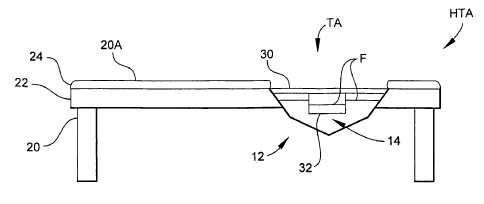
Referring now to Figures 1A and 1B, a hyperthermia treatment
apparatus, generally designated HTA, is illustrated according to one
embodiment. Hyperthermia treatment apparatus HTA primarily comprises a
treatment applicator, generally .designated TA, and associated electrical
circuitry, generally designated EC (see Figures 2C and 8, described in detail
hereinbelow). Treatment applicator TA has a body, generally designated 12,
constructed to form an open cavity 14 with which a biological target such as
tumor-afflicted tissue can be proximally disposed for exposure to RF
CA 02525769 2005-11-14
WO 2004/103456 PCT/US2004/012322
-5-
electromagnetic energy via electromagnetic coupling. In one embodiment,
body 12 is constructed from a clear polymeric material such as LEXAN~
material. The profile of body 12 can be polygonal as illustrated or can be
generally semispherical or semi-ovoid. The embodiments of hyperthermia
treatment apparatus HTA illustrated herein are particularly advantageous for
the treatment of tumors of the breast and chest wall. For this purpose,
treatment applicator TA can be mounted in a cut-out section of any suitable
patient support structure 20 (e.g., a table, bed or couch) such that its
cavity 14
opens upwards toward a top surface 20A of patient support structure 20. In
one embodiment, patient support structure 20 includes a base 22 and a
padding 24. By this configuration, a patient can lie comfortably in a prone
position on top surface 20A with the breast or chest wall to be treated
depending or facing downwardly into cavity 14.
In some embodiments, hyperthermia treatment apparatus HTA further
includes a tissue (e.g., breast or chest wall) support structure 30 that is
secured
to the top of treatment applicator TA by any suitable fastening means (not
shown) such as threaded screws, bolts and nuts, or clamps. In Figures 1A and
1 B, tissue support structure 30 includes a container 32 that extends into
cavity
14 to provide additional support for a breast. The use of tissue support
structure 30 will generally depend on breast size. Moreover, while container
32
is illustrated as being cup- or bowl-shaped, the size and shape of container
32
can generally depend on breast size and shape.
As further shown in Figures 1A and 1B, during the operation of
hyperthermia treatment apparatus HTA according to advantageous
embodiments, cavity 14 of treatment applicator TA and container 32 of tissue
support structure 30 (when used) are filled with a suitable fluid F such as
deionized water. The breast or other tissue can be immersed in fluid F during
treatment. As shown in Figure 1 B, the temperature of fluid F can be regulated
to prevent skin burns and improve patient comfort, by circulating fluid F
through
an inlet 34A and outlet 34B of cavity 14 as generally indicated by arrows and
distributing the heat evenly around the tissue. The arrows in Figure 1 B can
CA 02525769 2005-11-14
WO 2004/103456 PCT/US2004/012322
-6-
represent fluid flow through liquid conduits that communicate with any
suitable
temperature regulating system TRS. Water is useful as fluid F because its
dielectric constant is similar to that of the tissue of a patient, and thus RF
energy can be efficiently propagated and directed by treatment applicator TA
(in a manner described hereinbelow) with minimal reflected energy. The use of
water as fluid F is considered superior to air, at least in part because air
cannot
transfer heat as efficiently and its dielectric constant differs from water by
a
factor of about 10.
Referring now to Figures 2A- 2D, details of treatment applicator TA are
illustrated according to a four-antenna embodiment. Body 12 of treatment
applicator TA includes six body sections or walls defining cavity 14. In the
illustrated example, the body 12 sections include two opposing side sections
12E and 12F generally perpendicular to the plane of an aperture 36 of body 12;
two opposing side sections 12A and 12D angled relative to aperture 36; and
two angled bottom sections 12B and 12C. Aperture 36 is formed by the
respective top edges of perpendicular side sections 12E and 12F and angled
side sections 12A and 12B. Antennas ANTS -ANT4 are respectively disposed
in each of angled side sections 12A and 12B and angled bottom sections 12B
and 12C, although more or less antennas could be provided in angled side
sections 12A and 12B and angled bottom sections 12B and 12C. Antennas
ANTS - ANT4 can be secured to body 12 in any suitable manner, such as by
gluing antennas ANTS - ANT4 to the inside surfaces of angled side sections
12A and 12B and angled bottom sections 12B and 12C. Antennas ANTS -
ANT4 can have any design suitable for transmitting RF energy through a
selected fluid such as water. In the advantageous embodiments illustrated
herein, each antenna ANTS - ANT4 has a "bowtie" or "H" shape constructed
from a pair of generally C-shaped antenna elements 38A and 38B. For each
pair, antenna elements 38A and 38B (Figure 2C) are inverted with respect to
each other, with their corresponding legs opening away from each other.
Antennas ANTS - ANT4 are arrayed along angled side sections 12A and 12B
and angled bottom sections 12B and 12C to enable standing RF waves to be
CA 02525769 2005-11-14
WO 2004/103456 PCT/US2004/012322
-7-
coherently focused toward the tissue residing in or over cavity 14. Figure 2B
schematically depicts a coherent pattern of standing RF waves RF focused on
a tumor mass TM of a breast BR. As shown in Figure 2C, each antenna ANTS
-ANT4 communicates with electrical circuitry EC via respective low-loss output
cables OCR - OCa to provide RF energy as described hereinbelow.
Referring now to Figure 3, tissue support structure 30 includes a plate
42 from which container 32 extends downwardly. Plate 42 is sized to cover
cavity 14 and thus enable tissue support structure 30 to be mounted onto body
12, for example at a rim 44 thereof. Tissue support structure 30 can be
secured to body 12 by any suitable means, one example being the use of
screws (not shown) tapped through respective apertures 42A and 44A of plate
42 and rim 44, or bolts extending through apertures 42A and 44A and held by
nuts.
As further shown in Figure 3, in some embodiments, a magnetic coil
device MC can be mounted to the inside or outside of container 32 so as to
circumscribe the breast or other tissue to be treated. Magnetic coil device MC
can be coupled to any suitable magnetic resonance imaging (MRI) device MRI
to generate images of the tumor in the breast during treatment. Apart from
other known visual uses, the MRI images can be correlated to temperature,
and hence magnetic coil device MC can be used as a temperature-sensing
device. In other embodiments, a temperature-sensing device can be provided
in the form of a thermometer that is physically inserted into the breast, such
as
through a catheter as is understood by persons skilled in the art. The use of
magnetic coil device MC, however, is non-invasive and much less
discomforting for the patient undergoing treatment.
Referring now to Figure 4, treatment applicator TA is illustrated
according to a five-antenna embodiment. Body 52 of treatment applicator TA
includes five body sections or walls 52A - 52E defining cavity 14. In the
illustrated example, the body 12 sections include two opposing side sections
52D and 52E generally perpendicular to the plane of aperture 36 of body 52;
two opposing side sections 52A and 52C angled relative to aperture 36; and a
CA 02525769 2005-11-14
WO 2004/103456 PCT/US2004/012322
_$_
bottom section 52B generally parallel with aperture 36. Antennas ANTS -ANTS
are respectively disposed in angled side section 52A, bottom section 52B,
angled side section 52C, perpendicular side section 52D, and perpendicular
side section 52E, although more or less antennas could be provided in each
section 52A - 52E. Antennas ANTS - ANTS can be secured to body 52 in any
suitable manner, and can have any design, such as described hereinabove
with reference to Figures 2A - 2D and 3. In the embodiment illustrated in
Figure 4, side sections 52A and 52C include both angled portions 54A, 56A
and perpendicular portions 54B, 56B, respectively, and their corresponding
antennas ANTS and ANTS disposed over both portions 54A, 56A and 54B, 56B,
to provide additional directions over which standing RF waves propagate
toward the tissue residing in cavity 14.
Referring now to Figure 5, treatment applicator TA is illustrated
according to a six-antenna embodiment. Body 12 of treatment applicator TA is
similar to that shown in Figures 2A - 2D and 3. In Figure 5, however, two
additional antennas ANTS and ANTS are provided and are mounted at
perpendicular side sections 12E and 12F, respectively. Antennas ANTS -ANTS
can be secured to body 12 in any suitable manner, and can have any design,
such as the bowtie shape described hereinabove with reference to Figures 2A
- 2D.
For a given hyperthermia treatment, the selection of the four-, five- or
six-antenna embodiment of treatment applicator TA can depend on factors
including the type of tissue to be treated, such as the size and/or shape of a
breast; the type, location and advancement of the tumor to be treated; and the
pattern of standing RF waves determined to be optimal for the treatment of a
given tumor. The decision to employ tissue support structure 30 with treatment
applicator TA can also depend on these factors. For instance, the use of the
four-antenna embodiment of treatment applicator TA without tissue support
structure 30 can be indicated for a large-size breast afflicted with a
bilateral
disease.
CA 02525769 2005-11-14
WO 2004/103456 PCT/US2004/012322
_g_
Referring now to Figures 6 and 7, an alternate embodiment of treatment
applicator TA is illustrated in which tissue support structure 30 is provided
in
the form of a pillow 62 filled with a suitable fluid F such as deionized water
and
attached to a planar structure such as a silastic membrane 64. Similarto plate
42 of tissue support structure 30 illustrated in Figures 1 A, 1 B, 3 and 4,
membrane 64 is sized to cover cavity 14 and enable pillow 62 to be mounted
onto body 12 of treatment applicator TA. As shown in Figure 7, pillow 62 is
sized to be generally flush with top surface 20A of patient support structure
30.
Pillow 62 is useful for treating superficial or skin diseases, and post-
mastectomy chest wall recurrence. The patient can be comfortably positioned
prone on patient support structure 20, with the chest wall lying on pillow 62
in
operative alignment with antennas ANT of treatment applicator TA.
Referring now to Figure 8, a block diagram depicts one exemplary
embodiment of electrical circuitry EC suitable for driving antennas ANTS -ANT4
of hyperthermia treatment apparatus HTA (see, for example, Figures 1A and
1 B). The primary functions of electrical circuitry EC are to generate RF
signals
at a desired frequency (e.g., approximately 130 - 160 MHz), and divide the
power of the signals into separate channels CH, - CH4 for distribution to
corresponding antennas ANTS - ANT4 provided with hyperthermia treatment
apparatus HTA. In addition, advantageous embodiments of electrical circuitry
EC enable, in each channel CHI - CH4, attenuation of the amplitude of the RF
signal to control final output power in that channel CHI - CH4. Moreover, in
at
least some of the channels CH, - CH4, electrical circuitry EC enables
variation
of the,phase of the RF signal to establish RF standing wave patterns in cavity
14 of hyperthermia treatment apparatus HTA that are optimal for the
embodiment of hyperthermia treatment apparatus HTA being employed, the
type of tissue being treated, the characteristics of the tumor afflicting the
tissue,
the status of the patient (e.g., pre-surgical, post-mastectomy, and the like),
and
the type of therapy that is to be enhanced by hyperthermia treatment apparatus
HTA (e.g., chemotherapy, radiotherapy, and the like). In further embodiments,
electrical circuitry EC provides closed loop control of amplitude and phase in
CA 02525769 2005-11-14
WO 2004/103456 PCT/US2004/012322
-10-
each channel CHI - CH4 during a hyperthermia treatment procedure. In still
further embodiments, electrical circuitry EC enables impedance matching to
optimize the transfer of RF power to antennas ANTS - ANT4.
In the exemplary embodiment illustrated in Figure 8, electrical circuitry
EC comprises an RF signal generator 102 of any suitable type, one example
being an HP 8647A signal generator available from Hewlett-Packard, Palo Alto,
California. RF signal generator 102 generates the initial RF signal for the
system. The initial signal is then split by a 2-way power divider 104 to
provide a
reference signal over a reference line RL for a purpose described hereinbelow.
The main initial signal is then amplified by a pre-amp 106 and fed to a 4-way
power divider 108. At 4-way power divider 108, the amplified signal is split
into
four channels CHI - CH4, although more or less channels could be provided.
It will be noted that, for brevity, Figure 8 does not show all components
associated with first, second and third channels CHI - CH3. However, the
circuitry associated with first, second and third channels CHI -CH3 is similar
to
that of fourth channel CH4. All channels CHI - CH4 can include an
electronically variable attenuator 110. One primary difference in the present
embodiment is that first channel CHI does not include an electronically
variable
phase shifter 112, whereas each of second, third and fourth channels CH2
CH4 include phase shifter 112.
Continuing with the illustrated example of fourth channel CH4, the
divided RF signal dedicated for fourth channel CH4 is fed to variable
attenuator
110, where the amplitude of the signal and thus the final output power of
fourth
channel CH4 can be controlled. The output phase of fourth channel CH4 is
controlled by phase shifter 112. After the phase and amplitude of the signal
have been set, a high-power amplifier 114 amplifies the signal up to a
maximum power of, for example, 160 W. One example of a suitable high-
power amplifier 114 is available from LCF Enterprises, Post Falls, Idaho. Once
the signal has been appropriately conditioned, it is transmitted over a length
of
low-loss output cable OC4 to fourth antenna ANT4 from which it is outputted
into cavity 14 of treatment applicator TA (see, for example, Figure 2C).
CA 02525769 2005-11-14
WO 2004/103456 PCT/US2004/012322
-11-
Referring again to Figure 8, electronic circuitry EC can include a
circulator 116 positioned after high-power amplifier 114 to isolate high-power
amplifier 114 from the rest of fourth channel CH4 and allow high-power
amplifier 114 to operate reliably under any loading condition. Circulator 116
is
particularly useful in clinical applications, because the loading condition of
antennas ANTS - ANT4 varies from one treatment to another and can lead to
impedance mismatch. In addition, a high-pass filter 118 can be provided to
filter the signal at a desired cut-off frequency. In the present example, the
bandwidth of the system ranges from approximately 100 - 200 MHz, although
the actual bandwidth might be narrower due to the use of circulator 116 and
high-pass filter 118. The RF frequency should be low enough to ensure
sufficiently deep penetration into tissue where a tumor is located, as opposed
to other frequency ranges such as microwaves that are considered herein to
propagate at too high of a frequency to offer suitable penetration.
Electronic circuitry EC also includes a closed loop feedback circuit for
monitoring and adjusting amplitude and phase during operation. At the output
of fourth channel CH4, a dual directional coupler 120 taps off a portion of
the
forward power and reflected power in output cable OC4 and feeds these
sample signals to a switch 122 via respective sample lines SLR - SL2. An
example of a suitable dual directional coupler 120 is available from Bird
Electronic, Solon, Ohio. The respective dual directional couplers 120 of other
channels CHI - CH4 also provide sample signals to switch 122, as indicated by
additional sample lines SL~. Switch 122 connects a selected channel CHI -
CH4 to a vector voltmeter 124, which measures the amplitude and phase of the
channel CHI - CH4 being sampled. Switch 122 can be controlled to cycle or
scan through all of channels CHI - CH4 so that phase and amplitude
measurements for all channels CHI - CH4 are read from vector voltmeter 124
by a computer 126 several times per second. An example of a suitable
computer 126 is a DELL~ Model No. XP120C PC computer. Computer 126
receives the measurements made by vector voltmeter 124 as inputs for a
software algorithm executable by the central processing unit (CPU) of computer
CA 02525769 2005-11-14
WO 2004/103456 PCT/US2004/012322
-12-
126. The algorithm compares these measurements to predetermined set
points and makes appropriate adjustments by sending control signals over
control signal lines CL~ and CL2 to variable attenuator 110 and phase shifter
112, respectively.
The phase measurements for all channels CHI - CH4 should be made
with respect to the same reference signal. First channel CHI is arbitrarily
selected in the present embodiment to be the reference channel of the system,
since its phase is always zero and does not require a phase shifter 112.
Hence, first channel CHI would be the logical choice for providing the
reference
input to vector voltmeter 124. However, for some treatments, first channel CHI
might be turned off and therefore inactive. To ensure that vector voltmeter
124
can make measurements under this circumstance, a portion of the signal from
RF signal generator 102 (which is always ON during treatment) is routed by 2-
way power divider 104 to vector voltmeter 124 over reference line RL, prior to
the main signal being divided into channels CHI - CH4 at 4-way power divider
108.
The RF power system provided by electrical circuitry EC can be
calibrated to enable vector voltmeter 124 to accurately measure signals
sampled from each channel CH, - CH4. It can be seen from Figure 8 that while
samples are measured at point B corresponding to the selected input to vector
voltmeter 124 from switch 122, the phase and amplitude of the RF signal are of
greater interest at point A, where output cable OC4 attaches to antenna ANT4.
To calibrate each channel CHI - CH4, the input of dual directional coupler 120
can be connected to any signal generator, and point A of output cable OC4,
which usually is connected to antenna ANT4, can then be connected to the
reference signal port of vector voltmeter 124 in place of reference line RL,
thus
becoming the reference signal for vector voltmeter 124. Vector voltmeter 124
then measures the difference in phase and amplitude between point A and
point B over a band of frequencies. When the system is reconnected in the
standard operating configuration shown in Figure 8, computer 126 can retrieve
the values measured during calibration and add them to the vector voltmeter
CA 02525769 2005-11-14
WO 2004/103456 PCT/US2004/012322
-13-
124 readings at point B to reconstruct the amplitude and phase values at point
A. This process can be implemented by software executed in computer 126.
To increase the efficiency of power transfer from the RF energy source
to antennas ANTS - ANT4, electrical circuitry EC can provide for impedance
matching. As appreciated by persons skilled in the art, the amount of power
radiated from antennas ANTS - ANT4 is frequency-dependent. If the
impedance of any given antenna ANTS - ANT4 is not close to that of its
corresponding output cable OC, - OC4, which typically is a 50-Ohm
impedance, there will be an impedance mismatch and much of the RF energy
sent to that antenna ANTS - ANT4 will be reflected back into the system where
it is absorbed by a dummy load 128. The input impedance of any given
antenna ANTS - ANT4 depends on the material and the geometry of the load
placed inside treatment applicator TA. Since the load changes from treatment
to treatment, it is not always possible to know what frequency provides the
best
impedance match. This problem can be solved by scanning each individual
channel CHI - CH4 across the usable bandwidth of the system and recording
the impedance match (i.e., the ratio of reflected power to forward power) at
each frequency. While channels CHI - CH4 all match at similar frequencies,
they do not match at exactly the same frequency. The match of the entire
system at each frequency is taken to be the match of the worst channel at that
frequency. It is then suggested that the therapist use the frequency at which
the entire system has the best match.
During treatment, it is possible for the impedance to change due to, for
example, patient movement. As a result, it is possible for the impedance match
of the system to change during treatment. A matching algorithm, which can be
implemented by software executed by computer 126, can be run at any point
during a treatment to determine if it would be advantageous to change
frequencies. Inputs for the matching algorithm include the frequency setting
of
RF signal generator 102, the power setting for the amplifier of each channel
CHI - CH4 (e.g., high-power amplifier 114 of fourth channel CH4), and the
phase setting for each channel CHI - CH4. For each channel CHI - CH4,
CA 02525769 2005-11-14
WO 2004/103456 PCT/US2004/012322
-14-
computer 126 can display the forward power, reverse power, and phase
measured by vector voltmeter 124, as well as amplifier current. Vector
voltmeter 124 samples phase, forward power, and reverse power in each
channel CHI - CH4 at some interval (e.g., twenty times per second), makes a
comparison with the respective set values, and adjusts the respective voltages
over control signal lines CL~ and CL2 (e.g., 0 - 5 V) to control variable
attenuator 110 and variable phase shifter 112 associated with each channel
CHI - CH4.
It thus can be seen that electrical circuitry EC provides a 4-channel RF
power source for treatment applicator TA (Figures 1 - 7), with seven degrees
of
freedom or adjustability (four power settings ranging from approximately 0 -
160 W, three relative phase settings ranging from approximately +/- 180
degrees). If, in the present example, all four channels CHI - CH4 are
operating
at full power, the system can deliver a total output of 640 W.
It can be appreciated by persons skilled in the art that while electronic
circuitry EC illustrated by way of example in Figure 8 is configured to drive
the
four-antenna embodiment of treatment applicator TA (Figures 2A - 3),
electronic circuitry EC can be modified, or similar circuitry provided, so as
to
accommodate any of the other embodiments of treatment applicator TA (for
example, Figures 4 - 7). For instance, when using the five-antenna
embodiment of treatment applicator TA (Figure 4), the output of fourth channel
CH4 could be split using a coaxial 2-way splitter to drive two antennas ANT4
and ANTS (Figure 4) instead of one.
It can be further appreciated by persons skilled in the art that the
algorithms described hereinabove can be implemented by any suitable
software written in an appropriate language such as Visual Basic, C++, or the
like.
In operation, hyperthermia treatment apparatus HTA (see generally
Figures 1 A - 8) can be employed to heat any material that can benefit from
the
application of coherently focused RF energy coupled from a phased antenna
array and through a medium such as deionized water. As described
CA 02525769 2005-11-14
WO 2004/103456 PCT/US2004/012322
-15-
hereinabove, hyperthermia treatment apparatus HTA is particularly
advantageous for the treatment of locally advanced or inflammatory breast
cancer in presurgical patients, and of the recurrence of chest wall diseases
in
post-mastectomy patients. Depending on the nature of the tissue and tumor
contained therein to be treated, the configuration of treatment applicator TA
is
selected-e.g., whether to use the four, five, six, or other multiple antenna
embodiment of treatment applicator TA, whether to use tissue support structure
30 (see, for example, Figures 3, 4, 6 and 7), and whether container 32
(Figures
3 and 4) or pillow 62 (Figures 6 and 7) is used as tissue support structure
30.
Once treatment applicator TA has been selected, the channels CHI -
CH4 of electrical circuitry EC that are to be active are selected, as well as
the
desired settings (e.g., amplitude and phase) for the RF signals to be carried
in
each active channel CHI - CH4. In addition, the frequency setting of RF signal
generator 102 is selected. These various settings are selected so as to
provide
a beneficial standing RF wave pattern in cavity 14 of treatment applicator TA
that is tailored, for example, to the configuration chosen for treatment
applicator
TA. Software executed by computer 126 (Figure 8) can be provided to assist in
this optimization. The patient is then positioned on patient support structure
20
(Figures 1 A and 1 B) with the tumor-containing tissue supported on or in
treatment applicator TA as described hereinabove. In general, the tissue can
be characterized as being in operative alignment with antennas ANT, meaning
that the tissue is either immersed in cavity 14 or supported over or in close
proximity to cavity 14 as appropriate to effect electromagnetic coupling and
direct RF standing waves to the tumor. Electrical circuitry EC is then
operated
as described above to supply RF energy to treatment applicator TA, and
antennas ANT broadcast the RF energy through cavity 14 to the tumor-
containing tissue whereby the tumor is heated. In addition, treatment
applicator
TA is preferably connected with temperature regulation device TRD (Figure 1 B)
to circulate fluid F such as deionized water through cavity 14 at a
temperature
setting comfortable for the patient. The hyperthermia procedure proceeds in
CA 02525769 2005-11-14
WO 2004/103456 PCT/US2004/012322
-16-
this manner for a predetermined schedule (e.g., one hour per cycle, one cycle
every three weeks, four cycles total).
Hyperthermia treatment apparatus HTA is particularly advantageous as
a mechanism for enhancing tumor-related therapeutic procedures provided for
cancer patients. It is contemplated that the therapeutic procedure will
typically
be carried out prior to the use of hyperthermia treatment apparatus HTA, but
the practice of the embodiments disclosed herein is not limited to the order
in
which tumor-related therapy and hyperthermia treatment are performed. One
example is radiotherapy, the effects of which have been proven to be improved
through the application of heat to the tumor being treated. Another example is
chemotherapy.
In particular, certain types of chemotherapy are administered to patients
in liposomal encapsulations or coatings. When treatment applicator TA is
employed to focus RF energy at the tumor of a patient, the consequent heating
of the tissue can have a number of benefits. Heating promotes the
disintegration of the chemotherapy-carrying liposomes. Heating draws
liposomes out of the bloodstream and directly to the site of the tumor, thus
concentrating the chemotherapy-containing liposomes where they are most
needed. A tumor's blood vessels are much more leaky or chaotic than normal
blood vessels. Heating pulls the blood vessels apart more than usual, thereby
allowing the liposomes to leak out and pool into the tumor's interstitial
spaces.
Consequently, the chemotherapy is preferentially delivered to the tumor and
not to surrounding tissue. In normal tissues of the patient's body that remain
unheated during the hyperthermia treatment, the chemotherapy slowly leaks
out from the liposomes over a period of typically three or four weeks, a rate
sufficient to enable the liver and spleen of the patient to blunt any toxic
side
effects. Moreover, the heat provided by hyperthermia as disclosed herein
increases the rate of the chemotherapy's uptake into the cancer cell itself.
Heating further increases oxygen levels within the tumor, which is
advantageous for many chemotherapy agents whose proper functioning
critically relies on oxygen. Heating also boosts the potency of the
CA 02525769 2005-11-14
WO 2004/103456 PCT/US2004/012322
-17-
chemotherapy by interfering with mechanisms that control a cancer cell's
ability
to replicate. Finally, heating amplifies the level of DNA damage that
chemotherapy inflicts upon the cancer cell by inhibiting enzymes that normally
repair such DNA damage.
Presently, "melting" liposomes are being developed that melt quickly in
response to heating, thereby dumping their contents directly into a tumor
within
about twenty seconds of heating. Some of these liposomes have a precisely
determined melting point such as about 40°C (104°F). The effects
of
chemotherapy encapsulated in such liposomes can be advantageously
enhanced by performing hyperthermia treatment according to the embodiments
disclosed herein. For instance, referring to Figures 1A and 1 B, the bath of
fluid
F circulated in cavity 14 of treatment applicator TA can be maintained by
temperature regulating device at 40°C, which is warm enough to engage
the
benefits of heating but cool enough to prevent burning the skin of the
patient.
Data have been acquired from pre-clinical and phase I clinical studies on
human patients undergoing hyperthermia treatment using hyperthermia
treatment apparatus HTA in conjunction with chemotherapy infusion via
liposomes. In particular, twenty-one women afflicted with newly diagnosed
breast cancers participated in a twelve-week hyperthermia trial. It was found
that encapsulating the chemotherapy inside of liposomes enabled the delivery
of thirty times more chemotherapy to the tumor site as compared with more
conventional techniques, and without poisoning the rest of the body. Patients
generally experienced less nausea, fatigue, and cardiac toxicity than with
traditional chemotherapy. In addition, the results showed that the combined
therapy halted tumor growth in all patients and at least shrunk tumors in half
of
the patients. Eleven percent of the patients had complete pathologic
responses, meaning no cancer was found in the breast tissue upon analyzing
its surgical remains. Thirty-three percent of patients had complete clinical
responses, meaning visible signs of the tumor could no longer be detected.
Seventeen percent of patients were converted from mastectomy candidates to
lumpectomy candidates.
CA 02525769 2005-11-14
WO 2004/103456 PCT/US2004/012322
-18-
As one non-limiting example of a combined therapy/hyperthermia
treatment, a traditional cancer therapy (e.g., chemotherapy and/or radiation)
is
given to a patient and followed by a CT or other appropriate scanning
technique to locate the precise location of the tumor within the tissue. The
hyperthermia treatment is then given as described hereinabove. After the final
hyperthermia treatment is given, a radiation oncologist measures the tumor
shrinkage by any suitable means, and recommends the least invasive type of
surgery to remove the tumor. Surgery is followed by additional therapy and
hyperthermia treatment, if one or both procedures are indicated at this stage,
to
kill any undetected cancer cells in the tissue.
In the traditional order of cancer therapy, surgery is performed first and
chemotherapy and radiation performed last. It can be seen from the foregoing
disclosure that the methods disclosed herein can be characterized as reversing
that traditional order. Hyperthermia treatment apparatus HTA can be
implemented as part of a more recent therapeutic model termed "neo-adjuvant"
therapy, meaning the treatment occurs prior to surgery. In many cases, neo-
adjuvant therapy is a more logical sequence of treatment events, because it
requires less invasive surgery and offers patients a wider range of treatment-
related options. Moreover, the methods disclosed herein can further the
treatment goal of shrinking tumors enough for surgeons to successfully remove
them without damaging the surrounding tissue or leaving behind errant cancer
cells.
It can be appreciated that the embodiments disclosed hereinabove have
potential applications outside the immediate scope of cancer therapy, such as
cellular necrosis, chemical reaction kinetics, and catalysis.
It will be understood that various details of the invention may be
changed without departing from the scope of the invention. Furthermore, the
foregoing description is for the purpose of illustration only, and not for the
purpose of limitation, as the invention is defined by the claims as set forth
hereinafter.