Note: Descriptions are shown in the official language in which they were submitted.
CA 02526318 2005-11-18
WO 2004/103202 PCT/1L2004/000438
CONDENSING SKELETAL IMPLANT THAT FACILITATE INSERTION
FIELD AND BACKGROUND OF THE INVENTION
The disclosures herein relate generally to bone anchorage implants and more
particularly to a screw form dental implant having a combination of features
designed
to produce bone condensation while insertion is easy.
Many current screw-form dental implants are well designed for use in dense
bone. For example, the implant disclosed in U.S. Pat. No. 5,897,319 has sharp
cutting
features at their apical ends that readily facilitate self-tapping into hard
bone.
The osseous anatomy of the human jaw is complex. While the density of the
bone in the anterior regions of the mandible and maxilla is high, the
posterior regions,
particularly in the maxilla, are of significantly lower density. The height of
the bony
ridge in the posterior maxilla can be greatly reduced in partially or totally
edentulous
patients. This can lead to the need for use of shorter dental implants or
grafting
procedures in order to increase the height of bone available for implant
placement.
Dental implant stability in low-density bone, such as that found in the
posterior
regions of the mandible and maxilla and in regenerated bone, can be difficult
to
achieve. Compaction of low density bone, such as by the use of osteotomes, is
commonly performed in order to enhance the stability of implants at the time
of
surgical placement.
Implants of various tapers and with various thread profiles are known in the
art.
For example, U.S. Pat. No. 5,427,527 describes a conical implant design that
is placed
into a cylindrical osteotomy site in order to induce bone compression at the
coronal
aspect of the implant, i.e. at its widest end.
A variety of thread profiles and patterns are known in the art. The most
common design involves a symmetrical, V-shaped appearance such as that
illustrated
in U.S. Pat. No. 5,897,319. A variable thread profile is disclosed in U.S.
Pat. Nos.
5,435,723 and 5,527,183 which is mathematically optimized for stress transfer
under
occlusal loads. U.S. Pat. Nos. 3,797,113 and 3,849,887 describe dental
implants with
external thread-like features having a flat shelf facing the coronal end of
the implant.
U.S. Pat. No. 4,932,868 discloses a thread design with a flat surface disposed
toward
1
CA 02526318 2005-11-18
WO 2004/103202
PCT/1L2004/000438
the apical end of the implant. This thread is not variable over different
points of the
implant and does not produce both cutting and compression actions as described
herein. U.S. Pat. No. 5,007,835 discloses a screw-type dental implant with
rounded
threads for providing controlled radial osteocompressive force against the
walls of a
pre-tapped bone site. U.S. Pat. No. 5,628,630 discloses a method for designing
dental
implants to optimize and control stress transfer to surrounding bone including
a thread
design that changes from a sharp, highly angled profile at the apical end of
the implant
to a flat, nearly square profile at the coronal end, the goal being to control
the surface
area presented to occlusal forces. U.S. Pat. No. 6,402,515 describes a
condensing
implant with a gradually enlarged thread width to enhance stability in low
density
bone.
As an implant is designed to be more condensing its insertion becomes more
difficult. It is also more difficult to control the position of the implant
since a
condensing implant has a stronger tendency to slip into a region with the
lowest bone
density.
Therefore, what is needed is an implant that enhances stability in low density
bone such as that formed in the posterior mandible and posterior maxilla but
is easily
inserted and can be used also in regular bone and in hard bone. It is also
needed that
the implant will keep its path of insertion and will not slip towards regions
with low
bone density.
SUMMARY OF THE INVENTION
This invention is of a skeletal screw that can be easily inserted inside bone
and
can be use in soft bone and hard bone. The following description will focus on
dental
implants but all the details can be implemented also in orthopedics for other
regions of
the body. One embodiment, accordingly, provides a dental implant that is
particularly
suited for use in lower density bone but can be used also in hard bone. To
this end, a
dental implant having a variable profile thread includes a body having a
coronal end
and an apical end. The body includes a tapered core adjacent the apical end.
The core
is not forming a straight line in cross section. The core is like a circular
osteotome so
2
CA 02526318 2012-11-02
FRIED001-03CA
3
the difference between the diameter of the core just coronally to a thread and
the
diameter of the core just apically to this thread is smaller compared to a
regular
tapered implant with the same angle of tapering. A variable width helical
thread
extends along the tapered core. The thread has an apical side, a coronal side,
a lateral
edge and a base touching the core of the implant. A height defined between the
lateral
edge and the coronal edge. The width is defined by the length of the lateral
edge. The
variable width is expanded in the direction of the coronal end. As a result,
the least
width of the thread is adjacent the apical end and the greatest width of the
thread is
adjacent the coronal end. The variable height is expanded in the direction of
the apical
end. As a result, the least height of the thread is adjacent the coronal end
and the
greatest height of the thread is adjacent the apical end. The implant has
preferably a
two threads running along the implant. This implant has to cones one for the
outer
surface of the threads and the second for the inner surface of the threads
meaning the
core. The angle of the first cone is smaller than the angle of the second
cone. The
implant also has a spiral bone tap and coronal region with smaller tapering.
A principal advantage of this embodiment is that a dental implant is provided
that addresses the problems described above. It has a unique combination of
implant
body and thread profile that enhances stability in low-density bone but the
insertion is
easily done and the direction of the implant is dictated by the high apical
threads that
prevent slipping of the implant.
The coronal region of the implant is preferably converging coronally. This
region is to be placed below the bone level and the bone is covering this
region
because the implant is designed to allow insertion with a small diameter drill
and to
allow elastic expansion of the cortical bone. The presence of bone above the
implant
supports the gums to achieve an aesthetic result. In some preferred
embodiments the
implant is a one-piece implant preventing bone resorption. There are also
provided
several novel prosthetic systems that fit the new implant but can be also used
for other
implants.
Thus according to the teaching of the present invention there is provided a
dental implant comprising: a body; a coronal end of the body; an apical end of
the
CA 02526318 2005-11-18
WO 2004/103202
PCT/1L2004/000438
body. The apical end having a tapered core with helical thread extending along
the
tapered core, the apical end includes at least one region having coronal
thread which is
coronal to a coronal core segment which is coronal to an apical thread which
is coronal
to an apical core segment, the region is designed so when the most apical
aspect of the
border of the coronal core segment is continued by an imaginary straight line
apically
through the apical thread the line will be inside the apical core segment.
According to a further feature of the present invention, the core having a
variable width helical thread extending along the the core, the thread having
an apical
side, a coronal side and a width defined between the apical and coronal sides,
and the
variable width being progressively expanded substantially along the entire
threaded
region of the implant in the direction of the coronal end, so that a least
width of the
thread adjacent the apical end and a greatest width of the thread is adjacent
the coronal
end.
According to a further feature of the present invention, the apical end
includes
at least one region having a tapered variable profile helical thread extending
along the
core, the thread having an apical side, a coronal side, a lateral edge
connecting the
apical side and the coronal side, a base touching the core, a height defined
between the
lateral edge and the base, a variable length of the lateral edge being
progressively
expanded substantially along the region of the apical end in the direction of
the coronal
end, so that a least length of the lateral edge of the thread is adjacent the
apical end and
a greatest length of the lateral edge of the thread is adjacent the coronal
end, and a
variable height being progressively expanded substantially along the entire
threaded
region of the implant in the direction of the apical end, so that a least
height of the
thread is adjacent the coronal end and a greatest width of the thread is
adjacent the
apical end.
According to a further feature of the present invention, the apical side of
the
thread includes a flat shelf and the width of the thread is further defined by
a
circumferential face extending between the apical side and the coronal side.
According to a further feature of the present invention, the circumferential
face has a
flat face substantially perpendicular to the flat shelf and wherein the flat
face has a
4
CA 02526318 2005-11-18
WO 2004/103202
PCT/1L2004/000438
width that progressively expands from the apical end toward the coronal end.
According to a further feature of the present invention, the flat face narrows
at the
apical end and becomes sharp and thin.
According to a further feature of the present invention, the apical end
includes
a rounded region.
According to a further feature of the present invention, the thread is self-
tapping
adjacent the apical end.
According to a further feature of the present invention, the self-tapping
thread is
spaced from the rounded region.
According to a further feature of the present invention, the borders of the
core
segments are forming parallel lines.
According to a further feature of the present invention, the borders of the
core
segments are not straight lines.
According to a further feature of the present invention, wherein the lateral
edge
is parallel to the long axis of the implant.
According to a further feature of the present invention, the body of the
implant
is tapered and wherein the thread adjacent the apical end is self-tapping and
adapted to
cut bone.
According to a further feature of the present invention, the apical end
includes a
spiral tap, the spiral tap extends from one side of the implant to the
opposite side along
more then a third of the length of the implant.
According to a further feature of the present invention, the most coronal
aspect
of the coronal end is tapered coronally forming narrower coronal edge.
There is also provided according to the teachings of the present invention a
dental implant comprising: a body; a coronal end of the body; an apical end of
the
body; the apical end having a core, the apical end includes at least one
region having a
tapered variable profile helical thread extending along the core, the thread
having an
apical side, a coronal side, a lateral edge connecting the apical side and the
coronal
side, a base touching the core, a height defined between the lateral edge and
the base, a
variable length of the lateral edge being progressively expanded substantially
along the
5
CA 02526318 2005-11-18
WO 2004/103202
PCT/1L2004/000438
region of the apical end in the direction of the coronal end, so that a least
length of the
lateral edge of the thread is adjacent the apical end and a greatest length of
the lateral
edge of the thread is adjacent the coronal end, and a variable height being
progressively expanded substantially along the entire threaded region of the
implant in
the direction of the apical end, so that a least height of the thread is
adjacent the
coronal end and a greatest width of the thread is adjacent the apical end.
According to a further feature of the present invention, the apical side of
the thread
includes a flat shelf and the width of the thread is further defined by a
circumferential
face extending between the apical side and the coronal side.
According to a further feature of the present invention, the circumferential
face
has a flat face substantially perpendicular to the flat shelf and wherein the
flat face has
a width that progressively expands from the apical end toward the coronal end.
According to a further feature of the present invention, the flat face narrows
at the
apical end and becomes sharp and thin.
According to a further feature of the present invention, the apical end
includes a
rounded region.
According to a further feature of the present invention, the thread is self-
tapping
adjacent the apical end.
According to a further feature of the present invention, the self-tapping
thread is
spaced from the rounded region.
According to a further feature of the present invention, the lateral edge is
parallel to the long axis of the implant.
According to a further feature of the present invention, the core is tapered.
According to a further feature of the present invention, the thread adjacent
the
apical end is self-tapping and adapted to cut bone.
According to a further feature of the present invention, the apical end
includes a
spiral tap, the spiral tap extends from one side of the implant to the
opposite side along
more then a third of the length of the implant.
According to a further feature of the present invention, the most coronal
aspect
of the coronal end is tapered coronally forming narrower coronal edge.
6
CA 02526318 2005-11-18
WO 2004/103202
PCT/1L2004/000438
There is also provided according to the teachings of the present invention a
dental implant comprising: a body; a coronal end of the body; an apical end of
the
body; the apical end having a tapered core with helical tapered thread
extending along
the tapered core, the apical end includes at least one region where the angle
of the
tapered core is larger than the angle of the helical tapered thread.
According to a further feature of the present invention, the apical end having
coronal thread which is coronal to a coronal core segment which is coronal to
an apical
thread which is coronal to an apical core segment, the region is designed so
when the
most apical aspect of the border of the coronal core segment is continued by
an
imaginary straight line apically through the apical thread the line will be
inside the
apical core segment.
According to a further feature of the present invention, the core having a
variable width helical thread extending along at least one segment of the
core, the
thread having an apical side, a coronal side and a width defined between the
apical and
coronal sides, and the variable width being progressively expanded
substantially along
the segment of the implant in the direction of the coronal end, so that a
least width of
the thread adjacent the apical end and a greatest width of the thread is
adjacent the
coronal end.
According to a further feature of the present invention, the apical end
includes
at least one segment having a tapered variable profile helical thread
extending along
the core, the thread having an apical side, a coronal side, a lateral edge
connecting the
apical side and the coronal side, a base touching the core, a height defined
between the
lateral edge and the base, a variable length of the lateral edge being
progressively
expanded substantially along the segment of the apical end in the direction of
the
coronal end, so that a least length of the lateral edge of the thread is
adjacent the apical
end and a greatest length of the lateral edge of the thread is adjacent the
coronal end,
and a variable height being progressively expanded substantially along the
segment of
the implant in the direction of the apical end, so that a least height of the
thread is
adjacent the coronal end and a greatest height of the thread is adjacent the
apical end.
7
CA 02526318 2005-11-18
WO 2004/103202
PCT/1L2004/000438
According to a further feature of the present invention, the apical side of
the
thread includes a flat shelf and the width of the thread is further defined by
a
circumferential face extending between the apical side and the coronal side.
According to a further feature of the present invention, the circumferential
face has a
flat face substantially perpendicular to the flat shelf and wherein the flat
face has a
width that progressively expands from the apical end toward the coronal end.
According to a further feature of the present invention, the flat face narrows
at the
apical end and becomes sharp and thin.
According to a further feature of the present invention, the apical end
includes a
rounded region.
According to a further feature of the present invention, the thread adjacent
the
apical end is self-tapping.
According to a further feature of the present invention, the self-tapping
thread is
spaced from the rounded region.
According to a further feature of the present invention, the borders of the
core
segments are forming straight parallel lines.
According to a further feature of the present invention, the borders of the
core segments are not straight lines.
According to a further feature of the present invention, the lateral edge is
parallel to the long axis of the implant.
According to a further feature of the present invention, the body of the
implant
is tapered and wherein the thread adjacent the apical end is self-tapping and
adapted to
cut bone.
According to a further feature of the present invention, the apical end
includes a
spiral tap, the spiral tap extends from one side of the implant to the
opposite side along
more then a third of the length of the implant.
According to a further feature of the present invention, the most coronal
aspect
of the coronal end is tapered coronally forming narrower coronal edge.
According to a further feature of the present invention, the coronally tapered
aspect has a surface designed to be in contact with bone.
8
CA 02526318 2005-11-18
WO 2004/103202
PCT/1L2004/000438
According to a further feature of the present invention, the coronally tapered
aspect is designed to allow elastic expansion of the bone while inserting the
wider area
of the coronally tapered aspect inside the bone and after insertion of the
narrow area of
the coronally tapered aspect the bone relapses to cover the coronally tapered
aspect.
According to a further feature of the present invention, the implant has more
than one
thread.
According to a further feature of the present invention, the threads reach the
coronally tapered aspect.
According to a further feature of the present invention, the implant has
threads
on the coronally tapered region.
According to a further feature of the present invention, the implant includes
a
protruding element configured to protrude through the gums to allow the
connection to
a dental prosthesis.
According to a further feature of the present invention, the protruding
element
and the implant are one piece.
According to a further feature of the present invention, the protruding
element
includes at least one region with an anti-rotational element.
According to a further feature of the present invention, the protruding
element is
tapered coronally.
According to a further feature of the present invention, the protruding
element is
designed to get a wider collar that mimics the emergence profile of a natural
tooth.
According to a further feature of the present invention, the protruding
element is
configured to be attached to an abutment from the side.
BRIEF DESCRIPTION OF THE DRAWINGS
FIG. 1 is a side elevation view illustrating an embodiment of a dental implant
of
the present invention.
FIG. 2 is a cross-sectional view illustrating a regular tapered dental.
FIG. 3 is a cross-sectional view illustrating an embodiment of a dental
implant
of the present invention having a gradual condensing core.
9
CA 02526318 2011-11-22
WO 2004/103202
PCT/1L2004/000438
FIG. 4 is a cross-sectional view illustrating an embodiment of a dental
implant of the
present invention with rounded borders of the core segments.
FIG. 5 is a cross-sectional view of the novel implant.
FIG. 6 is a partial section taken from FIG. 5.
FIG. 7A is a side elevation view illustrating another embodiment of a dental
implant
of the present invention.
FIG. 7B is a side elevation view illustrating another side of the implant of
FIG. 7A.
FIG. 8 is a side elevation view illustrating another embodiment of a dental
implant of
the present invention with an inversed tapering of the coronal end.
FIG. 9 is a side elevation view illustrating another embodiment of a dental
implant of
the present invention as one piece with the abutment.
FIG. 10 is a side elevation view illustrating another embodiment of a dental
implant of
the present invention as one piece with the abutment.
FIG. 11 is a side elevation view illustrating another embodiment of a dental
implant of
the present invention with an abutment for cementation.
FIG. 12 is a side elevation view illustrating another embodiment of a one-
piece dental
implant with a coronally tapered coronal region.
FIG. 13A-D are side elevations views illustrating different types of anti-
rotational
element that can be used with the implant of FIG. 12.
FIG. 14 A is a side elevation view illustrating a full anatomical angled
abutment to be
fitted over the implant of FIG. 12.
FIG. 14B is a side elevation view illustrating a full anatomical straight
abutment to be
fitted over the implant of FIG. 12.
FIG. 15A is a side elevation view illustrating a bulky straight abutment to be
fitted
over the implant of FIG. 12.
FIG. 15B is a side elevation view illustrating a bulky angled abutment to be
fitted
over the implant of FIG. 12.
CA 02526318 2005-11-18
WO 2004/103202
PCT/1L2004/000438
FIG. 16A is a side elevation view illustrating a gingival anatomic collar to
be
fitted over the implant of FIG. 12.
FIG. 16B is a top view illustrating the collar of FIG. 16A.
FIG. 17A is a perspective view illustrating another embodiment of a one-piece
dental implant with a coronally tapered coronal region.
FIG. 17B is a perspective view illustrating another embodiment an abutment to
be fitted over the implant of FIG. 17A.
FIG. 17C is a perspective view of the implant of FIG. 17A with the abutment of
FIG. 17B.
FIG. 17D is a perspective view illustrating another embodiment a collar to be
fitted over the implant of FIG. 17A.
FIG. 17E is a perspective view of the implant of FIG. 17A with the collar of
FIG. 17D.
FIG. 17F is a perspective view of the implant of FIG. 17A with a ball
attachment.
FIG. 18 is a side elevation view illustrating an abutment with locking
mechanism.
FIG. 19 is a side elevation view illustrating another embodiment of an implant
with locking mechanism to the abutment.
FIG. 20A is a side elevation view illustrating another embodiment of a one-
piece dental implant with a coronally tapered coronal region configured to
allow the
abutment to be seated from the side.
FIG. 2013 is a perspective view illustrating a straight abutment to be seated
from
the side on the implant of FIG. 20A.
FIG 20C is a side elevation view of the abutment of FIG. 20B.
FIG. 20D is a side elevation view of the implant of FIG. 20A with the abutment
of FIG. 20B.
FIG. 20E is a side elevation view of the implant of FIG. 20A with an angled
abutment.
11
CA 02526318 2005-11-18
WO 2004/103202 PCT/1L2004/000438
FIG. 20F is a perspective view of the abutment of Fig. 20B with an external
anti-
rotational element.
FIG. 20G is a side elevation view illustrating another embodiment of a one-
piece
dental implant with a spherical coronal region configured to allow the
abutment to be
seated from the side or to be used as a ball attachment.
Fig. 20H is a side elevation view of an abutment with an angled ball
attachment.
12
CA 02526318 2005-11-18
WO 2004/103202
PCT/1L2004/000438
DESCRIPTION OF THE PREFERRED EMBODIMENTS
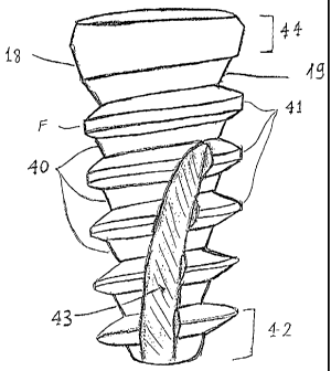
FIG. 1 illustrates an embodiment of the novel tapered condensing dental
implant. There are five elements in a dental implant that influence the
condensation,
insertion and stabilization of the implant. 1) The core of the implant 40. 2)
The
Threads 41. 3) The most apical region 42 which touches the bone first. 4) The
bone
tap 43. 5) The most coronal region 44 which engages the cortical bone and the
sometimes also the gums.
In order to have good stabilization in low density bone it is recommended to
use
small diameter drill and tapered implant. As the diameter of the drill is
smaller and the
implant is more tapered the bone is more preserved and more condensed
resulting in
improved stabilization, but the insertion is more difficult. In this case
controlling the
exact path of insertion of the implant becomes also more difficult since the
implant has
a tendency to slip towards the region with the lowest density. In order to use
a small
diameter drill and an implant with significant tapered configuration all five
elements of
the implant have to be designed to allow an easy insertion and good control on
the
final position of the implant.
In order to clarify the novelty of the new implant it will be compared to a
regular tapered implant like the implant illustrated in FIG. 2. The implant
has a coronal
end 12 and an apical end 14. The implant has five distinct regions. At the
most coronal
aspect is an implant-prosthetic interface region 16. Moving from the coronal
to the
apical ends the implant can have an optional mechanical stop region (not
shown), an
optional cylindrical region (not shown), a tapered region 22, and a bone
cutting end
region 24 which is self drilling and self tapping. An internal threaded
portion 25 is
provided for the attachment of prosthetic components.
The interface region 16 provides mechanical interlock between the implant and
the prosthetic components (not shown) attached to the implant. Interface
region 16 also
provides a means of applying torque to the implant and thus driving the
implant into
the selected site. The interface region 16 can be any of a number of known
interfaces,
including external splines or polygons, or internal geometric shapes such as
polygons
or Morse tapers.
13
CA 02526318 2011-11-22
WO 2004/103202
PCT/1L2004/000438
The optional mechanical stop region can be sharply tapered so that when the
implant is screwed into a prepared osteotomy, the stop limits inadvertently
placing the
implant too deeply.
The shape of the core can be seen in segments 10 in the spaces between the
threads in cross-sectional view FIG. 2. When connecting the outer border of
these
segments in all the tapered implants known in the field straight lines 8 are
formed as
illustrated in FIG. 2. This configuration causes strong resistance for
insertion. In the
present invention when connecting the outer border of these imaginary segment
lines
5 are formed as illustrated in FIG. 3. This configuration enables
gradual
condensation since the diameter of the lower aspect of each segment is close
to the
upper diameter of the previous apical segment. This gradual condensation of
the core
allow for easy insertion of the implant without loosing the final condensation
and
stability since the difference in the diameter between two adjacent core
segments is
the same as for a regular implant like the implant in FIG. 2. The final
condensation is
even larger since the core condenses the bone like a more tapered core. The
angles of
the imaginary segment lines 5 of the core segments in FIG. 3 of the novel
implant are
greater than the angles of the lines 8 of the regular tapered implant of FIG.
2. The
implant of FIG. 3 is tapered like the implant of FIG 2 (the angle between
lines 8) but
condenses the bone like a more tapered implant (the angle between imaginary
segment lines 5) and the condensation is gradually to facilitate insertion.
The imaginary segment lines 5 of FIG.3 which are the continuation of the
border of the core segments 10 are parallel and straight. This is one
preferred
embodiment, but there are other shapes of the border of the core segments that
will
function similarly. We can examine this character of the core of the implant
for
example in FIG. 4 that illustrates a dental implant with a rounded border of
the core
segment. By continuing the border of a core segment 4 positioned coronaly to a
thread
6 through the thread 6 by imaginary line 7. If the imaginary line enters
inside the core
segment 3 apically to the thread 6 it will function the same to allow gradual
condensation, but the condensation is strong only on the apical region of the
core
border. The preferred embodiment with straight
14
CA 02526318 2005-11-18
WO 2004/103202
PCT/1L2004/000438
border lines Fig. 3 allows for gradual condensation along all the border so
the insertion
is smoother.
The threads preferably have a variable profile. The tapered region 22 of FIG.
5
has on its external surfaces a thread 28 of novel profile. The external thread
28
includes a progressively changing profile. At the apical end 14, the thread 28
is sharp
narrow and high in order to facilitate cutting and self-tapping into bone. As
the thread
28 progresses towards the implant coronal end 12, its tip becomes increasingly
broad
or wider in the apical ¨coronal direction and increasingly lower in the
horizontal
direction in cross-sectional profile. The increasing breadth of thread 28
facilitates
compression of low-density bone previously tapped by the sharp apical thread
profile.
Bone compression increases the stability of the implant. The decreasing height
allows
easy insertion and dictates that the implant will keep its first direction
while it is
inserted. As the thread 28 progresses from coronal to apical ends, 12 and 14
respectively, of the implant, the thread 28 becomes sharper, thinner and
higher. Thread
28 is profiled so that a path cut or created in the bone is gradually
broadened by
compression due to the progressively broader thread 28. In this preferred
embodiment
the threads are tapered and the core is more tapered resulting in higher
threads at the
apical region. This configuration is suitable also for very dense bone. In
highly dense
bone sometimes the blood supply is compromised resulting in implant failure.
The
novel implant of FIG. 5 has high and spaced threads leaving spaces between
them after
insertion to hard bone following drilling with a wide drill. These spaces will
promote
blood vessels proliferation and bone regeneration.
FIG. 6 more particularly illustrates the variable profile thread 28. Each turn
T of
thread 28 is of a different profile from each other turn T of thread 28. For
example,
implant includes a plurality of turns T1, T2, T3, . . .
TN. Each turn
T includes an apical side A and a coronal side C and flat face F connecting A
and C.
The length of F varies by being continuously expanded in the direction of the
coronal
end 12. The length of A and C varies by being continuously expanded in the
direction
of the apical end 14.
CA 02526318 2011-11-22
WO 2004/103202
PCT/1L2004/000438
As such, a first turn T1, includes an apical side A1, a coronal side
CI,
and F 1. A second turn T2 includes an apical side A2, a coronal
side
C2, and a F2. The same pattern is repeated for turns T1,
T2,
T3, . . . TN. so that a least length F1, of the thread 28 is
adjacent the 5
apical end 14, and a greatest length FN is adjacent the coronal end 12.
The least
length AN, of the thread 28 is adjacent the coronal end 12, and a
greatest length
A1 is adjacent the apical end 14. The least length CN, of the thread
28 is
adjacent the coronal end 12, and a greatest length C1 is adjacent the
apical end 14.
The apical side of the thread can be a flat shelf perpendicular to the long
axis 9 of the
implant or with a non 90 degrees angle to the long axis of the implant as
illustrated in
FIGS. 5 and 6. In addition, the external thread 28 may have a flat shelf and
rounded
tip, which are most pronounced at the thread's coronal end 12. The flat shelf
provides
support against implant micro-motion imposed by axial loads, particularly
important
in low-density bone. The tip of the thread F can be flat or rounded. The angle
of the
each thread segment meaning the angle between A and C of Fig 6 is about 60
degree.
To allow cutting of the bone a more sharp angle is preferred at 30-40 degree
preferably at 35 degree. Preferably all the threads has the same angle between
A and
C. In another preferred embodiment the angle between A and C is gradually
increased
coronally to get more condensation for soft bone or gradually decreased
coronally for
hard bone.
In the preferred embodiments of FIGS. 1,5,6, a circumferential face F is
included on some turns of thread 28. The face F is preferably flat and is not
included
on the self-tapping portion of the thread 28, adjacent the apical end 14, but
is provided
as each turn progressively widens toward the coronal end 12. The face F
preferably
parallel to the long axis 9 of the implant but it can be also angled.
The threads are also tapered. The imaginary thread lines 23 connecting the
tips
of the threads are not parallel to the long axis 9 of the implant. The threads
are tapered
and at the same time become higher apically because the core of the implant is
more
tapered than the threads. The fact that the width of the apical region of the
implant is
smaller than the coronal region allows the use of a small drill therefore
preserving the
bone. The sharp apical threads enter the small hole in the bone and start
cutting the
bone. The next
16
CA 02526318 2005-11-18
WO 2004/103202
PCT/1L2004/000438
thread is wider in the coronal apical direction and the implant is wider
causing
compression of the bone but since the height of the thread is less than the
previous
thread the thread stays in the path created in the bone by the previous thread
therefore
preventing slipping of the implant to a region with even lower density bone.
The fact
that the height of the threads become smaller as going coronally allows for
gradual
compression of the bone and facilitate insertion. The combination of a gradual
tapered
compressing core as described above with a gradual compressing tapered thread
as
described here is the preferred embodiment. The implant preferably has more
than one
thread. An implant with double thread each thread with a double step allows
insertion
in half the turns needed for an implant with one thread while keeping the
outer surface
and the stability of the implant. The implant can have more than two threads.
The most apical region of the implant can have two preferred configurations.
One is smooth round design, this design is suitable for cases that the implant
is near
the Schneiderian membrane of the maxillary sinus or near the mandibular nerve
in
order to prevent damage to these delicate tissues. In this design the threads
start with a
distance from the apical end. The second design of the most apical region
illustrated in
FIGS. 7 A and B is to have sharp blades that cut the bone and allow easy
insertion.
There are several variations for the shape of the blades, which are well known
in the
dental implant field. Implants with this apical design are called self
drilling implants.
The bone tap of the implant influence the insertion. The presence of a bone
tap
allows the insertion of the implant without previous taping of the bone.
Implants with a
tap are called self tapping implants. The tap can be straight or oblique or
spiral. The
preferred design is the spiral bone tap to facilitate insertion. The tap 60 as
illustrated in
FIG. 7 is long and going through more than a third of the length of the
implant
crossing several threads. Preferably the tap extends along more than half of
the
implant. The tap is not straight but surrounding the implant. The tap starts
at on side of
the implant FIG. 7 A and extends to the other side FIG. 7B. The whole tap
can't be
seen from one place. This design of the tap facilitate insertion so when the
implant is
inserted only part of one thread is cutting the bone therefore the resistance
for insertion
is lower. This configuration together with the design of the thread as
described above
17
CA 02526318 2011-11-22
WO 2004/103202
PCT/1L2004/000438
also dictates that the implant will stay in its original path of insertion by
forcing the
next thread to go into the slot in the bone prepared by the previous thread.
This feature
is enhanced by the presence of a double thread. The implant can have more than
one
tap preferably two.
The most coronal region of the implant also influences the insertion and
stabilization of the implant. This region includes the interface region. There
are
several types of interfaces like splines whereas, the interface region 16 of a
single-
stage embodiment of FIG. 5, may optionally include a socket having a plurality
of
sides, e.g. a hex socket. Also, the embodiment of FIG. 1 does not include a
sharply
tapered mechanical stop as but instead includes a gradually tapered portion
18. The
gradually tapered portion 18 allows for more freedom in placement depth to
adjust the
distance that the trans-gingival collar protrudes from the bone. However, an
alternate
single-stage embodiment, can includes a coronal region 44 including a second
angled
portion 19 which acts as a stop.
When an implant is completely sharply tapered as are the implants described
above its most coronal region becomes very broad. This broad coronal is
appropriate
for regions with very low density cortical bone since it compress the cortical
bone. In
cases the cortical bone is not very soft this can interfere with the insertion
of the
implant. There are also clinical evidences that when the coronal region is
broad the
blood supply to the bone around the implant is disturbed resulting in higher
incidence
of bone resorbtion and implant failure. Therefore if the cortical bone is not
very soft
the coronal region preferably should be less tapered then the body of the
implant. The
most coronal part of the coronal region is even preferably inversed tapered 48
as
illustrated in FIG. 8.
The implant can include internal threads 63 for connection to the prosthetic
part as illustrated in FIG. 5. In case the bone is very narrow the core has to
be also
very narrow. When the core is very narrow it can't include internal threads,
so the
implant can come in one piece with abutment. In these embodiments the coronal
supragingival part serves for insertion of the implant and also as an abutment
to
support the future prosthetics. FIG. 8 illustrates such an embodiment with a
narrow
region 71 between
18
CA 02526318 2011-11-22
WO 2004/103202
PCT/1L2004/000438
the part of the implant that is to be inside the bone 72 and the abutment part
73 which
is tapered to allow connection to a prosthetic element like a crown. The
narrow region
71 allows good attachment of the gums to the implant therefore prevents bone
loss.
The abutment region can include an internal anti-rotational element or
external anti-
rotational element 76 that will serve for the insertion of the implant. FIG. 9
illustrates
another embodiment of the novel implant as one piece with the abutment. The
narrow
gingival region 71 is longer than the embodiment of FIG. 8. In this embodiment
an
internal hexagon 74 is used for the insertion of the implant. FIG. 10
illustrates another
embodiment like the embodiments of FIGS. 8 and 9 but the abutment element 73
is
wider and grinding is needed to get the shape of a normal abutment like the
dotted
line 77. This design allows easy preparation of the abutment in cases that the
implant
is placed with an angle to the path of insertion of the prosthetic element.
FIG. 11
resembles the implant of FIG. 10 but in this embodiment grinding the abutment
of the
implant is almost no needed. The implant has a round rod 80 protruding
coronally
above the gingival region 71. The abutment 82 has an internal bore 83 matching
the
round rod 80 of the implant. The abutment is tilted so after placing the
abutment on
the implant the angle of the abutment can be changed by rotating the abutment
82
around the rod 80. When the desired position of the abutment 82 is decided the
abutment 82 can be glued to the implant. In another preferred embodiment the
rod has
around its base an anti rotational element 87 matching an anti-rotational
element in
the abutment. This configuration prevents movements of the abutment while it
is
cemented to the implant and can also help in taking impression of the implants
to
prepare the abutments in a dental laboratory.
In another preferred embodiment illustrated in Fig. 12 the coronally tapered
region 90 is placed inside the bone so the bone can grow above this region.
The
tapered region 90 is below the bone level 91. The height of the coronally
tapered
region 90 is 0.5-4 mm. Preferably the height is 1-3 mm and for most cases 1.3-
2.5 mm
depending on the diameter of the implant.
The implant is preferably one piece because of two reasons: A. The coronal
region is narrow and placing a thread or a bore inside this region will reduce
the
19
CA 02526318 2005-11-18
WO 2004/103202 PCT/1L2004/000438
mechanical strength of the implant. B. The connection to a prosthetic element
result in
most cases with the creation of a micro-gap between the implant and the
prosthetic
element. This micro-gap can be colonized by bacteria that release toxins
resulting in
bone resorbtion. A one piece implant is mechanically strong and has no micro-
gap.
The thread of the implant has preferably high step. The most common implants
has thread step of about 0.6 mm. The present implant has preferably a thread
step of
1.5-2.5 mrn preferably the step is 2.1 mm. Preferably the implant has double
thread
meaning two threads with different beginnings running along the implant. This
configuration causes that for every point of one thread there is a thread at
the opposite
side of the implant at the same vertical level. The threads when are inserted
into the
bone are creating slots. The double thread creates two opposite steep slots in
the bone
for every bone segment. These slots facilitate the insertion of the implant
because the
bone is easily expanded. The presence of two opposite slots in the bone that
each one
is created by a thread of more than 1.5 mm and preferably of 2.1 mm thread
step
allows this expansion. A regular thread of 0.6 min will create almost
horizontal slots in
the bone resulting in crushing of the bone instead of expansion. Because of
the slots
the bone is not crushed but elastically expanded The threads begins preferably
at the
wider area of the coronally tapered region 92 so when this wider area reaches
the bone
the bone has already two points in the bone having between them approximately
the
diameter of this wide region so this wide region is pushing the bone at the
other
direction and the bone segments between the slots are displaced from each
other and
come back to their original location after the wide region is inserted more
inside the
bone. These bone segments between the slots can relapse to their original
location
because the coronal segment 90 is tapered coronally. This process will occur
for every
point along the bone where the coronally tapered region 90 is inserted inside
the bone
since this region is just above the beginning of the threads. The end result
is a tapered
region inside the bone covered with bone. Preferably the threads continue over
the
coronally tapered region 90 as illustrated in Fig. 12. In this configuration
the wider
region 92 is not a circle but resembles more an ellipse since the double
thread that
extends along the coronally tapered region reduce its diameter in one
direction. This
CA 02526318 2011-11-22
WO 2004/103202
PCT/1L2004/000438
configuration facilitate the insertion of the wide region 92 inside the bone
because the
longer diameter of this ellipse is inserted to the slots in the bone. The
insertion of a
coronally tapered region with more than one thread on it allows elastic
expansion of
the bone and the bone is covering this tapered region after insertion inside
the bone.
The best results are achieved if the height of the intra-bony coronally
tapered region
90 is close to the thread step. Preferably the height of the intra-bony
coronaly tapered
region 90 is higher than a half of the thread step.
In another preferred embodiment the threads are along the entire coronally
tapered region. The threads can be the same as the threads along the implant
but in
another preferred embodiment can be smaller in the thread step and the thread
height.
The presence of a small thread or micro-thread in this region can allow better
distribution of the forces to the cortical bone.
In operation, the implant can be placed into a pre-drilled osteotomy site that
either matches the external diameter of the implant body, that is, the
narrowest
diameter between threads, or into a site that is narrower than the external
diameter of
the implant. Placing the implant into a narrower site will provide additional
bone
compression, and therefore greater initial stability. The drill can be
straight or tapered.
Preferably the drill is straight and the diameter is dictated by the density
of the bone.
For soft bone the last drill has small diameter and even insertion can be done
without
drilling. In hard bone a wider drill should be used and the spaces between the
bone
and the core of the implant will be filled with blood vessels while the
implant is
stabilized by the high threads.
The implant of Fig. 12 is a one piece implant that has a protruding element 93
that extends from the bone level through the gums to the oral cavity. This
protruding
element is preferably tapered coronally and can serve for receiving a crown
like a
prepared tooth. The protruding element can serve for receiving an abutment.
This
tapered protruding element 93 preferably includes an anti-rotational element
of any
kind. Examples of anti-rotational elements are illustrated in Fig. 13 A-D. Fig
13A
illustrates several protrusions which can have an under-cut for receiving a
matching
transfer copping, Fig. 13B illustrates one or two protrusions or slots, Fig.
13C
21
CA 02526318 2005-11-18
WO 2004/103202
PCT/1L2004/000438
illustrates tapered slots, Fig. 13D illustrates a hexagon or any polygon and
any other
anti rotational option ellipse, stars, splines etc. The abutment preferably
includes a
matching anti rotational element. The anti rotational element can be used with
a mating
transfer coping for impressions and for the insertion of the implant. There
are several
The abutments can be bulky preferably having anatomical gingival aspect as
the abutments of Fig 15. This configuration should be prepared by the dentist
or at the
dental laboratory to desired shape. Fig 15A illustrates a straight bulky
abutment and
In another preferred embodiment the protruding element 93 of the implant can
receive a gingival anatomical collar. This collar matches the subgingival and
gingival
anatomy of different teeth and the protruding element extends through this
collar
coronally. The collar can be of different heights or can be seated at
different distances
22
CA 02526318 2005-11-18
WO 2004/103202 PCT/1L2004/000438
consideration. Preferably the collar is left above the bone level as described
above for
the abutments in Fig. 14, 15. An example of a collar is illustrated in Fig 16.
Fig 16A is
a side view and Fig 16B is a top view of a collar fitted on the protruding
element 93 of
the implant.
In case the protruding element is converging coronally by using different
sockets sizes inside the abutments and collars the distance from the bone to
the
abutment or collar can be determined. As the socket is larger the abutment or
collar
can be inserted more close to the bone. The collar or abutment can be inserted
at the
time of inserting the implant allowing the gums to heal around the collar to
receive the
right shape. In this case the collar or abutments serve as a healing cap. The
implant can
be left without a healing cap or can receive a standard healing cap that looks
like a
cylindrical bulky abutment.
The assembly of the abutments and collars with the implant is illustrated in
perspective views in Fig 17. Fig 17A is an example of a preferred embodiment
of the
implant, Fig. 17B is an example of an angled abutment Fig 17C illustrates the
implant
with abutment. Fig. 17D is a perspective view of a collar, Fig. 17E
illustrates the
implant with the collar. Fig. 17F illustrates the implant with a ball
attachment. The
height of the protruding element 93 is reduced before the ball attachment is
seated. The
abutments are above the bone level 91 leaving a narrow area 98 to allow the
gums to
grow and seal the bone from the oral cavity. The abutments and collars can be
made
from any biocompatible material such as titanium zirconium or gold or from
ceramic
materials.
There are several ways to assure a good connection between the protruding
element and the abutment or collar. The abutment or collar can be glued to the
protruding element. The abutment can be manufactured to fit very accurately to
the
protruding element so when using some force the abutment is tightly seated
over the
protruding element and the friction is keeping it in place. In these cases
preferably the
abutment has at least one point with an under-cut to allow the dentist to take
the
abutment out using a crown remover. In another preferred embodiment the
abutment
has a locking mechanism. Fig 18 illustrates an example of a locking mechanism.
The
23
CA 02526318 2005-11-18
WO 2004/103202
PCT/1L2004/000438
protruding element 93 can have a small notch 99 or a slot and the abutment can
have a
small hole 100 placed to fit the notch 99. Into this hole 100 a small pin can
be forced
from the side to get inside the notch 99 and lock the abutment to the
protruding
element preventing it from going coronally. In another preferred embodiment to
allow
to take the abutment out easily the hole can include a thread and the abutment
is locked
by a small screw coming from the side into the notch or the slot 99. In
another
preferred embodiment illustrated in Fig. 19 the protruding element can be made
of
several fingers 101 and a hole 102 in the center of the protruding element.
After an
hollow abutment 103 is seated on the protruding element a small pin is
inserted inside
the hole 102 and force the fingers 101 to push the inner aspect of the
abutment
therefore the abutment is strongly connected to the protruding element. In
another
preferred embodiment at the base of the protruding element below the point
where the
fingers are separated there is a thread 104 and instead of a pin a small screw
is screwed
inside the hole to the internal thread 104. The screw has a region that its
diameter is
slightly larger than the diameter of the hole 102 so as the screw is inserted
more deeply
the fingers are pushed stronger towards the abutment. This configuration
allows to take
the abutment out easily.
In another preferred embodiment of a one-piece implant illustrated in Fig. 20
the abutment is seated from the side. The implant illustrated in Fig. 20A has
a broad
region 105 above the narrow region 98. From this broad region protrudes a low
element of 0.5-3 mm height preferably of 1-2 mm with an anti-rotational
mechanism
like a hex 106. Above this low element 106 there is preferably a wide tapered
element
107. The abutment illustrated in Fig. 20B has a slot that fits the low element
106 and
the wide tapered element 107 of the implant. Fig 20B is a perspective view of
the
abutment looking from the side with the slot. Fig. 20C is a side view of the
abutment.
The dotted line 108 shows the internal slot from the side. Fig 20D shows the
abutment
of Fig 20 B-C on the implant of Fig. 20A. Fig. 20E illustrates the implant
with an
angled abutment. The abutments of Fig 20 are inserted from the side to fit the
anti-
rotational element 106 of the implant. The abutment can't move coronally
because of
the wide tapered element 107 but it can move to the side. To prevent the
movement to
24
CA 02526318 2005-11-18
WO 2004/103202
PCT/1L2004/000438
the side there are several ways 1) a matching cap 109 can be seated on top of
the
abutment or the crown can be used for this purpose. 2) The abutment can have
holes in
the walls of the slot below the height of the wide tapered element 107 so a
screw or a
pin can be inserted from the side below the wide tapered element 107 touching
the low
element 106. The hole 115 can be seen from the side in Fig 20C. 3) A ligature
can be
inserted between the holes 115 and the empty space below the wide tapered
element
filled with a dental filling material or just the filling material like
composite filling.
(the abutment can have a slot all around and a ligature is placed in the slot)
4) The
abutment can be manufactured to tightly fit the implant and to be inserted by
force.
The slot of the angled abutment of Fig. 20E can be on the left side of the
abutment of Fig 20E meaning at the opposite side to the direction of the tilt
of the
angled abutment leaving an empty space 110 below the wide tapered element 107
on
the left or the slot can be at the right side meaning at the direction of the
tilt of the
angled abutment leaving an empty space 111 below the wide tapered element 107
on
the right or the slot can be in other directions in relation to the direction
of the tilt of
the angled abutment. The empty space when using a matching cap 109 or a crown
is
closed. The empty space can be filled with a dental filling material. The
matching cap
109 or the crown can be cemented to abutment or the matching cap be tightly
fitted to
the abutment seated by friction. In a preferred embodiment the matching cap
109 or the
crown are screwed to the abutment. The matching cap 109 in Fig 20E or the
crown can
have a small hole preferably with a thread 112 so a small pin or a screw can
be inserted
through the hole to the empty space 111 (or 110 if the hole is in the other
direction).
This small screw is locking the abutment and the matching cap to the implant.
Because
there are at least two types of angled abutments, according to the location of
the hole in
respect to the direction of the tilt of the abutment, the dentist can decide
where to place
the screw for a screwed crown or bridge. The place of the screw is important
for the
esthetic result. If an implant is angled buccaly for all the common abutments
in the
market the screw is coming from buccal leaving a hole in the buccal aspect of
the
crown which is very difficult to cover. Screws coming from the side are known
but
demand a very difficult work from the laboratory. The embodiment of Fig 20
allows to
CA 02526318 2005-11-18
WO 2004/103202
PCT/1L2004/000438
have a simple screwed restoration from the side which is easily retrievable
and
esthetic. In another preferred embodiment the abutments of Fig. 20B and Fig
20E have
on their outer surface an anti-rotational mechanism to prevent the rotation of
the
matching cap or the crown. In these embodiments the matching cap and the crown
have also an internal anti-rotational mechanism fitting the anti-rotational
mechanism of
the abutment. Fig 20F is illustrating an embodiment of an abutment with an
anti
rotational mechanism like a hex 113. The wide tapered element 107 of the
implant can
also include an anti-rotational element preferably compatible with the anti-
rotational
element of the low region 106 of the implant for example both with a hex. In
another
preferred embodiment illustrated in Fig. 20G instead of the wide tapered
region there
is a spherical shape 114. This configuration allows the implant to be used as
a ball
attachment to support dental removable prosthesis. This preferred embodiment
enables
a variety of restorative possibilities: cemented restoration, screwed
restorations and
removable restorations. In the embodiment of fig. 20G the healing cup instead
of being
inserted from the side can have an internal elastic element fitted to hold the
ball 114 of
the implant so the healing cap is inserted and removed vertically by some
force. In
another preferred embodiment the implants of Fig 20 can also receive from the
side a
gingival collar as described in Fig. 16 and Fig. 17. In another preferred
embodiment
illustrated in Fig 20H the abutment (or the matching cap) has an angled ball
ZO attachment. This configuration allows the dentist to achieve parallelism
between the
ball attachments of several implants which is difficult to achieve in the
common
restorative systems.
The protruding element with the anti rotational element 106-107 can be also
used for the insertion of the implant and for impressions using matching
transfer
).5 copings. The advantage of this embodiment is that the abutment can't be
detached as
long as the crown is in place and there is no need to grind the protruding
element when
using angled abutments or short abutments as it is the case is some of the
previous
embodiments The embodiment of Fig. 20 is one example but any other
configuration
with a protruding element that has an under-cut can function similarly to
allow
;0 connection of an abutment from the side.
26
CA 02526318 2005-11-18
WO 2004/103202
PCT/1L2004/000438
All the embodiment demonstrating an anti-rotational element on the implant
preferably have a compatible anti-rotational element on the abutment or
collar. The
number of the protrusions or slots or angles of the anti-rotational element
don't have to
be the same for the implant and the abutment as long as the abutment can be
seated on
the implant.
All the abutments and collars described above can come in different heights,
different widths and different angles and to be seated at different heights
from the bone
level. They also can have different heights and widths of the subgingival part
and
different heights and widths of the supragingival part.
All the embodiments of implants of the present invention can have several
surfaces. The implant can have machine surface but preferably can have rough
surface
like TiUnite, S.L.A, Osseotite, Hydroxyapatite or bioactive surface that has
growth
factors and active proteins like B.M.P. The rough surface preferably is along
the intra-
bony part of the implant and preferably also extending to the narrow region
98of the
implant.
As a result, the above embodiments provide unique advantages by providing a
dental implant fixture particularly suited for use in lower density bone, such
as that
found in the posterior mandible and maxilla. The implant features a tapered
profile and
a unique external thread profile that offers superior stability when it is
implanted in
low density bone while insertion is easy. The implant tapers down in diameter
optionally beginning at a point about 1-3 mm from below the top surface of the
implant. The external thread is also tapered and changes profile from the
coronal to the
apical ends of the implant fixture, having a sharp, narrow and high profile at
the
extreme apical end, particularly suited for cutting into non-tapped bone, and
having a
broad, rounded and low profile at the coronal end, particularly suited for
compression
of bone tapped by the thread at the apical end. Further, the thread profile
optionally has
a flat shelf on its apical aspect, being most pronounced at the coronal end of
the
implant and being less pronounced at the apical end of the implant. At its
coronal end,
the implant has an optional flared region that acts as a mechanical stop,
serving to limit
over-insertion of the implant into soft bone. At its apical end, the implant
optionally
27
CA 02526318 2005-11-18
WO 2004/103202
PCT/1L2004/000438
has a round, blunt shape and a set-back thread in the event the implant comes
in
advertent contact with non-osseous structures. The implant can be of one piece
and
have coronally converging intra-bony region near the coronal cortical bone.
The combination of all the aspect described above the coronal region, the
core,
the threads and the apical region allows to produce an implant that is easily
inserted
although the drilling is minimal, to easily dictate the location of the
implant, to allow
good stabilization in the bone and to allow the bone to be above the intra-
bony
coronally tapered region. The presence of bone above this region supports the
gums
and maintain their desired configuration especially the height of the gums
between the
teeth called papilla which are very important for the esthetic result. This
bone is
preserved since the implant allows drilling with a small diameter drill and
the core is
tapered and the threads are tapered with variable thread design and the
coronal region
is inversed tapered. Only the combination of all the features and the
relationship
between them can lead to an implant that allows the best esthetic result.
Although illustrative embodiments have been shown and described, a wide
range of modification change and substitution is contemplated in the foregoing
disclosure and in some instances, some features of the embodiments may be
employed
without a corresponding use of other features. Accordingly, it is appropriate
that the
appended claims be construed broadly and in a manner consistent with the scope
of the
embodiments disclosed herein.
?,5
28