Note: Descriptions are shown in the official language in which they were submitted.
CA 02526327 2013-02-19
1
DEVICE FOR TRANSMITTING MULTIPLE OPTICALLY-ENCODED
STIMULATION SIGNALS TO MULTIPLE CELL LOCATIONS
FIELD OF THE INVENTION
The present invention relates generally to a device and method for stimulating
cells. More specifically, the present invention relates to a device and method
for
transmitting multiple optically-encoded stimulation signals to multiple
stimulation
sites, especially cell locations.
lo BACKGROUND OF THE INVENTION
In various medical fields, the use of artificial stimulation devices, or
prosthesis, to
stimulate damaged cells and/or tissue which are no longer responsive to
natural
stimuli is well known. These devices mimic natural impulses and act to re-
establish the natural stimulation path.
One of the best examples of the success of such an approach is the use of the
cochlear implant to restore partial hearing in profoundly deaf people. A
person is
diagnosed as profoundly deaf if either a very large number of hair cells or
auditory
neurons throughout the cochlea, the spiral-shaped cavity of the inner ear, are
damaged. Cochlear implants use electrical stimulation to directly excite the
remaining auditory neurons which connect the ear to the brain. In general,
such
implants include a microphone which picks up sound, an array of electrodes
surgically inserted into the cochlea, which electrically stimulates functional
auditory
neurons of the cochlea, and a signal transmission system which transmits the
sound information from the microphone to the array of electrodes. The whole
system is designed so that activation of the electrodes will fire up the
neurons,
which communicate with the patient's central nervous system, and thereby
transmit information about the acoustic signal to the brain.
CA 02526327 2005-11-09
In practice, implementation of existing cochlear implant technology is impeded
by
the size of the wires used to transmit information to the neurons. The minimum
diameter of such a wire being about 25 pm (P. Ake Oberg, Tatsuo Togawa,
Francis A. Spelman (eds.), Sensors in Medicine and Health Care,
Sensors Applications Volume 3, Wiley-VCH Verlag GmbH & Co. KGaA, 2004), the
number of wires is limited to less than one hundred (100) by the diameter of
the
auditory canal. By increasing the number of electrodes, it is hoped that the
resolution of the perceived acoustic signal can be improved. Moreover, by
decreasing the diameter of the wire, the risk of injury to the cochlea and its
inner
to structure, which includes the basilar membrane and the hair cells, is
reduced.
This risk of injury inherent with electrical charge is of import given the
increase in
popularity of cochlear implants and their growing consideration for use in
patients
with residual hearing. One other solution would be to develop a device which
uses
non-electrical artificial stimulation, for example optical or photo-
stimulation. US
Patent Application No. 2005/0216072 (MAHADEVAN-JANSEN) discloses a
system and methods for optical stimulation of neural tissues. However, one
major
drawback with this system and these methods lies in the probe: the probe
delivers
optical energy to the target neural tissue, one site at a time and at a
distance away
from the target neural tissue.
-)0
Applications of electrical stimulation systems are not limited to cochlear
implants.
They include brain neuro-stimulation (pain relief, tremor control, treatment
of
cerebral palsy, treatment of Parkinson's disease, visual cortex implants for
the
blind), spinal neuro-stimulation (pain relief, peripheral vascular flow
enhancement),
peripheral nerve stimulation (pain relief, phrenic nerve pacing), retinal
implants,
heart pacemakers, tissue-growth stimulation and inhibition, etc.
Functional Electrical Stimulation (FES) is used to produce, by means of
electrical
stimulation, contractions in muscles either injured or paralysed due to
central
nervous systemi lesions. In the case of FES, arrays of electrodes are
implanted
under the skin and used to choreograph movement in the patient's muscles.
CA 02526327 2005-11-09
3
Applications for this approach are found, for example, in cases of stroke,
spinal
cord injury, head injury, cerebral palsy, and multiple sclerosis. Here, too,
resolution is limited by the size of the wires used for electrical
stimulation.
Efforts are underway to develop visual prostheses, both retinal and cortical.
Retinal prostheses aim to restore some form of vision to patients that are
blind
owing to a degenerative condition, such as retinitis pigmentosa or age-related
macular degeneration, by bypassing the photoreceptor cells of the retina which
have become dysfunctional and electrically stimulating the relatively intact
retinal
ganglion cells which connect the eye to the visual cortex of the brain.
Electrical
stimulation of the retinal ganglion cells creates the sensation of a spot of
light (or
phosphene) in the spatial vicinity of the stimulation. Cortical prostheses may
be
used to treat patients with secondary blindness not due to retinal or optic
nerve
disease. The difficulty with cortical implants lies in the need for
intracranial
surgery and the complexity of brain geometry. Nevertheless, both types of
prostheses are faced with the problems inherent with electrical stimulation:
injury
incurred by neurons under chronic use and lack of specificity. U.S. Patent No.
6,458,157 (SUANING) discloses an apparatus in which all tissue-contacting
components may be fabricated from materials known to be well tolerated by
human tissue. While SUANING discloses attempts that have been made to limit
injury due to long-term use, the matter of specificity is not expressly
addressed.
In general, traditional methods and devices for direct electrical neuro-
stimulation
lack spatial, physiological and strength specificities. Furthermore, they are
prone
to electrical interference from the environment. For example, electrical
stimulation
of the visual cortex produces phosphenes (or blurred) spots rather than pixel-
like
(or well-defined) spots. Stimulating tactile sense through electrical
stimulation of
specific neuronal cells is practically impossible without stimulating muscles
and/or
a temperature response, producing hitching or pain. A stimulation device
permitting stimulation of specific neural ganglion cells would allow for
better control
of the stimulation process.
CA 02526327 2005-11-09
4
While certain cell, tissue, or system functions can be affected or controlled
through
electrical stimulation, a more efficient means of regulating these functions
would
be through the use of natural biochemical stimulators or inhibitors that are
target
specific. For example, insulin is produced naturally by the pancreas and is
used
by the body to activate glucose metabolism. Insulin production cannot be
induced
through electrical stimulation. Diabetics, who count for more than 5% of North
Americans, must inject themselves with insulin in order to metabolise the
glucose
present in their body. A more convenient means of regulating the level and
io production of insulin would greatly benefit diabetics. The same holds
true for
people that must take medications regularly either orally or through
injection.
Recent developments in nanotechnology (nanoshells, quantum dots (QDs),
micelles), photodynamic therapy and photo-imaging offer new possibilities for
is improving specificity. These new technologies provide ways to cage, tag
and
locate molecules thus allowing the regulation and monitoring of optical
stimulation
mechanisms. Of particular interest are molecular structures or compounds that
undergo changes in their properties (chemical affinity, conformal structure or
composition) upon exposure to light (photoactivated changes). Following
20 photoactivation, these molecules can react with other molecules or cells
or emit
light. In some cases, molecules undergo photoactivation only in the presence
of
certain other molecules or cells thus allowing these photoactivated molecules
to
be used as targets for locating, monitoring, imaging or destroying these other
molecules or cells when lighted. For example, U.S. Patent No. 6,668,190 (IEZZI
25 et al.) discloses a drug delivery system that includes a fluid channel
for delivering
a drug to one of a number of sites and a light channel for delivering light to
an area
near one of the sites for photoactivating caged and/or non-caged molecules of
the
drug to stimulate neurological tissue.
30 From all of the above, there is a need for an improved manner of
delivering either
electrical or optical stimulations to specific stimulation sites of any type.
CA 02526327 2011-01-27
SUMMARY OF THE INVENTION
It is an object of the present invention to propose a device that optically-
encodes
stimulation information and transmits this stimulation information to multiple
stimulation sites.
5
In accordance with one aspect of the present invention, there is therefore
provided a
stimulation device for transmitting stimulation information to a plurality of
physiological
stimulation sites in a body of a patient. The device includes light generating
means
for generating light having a plurality of wavelength components, encoding
means for
to separately encoding at least a portion of the stimulation information
into each of the
wavelength components, and a multiplexing arrangement for multiplexing the
wavelength components encoded by the encoding means into an encoded light
signal. The device further includes a primary waveguide having an input end
operationally connected to the multiplexing arrangement for receiving the
encoded
light signal, a light-guiding axis for guiding the encoded light signal
therealong and an
output end adapted to be implanted in the body of a patient proximate the
stimulation
sites, the output end having a plurality of output positions spatially
distributed along
said light-guiding axis. In addition to the above elements, the device also
has an
outcoupling arrangement provided at the output end of the primary waveguide.
This
outcoupling arrangement is wavelength-sensitive to transversally couple each
of the
wavelength components of the encoded light signal out of the primary waveguide
at a
different one of the output positions along the light-guiding axis, each of
the output
positions being associated with a corresponding one of the stimulation sites.
Thereby,
the output arrangement transmits a portion of the stimulation information
encoded in
each wavelength component to the corresponding stimulation site through the
associated output position.
CA 02526327 2013-02-19
,
6
In one embodiment of the device, the device preferably includes a number of
electrodes, each associated with one of the output positions, for transducing
a
corresponding wavelength component into an electrical stimulation signal.
In another embodiment of the device, the device preferably includes an optical
window in the primary waveguide at each of the output positions, in order to
output
an optical stimulation signal therefrom.
Embodiments of the device may be used for various applications, such as, non-
limitatively, stimulating cerebral neurons along a visual pathway, the
stimulation
information thereby stimulating a visual response; stimulating contraction of
muscle tissues; stimulating growth of tissues; stimulating biochemical
compounds
adapted for photoactivation by the wavelength components, and the like.
In accordance with one embodiment of the invention, there is also provided a
cochlear implant for transmitting stimulation information to auditory neurons
of the
cochlea in situ of a patient. The cochlear implant includes a light generating
means for generating light having a number of wavelength components, an
encoding means for separately encoding at least a portion of the auditory
stimulation information into each of the wavelength components, and a
multiplexing arrangement for multiplexing the wavelength components encoded by
the encoding means into an encoded light signal. The cochlear implant further
includes a primary waveguide having an input end operationally connected to
the
multiplexing arrangement for receiving the encoded light signal therefrom, a
light-
guiding axis for guiding the encoded light signal therealong and an output end
adapted to be implanted proximate the auditory neuron sites of the cochlea,
the
output end having a plurality of output positions spatially distributed along
the light-
guiding axis. In addition to the above elements, the device also has an
outcoupling
arrangement provided at the output end of the primary waveguide. This
outcoupling arrangement is wavelength-sensitive to transversally couple each
of
CA 02526327 2013-02-19
7
the wavelength components of the encoded light signal out of the primary
waveguide at a different one of the output, each of the output positions being
associated with a corresponding one of the auditory neuron sites of the
cochlea.
Thereby, the outcoupling arrangement transmits the at least a portion of the
stimulation information encoded in each wavelength component to the
corresponding neuron site through the associated output position. In one
embodiment, the cochlear implant preferably includes a number of electrodes,
each associated with one of the output positions, for transducing a
corresponding
wavelength component into an electrical stimulation signal. In
another
embodiment, the cochlear implant preferably includes an optical window in the
primary waveguide at each of the output positions, in order to output an
optical
stimulation signal therefrom.
Advantages of the present invention include enhanced transmission efficiency
(no
cross-talking) of optically multiplexed stimulation signals, enhanced
resolution
achieved through the smaller size of the surface area at the output position
interface and the increased number of output position interfaces.
Certain embodiments of the invention exhibit additional advantages: reduced or
eliminated risk of injury due to electrical charge (toxicity due to electrode
breakdown and heat damage), a more painless stimulus, and targeted and timed
delivery of treatment via photoactivation of biochemical compounds or
cellular/tissue functions at stimulation sites.
CA 02526327 2005-11-09
8
BRIEF DESCRIPTION OF THE DRAWINGS
Further aspects and advantages of the invention will be better understood upon
reading the description of preferred embodiments thereof with reference to the
following drawings:
FIG. 1 is a schematic illustration of an assembly for generating a multiplexed
multi-
wavelength encoded light signal for a device according to a preferred
embodiment
of the invention.
lo FIG. 2 is a schematic illustration of an assembly for generating a
multiplexed multi-
wavelength light signal, according to another preferred embodiment of the
invention.
FIG. 3 is a schematic illustration of an assembly for generating a multiplexed
multi-
is wavelength light signal, according to yet another preferred embodiment of
the
invention.
FIG. 4 is a front view illustration of the assembly of FIG. 3.
20 FIG. 5 is a schematic illustration of an assembly for generating a
multiplexed multi-
wavelength light signal showing the use therein of dichroic (or dielectric-
coated)
mirrors, according to yet another preferred embodiment of the invention.
FIG. 6 is a schematic illustration of a variant of the assembly of FIG. 5.
FIG. 6B is
25 an enlargement of section A of FIG. 6.
FIG. 7 is a cross-sectional side view of the output end of a device according
to a
preferred embodiment of the invention, showing a blazed optical grating
comprising a number of uniform Bragg gratings positioned at output positions
30 along the light guiding axis.
CA 02526327 2005-11-09
9
FIG. 8 is a cross-sectional side view of the output end of a device according
to
another preferred embodiment of the invention, showing a number of dielectric
reflectors positioned at output positions along the light guiding axis.
FIG. 9 is a cross-sectional side view of the output end of a device according
to
another preferred embodiment of the invention, showing dielectric reflectors
used
to transversally couple different wavelength components of the encoded light
signal out of the primary waveguide at different output positions along and
around
the light guiding axis. FIG. 9B is an enlargement of portion A of FIG. 9.
FIG. 10A is a cross-sectional side view of the output end of a device
according to
yet another preferred embodiment of the invention, showing outcoupling means
which use shaping in the device core to reflect part of the encoded light
signal out
is of the waveguide core; FIG 10B is a cross-sectional side view of a
refractive
variant to the embodiment of FIG. 10A.
FIG. 11 is a cross-sectional side view, according to a preferred embodiment of
the
invention, of an electrical wire extending along the primary waveguide and
used to
apply a polarization voltage to the photoelectric material of the electrodes.
FIG. 12 is a partially transparent perspective side view of the output end of
a
device according to yet another embodiment of the invention, showing a
metallic
cladding of the primary waveguide in electrical contact with the photoelectric
material of the electrodes.
FIG. 13 is a partially transparent perspective side view of a micro-structured
optical fiber having an air cladding composed of a number of air gaps and
fused
silica bridges, according to a preferred embodiment of the invention.
CA 02526327 2005-11-09
FIG. 14A is a partially transparent perspective side view of a micro-
structured
optical fiber having an air cladding composed of a number of air gaps and
fused
silica bridges with part of the cladding drilled and filed with an optically
transparent
material, according to a preferred embodiment of the invention; FIG. 14B is
cross-
5 sectional view of the micro-structured optical fiber of FIG. 14A.
FIG. 15A is a cross-sectional side view of the output end of a device
according to
an embodiment of the invention, showing the use of secondary fibers coupled to
output positions along the primary optical fiber and outcoupling light to
stimulation
io sites located away from the primary optical fiber; FIG. 15B is a cross-
sectional side
view showing the use of secondary fibers according to a different embodiment
of
the stimulation device.
FIG. 16A is a schematic illustration of a situation before induction of the
is photoactivation process of molecules; FIG. 16B is a schematic
illustration of the
situation during induction of the photoactivation process.
FIG. 17A is a schematic illustration of a situation before induction of the
photoactivation process of caged molecules; FIG. 17B is a schematic
illustration of
the situation during induction of the photoactivation process.
FIG. 18A is a schematic illustration of a situation during induction, by a
preferred
embodiment of the invention, of the photoactivation process of caged molecules
which act as a growth and/or migration factor for neurons (J. Q. Zheng,
Nature,
vol. 403 (2000) p. 89; US Patent Publication No. 2005/0203601; US Patent
Publication No. 2002/0051806). FIG. 18B is a schematic illustration of the
situation
after induction of the photoactivation process.
FIG. 19A is a schematic illustration of a situation where molecules capable of
being photoactivated (or nanoshells, micelles, quantum dots) are present in
the
immediate environment of a juvenile nerve cell; FIG. 19B is a schematic
illustration
CA 02526327 2005-11-09
11
of a situation where these molecules have been taken up by the mature nerve
cell
during the growth phase and may now be photoactivated by a preferred
embodiment of the stimulation device placed near the nerve cell.
Fig. 20A is a schematic illustration of a photo-excitation process of
molecules
taken in by a mature nerve cell induced by a preferred embodiment of the
stimulation device; FIG. 20B is a schematic illustration of the monitoring of
the
luminescence response of the photo-excitation process using this preferred
embodiment of the stimulation device.
Fig. 21A is a schematic illustration of a photo-excitation process of
molecules
taken in by a mature nerve cell induced by another preferred embodiment of the
stimulation device; FIG. 21B is a schematic illustration of the monitoring of
the
luminescence response of the photo-excitation process using this preferred
embodiment of the stimulation device.
FIG. 22 is a schematic illustration of a preferred embodiment of the
stimulation
device showing its use as a means to study living nerve tissue.
FIG. 23A is a schematic illustration of a situation where specific molecules
in the
vicinity of the nerve synapse are photoactivated by a preferred embodiment of
the
stimulation device; FIG. 23B is a schematic illustration of the photoactivated
molecules which transmit a nerve impulse by migrating to the nerve synapse and
stimulating an action potential.
FIG. 24A is a schematic illustration of a situation where different specific
molecules in the vicinity of the nerve synapse are photoactivated by the
different
wavelengths of light coupled out of the primary waveguide of a preferred
embodiment of the stimulation device; FIG. 24B is a schematic illustration of
new
molecules, created from the reaction of the photoactivated molecules, which
have
migrated to the nerve synapse thereby stimulating a nerve impulse.
CA 02526327 2005-11-09
12
FIG. 25A is a schematic illustration of a situation where different specific
molecules in the vicinity of the nerve synapse are photoactivated by the
different
wavelengths of light coupled out of the the same optical window of the primary
waveguide of a preferred embodiment of the stimulation device; FIG. 25B is a
schematic illustration of new molecules, created from the reaction of the
photoactivated molecules, which have migrated to the nerve synapse thereby
stimulating a nerve impulse.
io FIG. 26 is a schematic diagram of a preferred embodiment of the
stimulation
device of the invention illustrating the possibility of tailoring and fixing
the shape of
the optical fiber making it adaptable to cochlear implantation.
DESCRIPTION OF PREFERRED EMBODIMENTS OF THE INVENTION
In the following description, the terms "optical fiber" and "fiber" are used
in a
general manner and include all types of optical waveguides. The term "light"
is
used to refer to all electromagnetic radiation, including visible light.
Furthermore,
the term "optical" is used to qualify all electromagnetic radiation, including
light in
the visible spectrum.
The present invention relates to a stimulation device for transmitting
stimulation
information to a number of stimulation sites. It is understood throughout the
present application that the present device may be used for either the
electrical or
the optical stimulation of cells, molecules, etc, and that the expression
"stimulation
25 information" refers to any appropriate signal modulation accomplishing the
required stimulation. The stimulation sites may be embodied by cell sites or
any
other location where stimulation is needed, either in vitro or in vivo.
Generally, the device according to the present invention provides for the
encoding
30 of the stimulation information into different wavelength components
which are then
CA 02526327 2005-11-09
13
multiplexed into an encoded light signal. The encoded light signal is coupled
in the
input end of a primary waveguide and guided therein along a light guiding
axis.
Preferably, the primary waveguide is a length of optical fiber. The primary
waveguide has an output end adapted to be positioned proximate the stimulation
sites. Each wavelength component is coupled out of the primary waveguide at
different output positions along the light-guiding axis, each of the output
positions
being coupled to one of the stimulation sites. In this manner, independent
stimulation signals may be sent simultaneously to different stimulation sites,
improving the specificity of the stimulation process.
to
Various embodiments of components embodying the stimulation device according
to preferred embodiments of the invention will be described with reference to
the
appended drawings.
Devices according to preferred embodiments of the invention
The stimulation device according to the present invention first includes light
generating means for generating light having a plurality of wavelength
components. The light generating means may include a single monochromatic
light source, such as a light-emitting diode or laser diode, or a number of
such
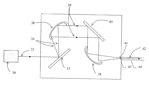
sources. Referring to FIG. 1, there is shown an embodiment of the invention
where
the light generating means are embodied by a single light source 30 generating
a
multi-wavelength light signal 32. The generated light 32 coming out of the
source
is collimated using standard collimation techniques adapted to the light
source 30.
The device further includes encoding means for encoding at least a portion of
the
stimulation information into individual wavelength components produced by the
light source. The expression "wavelength component" is used herein to refer to
either a single wavelength X or a finite wavelength band or channel AA. For
convenience, the wavelength components will generally be designated by the
symbol AX. In the embodiment of FIG. 1, the different wavelength components
(AXi, AX2, ..., AX) of the multi-wavelength light signal 32 are first
spatially
separated by a dispersive element 34 and with the help of a focussing element
38,
CA 02526327 2005-11-09
14
the separated wavelength components 36 are then redirected in a collimated
beam 39. The signal amplitude of each different wavelength component (Aki,
Ak2,
Akn) is then individually controlled with a spatial light modulator (SLM) 40.
The
spatial light modulator may for example be embodied by a liquid crystal
display
(LCD) linear array or a linear array of micro-mirrors. This control on the
signal
amplitude of each wavelength band (Aki, A2.2,
AX) allows the encoding of a
portion of the stimulation information into each of the separated wavelength
components 36. Depending on the target application of the device, each
wavelength component may be encoded with the same or different stimulation
io information as the other wavelength components.
The resulting collimated light beam with separated wavelength components 36
having different signal amplitudes along its transverse direction is then
multiplexed
into a unique encoded light signal 42 at the focal point 41 of another
focusing
element 38, preferably a cylindrical focussing element and enters the input
end 43
of the primary optical fiber 44. As will be readily understood by one skilled
in the
art, any alternative optical component of the optical arrangement may be used
in
order to multiplex the encoded wavelength components together.
Of course, the multiplexed encoded light signal may be obtained by a variety
of
different appropriate optical assemblies. By way of example, FIGs. 2 to 6 and
6A
show alternate manners of generating, encoding and multiplexing a plurality of
wavelength components according to preferred embodiments of the present
invention.
-)5
Referring to FIG. 2, there is shown an embodiment where different collimated
sources 30 are used to generate a multi-wavelength encoded light signal 42.
Each source 30 emits a collimated light beam 36 of a different spectral
bandwidth
AX selected to embody one wavelength component. The emitted collimated light
beam 36 is modulated at the source so as to encode the stimulation information
therein. The collimated light beam 36 from each source 30 is then multiplexed
CA 02526327 2005-11-09
using a focussing element 38, preferably a spherical mirror, into a unique
encoded
light signal 42 at the focal point 41 of the focusing element 38. This
arrangement
provides a more efficient means of coupling the encoded light signal 42 into
the
primary waveguide 44 by allowing focussing of the generated light beam 36
along
5 both (vertical and horizontal) axes.
Yet another embodiment is shown in FIGs. 3 and 4. Similar to the embodiment of
FIG. 2, different collimated sources 30 are used; each source emitting one
wavelength component in the form of a collimated light beam 36 of a different
lo spectral bandwidth Ak and each modulated to be encoded directly with the
required stimulation information. Unlike the arrangement shown in FIG. 2, the
sources 30 are arranged around the primary optical fiber 44. The collimated
light
beam 36 of each source is directed to a focussing element 38 that reflects it
towards a focal point 41 proximate the input end of the primary optical fiber
44.
15 The focussing element 38 is preferably a metallic-coated spherical
mirror. The
mirror coating is chosen to allow good reflectivity at all source wavelengths
whereas its radius of curvature is such that the beams are all directed and
focused
to fit into the core of the primary waveguide 44 and the numerical aperture.
The
different collimated light beams 36 originating from the different sources 30
are
multiplexed into a multi-wavelength encoded light signal 42 at this focal
point 41
which is then coupled into the primary optical fiber 44. This scheme
advantageously provides a more efficient coupling of the various light sources
into
the primary fiber over the one presented in FIG. 2 because the collimated
light
beams from the various light sources have similar optical paths.
-)5
Another possible arrangement for the generation of an encoded multiplexed
multi-
wavelength signal is provided in FIG. 5. Here different light sources 30 emit
a
collimated light beam 32 which includes at least one wavelength component Ak,
but may have a larger spectral width or different spectral profile. In the
illustrated
embodiment, the encoding takes place directly at the source through proper
modulation thereof, but in a variant embodiment, a spatial modulator may be
CA 02526327 2005-11-09
16
positioned downstream each source. Target, pre-modulated wavelength
components from each source light beam 32 is selected through reflection by an
appropriate dichroic mirror 37, or multi-wavelength partial reflector, placed
at an
angle (preferably 45 degrees) with respect to the light-guiding axis of the
primary
waveguide 44 along a common axis, thereby multiplexing the reflected
wavelength
components into the encoded light signal 42. This encoded light signal is then
coupled into the core 46 of the primary optical fiber 44 through focusing by a
lens
38 having a focal length and position appropriate to the numerical aperture
and
dimension of the fiber core 46.
Referring to FIGs. 6A and 6B, there is shown an alternative to the embodiment
of
FIG. 5 where the light sources 30 are positioned transversally to an input end
of
the primary optical fiber 44 at different positions along the length thereof,
such that
their modulated collimated light 32 is aligned with optically transparent
windows 54
is provided in the cladding 48 of the primary waveguide 44. Thus, the
source lights
32 are directly transmitted through the optical windows 54 into the core 46 of
the
primary waveguide 44 where the appropriate wavelength components are selected
and multiplexed by reflection using dichroic mirrors 38 provided directly in
the core
of the primary waveguide. To optimise coupling of the collimated sources 30,
lenses (not shown) can be placed between the sources 30 and the optical
windows 54. The focal length of the lenses should be appropriate to the
numerical
aperture and dimension of the fiber core 46
According to another preferred embodiment (not shown), modulated collimated
light from light sources may be first individually coupled into small
waveguides.
These small waveguides may then be bundled and simultaneously coupled into a
larger primary waveguide.
All of the embodiments described above provide different manners of sending an
encoded multiplexed light signal into the input end primary waveguide. It is
of
CA 02526327 2011-01-27
g
17
course understood that other assemblies achieving the same result would also
be
considered within the scope of the present invention.
In one preferred embodiment, for example shown in FIG. 7, the optical
waveguide 44
is a conventional fiber having a cladding 48 and a core 46. In another
embodiment,
illustrated in FIG.13, the optical fiber 44 is a micro-structured fiber having
an air
cladding 48 composed of air gaps 63 and fused silica bridges 64. Such an
optical
fiber allows a greater numerical aperture and thus higher modes of
electromagnetic
radiation (i.e., light) and greater light coupling capabilities. In both case,
the core 46 of
the fiber 44 defines its light-guiding axis. Coupling of the encoded light
signal at the
input end of the primary waveguide may be accomplished in any appropriate
manner,
as will be readily understood by one skilled in the art. The length of the
primary
waveguide is preferably selected as a function of the required distance to the
target
stimulation sites for a given application of the invention.
The outcoupling of the encoded light signal at the output end of the primary
waveguide will now be described according to several preferred embodiments of
the
invention.
Referring to FIG. 7, the output end of the primary waveguide of a device
according to
a preferred embodiment of the invention is shown. Outcoupling means, also
referred
to as outcoupling arrangement, for transversally coupling each wavelength
component of the encoded light signal out of the primary waveguide 44 at a
different
output position 50 along the light-guiding axis are provided. In the
embodiment of
FIG. 7, the outcoupling means include at least one reflecting element,
preferably an
optical grating 57. The optical grating may be a Bragg grating which is
chirped so that
different wavelengths are deviated, or reflected, at different positions along
the fiber
CA 02526327 2011-01-27
17a
44, and blazed (the fringes are at an angle with respect to the propagation
axis) so
that the deviated wavelengths are coupled out of the fiber 44 through its
cladding 48.
Standard, non-chirped, blazed Bragg gratings at different wavelengths may also
be
used if __________________________________________________________________
,
CA 02526327 2005-11-09
18
they are placed at different positions along the fiber. Long-period gratings
may
also be a preferred embodiment if the density of output positions 50 is not
important and if the spectral linewidth of the outcoupled light can be wider.
In other preferred embodiments shown in FIGs. 8 and 9, the output coupling
means include dielectric reflectors 58 placed at an angle inside the fiber
core 46.
Each dielectric reflector 58 reflects a specific wavelength with a specific
linewidth
so that only part of the spectrum is coupled out of the fiber 44 through its
cladding
48. A thorough description of the method and means used to introduce
reflective
and/or refractive components in an optical fiber is given in assignee's U.S.
patent
application filed on the 21 of October 2005, entitled "Optical Fiber Devices
Using
Component Insertion" by inventors Rene Beaulieu, Daniel Cantin, and Marc
Levesque.
In yet another preferred embodiment illustrated in FIG. 10A, shaping of the
waveguide core 46 and cladding 48 is used to provide reflecting elements 56.
These reflecting elements 56 reflect specific wavelength components of the
encoded light signal 42 towards output positions 50 and out of the fiber core
46.
Alternatively, according to the embodiment of FIG. 10B, shaping in the
waveguide
core 46 provides refracting elements 59 to refract specific wavelength
components
of the encoded light 42 through output positions and out of the fiber core 46.
For a number of applications, it is desirable to transform the optical
stimulation
information in each wavelength component into an electrical stimulation
signal,
Referring to FIG. 11, localized electrodes 52 may be provided on the outer
surface
of the primary waveguide for this purpose. The electrodes are preferably
composed of layers of photoelectric material deposited at the output positions
50
where light is coupled out of the fiber 44. The expression "photoelectric
material"
generally refers to a material whose electric properties are affected by
exposure to
light and includes photovoltaic and photoconductive material. Photovoltaic
materials are capable of producing a voltage when exposed to electromagnetic
CA 02526327 2005-11-09
19
radiation. Electrical conductivity of photoconductive material is affected
by
exposure to electromagnetic radiation. Preferably, this photoelectric material
is
biocompatible with the cells of the tissue to be stimulated.
In the particular case of photoconductive material, means to apply a
polarization
voltage to the fiber 44 are also provided. One of these means, shown in FIG.
11,
could be the use of a small electrical wire 61 running along the fiber
cladding 48
and making electrical contact 62 with the photoconductive material. Laser
micro-
machining could be used to produce a groove along the fiber 44 in order to
insert
the small electrical wire 61. Another means would be to micro-machine,
preferably
with a laser, a slot along the fiber-preform cladding 48 that would produce a
groove for the electrical wire 61 once the preform is pulled into an optical
fiber 44.
Finally, in another preferred embodiment shown in FIG. 12, the glass cladding
48
of the optical fiber 44 can be covered with a metallic cladding 47 that can be
laser-
machined to create an electrical contact 62 with the photoconductive material
52.
In this case, the metallic cladding 47 would be covered, or coated, with a non-
conductive material 49 to ensure its electrical insulation outside of the
electrode
regions. In the preferred embodiment of FIG. 12, laser micro-machining
techniques are used to provide grooves 45 in the metallic cladding 47 of the
fiber
44 which receive the electrodes 52. In another preferred embodiment, grooves
with a small length extent are made in the preform to be pulled into an
optical fiber
and when the pulling of the fiber is performed these grooves extend to fit the
length of the electrodes to be put on the fiber. If a photovoltaic material is
used for
the electrodes, it preferably includes GaAs crystal, which is preferred over
silicon
crystal owing to the smaller thickness required to achieve the same
efficiency.
Silicon crystal usually requires a thickness of several hundreds of microns to
obtain energy conversion efficiencies of over 10% while only a few microns are
sufficient in the case of GaAs. However, thin film materials produced through
deposition processes are preferable over crystalline material since the
required
thickness can be one micron or less owing to its higher absorptivity.
Furthermore,
the bonding of the electrode material to the fiber is also much easier in the
case of
CA 02526327 2005-11-09
,
=
/0
thin film materials since they can be directly sprayed into the laser micro-
machined
grooves of the optical fiber. The energy conversion efficiency of the material
in thin
film form is however less than that in crystalline form. New materials such as
photoconductive and photovoltaic polymers [for example, poly(p-
phenylenevinylene (PPV)] and dye-integrated titanium dioxide (Ti02) could
shortly
become preferred materials given their ease of integration into the optical
fiber
grooves. Many polymers could be integrated through wet coating processes while
TiO2 could be integrated with standard vacuum deposition processes. If signal
response times of over a few milliseconds are required, a pyroelectric
material,
to such as polyvinylidene fluoride (PVDF), may be preferable since it
may be
deposited easily using wet coating techniques and requires a thickness of a
few
tens of micrometers. Biocompatibility issues regarding the photovoltaic and
photoconductive material of electrodes can be addressed by coating the
photovoltaic and photoconductive material with biocompatible materials such as
polyimide.
Each deposition area therefore defines an "electrode". The density of the
electrodes provided on a given device depends on the selected manufacturing
techniques. Depending on the application, with a typical 125 pm-diameter
optical
fiber, it is possible to achieve an array of electrodes, each measuring 200 pm
long
by 50 pm to 90 pm wide, and spaced by 50 pm, leading to a density of 40
electrodes/cm. Up to 160 electrodes can be arranged on a 40 mm length,
allowing
very high resolution with a very small diameter. Evidently, if the charge
density
needs to be below a given value, the electrode size can be adjusted
accordingly.
,5
In some cases, for example where the density of axonic terminals of nerve
cells
are clustered into ganglia, the use of electrical stimulation may be
complicated by
the simultaneous creation of extraneous stimuli. Although an aim of multi-
electrode implants is improvement in the specificity of the stimulation, the
real
advantages to using multi-electrodes are limited by the current required to
attain
the threshold of perception. The current required is often greater in the case
of
CA 02526327 2005-11-09
/1
closely spaced multi-electrodes than for farther spaced single electrodes.
This
leads not only to increased extraneous stimuli, thus defeating the purpose,
but to
increased risk of injury to the patient.
One alternative to the problems of specificity and injury inherent with
electrical
stimulation is photo-stimulation. As suggested by an embodiment of the
invention
illustrated in FIG. 14A and 14B, the electrodes 52 used in transmitting
stimulation
information signals to the stimulation sites may be replaced by optical
windows 54
provided in the primary waveguide 44 at the output positions 50 of the
wavelength
components of the encoded light signal 42. These optical windows 54 allow the
light stimulation signal to be transversally coupled out of the optical fiber
44 by
refraction as illustrated in FIGs. 10B, 14A and 14B or by reflection as
illustrated in
FIGs. 7 to 10A. According to the embodiment of FIG. 14, an optical window 54
may be produced in the side of a primary waveguide 44 by laser micromachining
through the cladding 48 of the waveguide 44 and filling an air gap 63 with
appropriate optically transparent material, such as silica glass. According to
another preferred embodiment, the optically transparent material defining the
optical window 54 may simply be the optically transparent material of the
optical
waveguide 44 or fiber cladding 48 itself, providing it is made of a
transparent
material. In another preferred embodiment, the optical window is made of a
material having a refractive index higher than the refractive index of the
fiber core
so as to increase the output coupling efficiency through the optical window.
In yet
another embodiment, the optical window is made of material, which may include
a
dielectric coating that transmits specific wavelengths of light, for example
those
corresponding to specific photoactivated molecules, while reflecting others.
Finally,
these preferred embodiments of an optical window may be combined in such a
way as to optimize the desired results with respect to the requirements of the
application.
One other option, illustrated in FIGS. 15A and 15B, is to use secondary
waveguides 66 attached to the core 46 of the primary waveguide 44 at output
CA 02526327 2005-11-09
_
22
positions 50. The optical windows on the primary optical fiber may be used as
the
entry windows to the secondary optical fibers. The secondary waveguides may be
fused 68, preferably using silica powder as fusing agent, to the secondary
waveguides 66 at the output positions 50. In FIG. 15A, dielectric reflectors
58
ensure the coupling of light out of the primary fiber 44 and into the
secondary
fibers 66. The coupling of light into the secondary fiber 66 can be done
through
refraction means, by attaching the secondary fibers 66 to the primary
waveguide
core 46 at an angle, as shown in FIG. 15B, and matching the numerical
apertures
to the primary fiber 44. The use of secondary fibers is particulary
advantageous in
io cases where more specific or accurate positioning of the outcoupled
light and/or
access to more distant sites is necessary.
The secondary fibers are ideally
smaller than the primary fiber so that the distal part of the device remains
compact
allowing precise positioning without damaging the environment at sites of
interest
during the implantation surgery. Such damage can render the device completely
inoperative. In general, the connection between the primary fiber and the
secondary fibers may be done through laser micro-machining, including ablation
and fusion processes. The use of laser micro-ablated "V" grooved substrates
that
help to manipulate and align the fibers with respect to one another is
preferred.
Once the alignment is properly done, the fibers can be attached to the primary
fiber and/or the substrate, preferably by laser fusion. In the preferred
embodiment
illustrated in FIGs.15A and 15B, a capillary 70 is placed around the primary
fiber
44 to allow the attachment of a secondary fiber using laser fusion 68. Holes
are
drilled into the sides of the capillary 70 up to the core 46 of the primary
fiber 44 to
allow the passing through of the secondary fibers 66 prior their fusion. The
assembled fibers can then be packaged into a single device that can be
implanted
in a patient. This type of packaging provides added robustness to the device,
since
the primary fiber may be weakened following the laser ablation and/or fusion
processes used to attach the secondary fibers thereon. The secondary fibers
which are kept intact are less susceptible to breakage, more flexible, and can
be
coated with a material that enhances their robustness, such as polyimide which
is
also biocompatible. In the previous embodiments, the secondary fibers,
preferably
CA 02526327 2005-11-09
-
,3
their distal ends, may be equipped with electrodes composed of photoconductive
or photovoltaic materials for electrical stimulation.
In the following description the term "photoactivated molecules" refers to
both
caged molecules that become uncaged (or released) or are made chemically or
biochemically active when illuminated by light at specific wavelengths, and
molecules that reflect, absorb, or reemit characteristic luminescent light
when
illuminated with light of specific wavelength. These photoactivated molecules
may
be biochemical compounds, such as hormones, enzymes, neurotransmitters, etc,
or molecules caged in quantum dots, micro-spheres, nanoshells, micelles or
combinations of these.
Light coupled out of the waveguide can be used to photoactivate specific
molecules which then directly or indirectly stimulate or inhibit specific
cell, tissue,
or system functions. The wavelength of the light is chosen to match the
photoactivation wavelength of the photoactivated molecules. In this case,
modulation of the intensity of the light source will allow the modulation of
the
stimulation or inhibition of the function to be controlled. FIGS. 16 ¨ 18
depict the
photoactivation of caged molecules, such as those used in the regulation of
cell
growth and migration, placed in the vicinity of the cells to be stimulated or
inhibited
to maximise the coupling between the fiber and the cells and increase its
stimulation efficiency. FIGs. 19A and 19B depict the uptake of originally
inactive
but photoactivatable molecules by a nerve cell during growth. Cell processes
in
the nerve cell may be studied through either monitoring of luminescent
molecules
which are uncaged by certain nerve cell functions (such as nerve impulse) and
thus act like markers or measurement of changes in physical and chemical
properties (such as electrical activity) of the nerve cell resulting from the
photoactivation of specific molecules. FIG. 21 shows the use of a secondary
fiber
to more specifically photoactivate molecules (or excite luminescence) in a
certain
region of the nerve cell. FIG. 22 is a schematic illustration of a
preferred
CA 02526327 2005-11-09
,
/4
embodiment of the stimulation device showing its use as a means to study
living
nerve tissue grown in culture.
The case of neurostimulation, stimulation or inhibition that may be produced
via
photoactivation of molecules corresponding to neurotransmitters specific to
the
ganglion cells (or neuron types) to be stimulated, is shown in FIG. 23. These
molecules are biochemically inactive (or caged) prior to being illuminated.
Under
specific wavelength illumination, the caged molecules undergo either a
structural
or chemical change that makes them chemically active in the environment of the
cells to be stimulated or inhibited. In another preferred embodiment, the
active
molecules are placed inside micro-spheres, quantum dots, micelles, or
nanoshells
made of bio-resistant and bio-inert materials that change properties upon
illumination. Under illumination at specific wavelengths the bio-resistance of
the
micro-spheres decreases and the caged molecules are released and become
active.
These caged and photoactive molecules need to be placed in the vicinity of the
cells to be stimulated or inhibited so that they may perform their expected
functions properly. This may be accomplished by fabricating a channel in the
primary waveguide along its length. The molecules would then be injected in
solution form into the channel, exit the channel through a small opening in
the
optical fiber at the output position and thus be placed in the vicinity of the
stimulation site. If micro-structured fibers are used (see FIGs. 13 and14),
one or
more of the air gaps in the fiber can be used as injection channels much in
the
same manner. Another means of introducing the molecules is through
conventional injection into the blood stream of a solution containing the
molecules,
providing that the molecules can reach the specific stimulation area through
this
scheme. Otherwise, the molecules may be injected directly into the specific
area to
be stimulated using a syringe.
CA 02526327 2005-11-09
,
In one preferred embodiment, illumination of different molecules at different
wavelengths may preferably be performed simultaneously. Consider, for example,
the case where two neurons are located in proximity to one another and the
stimulation process of a particular neuron is independent of the stimulation
5 process of another neuron. It is possible to photoactivate this
particular neuron by
using light of a given wavelength to photoactivate specific molecules in the
vicinity
involved in its stimulation and to photoactivate the other neuron by using
light of a
different given wavelength to photoactivate different specific molecules also
in the
same vicinity but which are involved in the stimulation of this other neuron.
In this
10 way, the stimulation of both neurons may occur simultaneously but yet
independently ¨ there is no need to carry out the photoactivation at different
times
in order to limit crosstalk-like behaviour. In another preferred embodiment,
the
illumination at different wavelengths is performed sequentially. For example,
if one
photoactivated molecule needs to be put in the presence of another
15 photoactivated molecule to become effective, the illumination at the
photoactivation wavelength of the first molecule will have to be performed
prior to,
or simultaneously with, the illumination of the second photoactivated molecule
at
the second wavelength. This is illustrated in FIG. 24 and 25.
In some cases, it is known that light can stimulate the process of cellular
growth (J.
Q. Zheng, "Turning of nerve growth cones induced by localized increases in
intracellular calcium ions", Nature, vol. 403 (2002) p. 89). This may be done
by
using specific photoactivated growth factors (e.g. molecules, proteins or
hormones) to stimulate the growth of a specific type of cell placed in the
immediate
vicinity of the outcoupled light of the present device. For best results, stem
cells
may be added to the site at the time of surgical implantation of the device.
This
would be especially beneficial especially for cells that do not naturally grow
or
divide in adult patients, for example neurons. This is illustrated in FIGs.18
and 19.
Using this process, the coupling efficiency of the implanted device with the
natural
neuronal network may be increased by making specific neurons grow toward the
light outputs of the device.
CA 02526327 2005-11-09
26
Cells that contain photo-luminescent molecules, either naturally or by induced
uptake (see FIGs. 18 and 19) can be used to monitor cellular activities. These
molecules may be strategically chosen to control some of the cellular
functions
through direct or indirect detection of their presence, for example detection
through luminescence. Accordingly, this process could be used to monitor the
nerve impulse in neurons through polarisation of calcium, potassium or other
ions
or through the presence of neurotransmitters at synaptic connections
indicating
neuronal activities of specific neuron cells. This method may be used to
replace in
Jo lieu of electrical stimulation of neurons using implanted electrodes to
detect nerve
impulse. In this case, the proposed invention is used to provide illumination
at the
excitation wavelength from the proximal end to the photo-luminescent molecules
at the distal end and the luminescence signal emitted by the molecules is
collected
by the same device (see FIGs 20 and 21) working with light traveling in the
opposite direction e.g. to the proximal end. The luminescent light can then be
detected and analysed to measure the nerve impulse. To obtain the best
results,
the preferred embodiment uses, at the distal end of the device, output and
input
coupling techniques that are not dependent on the wavelength or dichroic
components that can handle both the excitation and luminescence wavelengths.
The same techniques described in preferred embodiments used to couple out the
light at the distal end can be used to couple the luminescent light into the
fiber up
to the proximal end. Another preferred approach, illustrated in FIG. 20, is to
use
two optical windows to provide illumination at the excitation wavelength and
to
collect the luminescence by a third window placed between them. The excitation
illumination can be performed with dielectric mirrors having high reflectivity
at the
excitation wavelength while the collection of the illuminescent light via the
collecting window can be performed using a dielectric mirror having a high
reflectivity at the luminescence wavelengths. Another approach illustrated, in
FIG.
21, is to use a secondary fiber to provide the excitation wavelength and a
window
in the primary fiber to collect the luminescent light. Yet another preferred
approach
is to use one secondary fiber to provide the excitation wavelength and another
one
CA 02526327 2005-11-09
27
to collect the luminescence. In this case, the two secondary fibers are placed
in
close vicinity to each other to ensure sufficient luminescent signal
collection (see
FIG 15). This monitoring technique of luminescent signal related to specific
biochemical concentration in the body can allow to diagnose pathologies,
control
concentration levels or presence of some compounds (glucose, iodine, ...), or
type
of cells (cancerous cells, stem cells, ...), or to stimulate their growth in a
specific
type of tissue while combined with photoactivated growth factors.
Photoactivated molecules can be used either directly or indirectly. Direct use
of
photoactivated molecules implies that once molecules are activated they will
react
chemically or biochemically with a cell to stimulate or inhibit one of its
functions.
Indirect use implies that once the molecules are activated they will react or
combine with one or many other molecules to produce a chemical or biochemical
compound that will react with the cell to stimulate or inhibit one of its
functions.
One preferred embodiment of direct use of photoactivated molecules is a
photoactivated neurotransmitter that could be used to initiate the stimulation
of a
nerve impulse to neuron cells, as shown in FIG. 23. One preferred embodiment
of
indirect use of photoactivated molecules is a photoactivated molecule that
will
combine with another molecule that could be naturally present or injected in
the
body to form an antagonist of neurotransmitter that could be used to inhibit
the
stimulation of a nerve impulse to neuron cells. Another preferred embodiment
of
indirect use of photoactivated molecules is the use of two different
photoactivated
molecules that will be activated at different wavelengths and that combine
together
to form a molecule that stimulates or inhibits a cell function, as shown in
FIG. 24
and 25. Yet another preferred embodiment of indirect use of photoactivated
molecules relates to a caged molecule that can be uncaged (or released)
through
photoactivation by one or more specific wavelength components, but that
becomes biologically active only once it is photoactivated by one or more
different
wavelength components. Some examples of applications of photoactivated
molecules include: control of insulin for diabetics (monitoring and
photoactivation),
control of the level of iodine compounds for hypo- and hyper-thyroidism
CA 02526327 2005-11-09
28
(monitoring and photoactivation), photodynamic therapy (creation of compounds
that can specifically link and kill cancerous cells through photoactivated
molecules), and stimulation of the growth of a specific type of cell.
In another embodiment, light is used to provide heat at the distal end of the
optical
fiber. The heat can be directly provided to molecules or cells by using the
scheme
illustrated in FIG. 7, 8, or 9 to couple out light 60 at specific wavelengths
that are
absorbed by the molecules or cells. If the absorbed wavelengths are mainly
converted into vibrational or rotational energy of the molecules rather than
reemitted as photons at longer wavelengths, the absorbed light heats the
molecules. This process is more likely to occur at wavelengths in the infrared
portion of the electromagnetic spectrum. One other preferred embodiment for
providing heat uses an indirect heating process through the heating of a
material
at the output position 50 placed on the cladding 48 of the fiber 44. The
heated
material can then be used to heat molecules or cells that are put into contact
with
it. The heating can be used to stimulate or inhibit specific cell functions in
the
vicinity of the fiber, activate specific molecules, or uncage caged molecules
in
micro-spheres, micelles, quantum dots or nanoshells that can be affected by
heat.
The use of other wavelengths that would not heat the fiber environment or the
heating material placed on the fiber would allow to monitor results of the
heating
process or to identify the presence of molecules, cells, micro-spheres,
micelles,
quantum dots or nanoshells to be heated. This monitoring or identification
process
could be done from the analysis of collected light through the same point as
the
heating point on the fiber or through other adjacent points in ways similar to
those
illustrated in FIGs 20 and 21.
It is also possible to use multiple points along the optical fiber where the
light can
be partly coupled out from the fiber to stimulate, or to monitor, similar or
different
cell functions. This way one optical fiber may have multiple devices connected
to it
that may be implanted at different places inside the body. The use of
different
CA 02526327 2005-11-09
=
/9
wavelength bands for each device can allow to control independently the
stimulation, or the monitoring, at each of the implanted positions inside the
body.
Of course, numerous modifications or combinations of these preferred
embodiments could be made to the device above without departing from the scope
of the present invention.
Method and Applications
In accordance with one application of the present invention, an embodiment of
the
ii) device described above may be used as a cochlear implant for
transmitting
auditory stimulation information to auditory neuron sites of the cochlea, in
situ of a
patient. Such a cochlear implant includes a light generating means for
generating
light having a number of wavelength components, an encoding means for
separately encoding at least a portion of the auditory stimulation information
into
each of the wavelength components, and a multiplexing arrangement for
multiplexing the wavelength components encoded by the encoding means into an
encoded light signal. The cochlear implant further includes a primary
waveguide
having an input end operationally connected to the multiplexing arrangement
for
receiving the encoded light signal therefrom, a light-guiding axis for guiding
the
encoded light signal therealong and an output end adapted to be positioned
proximate the auditory neuron sites of the cochlea.
In addition to the above
elements, the device also has outcoupling means provided at the output end of
the
primary waveguide. These outcoupling means transversally couple each of the
wavelength components of the encoded light signal out of the primary waveguide
at different output positions along the light-guiding axis, each of the output
positions being coupled to one of the auditory neuron sites of the cochlea.
In one embodiment, the cochlear implant preferably includes a number of
electrodes, each associated with one of the output positions, for transducing
a
corresponding wavelength component into an electrical stimulation signal. In
another embodiment, the cochlear implant preferably includes an optical window
in
CA 02526327 2005-11-09
the primary waveguide at each of the output positions, in order to output an
optical
stimulation signal therefrom.
The present invention can provide great improvements to the technology of
5 cochlear implants and address some of the drawbacks listed above. The
greater
number of electrodes afforded by the present invention helps to provide a
greater
resolution than most typical devices. Risk of injury to a patient's inner ear
can also
be reduced by using optical stimulation rather than electrical stimulation.
Optical
stimulation advantageously offers increased specificity through the use of
optical
io fibers with diameters smaller than those achievable with traditional
wires and
safety given that optical fibers may be fabricated out of plastic or glass
material,
which is relatively inert. Optical fibers are also very flexible and are
generally less
subject to mechanical fatigue than metallic wire conductors. Furthermore,
optical
fibers can be overcoated with biocompatible materials minimising adverse
IS reactions by host biological material and increasing the strength of the
fibers while
maintaining their compactness and flexibility. A preferred embodiment of the
present invention as a cochlear implant is illustrated in FIG. 26. As shown,
the
flexibility of optical fibers allows to shape and adapt the implant to the
particular
structural anatomy of the patient. Such a shape could be made permanent by
20 heating the fiber, preferably with a heat gun or a CO2 laser, while it
is rolled over a
cylindrical or conical shape.
Cochlear implant knowledge and technology is continually changing and
evolving.
Research is underway to design implants that would help people with deafness
25 due to surgical removal of their auditory nerves during tumor resection.
These
implants would stimulate the cochlear nucleus, the first stop after the
auditory
nerve in the auditory pathway to the brain. Some research is looking into
implants
that would stimulate the auditory nerve directly. The present invention would
certainly be of benefit to such applications given the compactness of the
device
30 and the increased number of electrodes that may be implanted with a single
device.
CA 02526327 2005-11-09
31
With reference to FIGS. 1, 7, and 8, the generation of an appropriate light
signal in
the particular application of a cochlear implant is illustrated. The multi-
wavelength
collimated light beam 32 coming out of a modulated light source 30 has its
different wavelengths components (AM , AX2, ,AXn) spatially separated 36 by
a
dispersive element 34. The light coming out of the light source 32 is
preferably
collimated by standard collimation techniques adapted to the light source 30
used.
The separated wavelengths components 36 are then redirected in a collimated
beam 39 with the help of a cylindrical focusing element 38. The signal
amplitude of
each different wavelength (AM , AX2, AXn) is then individually controlled
with a
spatial light modulator (SLM) 40. The resulting collimated light beam with
separated wavelengths having different signal amplitudes along its transverse
direction is then multiplexed in a unique encoded light signal into the
optical fiber
42 at the focal point 41 of another cylindrical focusing element 38. This
control on
the signal amplitude of each wavelength band (AX1, AiL2, AXn) allows to
control the electrical signal level generated at each electrode 52 or to
control the
light signal level coupled out 60 at each location 50 along the optical fiber
44.
In a preferred embodiment, the light source 30 may include a light emitting
diode
(LED) having a spectral content extending from 15 to 40 nm or it may include a
laser diode having similar extended spectral content. The light source 30 may
be
current modulated from a few hundred Hz up to 18 KHz to increase the
stimulation
response of the excited nerve cells and improve speech recognition of the
implant
patients. In one preferred embodiment, the dispersive element 34 is a blazed
grating used in reflective mode. The separated wavelength components of the
light
source 30 are then collimated with the use of cylindrical focussing elements
38,
preferably either a cylindrical mirror or lens 38. These cylindrical focusing
elements 38 must be adapted to both the grating dispersion angle of the
spectral
content of the light source 30 and the dimension of the spatial light
modulator 40.
Different wavelength components 36 may then travel in parallel separated paths
39 and their signal intensity may be individually varied with the use of a
linear
CA 02526327 2005-11-09
32
spatial light modulator 40 composed preferably of a LCD linear array having
refreshment rates from 120 to 400Hz. The array will have a number of elements
at
least equal to the number of electrodes 52 (160 in the current example) on the
optical fiber 44. Each element of the LCD array is used, for a specific
wavelength,
as a light attenuator in transmission mode that can be individually
controlled.
Another preferred spatial light modulator 40 is a linear array of micro-
mirrors
having dimensions in the range of 0.1-1mm and capable of angle position
changes
in the range of 1 to 5 degrees. Each micro-mirror of this array will control
the beam
direction of a specific wavelength. A change in direction of the beam will
modify
the amount of light coupled into the optical fiber 44 at that specific
wavelength and
then to the corresponding specific electrode 52 on the optical fiber of FIGS.
7 and
8. Another cylindrical mirror or lens 38 is used to focus the collimated multi-
wavelength encoded light signal 42 into the optical fiber 44 to form a
multiplexed
signal that will be demultiplexed by the blazed optical grating 57, or
dielectric
reflector 58, to provide the required signal to each output position 50.
The present invention is of course not limited to cochlear implants and may be
applied to any number of electrical and optical stimulation technologies, old
and
new.
In accordance with another aspect of the present invention, there is generally
provided a method for transmitting stimulation information to a plurality of
stimulation sites. For example, these stimulation sites may be embodied by
cerebral neuronal sites along a visual pathway ¨ the stimulation information
thereby stimulating a visual response, by muscle tissue sites whose
contraction is
to be stimulated or host tissue whose growth is to be stimulated. The use of
biochemical compounds adapted for photoactivation by the wavelength
components at these stimulation sites is also contemplated. These and more
examples will be described in more detail further below.
The method generally includes the following steps of:
CA 02526327 2005-11-09
33
a. generating light having a plurality of wavelength components.
This may be accomplished by activating a plurality of light sources, each
generating one of the wavelength components, or activating a light source
generating a multi-wavelength light signal which includes these wavelength
components.
b. separately encoding at least a portion of the stimulation information
io into each of the wavelength components.
If the wavelength components are generated by separate sources, this may for
example be accomplished by directly modulating the amplitude of each generated
wavelength component at the source. This modulation control may be timed so
that the wavelength components are encoded simulatenously or sequentially.
If the wavelength components are generated as a multi-wavelength light signal,
a
step of separating said multi-wavelength light signal into said wavelength
components may be performed between steps (a) and (b), so that the amplitude
of
each wavelength component may then be modulated separately.
c. multiplexing the wavelength components encoded by the encoding
means into an encoded light signal.
This for example accomplished by placing a focussing element in the path of
the
wavelength components, or by any other appropriate technique known in the art.
d. guiding the encoded light signal along a light-guiding axis of a primary
waveguide.
CA 02526327 2005-11-09
34
As mentioned above the primary waveguide is preferably an optical fiber having
a
core and a cladding.
e. transversally coupling each of the wavelength components of the
encoded light signal out of the primary waveguide at different output
positions along the light-guiding axis, each of these output positions
being coupled to one of the stimulation sites.
This may be accomplished by placing appropriate outcoupling elements at the
outcoupling end of the waveguide. In one embodiment, at least one blazed
optical
grating is provided in the optical fiber, an example of which may be a single
chirped Bragg grating having a period selected to reflect each of the
wavelength
components at one of the output positions along the light-guiding axis, a
plurality
of uniform Bragg gratings each positioned at one of these output positions and
is associated with one of the wavelength components, or a long-period
grating
having a period selected to reflect each of the wavelength components at one
of
these output positions. In another embodiment, a plurality of dielectric
reflectors
may be provided in the optical fiber oriented at an angle with respect to the
light-
guiding axis, each being positioned at one of the output positions and being
associated with one of the wavelength components.
An optional additional step of converting the wavelength components into
electrical
stimulation signals may also be provided. This step preferably includes
providing a
plurality of electrodes, each associated with one of the output positions.
Each
electrode is preferably made of a layer of photoelectric material deposited on
an
outer surface of the primary waveguide, in which case a polarization voltage
is
applied to this photoelectric material.
Alternatively, the transversal coupling of step (e) may be accomplished
through an
optical window provided in the primary waveguide at each of said output
positions.
CA 02526327 2005-11-09
As mentioned above, the teachings of the present invention may be used to
provide stimulation information to a variety of stimulation sites, depending
on the
particularities of the applications considered. For example, the proposed
invention
could beneficially be used as biofeedback implants in people with limb
prostheses.
S A major problem with the use of these prostheses is the lack of feedback,
or
sensation. With sensors placed on the prosthesis and the proposed invention
device implanted on sensitive nerves linked to the touch, one would be able to
obtain sensation about the pressure, temperature, texture, weight, and
position of
objects touched by the prosthesis. Position and tension sensors could also be
10 used to sense the position of the prosthesis in space and the strength
applied to
the motors used to activate it.
New prosthesis developments use metals, such as titanium rods, permanently and
directly implanted inside the bones to which the prosthesis can be solidly
attached.
is One can think of possibly inserting the proposed device in the body
through these
rods, using these rods as housing and connecting means for the device ¨ the
device being housed in the rods and the device output positions linked via the
implanted electrodes to nerves. Biofeedback signals from the prosthesis
sensors
could then be easily sent to the central nervous system through this new link.
Based on these biofeedback possibilities, one can extrapolate and imagine the
creation of new sensory input through the use of new interpretation schemas of
current nervous system inputs to the brain. For example, a capacitive sensor
linked with the proposed invention, implanted in the body to stimulate nerves
related to pressure sensation, may provide a sensation of the density of an
object
¨ following some training to establish the new interpretation schema in the
brain.
This type of new sense evolution is already commercially available: the vOICe
system, developed for blind people, encodes visual imagery information
captured
by camera into sound information via frequency and pitch. The sounds are fed
to
the ear of a blind patient using an earphone and, with some training, the
patient's
CA 02526327 2005-11-09
36
brain learns to interpret the sound information as visual information of the
image
captured by the camera.
Another field of application is in neurology where the proposed invention
could be
used to stimulate neuronal cells in live nerve tissue. This could allow
communication with specific nerve cells or groups of nerve cells providing a
better
understanding of their interactions within the network and of the network
itself.
From the high density of stimulation sites achievable with the proposed
invention,
one can conceive the possibility of constructing an artificial spinal cord to
conect
io members whose natural link has been severed following major injury.
Numerous modifications could be made to any of the embodiments above without
departing from the scope of the present invention as defined in the appended
claims.