Note: Descriptions are shown in the official language in which they were submitted.
CA 02526490 2011-08-25
-1-
COMPOSITIONS AND METHODS FOR TREATING AND PREVENTING
HEART TISSUE DEGENERATION, AND USES THEREOF
BACKGROUND OF THE INVENTION
[0003] Myocardial infarction (irreversible damage to heart tissue,
often due to heart
attack) is a common life-threatening event that may cause sudden death or
heart failure. The
ventricular dysfunction that arises after myocardial infarction results,
primarily, from a
massive loss of cardiomyocytes and gradual replacement of damaged
cardiomyocytes with
fibrotic non-contractile (scar) tissue. In most cases, the loss of
cardiomyocytes after
myocardial infarction is irreversible. Indeed, it is widely accepted that the
proliferative (and,
therefore, the regenerative) potential of adult mammalian cardiomyocytes is
quite limited
(Rumyantsev and Carlson, Growth and Hyperplasia of Cardiac Muscle Cells (New
York:
Harwood Academic Publishers, 1991)), although this view has recently been
challenged (Len
et al., Mol. Cell. Cardiol., 3:385-90, 2000; Kajstura et al., Am. J. Pathol.,
156:813-19, 2000;
Beltrami etal., N. Engl. J. Med., 344(23):1750-57, 2001).
[0004] Despite considerable advances in the diagnosis and treatment of
heart disease,
cardiac damage and dysfunction relating to myocardial infarction are still
among the major
cardiovascular disorders. Accordingly, it remains a major therapeutic
challenge to find new
effective approaches to improve cardiac function after myocardial infarction.
[0005] The potential to reactivate cardiomyocyte proliferation through
the
manipulation of putative cellular regulators, or the conversion of pluripotent
stem cells to
cardiomyocytes (Orlic et al., Nature, 410:701-05, 2001), offers an exciting
impetus for the
design of novel therapeutic interventions to enhance cardiac function during
disease
conditions. The bulk of evidence obtained over the past decade maintains,
however, that
mammalian cardiomyocytes proliferate throughout fetal development and into the
early
CA 02526490 2005-11-18
WO 2005/000403
PCT/US2004/015691
-2-
neonatal period, at which time DNA replication declines quickly and cell
division ceases
(Beinlich and Morgan, MoL Cell. Biochern., 119:3-9, 1993; Casscells et al., J.
Clin. Invest.,
85:433-41, 1990; Speir et al., Circ. Res., 71:251-59, 1992; Parker and
Schneider, Atinu. Rev.
Physiol., 53:179-200, 1991; Simpson, P.C., Annu. Rev. Physiol., 51:189-202,
1989).
Transition from hyperplastic growth (cell division) to hypertrophic growth
(increase in cell
size) then ensues. In the murine heart, cardiomyocyte division is reportedly
completed by
birth, with DNA synthesis in neonatal cells (through post-natal day 3)
contributing only to
binucleation (Soonpaa et al., J. MoL Cell. Cardiol., 28:1737-46, 1996). The
cessation of
myocyte proliferation is attributed to an arrest of the cell cycle (Brooks et
al., Cardiovasc.
Res., 39:301-11, 1998). In accordance with this hypothesis, adult rat
cardiomyocytes have
been shown to display a dual cell-cycle blockade, with approximately 80% of
cells arresting
in GO/G1, and 15%-20% of cells arresting in G2/M (Poolman and Brooks, MoL
Cell.
Cardiol., 29:A19 (Abstract), 1997; Poolman et al., Int. J. Cardiol., 67:133-
42, 1998).
[0006]
Progression through the cell cycle is tightly regulated, and involves cyclins
complexed with their catalytic partners, the cyclin-dependent kinases (cdks).
Among the
cyclins, cyclin A2 is unique in that it regulates progression through two
critical transitions:
cyclin A2 complexed with cdk2 is essential for the Gl/S transition, and cyclin
A2 complexed
with cdkl promotes entry into mitosis (Sherr and Roberts, Genes Dev., 9:1149-
63, 1995;
Pagano et al., EMBO j.,11:961-71, 1992). It is well-established that mammalian
cardiomyocytes cease to proliferate in the early neonatal period due to arrest
of the cell cycle.
Cyclin A2 is the only cyclin to be completely downregulated, at both the
message and protein
level, during cardiogenesis, in rat and human, in a manner that appears
coincident with this
Withdrawal of cardiomyocytes from the cell cycle (Yoshizumi et al., J. Clin.
Invest., 95:2275-
80, 1995).
[0007] Previously, it has been shown that zebrafish fully regenerate hearts
within 2
months of 20% ventricular resection, due to robust proliferation of
cardiomyocytes localized
at the leading epicardial edge of the new myocardium. This injury-induced
cardiomyocyte
proliferation was able to overcome scar formation, allowing cardiac muscle
regeneration. It
has been suggested that this regeneration of heart tissue in zebrafish is
related to the Mpsl
mitotic checkpoint kinase (Poss et al., Heart regeneration in zebrafish.
Science, 298:2188-90,
2002). It has also been shown that cardiomyocytes react to myocardial
infarction by
CA 02526490 2005-11-18
WO 2005/000403
PCT/US2004/015691
-3-
activating cyclins and cyclin-dependent kinases (Reiss et al., Myocardial
infarction is
coupled with activation of cyclins and cyclin-dependent kinases in myocytes,
Exp. Cell Res.,
225:44-54, 1996). However, prior to the present invention, it had not been
directly
demonstrated that regulation of cyclins, particularly cyclin A2, can induce
cardiomyocyte
mitosis once the timeline for cell-cycle exit (and, therefore, "terminal"
differentiation) has
been surpassed.
SUMMARY OF THE INVENTION
[0008] The inventors hypothesized that downregulation of cyclin A2
plays a crucial
role in cardiomyocyte cell-cycle exit, and that, conversely, the continued
expression of cyclin
A2 in the heart results in altered cell division and, importantly,
cardiomyocyte hyperplasia.
To determine whether the stimulation of myocyte mitotic divisions would have
an impact
upon cardiomyocyte cell-cycle withdrawal, the inventors created a cardiac-
specific cyclin A2
transgenic mouse model which constitutively expresses cyclin A2 in the
cardiomyocyte
lineage, from embryogenesis through adulthood. This model was previously shown
to
exhibit cardiomyocyte mitosis in differentiated cardiomyocytes. The inventors
now show
that the presence in the heart of cyclin A2 ¨ a protein that is normally
silenced in the post-
natal heart (and absent after birth) in mice, rats, and humans ¨ prevents
myocardial injury that
is induced by ligation of the left anterior descending coronary artery.
[0009] In particular, the inventors have observed that regeneration
of healthy
myocardium occurs within 2 weeks after myocardial infarction in cyclin A2
transgenic
animals, but not in control animals. Cyclin A2 induced cardiac enlargement in
the transgenic
animals, due to cardiomyocyte hyperplasia, when constitutively expressed from
embryonic
day 8 into adulthood. This hyperplasia was correlated with an increase in
cardiomyocyte
mitoses through post-natal development. The inventors' model represents an
improvement
over existing techniques for preventing and treating myocardial injury in that
it utilizes an
endogenous cell-cycle regulator, it induces native cardiomyocytes to
regenerate, and it does
not require introduction of another cell type (e.g., a stem cell) that may not
fully acquire the
characteristics of cardiomyocytes.
[0010] Accordingly, the present invention provides a method for
promoting
generation of heart tissue, by augmenting cyclin in heart tissue cells or in
side-population
CA 02526490 2005-11-18
WO 2005/000403
PCT/US2004/015691
-4-
(SP) progenitor cells. The present invention further provides a method for
preventing
degeneration of heart tissue, by augmenting cyclin in cells of the heart
tissue.
[0011] The present invention also provides a method for treating
heart tissue
degeneration in a subject, including the steps of: (a) obtaining or generating
a population of
cells selected from heart tissue cells, side-population (SP) progenitor cells,
and stem cells; (b)
augmenting cyclin in the cells; and (c) transplanting the cells containing
augmented cyclin,
and their progeny, if any, into the subject, in amounts effective to treat the
heart tissue
degeneration.
[0012] Further provided is a method for treating or preventing heart
tissue
degeneration in a subject, by administering to the subject an amount of a
cyclin-associated
agent effective to treat or prevent heart tissue degeneration.
[0013] Additionally, the present invention provides a therapeutic
composition,
including: (a) a cyclin-associated agent; and (b) optionally, a
pharmaceutically-acceptable
carrier. Also provided is a kit for use in delivering a cyclin-associated
agent to heart tissue
cells, side-population (SP) progenitor cells, or stem cells, that includes the
therapeutic
composition and a catheter.
[0014] The present invention further provides a heart tissue cell,
side-population (SP)
progenitor cell, or stem cell in which cyclin A2 is augmented. Also provided
are cell lines
comprising these cells, and in vitro and in vivo screening methods using the
cell lines.
[0015] The present invention further provides an in vitro method for
screening for at
least one cardiotoxic effect in a candidate drug that is potentially useful
for the treatment of a
pediatric disorder, including the steps of: (a) contacting at least one heart
tissue cell in which
cyclin A2 is augmented with a candidate drug that is potentially useful for
the treatment of a
pediatric disorder; and (b) assaying the at least one heart tissue cell for at
least one
cardiotoxic effect. Also provided is a drug screened by this method.
[0016] Furthermore, the present invention provides an in vitro method
for screening a
candidate agent for synergy with cyclin in the treatment or prevention of
heart tissue
degeneration, including the steps of: (a) contacting at least one heart tissue
cell in which
cyclin A2 is augmented with a candidate agent; and (b) assessing the ability
of the candidate
agent to enhance heart tissue generation. Also provided is an agent identified
by this method,
CA 02526490 2005-11-18
WO 2005/000403
PCT/US2004/015691
-5-
and a method for treating or preventing heart tissue degeneration in a subject
by
administering this agent to the subject, in combination with a cyclin-
associated agent, in
amounts effective to treat or prevent heart tissue degeneration.
[0017] Additionally, the present invention provides an in vivo method
for screening a
candidate agent for synergy with cyclin in the treatment or prevention of
heart tissue
degeneration, including the steps of: (a) contacting heart tissue cells in
which cyclin A2 is
augmented with a candidate agent; (b) transplanting the heart tissue cells and
their progeny, if
any, into a subject; and (c) assessing the ability of the candidate agent to
enhance survival of
the heart tissue cells and progeny thereof after transplantation. Also
provided is an agent
identified by this method, and a method for treating or preventing heart
tissue degeneration in
a subject by administering this agent to the subject, in combination with a
cyclin-associated
agent, in amounts effective to treat or prevent heart tissue degeneration.
[0018] The present invention also provides an in vitro method for
screening for a
candidate drug that has at least one toxic effect on stem cells, wherein the
toxic effect is
prevented or attenuated in the presence of augmented cyclin, by: (a)
contacting stem cells of
the stem cell line of the present invention with a candidate drug; (b)
contacting control stem
cells, that do not have augmented cyclin, with the candidate drug; and (c)
assaying the stem
cells of step (a) and the control stem cells of step (b) for at least one
toxic effect, wherein the
presence of a toxic effect in the control stem cells of step (b), but an
absent, or attenuated,
toxic effect in the stem cells of step (a), is indicative that the candidate
drug has at least one
toxic effect on stem cells, wherein the toxic effect is prevented or
attenuated in the presence
of augmented cyclin. Also provided is a drug screened by this method.
[0019] The present invention further provides an in vitro system for
use in screening
for at least one cardiotoxic effect in a candidate drug that is potentially
useful for the
treatment of a pediatric disorder, comprising a population of heart tissue
cells in which cyclin
A2 is augmented.
[0020] The present invention also provides an in vitro system for use
in screening a
candidate agent for synergy with cyclin in the treatment or prevention of
heart tissue
degeneration, comprising a population of heart tissue cells in which cyclin A2
is augmented.
[0021] Further provided is an in vitro system for use in screening for a
candidate drug
that has at least one toxic effect on stem cells, wherein the toxic effect is
prevented or
CA 02526490 2005-11-18
WO 2005/000403
PCT/US2004/015691
-6-
attenuated in the presence of augmented cyclin, comprising a population of
stem cells in
which cyclin A2 is augmented.
[0022] Additionally, the present invention provides a use of a cyclin-
associated agent
in the generation of heart tissue.
[0023] Finally, the present invention provides a use of a cyclin-associated
agent in the
treatment or prevention of heart tissue degeneration.
[0024] Additional aspects of the present invention will be apparent
in view of the
description which follows.
BRIEF DESCRIPTION OF THE FIGURES
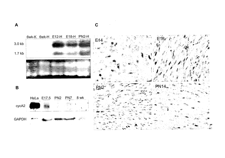
[0025] FIG. 1 shows that cyclin A2 mRNA and protein expression are
developmentally regulated in the normal mouse heart. (A) Cyclin A2 mRNA
expression in
normal mouse hearts was detected by Northern-blot analysis. RNA was extracted
from the
following tissues: 6-week heart (HA), 6-week kidney (KA), E12 heart (HE12),
E18 heart
(HE18), and PN2 heart (HPN2). The ethidium-bromide-stained ribosomal RNA bands
are
shown in the bottom panel as a loading control. (B) Immunoblot analysis of
cyclin A2
protein expression in normal mouse. Protein was extracted from mouse hearts at
E17.5, PN2,
PN7, and 8 weeks of age (labeled 'adult'), electrophoresed on 10% PAGE, and
detected by a-
cyclin A2 antibody (top). a-GAPDH antibody was used as a control for
equivalent loading.
(C) Cellular localization of cyclin A2 protein expression in the developing
heart. Immuno-
histochemical staining utilizing a-cyclin A2 antibody in ventricular tissue
sections from
selected stages (embryonic day 14 (E14) through post-natal day 14 (PN14)). The
brown
staining indicates positively-stained cardiomyocyte nuclei.
[0026] FIG. 2 illustrates that cyclin A2 mRNA and protein expression
are restricted to
transgenic mouse hearts. (A) Diagram of MHC-CYCA2 transgenic construct. The
inventors
cloned mouse cDNA, and used a standard construct for the purposes of this
invention. (B)
Representative Northern-blot analysis of control and transgenic samples. RNA
was isolated
from transgenic mice (t23 and t27) and normal mice (n1 and n2). The blot was
re-exposed
for a shorter period of time, to delineate detail in the Ht23 lane. 'H' and
'K' indicate heart and
kidney, respectively. The ethidium-bromide-stained ribosomal RNA bands are
shown in the
bottom panel as a loading control. (C) Immunoblot analysis of cell-cycle
protein expression
CA 02526490 2005-11-18
WO 2005/000403
PCT/US2004/015691
in transgenic (Tg) and normal (N) mice, at 2 weeks and 8 weeks. top panel: a-
cyclin A2;
second panel from top: cdk1; third panel from top: cdk2; bottom panel: GAPDH
sample
loading control. HeLa cell lysate was used as a positive control. (D)
Immunoprecipitation
analysis of cyclin A2 complexes in transgenic and control hearts, at 2 weeks
and 8 weeks. a-
cyclin A2 immunoprecipitated complexes (denoted by asterisks) were analyzed by
immunoblot analysis with a-cdkl (top) or a-cdk2 (bottom). The band visualized
at 29 kD
represents the immunoglobulin light chain. HeLa cell lysate was used as a
positive control.
[0027] FIG. 3 demonstrates that cyclin A2 transgenic mice ekhibit
cardiac
hyperplasia due to an increase in post-natal mitosis. (A) Enlargement of
hearts of cyclin A2
transgenic mice. Heart weight / body weight (HW/BW) ratios (mg/g) of normal
and
transgenic mice were plotted throughout development, from PN7 through 1.5
years of age.
The numbers of transgenic (Tg) and normal (N) mice examined at each age are
detailed as
follows: PN7 and PN14 ¨ 16 Tg, 15 N; 3-4 mo ¨ 5 Tg, 5 N; 6 mo ¨ 10 Tg, 10 N; 8-
12 mo ¨5
Tg, 5 N; 1.5 yr ¨ 1 Tg, 1 N. The asterisks indicate ages at which the HW/BW
difference
between transgenic and normal is statistically significant. (B) Myocyte cross-
sectional areas
were measured in normal versus transgenic littermates (lines 1 and 58, age 6
mo).
Hematoxylin- and eosin-stained sections of normal (N) and transgenic (Tg)
ventricular
myocardium were analyzed for cross-sectional areas, utilizing Image Tool
software.
(C) Myocyte lengths, measured in normal versus transgenic littennates (lines 1
and 58, age 6
mo). Pan-cadherin staining of intercalated disks in ventricular myocardium was
performed
for the measurements of cell lengths in longitudinal sections, utilizing Image
Tool software.
(D) Proliferating cell nuclear antigen (PCNA) expression in transgenic and
control hearts at
selected time points through development. The number of positively-stained
nuclei per unit
area was assessed and averaged over at least ten fields (field size = 16,800
pm2). With the
exception of E18, there was a significant increase in PCNA-stained nuclei per
unit area at
each developmental stage analyzed in the transgenic mice. (E) Detection of
phosphorylated
histone 113 in transgenic and normal myocardium. The number of mitotic nuclei
(as assessed
by phosphorylated histone-3 (H3P) staining) per field was significantly
enhanced in
transgenic hearts, as compared to normal hearts, at each developmental stage.
At least ten
fields were analyzed for each value. Green bars indicate transgenic mice, and
blue bars
indicate the normal controls. P values are indicated below each set. Note the
8-fold increase
in mitotic nuclei in transgenic versus normal mice at PN7.
CA 02526490 2005-11-18
WO 2005/000403
PCT/US2004/015691
-8-
[0028] FIG. 4 depicts visualization of mitotic nuclei in ventricular
myocardium from
PN7 normal and transgenic mice. (A), (B) Immunofluorescence was used to
localize H3P
staining (red) and a-sarcomeric actin (green). Different stages of mitosis
were observed in
cardiomyocytes: (C) prophase, (D) prometaphase, and (E) likely anaphase.
transgenic heart at a mid-ventricular cross-section of an 8-month old
transgenic mouse.
(B) MRI images of the heart at a mid-ventricular cross-section taken at
different points in the
cardiac cycle for the measurement of ejection fraction and fractional
shortening. Ventricle in
red indicates end-diastole, and ventricle in yellow indicates end-systole. (C)
Ejection fraction
and fractional shortening, as calculated from WiR1 analysis for normal (n = 3)
and transgenic
(n = 3) hearts.
[0030] FIG. 6 sets forth the amino acid sequence of mouse cyclin A2.
[0031] FIG. 7 shows the percent of left-ventricle-infarcted mice for
all groups. This
percentage was calculated by slicing each infarcted heart (from the ligation
site to the apex)
into 5 sections, measuring the mass of each slice, taking a thinner section
from each slice (-5
pm), and staining the slice with Masson trichrome to highlight areas of
fibrosis. The ratio of
the circumference of the infarcted area to the total circumference was
multiplied by the mass
of each slice, and the product of these was added for all 5 slices to obtain
infarct volume
percentage. The percentage of infarcted left ventricle was consistent between
groups; thus,
the inventors' surgical procedure was highly reproducible.
[0032] FIG. 8 illustrates the ejection fraction (EF) of infarcted
mice for all groups that
were assessed with serial MRI scans at 3 weeks and 3 months post myocardial
infarction.
The EF of the transgenic mice was significantly enhanced when compared to the
EF of the
wild-type controls and the non-transgenic littermate controls. EF was enhanced
in
transgenics, both at 3 weeks and 3 months post-infarct. P-values are given for
the
comparison to wild-type controls.
[0033] FIG. 9 sets forth representative MRI scans from 3 wild-type
mice (top 3
panels) and 3 transgenic hearts (bottom 3 panels). The scans show less left
ventricle (LV)
cavity dilatation, and higher ejection fractions (EFs), in the transgenic
hearts. Scans were
taken at the mid-ventricular level, to demonstrate that the LV cavity size is
notably smaller in
the transgenic mice. The smaller cavity size is indicative of remodeling that
is significantly
CA 02526490 2005-11-18
WO 2005/000403
PCT/US2004/015691
-9-
less than that which was observed in wild-type animals. The EFs, computed
volumetrically,
are given for each respective mouse beneath each scan.
[0034] FIG. 10 illustrates volumetric EFs computed using 3 transverse
images and 1
sagittal image. Three transverse images were scanned at equal distances from
the mid-point
of the long axis of the heart, as determined from a sagittal scan. It was
assumed that the
volume of an ellipsoid = 4/3Ah, where A = area and h = height; therefore,
total volume ¨
2/3A1h1 + 1.5A1 + 1.5A2 + 1.5A3 + 2/3A3h2. For each area (1, 2, 3), left-
ventricular, end-
systolic area was subtracted from left-ventricular, end-diastolic area, to
obtain the volumetric
EF.
[0035] FIG. 11 demonstrates that MRI tagging may be utilized quantitatively
to
analyze changes in regional wall motion of infarcted groups. For a mid-
ventricular scan, a
grid of absent signal was applied while acquiring the scan. Strain deformation
was then
measured for a given point on the cross-hairs of this grid, between systole
and diastole.
Examination of serial images permits a determination as to whether regional
wall motion
(i.e., the infarcted wall) exhibits improvement in contractility over time.
[0036] FIG. 12 depicts results of myocardial infarction. (A) Mitoses
were not
detected in wild-type infarcted hearts. Blue indicates DAN; green indicates
alpha-sarcomeric
actin. (B) Abundant mitoses were detected in cardiomyocytes of transgenic
infarcted hearts.
Clusters of mitotic cardiomyocytes are shown in the transgenic infarcted
myocardium. Red
indicates phosphohistone-H3-positive nuclei. Phosphohistone-H3 (H3P) is a
specific marker
of mitosis. Blue indicates DAPI staining of nuclei. Green indicates the
presence of alpha-
sarcomeric actin, which is specific for cardiomyocyte cytoplasm. (C, D)
Mitoses in the pen-
infarct zone. (E, F) Small, immature cardiomyocytes in the infarct zone
itself. The
photographs show a high nuclear-to-cytoplasmic ratio: the nuclei appear
mitotic, and the
green fluorescence of the cytoplasm indicates the presence of alpha-sarcomeric
actin. All
photographs were taken on the confocal microscope; thus, the signals are very
specific.
[0037] FIG. 13 illustrates detection of side-population (SP)
progenitor cells in the
mouse myocardium using ABCG2. ABCG2, a member of the ATP-cassette transporter
family of proteins, is a marker of side-population progenitor cells found in
mouse
myocardium. Although the protein becomes ubiquitinated as cardiomyocytes
proceed to
differentiate, ABCG2 typically displays a membrane pattern of expression, and
may be
CA 02526490 2005-11-18
WO 2005/000403
PCT/US2004/015691
-10-
localized to the cytoplasm. The inventors utilized antibody to ABCG2, denoted
by red in
these photographs, to detect SP progenitor cells in infarcted mice. SP cells
were noted in an
equal number of transgenic and wild-type mice. However, ABCG2 was shown to
have a
membrane-staining pattern in some photographs, and a cytoplasmic location in
other
photographs. This suggests that the transgenic cells may be behaving
differently from control
cells. (A, E, F) ABCG2 shows a membrane-staining pattern. (B, C) ABCG2 has a
cytoplasmic location. (D) ABCG2 is shown to have a cytoplasmic location. This
light-
microscopy photograph used DAB as a counterstain to ABCG2, in order to confirm
that a
non-specific, auto-fluorescent signal was not being detected.
[0038] FIG. 14 illustrates nuclear localization of cyclin A2 in "de novo"
myocardium
of transgenic infarcted mice. Cyclin A2 was noted in the nuclei of what appear
to be "de
novo" myocytes in the infarct zone of the transgenic mice; this was not
observed in controls.
Furthermore, even in the transgenic mice, nuclear localization of cyclin A2
was not typically
seen after early post-natal development (post-natal day 14); the transgene
protein product was
only noted in the cytoplasm at this point. (A) Red indicates cyclin A2; green
indicates alpha-
sarcomeric actin. (B) The section from (A) as viewed with a blue filter, so
that the red signal
of cyclin A2 can be localized to nuclei. The image from the blue filter was
merged with the
image from the green filter. Blue indicates DAPI staining of nuclei; green
indicates alpha-
sarcomeric actin.
DETAILED DESCRIPTION OF THE INVENTION
[0039] Genetic modulation, cell transplantation, and tissue
engineering promise
revolutionary approaches for myocardial regeneration and tissue repair after
myocardial
injury. Current data derived from animal models suggest that it may be
possible to treat heart
failure by inserting genetic materials or myogenic cells into injured
myocardium. See, e.g.,
U.S. Patent No. 6,534,052 and U.S. Patent Applications Nos. 20030022367,
20030054973,
and 20020197240.
[0040] One possible approach to cardiac regeneration involves
manipulation of
cellular proteins to promote cell-cycle re-entry and proliferation of
cardiomyocytes. This
approach has received considerable interest in recent years, due to the
identification of key
cell-cycle regulatory proteins and the publication of several reports
suggesting that
manipulation of these factors can reactivate DNA synthesis in vivo and in
vitro in the post-
CA 02526490 2005-11-18
WO 2005/000403
PCT/US2004/015691
-11-
mitotic ventricular myocardium (Kirshenbaum and Schneider, J. Biol. Clzein.,
270:7791-94,
1995; Agah et al., J. Clin. Invest., 100:2722-28, 1997; Soonpaa and Field,
Circ. Res., 83:15-
26, 1998). Prior to the present invention, however, no previous report
directly demonstrated
that regulation of these factors can induce cardiomyocyte mitosis once the
timeline for cell-
cycle exit (and, therefore, terminal differentiation) has been surpassed.
[0041] Previously, a significant gap in understanding of the
cardiomyocyte cell cycle
resulted from the limited number of studies that explore the effects of
putative cellular
regulators of the G2/M checkpoint. Cyclin A2 is unique among all cyclins in
that it has been
shown to regulate transition through both Gl/S and G2/M phases in cultured
cell lines (Sherr
and Roberts, Genes Dev., 9:1149-63, 1995). Cyclin A2 is normally silenced in
the heart
shortly after birth, when cardiomyocyte division ceases as the cells withdraw
from the cell
cycle. This was previously established in rat and human hearts (Yoshizumi et
al., J. Clin.
Invest., 95:2275-80, 1995), and the inventors have confirmed this in the
mouse. The
temporal pattern of cyclin A2 mRNA and protein levels implicates a crucial
role for cyclin
A2 as a regulator of cardiomyocyte cell-cycle exit.
[0042] To further elucidate the cardiomyocyte cell cycle, the
inventors generated a
mouse model of constitutive cyclin A2 expression in the myocardium, and tested
the impact
of deregulated cyclin A2 expression on cardiomyocyte proliferation and
terminal
differentiation. Phenotypic analysis revealed cardiac enlargement due to
hyperplasia in the
adult heart. More importantly, cardiomyocyte mitoses were significantly
enhanced during
post-natal development in the transgenic hearts, as compared with normal
hearts, with the
most dramatic difference occurring at PN7. However, cardiac enlargement in the
transgenic
mice became statistically significant during adulthood at 6 months of age,
implying that the
hyperplasia induced by constitutive cyclin A2 expression arose primarily
during post-natal
development, and not during embryo genesis.
[0043] Earlier studies had suggested that embryogenesis was
responsible for
hyperplasia in several other mouse models of altered or absent cell-cycle
proteins. For
example, Liao et al. (Circ. Res., 88:443-50, 2001) noted that cardiac
overexpression of cdk2
elicited cardiac enlargement at PN2, but that this did not persist in adults.
The cardiac
phenotype of p27KIPI knockout mice exhibited a significant increase in heart
weight, when
compared with wild-type, as analyzed between 2 and 35 days of age (Poolman et
al., Circ.
CA 02526490 2005-11-18
WO 2005/000403
PCT/US2004/015691
-12-
Res., 85:117-27, 1999). C-myc-overexpressing mice exhibited an enlargement in
cardiac size
that was most profound at 1 and 15 days of age (44% and 46%, respectively);
however, by 60
days of age, only a 34% increase was noted (Jackson et al., Mol. Cell. Biol.,
7:3709-16,
1990). In all of these studies, the investigators concluded that this
hyperplasia occurred
during fetal development, without acceleration of post-natal growth.
[0044] Cyclin D1 overexpression in the mouse heart has been shown to
promote
increased DNA synthesis in adult transgenic hearts, resulting in
multinucleation; karyokinesis
was not noted (Soonpaa et al., J. Clin. Invest., 99:2644-54, 1997).
Approximately 40%
enlargement was noted when the HW/BW ratios of adult transgenic mice were
compared
with those of non-transgenic mice (n =4 for each group, age not specified).
Although >60%
of the adult cyclin D1 transgenic cardiomyocytes exhibited a multinucleated
phenotype, the
authors concluded that it was unclear whether these cardiomyocytes retained
the ability to
undergo karyokinesis. This same group of investigators had previously
demonstrated
(Soonpaa et al., I Clin. Invest., 99:2644-54, 1997) that cardiomyocyte
division in the normal
mouse heart does not occur after birth, with DNA synthesis through PN2 and PN3
contributing only to binucleation.
[0045] The inventors' model of constitutive cardiac cyclin A2
expression directly
demonstrates that karyokinesis is induced in the transgenic heart after birth.
A recently-
published report, describing the effect of inhibition of the Rho family
GTPases, lends further
support to the association of cyclin A2 and cardiomyocyte proliferation:
expression of Rho
GDIa, an inhibitor of Rho family proteins, in the mouse myocardium resulted in
a decrease in
cellular proliferation in the embryonic heart that was associated with
downregulation of
cyclin A (Wei et al., Development, 7:1705-14, 2002).
[0046] As cyclin A2 has been shown to regulate both Gl/S and G2/M in
cultured
mammalian cell lines (Sherr and Roberts, Genes Dev., 9:1149-63, 1995), the
inventors
presumed that it plays a role in the regulation of both gap phases in vivo. In
Drosophila,
when regulators of both gap phases are overproduced (i.e., cyclin E and
string), cells are
unable to compensate for the shortening of both gap phases; the cell cycle as
a whole is
abbreviated, resulting in small cells with a faster generation time (Neufeld
et al., Cell,
93:1183-93, 1998). Previous investigators have demonstrated that
overexpression of Gl- to
S-phase cell-cycle regulatory proteins decreased cell size in vitro and in
vivo (Liao et al.,
CA 02526490 2005-11-18
WO 2005/000403
PCT/US2004/015691
-13-
Circ. Res., 88:443-50, 2001; Queue et al., Genes Dev., 8:1559-71, 1993). This
mechanism
may account, in part, for the cardiomyocyte hyperplasia with smaller cells
that was noted in
the inventors' model. However, the lack of any significant cardiac enlargement
in the
inventors' model during post-natal development, with an age-related increase
in the cardiac
size gap between transgenic and normal animals, points to the conclusion that
the most
dramatic effect of "de-silencing" cyclin A2 occurs after birth, when
cardiogenesis is normally
complete.
[0047] In order to test whether cyclin A2 can contribute to cardiac
repair, the
inventors induced myocardial infarction ND in transgenic mice, non-transgenic
littermates,
and wild-type mice (strain and age-matched) via ligation of the left anterior
descending artery
(LAD). In total, 89 mice were infarcted; there was an 83% survival rate, 1
week post-MI.
[0048] Functional analysis was performed utilizing fMRI to measure
volumetric
ejection fraction (EF); tagging was used to determine regional wall motion.
Cyclin A2
transgenic mice displayed markedly-enhanced EF, as compared with controls, at
3 weeks and
3 months post-MI. The percent of left ventricle (LV) infarcted was consistent
among all
groups. In the transgenic hearts, preservation of cardiac function was
observed at 3 months
of age, with a decline in EF and fractional shortening noted at 8 months of
age. This is
consistent with the observation of greater HW/BW differences between
transgenic and
normal animals after 5-6 months of age, as compared with younger animals. It
is expected
that hyperplastic hearts will display hypercontractility that will ultimately
progress to
hypocontractility with age.
[0049] The inventors also examined potential cellular and molecular
mechanisms that
may contribute to the apparent recovery of cardiac function. Assays of
proliferation included
immunofluorescence and confocal microscopy to detect phosphohistone 113 (H3P),
a mitosis-
specific marker. BRDU labeling was also performed. In these studies, the
inventors found
mitotic nuclei (as assessed by immunostaining for anti-phosphohistone H3) that
were
localized to cardiomyocytes in the infarct zone and in non-infarcted
myocardium in
transgenic mice; only 0-1 such mitosis was noted in non-transgenic and wild-
type hearts.
This suggests that cyclin A2 transgenic cardiomyocytes are able to re-enter
the cell cycle in
response to injury.
CA 02526490 2005-11-18
WO 2005/000403
PCT/US2004/015691
-14-
[0050] Cardiac progenitor cells were detected in the infarct zones of
both transgenic
animals and controls. These progenitor cells express ABCG2, a known marker of
"side-
population" (SP) cells. SP cells form a class of progenitor cells that are
identified on the
basis of Hoechst extrusion. They have been observed on many tissues, including
skeletal
muscle, bone marrow, liver, brain, and heart. In transgenic hearts alone,
nuclear localization
of cyclin A2 was detected in what appears to be "de novo" myocardium. These
results
indicate that cyclin A2 transgenic mice are able to repair damaged myocardium
either
through cell-cycle re-entry of pen-infarct zone cardiomyocytes or via the
induction of SP
cells with enhanced proliferative potential.
[0051] The expression of cyclin A2 in the nucleus of what appears to be "de
novo"
myocardium recapitulates the developmental paradigm noted in the post-natal
transgenic
model. The presence of nuclear cyclin A2, coupled with the findings of ABCG2
expression
in cells of similar location (as serial sections were analyzed), implies that
the SP cells of the
transgenic model, which normally reside in myocardium, exhibit a
"hyperproliferative"
phenotype. This may have exciting ramifications for cardiac regeneration.
[0052] In the inventors' transgenic model, then, constitutive cardiac
expression of
normally-silent cyclin A2 invoked an increase in cardiomyocyte mitosis in the
post-natal
heart, with resultant cardiac enlargement (due to hyperplasia) noted in the
adult heart. The
inventors' model differs from previous mouse models examining altered or
absent cell-cycle
regulators in that it specifically addresses control of the G2/M checkpoint,
in addition to the
Gl/S checkpoint. Furthermore, karyokinesis in post-natal cardiomyocytes is
specifically
demonstrated through the detection of histone-3 phosphorylation at Ser10, and
various stages
of cardiomyocyte mitosis may be observed. The enhanced HW/BW increase in the
transgenic heart, as compared with the normal heart during adulthood, suggests
that
cytokinesis is coupled with mitosis in the transgenic heart. The decline of
cardiomyocyte
mitoses noted between PN7 and PN14, and the scattered mitoses noted in the
adult heart,
indicate that cell cycle inhibitors are holding this mitotic process in check,
despite the
expression of cyclin A2.
[0053] Moreover, cyclin A2 transgenic mice have significantly
enhanced cardiac
function compared to non-transgenic littermates and wild-type controls at 3
weeks and 3
months post-MI. This suggests that cyclin A2 may confer the ability to re-
enter the cell cycle
CA 02526490 2005-11-18
WO 2005/000403
PCT/US2004/015691
-15-
for post-mitotic cardiomyocytes. Cyclin A2 also appears to confer a
hyperproliferative effect
on side-population progenitor cells that are normally found in myocardium.
[0054] In view of the foregoing, the present invention provides a
method for
promoting generation of heart tissue. As used herein, the term "promoting
generation of heart
tissue" includes activating, enhancing, facilitating, increasing, inducing,
initiating, or
stimulating the growth and/or proliferation of heart tissue, as well as
activating, enhancing,
facilitating, increasing, inducing, initiating, or stimulating the
differentiation, growth, and/or
proliferation of heart tissue cells. Thus, the term includes initiation of
heart tissue generation,
as well as facilitation or enhancement of heart tissue generation already in
progress.
"Differentiation" is the cellular process by which cells become structurally
and functionally
specialized during development. The terms "proliferation" and "growth", as
used herein,
refer to an increase in mass, volume, and/or thickness of heart tissue, as
well as an increase in
diameter, mass, or number of heart tissue cells. As further used herein, the
term "generation"
includes the generation of new heart tissue and the regeneration of heart
tissue where heart
tissue previously existed.
[0055] As used herein, the term "heart tissue" includes, without
limitation, the
myocardium of the heart (including cardiac muscle fibers, connective tissue
(endomysium),
nerve fibers, capillaries, and lymphatics); the endocardium of the heart
(including
endothelium, connective tissue, and fat cells); the epicardium of the heart
(including
fibroelastic connective tissue, blood vessels, lymphatics, nerve fibers, fat
tissue, and a
mesothelial membrane consisting of squamous epithelial cells); and any
additional connective
tissue (including the pericardium), blood vessels, lymphatics, fat cells,
progenitor cells (e.g.,
side-population (SP) progenitor cells), and nervous tissue found in the heart.
Cardiac muscle
fibers are composed of chains of contiguous heart-muscle cells, or
"cardiomyocytes", joined
end to end at intercalated disks. These disks possess two kinds of cell
junctions: expanded
desmosomes extending along their transverse portions, and gap junctions, the
largest of
which lie along their longitudinal portions.
[0056] The method of the present invention comprises, in one
embodiment,
augmenting cyclin in heart tissue cells. Heart tissue cells include any of the
cells of the
various tissues found in the heart, as described above. By way of example, the
heart tissue
cells of the present invention may include progenitor cells (e.g., heart-
tissue side-population
CA 02526490 2005-11-18
WO 2005/000403
PCT/US2004/015691
-16-
(SP) progenitor cells) and differentiated or post-mitotic cells. The term
"post-mitotic", as
used herein, refers to a cell that is in GO phase (a quiescent state), and is
no longer dividing or
cycling. In a preferred embodiment of the present invention, the heart tissue
cells are
cardiomyocytes. It is also within the confines of the present invention that
generation of
heart tissue may be promoted by augmenting cyclin in SP progenitor cells that
are derived
from non-heart tissue (e.g., spleen, bone marrow, skeletal muscle, brain,
liver, kidney, lung,
small intestine, etc.).
[0057] The heart tissue cells and SP progenitor cells of the present
invention may be
obtained from any animal, including amphibians, birds, fish, mammals, and
marsupials, but
are preferably obtained from a mammal (e.g., a human; a domestic animal, such
as a cat, dog,
monkey, mouse, and rat; or a commercial animal, such as a cow or pig).
Additionally, the
heart tissue cells and SP progenitor cells of the present invention may be
obtained from an
animal of any age, including a fetus, an embryo, a child, and an adult. In one
embodiment of
the present invention, the heart tissue cells or SP progenitor cells are
obtained from a
transgenic animal that overexpresses cyclin A2 in its heart tissue, as
described below. In
another embodiment, the heart tissue cells or SP progenitor cells are rat or
mouse cells. In a
preferred embodiment of the present invention, the heart tissue cells or SP
progenitor cells
are obtained from a human.
[0058] As described above, the method of the present invention
results in the
generation of new heart tissue and/or the regeneration of heart tissue where
such tissue used
to exist. In the case of regeneration, the heart tissue cells of the present
invention may be
obtained from, or found within, damaged or degenerated heart tissue (i.e.,
heart tissue which
exhibits a pathological condition). Causes of heart tissue degeneration
include, without
limitation, chronic heart damage, chronic heart failure, damage resulting from
injury or
trauma, damage resulting from a cardiotoxin, damage from radiation or
oxidative free
radicals, damage resulting from decreased blood flow, and myocardial
infarction (such as a
heart attack). Preferably, the degenerated heart tissue of the present
invention results from a
myocardial infarction or heart failure. Generation of new heart tissue and
regeneration of
heart tissue may be measured or detected by known procedures, including
Western blotting
for heart-specific proteins, electron microscopy in conjunction with
morphometry, simple
assays to measure rate of cell proliferation (including trypan blue staining,
the CellTiter-Blue
CA 02526490 2005-11-18
WO 2005/000403
PCT/US2004/015691
-17-
cell viability assay from Promega (Madison, WI), the MTT cell proliferation
assay from
ATCC, differential staining with fluorescein diacetate and ethidium bromide /
propidium
iodide, estimation of ATP levels, flow-cytometry assays, etc.), and any of the
methods,
molecular procedures, and assays disclosed herein.
[0059] As discussed above, cyclin is augmented in heart tissue cells or SP
progenitor
cells in accordance with the method of the present invention. Cyclins are
proteins, found in
certain eukaryotic cells, which help to regulate the cell cycle by causing
cells to begin mitosis
(a form of nuclear division). The proteins are generally produced during all
parts of the cell
cycle, but destroyed during mitosis. Examples of cyclins include, without
limitation, cyclin
A, cyclin B, cyclin C, cyclin D, and cyclin E. The mammalian A-type cyclin
family consists
of 2 members, cyclin Al (the germ-cell version of cyclin A) and cyclin A2 (the
somatic-cell
version of cyclin A, known as "cyclin A" in humans). Included in this family
is a 33-kD
protein identical to adenovirus el a-associated protein p60. Cyclin A proteins
regulate
p33cdk2 and p34cdc2, and are necessary for progression through the S phase of
the cell
cycle. Cyclin A2 promotes both Gl/S and G2/M transitions; cyclin Al is
expressed in mice
exclusively in the germline lineage, and is expressed in humans at highest
levels in the testis
and certain myeloid leukemia cells. Cyclin B is a 58-kD protein that is
regulated post-
transcriptionally and post-translationally in the cell cycle. In one preferred
embodiment of
the present invention, the cyclin is cyclin A2.
[0060] As used herein, "cyclin" includes both a "cyclin protein" and a
"cyclin
analogue". Unless otherwise indicated, "protein" shall include a protein,
protein domain,
polypeptide, or peptide, and any fragment or variant thereof having protein
function. The
variants preferably have greater than about 75% homology with the naturally-
occurring
protein sequence, more preferably have greater than about 80% homology, even
more
preferably have greater than about 85% homology, and, most preferably, have
greater than
about 90% homology with the protein sequence. In some embodiments, the
homology may
be as high as about 93-95%, 98%, or 99%. These variants may be substitutional,
insertional,
or deletional variants. The variants may also be chemically-modified
derivatives: proteins
which have been subjected to chemical modification, but which retain the
biological
characteristics of the naturally-occurring protein. In one embodiment of the
present
CA 02526490 2005-11-18
WO 2005/000403
PCT/US2004/015691
-18-
invention, the protein is mutated such that it has a longer half-life inside
the heart tissue cell
(e.g., it is modified at its ubiquitin-binding site).
[0061] A "cyclin analogue", as used herein, is a functional variant
of the cyclin
protein, having cyclin biological activity, that has 60% or greater
(preferably, 70% or greater)
amino-acid-sequence homology with the cyclin protein. As further used herein,
the term
"cyclin biological activity" refers to the activity of a protein or peptide
that demonstrates an
ability to promote generation of heart tissue, as described herein.
[0062] The cyclin A2 protein has the amino acid sequence set forth in
FIG. 6,
including conservative substitutions thereof. As used herein, "conservative
substitutions" are
those amino acid substitutions which are functionally equivalent to a
substituted amino acid
residue, either because they have similar polarity or steric arrangement, or
because they
belong to the same class as the substituted residue (e.g., hydrophobic,
acidic, or basic). The
term "conservative substitutions" includes substitutions having an
inconsequential effect on
the ability of cyclin to promote generation of heart tissue, particularly in
respect of the use of
said interaction for the identification and design of agonists of cyclin, for
molecular
replacement analyses, and/or for homology modeling.
[0063] It will be obvious to the skilled practitioner that the
numbering of amino acid
residues in proteins, and in the fragments, variants, analogues, and
peptidomimetics covered
by the present invention, may be different than that set forth herein, or may
contain certain
conservative amino acid substitutions that produce the same heart-tissue-
generating activity
as that described herein. Corresponding amino acids and conservative
substitutions in other
isoforms or analogues are easily identified by visually inspecting the
relevant amino acid
sequences, or by using commercially-available homology software programs.
[0064] In accordance with methods described herein, cyclin may be
augmented or
increased in heart tissue cells or side-population (SP) progenitor cells by
activating,
facilitating, inducing, or stimulating one or more functions, activities, or
effects (e.g.,
downstream effects of the cyclin in the cyclin signal transduction pathway) of
cyclin in the
cells, particularly those that result in promotion of heart-tissue generation,
or by increasing
the amount, expression, or level of cyclin in the cells. Furthermore, one or
more cyclin
functions, activities, effects, expression, and levels in a cell may be
augmented by targeting
cyclin directly, or by targeting cyclin indirectly, via an enzyme or other
endogenous molecule
CA 02526490 2005-11-18
WO 2005/000403
PCT/US2004/015691
-19-
that regulates or modulates the functions, activities, effects, expression,
and/or levels of
cyclin in the cell. Cyclin expression may also be augmented by engineering the
cyclin gene
so that cyclin is expressed on an inducible promoter. In such a case, cyclin
expression would
be sustained in the presence of a suitable inducing agent, but would shut down
once the
supply of inducer was depleted, thereby bringing about a decrease in the
amount or level of
cyclin in the cell. Cyclin also may be augmented in a cell by activating,
facilitating,
inducing, or stimulating the functions, activities, effects, expression, and
levels of
endogenous cyclin, or by introduction of an exogenous cyclin, particularly
where the cyclin is
under the control of a strong promoter.
[0065] Preferably, the functions, activities, effects, expression, and/or
levels of cyclin
in the heart tissue cells of the present invention are augmented or increased
by at least 10%.
More preferably, the functions, activities, effects, expression, and/or levels
of the cyclin are
increased by at least 20%. The functions, activities, effects, expression,
and/or levels of
cyclin are augmented in heart tissue cells or side-population (SP) progenitor
cells by an
amount effective to promote generation of heart tissue. This amount may be
readily
determined by the skilled artisan, based upon known procedures, including
analysis of
titration curves established in vivo, methods disclosed herein, and techniques
known to one of
skill in the art.
[0066] In the method of the present invention, the functions,
activities, effects,
expression, and/or levels of cyclin in heart tissue cells or side-population
(SP) progenitor
cells are preferably augmented by contacting the cells (i.e., treating the
cells) with a cyclin-
associated agent. As used herein, an "agent" shall include a protein,
polypeptide, peptide,
nucleic acid (including DNA, RNA, and an antisense oligonucleotide), antibody
(monoclonal
and polyclonal), Fab fragment, F(abt)2 fragment, molecule, compound,
antibiotic, drug, and
any combinations thereof, and may be an agent reactive with cyclin. The term
"reactive", as
used herein, means that the molecule or mimetic has affinity for, binds to, or
is directed
against cyclin. A Fab fragment is a univalent antigen-binding fragment of an
antibody, which
is produced by papain digestion. A F(abe)2 fragment is a divalent antigen-
binding fragment of
an antibody, which is produced by pepsin digestion.
[0067] As further used herein, the term "cyclin-associated agent" includes
a cyclin
protein, including an exogenous cyclin protein; a cyclin nucleic acid (i.e., a
nucleic acid
CA 02526490 2005-11-18
WO 2005/000403
PCT/US2004/015691
-20-
encoding a cyclin); a member of a cyclin signal-transduction pathway
(including upstream
and downstream effectors and activators, in either protein or nucleic acid
form); and a
modulator (e.g., inhibitor, activator, antagonist, or agonist) of a member of
the cyclin signal-
transduction pathway or system (i.e., a modulator which affects the
expression, activity,
function, and/or effect of a member of the cyclin signal-transduction
pathway), in either
protein or nucleic acid form, including a modulator of cyclin expression.
Additionally, as
used herein, a "member of a cyclin signal-transduction pathway" includes a
downstream
effector or an upstream regulator of cyclin in heart tissue cells or side-
population (SP)
progenitor cells.
[0068] By way of example, activity of cyclin in heart tissue cells or side-
population
(SP) progenitor cells may be augmented by contacting the cells with a small
molecule or
protein mimetic that stimulates cyclin activity and/or that is reactive with
cyclin. Similarly,
the level of cyclin in heart tissue cells or side-population (SP) progenitor
cells may be
augmented by directly or indirectly causing, inducing, or stimulating the
upregulation of
cyclin expression within a subject. Accordingly, in one embodiment of the
present invention,
activity of cyclin is increased in a subject by administering to the subject a
modulator of
cyclin expression.
[0069] In one embodiment of the present invention, the cyclin-
associated agent is a
protein. Examples of proteins for use in the present invention include,
without limitation,
cyclin proteins, members of the cyclin signal-transduction pathway (including
upstream and
downstream effector and activator polypeptides), modulators (e.g., inhibitors,
activators,
antagonists, or agonists) of a member of the cyclin signal-transduction
pathway/system,
cyclin-associated antibodies (e.g., IgA, IgD, IgE, IgG, IgM, single-chain
antibodies, and Fab'
fragments, such as scFv) that are capable of binding and inhibiting a negative
regulator of the
cyclin signal-transduction pathway, and cyclin-associated ligands (e.g., a
ligand for a member
of the cyclin signal-transduction pathway, and derivatives thereof).
Preferably, the cyclin-
associated protein is cyclin A2 protein.
[0070] Where the protein of the present invention is an antibody, the
protein is
preferably a mammalian antibody (e.g., a human antibody) or a chimeric
antibody (e.g., a
humanized antibody). More preferably, the antibody is a human or humanized
antibody. As
used herein, the term "humanized antibody" refers to a genetically-engineered
antibody in
CA 02526490 2005-11-18
WO 2005/000403
PCT/US2004/015691
-21-
which the minimum portion of an animal antibody (e.g., an antibody of a mouse,
rat, pig,
goat, or chicken) that is generally essential for its specific functions is
"fused" onto a human
antibody. In general, a humanized antibody is 1-25%, preferably 5-10%, animal;
the
remainder is human. Humanized antibodies usually initiate minimal or no
response in the
[0071] The cyclin-associated agent of the present invention may also
be a nucleic
acid. As used herein, a "nucleic acid" or "polynucleotide" includes a nucleic
acid, an
[0072] The "complement" of a nucleic acid refers, herein, to a
nucleic acid molecule
which is completely complementary to another nucleic acid, or which will
hybridize to the
CA 02526490 2005-11-18
WO 2005/000403
PCT/US2004/015691
-22-
single-stranded, or triple-stranded, it may have originated recombinantly or
synthetically, and
it may represent coding and/or noncoding 5' and/or 3' sequences.
[0073] The nucleic acid agent of the present invention, for example,
may be a
plasmid. Such a plasmid may comprise a nucleic acid sequence encoding cyclin
or another
cyclin-associated protein, although it is to be understood that other types of
nucleic acid
agents, such as recombinant viral vectors, may also be used for the purposes
of the present
invention. In one embodiment of the present invention, the nucleic acid (e.g.,
plasmid)
encodes at least one cyclin-associated protein. Preferably, the nucleic acid
encodes cyclin A2
protein.
[0074] The term "plasmid", as used herein, refers generally to circular
double-
stranded DNA, which is not bound to a chromosome. The DNA, for example, may be
a
chromosomal or episomal-derived plasmid. The plasmid of the present invention
may
optionally contain a terminator of transcription; a promoter; and/or a
discrete series of
restriction-endonuclease recognition sites, located between the promoter and
the terminator.
In the plasmid, a polynucleotide insert of interest (e.g., one encoding a
cyclin-associated
protein) should be operatively linked to an appropriate promoter. The promoter
may be its
native promoter or a host-derived promoter. The promoter may also be a tissue-
specific
promoter, such as a cardiomyocyte-specific promoter or other heart-tissue-
specific promoter.
The promoter may further be a regulatable promoter, which may be turned off
when the
expression of the gene is no longer desired. Examples of promoters for use in
the present
invention include the actin promoter and viral promoters. Other suitable
promoters will be
known to the skilled artisan.
[0075] In another embodiment of the present invention, the nucleic
acid (e.g.,
plasmid) encodes or comprises at least one gene-silencing cassette, wherein
the cassette is
capable of silencing the expression of genes that negatively affect the cyclin
signal-
transduction pathway/system. It is well understood in the art that a gene may
be silenced at a
number of stages, including, without limitation, pre-transcription silencing,
transcription
silencing, translation silencing, post-transcription silencing, and post-
translation silencing. In
one embodiment of the present invention, the gene-silencing cassette encodes
or comprises a
post-transcription gene-silencing composition, such as antisense RNA or RNAi.
Both
antisense RNA and RNAi may be produced in vitro, in vivo, ex vivo, or in situ.
CA 02526490 2005-11-18
WO 2005/000403
PCT/US2004/015691
-23-
[0076] For example, the cyclin-associated agent of the present
invention may be an
antisense RNA. Antisense RNA is an RNA molecule with a sequence complementary
to a
specific RNA transcript, or mRNA, whose binding prevents further processing of
the
transcript or translation of the mRNA. Antisense molecules may be generated,
synthetically
or recombinantly, with a nucleic-acid vector expressing an antisense gene-
silencing cassette.
Such antisense molecules may be single-stranded RNAs or DNAs, with lengths as
short as
15-20 bases or as long as a sequence complementary to the entire mRNA. RNA
molecules
are sensitive to nucleases. To afford protection against nuclease digestion,
an antisense
deoxyoligonucleotide may be synthesized as a phosphorothioate, in which one of
the
nonbridging oxygens surrounding the phosphate group of the deoxynucleotide is
replaced
with a sulfur atom (Stein et al., Oligodeoxynucleotides as inhibitors of gene
expression: a
review. Cancer Res., 48:2659-68, 1998).
[0077] Antisense molecules designed to bind to the entire mRNA may be
made by
inserting cDNA into an expression plasmid in the opposite or antisense
orientation.
Antisense molecules may also function by preventing translation initiation
factors from
binding near the 5' cap site of the mRNA, or by interfering with interaction
of the mRNA and
ribosomes (e.g., U.S. Patent No. 6,448,080, Antisense modulation of WRN
expression; U.S.
Patent Application No. 2003/0018993, Methods of gene silencing using inverted
repeat
sequences; U.S. Patent Application No., 2003/0017549, Methods and compositions
for
expressing polynucleotides specifically in smooth muscle cells in vivo; Tavian
et al., Stable
expression of antisense urokinase mRNA inhibits the proliferation and invasion
of human
hepatocellular carcinoma cells. Cancer Gene Ther.,10:112-20, 2003; Maxwell and
Rivera,
Proline oxidase induces apoptosis in tumor cells and its expression is absent
or reduced in
renal carcinoma. 1 Biol. Chem., 278:9784-89, 2003; Ghosh et al., Role of
superoxide
dismutase in survival of Leishmania within the macrophage. Biochem. 1, 369:447-
52, 2003;
and Zhang et al., An anti-sense construct of full-length ATM cDNA imposes a
radiosensitive
phenotype on normal cells. Oncogene, 17:811-8, 1998).
[0078] Oligonucleotides antisense to a member of the cyclin signal-
transduction
pathway/system may be designed based on the nucleotide sequence of the member
of interest.
For example, a partial sequence of the nucleotide sequence of interest
(generally, 15-20 base
pairs), or a variation sequence thereof, may be selected for the design of an
antisense
CA 02526490 2005-11-18
WO 2005/000403
PCT/US2004/015691
-24-
oligonucleotide. This portion of the nucleotide sequence may be within the 5'
domain. A
nucleotide sequence complementary to the selected partial sequence of the gene
of interest, or
the selected variation sequence, then may be chemically synthesized using one
of a variety of
techniques known to those skilled in the art, including, without limitation,
automated,
synthesis of oligonucleotides having sequences which correspond to a partial
sequence of the
nucleotide sequence of interest, or a variation sequence thereof, using
commercially-available
oligonucleotide synthesizers, such as the Applied Biosystems Model 392 DNA/RNA
synthesizer.
[0079] Once the desired antisense oligonucleotide has been prepared,
its ability to
augment cyclin then may be assayed. For example, the antisense oligonucleotide
may be
contacted with heart tissue cells or SP progenitor cells, and the levels of
cyclin expression or
activity in the cells may be determined using standard techniques, such as
Western-blot
analysis and immunostaining. Alternatively, the antisense oligonucleotide may
be delivered
to heart tissue cells or SP progenitor cells using a liposome vehicle, then
the levels of cyclin
expression or activity in the cells may be determined using standard
techniques, such as
Western-blot analysis and immuno staining. Where the level of cyclin
expression in the cells
is increased in the presence of the designed antisense oligonucleotide, it may
be concluded
that the oligonucleotide could be an appropriate cyclin-associated agent for
use in
augmenting cyclin in heart tissue cells or SP progenitor cells.
[0080] It is within the confines of the present invention that
oligonucleotides
antisense to a member of the cyclin signal-transduction pathway/system may be
linked to
another agent, such as a drug or a ribozyme, in order to increase the
effectiveness of
treatments using cyclin-associated agents and/or to increase the efficacy of
targeting.
Moreover, antisense oligonucleotides may be prepared using modified bases
(e.g., a
phosphorothioate), as discussed above, to make the oligonucleotides more
stable and better
able to withstand degradation.
[0081] The cyclin-associated agent of the present invention also may
be an interfering
RNA, or RNAi, including cyclin small interfering RNA (siRNA). As used herein,
"RNAi"
refers to a double-stranded RNA (dsRNA) duplex of any length, with or without
single-strand
overhangs, wherein at least one strand, putatively the antisense strand, is
homologous to the
target mRNA to be degraded. As further used herein, a "double-stranded RNA"
molecule
CA 02526490 2005-11-18
WO 2005/000403
PCT/US2004/015691
-25-
includes any RNA molecule, fragment, or segment containing two strands forming
an RNA
duplex, notwithstanding the presence of single-stranded overhangs of unpaired
nucleotides.
Additionally, as used herein, a double-stranded RNA molecule includes single-
stranded RNA
molecules forming functional stem-loop structures, such that they thereby form
the structural
[0082] In one embodiment of the present invention, RNAi is produced ill
vivo by an
expression vector containing a gene-silencing cassette coding for RNAi. See,
e.g., U.S.
Patent No. 6,278,039, C. elegans deletion mutants; U.S. Patent Application No.
2002/0006664, Arrayed transfection method and uses related thereto; WO
99/32619, Genetic
inhibition by double-stranded RNA; WO 01/29058, RNA interference pathway genes
as tools
[0083] In a further embodiment of the present invention, the plasmid
is an expression
CA 02526490 2005-11-18
WO 2005/000403
PCT/US2004/015691
-26-
portions of the mature transcripts expressed by the plasmid may include a
translation-
initiating codon at the beginning, and a termination codon appropriately
positioned at the end
of the polypeptide to be translated.
[0084] By way of example, the cyclin-associated gene to be expressed
from the
expression plasmid may be under the specific regulatory control of certain
types of
promoters. In one embodiment, these promoters are constitutive promoters.
Genes under the
control of these constitutive promoters will be expressed continually. In
another
embodiment, the promoters are inducible promoters. Genes under the control of
these
inducible promoters will be expressed only upon the presence of an inducer
molecule or the
absence of an inhibitor molecule, thereby providing a method to turn off
expression of the
gene when it is not desired. In yet another embodiment, the promoters are cell-
type-specific
promoters or tissue-specific (e.g., heart-tissue-specific) promoters. Genes
under the control
of cell-type-specific promoters will be expressed only in certain cell types,
preferably only in
cardiomyocytes.
[0085] In another embodiment of the present invention, the cyclin-
associated agent is
a modulator (e.g., inhibitor, activator, antagonist, or agonist) of cyclin
expression/activity,
including a modulator of a member of the cyclin signal-transduction
pathway/system. The
modulator of the present invention may be a protein, polypeptide, peptide,
nucleic acid
(including DNA or RNA), antibody, Fab fragment, F(a13')2 fragment, molecule,
compound,
antibiotic, or drug, including an agent reactive with cyclin, and an agent
that induces or
upregulates cyclin expression or activity.
[0086] Modulators of cyclin or a member of the cyclin signal-
transduction pathway/
system may be identified using a simple screening assay. For example, to
screen for
candidate modulators of cyclin, heart tissue cells or SP progenitor cells may
be plated onto
microtiter plates, then contacted with a library of drugs. Any resulting
increase in, or
upregulation of, cyclin expression then may be detected using a luminescence
reporter,
nucleic acid hybridization, and/or immunological techniques known in the art,
including an
ELISA. Additional modulators of cyclin expression may be identified using
screening
procedures well known in the art or disclosed herein. It is within the
confines of the present
invention that the modulator of cyclin expression may be linked to another
agent, or
administered in combination with another agent, such as a drug or a ribozyme,
in order to
CA 02526490 2005-11-18
WO 2005/000403
PCT/US2004/015691
-27-
increase the effectiveness of treatments using cyclin-associated agents and/or
increase the
efficacy of targeting. Additional cyclin-associated agents may be identified
using screening
procedures well known in the art, and methods described herein.
[0087] It is also within the confines of the present invention to
augment cyclin in
heart tissue cells or side-population (SP) progenitor cells by contacting the
cells with stem
cells (e.g., hematopoietic stem cells or heart-derived stem cells) containing
augmented cyclin.
The stem cells may be obtained from any animal, but are preferably obtained
from a mammal
(e.g., human, domestic animal, or commercial animal).
[0088] The efficacy of this technique could be assessed, for example,
using a cyclin
A2 mouse model, in which all cells of the transgenic animal contain an a-MHC-
cyclin A2
transgene (as described below). By way of example, female wild-type mice may
be subjected
to myocardial infarction via ligation of the left-anterior descending (LAD)
artery. These
mice then may be lethally irradiated. Hematopoietic stem cells (HSCs) purified
from male
cyclin A2 transgenic mice then may be injected, via the tail vein, into the
infarcted, female
wild-type mice. For a control group, HSCs from wild-type male mice may be
injected into a
separate group of infarcted, female wild-type mice. Fluorescence in situ
hybridization
techniques may be utilized to identify the Y-chromosome, for the purpose of
confirming that
transdifferentiated stem cells are donor-derived (Gussoni et al., Dystrophin
expression in the
mdx mouse restored by stem cell transplantation. Nature, 401:390-94, 1999).
[0089] It is expected that HSCs derived from a-MHC-cyclin A2 transgenic
mice will
transfer cyclin A2 upon fusing with native heart tissue cells (e.g.,
cardiomyocytes) or SP
progenitor cells, and thereby contribute to cardiac regeneration. Thus, cyclin
may be
augmented in heart tissue cells or side-population (SP) progenitor cells by
contacting the cells
with stem cells in which cyclin is already augmented. Furthermore, it is
expected that stem
cells which transdifferentiate into heart tissue cells (e.g., cardiomyocytes)
will retain
proliferative potential, and augmented cyclin, instead of transdifferentiating
into post-mitotic
heart tissue cells. Thus, cyclin also may be augmented in heart tissue cells
by augmenting
cyclin in stem cells, and allowing such stem cells to differentiate into heart
tissue cells that
retain proliferative potential and that retain augmented cyclin.
[0090] As discussed above, the present invention contemplates the use of
proteins and
protein analogues generated by synthesis of polypeptides in vitro, e.g., by
chemical means or
CA 02526490 2005-11-18
WO 2005/000403
PCT/US2004/015691
-28-
in vitro translation of mRNA. For example, cyclin may be synthesized by
methods
commonly known to one skilled in the art (Modern Techniques of Peptide and
Amino Acid
Analysis (New York: John Wiley & Sons, 1981; Bodansky, M., Principles of
Peptide
Synthesis (New York: Springer-Verlag New York, Inc., 1984)). Examples of
methods that
may be employed in the synthesis of the amino acid sequences, and analogues of
these
sequences, include, but are not limited to, solid-phase peptide synthesis,
solution-method
peptide synthesis, and synthesis using any of the commercially-available
peptide
synthesizers. The amino acid sequences of the present invention may contain
coupling agents
and protecting groups, which are used in the synthesis of protein sequences,
and which are
well known to one of skill in the art.
[0091] In accordance with the method of the present invention, cyclin
in heart tissue
cells or side-population (SP) progenitor cells may be augmented, and cells may
be contacted
with a cyclin-associated agent (e.g., by introducing a cyclin-associated agent
directly into the
cells) ¨ including stem cells containing a cyclin-associated agent ¨ either in
vitro, or in vivo
in a subject. Where cells are contacted with a cyclin-associated agent in
vitro, the agent may
be added directly to the cell-culture medium. Alternatively, a cyclin-
associated agent may be
contacted with heart tissue cells or side-population (SP) progenitor cells in
vivo in a subject,
by introducing the agent into the subject (e.g., by introducing the agent
directly into heart
tissue or heart tissue cells of the subject) and/or administering the agent to
the subject. The
subject may be any animal, including amphibians, birds, fish, mammals, and
marsupials, but
is preferably a mammal (e.g., a human; a domestic animal, such as a cat, dog,
monkey,
mouse, and rat; or a commercial animal, such as a cow or pig). In a preferred
embodiment,
the subject is a human.
[0092] The cyclin-associated agent of the present invention
(including stem cells
containing the agent) may be contacted with heart tissue cells or SP
progenitor cells, either in
vitro, or in vivo (including in situ) in a subject, by known techniques used
for the introduction
and administration of proteins, nucleic acids, and other drugs. Examples of
methods for
contacting the cells with (i.e., treating the cells with) a cyclin-associated
agent (in protein or
nucleic acid form, and including protein or nucleic acid contained within stem
cells) include,
without limitation, absorption, electroporation, immersion, injection
(including
microinjection), introduction, liposome delivery, stem cell fusion (including
embryonic stem
CA 02526490 2005-11-18
WO 2005/000403
PCT/US2004/015691
-29-
cell fusion), transduction, transfection, transfusion, vectors, and other
protein-delivery and
nucleic-acid-delivery vehicles and methods.
[0093] When the heart tissue cells (including heart-tissue SP
progenitor cells) are
localized to a particular portion of a subject, it may be desirable to
introduce the agent
directly to the cells, by injection or by some other means (e.g., by
introducing the agent into
the blood or another body fluid). Preferably, where heart tissue cells are
contacted with a
cyclin-associated agent (including stem cells containing the agent) in vivo in
a subject,
contacting is accomplished via a catheter inserted directly into the subject's
heart tissue. A
catheter would be useful in achieving targeted delivery of the agent to heart
tissue cells.
Targeted delivery is especially appropriate for cardiomyocytes, which are
joined by
intercalated disks. These disks should allow passage of the agent from one
cardiomyocyte to
adjoining cardiomyocytes, thereby aiding in the distribution of the agent
throughout the heart
tissue.
[0094] Where a cyclin-associated agent is a protein, it may be
introduced into heart
tissue cells or SP progenitor cells directly, in accordance with conventional
techniques and
methods disclosed herein. Additionally, a protein agent may be introduced into
heart tissue
cells or SP progenitor cells indirectly, by introducing into the cells a
nucleic acid encoding
the agent, in a manner permitting expression of the protein agent. The cyclin-
associated
agent may be introduced into cells, in vitro or in vivo, using conventional
procedures known
in the art, including, without limitation, electroporation, DEAE dextran
transfection, calcium
phosphate transfection, monocationic liposome fusion, polycationic liposome
fusion,
protoplast fusion, creation of an in vivo electrical field, DNA-coated
microprojectile
bombardment, injection with recombinant replication-defective viruses,
homologous
recombination, in vivo gene therapy, ex vivo gene therapy, viral vectors, and
naked DNA
transfer, or any combination thereof. Recombinant viral vectors suitable for
gene therapy
include, but are not limited to, vectors derived from the genomes of such
viruses as
retrovirus, HSV, adenovirus, adeno-associated virus, Semiliki Forest virus,
cytomegalovirus,
lentivirus, and vaccinia virus.
[0095] By way of example, exogenous cyclin may be contacted with
heart tissue cells
or SP progenitor cells using an adenovirus vector, such as a replication-
deficient (El, E3
deleted) adenovirus vector containing a transgene encoding an exogenous cyclin
(e.g., human
CA 02526490 2005-11-18
WO 2005/000403
PCT/US2004/015691
-30-
cyclin A2) and a strong promoter (e.g., the constitutively-active
cytomegalovirus (CMV)
promoter). To assess the efficacy of this method, the vector may be prepared,
and
administered to a test animal as previously described (Chatterjee et al.,
Viral gene transfer of
the antiapoptotic factor Bc1-2 protects against chronic postischemic heart
failure. Circulation,
106 (12 Suppl. 1):1212-1217, 2002). Cardiac function at 2-, 4-, or 6-week
intervals then may
be assessed utilizing echocardiography, and regional wall motion may be
assessed utilizing
sonomicrometry (Chatterjee et al., Viral gene transfer of the antiapoptotic
factor bc1-2
protects against chronic postischemic heart failure. Circulation:1212-1217 ,
2002). Mitoses
may be assayed using double-irnmunofluorescence staining with anti-
phosphohistone-3, to
identify mitotic nuclei; cardiomyocyte cytoplasm may be identified with
antibody to alpha-
sarcomeric actin.
[0096] The amount of nucleic acid to be used in the method of the
present invention
is an amount sufficient to express an amount of protein effective to promote
generation of
heart tissue. These amounts may be readily determined by the skilled artisan.
It is also
within the confines of the present invention to use an ex vivo approach,
wherein a nucleic
acid encoding a protein agent is introduced into suitable heart tissue cells
or SP progenitor
cells in vitro, using conventional procedures, to achieve expression of the
protein agent in the
cells. Cells expressing protein agent are then introduced into a subject to
generate heart
tissue in vivo.
[0097] In accordance with the method of the present invention, a cyclin-
associated
agent, including stem cells containing the agent, may be administered to a
human or animal
subject by known procedures, including, without limitation, oral
administration, parenteral
administration, transdermal administration, and by way of a catheter. For
example, the agent
may be administered parenterally, by intracranial, intraspinal, intrathecal,
or subcutaneous
injection. The agent of the present invention also may be administered to a
subject in
accordance with any of the above-described methods for effecting in vivo
contact between
heart tissue cells or SP progenitor cells and cyclin-associated agents.
Preferably, the agent is
administered to the subject by way of targeted delivery to heart tissue cells
via a catheter
inserted into the subject's heart.
[0098] For oral administration, a formulation comprising the cyclin-
associated agent
may be presented as capsules, tablets, powders, granules, or as a suspension.
The
CA 02526490 2005-11-18
WO 2005/000403
PCT/US2004/015691
-31-
formulation may have conventional additives, such as lactose, mannitol,
cornstarch, or potato
starch. The formulation also may be presented with binders, such as
crystalline cellulose,
cellulose derivatives, acacia, cornstarch, or gelatins. Additionally, the
formulation may be
presented with disintegrators, such as cornstarch, potato starch, or sodium
carboxymethylcellulose. The formulation also may be presented with dibasic
calcium
phosphate anhydrous or sodium starch glycolate. Finally, the formulation may
be presented
with lubricants, such as talc or magnesium stearate.
[0099] For parenteral administration (i.e., administration by
injection through a route
other than the alimentary canal) or administration through a catheter, a
cyclin-associated
agent may be combined with a sterile aqueous solution that is preferably
isotonic with the
blood of the subject. Such a formulation may be prepared by dissolving a solid
active
ingredient in water containing physiologically-compatible substances, such as
sodium
chloride, glycine, and the like, and having a buffered pH compatible with
physiological
conditions, so as to produce an aqueous solution, then rendering said solution
sterile. The
formulation may be presented in unit or multi-dose containers, such as sealed
ampoules or
vials. The formulation may be delivered by any mode of injection, including,
without
limitation, epifascial, intracapsular, intracranial, intracutaneous,
intrathecal, intramuscular,
intraorbital, intraperitoneal, intraspinal, intrasternal, intravascular,
intravenous,
parenchymatous, subcutaneous, or sublingual, or by way of a catheter.
[00100] For transdermal administration, an agent may be combined with skin
penetration enhancers, such as propylene glycol, polyethylene glycol,
isopropanol, ethanol,
oleic acid, N-methylpyrrolidone, and the like, which increase the permeability
of the skin to
the agent, and permit the agent to penetrate through the skin and into the
bloodstream. The
agent/enhancer composition also may be further combined with a polymeric
substance, such
as ethylcellulose, hydroxypropyl cellulose, ethylene/vinylacetate, polyvinyl
pyrrolidone, and
the like, to provide the composition in gel form, which may be dissolved in
solvent, such as
methylene chloride, evaporated to the desired viscosity, and then applied to
backing material
to provide a patch.
[001011 It is believed that, by promoting generation of heart tissue,
the method
described herein will be particularly useful in repopulating degenerated
(damaged or injured)
heart tissue in a subject, through either in vitro generation of heart tissue
and subsequent
CA 02526490 2005-11-18
WO 2005/000403
PCT/US2004/015691
-32-
transplant thereof into a subject (e.g., a subject in need thereof), or in
vivo I in situ generation/
regeneration of heart tissue. Accordingly, the present invention provides a
method for
treating heart tissue degeneration in a subject (e.g., a subject in need of
treatment), by
promoting generation of heart tissue in accordance with the methods described
herein, and
transplanting the heart tissue into the subject, thereby treating the heart
tissue degeneration.
As used herein, the term "transplanting the heart tissue into the subject"
includes grafting the
heart tissue onto the subject's heart, particularly where the subject's heart
tissue is
degenerated. The heart tissue transplanted into the subject would, of
necessity, include some
or all of the heart tissue cells in which cyclin was augmented, as well as
some or all of the
new heart tissue generated by the present method ¨ which would also comprise
heart tissue
cells, or side-population (SP) progenitor cells, or stem cells in which cyclin
is augmented.
[00102] As used herein, "heart tissue degeneration" means a condition
of deterioration
of heart tissue, wherein the heart tissue changes to a lower or less
functionally-active form.
As described above, heart tissue damage or degeneration may be caused by, or
associated
with, a variety of disorders, conditions, and factors, including, without
limitation, chronic
heart damage, chronic heart failure, injury and trauma, cardiotoxins,
radiation, oxidative free
radicals, decreased blood flow, and myocardial infarction. Preferably, the
heart tissue
degeneration of the present invention was caused by myocardial infarction or
heart failure.
[00103] By way of example, the method of the present invention may
comprise the
following steps: (a) obtaining or generating a population of heart tissue
cells, side-population
(SP) progenitor cells, or stem cells; (b) augmenting cyclin in the cells; and
(c) transplanting
into the subject the cells containing augmented cyclin, and their progeny, if
any, in an amount
effective to treat the heart tissue degeneration. As discussed above, heart
tissue cells
containing augmented cyclin would include the original heart tissue cells in
which cyclin was
augmented and any progeny which contributed to the formation of the newly-
generated heart
tissue. Thus, heart tissue generation may initially arise in vitro, in the
culture of heart tissue
cells, SP progenitor cells, or stem cells, but may continue in vivo once
transplanted into the
subject. In one embodiment of the invention, the subject is a non-human
animal. In another
embodiment, the subject is a human. Preferably, the subject has heart tissue
degeneration. In
one embodiment of the invention, the subject is a candidate for, or is
recovering from,
CA 02526490 2005-11-18
WO 2005/000403
PCT/US2004/015691
-33-
myocardial infarction. In another embodiment, the subject has, or is a
candidate for, chronic
heart failure.
[00104] The heart tissue generated by the method of the present
invention may be
transplanted into a subject (e.g., a subject in need of treatment) by standard
procedures
known in the art, as well as methods described herein. By way of example,
heart tissue cells,
side-population (SP) progenitor cells, or stem cells may be contacted with a
cyclin-associated
agent, to promote heart tissue generation. At an appropriate time post-
contact, the heart
tissue/cells may be prepared for transplantation (e.g., partially triturated),
and then
transplanted into a subject. To accommodate transplanted tissue, the subject
may be suction-
lesioned prior to implantation.
[00105] In one embodiment of the present invention, the transplanted
heart tissue cells,
side-population (SP) progenitor cells, or stern cells contain a transgene that
has been
engineered to express a cyclin-associated agent on an inducible promoter. In
this
embodiment of the present invention, the agent may be expressed in the
presence of a suitable
inducer, thereby permitting propagation of the heart tissue cells, SP
progenitor cells, or stem
cells in vitro. Once the cells are transplanted into the subject, however, the
inducing agent
would be withdrawn, resulting in decreased expression of the agent, and
thereby preventing
hyperplasia. Expression of the agent would be sustained in the presence of the
inducer, and
would be shut down once the supply of inducer was depleted (e.g., upon
transplant into a
subject).
[00106] In the method of the present invention, the heart tissue is
transplanted into a
subject (e.g., a subject in need of treatment) in an amount effective to treat
the heart tissue
degeneration. As used herein, the phrase "effective to treat the heart tissue
degeneration"
means effective to ameliorate or minimize the clinical impairment or symptoms
of the heart
tissue degeneration. For example, where the heart tissue degeneration results
from a
myocardial infarction, the clinical impairment or symptoms of the myocardial
infarction may
be ameliorated or minimized by increasing the number of cardiomyocytes in the
subject,
increasing heart muscle mass, reducing muscle atrophy, and restoring cardiac
function
(including ventricular function). The amount of heart tissue effective to
treat nervous tissue
degeneration in a subject (e.g., a subject in need of treatment) will vary
depending upon the
particular factors of each case, including the type of heart tissue
degeneration, the subject's
CA 02526490 2005-11-18
WO 2005/000403
PCT/US2004/015691
-34-
weight, the severity of the subject's condition, the types of cells in the
heart tissue, and the
method of transplantation. This amount may be readily determined by the
skilled artisan,
based upon known procedures, including clinical trials, and methods disclosed
herein.
[00107] The method of the present invention may also be used either to
treat heart
tissue degeneration in vivo in a subject, or to prevent heart tissue
degeneration in vivo in a
subject. As the inventors have demonstrated, augmented cyclin in heart tissue
cells or SP
progenitor cells has the ability to promote generation (including
regeneration) of heart tissue
following injury or degeneration, whether the cyclin is augmented prior to the
heart tissue
degeneration or after. Additionally, the inventors have demonstrated that
augmented cyclin
in heart tissue cells has a protective effect on the cells, aiding in the
prevention of heart tissue
degeneration (e.g., degeneration resulting from myocardial injury) that arises
after cyclin has
been augmented in the cells. In particular, it appears that augmented cyclin
in heart tissue
cells conditions them to respond to degeneration, such as damage or injury,
essentially by
repairing themselves.
[00108] Accordingly, the present invention provides a method for preventing
future
heart tissue degeneration, comprising augmenting cyclin in stem cells, or side-
population
(SP) progenitor cells, or in cells of heart tissue, either in vitro or in vivo
in a subject.
Furthermore, the present invention provides a method for treating or
preventing heart tissue
degeneration in a subject (e.g., a subject in need), by promoting generation
of heart tissue, in
accordance with the methods described herein, through in vivo augmentation of
cyclin in
heart tissue cells (e.g., cardiomyocytes, heart-tissue SP progenitor cells,
etc.) of the subject.
By way of example, the method of the present invention may comprise
administering to the
subject an amount of a cyclin-associated agent (including stem cells
containing a cyclin-
associated agent) effective to treat or prevent the heart tissue degeneration.
This amount may
be determined by a skilled artisan.
[00109] In one embodiment of the invention, the subject has heart
tissue degeneration.
Preferably, the subject is recovering from a myocardial infarction, or has
chronic heart
failure. In another embodiment of the invention, the subject is believed to be
a candidate for,
or at risk of developing, heart tissue degeneration in the future (e.g., based
on certain health
indicators, including those based on family history and/or personal history,
such as smoking,
CA 02526490 2005-11-18
WO 2005/000403
PCT/US2004/015691
-35-
alcohol consumption, high fat intake, high cholesterol, etc.). Preferably, the
subject is a
candidate for a myocardial infarction or chronic heart failure.
[00110] In view of the foregoing methods, the present invention also
provides use of a
cyclin-associated agent in the generation of heart tissue. Additionally, the
present invention
provides use of a cyclin-associated agent in the treatment or prevention of
heart tissue
degeneration.
[00111] The present invention also provides a therapeutic composition
comprising a
cyclin-associated agent and, optionally, a pharmaceutically-acceptable
carrier. As described
above, the cyclin-associated agent may include a cyclin protein or nucleic
acid, a cyclin-
associated protein, a cyclin-associated nucleic acid, a member of the cyclin
signal-
transduction pathway (including upstream and downstream effectors and
activators, in
protein or nucleic acid form), and a modulator (e.g., inhibitor, activator,
antagonist, or
agonist) of a member of the cyclin signal-transduction pathway/system (i.e., a
modulator
which affects the expression and/or activity of cyclin or a member of the
cyclin signal-
transduction pathway). In a preferred embodiment, the cyclin-associated agent
is a nucleic
acid encoding cyclin A2.
[00112] In accordance with the therapeutic composition of the present
invention, the
pharmaceutically-acceptable carrier must be "acceptable" in the sense of being
compatible
with the other ingredients of the composition, and not deleterious to the
recipient thereof.
The pharmaceutically-acceptable carrier employed herein is selected from
various organic or
inorganic materials that are used as materials for pharmaceutical
formulations, and which
may be incorporated as analgesic agents, buffers, binders, disintegrants,
diluents, emulsifiers,
excipients, extenders, glidants, solubilizers, stabilizers, suspending agents,
tonicity agents,
vehicles, and viscosity-increasing agents. If necessary, pharmaceutical
additives, such as
antioxidants, aromatics, colorants, flavor-improving agents, preservatives,
and sweeteners,
may also be added. Examples of acceptable pharmaceutical carriers include,
without
limitation, carboxymethyl cellulose, crystalline cellulose, glycerin, gum
arabic, lactose,
magnesium stearate, methyl cellulose, powders, saline, sodium alginate,
sucrose, starch, talc,
and water, among others.
[00113] Formulations of the therapeutic composition of the present
invention may be
prepared by methods well-known in the pharmaceutical arts. For example, a
cyclin-
CA 02526490 2005-11-18
WO 2005/000403
PCT/US2004/015691
-36-
associated agent may be brought into association with a carrier or diluent, as
a suspension or
solution. Optionally, one or more accessory ingredients (e.g., buffers,
flavoring agents,
surface active agents, and the like) also may be added. The choice of carrier
will depend
upon the route of administration. The therapeutic composition would be useful
for
administering the cyclin-associated agent of the present invention to a
subject to treat or
prevent heart tissue degeneration, as discussed above. The cyclin-associated
agent is
provided in an amount that is effective to treat or prevent the heart tissue
degeneration in a
subject to whom the therapeutic composition is administered. This amount may
be readily
determined by the skilled artisan.
[00114] In one embodiment of the present invention, the cyclin-associated
agent is a
protein that is expressed in a target heart tissue cell or stem cell using an
expression
construct. Expression of the protein may be controlled by methods known in the
art,
including the use of attenuators, downregulators, inhibitors, and other
molecules known to
inhibit protein expression. By way of example, where the therapeutic
composition of the
present invention is administered to a subject, such that the composition
expresses a cyclin-
associated protein in the subject, this expression may be shut off in vivo by
subsequently
administering to the subject an attenuator, downregulator, inhibitor, or other
molecule that
will inhibit expression of the exogenous molecule. Control of expression of
the cyclin-
associated protein is also advantageous, in that it allows one to turn off the
expression of the
protein when desired, thereby minimizing any harmful side-effects in a subject
to whom the
composition is administered. Continuous expression of such a protein, beyond
an appropriate
time limit, may harm the subject. For example, a significant interference with
a cyclin signal-
transduction pathway may cause neoplasia or apoptosis.
[00115] The therapeutic composition of the present invention may
further comprise a
vehicle for assisting in the delivery of the composition to target heart
tissue cells, SP
progenitor cells, or stem cells. A variety of biological delivery systems
(e.g., antibodies,
bacteria, liposomes, and viral vectors) currently exist for delivering drugs,
genes,
immunostimulators, pro-drug converting enzymes, radiochemicals, and other
therapeutic
agents to the vicinity of target cells: see, e.g., Ng et al., An anti-
transferrin receptor-avidin
fusion protein exhibits both strong proapoptotic activity and the ability to
deliver various
molecules into cancer cells. Proc. Natl. Acad. Sci. USA, 99:10706-11, 2002;
Mastrobattista et
CA 02526490 2005-11-18
WO 2005/000403
PCT/US2004/015691
-37 -
al., Functional characterization of an endo some-disruptive peptide and its
application in
cytosolic delivery of immunoliposome-entrapped proteins. I Biol. Chem.,
277:27135-43,
2002; Fefer, "Special delivery" to cancer cells. Blood, 99:1503-04, 2002;
Kwong et al., The
suppression of colon cancer cell growth in nude mice by targeting 13-
catenin/TCF pathway.
Oncogene, 21:8340-46, 2002; Huser et al., Incorporation of decay-accelerating
factor into the
baculovirus envelope generates complement-resistant gene transfer vectors.
Nat. BiotechnoL,
19:451-55, 2001; Lu et al., Polymerizable Fab' antibody fragments for
targeting of anticancer
drugs. Nat. BiotechnoL, 17:1101-04, 1999; and Chu et al., Toward highly
efficient cell-type-
specific gene transfer with retroviral vectors displaying single-chain
antibodies.
71:720-25, 1997. For example, U.S. Patent No. 6,491,905 provides a prokaryotic
cell stably
carrying a vector that includes a DNA sequence encoding a purine nucleotide
phosphorylase
or hydrolase, and the use of such a cell, together with a purine pro-drug, to
treat tumors.
[00116] In one embodiment of the present invention, the vehicle is a
liposome.
Liposomal vesicles may be prepared by various methods known in the art, and
liposome
compositions may be prepared using any one of a variety of conventional
techniques for
liposome preparation known to those skilled in the art. Examples of such
methods and
techniques include, without limitation, chelate dialysis, extrusion (with or
without freeze-
thaw), French press, homogenization, microemulsification, reverse phase
evaporation, simple
freeze-thaw, solvent dialysis, solvent infusion, solvent vaporization,
sonication, and
spontaneous formation. Preparation of the liposomes may be carried out in a
solution, such
as an aqueous saline solution, aqueous phosphate buffer solution, or sterile
water. Liposome
compositions also may be prepared by various processes involving shaking or
vortexing.
[00117] The therapeutic composition of the present invention may be
incorporated into
the layers of a liposome, or enclosed within the interior of the liposome. The
liposome
containing the composition then may be fused with a target heart tissue cell,
SP progenitor
cell, or stem cell, in accordance with known methods of fusion of liposomes to
cell
membranes, such that the composition protein is delivered into the membrane of
the cell or
into the interior of the cell, as the case may be.
[00118] The present invention also provides a kit for use in
delivering a cyclin-
associated agent to heart tissue cells, SP progenitor cells, or stem cells,
particularly cells in a
subject. The kit comprises a therapeutic composition and a catheter. As
described above, the
CA 02526490 2005-11-18
WO 2005/000403
PCT/US2004/015691
-38-
therapeutic composition may comprise a cyclin-associated agent; optionally, a
pharmaceutically-acceptable carrier; and, optionally, a liposome, viral
vector, or other
vehicle.
[00119]
The present invention further provides a cell (e.g., a heart tissue cell, a
side-
population (SP) progenitor cell, a stem cell, etc.) in which the
expression/level of at least one
cyclin, and/or at least one function, activity, or effect of at least one
cyclin, have been
augmented. Preferably, the cell is a heart tissue cell (e.g., a cardiomyocyte,
a heart-tissue SP
progenitor cell, etc.) or a side-population (SP) progenitor cell found in non-
heart tissue. The
cell may be obtained from, or located in, any animal. In a preferred
embodiment, the cell is a
human cell. In another preferred embodiment, the cell is obtained from, or
located in, a
transgenic animal that overexpresses cyclin A2 in its heart tissue, as
described below.
Additionally, in one embodiment of the present invention, the cyclin is cyclin
A2. In another
embodiment, the cell overexpresses at least one cyclin, particularly cyclin
A2. In a further
embodiment of the present invention, the heart tissue cell, SP progenitor
cell, or stem cell
comprises the therapeutic composition described above.
[00120]
The heart tissue cell of the present invention may be any cell found in heart
tissue, as described above. In one embodiment of the present invention, the
heart tissue cell
is a cardiomyocyte. The cardiomyocyte may be of adult origin; thus, initially,
it would
contain no, or an insignificant amount of, cyclin, but would be manipulated to
contain a
substantial amount of cyclin, or substantially more cyclin that it contained
prior to
manipulation. The cardiomyocyte may also be of pre-adult or prenatal origin;
thus, initially,
it would contain a base level of cyclin, but would be manipulated to contain
augmented
functions, activities, effects, expression, and/or levels of cyclin.
Similarly, the SP progenitor
cell or stein cell of the present invention may initially contain a base level
of cyclin.
However, it would be manipulated to contain augmented functions, activities,
effects,
expression, and/or levels of cyclin. In one embodiment of the present
invention, the
functions, activities, effects, expression, and/or levels of cyclin in the
cell are augmented by
contacting the cell with a cyclin-associated agent. In a preferred embodiment
of the
invention, the cyclin is cyclin A2, and the cells are contacted with a cyclin-
A2-associated
agent. The agent may be delivered to the heart tissue cell, SP progenitor
cell, or stem cell in
vitro, in vivo, ex vivo, or in situ, in accordance with methods described
above. In one
CA 02526490 2005-11-18
WO 2005/000403
PCT/US2004/015691
-39-
preferred embodiment, the agent is delivered to a heart tissue cell in situ,
through a catheter.
The present invention also provides a cell line comprising the cell of the
present invention
and the progenies thereof.
[00121] Cardiotoxicity is a side-effect that can occur after treatment
with anticancer
and other drugs, and which may have severe clinical implications. The heart
tissue cell line
of the present invention provides a population of cells that may be useful in
an in vitro assay
that screens for cardiotoxic effects (i.e., poisonous or deleterious effects
upon the heart) of a
candidate drug that is potentially useful for the treatment of a pediatric
disorder. It is known
that the levels of certain cyclins are high in the fetal heart, but diminish
(often rapidly) after
birth, and ultimately disappear by adulthood (Kim et al., Korean J. Intern.
Med., 13(2):77-82,
1998; Kang and Koh, J. Mol. Cell Cardiol. , 33(10):1769-71, 1997; Yoshizumi et
al., J. Clin.
Invest., 95(5):2275-80, 1995). The inventors, themselves, have demonstrated
that the levels
of cyclin A2 are high in the fetal heart, but diminish rapidly after birth, in
the perinatal stage.
Thus, heart tissue cells having augmented cyclin, particularly cyclin A2,
provide a convenient
model of prenatal heart cells, and provide a useful assay for identifying
drugs that may be
cardiotoxic to newborns and children of early age.
[00122] The heart tissue cell line of the present invention also may
be useful in an in
vitro assay that screens candidate agents for synergy with cyclin in the
treatment of heart
tissue degeneration. In such a system, the tissue-generating effects of
augmented cyclin,
particularly cyclin A2, would be enhanced or improved in the presence of such
a synergistic
agent.
[00123] Accordingly, the present invention further provides an in
vitro system for use
in screening for at least one cardiotoxic effect in a candidate drug that is
potentially useful for
the treatment of a pediatric disorder. This in vitro system comprises a
population of heart
tissue cells in which a cyclin (preferably, cyclin A2) is augmented. In one
embodiment of the
invention, the heart tissue cells are obtained from the above-described heart
tissue cell line.
Additionally, the present invention provides an in vitro system for use in
screening a
candidate agent for synergy with cyclin in the treatment of heart tissue
degeneration. This in
vitro system comprises a population of heart tissue cells in which a cyclin
(preferably, cyclin
A2) is augmented. In one embodiment of this invention, the heart tissue cells
are obtained
from the above-described cell line.
CA 02526490 2005-11-18
WO 2005/000403
PCT/US2004/015691
-40-
[00124] Furthermore, the stem cell line of the present invention may
be useful in an in
vitro assay that screens candidate therapeutics for toxic effects on stem
cells, wherein the
toxic effects are diminished in the presence of augmented cyclin. Accordingly,
the present
invention further provides an in vitro system for use in screening for a
candidate drug that has
at least one toxic effect on stem cells, wherein the toxic effect is prevented
or attenuated in
the presence of augmented cyclin. This in vitro system comprises a population
of stem cells
in which a cyclin (preferably, cyclin A2) is augmented. In one embodiment of
the invention,
the stem cells are obtained from the above-described stem cell line.
[001251 The in vitro assays and cell lines of the present invention
may be used in
various screenings, as described above. Thus, the present invention also
provides an in vitro
method for screening for at least one cardiotoxic effect in a candidate drug
that is potentially
useful for the treatment of a pediatric disorder, comprising the steps of: (a)
contacting heart
tissue cells in which a cyclin (preferably, cyclin A2) is augmented (e.g.,
cells obtained from
the above-described heart tissue cell line) with a candidate drug that is
potentially useful for
the treatment of a pediatric disorder; and (b) assaying the heart tissue cells
for one or more
cardiotoxic effects, if any. Examples of a cardiotoxic effect may include,
without limitation,
heart tissue degeneration, leakage of lactate dehydrogenase, changes in cell
morphology, cell
membrane lysis, cellular viability, alterations in spontaneous beating
activity (Mbugua et al.,
In vitro Cell Dev. Biol., 24(8):743-52, 1988), depolarization of cell
membranes, diminished
contractile function of heart tissue cells, a decrease in the number of heart
tissue cells, and
downregulation of cyclin. Cardiotoxic effects may be measured or detected by
known
techniques, including Western blotting for heart-specific proteins, electron
microscopy in
conjunction with morphometry, simple assays to measure rate of cell
proliferation, including
those described above, and any of the methods, molecular procedures, and
assays disclosed
herein. The present invention also provides a drug screened or identified by
this method.
[00126] Additionally, the present invention provides an in vitro
method for screening a
candidate agent for synergy with cyclin in the treatment or prevention of
heart tissue
degeneration, comprising the steps of: (a) contacting heart tissue cells in
which a cyclin
(preferably, cyclin A2) is augmented (e.g., cells obtained from the above-
described heart
tissue cell line) with a candidate agent; and (b) assessing the ability of the
candidate agent to
enhance heart tissue generation (e.g., cyclin-mediated heart tissue
generation). Where the
CA 02526490 2005-11-18
WO 2005/000403
PCT/US2004/015691
-41-
candidate agent is shown to enhance heart tissue generation, it may have
synergy with cyclin
in the treatment or prevention of heart tissue degeneration. Enhanced heart
tissue generation
may be detected, for example, by detecting increased proliferation of heart
tissue cells or by
detecting an increased rate of division of heart tissue cells. The present
invention also
provides an agent identified by this method. The present invention further
provides a method
for treating or preventing heart tissue degeneration in a subject (e.g., a
subject in need), by
administering to the subject a cyclin-associated agent in combination with the
agent identified
by the above-described screening method, in amounts effective to treat or
prevent heart tissue
degeneration. Such amounts may be readily determined by the skilled artisan.
[00127] The present invention also provides an in vivo method for screening
a
candidate agent for synergy with cyclin in the treatment or prevention of
heart tissue
degeneration, comprising the steps of: (a) contacting heart tissue cells in
which a cyclin
(preferably, cyclin A2) is augmented (e.g., cells obtained from the above-
described heart
tissue cell line) with a candidate agent; (b) transplanting the heart tissue
cells and their
progeny, if any, into a subject; and (c) assessing the ability of the
candidate agent to enhance
survival of the cells and progeny thereof after transplantation. The ability
of the candidate
agent to enhance survival of the heart tissue cells, following
transplantation, may be assessed
by determining whether cells from the cell line are more easily implanted, or
more readily
incorporated into heart tissue of the subject, in the presence of the
candidate agent. Where
implantation and/or incorporation are enhanced in the presence of the
candidate agent, it may
be concluded that the candidate agent has synergy with cyclin in the treatment
or prevention
of heart tissue degeneration. The present invention also provides an agent
identified by this
screening method. The present invention further provides a method for treating
or preventing
heart tissue degeneration in a subject (e.g., a subject in need), comprising
administering to the
subject a cyclin-associated agent in combination with the agent identified by
the above-
described screening method, in amounts effective to treat or prevent heart
tissue
degeneration. Such amounts may be readily determined by the skilled artisan.
[00128] The present invention further provides an in vitro method for
screening for a
candidate drug that has at least one toxic effect on stem cells, wherein the
toxic effect is
prevented or attenuated in the presence of augmented cyclin, comprising the
steps of: (a)
contacting stem cells in which a cyclin (preferably, cyclin A2) is augmented
(e.g., cells
CA 02526490 2005-11-18
WO 2005/000403
PCT/US2004/015691
-42-
obtained from the above-described stem cell line) with a candidate drug; (b)
contacting
control stem cells, that do not have augmented cyclin, with the candidate
drag; and (c)
assaying the stem cells of step (a) and the control stem cells of step (b) for
at least one toxic
effect, wherein the presence of a toxic effect in the control stem cells of
step (b), but an
absent, or attenuated, toxic effect in the stem cells of step (a), is
indicative that the candidate
drug has at least one toxic effect on stem cells, wherein the toxic effect is
prevented or
attenuated in the presence of augmented cyclin. Toxic effects on stem cells
may include,
without limitation, changes in cell morphology, cell membrane lysis, changes
in cell viability,
and depolarization of cell membranes. Such toxic effects may be measured or
detected by
known techniques, including Western blotting for stem-cell-specific proteins,
electron
microscopy in conjunction with morphometry, simple assays to measure rate of
cell
proliferation, including those described above, and any of the methods,
molecular procedures,
and assays disclosed herein. The present invention also provides a drug
screened or
identified by this method.
(
[00129] The present invention is described in the following Examples, which
are set
forth to aid in the understanding of the invention, and should not be
construed to limit in any
way the scope of the invention as defined in the claims which follow
thereafter.
EXAMPLES
[00130] In the following Examples, data are expressed as mean + s.e.m.
A Student's t-
test was used for data comparison, using a significance level of P<0.05.
EXAMPLE 1 ¨ GENERATION OF TRANS GENIC MICE
[00131] Mouse cyclin A2 cDNA was subcloned into a vector (clone 26
from Dr.
Jeffrey Robins, University of Cincinnati, Cincinnati, OH) containing alpha-
myosin heavy-
chain promoter and the human growth hormone polyadenylation site (Subramaniam
et al., J.
Biol. Chem., 266:24613-20, 1991). Transgenic mice were then generated
according to
previous protocols on a B6CBA background (Behringer et al., Development,
117:823-33,
1993). Specifically, purified insert DNA was microinjected into C57B16/J
zygotes,
according to the inventors' previous protocols (Behringer et al., Development,
117:823-33,
1993). In brief, the transgene was injected into the male pronuclei of
fertilized eggs, at a
concentration of 5 ng/ 1. The microinjected embryos were then cultured in
vitro, to the two-
CA 02526490 2005-11-18
WO 2005/000403
PCT/US2004/015691
-43-
cell stage, and then re-implanted into pseudopregnant CD-1 female mice. All
manipulations
were performed according to Institutional Animal Care and Use Guidelines. Pups
derived
from the microinjected embryos were screened for the presence of the transgene
by genomic-
DNA-blot hybridization (Behringer et al., Development, 117:823-33, 1993),
utilizing cyclin
A2 cDNA as a probe. Positive animals were then used to establish six lines of
transgenic
mice, which were maintained on a B6CBA background (Behringer et al.,
Development,
117:823-33, 1993). Phenotypic characterization in this study was carried out
using the Fl
generation.
EXAMPLE 2¨ ASSESSMENT OF HEART SIZE / BODY WEIGHT RATIOS
[00132] Following body-weight determination for each mouse, the heart was
removed
after anesthetizing with avertin. KC1 (3.0 M) was injected into the beating
heart, to induce
diastolic arrest. The heart was gently perfused with 1X PBS, and all fat
tissue was removed
before heart-weight determination was made. Heart-to-body weight ratios were
measured for
neonatal (PN7, PN14) and adult (3-18 months) transgenic mice, and normal
littermate
controls.
EXAMPLE 3¨ ASSESSMENT OF CELL SIZES
[00133] Whole ventricular sections from adult (6 months) transgenic
and normal
littermate controls were fixed in 4% paraformaldehyde and embedded in
paraffin. Sequential
transverse sections (4 Am) were cut and stained with hematoxylin and eosin.
Using digital
pictures of these sections, cell-analysis software (UTHSCSA Image Tool) was
employed to
measure cross-sectional areas of myocytes. Similar fields, at 40x
magnification for both
transgenic and non-transgenic sections, were utilized. At least 200
cells/heart were measured
at 6 months of age, for each of two lines (lines 1 and 58), in both transgenic
and normal
littermate controls. Only cells with clearly-delineated borders were used in
these
measurements.
[00134] Cell lengths were measured utilizing the same program, after
immunostaining
for pan-cadherin (Sigma, St. Louis, MO), an antibody to a structural protein
found in the
intercalated discs (Bianchi et al., Circulation, 104(12 Suppl. 1):1319-24,
2001), was
performed. Similar sections of myocytes, in longitudinal sections, at 40x
magnification for
both transgenic and non-transgenic hearts, were used. Only those myocytes
which could be
visualized from end to end were used in this determination. At least 200 cell
lengths/heart
CA 02526490 2005-11-18
WO 2005/000403
PCT/US2004/015691
-44-
were measured at 6 months of age, for lines 1 and 58, in both transgenic and
normal
littermate controls.
EXAMPLE 4¨ ASSESSMENT OF MYOCYTE NUMBER
[00135] The weights of 10 transgenic and 10 non-transgenic mice (aged
6 months,
male) were obtained. The ventricles were separated from these hearts using a
dissecting
microscope, and ventricular weights were obtained. The average ventricular
weight / total
heart weight ratio was computed for transgenic and non-transgenic hearts. From
this
determination, the ventricular weight was computed for the hearts used in
measurements of
myocyte cross-sectional area and length. Ventricular weight was multiplied by
the known
value for specific gravity of muscle tissue (1.06 gm/ml), to obtain
ventricular volume
(Mendez and Keys, Metabolism, 9:184-88, 1960). The calculated ventricular
volume was
multiplied by 0.82 to determine the fraction occupied by myocytes (Jackson et
al., MoL Cell.
Biol., 7:3709-16, 1990). The average volume of each myocyte was calculated by
multiplying
myocyte cross-sectional area by length. The number of myocytes per ventricle
was computed
by dividing the myocyte fraction of ventricular volume by the average myocyte
volume.
EXAMPLE 5¨ ASSESSMENT OF CARDIOMYOCYTE DNA SYNTHESIS
[00136] Whole ventricular sections from embryonic, post-natal, and
adult transgenic
and normal littermate controls were fixed and embedded in paraffin, as
described above.
Stages analyzed included E18, PN2, PN7, PN14, and 6 months. Sequential
transverse (4 gm)
sections were cut and analyzed by immunohistochemistry, as performed
previously in the
inventors' laboratory (Behringer et al., Development, 117:823-33, 1993).
Immunostaining
with antibody (1:100) to proliferating cell nuclear antigen (PCNA)
(Pharmingen, San Diego,
CA) was performed as an indicator of DNA synthesis (Haracska et al., MoL Cell.
Biol.,
3:784-91, 2002). Sections were analyzed on a Nikon microscope, under bright-
field optics.
Similar fields for each transgenic versus normal littermate control were
compared at 40x
magnification, and the number of nuclei staining positively for PCNA were
counted per
16,800 Rm2.
EXAMPLE 6¨ ASSESSMENT OF CARDIOMYOCYTE
NUCLEI PER UNIT AREA
[00137] Whole ventricular sections from 6-month-old transgenic and non-
transgenic
mice, from lines 1 and 58, were prepared, as above. Imrnunofluorescence
staining with
CA 02526490 2005-11-18
WO 2005/000403
PCT/US2004/015691
-45-
antibody to a-sarcomeric actin (1:200), to delineate cardiomyocytes, was
performed as above;
nuclei were stained with DAPI (Molecular Probes, Eugene, OR). Nuclei within
cardiomyocytes were counted for each field (16,800 p,m2), in similar sections
from both
transgenic and non-transgenic mice.
EXAMPLE 7¨ ASSESSMENT OF MITOSIS
[00138] Whole ventricular sections, at various developmental stages,
were prepared as
above. Immunofluorescence staining with antibody (1:50) to phosphorylated
histone-3 (113P,
Upstate Biotechnology, Lake Placid, NY) was performed. Phosphorylated histone-
3 is a
mitosis-specific marker (Wei et al., Proc. Natl. Acad. Sci., 95:7480-84,
1998).
Cardiomyocytes were stained with antibody (1:200) to a-sarcomeric actin
(Sigma, St. Louis,
MO). Anti-rabbit rhodamine (Molecular Probes, Eugene, OR) was used as the
secondary
antibody for the H3P, and anti-mouse IgG FITC (Sigma, St. Louis, MO) was used
as the
secondary against a-sarcomeric actin. Similar fields of ventricular
myocardium, for each
transgenic versus normal littermate control, were compared at 40x
magnification, and the
number of cardiomyocyte nuclei staining positively for H3P was counted per
16,800 pm2.
The measurements for at least 10 fields were averaged at each developmental
stage analyzed.
These were counted directly, as viewed on a Nikon photomicroscope under
fluorescent-field
optics. Rotational analysis for localization to cardiomyocytes was performed
using confocal
microscopy, through 10-micron-thick sections.
EXAMPLE 8¨ ASSESSMENT OF CARDIAC FUNCTION:
MRI IMAGE ACQUISITION AND ANALYSIS
[00139] All imaging experiments were performed on a 9.4 Tesla Bruker
WB400
microimaging system, with 30 mm quadrature RF coil (Bruker NMR Inc.,
Bellerica, MA).
The mice were anesthetized with isoflurane (1.5% vol. in 2 L / min air flow).
The heart rate
was ¨450 bpm. Quantification of ventricles was based on bright blood 2D image
stacks that
were acquired using ECG-gated fast gradient echo cine sequence. The
acquisition parameters
were 250 ms repetition time, 1.8 ms echo time, 30 flip angle, 0.1 mm in-plane
resolution, 1
mm slice thickness, and 4 min / slice scan time. Eight cardiac points were
sampled over the
cardiac cycle. The short axis images were acquired, from which the left
ventricle and
myocardium were semi-automatically segmented using region-growing algorithm
and
histogram-based thresholding (Tang et al., Ann. NY Acad. Sci., 904:32-41,
2000).
CA 02526490 2005-11-18
WO 2005/000403
PCT/US2004/015691
-46-
[00140] Discussed below are results obtained by the inventors in
connection with the
experiments of Examples 1-8:
[00141] To demonstrate that cyclin A2 is silenced in the mouse heart,
shortly after
birth, concomitant with withdrawal from mitosis, the relative levels of cyclin
A2 mRNA and
protein expression were assayed at selected times during murine cardiac
development.
Northern-blot analysis revealed that cyclin A2 transcripts of 3.0 kb and 1.7
kb were observed
at embryonic day 12 (E12), E18, and PN2, but that expression was absent by 6
weeks of age
(FIG. 1A). Immunoblot analysis could not detect cyclin A2 protein in lysates
of total protein
of mouse hearts at PN2, and at later time points (FIG. 1B). The number of
ventricular
cardiomyocyte nuclei expressing cyclin A2 protein, as detected by
immunohistochemical
analysis, was high at E14, with a noticeable decline at E18, a further
diminution at PN2, and
complete absence by 2 weeks of age (FIG. 1C). This temporal pattern of
decreased
expression of cyclin A2 mRNA and protein levels are consistent with the
previously
described silencing of cyclin A2 in the hearts of rats and humans shortly
after birth
(Yoshizumi et al., .1 Gin. Invest., 95:2275-80, 1995), which also coincides
with
cardiomyocyte cell-cycle withdrawal.
[00142] To test the hypothesis that sustained cyclin A2 expression
would modulate
cardiomyocyte proliferation, transgenic mice that constitutively express
cyclin A2 in the
cardiomyocyte lineage were generated (Behringer et al., Development, 117:823-
33, 1993).
The a-myosin heavy-chain (MHC) promoter was chosen, as it is expressed
throughout
embryogenesis, from E7.5, and continues to be expressed through adulthood
(Subramaniam
et al., J. Biol. Chem., 36:24613-620, 1991) (FIG. 2A). Eight founders were
identified after
screening 60 pups derived from microinjected embryos; six gave rise to
transgenic lineages.
There was no obvious morbidity in the MHC-CYCA2 founder mice. Cyclin A2
expression
in at least 3 hearts from each line, at 5-7 months of age, was assessed by
Northern analysis.
A representative blot is shown in FIG. 2B.
[00143] Lines 1, 2, 44, and 58 consistently expressed cyclin A2 mRNA,
although there
was some variation in the levels of expression from animal to animal within
the same line.
Other adult organs in which cyclin A2 expression has not been detected, such
as the kidney
(Ravnik and Wolgemuth, Dev. Biol., 173(1):69-78, 1996), failed to show cyclin
A2
expression in transgenic mice, consistent with the previously described
myocardial specificity
CA 02526490 2005-11-18
WO 2005/000403
PCT/US2004/015691
-47-
of the MHC promoter (Subramaniam et al., I Biol. Chem., 36:24613-620, 1991).
The
expected size of the transgenic mRNA transcript was 2.3 kb, as the inventors'
transgene had
0.6 kb of human growth hormone poly-adenylation signal attached to the 3' end
of the 1.7-kb
cyclin A2 cDNA. Interestingly, a 3.0-kb band was also visualized in the lanes
containing
transgenic heart mRNA. This size was consistent with the larger endogenous
transcript, and
was absent from the lanes containing non-transgenic heart mRNA. Cyclin A2
protein,
assayed by immunoblot analysis, was expressed in transgenic hearts from line
58 at ages 2
weeks and 8 weeks, and was absent from non-transgenic hearts at both time
points (FIG. 2C).
[00144] To determine if the continued expression of cyclin A2 altered
the expression
of cdkl and cdk2, control and transgenic lysates were analyzed by immunoblot
analysis at 2
weeks and 8 weeks of age (FIG. 2C). Average 1.6-fold and 1.2-fold increases in
the levels of
cdkl protein, at 2 weeks and 8 weeks of age, respectively, were observed in
the lysates of
total transgenic hearts. These levels were notably different from the low
levels of
constitutive expression of cdkl in control littermates. Immunoblot analysis
demonstrated low
levels of expression of cdk2 in non-transgenic hearts, consistent with
previous work reported
by other investigators (Oh et al., Proc. Natl. Acad. Sci., 98(18):10308-313,
2001).
Interestingly, average 2.5-fold and 2.1-fold increases in the expression of
cdk2 in the
transgenic heart were observed at 2 weeks and 8 weeks of age, respectively.
[00145] To ascertain whether this temporally- and ectopically-
expressed cyclin A2
actually complexed with its normal cdkl or cdk2 partner, immunoprecipitation
followed by
immunoblot analysis was performed using total heart lysates (FIG. 2D). Both
cyclin A2 /
cdkl and cyclin A2 / cdk2 complexes were clearly detected at 2 weeks of age in
transgenic
hearts, but never in non-transgenic hearts. The cyclin A2 / cdk2 complex was
still detected at
8 weeks in the transgenic hearts, but not in non-transgenic hearts.
[00146] Cyclin A2 transgenic mice were fertile, appeared healthy, and were
not prone
to alteration in morbidity and mortality over one year of observation. No
gross
morphological abnormalities were noted in the transgenic hearts. However, the
heart-weight-
to-body-weight ratio (HW/B1N) of adult transgenic mice was significantly
increased when
compared to normal hearts. Cardiac enlargement was actually noted across all
lines that
expressed cyclin A2 mRNA. However, a closer analysis of this phenotype was
undertaken in
lines 1 and 58. HW/BW ratios were determined at selected ages from post-natal
development
CA 02526490 2005-11-18
WO 2005/000403
PCT/US2004/015691
-48-
through adulthood (specifically, PN7 to 1.5 years of age). There was no
significant cardiac
enlargement noted at PN7 and PN14. However, the difference between the HW/BW
ratios of
transgenic versus non-transgenic mice increased with age (FIG. 3A), with
statistical
significance noted after 6 months of age.
[00147] Microarray magnetic resonance imaging (MRI) analysis is the most
technologically advanced modality currently available for the assessment of
cardiac mass and
function, and is presently the most accurate and reliable method for
noninvasively
quantifying left ventricular mass and function in mice (Wiesmann et al., Am.
J. Physiol.,
278:H653-57, 2000; Slawson et al., Magn. Reson. Med., 39:980-87, 1998). MRI
images at
end-diastole (at mid-ventricular level) were, therefore, utilized for the
assessment of cardiac
size in transgenic and normal gender-matched litterrnates, in line 58, at 8
months of age. The
transgenic (n = 3) mouse hearts occupied an average of 41.0 + 0.01 % of the
chest area
compared with 30.5 + 0.01 % occupied by the normal (n = 3) mouse hearts (p =
0.0083).
Thus, the MRI analysis confirmed that the area of the chest cavity occupied by
the heart is
larger in the living transgenic mice than in normal littermates.
[00148] The inventors went on to examine the possibility that the
increase in cardiac
size in MHC-CYCA2 mice was due to an increase in connective tissue content.
Transverse
sections from adult (6 months) transgenic and non-transgenic hearts, from
lines 1 and 58,
were stained with Masson trichrome, to identify fibrotic tissue. Histologic
examination
revealed no evidence of increased fibrosis in the cyclin-A2-expressing hearts
(data not
shown). Therefore, the increased cardiac size of the transgenic hearts was not
secondary to
changes in the connective tissue content of the heart.
[00149] To determine if the continued expression of cyclin A2 resulted
in hyperplasia,
which would contribute to the enlarged heart phenotype, the inventors
calculated the total
number of ventricular cardiomyocytes present in the transgenic versus control
hearts, at 6
months of age. The first step was to measure cross-sectional areas and myocyte
lengths in
transgenic and normal mice, at 6 months of age, in lines 1 and 58 (FIGS. 3B,
3C). Myocyte
cross-sectional areas were, in fact, slightly reduced, in a statistically-
significant manner, in
the transgenic hearts, as compared with normal hearts, with an average of
209.7 + 2.32 1..tm2
in the line 1 transgenic mice and 228.2 + 2.29 [tm2 in the line 1 non-
transgenic mice,
P<0.0001. In line 58, there was an average cross-sectional area of 193.2 +
2.04 pun2 in the
CA 02526490 2005-11-18
WO 2005/000403 PCT/US2004/015691
-49-
transgenic and an average of 231.0 + 2.26 m2 in the non-transgenic, P<0.0001.
Myocyte
length was found to be shorter in the transgenic hearts, as compared to normal
hearts, with an
average length of 48.4 + 0.599 gm in the line 1 transgenic heart, and 51.9 +
0.583 gm in the
line 1 non-transgenic, P<0.0001. In line 58, there was an average length of
46.2 + 0.522 gm
in the transgenic mice, and an average length of 50.2 + 0.567 gm in the non-
transgenic mice,
P<0.0001.
[00150]
The measurements of cardiomyocyte cross-sectional areas and lengths were
utilized to calculate the average myocyte volume in representative hearts from
lines 1 and 58
in which the original measurements were made (Table 1). The quotient of the
total
ventricular volume to the average myocyte volume gave an estimate of the
number of
myocytes present in each ventricle. There was an average increase of 67.3% in
the number of
myocytes present in the line 58 transgenic heart, as compared to its normal
littennate control,
at 6 months of age. In contrast, an average increase of 43.4% was noted in the
line 1
transgenic heart, as compared to its normal littermate control, at 6 months of
age. The
reduction in cardiomyocyte size in the transgenic mouse heart, coupled with
the overall
increase in cardiac size, demonstrates that constitutive expression of cyclin
A2 elicits
cardiomyocyte hyperplasia.
Table 1. Ventricular myocyte number in transgenic (Tg) versus normal (N)
hearts.
Heart Ventricular Ventricular Myocyte Myocyte
Volume Number of Averal
Weight (g) Weight (g) Volume Fraction of (ml)
Myocytes Percei
(ml) Ventricular
Increm
Volume
N 0.113 0.098 0.092 0.075 1.18 0.025 x10⁻⁸ 6.36
0.136 x 10⁶
Line
43.4%
1 Tg 0.143 0.120 0.113 0.093 1.02 0.022 x 10⁻⁸
9.12 0.195 x 10⁶
N 0.131 0.113 0.107 0.088 1.16 0.025 x 10⁻⁸
7.59 0.165 x 10⁶
Line
67.3%
58 Tg 0.174 0.146 0.138 0.113 0.893 0.019 x 10⁻⁶
12.7 0.310 x 10⁶
Average myocyte volume in representative hearts from line 1 and line 58 was
calculated utilizing measurements
from myocyte cross-sectional areas and lengths. The number of myocytes per
ventricle was calculated as the
quotient of myocyte fraction of ventricular volume to the average myocyte
volume.
[00151] As hyperplasia should be coupled with an increase in DNA
synthesis, the
inventors assayed the expression of proliferating cell nuclear antigen (PCNA)
in normal and
CA 02526490 2005-11-18
WO 2005/000403
PCT/US2004/015691
-50-
transgenic mice, at different stages, by immunohistochemical analysis. PCNA is
a
component of the DNA replication fork, and is required for both DNA synthesis
and repair
(Haracska et al., MoL Cell. Biol., 3:784-91, 2002). The number of PCNA-stained
nuclei per
unit area was similar in both normal and transgenic mice, through El 8;
however, by PN2,
there was a markedly higher level of expression in transgenic hearts than in
normal hearts
(FIG. 3D). This elevation in PCNA expression in transgenic hearts persisted to
6 months of
age (a stage when PCNA expression is virtually undetected in non-transgenic
hearts), albeit at
more modest levels. These results suggested that cyclin A2 expression was
correlated with
an increase in DNA synthesis.
[00152] To examine the possibility that the increase in DNA synthesis could
result in
increased multinucleation of transgenic myocytes, cardiomyocyte nuclear
density was
measured by counting cardiomyocyte nuclei per unit area in transverse
ventricular sections
from adult (6 months) myocardium, taken from transgenic and non-transgenic
littermates
from lines 1 and 58. Cardiomyocytes were identified by staining with an
antibody to a-
sarcomeric actin, and DAPI was used to highlight nuclei. There was no
significant change in
the number of cardiomyocyte nuclei per unit area in transgenic hearts, as
compared with
normal hearts, for both lines 1 and 58. The numbers for line 1 were 32.70 +
1.86 in
transgenic mice and 31.60 + 1.80 in normal mice. The corresponding numbers for
line 58
were 30.60 + 1.76 in transgenic and 32.20 + 2.05 in normal mice. These data
further support
the conclusion that increased DNA synthesis in the transgenic hearts results
in myocyte
hyperplasia.
[00153] Because cyclin A2 regulates progression through the G2/M
transition, in
addition to the Gl/S checkpoint of the cell cycle, the inventors sought to
determine whether
there was an increase in cardiomyocyte mitoses in the inventors' transgenic
model. The
expression of phosphorylated histone-3, a mitosis-specific marker, was assayed
throughout
development, in transgenic and normal hearts, utilizing an anti-phosphohistone-
3 antibody.
Phosphorylated histone-3 on Ser10 is an established marker for chromosome
condensation
during mitotic prophase in animal cells (Wei et al., Proc. Natl. Acad. Sci.,
95:7480-84, 1998).
The phosphorylation of histone-3 begins in late G2, and is completed by early
prophase;
contrastingly, its dephosphorylation begins in anaphase, and is completed by
early telophase
(Hendzel et al., Chrornosotna, 106:348-60, 1997).
CA 02526490 2005-11-18
WO 2005/000403
PCT/US2004/015691
-51-
[00154] Histologic sections from transgenic and non-transgenic hearts,
at El 8, PN2,
PN7, PN14, and 6 months of age, were co-stained with antibody to a-sarcomeric
actin, in
order to localize the anti-phosphohistone-3-stained mitotic nuclei to
cardiomyocytes. There
was a significantly increased number of cardiomyocyte mitoses noted throughout
all
developmental time points examined (embryogenesis through post-natal stages)
in the
transgenic hearts (line 58) as compared with normal littermate hearts (FIG.
3E). The data
from the 6-month time point are not shown, as there were only a few scattered
mitoses noted
in transgenic hearts, and none was noted in non-transgenic hearts. The most
dramatic
elevation in the number of mitoses in transgenic hearts, as compared with
normal hearts, was
noted at PN7, at which point there was an 8-fold increase in the number of
mitotic divisions.
[00155] Images obtained using confocal microscopy (similar ventricular
sections of
PN7, in non-transgenic (FIG. 4A) and transgenic (FIG. 4B) mice) illustrated
the dramatic
elevation in numbers of mitotic nuclei noted in the transgenic cardiomyocytes.
Closer
analysis with the confocal microscope revealed that several stages of mitosis
¨ prophase,
prometaphase, and (likely) anaphase ¨ could be observed in transgenic
cardiomyocytes, as
illustrated in FIGS. 4C-4E. Rotational analysis of a 10-micron-thick
histologic section
further demonstrated the localization of a mitotic prometaphase nucleus, and
ensured that it
was embedded in an a-sarcomeric actin-stained cardiomyocyte (data not shown).
[00156] Cardiac-function analysis was undertaken in transgenic (n = 3)
and non-
transgenic littermate controls (n = 3) from line 58 males, at 8 months of age,
utilizing MRI
(FIGS. 5A, 5B; data not shown). Ejection fraction was computed from the
difference
between the end-diastolic cavity area and the end-systolic cavity area, at mid-
ventricular
level. Fractional shortening was assessed by measuring thickening of the
endocardium
during systole (FIG. 5C). These indices are easily measured in murine MRI
analysis, given
the clear delineation between blood and the endocardial border. There was a
mild, but
statistically-significant, decrease in both ejection fraction and fractional
shortening in the
transgenic hearts, as compared with their littermate control hearts, at this
time point.
Echo cardiographic analysis at an earlier time point (3 months of age) was
performed among 3
lines for transgenic (n = 6) and non-transgenic (n = 6) mice, and there was no
difference
observed in fractional shortening (data not shown).
CA 02526490 2005-11-18
WO 2005/000403
PCT/US2004/015691
-52-
EXAMPLE 9¨ SURGICAL PROCEDURES
[00157] The following surgical procedures were used on mice that were
studied in
Examples 10-12. Cyclin A2 transgenic mice were maintained in a B6CBA
background.
Non-transgenic littermates and wild-type mice were used as two independent
control groups.
At 8 weeks of age, mice underwent LAD ligation to induce anterolateral MI.
This was
performed in a blinded manner. Each mouse was anesthetized and intubated, and
subsequently underwent thoracotomy with LAD ligation under a surgical
microscope. 30
transgenic mice, 31 non-transgenic mice, and 28 wild-type mice were infarcted
with an
overall 83% survival rate at 1 week post-infarct. There were no statistically-
significant
differences in mortality among the groups. All manipulations were performed
according to
Institutional Animal Care and Use Guidelines.
EXAMPLE 10¨ MOLECULAR ANALYSIS
[00158] The infarcted mice from Example 9 were given weekly serial
intraperitoneal
bromodeoxyuridine (BRDU) injections, each at a concentration of 100 jig BRDU/g
mouse.
To examine response to the induced MI in the different groups, mice were
sacrificed at 1
week, 2 weeks, 3 weeks, and 3 months of age, as follows. Each mouse was
anesthetized with
avertin. 3 M KC1 was injected into the beating heart to induce diastolic
arrest. The hearts
were perfused with 1X phosphate buffered saline (PBS), and fat tissue was
removed. The
hearts were then fixed in 4% paraformaldehyde overnight. The atria were
removed under a
dissecting microscope. Thereafter, the ventricles were sectioned into serial 1-
mm thick slices
(with the first slice at the level of ligation of the LAD), dehydrated through
ethanol series,
and embedded in paraffin. Sequential transverse sections (5 pcm) were then
cut.
[00159] Co-immunofluorescence staining was performed utilizing anti-
alpha-
sarcomeric actin antibody with either anti-phosphorylatedhistone-3, anti-BRDU,
anti-cyclin
A2, or anti-ABCG2 antibody. Anti-mouse IgM FITC was used as the secondary
antibody
against alpha-sarcomeric actin for localizing nuclear proteins to
cardiomyocyte nuclei.
Rhodamine-conjugated antibody was used as the secondary against all other
antibodies.
Nuclei were stained with DAPI. Analysis was done under 40x and 100x
magnification and
fluorescent-field optics. 3-D analysis for definitive localization of signal
to cardiomyocytes
was performed using confocal microscopy.
CA 02526490 2005-11-18
WO 2005/000403
PCT/US2004/015691
-53-
[00160] The inventors detected cardiomyocyte mitoses in infarcted
transgenic hearts
only, and were able to localize these mitoses to cardiomyocytes utilizing
antibody to alpha-
sarcomeric actin. The inventors also observed an increase in DNA synthesis in
infarcted
transgenic hearts through BRDU labeling.
[00161] Additionally, the inventors noted that ABCG2-positive (side-
population
progenitor) cells homed to the infarct region in several transgenic and
control mice. These
ABCG2-positive cells also appear to express alpha-sarcomeric actin, implying
that they are
differentiating into a cardiac lineage. Although these cells were noted in
transgenics and
controls, the nuclear localization of cyclin A2 was only observed in
transgenic
cardiomyocytes. In the normal heart, cyclin A2 is completely silenced at both
the message
and protein level after birth in rats, humans, and mice. In cyclin A2
transgenic mice, the
cyclin A2 transgenic protein product is expressed in the nucleus only during
the first two
weeks after birth, and is localized to the cytoplasm thereafter. Results are
shown in FIGs. 12-
14.
EXAMPLE 11¨ ASSESSMENT OF CARDIAC FUNCTION
[00162] MRI image acquisition was performed (in a blinded manner) on a
9.4 Tesla
Bruker WB400 microimaging system, with 30-mm quadrature RF coil (Brucker NMR
Inc.,
Bellerica, MA). The mice were anesthetized with isoflurane (1.5% volume in 2
L/min air
flow). The heart rate was ¨450bpm. Quantification of ventricles was based on
bright blood
2-D image stacks acquired using ECG-gated fast-gradient echo cine sequence.
Functional
magnetic resonance imaging (fMRI) was done at 1 week, 3 weeks, and 3 months
post-MI.
For calculation of volumetric ejection fraction, serial transverse sections
were taken at 3
levels perpendicular to the vertical axis (from the apex to the aorta of the
heart), and sagittal-
section imaging was performed. The midline fNIRI slice was also scanned using
a tagging
technique, in order to assess regional wall motion over time.
[00163] Based upon their MI studies, the inventors have determined
that cyclin A2
transgenic mice have significantly better ejection fraction (EF) at both 3-
week and 3-month
timepoints, as compared with controls. The inventors have also observed that
there is better
regional wall motion in the transgenic mice; this may be quantified using the
MRI tagging
technique. Results are set forth in FIGs. 8-11.
CA 02526490 2005-11-18
WO 2005/000403
PCT/US2004/015691
-54-
EXAMPLE 12¨ ASSESSMENT OF INFARCTION SIZE
[00164] In order to determine the extent of infarction, 5-Am serial
paraffin-embedded
sections of the heart underwent Masson's trichrome staining. Imagetool
(UTHSCSA, Texas)
was utilized to measure the circumference of infarcted left ventricle (LV)
relative to non-
infarcted LV in each section. Based on these measurements and the mass of each
slice used
to generate the section, the infarction percent was calculated for each heart.
[00165] Assessment of infarction size was repeated using gadolinium-
enhanced multi-
slice MR imaging. Gadolinium (0.1 ml 1:5 gado:saline 0.9%) was infused
intraperitoneally,
and images of hearts at the end of diastole were taken at 5 levels
perpendicular to the vertical
axis, for assessment of infarction size. The infarction region was determined
based on the
decreased regional wall motion shown in fMRI cine images; an estimation of
infarction size,
using the ratio between circumference of infarcted wall and circumference of
non-infarcted
wall, was made on the basis of 5 images. Results are shown in FIG. 7.
[00166] While the foregoing invention has been described in some detail for
purposes
of clarity and understanding, it will be appreciated by one skilled in the
art, from a reading of
the disclosure, that various changes in form and detail can be made without
departing from
the true scope of the invention in the appended claims.