Note: Descriptions are shown in the official language in which they were submitted.
CA 02526533 2005-11-18
WO 2004/106555
PCT/US2004/016228
1
DESCRIPTION
= HIGH-THROUGHPUT METHODS OF SCREENING DNA
FOR DELETIONS AND OTHER MUTATIONS
Cross-Reference to Related Application
The subject application claims priority to U.S. Provisional Application Serial
No.
60/472,863, filed May 22, 2003.
1. Background
[0001] Since ancient times, mankind has been trying to improve the
quality and yield of
crops. In the 20th century, plant breeders developed mutation breeding as a
tool for crop
improvement. Breeders painstakingly examined progeny plants or seeds from
mutagenized
source material in hopes of finding lines with superior yield, grain quality,
pest and stress
resistance, and the like. Although these efforts had some degree of success
(for example the
dwarfing mutations that contributed to the Green Revolution of the 1960's),
there was often
little or no understanding of the underlying genetic changes that contributed
to the improved
traits. Success more or less reflected the random nature of mutagenesis and
the ability of the
investigators to recognize a plant with improved characteristics hidden among
relatively large
populations.
[0002] The recent convergence of multiple disciplines, such as molecular
genetics,
biochemistry and information science, has created a virtual explosion in the
understanding of
genes and their functions. The genomes of many organisms, including the plants
Arabidopsis
and rice, have been sequenced in their entirety; multiple varieties of
transgenic crops with
improved traits are now on the market or in development. However, given the
cost of
development and registration, as well as political opposition in some
quarters, transgenic
technology may not always provide the best solution for the goals of crop
improvement.
Although traditional crop breeding has benefited from themewer technologies,
particularly in
the areas of marker-assisted breeding and the identification of multiple loci
in quantitative
traits, classical mutation breeding has seen relatively few changes. By
combining the
expanding knowledge of gene function, the tools of molecular biology, and the
techniques of
CA 02526533 2005-11-18
WO 2004/106555
PCT/US2004/016228
2
classical mutagenesis, it is possible to create novel, non-transgenic
approaches to crop
improvement. Despite this potential, relatively few advances in that regard
have been made
thus far.
[0003] Although the entire genomes of some plants and other organisms
have been
sequenced, another great challenge that remains is identifying the function of
genes that have
not yet been characterized beyond the sequence level. One approach for
identifying a gene's
function is to "knock-out" the gene and observe the effect(s) this has on the
plant. However,
there are some limitations to the currently available techniques available for
such approaches.
For example, RNAi can be used in an attempt to silence a specific plant gene,
but this
"silencing" is often partial. Thus, it can be difficult to assess the effects
of a gene "silenced"
in this manner if partial expression remains.
[0004] A gene can also be "knocked out" by insertion of T-DNA or a
transposable element
into the gene or its regulatory region. Libraries of insertion mutants or
lines can be generated
(with each affecting a certain gene or genes), but a tremendous number of such
insertion lines
are typically required to span an entire genome. Creating insertions in small
genes, or
obtaining lines with insertions in two or more closely-linked genes, is also
especially difficult
or impossible. Furthermore, despite the fact that many of these techniques can
be readily
applied in the model plant Arabidopsis, and to a lesser extent in a limited
number of other
species such as maize, their utility in most crop species is often severely
restricted or
nonexistent.
[0005] U.S. Patent No. 5,994,075 relates generally to methods for
identifying a mutation in a
gene of interest without a phenotypic guide. Some methods of inducing
mutations in
organisms and screening those organisms for mutations in genes of interest are
known
(Ballinger and Benzer S, 1989 Proc Natl Acad Sci U S A 86:9402-9406, Zwaal et
al. 1993
Proc Natl Acad Sci USA 90:7431-7435), but those methods all have various
limitations. For
example, the presence of a T-DNA or transposon insertion (mentioned above) can
be detected
by polymerase chain reaction (PCR), which is a well-known technique that can
be used for
amplifying a targeted genetic region. However, transgenic and endogenous
elements such as
these are not widely available, and the techniques that use them have low
detection sensitivity
or require multiple screens to recover mutations.
[0006] Libraries of mutants can be generated in many ways, with the goal
being mutants that
span the genome. Various mutagens can be used to cause deletion mutants or
other
CA 02526533 2005-11-18
WO 2004/106555
PCT/US2004/016228
3
deleterious mutations. Point mutations can be difficult to screen and
identify, although some
techniques are reportedly available for such purposes. See, e.g., McCallum,
C.M., L. Comai,
E.A. Grene and S. Henikoff, "Targeting Induced Local Lesions in Genomes
(TILLING) for
plant functional genomics," Plant Physiology (June 2002), 123(2):429-442.. See
also WO
01/75167. PCR and sequencing of the amplicon is another technique, but this is
obviously
laborious and not amendable to high throughput.
[0007] Another approach for identifying mutations of interest is to use
peptide nucleic acid
(PNA) probes designed to target a certain sequence (typically of 18 residues
or fewer) where
a point mutation might occur. PNAs are nucleic acid analogues that can be
designed to
selectively bind conventional nucleic acids of complementary sequence to form
hybrids that
are more stable against dehybridisation by heat than are similar hybrids
between conventional
nucleic acids.
[0008] As explained in U.S. Patent No. 5,891,625, a PNA probe can be
designed as a
diagnostic to bind strongly to a particular gene of a healthy individual but,
in the case of a
mismatch in the gene, lacks stable binding in individuals having a mutation in
the gene.
Thus, in a healthy individual, the PNA probe binds strongly to the gene and is
effective to
block PCR directed to that gene. On the other hand, the PNA probe will not
maintain
hybridization with an oncogenic mutation, allowing PCR amplification (with a
resulting
observable band) to proceed, thereby resulting in a PCR product that signals a
dectectable
oncogenic mutation. Alternatively, or in addition, such a PNA probe may be
labeled,
whereby the presence or absence of a label signals the absence or presence of
a mutation.
[0009] DE 19733619 relates to the diagnosis of malignant tumors and to
methods of assaying
a small tissue sample from a known individual. The methods described therein
generally
involve the use of a PNA probe to detect oncogenic gene mutations. More
specifically, the
methods comprise: performing PCR using a complementary wild type analog PNA
oligonucleotide which suppresses the amplification of surplus wild type
alleles along with an
oligodeoxynucleotide primer pair; and identifying the mutations or variations
using PCR-
RFLP (restriction fragment length polymorphism) and a known sequence for a
restriction
enzyme carrying oligonucleotide. When used for cancer detection, the PNA probe
blocks
PCR amplification of a sample from a cancer-free individual but permits PCR
amplification
of DNA from oncogenic cells having the known mutation(s). This method is said
to be an
CA 02526533 2005-11-18
WO 2004/106555
PCT/US2004/016228
4
improvement over PNA-mediated PCR clamping. Various limitations of PCR
clamping are
discussed in this reference, which adds a second step (PCR-RFLP) as an
improvement
[0010] The above-described PNA procedures, however, do not involve or
suggest pooling
DNA or using DNA samples from multiple sources. While these PNA procedures may
be
suitable for detecting point mutations in a given individual, different
considerations are
involved when screening large numbers of samples from multiple sources for
unknown
mutations (or deletions). This latter would be the case for screening a large
collection of
plants (1000+) that were subject to random mutagenesis. In this regard,
screening an
individual for cancer is quite different from screening large numbers of
mutated plants, for
example. Because the rate of mutation resulting from treatment with chemical
mutagens and
the like is relatively very low, high-throughput methods are needed in this
context to screen
large numbers of plants for unknown mutations.
[0011] PCR-based techniques have been developed to screen pooled samples
for deletion
mutants; however, this art has consistently taught that the extension time in
the PCR
procedure (i.e., the length of time that the polymerase is allowed to extend
the DNA strand)
must be shortened so as to preferentially amplify the shorter product from the
deletion mutant
but not the longer wild-type PCR product. In high-throughput versions of such
screenings,
samples from hundreds of mutated plants, for example, are pooled, and the pool
is
subsequently screened for the presence of a mutant. With this number of
amplifications in
mind, it is understandable that those in the art perceived it necessary to
suppress the signal
from the predominant wild-types by limiting the extension step of the PCR.
See, e.g., U.S.
Patent No. 6,484,105; WO 98/50539; U.S. Patent No. 6,358,690; WO 99/51774,
U.S. Patent
No. 5,994,075; Xin Li et al. (The Plant Journal (2001), 27(3), 235-242), and
Li & Zhang
(Funct. Integr. Genomics (2002) 2:254-258). Many of these references involve
attempts to
identify the function of unknown genes having deletions therein.
[0012] Another limitation to these PCR techniques is that they are not
sensitive enough to
detect small deletions. That is, the PCR amplicon of a deletion mutant missing
only 100 or
so basepairs would not have a noticeably different band (as compared to the
wild-type
amplicon) on a typical gel (having resolving power to about 600 basepairs).
[0013] Edgley et al. note that only a small fraction of the sequenced
nematode genes have
been mutagenized. Nucleic Acids Research, 2002, Vol. 30, No. 12, e52. Edgely
et al.
attempt to "knock out" additional genes of nematodes (Caenorhabditis elegans)
to create
CA 02526533 2005-11-18
WO 2004/106555
PCT/US2004/016228
larger libraries to study the function of these genes, and to possibly find
corresponding genes
in humans. Edgley et al. used trimethylpsoralen (TMP)/ultraviolet light (UV)
mutagenesis,
which is reported to typically produce deletions in the range of 50-600
basepairs. TMP/UV
mutagenesis is more suited for mutagenizing nematodes than plants, due to the
inability of
UV to penetrate plant seeds for example. This reference describes a technique
using PCR
and a third primer between the two external PCR primers to amplify DNA pooled
from
various nematodes mutated with TMP/UV. This technique was based on "nested
PCR,"
which uses one set of primers in an initial round of PCR, followed by a second
round of PCR
using primers just inside of the first primers. The second step, which makes
use of the nested
primers, is performed so as to virtually eliminate the chances that a non-
target amplicon
produced in the first round would also be amplified in the second round. In
the Edgley et al.
approach, a third primer is used in the first round of PCR, wherein the third
primer binds (in
wild types) between the first set of primers. For wild-type templates, the
third primer inhibits
amplification between the two external primers, but allows amplification
between the third
primer and one of the external primers to occur. Thus, for wild type DNA,
there is only
amplification of a relatively short amplicon, which lacks a binding site for
one of the nested
primers for the second round.) However, if a deletion mutation removes the
binding site of
the third primer, PCR amplification between the two external primers occurs,
resulting in a
long amplicon. In the second round of PCR using the nested primers, only
amplification of
the long amplicon (containing the deletion mutation) occurs, thus signaling
the presence of a
deletion. The Edgely et al. approach is used for detecting small deletions in
relatively short
PCR amplicons in an organism amenable to mutagenesis Methods that produce such
deletions.
2. Brief Summary of the Invention
[0014] The subject invention relates to high-throughput methods of
screening for mutations,
including deletions, in DNA. Preferably, plants are screened for desired
mutations. These
methods offer various unexpected advantages over previous methods, as
explained in more
detail below. The subject invention includes mutating tissues from plants
(preferably seeds)
or other organisms, extracting DNA samples therefrom to obtain a plurality of
DNA
sequences; amplifying the plurality of DNA sequences; and assaying the
plurality of DNA
sequences for the presence of mutated amplicons. Methods of the subject
invention include
CA 02526533 2005-11-18
WO 2004/106555
PCT/US2004/016228
6
highly sensitive means for detecting individual mutants in large pools of DNA
from multiples
sources.
[0015] In one embodiment, the subject invention provides "full-extension
PCR" methods of
using polymerase chain reaction (PCR) to detect deletion mutants (preferably
in a pool of
DNA samples). Thus, the subject invention includes a method of detecting
mutagenized
DNA, comprising: subjecting a plurality of DNA sequences to mutagenesis;
amplifying the
plurality of DNA sequences to allow full extension of non-mutagenized DNA, and
less than
full extension of mutagenized DNA, in the plurality of DNA sequences; and
assaying the
plurality of DNA sequences for the presence of mutated amplicons by detecting
size
differences between amplicons from the mutagenized DNA and the non-mutagenized
DNA.
In preferred methods, the extension step of the PCR reaction is allowed to
progress to
completion (thereby fully amplifying wild-type DNA), rather than being
shortened to favor
amplification of mutants and the production of truncated amplicons. One
unexpected
advantage this method provides over current (heretofore) teachings is that it
includes a built-
in positive control, which confirms that the PCR is proceeding correctly.
[0016] The subject invention also includes methods of detecting
mutagenized DNA,
comprising: subjecting a plurality of DNA sequences to mutagenesis; amplifying
the
plurality of DNA sequences to allow full extension of mutagenized DNA, and
less than full
extension of non-mutagenized DNA, in the plurality of DNA sequences; and
assaying the
plurality of DNA sequences for the presence of mutated amplicons by detecting
size
differences between amplicons from the mutagenized DNA and the non-mutagenized
DNA.
[0017] In preferred embodiments, the subject invention provides methods
of optimizing and
improving the general strategies discussed above. In one such example of a
preferred
embodiment, the subject invention provides unique methods of blocking PCR
amplification
of wild-type DNA (preferably from plants, in a mixed pool), which results in
the preferential
amplification of mutant DNA. Thus, in situations where it is desirable to
preferentially
amplify mutants (preferably deletions) in mixed pools of plant DNA, the
subject invention
provides the unique approach of using peptide nucleic acid (PNA) probes to
block PCR
amplification of wild-type DNA. This approach is novel in this context and
provides several
unexpected advantages over techniques that are currently (heretofore) used to
selectively
amplify deletion mutants in large pools of mixed DNA.
CA 02526533 2005-11-18
WO 2004/106555
PCT/US2004/016228
7
[0018] In a further preferred embodiment, the subject invention provides
methods of using
PCR and a "poison primer" to yield a unique amplicon that signals the presence
or absence of
a mutation that removes the binding site of the poison primer. These methods
provide
surprising advantages (described below in more detail), and are surprisingly
applied to plants
in preferred embodiments. These methods are also preferably coupled with the
novel use of
preferred mutagens.
[0019] In yet another preferred embodiment, the subject invention
provides a system referred
to herein as gene mutation scanning (GMS). This approach can involve a first
PCR step to
amplify genomic DNA of interest. Generally with this approach, DNA (preferably
genomic
DNA) is digested by at least one restriction enzyme. One or more primers are
designed and
hybridized to each restriction fragment in such a manner so that PCR
amplification (from a
second PCR step if a first PCR step is used before restriction digestion)
occurs only if there
was a mutation that removed a restriction site.
[0020] In a preferred embodiment, fast neutron mutagenesis is utilized to
create a mutant
population to be screened.
[0021] One of skill in the art will recognize that these methods can be
adapted to a wide
variety of applications. Preferably, the methods of the subject application
are used to screen
deletion (and other) mutants of plants.
3. Brief Description of the Figures
[0022] Figure 1 is a diagram of steps taken using the GenomeWalkerTM
method to obtain
DNA flanking the target gene for long PCR screening.
[0023] Figure 2 shows PCR products of mixtures of wild type (non-
transgenic canola with
gene "deleted") and transgenic canola containing a 7 kb Aspergillus A9
desaturase/PAT insert
using primers that produce a 16 kb amplicon containing the transgenic gene
inserts. The
amplicon with the "deleted" gene (lacking the PAT insert) is detected at a
dilution of
1/1000th.
[0024] Figure 3 is a basic illustration of the PNA approach.
[0025] Figures 4 and 5 show detection of the Arabidopsis sng1-8 deletion.
See Lehfeldt et
al. 2000 Plant Cell 12:1295-1306.
[0026] Figure 6 shows an agarose gel exemplifying a PNA method of the
subject invention
used to identify a deletion in canola.
CA 02526533 2005-11-18
WO 2004/106555
PCT/US2004/016228
8
[0027] Figure 7 shows an agarose gel exemplifying a poison primer method
of the subject
invention used to identifying a deletion in canola.
[0028] Figure 8 shows a basic illustration of one type of Gene Mutation
Scanning approach.
[0029] Figure 9 shows a basic illustration of a variation of the Gene
Mutation Scanning
approach.
[0030] Figure 10 shows an agarose gel exemplifying a GMS method of the
subject invention
used to identifying a deletion in canola. Genomic DNA was first amplified, and
the resulting
amplicon was then digested with a restriction enzyme.
[0031] Figure 11 shows an agarose gel exemplifying a GMS method of the
subject invention
used to identifying a deletion in canola. With the approach used to generate
this gel, genomic
DNA was digested with a restriction enzyme (without prior amplification as in
the technique
used to generate the gel of Figure 10).
[0032] Figure 12 illustrates the concept used to produce the results
shown in Figure 11.
4. Brief Description of the Sequences
[0033] SEQ ID NO:1 is a primer used in Example 5.
[0034] SEQ ID NO:2 is a primer used in Example 5.
[0035] SEQ ID NO:3 is a PNA probe used in Example 5.
[0036] SEQ ID NO:4 is a PNA probe used in Example 5.
[0037] SEQ ID NO :5 is primer D199 (sense primer, aligns at approximately
residue [2,101-]
of GENBANK Accession No. AJ245480).
[0038] SEQ ID NO:6 is primer D200 (antisense primer, aligns at
approximately residue [-
19,162] of GENBANK Accession No. AJ245480).
[0039] SEQ ID NO:7 is PNA probe Q108.
[0040] SEQ ID NO:8 is primer D249 (sense primer for poison primer test,
aligns at
approximately residue [14,828-] of GENBANK Accession No. AJ245480).
[0041] SEQ ID NO:9 is primer D245 (sense primer, aligns at approximately
residue [2,243-]
of GENBANK Accession No. AJ245480).
[0042] SEQ ID NO:10 is primer D190 (Sense primer, aligns at approximately
residue [-
16,230] of GENBANK Accession No. AJ245480).
CA 02526533 2005-11-18
WO 2004/106555
PCT/US2004/016228
9
[0043] SEQ ID NO:11 is primer D195 (Sense primer, aligns at approximately
residue
[11,851-] of GENBANK Accession No. AJ245480).
[0044] SEQ ID NO:12 is primer D201 (Sense primer, aligns at approximately
residue
[12,650-] of GENBANK Accession No. AJ245480).
[0045] SEQ ID NO:13 is primer D209 (Sense primer, aligns at approximately
residue
[15,443-] of GENBANK Accession No. AJ245480).
5. Detailed Description of the Invention
[0046] One method of increasing genetic diversity in crops to develop
varieties with
improved qualities and traits is through mutagenesis. As described herein,
plants and/or
seeds can be mutagenized and their progeny / descendants screened for,
preferably, deletions
in genes of interest. Methods of screening for plant mutants typically require
breeders to
examine thousands of plants or seeds for phenotypic variations, which usually
appear in less
than 25% of individual plants carrying some rare, beneficial mutation.
Instead, preferred
embodiments of the subject invention provide the same (or better) end results
much more
quickly using microtiter plates (and the like) to screen for desired mutants
with traits of
interest in almost any type of plant.
[0047] The subject invention relates to high-throughput methods of
screening for mutations,
including deletions, in DNA. Preferably, plants are screened for desired
mutations in genes
of interest. These methods offer various unexpected advantages over previous
methods. The
subject invention can include mutating plants or tissues therefrom (preferably
seeds or pollen)
to create M1 seeds and plants, pollinating the M1 plants to obtain M2 seeds,
extracting DNA
samples from M2 seeds or plants, and pooling the DNA samples from up to a
thousand or so
individuals. Methods of the subject invention provide highly sensitive means
for detecting
individual mutants in large pools.
[0048] The methods described in more detail below can be adapted to a
wide variety of
applications, including cancer detection. Preferably, the methods of the
subject invention are
used to screen deletion (and other) mutants of plants. Preferred plants for
use with methods
of the present invention include, but are not limited to, Arabidopsis, corn or
maize, Brassica
species (e.g. canola), wheat, cotton, soybeans, sorghum, fruits, vegetables
(including
tomatoes), legumes, castorbeans, grasses, rice, barley, and sunflowers.
Mutagenesis is not
required for some applications of the subject invention. For example, methods
discussed
CA 02526533 2005-11-18
WO 2004/106555
PCT/US2004/016228
below (preferably the PNA method and the GMS method) can be used to study gene
evolution in pedigrees, wild germplasm, and species variants and varieties.
[0049] If mutagenesis is desired, fast neutron mutagenesis (FN) is the
preferred methodology
for creating mutant populations to be screened according to the methods
described herein.
This mutagen is well known. See, e.g., Bruggemann et al. Plant J (1996) 10:755-
760, Xin Li
et al. (The Plant Journal (2001), 27(3), 235-242), and Li & Zhang (Funct.
Integr. Genomics
(2002) 2:254-258). Although FN techniques are known, the combination of FN
with the
methods described herein is novel and surprisingly advantageous. While FN is a
preferred
mutagen, many other type of mutagens can be used or adapted for use according
to the
teachings of the subject invention. Other preferred mutagens are those that
cause detectable
(as described herein) deletions in a gene or regulatory element of interest.
Aside from FN,
other radiation-type mutagens include X-rays, gamma-rays, UV, and the like. FN
can
typically be expected to produce deletions in the approximate size range of a
few hundred
base pairs to several thousand base pairs or more. Trymethylpsoralen (TMP)/UV
generates
smaller deletions than FN, as discussed above in the Background Section
(Section 1).
Chemical mutagens (most of which produce point mutations, but some of which
also cause
deletions at some frequency) include: diepoxybutane (DEB), diepoxyoctane
(DEO), ethyl
methanesulfonate (EMS), N-ethyl-N-nitrosourea (ENU), N-methyl-N nitrosourea
(MNU),
methylrnethane sulfonate (MMS), and the like.
[0050] As previously described herein, gene knockouts have been
associated with certain
improvements in desirable attributes and traits of crops and other plants of
interest. One
skilled in the art will recognize that the present invention may be used to
detect knockouts of
certain genes and/or regulatory regions thereof that can improve traits and
qualities in crops
and other plants of interest. For example, certain proteins or metabolites are
known to impart
undesirable flavors, allergenic properties, or reduced nutritional quality to
various parts of
plants that are consumed by humans or farm animals. Thus, knocking out a
particular gene in
a pathway that gives rise to the undesirable protein or metabolite may act to
improve the
desirable qualities of the plant.
[0051] Alternatively, and according to the subject invention, one can
screen for deletions in
regulatory elements of genes of interest. Desirable deletions of this type can
either, for
example, knock out promotion or expression of an undesirable gene or a gene in
an
undesirable pathway, or they can knock out a regulatory element or gene
responsible for the
CA 02526533 2005-11-18
WO 2004/106555
PCT/US2004/016228
11
suppression of the activity of a gene (or genes) responsible for desirable
qualities. Thus,
desirable types of fatty acids, oils, proteins, and/or carbohydrates, for
example, can be
increased in this manner.
[0052] There are many specific examples of such targets. For example,
allergenic proteins
are known to exist in peanuts and other crops. Glucosinolates and sinapine
impart an off-
flavor to some products derived from Brassica species. Excess polyunsaturated
oils can
cause oil to become rancid. Another example is phytic acid, which can inhibit
uptake of
nutrients and also contributes to phosphate pollution.
[0053] In any of the approaches described below, as would be known in the
art in light of the
subject disclosure, running PCR products on a gel and observing the presence
or absence of a
band is not necessary. The presence of PCR-amplified product can simply be
detected in a
well be known techniques (such as by the use of dyes, stains, fluorescens and
luminescens, or
any other means for detecting the presence or absence of an amount of DNA).
[0054] Furthermore, as will be clear to one skilled in the art having the
benefit of the subject
disclosure, mutagenizing DNA is not required. Methods of the subject invention
can be
applied in other situations, such as detecting oncogenes and the like. Thus,
DNA from a
variety of sources (including humans) can be screened according to the subject
invention.
Likewise, DNA from a variety of sources can be mutagenized and screened
according to the
subject invention. Examples of such sources are listed below in Section 5A.
Mutagenizing
tissues and tissue samples from such organisms (including the whole organism
itself) is one
way of "mutagenizing DNA" for use according to the subject invention. It
should also be
understood that nucleic acids other than DNA (RNA, for example) can be used in
place of
"DNA" as referred to throughout this disclosure.
[0055] As described below, the subject invention includes a method of
detecting
mutagenized DNA, comprising subjecting a plurality of DNA sequences to
mutagenesis;
amplifying the plurality of DNA sequences to allow full extension of non-
mutagenized DNA,
and less than full extension of mutagenized DNA, in the plurality of DNA
sequences; and
assaying the plurality of DNA sequences for the presence of mutated amplicons
by detecting
size differences between amplicons from the mutagenized DNA and the non-
mutagenized
DNA. The subject invention also includes methods of detecting mutagenized DNA,
comprising: subjecting a plurality of DNA sequences to mutagenesis; amplifying
the
plurality of DNA sequences to allow full extension of mutagenized DNA, and
less than full
CA 02526533 2005-11-18
WO 2004/106555
PCT/US2004/016228
12
extension of non-mutagenized DNA, in the plurality of DNA sequences; and
assaying the
plurality of DNA sequences for the presence of mutated amplicons by detecting
size
differences between amplicons from the mutagenized DNA and the non-mutagenized
DNA.
5A. Full-extension PCR approach
[0056] In one embodiment, the subject invention provides methods of using
polymerase
chain reaction (PCR) to detect deletion mutants in a collection of DNA samples
(preferably
pooled DNA samples) wherein the extension step of the PCR reaction is allowed
to progress
to completion (to fully amplify wild-type DNA), rather than being shortened to
favor
amplification of deletion mutants and the production of truncated amplicons.
This is directly
contrary to, and has unexpected advantages over, current (heretofore)
teachings.
[0057] Polymerase Chain Reaction (PCR) is a repetitive, enzymatic, primed
synthesis of a
nucleic acid sequence. This procedure is well known and commonly used by those
skilled in
this art (see, e.g., Mullis, U.S. Patent Nos. 4,683,195, 4,683,202, and
4,800,159; Saiki et al.,
1985, and PCR Protocols: A Guide to Methods and Applications (Innis, M.,
Gelfand, D.,
Sninsky, J., and White, T., Eds.), Academic Press, San Diego (1990)). PCR is
based on the
enzymatic amplification of a DNA fragment of interest that is flanked by two
oligonucleotide
primers that hybridize to opposite strands of the target sequence. The primers
are oriented
such that extension from the 3' hydroxy terminus of each primer is directed
towards the
other. Repeated cycles of heat denaturation of the template, annealing of the
primers to their
complementary sequences, and extension of the annealed primers with a DNA
polymerase
result in the amplification of the segment defined by the 5' ends of the PCR
primers. Since
the extension product of each primer serves as a template for the other
primer, each cycle
essentially doubles the amount of DNA fragment produced in the previous cycle.
This results
in the exponential accumulation of the specific target fragment, up to several
million-fold in a
few hours. By using a thermostable DNA polymerase such as Taq polymerase,
which is
isolated from the thermophilic bacterium Thermus aquaticus, the amplification
process can
be completely automated.
[0058] According to the subject invention, preferred polymerases are able
to (quickly)
amplify long segments (16 kb-20 kb for example) of DNA by PCR. This offers
advantages
that the art taught were not possible, as the art taught that the PCR
extension step must be
shortened to prevent amplification of the wild-type (full-length) DNA. An
example of a
CA 02526533 2005-11-18
WO 2004/106555
PCT/US2004/016228
13
preferred polymerase for long-range PCR is discussed in the Examples section,
below. One
advantage of the subject invention is that it allows for amplifying longer
regions of DNA, and
thus increases the window that can be screened for a deletion, as compared to
prior methods.
[0059] Without shortening the extension step, which allows amplification
of wild-type DNA,
this aspect of the subject invention also provides a built-in control. This
allows one to easily
check, without performing a separate reaction, whether the PCR is in fact
proceeding
correctly (should no mutants be detected in a given sample).
[0060] Thus, the subject invention includes the following:
[0061] A method of detecting a deletion mutant in a pool of DNA from a
plurality of plants,
wherein said method comprises the steps of:
[0062] a) subjecting a plurality of plant seeds to mutagenesis to
obtain M1 seeds;
[0063] b) planting a plurality of said M1 seeds and growing M1 plants;
[0064] c) pollinating a plurality of said M1 plants to produce M2
seeds;
[0065] d) obtaining a DNA sample from each of a plurality of said M2
seeds (or from
M2 plants obtained by the further step of growing M2 plants from a plurality
of said
M2 seeds);
[0066] e) pooling said DNA samples to create pooled DNA;
[0067] assaying said pooled DNA by PCR to obtain PCR samples,
wherein said PCR
comprises the steps of
[0068] a. hybridizing primers, and
[0069] b. performing PCR amplification to allow full amplification
of wild-type
DNA present in said pooled DNA;
[0070] g) running said PCR samples on a resolving gel; and
[0071] h) analyzing said gel for the presence of a band indicative of
a deletion mutant
present in said pooled DNA.
[0072] This method, and the other methods of the subject invention, can
be followed by steps
of tracing back the PCR results to the M2 (for example) family that was the
source of the
DNA sample where the deletion was identified. (U.S. Patent No. 6,484,105, for
example,
discusses how databases can be used for this; e.g. insertion libraries in
Drosophphila or C.
elegans require such a structure to trace back from a DNA "hit" to recover the
organism
containing the mutation.) Likewise for all the methods of the subject
invention, this can be
followed by planting the M2 seed (for example) and examining plants having the
deletion for
CA 02526533 2005-11-18
WO 2004/106555
PCT/US2004/016228
14
the trait of interest. Thus, the mutant DNA is traced back to the seed, for
example, to recover
a viable line that contains the deletion. In the case of a recessive
phenotype, those lines
would need to be screened for homozygous individuals where the trait could be
assessed.
[0073] Steps g) and h) above are directed to a gel electrophoresis.
However, steps g) and h)
can be omitted and replaced with the step of simply detecting the presence or
absence of
PCR-amplified DNA. This can be done by a plate-based fluorescent assay, for
example, as is
known in the art.
[0074] Other variations of this preferred method can also be conducted
according to the
subject invention. For example, with reference to steps e) and f) above, the
DNA samples do
not actually have to be pooled. These steps can instead be performed on a
"collection" of
DNA samples, with each sample being in a microtiter plate well, for example.
Thus, the
DNA samples do not actually have to be mixed to form a single pool. Instead, a
collection (a
plurality of DNA samples) can be screened using known techniques, in light of
the subject
disclosure.
[0075] In addition, steps a)-d) can be substituted with the simple steps
of obtaining a DNA
sample from tissue from a plurality of plants. This tissue can be from pollen,
seedlings,
leaves, and the like, in addition to M2 seeds. For that matter, further
generation of plants
and/or seeds can be produced (e.g., M3, M4, M5, M6, etc.) and used as the
source of the
DNA sample. M1 seed can also be used according to the subject invention.
Instead of using
seed as the source of mutagenized DNA, mutated pollen can be produced and then
used in
step c), thereby removing or altering exemplified steps a)-c).
[0076] Still further, the methods of the subject invention are not
limited to plant DNA. DNA
from almost any organism (bacteria, fungi, nematodes, and animals including
mice, rats,
hamsters, and humans) can be used according to the subject invention. Almost
any organism
(cells therefrom) can be mutated and screened according to the subject
invention, together
with what was known in the art. The mutation step can also be eliminated for
diagnostic
applications, such as cancer detection in humans. Pooling of such samples
might be
eliminated in such applications.
[0077] Where mutagenesis is to be used, fast neutron (FN) mutagenesis is
preferred. As used
herein, a "plurality" can include 10, 100, 1000, or more. PCR conditions and
time can be
adjusted to allow for more or less amplification of the wild-type DNA. Some
amplification
of wild-type DNA is surprisingly desirable to provide a control; however,
conditions can be
CA 02526533 2005-11-18
WO 2004/106555
PCT/US2004/016228
adjusted so that only a faint band is obtained for wild-type to confirm that
the PCR is
properly functioning. It should also be understood that "wild type" as used
herein typically
refers to full-length DNA, as compared to deletion mutants. However, it should
be
understood that wild type DNA can actually be shorter than the target being
screened for
according to the subject invention. This is exemplified by Figure 2 and in
Example 4, which
uses a transgenic plant containing a heterologous PAT insert as the "wild
type"; DNA
lacking this insert is the target of that screen. "Wild type" is used
throughout this disclosure
for ease of reference and to avoid confusion, but with the foregoing
understood.
5B. PNA Approach
[0078] Although it was surprisingly found to be unnecessary (and
advantageous) to limit or
eliminate the amplification of wild-type DNA in mixed pools, there can be some
advantages
to preventing or inhibiting the amplification of wild-type (full-length) DNA.
The subject
invention provides unique methods of blocking PCR amplification of DNA from
wild-type
plants in a mixed pool so that deletion mutants are preferentially amplified.
Thus, in
situations where it is desirable to preferentially amplify deletion mutants in
mixed pools of
plant DNA, the subject invention provides the unique approach of using peptide
nucleic acid
(PNA) probes to block PCR amplification of wild-type DNA in mixed pools of
multiple plant
DNA. This approach is novel in this context and provides several unexpected
advantages
over techniques that are currently (heretofore) used to selectively amplify
deletion mutants in
large pools of mixed DNA.
[007.9] PNA probes, themselves, are well-known in the art. See, e.g., U.S.
Patent No.
5,891,525. In practice, the maximum size of PNA probes is approximately 18
nucleic acid
residues. PNA probes according to the subject invention can be designed to
target almost any
gene of interest so that deletions in the gene of interest can be detected.
Alternatively, LNA
probes, for example, can be adapted for use in the same manner as the
exemplified (and
preferred) PNA probes. Thus, the subject invention includes the use of LNA
probes in place
of PNA probes according to the other teachings of this invention.
[0080] This approach is more sensitive to smaller deletions than is the
PNA-free PCR
approach discussed above. This is discussed at the end of Example 5.
[0081] Thus, the subject invention includes:
CA 02526533 2005-11-18
WO 2004/106555
PCT/US2004/016228
16
[0082] A method of detecting a deletion mutant in a pool of DNA from a
plurality of plants,
wherein said method comprises the steps of:
[0083] a) subjecting a plurality of plant seeds to mutagenesis to
obtain M1 seeds;
[0084] b) planting a plurality of said M1 seeds and growing M1 plants;
[0085] c) pollinating a plurality of said M1 plants to produce M2
seeds;
[0086] d) obtaining a DNA sample from each of a plurality of said M2
seeds (or from
M2 plants obtained by the further step of growing M2 plants from a plurality
of said
M2 seeds);
[0087] e) pooling said DNA samples to create pooled DNA;
[0088] f) providing a PNA probe to said pooled DNA wherein said PNA
probe is
designed to hybridize to a gene or regulatory element of interest;
[0089] assaying said pooled DNA by PCR wherein PCR amplification
proceeds in
the absence of bound PNA, and PCR amplification does not proceed in the
absence of
bound PNA (thereby indicating the presence of a deletion that removes the PNA
binding site);
[0090] h) running said PCR samples on a resolving gel; and
[0091] i) analyzing said gel for the presence of a band indicative of
a deletion mutant
present in said pooled DNA.
[0092] Steps h) and i) can be omitted and replaced with a step of simply
detecting the
presence or absence of PCR-amplified DNA. A plate-based fluorescent assay, for
example,
can be used.
[0093] Fast neutron (FN) mutagenesis is preferred. Hybridization and
other conditions can
be adjusted to allow for partial blockage of the wild-type PCR by the PNA
probe. Thus, a
positive control can be obtained in this manner. Complete blockage of the wild-
type PCR by
the PNA probe is not necessary.
[0094] This approach can also be used to detect mutants other than
deletion mutants. Point
mutants, for example, can "knock out" the PNA binding site (or a primer
binding site) which
would also be detectable by the subject PNA method. (The full-extension PCR
approach is
more suited to detect deletions rather than point mutations.) Thus, the
subject PNA method
lends itself to diagnostic applications, such as cancer detection, in which
case the
mutagenizing step a) would not be performed. Methods omitting step a) are
within the scope
of the subject invention, as well.
CA 02526533 2005-11-18
WO 2004/106555
PCT/US2004/016228
17
[0095] As with the fall-extension PCR approach, the subject PNA approach
is not limited to
plant DNA but can be used with other organisms. Steps a)-d) can be replaced
with the step of
obtaining DNA from an organism that was subject to mutation. Ml, M3, M4, M5
and seeds
from even subsequent generations (where either mutated seed, pollen, or plant
was a parent)
can be used in any of the exemplified steps. Again, the DNA samples do not
have to be
mixed in a single pool; a collection of DNA samples in separate wells or
microtiter plates, for
example, can be used. However, in some applications, a sample from a single
individual
could also be screened according to the subject invention. Instead of using M1
seed, mutated
pollen can be obtained and used in step c), thereby removing or altering
exemplified steps a)-
c).
5C. "Poison Primer" Approach
[0096] In a further preferred embodiment, the subject invention provides
methods of using
PCR and a "poison primer" followed by a nested PCR reaction. These methods are
surprisingly applied to plants, preferably, and are coupled with the novel use
of preferred
mutagens.
[0097] As described in Example 6, this approach modifies the traditional
two-stage nested
PCR procedures by adding at least one additional primer that binds between the
two outer
primers of the primary PCR. This "poison primer" is designed to hybridize to
the target gene
and, together with one of the outer primers, leads to the formation of a PCR
"poison product"
that is substantially smaller than the size of a product formed between the
two outer primers
in the next step. Due to the kinetic advantage (as discussed in Example 5),
the shorter poison
product will predominate in the primary PCR. When this primary product is used
as a
template in the secondary reaction, little or no full-length product is formed
because a
binding site for one of the two nested primers does not exist on the poison
product. If
however, a deletion removes the binding site for the poison primer (but not
any binding sites
for the nested or outer primers), the poison product can no longer form in the
primary PCR.
In the secondary PCR, both nested primers will bind to the primary template
and lead to
formation of a product that signals the presence of the deletion.
[0098] Use of this approach in the context of pooled plant DNA,
especially when combined
with FN mutagenesis, is novel. TMP/LTV, for example, is not suited for
mutagenizing seeds
and plants due to its lack of penetrability. Furthermore, TMP/UV mutagenesis
generates
CA 02526533 2005-11-18
WO 2004/106555
PCT/US2004/016228
18
smaller deletions, and smaller PCR windows (closer primers) must be used. As
is apparent
based on the subject disclosure, it is presently taught that screening large
(long) windows
with PCR is preferred (though not essential), because larger windows are more
likely to
"catch" a given deletion (in fewer steps). Thus, the use of mutagens such as
FN that generate
large deletions, coupled with this approach, is a novel and unexpected
combination.
[0099] In a highly preferred embodiment of this approach, which offers
further advantages
over prior nested PCR approaches, two PCR steps are not required. The results
of the first
PCR amplification with the "poison primer" can be assayed for the presence of
a relatively
long amplicon, thus indicating the presence of a mutation that removed the
binding site of the
poison primer (which would block amplification/production of the long
amplicon). This
approach is much more efficient, cheaper, and quicker than a nested PCR
approach and other
related approaches as previously taught in the art.
[00100] Thus, the subject invention includes:
[00101] A method of detecting a deletion mutant in a pool of DNA from a
plurality of plants,
wherein said method comprises the steps of:
[00102] a) subjecting a plurality of plant seeds to mutagenesis (fast
neutron mutagenesis
is preferred) to obtain Ml seeds;
[00103] b) planting a plurality of said Ml seeds and growing Ml plants;
[00104] c) pollinating a plurality of said Ml plants to produce M2
seeds;
[00105] d) obtaining a DNA sample from each of a plurality of said M2
seeds (or from
M2 plants obtained by the further step of growing M2 plants from a plurality
of said
M2 seeds);
[00106] e) pooling said DNA samples to create pooled DNA;
[00107] providing a first pair of PCR primers designed to hybridize
to said DNA;
[00108] providing a third primer designed to hybridize between said
first pair of PCR
primers;
[00109] h) performing PCR amplification with said pooled DNA, said
first primer pair,
and said third primer (whereby amplification occurs from said first primer
pair in the
absence of binding of the third primer, the absence of binding of the third
primer
being due to a deletion that removes the binding site of said third primer);
[00110] i) providing a second pair of PCR primers designed to be nested
within said first
PCR primers;
CA 02526533 2005-11-18
WO 2004/106555
PCT/US2004/016228
19
[00111]
performing PCR amplification with said second pair of PCR primers and said
pooled DNA; and
[00112] k)
assaying said pooled DNA for the presence of an amplicon resulting from
amplification from said second PCR primer pair (wherein the lack of said
amplicon
indicates the presence of a deletion that removed the binding site of said
third primer).
[00113] This approach can also be used to detect mutants other than
deletion mutants. Point
mutants, for example, can "knock out" the "poison primer" binding site which
would also be
detectable by the subject method. Steps a)-d) can be replaced with the step of
obtaining DNA
from Ml, M3, M4, and M5 plants and seeds. Seeds and plants from even
subsequent
generations can be substituted for use in the exemplified steps. Again, the
DNA samples do
not have to be mixed in a single pool; a collection of DNA ,samples in
separate wells or
microtiter plates, for example, can be used. Instead of using M1 seed, mutated
pollen can be
used in step c), thereby removing or altering exemplified steps a)-c).
[00114] This approach is also not limited to plants, although such
applications are preferred.
One skilled in the art will recognize that DNA from other organisms, including
humans, can
be used in the methods of the present invention. Furthermore, in some
applications, the
mutagenesis step is not essential, as would be the case if these methods are
used to detect an
oncogene, for example.
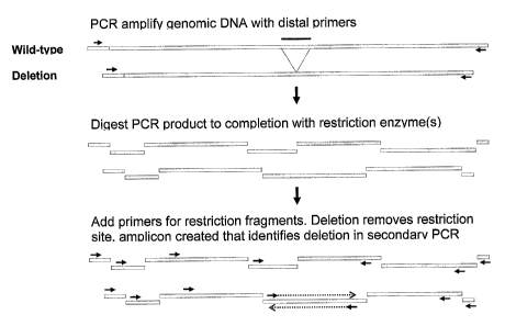
5D. Gene Mutation Scanning
[00115] In yet another preferred embodiment, the subject invention
provides a system referred
to herein as gene mutation scanning (GMS). In this approach, a first PCR step
can be
performed. Generally with this approach, DNA (including genomic DNA amplified
by said
first PCR step, if that is desired) is subjected to digestion by at least one
restriction enzyme.
In preferred embodiments of this approach, one or more primers are designed
and hybridized
to each restriction fragment, followed by a PCR step (a second PCR step if a
first PCR step is
performed), so that amplification by the (second) PCR step occurs only in the
presence of a
deletion that removed a restriction site. This approach is explained in much
more detail
below in Example 7.
[00116] Thus, the subject invention includes:
[00117] A method of detecting a mutant (including deletion mutants and
point mutants) in a
pool of DNA from a plurality of plants, wherein said method comprises the
steps of:
CA 02526533 2005-11-18
WO 2004/106555
PCT/US2004/016228
[00118] a) subjecting a plurality of plant seeds to mutagenesis to
obtain Ml seeds;
[00119] b) planting a plurality of said Ml seeds and growing Ml plants;
[00120] c) pollinating a plurality of said Ml plants to produce M2
seeds;
[00121] d) obtaining a DNA sample from each of a plurality of said M2
seeds (or from
M2 plants obtained by the further step of growing M2 plants from a plurality
of said
M2 seeds);
[00122] e) pooling said DNA samples to create pooled DNA;
[00123] providing a first pair of PCR primers designed to hybridize
to said DNA;
[00124] performing PCR amplification with said pooled DNA and said
first primer
pair to obtain a primary amplicon;
[00125] h) digesting the amplified PCR product with at least one
restriction enzyme to
obtain a plurality of restriction fragments;
[00126] i) providing a plurality of RF primers, each said RF primer
being designed to
hybridize to a restriction fragment, wherein a deletion of a restriction site
in said
primary amplicon allows PCR amplification by two or more of said RF primers of
a
secondary amplicon, and wherein no amplification of a secondary amplicon by
two or
more of said RE primers occurs in the absence of a deletion removing a
restriction site
in said primary amplicon;
[00127] performing PCR amplification with said restriction fragments
and said RF
primers; and
[00128] k) assaying said pooled DNA (by gel electrophoresis or
microtiter-based
fluorescent assay, for example) for the presence or absence of a secondary
amplicon
resulting from amplification from said RF primers (wherein the presence of
said
secondary amplicon indicates the presence of a mutation (including a deletion)
that
removed a restriction site from said primary amplicon).
[00129] The subject approach does not require the first PCR amplification
of steps f) and g).
That is, genomic DNA can be digested directly with the restriction enzymes,
and the resulting
fragments can be used for the PCR amplification of steps i) and j).
Furthermore, this
approach can also be used to detect mutants other than deletion mutants. Point
mutants, for
example, can "knock out" the restriction enzyme cut site (or a primer binding
site) which
would also be detectable by the subject method. The subject approach is also
not limited to
plant DNA but can be used with DNA from other organisms. Steps a)-d) can be
replaced
CA 02526533 2005-11-18
WO 2004/106555
PCT/US2004/016228
21
with the step of obtaining DNA from an organism that was subject to mutation.
Mutagenesis,
however, is not a required step. This would be the case if these methods are
adopted for
diagnosing cancer, for example. Because applications in the plant breeding
context are
preferred, Ml, M3, M4, and M5 plants and seeds, and seeds and plants from even
subsequent
generations (where either mutated seed, pollen, or plant was a parent) can be
used in any of
the exemplified steps. Instead of using M1 seed, mutated pollen can be
produced and used in
step c), thereby removing or altering exemplified steps a)-c). Again, the DNA
samples do not
have to be mixed in a single pool; a collection of DNA samples in separate
wells or microtiter
plates, for example, can be used. DNA from a single individual can also be
tested alone, if
desired.
[00130] All patents, patent applications, provisional applications, and
publications referred to
or cited herein are incorporated by reference in their entirety to the extent
they are not
inconsistent with the explicit teachings of this specification.
[00131] Following are examples that illustrate procedures for practicing
the invention. These
examples should not be construed as limiting. All percentages are by weight
and all solvent
mixture proportions are by volume unless otherwise noted.
Example 1 ¨ Creation of fast neutron-mutagenesis-derived canola seed bank and
DNA
libraries
[00132] This example shows how a collection of seed and corresponding DNA
samples were
created. Canola seed (Brassica napus) was treated with 50-60 Gy of fast
neutrons (KFKI
Atomic Energy Research Institute, Budapest, Hungary). The resulting M1 seeds
were
planted and each M1 plant was harvested individually to give an M2 family.
Each M2 family
was placed in a numbered envelope for long-term storage and retrieval. Seeds
of five M2
families, sampled at the rate of approximately 4-5 M2 seeds per family, were
placed in each
well of a 96 - 2 ml deep-well plate (Fisher). A 4-mm tungsten ball was added
to each well
and the (dry) seeds were ground by agitating the plate on a shaker (Kleco).
DNA was
extracted and purified using a commercial kit (Qiagen DNeasy Plant 96)
following the
manufacturer's protocol with modifications. Briefly, the ground seed material
in each well
was incubated for 1 h at 60 C in 500 1.11 of buffer AP1 with RNase and reagent
DX as
CA 02526533 2005-11-18
WO 2004/106555
PCT/US2004/016228
22
specified by the manufacturer plus 10 mM of the antioxidant sodium
metabisulfite. 150 pi of
AP2 solution (Qiagen) was added, the mixture was held at ¨20 C for 10 min and
then
centrifuged at 5600 x g for 5 min. Approximately 450 p,1 of the supernatant
from each well
was transferred to the corresponding well of a new plate followed by 1.5
volumes of
AP3/ethanol buffer (Qiagen). The mixtures were transferred to DNeasy 96 spin
columns
followed by a wash step as specified by the manufacturer but with the addition
of a final
wash using 800 1 of absolute ethanol per column and a 20 min centrifugation
at 5600 x g to
dry the columns. DNA was eluted with 100 pl of buffer AE (Qiagen) to a primary
plate. A
second 100 pi elution was saved to a back-up plate.
Example 2 - Pooling DNA and screening with primers for deletions ,
[00133] This example shows how DNA from the primary plates described in
Example 1 were
pooled and screened for deletions in a target gene. DNA from 3 wells of a
primary plate
were pooled into a single well of a secondary plate. At a sampling rate of 5
seeds per M2
family and 5 M2 families per well in the primary plate, a single well in a
secondary plate
represents 60 M2 families and a heterozygous gene deletion event in a single
seed would be
represented at approximately 1 in 600 non-deleted gene copies. PCR
amplifications were
performed in 200 I wells in a 25 1 reaction volume, using 2.0 1 of the
pooled DNA (-50
ng) as a template plus other components as recommended by the manufacturer
(TaKaRa): lx
LA PCR buffer, 3.0 mM MgC12, 400 p,M each dNTP, 0.2 M of each primer, and 1.2
U LA
Taq. The thermocycler (MJ RESEARCH) was programmed for 94 C, 2 mM followed by
30
cycles of 94 C, 20 sec; 68 C, 15 min. The primers as used supported
amplification of an
approximately 16 kb product. PCR products were visualized by running
approximately one-
half the reaction volume on a 0.5% agarose gel (SEA CHEM GOLD). In an initial
screen of
88 samples representing DNA from 5,280 M2 families, seven reactions yielded
secondary
products of approximately 11 kb. Because of the expected low frequency of
deletions and the
indication that these products were all the same size, it is likely these
secondary products
represent the same natural polymorphic allele. PCR analysis of respective
primary samples
for these products demonstrated that the PCR product identified in each
secondary pooled
sample originated in a single primary DNA sample derived from five M2
families.
CA 02526533 2005-11-18
WO 2004/106555
PCT/US2004/016228
23
Example 3 ¨ Preparation and Pooling of DNA for Detection of Mutant Lines Using
Full-
Extension PCR
[00134] Gene targets are prepared by identification of an amplicon that
contains the gene plus
upstream and downstream flanking regions. Amplicon identification is achieved
using a
Universal GenomeWalker Kit (Clontech, Palo Alto, CA). The first step in this
procedure is
to obtain clean, high average molecular weight genomic DNA from canola tissue.
Canola
seeds are sown on 0.8% agar medium supplemented with 1/2 strength MS media and
grown
in a growth chamber with 12 hour light period at 25 C and a 8 hour dark period
at 15 C.
After 7 days the seedlings are washed, blotted dry, ground to a fine powder in
liquid N2 with
a mortar and pestle, and immersed (1g/10m1) in a CTAB buffer solution
containing (100 mM
Tris-HCL, pH 7.5, 25 mM EDTA, 2 M NaC1, 1.5% PVP 40[polyvinylpyrrolidone,
Sigma, St.
Louis, MO], and 2.5% CTAB [Hexadecyltrimethylammonium bromide, Sigma]. The
solution is incubated for 2 hours at 65 C. After cooling to room temperature,
4.5 ml
chloroform/octanol (24:1) is added and gently mixed for 5 min until both
layers are mixed
and dispersed, then centrifuged for 10 min at 2000 rpm. The top (aqueous)
phase is
transferred to a 15 ml polypropylene tube containing 6 ml ixopropanol and
allowed to stand
for 1 hour. The samples are gently mixed and then centrifuged at 10,000 rpm
for 20 min to
pellet the DNA. The supernatant is poured off and the pellet is re-suspended
in 1 ml TB
buffer. RNA is digested by adding 2 1 of a 10 lug/u1 RNAse A solution and
incubated at
37 C for 1 hour. Polysaccharides and other debris are pelleted by centrifuging
at 14,500 rpm
for 10 min. The supernatant is separated to two 0.5 ml samples and to each is
added 150 010
M Ammonium Acetate and 500 ul isopropanol. The DNA is spooled with a glass
rod, rinsed
twice with 70% ethanol, blotted dry and resuspended in TB buffer. The
concentration is
determined using a Pico Green dsDNA Quantitation Kit (Molecular Probes,
Eugene, OR)
and the solution is diluted with more TB buffer to give a final concentration
of 200 ng DNA/
14 The DNA is also tested for size and purity by running on a 0.5%
agarose/ethidium
bromide gel. This DNA is used to make the restriction digest libraries that
are used for
genome walking and as a template for PCR reactions.
[00135] Restriction digest libraries of Dra 1, EcoR V, Pvu 11 and Stu 1
are prepared by
incubation at 37 C for 2 hours of 2.5 lug canola genomic DNA, the restriction
enzyme (80
units) and the respective restriction enzyme buffer in deionized water in a
total of 100 IA. A
control library using human genomic DNA and one of the restriction enzymes is
also
CA 02526533 2005-11-18
WO 2004/106555
PCT/US2004/016228
24
produced. After incubation 5 !_t1 of the solutions are run on a 0.5%
agarose/ethidium bromide
gel to determine if the cuts are complete. The remaining 95 pl is mixed with
phenol,
vortexed at slow speed and spun briefly in a microcentrifuge. The upper
(aqueous) layer is
transferred to a clean 1.5 ml tube and mixed with 190 pi ice cold 95% ethanol,
9.5 pl. 3 M
sodium oxaloacetate (pH5.2) and 20 lig glycogen. After centrifugation at
13,100 rpm for 15
min, the supernatants are decanted, and the pellets are washed with 100 [A ice
cold 80%
ethanol. The pellets are air-dried and dissolved in 20 pi 10 mM TB with 0.1 mM
EDTA pH
7.5. Use 5 pl to run on 0.5% agarose/ethidium bromide gel to determine
approximate
quantity of DNA after purification. The final step in library preparation is
ligation of the
purified DNA. A ligation reaction of 4 p1 digested, purified DNA, 1.9 1
GenomeWalker
Adaptor (25 M), 1.6 pi 10x Ligation Buffer and 0.5 1 T4 DNA Ligase (6 units/
1) is
incubated at 16 C overnight. The reactions are stopped by incubating at 70 C
for 5 min and
then diluted by adding 72 jtl 10 mM TB with 1 mM EDTA, pH 7.5. Each library
solution is
aliquoted and stored at minus 20 C.
[00136] Genome walking is conducted by utilizing primary and secondary PCR
reactions.
Primers are designed, according to the GenomeWalker Kit specifications, for
primary and
secondary (or nested) PCR from the 5' (for walking upstream) and 3' (for
walking
downstream) ends of the gene to be walked. TaKaRa LA TaqTm (LA Taq, Takara
Shuzo
Co., Japan) is the DNA polymerase used for the PCR reactions. A primary PCR
reaction
consists of 0.5 pl restriction digest library (reactions for all 4 libraries
are run concurrently),
0.5 IA of primary adaptor primer, 0.5 pl of primary gene primer, 2.5 pl 10x LA
PCR buffer
(with 25 mM Mg), 4 1 dNTP mix (2.5 mM each), 0.7 1 MgC12, 0.25 1 LA Taq (5
U/ 1)
and H20 to make 25 1 total. Positive controls (with the human DNA restriction
library
constructed along with the other libraries and a human DNA restriction library
supplied in the
GenomeWalker Kit) are run with the supplied adaptor and gene primers. In
addition, a
negative control with the gene primer omitted is run to test if any products
are produced with
the adaptor primer alone. The primary reaction is performed using a PTC-220
DNA Engine
DyadTM Peltier Thermal Cycler (Dyad, MJ Research, Boston) with the following
cycling
parameters:
[00137] 1. 94 C, 25 sec
[00138] 2. 72 C, 3 min
CA 02526533 2005-11-18
WO 2004/106555
PCT/US2004/016228
[00139] 3. cycle 6 more times to step 1
[00140] 4. 94 C, 25 sec
[00141] 5. 65 C, 3 min
[00142] 6. cycle 31 more times to step 4.
[00143] 7. 65 C 7 min
[00144] 8. hold at 4 C
[00145] Eight ill of the primary PCR reaction products are separated on a
1.0%
agarose/ethidium bromide gel. When products and/or a smear are observed,
proceed to the
secondary reaction. The secondary PCR reaction consists of 0.5 111 of a 1/50
dilution of the
primary PCR reaction, 0.5 ill of secondary adaptor primer, 0.5 IA of secondary
gene primer,
2.5 ill 10x LA PCR buffer (with 25 mM Mg), 4 111 dNTP mix (2.5 mM each), 0.7
j.il MgCl2,
0.25 1,L1 LA Taq (5 U/ 1) and H20 to make 25 1 total. The secondary reaction
is preformed
on the Dyad with the following cycling parameters:
[00146] 1. 94 C, 25 sec
[00147] 2. 72 C, 3 min
[00148] 3. cycle 4 more times to step 1
[00149] 4. 94 C, 25 sec
[00150] 5. 65 C, 3 min
[00151] 6. cycle 19 more times to step 4.
[00152] 7. 65 C 7 min
[00153] 8. hold at 4 C
[00154] Five .1 of the secondary PCR reaction products are separated on a
1.0%
agarose/ethidium bromide gel. Seconday PCR products are either sequenced
directly from
the PCR reaction mixture after purification using a Performa DTR Gel
Filtration Cartridge
(Gaithersbury, MD) or sequenced after separation on and purification from
(using a
QIAquick Gel Extraction Kit, Qiagen Inc., Valencia, CA) a 0.5% agarose/ethidum
bromide
gel. Sequencing is performed using the Big DyeTM terminator cycle sequencing
ready
reaction (Applied Biosystems, Foster City, CA) according to the manufacturer's
suggested
protocol. The 5' sequence of the secondary PCR product is used to design
primers for PCR of
the PCR fragment plus the gene to show that they are contiguous, and to design
primary and
secondary primers for the next genome walking PCR reactions. The genome
walking
CA 02526533 2005-11-18
WO 2004/106555
PCT/US2004/016228
26
reactions and consequent PCR reactions are continued both upstream and
downstream from
the gene until the desired size of amplicon is identified. Figure 1 shows an
approximate 16
kb sample amplicon obtained for a sample gene from canola variety Nex 710.
[00155] A sense primer and an antisense primer designed from "US3" and
"DS4" (upstream
and downstream fragments, respectively, from the gene of interest; see Figure
1) are used to
obtain an approximate 16 kb PCR product. The PCR reaction mixture consists of
0.5 1
Canola Nex 710 genomic DNA (10 to 200 ng/ 1) as template, 0.5 1 each of
primers, 2.5 ill
10x LA PCR buffer (with 25 mM Mg), 4 pi dNTP mix (2.5 mM each), 0.7 pl MgCl2,
0.25 1
LA Taq (5 U/ 1) and H20 to make 25 pl total. The PCR reaction is run on the
Dyad
Thermal Cycler with the following cylicing parameters:
[00156] 1. 94 C 2 min
[00157] 2. 94 C 20 sec
[00158] 3. 68 C 15 min
[00159] 4. cycle 29 more times to step 2
[00160] 5. hold at 4 C
Example 4 - Sensitivity of detection of a deleted gene in canola
[00161] To illustrate how the procedures of the subject invention work in
practice, a
transgenic plant DNA (with a marker gene insert) was used to simulate wild-
type plant DNA,
and wild-type plant DNA without the marker gene insert (to simulate a deletion
event) was
used to illustrate how a deletion would be identified.
[00162] Sensitivity of detection of a deleted gene in canola was measured
by the following
procedure. Genomic DNA from a transgenic canola line that contains a gene for
Aspergillus
A9 desaturase (US Patent No. 6,495,783) and a gene for phosphenothricin
transferase (PAT,
selectible marker gene) was used to make restriction digest libraries for
genome walking.
Genome walking was performed both upstream and downstream of the 7 kb
Aspergillus A9
desaturase/PAT insert. Primers were designed from the two outmost fragments
that produce
a 16 kb PCR product using the PCR reaction and cycling parameters described
above. PCR
reactions were performed using various dilutions of the wild type DNA (from
non-transgenic
canola, in this case respresenting DNA containing the "deleted" gene) with DNA
from the
Aspergillus A9 desaturase/PAT transgenic canola as templates. Dilutions of
1/10th to 1/1000th
CA 02526533 2005-11-18
WO 2004/106555
PCT/US2004/016228
27
are tested. Figure 2 shows the PCR products from this experiment. The PCR
product from
the "deleted" gene amplicon is clearly observed at a dilution of 1/1000th.
[00163] Figure 2 shows PCR products of mixtures of wild type (non-
transgenic canola with
gene "deleted") and transgenic canola containing a 7 kb Aspergillus A9
desaturase/PAT insert
using primers that produce a 16 kb amplicon containing the transgenic gene
inserts. The
amplicon with the "deleted" gene is detected at a dilution of 1/1000th.
Example 5 ¨ Use of PNA Probes in the Detection of Deletion Mutants in Mixed
DNA Pools
[00164] This example shows how peptide nucleic acid (PNA) oligomers can be
used to
enhance the long-range PCR-based detection of DNA from a deletion mutant that
is present
as a minor fraction in a pool of DNA derived primarily from wild-type plant
material.
Figure 3 shows a basic illustration of this technology, which is surprisingly
and
advantageously applied presently to the context of pooled plant DNA.
[00165] The Arabidopsis sng1-8 deletion event removes ¨ 6 kb of DNA from
the SNG1 locus
(Lehfeldt et al. 2000, Plant Cell 12:1295-1306). Using published sequence
information from
the Arabidopsis genome, PCR primers upstream
(GAATTATCTACTATGTGAGCTATTTGTTCCTGAG) (SEQ ID NO:1) and downstream
(CCTTCATCTAATCAGAACATGTAAGTAGAATGTG) (SEQ ID NO:2) of the sng1-8
deletion were designed to produce a 19.1 kb amplicon that included the SNG1
gene and
flanking regions. To inhibit PCR amplification of the wild-type SNGI DNA, two
PNA
oligomers (CAAACTGAACCAAACCCG and TGGTTTCGGTATGATCCA) (SEQ ID
NO:3 and SEQ ID NO:4, respectively) that were complementary to a region of the
SNG1
gene known to be removed by the sng1-8 deletion, were synthesized (Applied
Biosystems)
for addition to PCR. PCR conditions were as recommended by the manufacturer of
LA Taq
(Takara) except that a 25 I reaction volume, 3.2 mM Mg, 100 ng of template
DNA, and 3.0
tM of each PNA oligomer were used. In addition, thermocycler parameters were
modified
from the 2-step protocol recommended by the manufacturer to include a 75 C
annealing step
for the PNA oligomers: a single 94 C 1 min denaturation step was followed by
30 cycles of
94 C 20 sec, 75 C 30 sec, 66 C 12 min.
[00166] Under the conditions described above, the addition of the PNA
oligomers to the
reaction mix specifically inhibited synthesis of the 19.1 kb wild-type product
but did not
inhibit production of the 13 kb sng1-8 mutation-derived product. The mutant
product could
CA 02526533 2005-11-18
WO 2004/106555
PCT/US2004/016228
28
be detected from a PCR reaction where only 0.1 ng of sng1-8 DNA (0.1% of the
total) was
provided as a template. It should be noted that the 13 kb mutant product was
still detectable,
albeit at a lower level, from a nearly identical PCR that did not contain PNA
oligomers. See
Figure 4 and 5. The relative abundance of the 13 kb mutant product in this
case is due to its
kinetic advantage over the larger 19.1 kb wild-type product in PCR. Had the
sng1-8 deletion
been smaller (1 kb for example), the mutant product would have little kinetic
advantage over
the wild-type product and so would not likely have been detected without the
addition of
PNA oligomers to suppress formation of the wild-type product. (That is, with a
large
deletion, PCR amplification of a full-length wild-type segment takes longer
for the
polymerase to produce than a much shorter deletion mutant of that segment.
Thus, with both
templates - wild-type and deletion - in the same PCR, the shorter amplicon
will be relatively
much more abundant than the long wild-type after the PCR is allowed to proceed
for some
time. However, this would not be the case where the wild type and the deletion
are close to
the same size, as it would take the polymerase approximately the same time to
traverse both
of these segments.) This illustrates an advantage of the subject PNA approach.
[00167] The PNA method was also used to detect a deletion in canola. These
methods were
exemplified using wild-type canola lines and a canola line having an
approximately 2,325 bp
deletion beginning with residue 13,544 of the 87,844 bp region in the canola
genome
corresponding to the sequence available from GENBANK as Accession No.
AJ245480.
Primers and PNA probes were designed to target this region. Eight samples,
each containing
approximately 15 ng of genomic DNA (pooled from samples representing 15 M2
families
sampled at the rate of approximately 5 seeds per family) were subjected to PCR
using
primers D199 (SEQ ID NO:5) and D200 (SEQ ID NO:6) plus 1 ,M of PNA probe Q108
(SEQ ID NO:7) (Applied Biosystems). (The approximate location of these primers
[and
other primers described below] with respect to the sequence of GENBANK
Accession No.
AJ245480 are provided above in the Brief Description of the Sequences
section.) PCR
conditions were those recommended by the manufacturer for LA Taq (Takara) in a
25 Al
volume. Thermocycler settings were: 94 C, 1 minute followed by 30 cycles of 94
C, 20
seconds; 78 C, 30 seconds; 66 C, 12 minutes. PCR products (5 Al each) were
separated on a
0.4% agarose gel also containing X Hind III markers. See Figure 6. The band
for the
amplicon with the deletion is clearly visible in lane 5. (Lane 6 contains a
PCR artifact).
CA 02526533 2005-11-18
WO 2004/106555
PCT/US2004/016228
29
Thus, the PNA probe suppressed amplification of the wild-type DNA samples, but
the
deletion was amplified, thereby identifying the plant tissue sample that had a
deletion.
Example 6 ¨ Use of "Poison Primer" to Detect Deletion Mutants in Pool of Mixed
Plant
DNA
[00168]
This example indicates how the use of additional primers in PCR can enhance
the
detection of deletions in genes of interest. As in Example 5, primers for long
PCR are
designed to bind to DNA flanking the target gene so that a product, which
includes the target
gene and is up to 20 kb in length, is generated by long PCR from these
opposing "outer"
prirriers. A second pair of nested primers is also designed to bind to
sequences just inside the
outer primers. In a typical nested PCR procedure, a primary reaction is run
using genomic
DNA as the template and a set of outer primers for amplification. A small
amount of the
primary PCR product is then used as template in a secondary PCR that utilizes
a set of nested
primers for amplification. Because the majority of molecules that serve as
templates for the
nested primers in the secondary reaction are those produced from the pair of
outer primers in
the primary reaction, this two-stage process typically results in an abundant
yield of the
expected PCR product, with little or no non-specific PCR products being
formed.
[00169]
This example modifies the traditional two-stage nested PCR by adding at
least one
additional primer that binds between the two outer primers of the primary PCR.
This
"poison primer" is designed to hybridize to the target gene and, together with
one of the outer
primers, leads to the formation of a PCR "poison product" that is only about
half the size of a
product formed between the two outer primers with no poison primer
therebetween. Due to
the kinetic advantage (as discussed in Example 5), the shorter poison product
will
predominate in the primary PCR. When this primary product is used as a
template in the
secondary reaction, little or no full-length product is formed because a
binding site for one of
the two nested primers does not exist on the poison product. If however, a
deletion removes
the binding site for the poison primer (but not any binding sites for the
nested or outer
primers), the poison product can no longer form in the primary PCR. In the
secondary PCR,
both nested primers will bind to the primary template and lead to formation of
a product that
signals the presence of the deletion.
[00170] The "poison primer" method was used to detect a deletion in
canola. These methods
used the same eight samples as described in the preceding Example. Again, the
eight
CA 02526533 2005-11-18
WO 2004/106555
PCT/US2004/016228
samples, each containing approximately 15 ng of genomic DNA (pooled from
samples
representing 15 M2 families sampled at the rate of approximately 5 seeds per
family) were
subjected to PCR using primers D199 (SEQ ID NO:5) and D200 (SEQ ID NO:6) plus
the
poison primer D249 (SEQ ID NO:8). Thermocycler settings were: 94 C, 1 minute
followed
by 30 cycles of 94 C, 20 seconds; 78 C, 30 seconds; 68 C, 12 minutes. The PCR
products (1
Al each) were separated on a 0.4% agarose gel also containing X Hind III
markers. See
Figure 7. The band for the amplicon with the deletion is indicated in lane 5.
(The
preferential amplification of the deletion worked so well that even less DNA
could have been
loaded into this well; this would have yielded a more distinct deletion band.)
Some wild-type
product is visible in lanes 1, 4, and 6-8. The wild-type product was
suppressed in lanes 2 and
3 to the extent that no wild-type bands are visible in those two lanes. This
illustrates that the
band for the amplicons with the deletion (being smaller than the wild-type
amplicons in some
of the other lanes) are clearly identifiable and that amplification of wild-
type DNA can be
completely or sufficiently suppressed using this method of the subject
invention.
Example 7¨ Gene Mutation Scanning (GMS)
[00171] This example describes a screening method, with two variations,
that can be used to
detect deletions or other mutations (including point mutants) within pooled
samples of DNA
from multiple lines. These mutations (including deletions) must occur within a
window
defined by long-range PCR from primers flanking a target gene as in previous
examples. In
this method, the initial long PCR product is digested with one or more
restriction enzymes.
The resulting restriction fragments are then subjected to a second round of
PCR using primers
that would not support PCR amplification except in rare cases where a mutation
or deletion
event removed one or more of the recognition sites for the restriction
enzyme(s) used in the
previous step.
[00172] To be successful, the GMS method requires complete or nearly
complete sequence
information for the target gene (including introns) and all flanking DNA
contained within the
initial long PCR product. This information can be obtained, for example, by
sequencing the
insert from one or more large genomic clones (BAC, cosmid, etc) that contain
the target gene
and its flanking DNA. Such clones can be identified by screening libraries of
these clones by
standard methods that utilize the sequences derived from the target gene as a
probe.
Alternatively, the information can be obtained by fully sequencing many of the
intermediate
CA 02526533 2005-11-18
WO 2004/106555
PCT/US2004/016228
31
products obtained while chromosome walking from the two ends of the target
gene, for
example by the GenomeWalkerTM method.
[00173] A collection of M2 families, derived from a mutagenesis utilizing a
deletion-inducing
agent such as fast neutron radiation, is generated as in previous examples.
Using the sequence
information obtained by one of the above methods, primers that bind to regions
flanking the
target gene are designed for long PCR. In addition, one or more restriction
enzymes are
chosen to digest the initial PCR product into several fragments. Fragment
lengths ranging
from approximately 500 bp to 2000 bp are preferred, although this method can
utilize
restriction fragment lengths outside of that range. Finally, PCR primers that
bind to the
restriction fragments (RF primers) are designed. The position of these RF
primers on the
restriction fragments is determined by the choice of two possible variations
of the method.
[00174] In the first variation, it is desired to identify deletions that
remove one, or possibly
two of the multiple restriction sites that occur within the initial long PCR
product. For
example a single targeted restriction site (TRS) within an exon of the target
gene could be
selected to identify deletions that remove some or all of the target gene.
Such deletions
would be expected to generate complete loss of function mutations.
Alternatively, a TRS
upstream and a TRS downstream of the target gene could be chosen to be
screened
simultaneously in order to identify deletions that occur in the flanking
regions. A deletion of
this type could potentially lead to a partial loss or gain of function in the
target gene if it left
the coding region and minimal regulatory elements intact but removed other
regulatory
elements, such as enhancers or suppressors, that may exist in the flanking
DNA.
[00175] In the first variation, one RF primer is positioned on each
restriction fragment as far
from the TRS as possible. See Figure 8. Sense RF primers are positioned near
the upstream
end of fragments upstream of the TRS, and antisense RF primers are positioned
near the
downstream end of fragments downstream of the TRS, so that a series of nested
primers
"pointing towards" the TRS is created. Under this arrangement, the initial
long PCR product,
fully digested by the selected enzyme(s), would not be a suitable template for
the second
round PCR using the RF primers. However, if a variant existed within the
pooled DNA
sample where a deletion had removed the TRS but not the binding sites for at
least one sense
and one antisense RF primer, then a PCR product could accumulate that would
signal the
presence of the deletion. In cases where more than one TRS is selected, the
initial long PCR
product would be divided into separate sub-section for each TRS. Within each
sub-section,
CA 02526533 2005-11-18
WO 2004/106555
PCT/US2004/016228
32
sense RF primers would be positioned near the upstream end of fragments
upstream of the
respective TRS and antisense RF primers would be positioned near the
downstream end of
the downstream fragments. In these cases, only deletions that removed the TRS
and left
intact at least one sense and one antisense RF primer binding site within the
sub-section for
the TRS would be detected.
[00176] In the second variation, it is desired to identify deletions that
remove any one or more
of the restriction enzyme recognition sites within the initial long PCR
product. In this
variation, most of the restriction fragments would have two RF primers
designed to bind near
their centers, with the sense primers positioned slightly downstream of the
antisense primers.
See Figure 9. Under this arrangement the DNA polymerase could extend a product
from the
primers to the ends of the restriction fragments, but the products would not
overlap and
therefore exponential amplification of DNA would not be supported. However, in
cases
where a rare deletion event removed one or more restriction sites but left at
least one sense
and one antisense priming site intact, PCR would be supported.
[00177] Two approaches were used to exemplify the GMS methodology for
detecting a
deletion in canola. These methods were exemplified using the same known
deletion and
eight samples used in the PNA and poison primer Examples above. In the first
approach (see
Figure 10), pooled Brassica napus genomic DNA (for wild-type lines and for the
deletion
line) was first amplified with two primers D199 (SEQ ID NO:5) and D200 (SEQ ID
NO:6).
The resulting PCR amplicon was digested with Nco I restriction enzyme. In the
second
approach (see Figure 11), pooled Brassica napus genomic DNA was digested with
Nco I
restriction enzyme. For both approaches, the digested product was amplified
with the
following five primers (using Multiplex PCR): D195 (SEQ ID NO:11), D201 (SEQ
ID
NO:12), D209 (SEQ ID NO:13), D190 (SEQ ID NO:10), and D200 (SEQ ID NO:6). The
PCR products were then resolved on 0.8% agarose gel. See Figures 10 and 11. In
both
figures, the amplicon resulting from the removal of a restriction site (the
deletion) is apparent
in lane 5. This shows that the removal of the restriction site resulted in
proper alignment of
the GMS primers of the subject invention, thereby resulting in amplification
of DNA from a
tissue sample having a deletion. The absence of this amplicon in the other
lanes signifies that
the wild-type restriction sites were maintained and, thus, amplification from
the isolated
GMS primers did not occur for the wild-type samples. The concept used to
produce the
results shown in Figure 11 is illustrated in Figure 12. The amplicons shown at
the bottom of
CA 02526533 2005-11-18
WO 2004/106555 PCT/US2004/016228
33
Figure 12 illustrate non-detectable deletions which do not eliminate central
restriction sites
or primer sites. The amplicons above those illustrate detectable deletions
which must
eliminate central restriction sites but not outer primer sites. This is
similar for what is
illustrated in Figure 9.
CA 02526533 2006-11-08
1
SEQUENCE LISTING
<110> Dow AgroSciences LLC
<120> High-Throughput Methods of Screening DNA for Deletions and Other
Mutations
<130> 1003-172
<140> 2,526,533
<141> 2004-05-21
<150> US 60/472,863
<151> 2003-05-22
<160> 13
<170> PatentIn version 3.2
<210> 1
<211> 34
<212> DNA
<213> Artifical Sequence
<220>
<223> Primer used according to the subject invention
<400> 1
gaattatcta ctatgtgagc tatttgttcc tgag 34
<210> 2
<211> 34
<212> DNA
<213> Artifical Sequence
<220>
<223> Primer used according to the subject invention
<400> 2
ccttcatcta atcagaacat gtaagtagaa tgtg 34
<210> 3
<211> 18
<212> DNA
<213> Artifical Sequence
<220>
<223> PNA probe used according to the subject invention
<400> 3
caaactgaac caaacccg 18
CA 02526533 2006-11-08
2
<210> 4
<211> 18
<212> DNA
<213> Artifical Sequence
<220>
<223> PNA probe used according to the subject invention
<400> 4
tggtttcggt atgatcca 18
<210> 5
<211> 30
<212> DNA
<213> Artifical Sequence
<220>
<223> Primer D199 (sense primer, aligns at approximately residue [2,101-] of
GENBANK Accession No. AJ245480).
<400> 5
acgtcctcct caacctcgtt aagacacttg 30
<210> 6
<211> 31
<212> DNA
<213> Artifical Sequence
<220>
<223> primer D200 (antisense primer, aligns at approximately residue [-19,162]
of GENBANK Accession No. AJ245480).
<400> 6
cttcttcatc agcttgctaa ggaggggtaa g 31
<210> 7
<211> 18
<212> DNA
<213> Artifical Sequence
<220>
<223> PNA probe Q108.
<400> 7
CA 02526533 2006-11-08
3
aggtgcagcc agctacat 18
<210> 8
<211> 30
<212> DNA
<213> Artifical Sequence
<220>
<223> Primer D249 (sense primer for poison primer test, aligns at
approximately
residue [14,828-] of GENBANK Accession No. AJ245480).
<400> 8
gtttgtagag atgtcaactg ggtgggcagt 30
<210> 9
<211> 30
<212> DNA
<213> Artifical Sequence
<220>
<223> Primer D245 (sense primer, aligns at approximately residue [2,243-] of
GENBANK Accession No. AJ245480).
<400> 9
gaatagagct gatgggtcac tgacaaggag 30
<210> 10
<211> 30
<212> DNA
<213> Artifical Sequence
<220>
<223> Primer D190 (Sense primer, aligns at approximately residue [-16,230] of
GENBANK Accession No. AJ245480).
<400> 10
cactgtctgc ttaggactag ctgcatccat 30
<210> 11
<211> 30
<212> DNA
<213> Artifical Sequence
<220>
<223> Primer D195 (Sense primer, aligns at approximately residue [11,851-] of
GENBANK Accession No. AJ245480).
<400> 11
tggcctaggc catgtataac attaaaacag 30
CA 02526533 2006-11-08
4
<210> 12
<211> 32
<212> DNA
<213> Artifical Sequence
<220>
<223> Primer D201 (Sense primer, aligns at approximately residue [12,650-] of
GENBANK Accession No. AJ245480).
<400> 12
cggagaacaa gttatatgcc cattatgaca ct 32
<210> 13
<211> 30
<212> DNA
<213> Artifical Sequence
<220>
<223> Primer D209 (Sense primer, aligns at approximately residue [15,443-] of
GENBANK Accession No. AJ245480).
<400> 13
gcaccttctg tgctacaaca actaatcttt 30