Note: Descriptions are shown in the official language in which they were submitted.
CA 02526661 2005-11-22
WO 2004/104598 PCT/KR2004/001242
DIAGNOSTIC KIT FOR LIVER CIRRHOSIS COMPRING AN ANTIBODY
SPECIFIC FOR HUMAN PROTOONCOGENIC PROTEIN
Technical Field
The present invention relates to a method for detecting human HCCR-2
protein present in a serum sample by an antigen-antibody binding reaction
using an antibody specifically binding to a protein expressed from human proto-
oncogene HCCR-2, and a kit for diagnosing liver cirrhosis using the same.
1o Background Art
Liver cirrhosis is referred to as liver fibrosis in a broad sense in that it
causes tubercles in liver of a patient. As an end-stage chronic liver disease,
the liver cirrhosis causes persistent and recurring diffuse liver injuries,
leading to
fibrosis and formation of tubercles in liver cells (Ikeda et al., Hepatology
18:47-
53, 1993; Minami H and Okanoue T, Int Med. 80: 646-649, 1997; Kiyosawa IC,
Hepatology Research 24: S40-S45, 2002). Major factors for inflammation of
liver include virus, medicinal constituents, alcohol, metabolic diseases,
chronic
cholestasis, hepatic vein bloods flow occlusion. Liver cirrhosis accounts for
60-
90% of liver cancer cases, and liver cancer arises in 5-20% of liver cirrhosis
2o patients, suggesting that liver cirrhosis increases the risk of developing
liver
cancer. Hence, there is a pressing need for early diagnosis of liver cirrhosis
and elimination of major factors causing liver cirrhosis (Velazpuez et al.,
Hepatology 37(3): 520-527, 2003). In addition to the viral factor, risk
factors
associated with liver cirrhosis include alcohol abuse, chemical substances,
1
CA 02526661 2005-11-22
WO 2004/104598 PCT/KR2004/001242
hemochromatosis and so on. Therefore, it is quite important to perform early
detection, elimination, inoculation and quarantine of such diseases and risk
factors thereof.
Liver cirrhosis presented with clinically normal features without
complications is referred to as compensated liver cirrhosis. Liver cirrhosis
concomitant with a variety of complications is referred to as decompensated
liver cirrhosis.
Abdominal laparoscopic examination and liver organ examination has
gained popularity as the foremost tests in diagnosing liver cirrhosis. To
directly
to observe the internal structure of liver, the abdominal laparoscopic
technique is
performed in the interior of the abdomen through a small incisions that is, an
endoscope is inserted through a small entrance incision in the abdominal
cavity.
When a hard and lumpy surface is visualized through the abdominal
laparoscopic examination, the liver organ is diagnosed as being affected by
cirrhosis. Also, when liver fibrosis is visualized through the liver organ
examination, the presence or severity of liver cirrhosis is diagnosed.
However,
such examination can be a psycho I og i ca I burden to a patient. Also, an
accurate diagnosis for liver cirrhosis is often based on a combination of a
variety of methods or techniques, including doctor's medical advice, blood
test,
2o ultrasonography, computerized tomography (CT).
In a case of liver cirrhosis, the level of serum a I an i ne am i not
ransferase
(ALT) (equivalent to conventional GOT or GPT level) is not so high and is
substantially normal or less than two times the normal level, which makes it
impossible to be used as effective diagnostic indicator for liver
inflammation.
2
CA 02526661 2005-11-22
WO 2004/104598 PCT/KR2004/001242
In particular, even in cases involving compensated liver cirrhosis, there are
considerable liver cells that normally function. Thus, the level of albumin or
bilirubin falls under a substantially normal range. On the other hand, in
cases
involving decompensated liver cirrhosis, the level or albumin may decrease or
the level of bilirubin may increase. The presence of normally functioning
liver
cells is quite important for liver cirrhosis or advanced chronic liver disease
patients, and albumin or bilirubin is only a rough indicator for the presence
of
normal liver cells. If normal liver cells, which produce blood coagulating
factors,
are insufficient, coagulation of blood may be delayed. A prothrombin time (PT)
1o examination is performed to directly evaluate a complete coagulation of
blood,
which also acts as an indicator to evaluate remaining liver function. Liver
cirrhosis results in an enlargement of the spleen, in which a large quantity
of
platelets is entrapped. Thus, a reduction in the platelet level due to unknown
cause is suspected of having liver cirrhosis.
Serological marker examination against hepatitis virus like in chronic
hepatitis also importantly acts as an indicator for liver cirrhosis. In Korea,
approximately 60% of liver cirrhosis is caused by hepatitis B virus (HBV) and
approximately 20% of liver cirrhosis is caused by hepatitis C virus (HCV).
Thus, it can be said that a positive HBV or HCV marker suggests a high risk
2o factor for chronic liver diseases. In addition, when a patient is presented
with
other lesions suggesting liver cirrhosis, it is natural to diagnose the
patient's
lesion as liver cirrhosis.
A non-specific increase in the serum alpha-fetoprotein (APF) level,
3
CA 02526661 2005-11-22
WO 2004/104598 PCT/KR2004/001242
which is an assay for early detection of liver cancer, may be observed in some
patients having chronic hepatitis or cirrhosis and not having HCC lesions .
(Adinolfi A et al., J Med Genet. 12(2 " 138-151, 1975; Lock AS and Lai CL
Hapatology 9(1 ). The level of alpha-fetoprotein (AFP) has been
advantageously used in diagnosing liver cirrhosis as one of screening factors
for early detection of liver cirrhosis based on the finding that the
concentration
of AFP in normal adult sera is almost zero but drastically increases in many
patients with liver cirrhosis. In fact, however, the concentration of AFP may
increase in chronic hepatitis or liver cirrhosis. For these reasons, although
1o AFP testing is regarded as an essential step in screening patients with
HCCs
among patients with chronic liver diseases to some extents, diagnosis of liver
cirrhosis based on the AFP level requires a number of considerations, which
means that there is a need for research into new useful diagnostic methods.
Thus, as part of such research, in order to achieve early diagnosis of and
effective prediction of liver cirrhosis, development of a new serological test
with
improved sensitivity and specificity would be highly desirable.
Accordingly, in order to develop a method for effective early diagnosis of
liver cirrhosis, the present inventors undertook earnest research and studies
and identified that liver cirrhosis could be effectively detected by an
antigen-
2o antibody binding reaction using antibodies specifically binding to a
protein
expressed from human proto-oncogene HCCR-2 deposited at the GenBank as
Accession No. AF315598, as described in Korean Patent Publication No. 2002-
41550. This finding has led the present inventors to complete the present
invention.
4
CA 02526661 2005-11-22
WO 2004/104598 PCT/KR2004/001242
Disclosure of the Invention
To solve the above problems, it is an object of the present invention to
provide a method for effectively detecting liver cirrhosis in an early stage
using
human proto-oncogene HCCR-2 in serum and a kit for diagnosing liver cirrhosis
using the same.
To accomplish the above object of the present invention, there is
provided a method for diagnosing liver cirrhosis by measuring the level of
expression of HCCR-2 protein in a sample in vivo by an antigen-antibody
to binding reaction using an antibody specifically binding to a protein
expressed
from human proto-oncogene HCCR-2, and a kit for diagnosing liver cirrhosis
using the same.
Brief Descrilotion of the Drawings
FIG. 1 illustrates results of immuno-blotting performed on HCCR-2
recombinant protein isolated and purified from Escherichia coli (E. coli) BL21
strain transformed into pMAL-p2X/HCCR-2 vector according to the present
invention; and
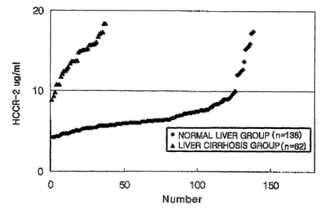
FIG. 2 illustrates diagnosis results of detecting the expression of HCCR-
2 protein in liver cirrhotic serum and normal serum by a diagnostic kit using
HCCR-2 polyclonal antibody according to the present invention.
Best mode for carrying out the Invention
Hereinafter, embodiments of the present invention will be described in
5
CA 02526661 2005-11-22
WO 2004/104598 PCT/KR2004/001242
detail with reference to the attached drawings.
The human proto-oncogene HCCR-2 (GenBank Accession No.
AF315598; Korean Patent Publication No. 2002-41550), which is positioned in
the long arm of Chromosome No. 12 (12q) and has an open reading frame, acts
as a carcinogenic gene when over-expressed in mammals, and a protein having
a size of about 36 kDa (to be referred to as HCCR-2 protein) is derived
therefrom.
The HCCR-2 protein specific antibody is preferably purified from
antiserum obtained by immunizing HCCR-2 antigen protein in an animal. More
1o preferably, the HCCR-2 protein specific antibody is a polyclonal antibody
purified from the serum obtained by immunizing HCCR-2 antigen protein in a
rabbit.
To synthesize an antibody specifically recognizing HCCR-2 protein,
HCCR-2 protein is first acquired. HCCR-2 protein can be synthesized using
known amino acid sequences or produced in recombinant protein types by
genetic engineering methods. For example, HCCR-2 recombinant protein can
be prepared by a method comprising preparing an expression vector of
expressing HCCR-2 protein in the form of a recombinant protein using the base
sequence of the HCCR-2 gene set forth in the NIH program GenBank database;
obtaining a transformant by transforming the expression vector into E.coli to
produce HCCR-2 recombinant protein; and cultivating the transformant to
isolate/purify the human proto-oncogene HCCR-2 recombinant protein.
In a preferred embodiment of the present invention, a recombinant
protein having maltose fused to N-terminal is produced from E. coli BL21
6
CA 02526661 2005-11-22
WO 2004/104598 PCT/KR2004/001242
transformed into pMAL-p2X/HCCR-2 vector containing amino acid sequences
112-304 of a HCCR-2 gene, and then treated with an appropriate enzyme to
isolate/purify HCCR-2112-304 protein from which maltose is removed to then be
used as antigen protein. In order to identify that the thus produced protein
is
HCCR-2112-304 recombinant protein, specific detection of HCCR-2
recombinant protein having a molecular weight of about 66 kDa (45 kDa of
pMAL plus 16 kDa of HCCR-2112-304) is observed by Western blot analysis.
The HCCR-2 recombinant protein isolated and purified from the E. coli
transformant is used as an antigen for screening and analyzing fused cell
lines
1o that produce polyclonal antibodies by immunization and cell fusion of
rabbits.
The immunized rabbits necessary for development of polyclonal
antibodies are generated by well mixing the HCCR-2 recombinant protein and
an equivalent amount of Freund's complete adjuvant until the mixture is
emulsified and injected into rabbits intraperitoneally, followed by booster
injection for the purpose of increasing the immunogenicity of the rabbits.
Preferably, only the HCCR-2 recombinant protein is further administered three
times into the rabbits via an intraperitoneal route, rather than through the
use of
the adjuvant.
The rabbit sera immunized through injection of the HCCR-2 recombinant
2o protein antigen are collected and antibody titers are measured.
Diagnosing cancer by a antigen-antibody binding reaction using the thus
produced HCCR-2 specific antibody protein can be made by determining the
expression of this protein in a sample in vivo, taken from a sample. The level
of expression can be detected by technique known in the art, including enzyme-
7
CA 02526661 2005-11-22
WO 2004/104598 PCT/KR2004/001242
linked immunosorbent assay (ELISA), radioimmunoassay (RIA), sandwich
assay, and Western blotting or immunoblotting analysis on a polyacrylamide
gel.
As the sample in vivo (specimen), tissues, sera or platelets, most
preferably sera, are preferably used.
In a preferred embodiment, the ELISA technique using HCCR-2 protein
specific antibodies is carried out through the following steps:
1 ) placing an HCCR-2 specific antibody into a reactor coated with a
sample and a control group to induce an antigen-antibody reaction;
2) detecting an antigen-antibody reaction product using a secondary
1o antibody-label conjugate and a color-developing substrate solution of a
labeling
substance; and
3) comparing the detection result of the sample with that of the control
group.
A large amount of the sample can be analyzed using known technique
such as ELISA, biological microchip or automated microarray system. The
biological micro chip can detect an antigen for the HCCR-2 specific antibody
protein by fixing the HCCR-2 specific antibody protein on a biological
microchip,
causing a reaction between the same and a sample in vivo collected from an
individual.
2o Also, the present invention provides a kit for diagnosing liver cirrhosis
comprising an antibody specifically reacting with the HCCR-2, thereby
effectuating early diagnosis of liver cirrhosis.
The diagnostic kit according to the present invention comprises:
1 ) a specific antibody against HCCR-2;
CA 02526661 2005-11-22
WO 2004/104598 PCT/KR2004/001242
2) a positive control group containing a HCCR-2 standard antigen and a
negative control group containing antiserum of an animal into which the
antigen
is not injected;
3) a secondary antibody conjugate having a conjugated labeling
substance producing a color by reacting with a substrate;
4) a color-developing substrate solution reacting with the labeling
substance to produce a color;
5) a washing solution to be used in each step; and
6) an enzymatic reaction stopping solution.
1o The diagnostic kit can diagnose liver cirrhosis by quantitatively or
qualitatively analyzing an antigen of the antibody protein by the antigen-
antibody binding reaction. The antigen-antibody binding reaction can be
detected by technique generally known in the art, including ELISA, RIA,
sandwich assay, Western blotting on a polyacrylamide gel, and immunoblotting
analysis. For example, the diagnostic kit can be provided to be used for ELISA
using a 96-well microtiter plate coated with recombinant monoclonal antibody
protein.
Examples of the reactor useful to coat the antibody protein thereon
include a nitrocellulose membrane, a 96-well plate made of a polyvinyl resin,
a
96-well plate made of a polystyrene resin, and slide glass.
As described above, the antibody according to the present invention is
preferably purified from the antiserum obtained by immunizing HCCR-2 antigen
protein in an animal. The HCCR-2 protein specific antibody is preferably
coated at a rate of about 1 to 10 fig/ 100 ,u,~ for each reactor.
9
CA 02526661 2005-11-22
WO 2004/104598 PCT/KR2004/001242
The control groups contained in the diagnostic kit according to the
present invention include positive control groups and negative control groups.
The positive control groups are mixtures containing HCCR-2 protein standard
antigen, and the negative control groups are animal sera uninfected with
HCCR-2 protein antigen. In Examples of the present invention, HCCR-2
protein standard antigen solutions having various protein concentrations of 0
ng/m.~ (A), 20 ng/m.~ (B), 40 ng/m.~ (C), 80 ng/m.~ (D), 160 ng/m.~ (E), 320
ng/m.~
(F) and 640 ng/m.~ (G), were used.
As the secondary antibody labeling substance, known labeling
1o substances inducing color development are preferably used, and examples the
labeling substance useful in the present invention include horseradish
peroxidase (HRP), alkaline phosphatase, colloid gold, fluorescein such as poly
L-lysine-fluorescein isothiocyanate (FITC) or rhodamine-B-isothiocyanate
(RITC), and a dye. In the present invention, goat anti-rabbit IgG-HRP
conjugate (IgG-HR.P conjugate), for example, is used.
Preferably, the chromogen used varies according to the labeling
substance involving the color development, and usable examples thereof
include 3,3',5,5'-tetramethyl bezidine (TMB), 2,2'-azino-bis(3-
ethylbenzothiazoline-6-sulfonic acid) (ABTS), and o-phenylenediamine (OPD).
2o More preferably, the chromogen is provided in a state in which it is
dissolved in
a buffer solution (0.1 M NaAc, pH 5.5). The chromogen such as TMB is
decomposed by HRP used as the labeling substance of the secondary antibody
conjugate to produce a color-developing precipitate. The level of
precipitation
of the color-developing precipitate is observed by naked eye, thereby
CA 02526661 2005-11-22
WO 2004/104598 PCT/KR2004/001242
determining the presence of HCCR-2 protein antigen.
The washing solution preferably include a phosphate buffer solution,
NaCI, and Tween 20, more preferably a buffer solution containing 0.02 M
phosphate buffer solution, 0.13 M NaCI, and 0.05% Tween 20. Following
antigen-antibody binding reaction, an appropriate amount of the washing
solution is supplied into the reactor having undergone the reaction between
the
secondary antibody and the antigen-antibody conjugate. Washing with the
washing solution is repeatedly performed 3 to 6 times. 0.1 % BSA containing
phosphate buffer is preferably used as the blocking solution and 2 N sulfuric
acid solution is preferably used as the enzymatic reaction stopping solution.
The method of diagnosing liver cirrhosis in an early stage by detecting
HCCR-2 antigen in a specimen sample using the diagnostic kit will now be
described. HCCR-2 polyclonal antibodies and specimen samples in the positive
and negative control groups are reacted, respectively, and washed with the
washing solution. Then, the secondary antibody conjugate labeled with a
labeling substance producing a color by the reaction with the substrate is
added
to the resultant product and washed again with the washing solution. Then,
the substrate containing solution is added to the resultant production to
induce
color development. Then, the absorbance at 450 nm is measured. Here, the
2o average absorbance of the standard antigen solution A should be greater
than
or equal to 0.000 and less than or equal to 0.200. The average absorbance of
the standard antigen solution F should be greater than or equal to 1.200 and
less than or equal to 3.000. The mean value between the absorbance values
of the standard antigen solutions A and F is set as a cut-off value to then be
11
CA 02526661 2005-11-22
WO 2004/104598 PCT/KR2004/001242
used to determine samples as positive or negative samples. When the
absorbance of a sample is greater than that of the standard antigen solution
F,
the sample is diluted and the absorbance thereof is then measured again. The
sample having absorbance above the cut-off value is identified as being
positive,
and the sample having absorbance below the cut-off value is identified as
being
negative.
It was confirmed that the liver cirrhosis diagnostic kit containing the
human proto-oncogene HCCR-2 specific antibody according to the present
invention, which is a new immunological diagnostic tool using patient's serum,
1o had higher accuracy and reproducibility. Conventionally, an AFP test tool
used
for diagnosis of liver cancer has also been used as a serological diagnostic
kit
for liver cirrhosis, which is, however, very poor in accuracy. However, the
serological HCCR-2 test for early diagnosis of liver cirrhosis according to
the
present invention showed a diagnosing accuracy of about 95.1 %, which is
statistically significantly higher than the conventional AFP test. Therefore,
the
diagnosing method and kit according to the present invention can be very
advantageously used for early diagnosis of liver cirrhosis and diagnosis of
small
HCC because they have high accuracy and reproducibility.
The present invention will now be illustrated in detail by the following
2o Examples, without in anyway being limited in scope to the specific
embodiments
described in Examples.
<Example 1 > Production of HCCR-2 Polyclonal Antibody
<1-1> Production of HCCR-2 Recombinant Protein
12
CA 02526661 2005-11-22
WO 2004/104598 PCT/KR2004/001242
In order to produce human proto-oncogene HCCR-2 specific antibody
as a labeling substance for diagnosis of liver cirrhosis, pET-32b(+)/HCCR-2
vector was prepared by inserting part of human proto-oncogene HCCR-2
corresponding to amino acid sequence ID NOS: 112-304 of GenBank
Accession No. AF315598 into multicloning sites of pET-32(+) vector (New
England biolabs, MA). The vector was transformed into E. coli BL21 (DE3)
(Novagen, WI) and treated with 1 mM isopropyl (3-D-thiogalacto-pyranoside
(IPTG) (manufactured by Sigma Chemical Co.) to induce expression, producing
66 KDa HCCR-2112-304 fused protein having 45 KDa maltose protein fused
1o thereto and purifying the same with an amylase resin kit (New England
biolabs,
MA ) (International Patent Application Publication No. WO 02/44370 A1 ). The
purified recombinant protein was subjected to immonoblotting, confirming that
a
large amount of about 66 kDa fusion protein was included (FIG. 1 ). The thus
isolated and purified HCCR-2 recombinant protein was used as an antigen in
the antigen-antibody binding reaction for screening and analyzing fused cell
lines producing polyclonal antibodies for immunization of rabbits.
<1-2> Immunization of Rabbits
In order to yield immunized rabbits necessary for production of
2o antibodies specific to the HCCR-2 recombinant protein prepared in Example
<1
1 >, New Zealand White (NZW) rabbits (average weight of about 2.5 kg) was
used. 200 pg/E of the HCCR-2 recombinant protein and 200 pg/~ of Freund's
complete adjuvant (manufactured by Sigma Chemical Co.) were mixed and
emulsified. 2 m~E of the resultant emulsion was subcutaneously given at 8
13
CA 02526661 2005-11-22
WO 2004/104598 PCT/KR2004/001242
dorsal sites at a rate of 0.25 m~ per rabbit. After the first injection,
booster
injection was performed several times for 2 weeks at two-week intervals in the
same manner as in the first injection by emulsifying the immunized antigen
using Freund's incomplete adjuvant.
<1-3> Isolation of Rabbit Serum and Screening of Specific
Polyclonal Antibody
At a 2-week interval after day 5 from the last inoculation, blood was
collected from the artery of the rabbits immunized in Example <1-2> and serum
1o was isolated therefrom to be stored at -20 °C until it is used in
various
experiments. Antibody specificity of the serum was examined, and the result
showed that it specifically reacted with only human proto-oncogene HCCR-2
recombinant protein. In order to screen sera specifically reacting with the
HCCR-2 protein antigen among the sera isolated and purified from the E. coli
transformant prepared in Example <1-1 > was subjected to ELISA.
In detail, in order to suppress non-specific immune responses, 96-well
plate (Falcon Co., USA) was coated by applying 1 % skim milk-PBS thereto and
allowing the same to stand at room temperature for 1 hour, and the recombinant
proteins were coated onto each well in an amount of 1 pg/well. The rabbit
2o serum fluid was diluted with a 2% BSA containing PBS solution in various
concentrations of 1 x 10³, 1 x 10⁴, 1 x 10⁵, 1 x 10⁶, and 1 x 10', and the
resulting diluted solution was added to each well in an amount of 100 ~,I per
well.
Thereafter, an antigen-antibody binding reaction was induced to take place at
37°C for 2 hours, followed by washing three times with PBS solution.
Then,
14
CA 02526661 2005-11-22
WO 2004/104598 PCT/KR2004/001242
each 100 ~,I of goat anti-rabbit IgG (manufactured by Sigma Co., USA) as a
secondary antibody, diluted to 1/10,000 with a PBS buffer solution containing
2% (W/~ BSA was added to each well and reacted at 37°C for 1 hour.
Thereafter, 100 lul of a substrate solution obtained by mixing 10 ml of 0.1
M phosphate buffered citrate (pH 5.0), 1 mg of 3.3', 5.5'-tetramethylbenzidine
(TMB) (manufactured by Sigma Co., USA) and 20 ~.I of 35% hydrogen peroxide,
was added to each well to induce an enzymatic reaction. The enzymatic
reaction was maintained at room temperature for 15 minutes and the same
amount of 2 N sulfuric acid solution was then added thereto to stop the
1o enzymatic reaction. The extent of color development was observed at 450 nm.
From polyclonal antibodies having HCCR-2 protein specific antibodies, serum
with antibody titer of 10 times greater than that of the negative control
group, as
measured by ELISA, were further screened, and characteristics of the screened
cells were analyzed.
<Example 2> ELISA using HCCR-2 Polyclonal Antibody
The liver cirrhosis was diagnosed by ELISA using HCCR-2 polyclonal
antibody in the following manner. First, wells were coated with the sample;
Second an ELISA plate wells were coated with HCCR-2 specific polyclonal
2o antibodies isolated and purified from the rabbit. Third, step detecting the
presence of HCCR-2 antigen-antibody binding in the sample.
<2-1 > Coating with sample on the wells
Liver cirrhosis and hepatitis sera and normal liver serum were used as
CA 02526661 2005-11-22
WO 2004/104598 PCT/KR2004/001242
specimen samples. To obtain a standard curve for measurement of cut-off
values, standard antigen solutions A, B, C, D, E, F and G were prepared by
diluting HCCR-2 recombinant protein at various concentrations of 0 ng/ml~, 20
ng/ml, 40 ng/ml, 80 ng/ml, 160 ng/ml, 320 ng/ml and 640 ng/ ml, respectively.
Each 100 ~,I of the respective specimen samples was distributed to each
96-well ELISA flat-bottomed plate and reacted at 37°C for 4 hours,
followed by
washing 4 times with a washing buffer solution (PBS including 0.05% Tween 20,
Ph 7.4). Here, the standard antigen solutions prepared above were used as
positive control groups and normal rabbits sera were used as negative control
to groups.
<2-2> Addition of HCCR-2 Polyclonal Antibody
HCCR-2 protein specific polyclonal antibodies according to example <1
3> were placed in each well coated with the specimen samples, covered with a
lid and allowed to stand at 4 C for 16 to 18 hours. The polyclonal antibodies
were diluted in 0.5 M carbonate buffer (pH 9.6) in a concentration of 5 ~,g/ml
and 100 ~,I of the diluted solution was added to each well. As a control
group,
the normal rabbit serum that is not infected with HCCR-2 protein was 500-fold
diluted in a carbonate bufifer solution and distributed to each well (100 ~,I
/well).
2o Then, the wells of the plate were washed 4 times with a washing buffer
solution. In order to block non-specific protein binding sites, a blocking
solution
(PBS buffer solution (pH 7.4) containing a 2% BSA) was distributed to each
well
(300 ~,I /well) and allowed to stand at 37°C at 2 hours.
16
CA 02526661 2005-11-22
WO 2004/104598 PCT/KR2004/001242
<2-3> Detection of Antigen-Antibody Complex
100 ~I of a 10,000-fold dilution of horsedarish peroxidase conjugated
goat anti-rabbit IgG secondary antibody was added to each well, and the plate
was allowed to stand at 37 C for 1 hour, followed by washing 4 times with a
washing buffer solution. Subsequently, 1 mg of 3.3',5.5'-tetramethylbenzidine
(TMB) (Sigma Co., USA) as a substrate was dissolved in 10 ml of a citrate
buffer solution (pH 5.0) and 2 ~,I of 35% hydrogen peroxide was added thereto,
thereby preparing a substrate solution. 100 ~,I of the prepared substrate
solution
was distributed to each well and reacted at room temperature for 15 minutes
to without exposure to light. Thereafter, 50 ~.I of 2 N H2S04 solution was
added
to stop the reaction and the absorbance at 450 nm was measured.
For each specimen sample, the absorbance by HCCR-2 antigen was inferred
as the remainder obtained by subtracting the absorbance of wells coated with
only HCCR-2 fusion protein as the positive control group and PBS as the
negative control group from the absorbance of the sample.
In the same manner, after the absorbance values of the standard solutions were
calculated the mean value between the absorbance values of the standard
antigen solutions A and F was set as a cut-off value. The sample having
absorbance above the cut-off value is identified as being positive, and the
2o sample having absorbance below the cut-ofif value is identified as being
negative.
Here, the average absorbance of the standard antigen solution A should
be greater than or equal to 0.000 and less than or equal to 0.200. The
average absorbance of the standard antigen solution F should be greater than
17
CA 02526661 2005-11-22
WO 2004/104598 PCT/KR2004/001242
or equal to 1.200 and less than or equal to 3.000.
The cut-off value was determined as 10 ~,g/ml from the standard curve
prepared using the HCCR-2 standard antigen solutions. Based on the cut-oil
value, the absorbance values of the respective samples were compared to
determine whether they are positive or negative. The comparison results of
concentrations of HCCR-2 protein in blood collected from the liver cirrhosis
group and normal group are shown in FIG. 3.
<Example 3> Confirmation of Liver Cirrhosis Diagnostic Efficiency by
1o ELISA using HCCR-2 Polyclonal Antibody
<3-1 > Diagnosing accuracy of AFP and HCCR-2 Kits for Liver
Cirrhosis Patient Group
In order to confirm the liver cirrhosis diagnostic efificiency of ELISA
diagnostic kit and method using the HCCR-2 polyclonal antibody, measurement
by ELISA using the HCCR-2 specific antibody described in Example 2 and
measurement by the conventional alpha-fetoprotein (AFP) test, which has
conventionally been used for diagnosis of HCC or liver cirrhosis, were
compared. AFP levels were measured using ELSA2-AFP kit commercially
available from CIS Bio International, France.
2o To determine whether samples are positive or negative based on
diagnosing results, the cut-ofif value for AFP was set to 20 ng/ml and the cut-
ofif
value for HCCR-2 was set to 10 ~,g/ml, which was obtained from the standard
curve. A difference in the reactivity between AFP positives and HCCR-2
positives within each of the liver cirrhosis and normal maternal groups was
18
CA 02526661 2005-11-22
WO 2004/104598 PCT/KR2004/001242
compared with data measured by McNemar test. From liver cirrhosis and
normal groups, HCCR-2 sensitivity, specificity, pseudo-positive/negative rate
and 95% confidence intervals were inferred.
A difference in the HCCR-2 positive reactivity between each of the
respective groups was compared with data measured by Fisher's exact test. A
difference between AFP positive reactivity and HCCR-2 positive reactivity of
each group was compared with data measured by McNemar test. SAS variance
6.12 (SAS/STAT Software: Changes and Enhancements through Release 6.12.
Cary, NC: SAS Institute, 1997) was used in all statistical analyses with a
to significance level of 0.05.
Table 1 shows comparison results of diagnosing accuracy of HCCR-2
and AFP kits for 61 out of 62 liver cirrhosis patients, exclusive of a single
patient
in HCC group consisting of 62 liver cirrhosis patients. The results showed
that
the diagnosing accuracy based on HCCR-2 was 95.1 %, which is significantly
higher than that based on AFP, 18.0% (chi_M "2=43.614, df=1, P=0.0001,
McNemar test).
Table 1
AFP
Positive Negative Total
Positive 9 42 58 (95.1 %)
HCCR-2
Negative 2 2 3
Total 11 (18.0%) 50 61 (100%)
19
CA 02526661 2005-11-22
WO 2004/104598 PCT/KR2004/001242
<3-2> Confirmation of Utility of HCCR-2 Kit as Diagnostic Kit for
Liver cirrhosis
As shown in Tables 2 and 3, 62 liver cirrhosis patients and 138 normal
persons were diagnosed with ELISA using the HCCR-2 specific antibody
described in Example 2. Sensitivity of the HCCR-2 kit according to the present
invention was 95.2% and specificity thereof was 91.3%, pseudo-positives and
pseudo-negatives were 16.9% and 2.3%, respectively. Also, the overall
diagnosing accuracy was 92.5, confirming that the HCCR-2 kit according to the
present invention was very useful as a diagnostic tool.
to
Table 2
HCCR-2
Positive Negative Total
Liver cirrhosis59 3 62
Group
Normal 12 126 138
Total 71 129 200
Table 3
Measure Value (%) 95% Confidence Interval
Sensitivity 95.2 89.8 ~ 100
Specificity 91.3 86.6 ~ 96.0
Pseudo-positivity 16.9 8.1 ~ 25.6
Pseudo-negativity 2.3 0 ~ 4.9
Accuracy 92.5 88.9 ~ 96.2
CA 02526661 2005-11-22
WO 2004/104598 PCT/KR2004/001242
As described above, since the liver cirrhosis diagnostic kit containing the
human proto-oncogene HCCR-2 specific antibody according to the present
invention, which is a new immunological diagnostic tool using patient's serum,
has higher accuracy and reproducibility than the conventional AFP test tool,
it
can be advantageously used early diagnosis of liver cirrhosis.
21