Note: Descriptions are shown in the official language in which they were submitted.
CA 02526845 2005-11-22
WO 2005/028678 PCT/US2004/017951
-1-
METHODS AND MATERIALS FOR IDENTIFYING AGENTS WHICH MODULATE BONE
REMODELING AND AGENTS IDENTIFIED THEREBY
Inventors: Moitreyee Chatterjee-Kishore, John Allen Robinson,
Bheem M. Bhat, and Frederick James Bex III
BACKGROUND OF THE INVENTION
Bone disorders that involve bone mineral loss are a large contributor to
health care costs and poor health in the aging population in the United
States.
l0 Osteoporosis is the leading condition resulting in the large healthcare
costs.
Bone mineral loss results from an imbalance in bone remodeling homeostasis
and maintenance of normal serum calcium levels. Serum calcium depends on the
interplay of intestinal calcimn absorption, renal excretion and skeletal
mobilization
or uptake of calcium. Although serum calcium represents less than 1% of total
body
15 calcium, the serum level is extremely important for maintenance of normal
cellular
functions.
Serum calcimn regulates and is regulated by three major hormones.
Parathyroid hormone (PTH) and 1,25-dihydroxyvitamin D are the major regulators
of calcium and bone homeostasis. PTH acts on the kidney to increase calcium
2o reabsorption, phosphate excretion and 1,25-dihydroxyvitamin D production.
PTH
increases bone resorption. 1,25-dihydroxyvitamin D is a potent stimulator of
bone
resorption and an even more potent stimulator of intestinal calcium (and
phosphate)
absorption. 1,25-dihydroxyvitamin D is also necessary for bone mineralization.
The third hormone involved in serum calcium regulation is calicitonin.
Calcitonin
25 modulates calcium homeostasis to a lesser extent than PTH and 1,25-
dihydroxyvitamin D.
A number of feedback loops operate to control the level of serum calcimn
and the two major homeostatic hormones. A calcium-sensing receptor, identified
in
parathyroid and kidney cells, but also found in other tissues that senses
extracellular
3o calcium, plays a critical role in calcium homeostasis. Low serum calcium
lepels
stimulate 1,25-dihydro~yvitamin D synthesis directly through stimulation of
PTH
release (and synthesis). To prevent an elevated level of serum calcium, a
second set
of feedback loops operate to decrease PTH and 1,25 dihydroxyvitamin D levels.
These feedback loops maintain serum calcium within a narrow physiological
range,
35 regardless of the amount of calcium consumed by the individual.
In addition to calcium homeostasis and hormonal control of calcium, bone
mineralization is also greatly influenced by cellular bone remodeling. Bone
consists
of extracellular matrix (largely mineralized), collagen and cells. Collagen
fibers are
CA 02526845 2005-11-22
WO 2005/028678 PCT/US2004/017951
-2-
of type I and comprise 90% of the total protein in bone. Within the collagen
fibers
are spindle or plate-shaped crystals of hydroxyapatite, [3Ca3(P04)z~ ' (OH)z.
These
spindle or plate shaped crystals are the calcium-phosphate containing compound
derived from the serum calcium and phosphate. Hydroxyapatite is also found on
the
"ground substance". The ground substance is composed primarily of
glycopxoteins
and proteoglycans. These highly anionic complexes have a high ion binding
capacity and therefore are believed to play an important role in
calcification.
In addition to collagen, there are several cellular players that play an
enormous role in bone remodeling and mineralization. The principal cells in
bone
to are osteoclasts and osteoblasts (which also include bone-lining cells and
osteocytes).
Osteoclasts axe the cells responsible for resorption of the bone and are
derived from
haematapoietic stem cells. Osteoblasts are derived from local mesenchymal
cells
and are directly responsible for bone formation. Osteoblasts are indirectly
responsible for regulating osteoclastic bone resorption via paracrine factors.
Bone is continually undergoing renewal; this is called bone remodeling. hi a
normal adult, new bone is laid down by osteoblasts. New bone production is
equally
matched by osteoclast cell bone resorption. Most of the bone turnover occurs
on
bone surfaces, especially at endosteal surfaces. The rate of remodeling
differs in
different locations due to physical loading on a particular bone, proximity to
a
synovial joint or the presence of hematopoietic rather than fatty tissue in
the
marrow, and even the type of bone. Trabecular bone remodels 3-10 times more
rapidly than cortical bone.
Remodeling follows an ordered sequence referred to as the basic
multicellular unit of bone turnover or bone remodeling unit (BMU). In this
cycle,
bone resorption is initiated by the recruitment of osteoclasts, which act on
matrix'
exposed by proteinases derived from bone lining cells. A resorptive pit (i.
e.,
Howship's lacuna) is created by the osteoclasts. The pit results from the
release of
lysosomal enzymes from the osteoclasts into the poclcets, which result in
matrix
resorption. This resorptive phase is then followed by a bone formation phase
where
osteoblasts fill the lacuna with osteoid. The osteoid is then mineralized with
hydroxyapatite to form new bone matrix. It is the uncoupling of this
remodeling
cycle which can result in a detrimental net bone change that is observed in
osteoporosis and other bone mineralization disorders.
Loss of bone mineral has no clinical effect itself, unless a fracture occurs.
Common sites of fracture due to osteoporosis or bone mineralization loss
disorders
include fractures of the spine, wrist, hip or pelvis after minor trauma.
Fractures can
also manifest in loss of anterior height (i.e., wedge fractures), loss of
midvertebral
CA 02526845 2005-11-22
WO 2005/028678 PCT/US2004/017951
-3-
height (i.e,, codfish vertebrae) or loss of anterior, middle and posterior
height (i.e.,
compression or crush fractures). Other diseases that include bone loss include
osteomalacia and Rickets.
Increased bone creation can also cause fractures. Paget's disease is a
condition in which localized areas of bone show increased bone turnover due to
overactive osteoclasts. The increased remodeling results in potential limb
deformity, bone pain and increased fracture risk.
Currently, methods of preventing or inhibiting bone loss include exercise, a
daily dietary calcium intake of 800-1200 mglday in women, and avoidance of
to corticosteroids, which deleteriously affect calcium metabolism (e.g~.,
inhibits
osteoblastic bone formation). Vitamin D supplementation may be recommended
when there is an indication of calcium malabsorption. In women, estrogen
replacement therapy is also a common treatment, as it reduces
osteoclastogenesis by
decreasing production of cytokines such as IL-1 and RANK. Finally,
15 bisphosphonates are an effective means of treating bone loss. These
compounds act
by inhibiting osteoclast function. However, no treatment exists that enhances
bone
mineralization, and the existing treatments are not greatly effective at
inhibiting
bone loss in affected populations. Most treatments only slow the progression
of
bone loss, but affected individuals will continue, despite treatment, to lose
bone
20 mass density.
In view of the complexity of serum calcium homeostasis and bone
remodeling homeostasis, the feedback mechanisms that control them, and the
current treatments available for treating bone disorders, additional methods
of
treating bone remodeling disorders are needed. Methods for screening agents,
25 which modulate bone remodeling and mineralization are also needed.
SUMMARY OF THE INVENTION
This invention is directed towards providing new reagents, which modulate
bone remodeling and/or mineralization. The invention further provides for new
3o research tools that can screen for compounds and compositions that modulate
bone
remodeling andlor mineralization based on the newly elucidated pathway which
modulates bone remodeling, the Wnt pathway.
One aspect of the invention is directed to a gene expression profile of bone
cells subjected to bone load, and wherein bone load has been modulated by a
Wnt
35 pathway modulator. The gene expression profile encompasses any two or more
genes of any of Tables 1-5 or 12 or any of the genes and proteins derived
there from
involved in the pathway model of FIG. 16. Preferably, the Wnt pathway
modulator
CA 02526845 2005-11-22
WO 2005/028678 PCT/US2004/017951
-4-
is an agonist of the Wnt pathway. More preferably, the agonist is a GSK-3
inhibitor
or a Wnt 3A, Wnt 3A mimetic, or Wnt 3A agonist. Other preferred modulators are
discussed herein. Preferable GSK-3 inhibitors include lithium chloride or
other
lithium salt, a maleimide, a muscarinic agonist, an aloisine, a hymeninidisine
or an
inidirubin. The preferred maleimide is 3-(2,4-dichlorophenyl)-4-(1-methyl-1H
indol-3-yl)-1H pyrrole-2,5-dione or 3-(3-chloro-4-hydroxyphenylamino)-4-(2-
nitrophenyl)-lHpyrrole-2,5-dione.
In another aspect of the invention, the gene profiles are derived from
cultured cells, and preferably bone cells. Preferable bone cells are
osteoblasts,
l0 osteoclasts, osteocytes, preosteoblasts, osteoprogenitor cells, or
mesenchymal stem
cells, or any combination of these cells.
Another object of the invention provides a method of identifying Wnt
pathway modulating agents and thereby modulate bone remodeling comprising the
steps of:
(A) obtaining a gene expression profile of bone cells exposed to a
candidate agent; and
(B) comparing the gene expression profile of step (A) with a preferred
gene expression profile thereby determining whether the Wnt
pathway was modulated.
2o In yet another aspect of the invention, the gene expression profiles can be
from cultured cells or cells obtained from animals (ih vivo). The cells are
preferably
bone cells or stem cells, such as osteoblasts, osteoclasts, osteocytes, or
mesenchymal
cells. The profiles obtained include data from mechanically loaded cells or
unloaded cells. Additional profiles can be prepared from cells expressing an
LRPS
mutation (HBM cells) that yields a high bone mass phenotype.
It is a further object of the invention to provide a method of preparing a
bone
loading gene expression profile comprising the steps of:
(A) obtaining a gene expression profile of a bone cell population which is
not exposed to mechanical stress and a gene expression profile of a
bone cell population which is exposed to mechanical stress; and
(B) comparing the gene expression profile without mechanical stress with
the gene expression profile with exposure to mechanical stress
thereby obtaining a bone loading gene expression profile.
This method can further comprise the steps of:
(C) obtaining a gene expression profile of a bone cell population to which
a Wnt pathway modulator and mechanical stress have been
administered;
CA 02526845 2005-11-22
WO 2005/028678 PCT/US2004/017951
-5-
(D) comparing the gene expression profile of step (C) with the gene
expression profiles of steps (A) and (B) thereby obtaining an
augmented bone loading gene expression profile.
This method preferably uses osteoclasts, osteoblasts or other bone cells.
In a further aspect of the invention, a modulator of the above method is a
Wnt pathway agonist or antagonist. Preferable agonists include Dkl~
antagonists
(preferably Dkkl antagonists), Wnt 3A agonists or mimetics (as well as Wnt 3A)
GSI~-3 antagonists, LRPS agonists, LRP6 agonists, (i-catenin agonists.
Another obj ect of the invention provides for a method ~of screening agents
to that enhance bone remodeling due to mechanical load comprising the steps o~
determining effect of a candidate agent on the load response of a cultured
bone cell
by comparing data sets from a gene expression profile generated in the absence
of
the candidate agent and in the presence of the candidate agent. Preferably
such
screening tools and methods comprise reference compounds (controls). Positive
15 controls include for example GSI~-3 inhibitors, and parathyroid hormone
and. Other
reference samples would be evident from the disclosure.
The agents identified by the above method can be used to treat such
conditions and diseases as osteoporosis, a bone fracture, chondrodystrophies,
a drug-
induced bone disorder, high bone turnover, hypercalcemia, hyperostosis, '
20 osteoarthritis, osteomyelitis, and Paget's disease. Preferred bone
fractures include
but are not limited to hip fracture, Colle's fracture, or a vertebral crush
fracture.
Preferred drug-induced disorders include but are rat limited to glucocorticoid
induced osteoporosis, heparin-induced osteoporosis, an aluminum hydroxide
induced osteomalacia, anticonvulsant induced osteomalacia, or glutethimide
induced
25 osteomalacia.
In yet another aspect, the invention relates to a composition comprising a
plurality of probes, which correspond to genes of a bone loading gene
expression
profile. The plurality of probes preferably comprises probes that bind to
nucleic
acid sequences of connexin 43, COX-2, eNOS, SFRP1, Jun and Fos or any of the
30 genes listed in Tables 1-5, 11 or 12.
Another aspect of the invention contemplates modulating bone
mineralization in a cell using a reagent that produces one of the above bone
load or
mechanical load expression profiles. Preferred reagents are GSK-3 antagonists,
such as, but not limited to a maleimide, a muscarinic agonst, an aloisine, a
35 hymeninidisine or an inidirubin. Also preferred are Wnt 3A, its mimetics or
functional variants thereof, and Wnt 3A agonists.
These reagents, in another aspect, can be combined with already approved
CA 02526845 2005-11-22
WO 2005/028678 PCT/US2004/017951
_6_
therapies. For example, agonists of the Wnt pathway can be combined with
existing
bone mineralization modulating agents such as but not limited to parathyroid
hormone, estrogen, vitamin D, a vitamin D analog, a selective estrogen
receptor
modulator, a glucocorticoid, a calcium preparation or a bisphosphonate.
In another object of the invention provides for a composition comprising a
plurality of reagents (e.g., immunoglobulins or other protein-binding ligands)
which
recognize bind to two or more proteins encoded by the genes of Tables 1-5, 11
or
12. Preferable proteins recognized and bound by these reagents are two or more
proteins are eNOS, comlexin 43, SFRP1, cyclin D1, WntlOB, Jun, Fos, and COX-2.
to Another aspect of the invention provides for a composition for studying
bone
load modulation comprising (A) a substrate; and (B)a plurality of bone cell
lysate
two or more lysates from (i) cells without mechanical stress, (ii) cells with
mechanical stress, (iii) HBM cells without mechanical stress, (iv) HBM cells
with
mechanical stress, and (v) any of the prior cells with a Wnt pathway
modulator.
These compositions can then be utilized to screen reagents that bind to the
proteins.
Another object of the invention contemplates a method of determining
whether a compound or a composition enhances the effect of bone load on bone
cell
activitylfunction andlor mineralization comprising
(A) administering the compound or the composition
to a cell line;
(B) administering thereafter a mechanical stimulus
to the cell line;
(C) obtaining a cell lysate from the cell line;
(D) contacting the cell lysate to a solid substrate
(e.g., plate, slide, bead,
and the lilce) under suitable conditions to allow binding of proteins in
the cell lysate to the solid substrate; and
(E) determining whether the compound or the composition enhances the
effect of bone load on bone cell activity/function andlor
mineralization by comparing the pattern obtained from step (D) with
an expression pattern obtained from a cell lysate of cells to which
mechanical load stimulus only was administered.
BRIEF DESCRIPTION OF THE DRAWINGS
FIG. 1. FIG, lA shows a dose dependent activation ofTCF-signal by a
GSK-3 inhibitor in HEK-293A cells. The graph shows that between 30 ~M and 60
~,M concentration of iGSK-3 activates transfected TCF-reporter, and hence Wnt
signaling in 293A cells. FIG. 1B shows a comparison of dose dependent
activation
of TCF-signal by GSK-3 inhibitor in HEK-293A cells and U20S bone cells. The
data indicates that in addition to 293A cells, iGSK-3 inhibitor activates TCF-
signal
CA 02526845 2005-11-22
WO 2005/028678 PCT/US2004/017951
in U20S bone cells. U20S cells are more responsive that 293A cells to iGSK-3
mediated TCF-signal activation. The TCF-induction starts at lower dose (10
p.M)
than in 293A cells and peaks at 30 ~,M unlike 293A cells.
FIG 2. GSK-3 inhibitor can be used to release Dkkl mediated inhibition of
TCF-signal in U20S cells. As demonstrated, Wntl and Wnt3A activates TCF-
signal about 10-15X over control. Addition of Dkkl inhibit Wnt mediated TCF
signal. GSK-3 inhibitor can reverse the inhibition. This demonstrates that
this and
other GSK-3 inhibitors can be used as controls or active agents in Dkkl-
antagonist
reporter assays. Other Wnt antagonists can be calibrated by using GSK-3
inhibitors.
to FIG. 3. Effects of local administration of iGSK-3 on, mouse calvarial
thickness. H&E stained transverse section of parietal bone from mouse treated
18
days after achninistration of a local iGSK-3 injection. The local anabolic
effect of 1
mglkgld iGSK-3 on the right hemicalvarium is evident.
FIG. 4. Local Effect of iGSK-3 on mouse calvariae thickness represented by
percent change from the non-injected side of the calvariae. Quantification of
calvarial bone thickness in mice treated with human PTH (hPTH), iGSK-3, and
vehicle (50% DMSO containing 2% Tween 80 and 0.5% methylcellulose). Human
PTH (1-34) at 20 ~,g/kglday, served as a positive control and produced a
significant
increase in calvarial thickness. A significant increase in calvarial thickness
was
observed on the right hemicalvarium injected with iGSK-3 for 18 d when
compared
to the left non-injected hemicalvarium of the same animal (11.8%, p<0.005).
FIG. 5. Local Effect of 18 day iGSK-3 treatment on calvarial thickness
compared to vehicle treated calvaria. Qumtification of calvarial bone
thickness in
mice treated with hPTH, iGSK-3, and vehicle (50% DMSO containing 2% Tween
80 and 0.5% methylcellulose). Human PTH (1-34) at 20 ~,gflcgfday, served as a
positive control and produced a significant increase in calvarial thickness.
An
increase (6%) in calvarial thickness was observed on the right hemicalvarium
injected with iGSK-3 for 18 d when compared with vehicle alone.
FIG. 6. Local effect of 7 day PTH 1-34 and iGSK-3 treatment on calvarial
thickness compared to vehicle treated calvaria (upper panel). Quantification
of
calvarial bone thickness in mice treated fox 7 days with hPTH, iGSK-3, and
using a
different vehicle (i.e., 10% DMSO containing 2% Tween 80 with 0.5%
methylcellulose) there was a statistically significant 10% increase in
calvarial
thickness compared to vehicle control treated calvaria (lower panel).
FIG. 7. The effects of iGSK-3 on endogenous alkaline phosphatase activity
(ALPase) and (3-catenin protein expression on mouse calvariae. The effect of
iGSK
3 on calvarial bone was assessed by ALPase enzyme histochemical staining and
(3
CA 02526845 2005-11-22
WO 2005/028678 , PCT/US2004/017951
_g_
catenin expression by immunohistochemistry. ALPase activity was markedly
enhanced in osteoblasts following either iGSK-3 or PTH administrations ~n~per
panel). T_mmunohistochemistry of calvaria injected with iGSK-3
revealed~s~trong (3-
catenin expression in osteoblastic cells lining the periosteum. In contrast,
PTH had
no effect on levels of [3-catenin expression (bottom).
FIG. 8. Effects of strain on gene response of an expanded list of genes in
MC3T3 cells immediately following load. Cyclin D1, Connexin 43, SFRP1, Wnt
10B, COX-2 and eNOS gene expression is induced, as well as Frizzled 2, Fos and
Jun expression with the application of load. There was minimal induction
of'WISP2
gene expression following 5 hr of load.
FIG. 9. Effect of load alone on activation of the (3-catenin pathway with
iGSK-3 and load in combination with iGSK-3. The data demonstrate that load
alone
induced the expression of each of the genes (except WISP2) compared to non-
loaded controls. The GSK-3 inhibitor (5 ~M) alone induced the expression of
Frizzled 2 and WISP2, but had no effect on Connexin 43, Cyclin Dl, Wnt l OB,
SFRP1, COX-2, eNOS, Fos or Jun. However, treatment of the MC3T3 cells with 5
~M GSK-3 inhibitor in the presence of load caused a synergistic induction of
gene
expression for each of the target genes.
FIG. 10. Dose dependent effects of iGSK-3 on Wnt target gene expression
2o in the presence of load. The data demonstrate that load alone induced the
expression
of each of the genes compared to non-loaded controls. The GSK-3 inhibitor
alone
had no effect on gene expression for the genes listed at any concentration
(data not
shown). However, treatment of the MC3T3 cells with increasing concentrations
(0.05-20 ~.M) of the GSK-3 inhibitor in the presence of load caused a dose-
dependent synergistic induction of gene expression fox each of the target
genes.
FIG.11. ~'h vivo loading effects on calcein labeling. Female mice were
loaded with 6N of force while the male mice were loaded with 7N. A robust bone
formation response was observed as demonstrated by the increased calcein
labeled
surface in the tibia of both non-transgenic and HBM transgenic and in both
sexes of
loaded mice compared to non-loaded controls.
FIG.12. TaqMan~ data showing expression of COX 2, PTGS and eNOS in
unloaded and loaded tibiae from non-TG and LRPS G 171 V TG mice. Load
induced increase of mRNA levels for all three genes was higher in LRPS G171V
TG
mice than in non-TG mice.
FIG 13. FIG. 13A depicts TaqMan~ data showing expression of Wnt
related and Wnt target genes in non-TG and LRPS G 171V TG (HBM TG) mice at 4
hr post load. Load induces an increase in transcription of (3-catenin target
genes in
CA 02526845 2005-11-22
WO 2005/028678 PCT/US2004/017951
-9-
both non-TG and LRPS 6171 V TG mice. However, this induction is more
significant in the LRPS G171V TG mice. FIG. 13B depicts TaqMan~ data showing
expression of Wnt related and Wnt target genes in non-TG and LRPS G 171 V TG
(HBM TG) mice at 24 hr post-load.
FIG. 14. TAQMAN~ data showing expression of RANKL and OPG, at 4
and 24 hr post load, in non-TG and G 171 V LRPS TG (HBM TG) mice. RANKL
gene transcription is not induced significantly in either non-TG or LRPS 6171
V TG
mice. OPG gene transcription is induced only in the LRPS 6171 V TG mice and
not
in the non-TG mice.
1o FIG. 15. Effects of inhibiting COX-2 expression on load induced gene
expression. One hour prior to loading (3,400 ~,s strain for 5 hrs), the COX-2
inhibitor, NS-398, was added to the cells at various concentrations (1-60
~,M). The
COX-2 inhibitor was demonstrated to block the induction of Connexin 43, Cyclin
D1, Wnt 10b, SFRP1 and COX-2 gene expression induced by load, while having no
15 effect on Frizzled 2, eNOS, Fos and Jun. These data demonstrate that COX-2
expression plays an important role in mediating the response of Wnt target
gene
expression upon application of a load stimulus.
FIG. 16. Model describing the involvement of LRPS in the activation of the
Wnt/(3-catenin pathway.
2o FIG.17. Natural Wnt Ligand (Wnt 3A) Synergistically Induces (3-catenin
Target Gene Expression.
DETAILED DESCRIPTION OF THE INVENTION
The methods, compositions and assays disclosed herein are for identification
25 and analysis of compounds and compositions and their use to treat bone
mineralization disorders and diseases. Such disorders and diseases include but
are
not limited to a bone development disorder, a bone fracture (e.g., fractures
of the
spine, hip, wrist or pelvis, wedge fractures, compression and crush
fractures), age
related loss of bone, a chondrodystrophy (e.g., achondroplasia, thanatophoric
3o dysplasia, Jackson-Weiss syndromes with mutations in FGFR-2, and Pfeiffer
syndrome with mutations in FGFR-1), a drug-induced bone disorder (e.g.,
glucocorticoid induced bone loss), high bone turnover, hypercalcemia,
hyperostosis,
osteomyelitis, osteoporosis, osteopetrosis, loss of midvertebral, anterior,
middle, or
posterior height, Paget's disease, or any of the other disorders and diseases
discussed
35 herein.
Definitions and Abbreviations
CA 02526845 2005-11-22
WO 2005/028678 PCT/US2004/017951
-10-
1.1 Definitions
By "subj ect" is meant to any animal. Preferred animals include avians, fish,
mammals and rodents. Other categories of animals include domesticated animals
or
agricultural animals (e.g., poultry such as chickens, turkeys, ducks, and
quail as well
as pigs, sheep, goats, cattle, buffalo and the like). Preferred mammals
include
equines, porcines, ovines, caprines, bovines, and primates, with the preferred
primate being humans.
By "agent" or "reagent" is meant to include a compound or composition that
preferably modulates the Wnt pathway or a member thexeof.
1o By a "reference compound" is meant to include a compound which
modulates the Wnt pathway and more preferably both the Wnt pathway and bone
remodeling that can serve as a control. Reference compounds include but are
not
limited to parathyroid hormone (PTH) and GSK inhibitors.
By "modulate" or "regulate" is meant the ability to alter by either up-
15 regulating or down-regulating the activity of a protein, nucleic acid
encoding a
protein, a pathway (e.g., the Wnt pathway), a protein within a pathway and the
like.
By "bone cell modulation" is meant to include modulation of bone density
andlor bone mineralization. Modulation of bone cells can be determined in
vitro by
assessing changes in bone mineralization, alkaline phosphatase induction or
20 induction of osteoblasts. In vivo, bone modulation can be assessed by any
of the
same methods studied ifz vitro as well as studying changes in bone mass
density by
bone scans or changes in Wnt pathway activity by staining tissue samples for
(3-
catenin or other marker for bone modulation discussed herein.
The terms "force", "load", "st'ress" and "strain" are used interchangeably
25 herein and are relate to the principles of force which in mechanics is any
action that
tends to maintain or alter the position of a body or to distort it and this
term is used
interchangeably with load in this document. Force as a measure per unit area
is
defined as "stress" and is also referred to in this document as "mechanical
stress"
and can be classified as compressive, tensile or shear depending on how the
forces
30 (load) are applied. Specifically, compressive stresses are developed if
loads are
applied so that the material becomes shorter, whereas tensile stresses are
developed
when the material is stretched. Shear stresses are developed when one region
of a
material slides relative to an adjacent region. The result of stress is
defined as
deformation and the percentage of the relative deformation or change in length
is
35 termed "strain". If for example a material is stretched to 101% of its
original length
it has a strain of 0.01 or 1 %. Since strain has no units it is either
reported as relative
deformation where a strain of 0.01 is equal to 1% deformation or in terms of
CA 02526845 2005-11-22
WO 2005/028678 PCT/US2004/017951
-11-
microstrain where 10,000 microstrain is equal to 0.01 strain or 1% deformation
(Turner et al., Bohe, 14: 595-608 (1993)).
By "Wnt pathway" is meant to include any of the proteins dowxnstream ox
upstream of Wnt protein activity (refer to FIG. 16). For example, this could
include
LRPS, L.RP6, Dkk, GSK-3, WntlOB, Wnt6, Wnt3 (e.g., Wnt 3A), Wntl or any of
the other proteins discussed herein, and the genes that encode these proteins.
Discussion of the Wnt pathway also is meant to include all of the pathways
downstream of Wnt which are involved in bone remodeling, such as the LRPS or
HBM pathways, the Dklc pathway, the (3-catenin pathway, the MAPKAI'K2
1o pathway, the OPGfRANK pathway, and the like.
By "GSK inhibitor" is meant any agent which inhibits GSK activity. These
can include non-selective GSK inhibitors, such as LiCl or other lithium salts,
as well
as selective GSK inhibitors. Preferred GSK inhibitors are GSK-3 inhibitors.
More
preferred GSK inhibitors are GSK-3 isoform specific inhibitors, such as GSK-
3~i or
GSK-3a inhibitors. Additional inhibitors include but are not limited to
monoclonal
or polyclonal antibodies or immunogenically active fragments thereof, peptide
aptamers, a GSK binding protein, an antisense molecule to a GSK nucleic acid,
an
RNA interference molecule, a morpholino oligonucleotide, a peptide nucleic
acid
(PNA), a ribozyrne, and a peptide.
2o By "Dkk1 antagonist" is meant to include but not limited to monoclonal or
polyclonal antibodies or immunogenically active fragments thereof, peptide
aptamers, a GSK binding protein, an antisense molecule to a GSK nucleic acid,
an
RNA interference molecule, a morpholino oligonucleotide, a peptide nucleic
acid
(PNA), a ribozyme, and a peptide the inhibit Dkkl activity in the Wnt pathway.
By "Wnt 3A agonist" is meant to include reagents which upregulate Wnt 3A
synthesis and/or activity. By "Wnt 3A mimetic" is meant a molecule that mimics
Wnt3A activity, preferably in a manner to that seen in Example 9. By "Wnt 3A
variant" would include any functional variant which when administered with
load
can enhance activation with a Wntl(3-catenin response.
3o By "bone disorder" and "bone disease" is meant to include disorders wherein
bone mineralization homeostasis has been adversely disrupted in the subject.
Adverse disruption can be in the form of increased bone mineralization and
decreased bone mineralization. Bone disorders include any of the disorders
discussed herein. Preferable bone disorders include loss of bone mass or loss
of
bone mineralization homeostasis. For examples, preferable bone disorders and
diseases include but are not limited to osteoporosis, bone fractures,
chondrodystrophies, a drug-induced bone disorder, high bone turnover,
CA 02526845 2005-11-22
WO 2005/028678 PCT/US2004/017951
-12-
hypercalcemia, hyperostosis, osteoarthritis, osteomyelitis and Paget's
disease.
Preferred fractures include but are not limited to hip fractures, Colle's
fracture or a
vertebral crush fracture. Preferred drug-induced disorders include but are not
limited to glucocorticoid induced osteoporosis, heparin-induced osteoporosis,
an
aluminum hydroxide induced osteomalacia, anticonvulsant induced osteomalacia
or
glutethimide induced osteomalacia. '
By "bone cell" is meant to include cells from tissue culture ("cultured cell")
or cells obtained from bone tissue. Such cells include but are not limited to
osteoblasts, preosteoblasts, osteoprogenitor cells, osteoclasts, osteocytes,
1o mesenchymal stem cells or any combination thereof. By bane tissue would
mean to
include a combination of these cells, as may be obtained from a bone biopsy.
By "bone remodeling" is meant the process of bone growth and turnover. By
"bone remodeling agent" is meant a compound or a composition that modulates
bone remodeling. Preferably the agent enhances bone remodeling such that bone
15 mineralization is enhanced and bone resorption is inhibited. Thus, such
agents may
also include "bone mineralization,modulators". Bone remodeling can be studied
both in vivo and ih vitro.
By "bone mineralizatian" is meant the process hydroxyapatite formation in
bone. Reagents which modulated bone mineralization are contemplated herein
20 wherein the amount of hydraxyapatite forming in bone is modulated. For
example,
a bone mineralization agonist would be one that enhances the amount of
hydroxyapatite formation in a subject in need thereof. Bone remodeling can be
studied bath ih vivo and in vitro.
By "LRPS pathway" and "HBM pathway" is meant any proteins/genes '
25 including LRPS or the HBM mutant and proteins downstream of LRPS or the HBM
mutant involved in signaling relative to bone remodeling. Preferred agents of
the
invention are agonists of the LRP5 pathway that would be useful in treating a
bone
loss related disorder. Also contemplated are agents that are agonists of the
related
LRP6 pathway. Because of the great similarity between LRPS and LRP6, all
3o mention of LRPS and HBM modulation are also contemplated with respect to
LRP6.
By "HBM" is meant to include high bone mass, as well as the phenotype
associated with the HBM1 kindred. In human LRPS, there is a mutation of G171V
that produces the phenotype observed in the HBM1 kindred. Any mutation at this
site however is contemplated in the human LRPS gene or in any mammalian LRPS
35 gene or the equivalent site in the beta propellers of LRP6.
By "HBM phenotype" is meant to include all mutations that result in a
phenotype such as that observed with the HBMl kindred. The mutations can be at
CA 02526845 2005-11-22
WO 2005/028678 PCT/US2004/017951
-13-
residue 171 of human LRPS or at other sites in LRPS or similar sites in LRP6
which
induce high bone mass when expressed in an aiumal.
By "j3-catenin pathway" is meant any proteinsJgenes including (3-catenin and
proteins downstream of (3-catenin involved in signaling relative to bone
remodeling.
Preferred agents of the invention are those that activate the (3-catenin
pathway (i.e.,
(3-catenin agonists).
By "MAPI~APK2 pathway" is meant any proteins/genes including
MAPKAPI~ and proteins downstream of MAPKAPK2 involved in signaling
relative to bone remodeling.
By "OPGIRANI~L, pathway" is meant any proteins/genes including
OPG/RANKI, and proteins downstream of OPG and RANI~I, involved in signaling
relative to bone remodeling.
By "Dkk pathway" is meant to include any proteinslgenes involved in Dkk-1
and LRPS and/or LRP6 interaction that is part of the Wnt pathway. Dkk-1
inhibits
LRPS activity. Thus for bone loss disorders, Dkk-1 antagonists are preferred.
A "protein" means a polymer of amino acid residues linked together by
peptide bonds. The term, as used herein, refers to proteins, polypeptides, and
peptides of any size, structure, or function. Typically, however, a protein
will be at
least six amino acids long. Preferably, if the protein is a short peptide, it
will be at
least about 10 amino acid residues long. A "protein" also includes naturally
occurring, recombinant, or synthetic proteins. Use of the term may also be
referring
to a protein fragment. A protein may be a single molecule or may be a multi-
molecular complex. The term protein may also apply to amino acid polymers in
which one or more amino acid residues are an artificial chemical analogue of a
corresponding naturally occurring amino acid. An amino acid polymer in which
one
or more amino acid residues is an "unnatural" amino acid, not corresponding to
any
naturally occurring amino acid, is also encompassed by the use of the term
"protein".
Preferably the proteins possess biological activity with respect to bone
remodeling
andJor bone mineralization.
A "fragment of a protein" or "protein fragment" means a protein/polypeptide,
which is a portion of another protein. For instance, fragments of proteins may
be
polypeptides obtained by digesting full-length protein isolated from cultured
cells.
A fragment of a protein will typically comprise at least six amino acids. More
typically, the fragment will comprise at least ten amino acids. Preferably,
the
fragment comprises at least about 16 amino acids. Such protein fragments
preferably have biological activity. Such biological activity preferably is
the
modulation of the Wnt pathway, which results in modulation of bone
mineralization.
CA 02526845 2005-11-22
WO 2005/028678 PCT/US2004/017951
-14-
By "immunoglobulin'.' is meant to include an antibody, and antibody
fragment, and recombinant proteins that are a portion of an antibody. The use
of the
term "antibody" means an immunoglobulin, whether natural, or wholly or
partially
synthetically produced. All derivatives thereof that maintain specific binding
ability
to an antigen are also included in the term. The term also covers any protein
having
a binding domain, which is homologous or largely homologous to an
inununoglobulin binding domain. These proteins may be derived from natural
sources, or partly or wholly synthetically produced. An antibody may be
monoclonal or polyclonal. The antibody may be a member of any immunoglobulin
to class, including any of the human classes: IgG, IgM, IgA, IgD, and IgE, as
well as
subclasses (e.g., IgGl, IgG2). Derivatives of the IgG class, however, are
preferred
in the present invention.
The term "antibody fragment" refers to any derivative of an antibody, which
is less than full-length. Preferably, the antibody fragment retains at least a
i5 significant portion of the full-length antibody's specific binding ability.
Examples of
antibody fragments include, but are not limited to, Fab, Fab', F(ab')Z, scFv,
Fv, dsFv
diabody, and Fd fragments. The antibody fragment may be produced by any means.
For instance, the antibody fragment may be enzymatically or chemically
produced
by fragmentation of an intact antibody, or it may be recombinantly produced
from a ,
2o gene encoding the partial antibody sequence. Alternatively, the antibody
fragment
may be wholly or partially synthetically produced. The antibody fragment may
optionally be a single chain antibody fragment. Alternatively, the fragment
may
comprise multiple chains, which are linked together, for instance, by
disulfide
linkages. The fragment may also optionally be a multimolecular complex. A
25 functional antibody fragment will typically comprise at least about 50
amino acids
and more typically will comprise at least about 200 amino acids, or any length
in
between these values.
"Single-chain Fvs" ("scFvs") are recombinant antibody fragments consisting
of only the variable light chain (VL) and variable heavy chain (VH) covalently
30 comiected to one another by a polypeptide linker. Either VL or VH may be
the NH2 -
terminal domain. The polypeptide linleer may be of variable length and
composition
so long as the two variable domains are bridged without serious steric
interference.
Typically, the linlcers are comprised primarily of stretches of glycine and
serine
residues with some glutamic acid or lysine residues interspersed for
solubility.
35 "Diabodies" are dimeric scFvs. The components of diabodies typically have
shorter peptide linkers than most scFvs, and they show a preference for
associating
as dimers.
CA 02526845 2005-11-22
WO 2005/028678 PCT/US2004/017951
-15-
An "Fv" fragment is an antibody fragment that consists of one Vn and one
VL domain held together by non-covalent interactions. The term "dsFv" is used
herein to refer to an Fv with an engineered intermolecular disulfide bond to
stabilize
the VH-VL pair.
A "F(ab')2" fragment is an antibody fragment essentially equivalent to that
obtained from inununoglobulins (typically IgG) by digestion with the enzyme
pepsin at pH 4.0-4.5. The fragment may also be recombinantly produced.
A "Fab" fragment is an antibody fragment essentially equivalent to that
obtained by reduction of the disulfide bridge or bridges joining the two heavy
chain
l0 pieces in the F(ab')2 fragment. The Fab' fragment may also be recombinantly
produced.
A "Fab" fragment is an antibody fragment essentially equivalent to that
obtained by digestion of immunoglobulins (typically IgG) with the enzyme
papain.
The Fab fragment may also be recombinantly produced. The heavy chain segment .
of the Fab fragment is the Fd piece.
The term "protein-capture agent" means a molecule or a multi-molecular
complex, which can bind a protein to itself. Protein-capture agents preferably
bind
their binding partners in a substantially specific manner. Protein-capture
agents with
a dissociation constant (IUD) of less than about 10-6 are preferred.
Antibodies or
2o antibody fragments are highly suitable as protein-capture agents. Antigens
may also
serve as protein-capture agents, since they are capable of binding antibodies.
A
receptor that binds a protein ligand is another example of a possible protein-
capture
agent. Protein-capture agents are understood not to be limited to agents,
which only
interact with their binding partners through non-covalent interactions.
Protein-
capture agents may also optionally become covalently attached to the proteins,
which they bind. For instance, the protein-capture agent may be photo-
crosslinked
to its binding partner following binding.
The term "binding partner" means a protein that is bound by a particular
protein-capture agent, preferably in a substantially specific manner. In some
cases,
3o the binding partner may be the protein normally bound ira vivo by a protein
that is a
protein-capture agent. In other embodiments, however, the binding partner may
be
the protein or peptide on which the protein-capture agent was selected
(through ifa
vitro or in vivo selection) or raised (as in the case of antibodies). A
binding partner
may be shared by more than oye protein-capture agent. For instance, a binding
, partner that is bound by a variety of polyclonal antibodies may bear a
number of
different epitopes. One protein-capture agent may also bind to a multitude of
binding partners (for instance, if the binding partners share the same
epitope).
CA 02526845 2005-11-22
WO 2005/028678 PCT/US2004/017951
-16-
"Conditions suitable for protein binding" means those conditions (in terms of
salt concentration, pH, detergent, protein concentration, temperature, etc.)
which
allow for binding to occur between a protein and its binding partner in
solution.
Preferably, the conditions are not so lenient that a significant amount of non-
specific
protein binding occurs.
An "array" is an arrangement of entities in a pattern on a substrate. Although
the pattern is often a two-dimensional pattern, the pattenl may also be a
three-
dimensional pattern for a greater application of the material to the array
substrate.
The term "substrate" refers to the bulk, underlying, and core material of the
l0 arrays of the invention. The substrate is the material to which nucleic
acids,
antibodies, immunoglobulins and other compounds are affixed.
The terms "micromachining" and "microfabrication" both refer to any
number of techniques that are useful in the generation of microstructures
(structures
with feature sizes of sub-millimeter scale). Such technologies include, but
are not
15 limited to, laser ablation, electrodeposition, physical and chemical vapor
deposition,
photolithography, and wet chemical and dry etching. Related technologies such
as
injection molding and LIGA (e.g., X-ray lithography, electrodeposition, and
molding) are also included. Most of these techniques were originally developed
for
use in semiconductors, microelectronics, and Micro-ElectroMechanical Systems
20 (MEMS) but are applicable to the present invention as well.
The term "coating" means a layer that is either naturally or synthetically
formed on or applied to the surface of the substrate. For instance, exposure
of a
substrate, such as silicon, to air results in oxidation of the exposed
surface. W the
case of a substrate made of silicon, a silicon oxide coating is formed on the
surface
25 upon exposure to air. In other instances, the coating is not derived from
the
substrate and may be placed upon the surface via mechanical, physical,
electrical, or
chemical means. An example of this type of coating would be a metal coating
that
is applied to a silicon or polymer substrate or a silicon nitride coating that
is applied
to a silicon substrate. Although a coating may be of any thickness, typically
the
3o coating has a thickness smaller than that of the substrate.
An "interlayer" is an additional coating or layer that is positioned between
the first coating and the substrate. Multiple interlayers may optionally be
used
together. The primary purpose of a typical interlayer is to aid adhesion
between the
first coating and the substrate. For example, titanium or chromium interlayers
are
35 utilized to adhere a gold coating to a silicon or glass surface. However,
other
possible functions of an interlayer are also anticipated. For instance, some
interlayers may perform a role in the detection system of the array (such as a
CA 02526845 2005-11-22
WO 2005/028678 PCT/US2004/017951
-17-
semiconductor or metal layer between a nonconductive substrate and a
nonconductive coating).
An "affinity tag" is a functional moiety capable of directly or indirectly
immobilizing a polypeptide onto an exposed functionality of the organic
thinfilin.
Preferably, the affinity tag enables the site-specific immobilization and thus
enhances orientation of the polypeptide or nucleic acid onto the organic
thinfilm. In
some cases, the affinity tag may be a simple chemical functional group. Other
possibilities include nucleic acids, amino acids, poly(amino acid) tags, or
full-length
proteins. Still other possibilities include carbohydrates and nucleic acids.
For
to instance, the affinity tag may be a polynucleotide that hybridizes to
another
polynucleotide serving as a functional group on the organic thinfilin or
another
polynucleotide serving as an adaptor. The affinity tag may also be a synthetic
chemical moiety. If the organic thinfilm of each of the patches comprises a
lipid
bilayer or monolayer, then a membrane anchor is a suitable affinity tag. The
affinity
tag may be covalently or noncovalently attached to the protein. For instance,
if the
affinity tag is covalently attached to the polypeptide, it may be attached via
chemical
conjugation or as a fusion protein. The affinity tag may also be attached to
the
protein via a cleavable linkage. Alternatively, the affinity tag may not be
directly in
contact with the polypeptide. The affinity tag may instead be separated from
the
2o protein by an adaptor. The affinity tag may immobilize the protein to the
organic
thinfilm either through non-covalent interactions or through a covalent
linlcage.
An "adaptor", for purposes of this invention, is any entity that links an
affinity tag to the immobilized protein of a patch of the array. The adaptor
may be,
but need not necessarily be, a discrete molecule that is non-covalently
attached to
both the affinity tag and the protein. The adaptor can instead be covalently
attached
to the affinity tag or the protein or both (via chemical conjugation or as a
fusion
protein, for instance). Proteins such as full-length proteins, polypeptides,
or
peptides are typical adaptors. Other possible adaptors include carbohydrates
and
nucleic acids.
The term "fusion protein" refers to a protein composed of two or more
polypeptides that, although typically unjoined in their native state, are
joined by
their respective amino and carboxyl termini through a peptide linkage to form
a
single continuous polypeptide. It is understood that the two or more
polypeptide
components can either be directly joined or indirectly joined through a
peptide
linker/spacer.
The term "normal physiological condition" means conditions that are typical
inside a living organism or a cell. While it is recognized that some organs or
CA 02526845 2005-11-22
WO 2005/028678 PCT/US2004/017951
-18-
organisms provide extreme conditions, the infra-organismal and infra-cellular
environment normally varies around pH 7 (i. e., from pH 6.5 to pH 7.5),
contains
water as the predominant solvent, and exists at a temperature above
0°C. and below
50°C. It will be recognized that the concentration of various salts
depends on the
organ, organism, cell, or cellular compartment used as a reference. Normal
physiological condition may further encompass both loaded and unloaded states
in
bone tissue and bone cells.
"Proteomics" means the study of or the characterization of either the
proteome or some fraction of the proteome. The "proteome" is the total
collection of
to the intracellular proteins of a cell or population of cells and the
proteins secreted by
the cell or population of cells. This characterization most typically includes
measurements of the presence, and usually quantity, of the proteins that have
been
expressed by a cell. The function, structural characteristics (such as post
translational modification), a~ld location within the cell of the proteins may
also be
studied. "Functional proteomics" refers to the study of the functional
characteristics,
activity level, and structural characteristics of the protein expression
products of a
cell or population of cells.
1.2 Abbreviations
2o ACPS acid phosphatase 5
Akt-3 protein kinase B (PKB) or RAC-PK
A1PASE alkaline phosphatase
AP 1 adaptor-related protein 1
AP1B1 adaptor protein complex AP-1, beta 1 subunit
AXIN axin
b.i.d. bis is2 die (twice daily)
BGN bone specific biglycan
BMP1 bone morphogenetic protein 1
BMP4 bone morphogenetic protein 4
BMU bone remodeling unit
BSA bovine serum albumin
BTG2 B-cell translocation gene 2, anti-proliferative
CBFB core binding factor beta
CCND 1 cyclin D 1
CCND3 cyclin D3
CCNI cyclin I
CELSR2 cadherin EGF LAG seven-pass G-type receptor 2
CA 02526845 2005-11-22
WO 2005/028678 PCT/US2004/017951
-19-
CHUK/IKK alpha conserved helix-loop-helix ubiquitous
kinase, IkB
kinase alpha
CKl alpha casein kinase 1, alpha 1
CKB creative kinase, brain
CNK1 ~ connector enhancer of KSR-like i
Co11A1 collagen, type 1, alpha 1
Co13A1 collagen, type 3, alpha 1
Co16A3 collagen, type VI, alpha 3
Connx43 Connexin 43
COX-2 cyclooxygenase-2
CRABP2 cellular retinoic acid binding protein
II
CSF1R colony stimulating factor 1 receptor
CSPG2 chondroitin sulphate proteoglycan
CTGF connective tissue growth factor
CTSK cathepsin K
CX3CR1 chemokine (C-X3-C) receptor 1
Cyclin D1 see also CCND1
DELTEX deltex homolog 2 (D~osoplaila), see
EphB2
DMSO dimethyl sulphoxide
DVL1 disheveled, dsh homolog (D~osophila)
EDTA ethylenediaminetetra acetic acid
EGTA ethylene glycol-O-O'-bis(2-amino-ethyl)-N,N,N'N'-
tetraacetic acid
EPHB2 connector enhancer of KSR-like (D~osophila
kinase
suppressor of ras)
EPHB6 Eph receptor B6
ERBB3 GRO1 oncogene
ERK also known as mitogen activated protein
l~.nase p44l42
(MAPK)
FAP fibroblast activation protein, alpha
FBLN1 fibulin 1
FBS fetal bovine serum
FGF-2 Fibroblast growth factor 2 (basic)
FGF-7 Fibroblast growth factor 7 (keratinocyte
growth factor)
FOS FBJ murine osteosarcoma viral oncogene
homolog
FOSL1 Fos-like antigen 1
Frizzled2 Frizzled (DrosoplZila) homolog 2, also
called FZD2
CA 02526845 2005-11-22
WO 2005/028678 PCT/US2004/017951
-20-
FZD2 Frizzled (D3~osophila) homolog 2
G171V glycine to valine mutation at position 171 of human
LRPS
GADD45A growth arrest and DNA-damage inducible, alpha
s GADD45B growth arrest and DNA-damage inducible 45, beta
GADD45G growth arrest and DNA-damage inducible 45, gamma
GAS6 growth arrest-specific 6 '
GJAl gap junction membrane channel protein alpha
1 (also
known as Connexin 43)
1o GJB3 gap junction membrane channel protein beta 3
GSK-3 glycogen synthase kinase-3
GSK-3a glycogen synthase kinase-3, alpha isoform
GSK-3(3 glycogen synthase kinase-3 ,beta isoform
iGSK GSK inhibitor
15 iGSK-3 GSK-3 inhibitor
HBM high bone mass
HERPUD 1 homocysteine-inducible, endoplasmic reticulum
stress-inducible, ubiquitinOlike domain member
1
HRT hormone replacement therapy
20 i.m. intramuscular
i.v. intravenous
IDB2 inhibitor of DNA binding 2
~B3 inhibitor of DNA binding 3
IGF2 insulin-like growth factor 2 (somatomedin A)
25 IGF2R insulin-like growth factor 2 receptor
IGFBP6 insulin-like growth factor binding protein 6
IL-1 interleulcin-1
IL1R1 interleulcin-1 receptor, type I
IL1RL1 interleukin 1 receptor-like 1
3o IL4RA interleukins 4 receptor, alpha
IL-6~ interleukin-6
ITGAS integrin alpha 5 (fibronectin receptor alpha)
ITGBS integrin, beta
ITGBL1 integrin, beta-like 1
35 JNK c jury amino kinase pathway
JUN v jun avian sarcoma virus 17 oncogene homolog
JUND 1 Jun proto-oncogene related gene d1
CA 02526845 2005-11-22
WO 2005/028678 PCT/US2004/017951
-21-
LBD ligand binding domain of LRPS
LDLR low density lipoprotein receptor
LOX lysyl oxidase
LRPS low density lipoprotein receptor-related
protein 5
LRP6 low density lipoprotein receptor-related
protein 6
LSP1 lymphocyte-specific protein 1
LUM lumican
MAPK mitogen activated protein kinase (p42,44)
(ERK)
MAPKAPK2 mitogen-activated protein kinase-activated
protein
to kinase 2, also called MK2
MCC mutated in colorectal cancers
MDSC mesenchyrne derived stem cells
MET met proto-oncogene (hepatocyte growth factor
receptor)
MMP-14 matrix metalloproteinase 14
MMP-9 matrix metalloproteinase 9
MSX1 homeo box, msh-like 1
MYBL1 v-nayb myeloblastosis viral oncogene homolog
(avian)-like 1
MYC v-myc avian myelocytomatosis viral oncogene
homolog
MYCS Myc-like oncogene, s-myc protein
NCAMl neural cell adhesion molecule 1
NFATC 1 nuclear factor of activated T-cells, cytoplasmic
1
NFKB1 nuclear factor of kappa light chain gene
enhancer in
B-cells 1, p105
Non-TG non-transgenic
NOS3 nitric oxide synthase 3 (NOS3), also known
as eNOS
NR4A1 nucleax receptor subfamily 4, group A, member
1
3o OGN osteoglycin
OPG osteoprotegerin
OSMR oncostatin M receptor
p.o. pef os (by mouth)
PCOLCE procollagen c-proteinase enhancer protein
PDGFA Cluster Incl. M29464:Platelet derived growth
factor
alpha
CA 02526845 2005-11-22
WO 2005/028678 PCT/US2004/017951
-22-
PDGFRA platelet-derived growth factor receptor
alpha
polypeptide
PKA protein kinase A
PKC protein kinase C
PLAT tissue-type plasminogen activator,
t-PA
PRDC-PENDING protein related to DAC and Cerberus
PTGIS prostaglandin synthase
PTGS 1 prostaglandin-endoperoxide synthase
1, also called
. COX-1
to PTGS2 prostaglandin-endoperoxide synthase
2 (prostaglandin
GlH synthase or cyclooxygenase 2) or
COX-2
PTH parathyroid hormone
q.d. quaque die (every day)
q.h. quaque ho~a (e.g., q24, q6h)
q.o.d. quaque alte3a die (every other day)
RAMP3 receptor (calicitonin) activity modifying
protein 3
RANK receptor activator of NF-kB
RANI~I, receptor activator of NF-kB ligand
RNAi RNA interference
2o RIJNX1 runt related transcription factor 1
RI1NX2/CBFAl runt related transcription factor 2
s.c. subcutaneous
S 100A10 calcium binding protein similar to
calpactin
SDC1 syndecan 1
SDFl stromal derived factor 1
SERM selective estrogen receptor modulator
SERPINE1 serine (or cysteine) proteinase inhibitor,
Glade E
(nexin, plasminogen activator inhibitor
type 1),
member 1
SFRP1 secreted frizzled-related protein 1
SFRP4 secreted frizzled-related protein 4
shRNA small hairpin RNA
siRNA short interfering RNAs
SPARC sparc/osteonectin
SPARCLl SPARC-like 1 (mast9, Kevin)
SPPl secreted phosphoprotein 1
SPR surface plasmon resonance
CA 02526845 2005-11-22
WO 2005/028678 PCT/US2004/017951
-23-
STAT1 signal trandsducer and activator of transcription
1
STAT3 RIKEN cDNA 1110034002 gene
TANK TRAF family member-associated Nf kappa B
activator
TG transgenic
TGFB1 transforming growth factor, beta 1
TGFBR2 transforming growth factor, beta receptor
II
THBD thrombomodulin
THBS 1 thrombospondin 1
to TIEG TGFB inducible early gene
TIMP1 tissue inhibitor of metalloproteinase
TIMP2 tissue inhibitor of metalloproteinase 2
TIl~,rIP3 tissue inhibitor of metalloproteinase 3
TNF tumor necrosis factor
TNFRSF10B tumor necrosis factor receptor superfamily,
member
lOb
TNFRSF11B tumor necrosis factor receptor superfa~nily,
member
l lb (osteoprotegerin)
TNFSF11 tumor necrosis factor (ligand) superfamily,
member 11
(see RANI~L)
TOB 1 transducer of ErbB-2.1
TRAF3 TNF receptor-associated factor 3
TUNEL terminal deoxynucleotidyl transferase dUTP
nick end
labeling
UNK D83402 prostaglandin I2 (prostacyclin) synthase
VCAM1 vascular cell adhesion molecule 1
VEH vehicle
WIF Wnt inhibitory factor
WISP1 WNT1 inducible pathway protein 1
WISP2 WNT1 inducible signaling pathway protein
2
wk week
Wnt wingless-type MMTV integration site
Wnt 3A wingless-type MMTV integration site family
member
3A
Wnt6 wingless-type MMTV integration site family
member
6
CA 02526845 2005-11-22
WO 2005/028678 PCT/US2004/017951
-24-
WntlOB wingless-type MMTV integration site family member
lOB
2. Bone Load Gene E~ression Profile
One novel aspect of the invention is the elucidation that the Wnt pathway is
involved in bone mineralization homeostasis and that by modulating this
pathway,
mineralization can also be modulated. Using both ih vivo and ifz vitf°o
assays, a gene
expression profile of bone load was elucidated. Most typically a gene
expression
profile (i.e., the identification of which genes are up- and down-regulated),
and more
particularly a gene signature profile (i. e., the quantities of genes'
transcripts
up-r egulated a.nd down-regulated relative to each other) was developed for a
wide
variety of genes directly or indirectly associated with activation of the Wnt
sig~ialing
pathway.
Performing the gene expression analysis as disclosed herein (see additional
section below as well as the examples), it was discovered that numerous genes
are
up-regulated in response to bone load and enhancement of bone load, most
especially including COX-2, eNOS, Connexin 43, Fos, Jun and SFRP1 (additional
genes are listed in the tables below). It was fizrther determined that ~3-
catenin is an
essential component in the canonical Wnt pathway. Upon activation of this
pathway, (3-catenin is no longer phosphorylated. The unphosphorylated form of
(3-
catenin accumulates in the cytoplasm and translocates into the nucleus. Once
in the
nucleus, ~3-catenin can then relieve inhibitors of targeted transcription
factors,
including TCF and LEF, and in turn activate transcription.
Signaling pathway agonists (i.e., Wnt pathway agonists) include but are not
limited to GSK inhibitors. Additional signaling pathway inhibitors include but
are
not limited to Wnt 3A, Wnt 3A mimetics, Wnt 3A agonists, PKC inhibitors (e.g.,
SQ22536), PKA inhibitors (e.g., H89, Calbiochem), MEK1/2 inhibitors (e.g.,
U0126, PD98059 of Calbiochem), P38 MAPK inhibitors (e.g., SB203580,
Calbiochem), JNK inhibitors (SP-600125 of Calbiochem), MAPKAPK2 inhibitors
(Calbiochem Cat. No. 3850880), calcium mobilization inhibitors (e.g., TMB-8
hydrochloride), G-protein coupled signaling inhibitors (e.g., pertussis
toxin), nitric
oxide synthase inhibitors (e.g., L-NAME), and COX-2 inhibitors (e.g., NS-398,
indomethacin).
Thus, the agonists and antagonists discussed above can be used both as
research tools to study (1) the Wnt pathway, (2) Wnt pathway signaling as
related to
bone homeostasis, (3) Wnt pathway regulation with respect to bone homeostasis,
(4)
contribution of other signaling pathways in conjunction with the Wnt pathway
CA 02526845 2005-11-22
WO 2005/028678 PCT/US2004/017951
-25-
signaling, (5) bone load response and gene expression profiles of bone load
both ih
vivo and ifz vzt~o, (6) and bone homeostasis and modulation thereof. The
reagents
can be used, for example, to identify new bone anabolic gene targets; they can
also
be used to treat subjects in need of bone homeostasis modulation. For example,
Wnt
pathway agonists can be used to treat bone loss, and Wnt pathway antagonists
can
be used to treat disorders with elevated bone mineralization, such as is seen
in
osteopetrosis.
2.1 Gene Expression Profiling
Gene expression profiling is performed by analyzing transcription of genes
1a into RNA. A preferred method of doing this is via real-time PCR and TaqMan~
methodology. Real-time PCR offers a rapid and reproducible method of preparing
a
transcriptional profile and gene transcriptional signature in response to a
stimulus,
especially at time points immediately after the stimuli. This method therefore
is
particularly useful for analyzing bone cell response to bone load. The signal
15 detected is in direct proportion to the amount of PCR product in a
reaction. By
recording the amount of fluorescence emission at each cycle, it is possible to
monitor the PCR reaction during the-exponential phase of PCR, wherein the
first
significant increase in the PCR product correlates to the intial amount of
target
template. '
20 Real-time PCR and the use of TaqMan~ technology therefore also allows
the analysis of multiple targets on the same plate, as long as all the primer
sets
utilize the same thermal cycling parameters. Consequently analysis of a
plurality of
genes, such as the genes that have been shown to be up- and down-regulated in
response to bone stress stimuli, can be assessed. Methods of using real-time
PCR
25 are disclosed herein and in the examples. Additional methods would be known
to
the skilled artisan. See, for example, RAPID CYCLE REAL-TIME PCR: METHODS AND
APPLICATION (S. Meuer et al., eds., Springer Verlag 2001) and RAPID CYCLE REAL-
TIME PCR-METHODS AND APPLICATIONS (W. Dietmaier et al., eds., Springer
Verlag 2002).
3o Although real-time PCR is a preferred method of performing gene
expression profiling, other methods of RNA analysis and quantification can
also be
employed. Additional means for analyzing RNA expression are known in the art
and including eTAG (ACLARA Biosciences), Northern blot analysis, S 1 nuclease
analysis, RNase protection assays and Western blot (viewing changes at the
protein
35 level). Methods for doing these assays are known in the art. See for
example,
USING ANTIBODIES: A LABORATORY MANUAL, Harlow, Ed and Lane, David (Cold
Spring Harbor Press, 1999); Sambrook et al., MOLECULAR CLONING: A
CA 02526845 2005-11-22
WO 2005/028678 PCT/US2004/017951
-26-
LABORATORY MANUAL (2nd Ed. Cold Spring Harbor Laboratory Press, 1989); and
Maniatis et al. , MOLECULAR CLONING, A LABORATORY MANUAL, (Cold Spring
Harbor Laboratory, Cold Spring Harbor, NY 1982).
Gene expression profiling can be performed on cells grown in culture for ire
vitYO analysis of bone loading, as well as irz vivo analysis of transcription
in cells
obtained from bone tissue. Methods of administering bone stimuli for both in
vivo
and in vitro analysis is discussed further below. Briefly, gene expression
profiles
and signatures were obtained for unloaded cells, cells to which load has been
administered, cells to which agents which modulate the Wnt pathway have
to administered, HBM cells at rest and to which have been administered load,
and from
cells from the prior categories from either HBM transgenic (TG) or normal
animals.
The compilation of gene expression profiles obtained from each population of
cells
has provided both single gene profile and gene signature sets by which agent
screening can be preformed, as well as an optimized set gene expression
profile,
which provides a set of up and doom regulated genes that is the same set of
genes
which is found to be up- and down-regulated in response to bone stimulus in
nature.
Bone gene expression profiles were obtained for the following set of
parameters:
(1) ira vitro cell cultures absent load,
(2) in vitro cell cultures subjected to a load stimulus,
(3) ih vitro cell cultures subjected to a load stimulus after administration
of a compound that modulates Wnt pathway activity,
(4) cells obtained from HBM animals subjected to load,
(5) cells obtained from HBM TG mimals subj ected to load animals AND
a compound that modulates the Wnt pathway,
(6) cells obtained from nan-TG animals subjected to load,
(7) cells obtained from non-TG animals subjected to load and a Wnt
pathway modulator, and
(8) cells obtained from either TG or non-TG animals not subject to load.
3o Based on the data obtained for each set of cells, gene expression profiles
(i.e., an
indication of the genes that are up- and down-regulated) and gene expression
signatures (i.e., the degree of up regulation and down regulation of gene
expression
as compared to resting state) was obtained. From that data, a core set of
genes was
obtained which constitutes genes that are always up- or down-regulated in
response
to bone load.
The tables below brew down the gene expression profiles obtained for each
of the parameters above.
CA 02526845 2005-11-22
WO 2005/028678 PCT/US2004/017951
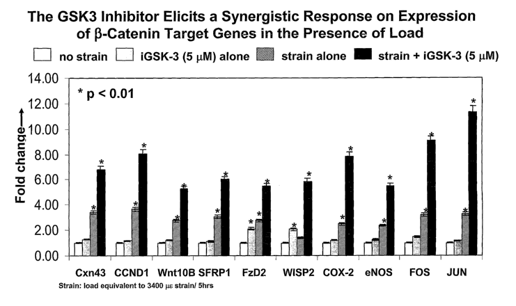
-27-
TABLE 1
HBM Gene Expression Profile
Observed Effect of HBM
Gene Pathway Genotype on Gene Expression
ACPS HBM Up-regulated in HBM cells
Co11A1 HBM No significant affect
Connexin 43 Wnt No significant affect
CTSI~ HBM Up-regulated in HBM cells
Cyclin D 1 Wnt No significant affect
ENOS Load Sensor No significant affect
Frizzled 2 Wnt No significant affect
GADD45A HBM Down-regulated in HBM cells
IGF2 HBM Down-regulated in HBM cells
IGFBP6 HBM Up-regulated in HBM cells
IL-6 Load Sensor Down-regulated in HBM cells
IL-8 Stress & OsteoclastDown-regulated in HBM cells
Function
MI~2 Stress & OsteoclastDown-regulated in HBM cells
Function
OPG Stress ~ OsteoclastNo significant affect
Function
Osteonectin HBM No significant affect
PTGS2 Load Sensor No significant affect
RA1VI~L Stress ~ OsteoclastNo significant affect
Function
SFRP 1 Wnt Up-regulated in HBM cells
SFRP4 Wnt Up-regulated in HBM cells
TGF j3 HBM Up-regulated in HBM cells
TIMP3 HBM Up-regulated in HBM cells
WISP2 Wnt Up-regulated in HBM cells
WntlOB Wnt Up-regulated in HBM cells
By "stress " in Table 1 is meant a gene
and osteoclast that is a stress
function
responsive gen e as well as a
gene that is required
for osteoclastogenesis
and
function. By load sensor" as
" used in Table
1, is meant a
gene known in
the
literature to By "HBM signature" as used
respond to for Table 1
mechanical
load.
and throughout
the application
is meant to
include a set
of genes that
is differentially
to expressed
in cell lines
expressing
the HBM mutation
or in affected
individuals
of the
human HBM1 kindxed.
CA 02526845 2005-11-22
WO 2005/028678 PCT/US2004/017951
-28-
TABLE 2
Effect of Load on Gene Expression Iya vivo Com~"arin~ HBM TG and Non-TG
Animals
Gene Pathway Effect of Load on Gene Expression
ACPS HBM Up-regulated equally in the males
and is more
significantly induced in female
HBM-TG
Co11A1 HBM No significant change in either
Connexin Wnt Up-regulated; More significant
43 in HBM-TG
CTSK HBM Up-regulated in both animals equally
Cyclin D1 Wnt Up-regulated; More significant
in HBM-TG
ENOS Load Sensor Up-regulated; More significant
in HBM-TG
Frizzled Wnt Up-regulated; More significant
2 in HBM-TG
GADD45A HBM Down-regulated in both animals
IGF2 HBM Up-regulated in both male animals
IGFBP6 HBM Up-regulated; More significant
in HBM-TG
IL-6 Load Sensor Up-regulated; More significant
in HBM-TG
IL-8 Stress & Up-regulated; More significant
in HBM-TG
Osteoclast
Function
LRPS - No significant change in either
MK2 Stress & Up-regulated in non-TG animals
only
Osteoclast
Function
OPG Stress & Up-regulated in HGM-TG animals
only
Osteoclast
Function
OsteonectinHBM Up-regulated; More significant
in HBM-TG
PTGS Load Sensor Up-regulated; More significant
in HBM-TG
RANI~L, Stress & No significant change in either
Osteoclast
Function
SFRP 1 Wnt Up-regulated; More significant
in HBM-TG
SFRP4 Wnt Up-regulated; More significant
in HBM-TG
TGF(3 HBM No significant change in either
TIMP3 HBM No signiEcant change in either
WISP2 Wnt Up-regulated; More significant
in HBM-TG
WntlOB Wnt Up-regulated; More significant
in HBM-TG
TABLE 3
Effect of Load on Gene E~ression In vitf-o
MC3T3 Cell Response
Gene Gene type to Gravitational Load
AP 1B 1 Stress regulated gene Up-regulated
AXIN Wnt pathway component Up-regulated
CA 02526845 2005-11-22
WO 2005/028678 PCT/US2004/017951
-29-
MC3T3 Cell Response
Gene Gene type to Gravitational
Load
BMP1 Observed to be induced Up-regulated
by iGSK-3
CBFB Osteoblast function Up-regulated
CCND 1 Wnt target gene Up-regulated
CCND3 Cell cycle Up-regulated
CELSR2 G-type receptor Up-regulated
CHUKIIKK alphaFacilitates (3-catenin Up-regulated
nuclear
translocation
CKl alpha Wnt pathway component Up-regulated
Kinase Up-regulated
CRABP2 Osteoblast differentiation Up-regulated
CSF1R Osteoclastogenesis Up-regulated
CTGF Growth factor Up-regulated
DVLl Wnt signaling intermediate Up-regulated
EPHB6 Wnt target gene Up-regulated
FOSLl Stress regulated gene Up-regulated
GADD45B Cell cycle Up-regulated
GADD45G Cell cycle Up-regulated
GJA1 Wnt target gene Up-regulated
GJB3 Wnt target gene Up-regulated
HERPUD 1 Wnt target gene Up-regulated
IGFBP6 IGF binding protein Up-regulated
IL1R1 IL-1 mediated signaling, Up-regulated
inflammation
IL1RL1 TL-1 mediated signaling, Up-regulated
Inflammation
IL4RA Inflammation Up-regulated
ITGAS Integrin signaling Up-regulated
JUN Stress regulated gene Up-regulated
JUND1 Stress regulated gene Up-regulated
LDLR Lipoprotein receptor Up-regulated
LOX Lysyl oxidase Up-regulated
MAPKAPKZ Kinase in stress regulated Up-regulated
signaling
MSX1 Wnt target gene Up-regulated
MYCS Wnt target gene Up-regulated
NCAM 1 Wnt target gene Up-regulated
NFATC 1 Inflammation Up-regulated
NFKB1 Inflammation, proliferation Up-regulated
PDGFA Growth factor, osteoblast Up-regulated
development
PRDC-PENDING Cereberus life protein Up-regulated
PTGS 1 Inflammation Up-regulated
PTGS2 Wnt target gene Up-regulated
RAMP3 Calcium signaling Up-regulated
RUNX Osteoblast function Up-regulated
RUNX2lCBFAI Osteoblast function Up-regulated
SDC1 Proteoglycan required for Up-regulated
Wnt
signaling
CA 02526845 2005-11-22
WO 2005/028678 PCT/US2004/017951
-30-
MC3T3 Cell Response
Gene Gene a to Gravitational
Load
SERPINE1 Protease Up-regulated
SPARCL1 Osteoblast function Up-regulated
STAT3 Proliferation and cell Up-regulated
growth
TANK Inflarmnation, NF-kB Up-regulated
signaling
TGFB1 TGF beta signaling gene Up-regulated
THBD Endothelial cell functionUp-regulated
TIEG TGF beta signaling gene Up-regulated
TIMP1 Matrix metalloproteinaseUp-regulated
TMP3 Matrix metalloproteinaseUp-regulated
TNFRSFIIBIOPG Wnt target gene Up-regulated
TRA,F3 NF-kB signaling Up-regulated
WISP1 Wnt target gene Up-regulated
The above listed genes were modulated in response to application of
gravitational load to cultured MC3T3 cells.
TABLE 4
The Effects of Load Using the FlexerCell in the Presence and Absence of iGSK-
3; a
Wnt/~i-catenin Pathwa~Activator
Treatment/GENECCND1 CXN43 SFRPl WntlOb eNOS COX-2 FOS
No iGSKINo 1.00 1.00 1.00 1.00 1.00 1.00 1.00
load
No iGSK + 3.64 3.39 3.05 2.76 2.35 2.48 3.20
load
iGSK 0.05 3.80 4.27 3.04 3.70 2.54 2.56 3.66
NM +
load
iGSK 0.2 NM 4.39 4.42 3.36 3.53 2.65 2.74 3.76
+
load
iGSK 1 NM+load5.17 4.76 3.59 ' 3.69 3.06 3.16 4.51**
iGSK 5 ~M 6.93* 5.3_8 5.41* 4.40* 4.33* 6.50**6.20**
+ load
iGSK 20 ~M 7.13** 7.72**10.00**6.95** 5.95**8.17**10.77**
+
load
"*" indicates a near 2 fold induction over load. "*'~" indicates equal to or
>2 fold
to induction over fold.
For additional genes that get up and down regulated, see the Examples and
other Tables provided herein.
CA 02526845 2005-11-22
WO 2005/028678 PCT/US2004/017951
-31-
3. Methods of Studying Bone Loading In. vivo
3.1 Bone Studies
To understand the mechanism underlying the anabolic nature of the HBM
mutations, HBM transgenic (TG) mice were subjected to in vivo mechanical
loading
to look for changes in gene expression as compared to their non-traxlsgenic
(non-
TG) control littermates. This was performed by obtaining tibias or calvaria
from the
animals to which bone load stimuli has been administered, but other suitable
bones
can be used, including but not limited to ulnas, femurs and vertebrae. RNA was
obtained from the HBM TG and non-TG littermate mice after load stimuli was
to administered: RNA was then extracted from the calvaria or tibias (or other
bones)
and compared between the animals (i.e., HBM TG and non-TG animals at rest and
after load stimuli).
It was observed that the HBM mice had significantly greater Wnt pathway
gene response than their non-TG littermate controls. From this observation, it
was
concluded that the HBM mutation causes the bone to be more sensitive to
mechanical loading. One signature set of genes produced in response to a load
stimuli in vivo comprises up-regulation of comiexin 43, osteonectin,
osteoprotegerin,
eNOS, COX-2, prostacyclin synthase (PTGS), interleukins-6 (IL-6), cyclin Dl,
Wnt
lOB, SFRP1 and SFRP4. Additional genes also were up-regulated as discussed in
2o greater detail below and in the examples.
Methods of inducing bone load stimuli include the four-point load system
discussed in the Examples. Additional in vivo methods of administering load
are
know in the art (e.g., three-point load system) and can also be used as would
be
known to the artisan of ordinary skill.
With the above expression profile obtained in the HBM TG mice or with any
combination of the additional genes discussed herein, agents can be screened
in the
non-TG and HBM TG animals to ascertain whether a particular agent enhances
activation of the Wnt pathway and thereby bone mineralization. Several
positive
controls for studying agents which enhance mineralization include PTH, and a
GSI~-3 inhibitor which enhances mineralization via activation of the Wnt
pathway,
3-(3-chloro-4-hydroxyphenylamino)-4-(2-ntrophenyl)-1-H pyrrole-2,5-dione.
Other GSK-3 inhibitors described herein can also be used as positive controls.
W addition to gene expression profiles and signatures obtained from animals
subjected to load stimuli andlor Wnt pathway modulation, animals can also be
studied for changes in bone pathology as a result of load and/or Wnt pathway
modulation. For example, changes in calvaria thickness (or thickness changes
in
other bones) and protein expression of any of the RNAs or proteins listed in
any of
CA 02526845 2005-11-22
WO 2005/028678 PCT/US2004/017951
-32-
the Tables herein as being up- or down-regulated in response to bone load
stimulus
alone or in combination with one or more compounds that modulate bone
remodeling.
Bone calvaria analysis can be performed for example by administering a test
agent to an animal in an amount of about 0.01 mg/kg/day to about 100
mg/kg/day.
More preferably, the agent is provided in an amount of 0.1 mg/kg/day to about
50
mglkg/day. For example animals can be administered the agent at about 0.5, 10
and
50 mgJkg/day. Typically, animals are done in batches of 6 mice per group
(total of
72 mice in a study) and studied 5, 15 and 30 days post administration.
Parathyroid.
l0 hormone (PTH) can be used as a positive control, as can the GSK-3
inhibitor, 3-(3-
chloro-4-hydroxyphenylamino)-4-(2-nitrophenyl)-lHpyrrole-2,5-dione. After bone
load stimulus in the presence and absence of these reagents, differences in
calvaria
size can be measured.
Other methods of studying pathological changes to bone would be evident to
one of skill in the art. These pathological changes in bone can then be
compared to
the gene expression and gene signature profiles obtained both in vivo and iyz
vitro
and the data further correlated. As previously discussed, the gene expression
profiles can be obtained by any of they methods discussed herein or as would
be
evident to one of ordinary skill.
2o Although any of the genes in the discussed above can be assayed for
modulated activity in response to a bone load stimulus, preferred genes for
evaluation include but are not limited to SFRP1, TIMP3, GJA1, CTSK, Co11A1,
CCND1, TIMP2, GADD45A, WISP2, FZD2, SFRP4, IGFBP6, LRPS, LRP6, IL6,
IGF2, SPARC, MAPKAPK2, TNF, TNFRSF11B, TNFSF11, PTGS2 (COX-2),
eNOS, GRO1 and WntlOB. See also the genes that are listed in any of the Tables
herein as being up- or down-regulated in response to bone load stimulus alone
or in
combination with one or more compounds that modulate bone remodeling.
4. Methods of Studying Bone Loading-Ire vitro
One aspect of the invention is the study of the effect of bone load in vitro
and
means by which the benefits of bone load (i. e., increased bone
mineralization) can
be enhanced. Studying bone load enhancement can be done both ih vivo (as
discussed above) and in vitYO. Preferably bone load enhancement is first
performed
ii2 vitf~o followed then with in vivo experiments, such as those discussed
above.
Consequently, one aspect of the invention involves placing cells under
conditions, which simulate load stimuli. There are several methods available
for
placing strain on cell cultures to mimic the bone load response observed ifz
vivo.
CA 02526845 2005-11-22
WO 2005/028678 PCT/US2004/017951
-33-
These methods include but are not limited to fluid shear, hydrostatic
compression,
uniaxial stretch, biaxial stretch, gravitational loading and load induced
using a
Flexercell~ or equivalent system.
4.1 Bone Load Stimuli
Preferred genes which are modulated by a bone load stimuli, such as those
provided by any of the above methods, include but are not limited to SFRP 1,
connexin 43, CCND1, WntlOb, Jun, Fos, PTGS2 (COX-2) and eNOS. Additional
genes that can be monitored for increases in their activity (e.g., increased
mRNA
transcripts and protein) as reflected in many of the Tables herein. At least
six genes
to that have been shown to be consistently up-regulated in response to bone
load (i.e.,
Jun, Fos, eNOS, SFRP1, COX-2 and Connexin 43) are also enhanced by the
addition of an agent which activates the Wnt pathway. Other genes, such as
Wnt2,
are not enhanced by the addition of reagents that activated the Wnt pathway
(e.g.,
GSK-3 inhibitors and Wnt 3A and its agonists, mimetics, and variants) and only
respond to bone load.
4.1.1 Fluid Shear Stimulus
One method of inducing bone load is by fluid shear. Fluid shear involves a
cone plate viscometer that generates continuous laminar shear by a stirring
mechanism. Alternatively, a flow loop apparatus can produce such shear in a
parallel flow culture chamber. The latter method and apparatus is exemplified
by
the Streamer system produced by Flexcell International Corporation. The flow
loop
apparatus also is known to produce a reproducible and consistent stimulus. The
only
drawbacks are that the end points are typically short-lived and whether these
changes impact the function of differentiated osteoblasts (Basso et al., Bone
30(2):
347-51 (2002)).
4.1.2 Hydrostatic Comt~ression Stimulus
A second method of inducing bone load is use of hydrostatic compression.
Hydrostatic compression utilizes compressed air to generate a continuous or
intermittent force that is believed to localize the force specifically to
regions where
3o the cells interact with the extracellular matrix protein/adhesion proteins.
4.1.3 Uniaxial Stretch Stimulus
A third means of inducing bone load in vitro is use of a uniaxial stretch
stimulus. The uniaxial stretch method utilizes stretch force in one direction.
The
method involves growing cells in a tissue culture on a treated strip of
polystyrene
film or other film, which is fixed to a flexible layer of silicone. The layer
of silicone
is further attached to two metal bars. The metal bars can be manipulated
relative to
each other using an electromagnet or some other moving means. This method does
CA 02526845 2005-11-22
WO 2005/028678 PCT/US2004/017951
-34-
not create any fluid shear. The lack of fluid shear makes this method less
preferred,
because interstitial fluid flow may play a larger role in bone remodeling than
mechanical stretch. Accordingly, this method may not fully mimic what occurs
ih
vivo despite the reproducible and consistent stimulus produced (Basso et al.,
Bone
30(2): 347-51 (2002)).
4.1.4 Biaxial Stretch Stimulus
Biaxial stretch is essentially the Flexercell~ system discussed herein. This
method utilized a collagen coated silastic membrane upon which the cells are
grown.
The plates are then placed in a special tray, which is attached to a vacuum
pump.
to The vacuum pump stretches and relaxes the membrane, by stretching or
otherwise
distorting the cell membrane. Additionally, any media or fluid movement will
fuxther add fluid shear.
4.1.5 Gravitational Load Stimulus
Gravitational loading is another method by which bone load can be induced
15 in vita~. Essentially, force is placed on the cells causing the cells to
flatten. For
additional details, see for example, Hatton et al., .I. Bone ~ Min. Res.
18(1): 58-66
(2003); and Fitzgerald et al., Exp. Cell. Res. 228: 168-71 (1996).
Specifically, the
cells are grown on plates or cover slips and then are exposed to increasing G
forces.
4.1.6 Flexercell~ Stimulus
20 One preferred method for assessing reagent-based enhancement of the Wnt
pathway and bone mineralization is using the Flexercell~ system, a biaxial
stretch
stimulus. Briefly, bone cells (e.g., MC3T3 cells) are exposed to 3,400 ~,c.
Loads of
about 50 ~,E to about 5,000 ~.E (and any value in between) can be used as well
for
mechanical load stimuli. Any stimulus in this range mimics physiological bone
load
25 stimuli. Stimuli above 5,000 ~.s result in pathophysiological loads and
therefore are
not preferred. The cells also can be exposed to a Wnt pathway modulator (e.g.,
a
GSK inhibitor) prior to exposure to biaxial stretch.
The genes up-regulated by the administration of the load alone or with a
GSK-3 inhibitor include, but are not limited to COX-2, eNOS, comiexin 43, and
30 SFRP1. The expression profile obtained i~z vitro from the Flexercell~
studies
mimics the i3z vivo loading gene expression profile (i.e., RNA analysis
performed on
cells from HBM TG mice tibia wherein the mice were subjected to bone load
using
a four-point system). Thus, this mechanical load assay, or the use of other
mechanical load means with the variety of cell lines disclosed herein, can be
used to
35 identify small molecules, peptides, immunoglobulins, and the like that
modulate,
and preferably activate, the canonical Wnt pathway and which mimic the HBM
phenotype.
CA 02526845 2005-11-22
WO 2005/028678 PCT/US2004/017951
-35-
The ih vitro methods of inducing mechanical stress stimuli on cells can also
be used to study cell proliferation and apoptosis, which is relevant to bone
remodeling and the need for osteoblast and osteoclast proliferation and
osteoclast
resorption. For example, HBM and unaffected osteoblastic cells can be seeded
into
bioflex 6 well plates and cultured for 2-3 days in growth media containing 10%
FBS
until the cells are about 60% confluent. Twenty-four hours prior to mechanical
loading, the media is replaced with 1 mL of basal media containing about 2 to
about
4% FBS. The cells are then subjected to about 50 to about 5,000 ~,s of load
for
about 1 to about 5 hours.
to Following load, the cells are cultured for an additional period of time.
Subsequently, cell number and proliferation can be assessed using a number of
commercial assays or assays lmown in the art, including but not limited to
[3H]-
thymidine incorporation, 5-bromo-2'-deoxyuridine (BrdU) incorporation, 3-(4,5
dimethylthiazol-2-yl)-5-(3-carboxymethoxyphenyl)-2-(4-sulfophenyl)-2H
trazolium
15 alts (MTS) assay, TUNEL assay (i.e., terminal deoxynucleotidyltransferase
dUTP
nick end labeling) or Annexin V assay.
Additional Wnt pathway agonists include other GSK-3 inhibitor compounds
as discussed herein, natural Wnt pathway ligands, synthetic ligands, small
molecules
as well as known antagonists cerebrus, SFRP and WIF (Wnt Inhibitory Factor)
can
20 b~w'a~nalyzed using the in vitro bone load methods described above for
their ability to
enhance bone load. Known Wnt pathway activators include Wntl and Wnt3A,
small molecule Wnt mimetics, peptide aptamers that interact with LRPS and
activate
Wnt signaling. Preferred peptide aptamers include:
A tamer Aptamer,Sequence (Amino to Carboxy Terminus)
2,6~ METDTLLLWVLLLWVPGSTGDGSMSDKIIHLTDDSFDTDVLKADGAILV
DFWAEWCGPNSGGGGMIWEAWSCYACGTSGPCKMIAPILDEIADEYQGK
LTVAKLNTDQNPGTAPKYGIRGIPTLLLFKNGEVAATKVGALSKGQLKE
FLDANLA
In another embodiment, Wnt antagonists can be screened or used to treat
individuals
25 wherein bone delnineralization (e.g., osteopetrosis) is needed. Wnt
antagonists
include but are not limited to Dkkl antagonists.
4.2 Cell Cultures
The cells to which ih vitro loading experiments can be performed include but
are not limited to the following human cell lines: U20S cells (ATCC), MG-63
cells
30 (ATCC), SAOS-2 cells (ATCC), HOS-TE85 cells (ATCC), HOB03CE6 cells
(Wyeth), HOB01C1 preosteocytes (Wyeth), and human primary osteoblasts.
Additionally, cells can be cultured from any mammalian system. Preferred
animal
lines for study include rat and mouse bone cells. For example, mouse bone
cells,
CA 02526845 2005-11-22
WO 2005/028678 PCT/US2004/017951
-3 6-
which can be used with any of the above methods include but are not limited to
MC3T3 cells (ATCC) as discussed in the examples and primary osteoblasts or any
cell line analogous to the human cell lines above. Rat cells that can be used
with
any of the disclosed methods of inducing stress ifa vity~o include but are not
limited to
UMR-106 cells (ATCC), ROS 17/2.8 cells and primary osteoblasts or any cell
line
analogous to the human cell lines above. Methods of culturing the cells would
be
known to the skilled artisan. See, e.g., IAN FRESHNEY, CULTURE of ANIMAL CELLS
-
A MANUAL of BASIC TECHNIQUE (4th ed., Wiley-Liss, New York, 2000).
In another aspect of the invention cells can be taken from bones and can
l0 include osteoblasts, osteoclasts and osteocytes as well as progenitor and
stem cells.
Preferred osteoblasts and their progenitor and stem cells include mature
osteoblasts,
preosteoblasts (mature and immature), and mesenchymal stem cells (also
referred to
as mesenchyme-derived stem cells, MDSC).
In another aspect of the invention, human cell lines obtained from HBM and
15 unaffected individuals can be used in conjunction with the bone load
methodologies
discussed herein. These cell lines can be used to investigate the gene
inductions
identified from the in vivo loading experiments performed on HBM and non-
transgenic mice.
4.3 TCF Luciferase Assays
2o A TCF-luciferase assay system can also be used to monitor Wnt signaling
activity. Constructs for the TCF-luciferase assays can be prepared as would be
known in the art. For example, Wnt pathway proteins such as LRPS, LRP6 and
HBM amongst others, can be expressed in pcDNA3.1, using I~ozak and signal
sequences to target peptides for secretion.
25 Once constructs have been prepared, cells such as osteoblasts and HEK2,93
cells are seeded in well plates and transfected with construct DNA, CMV (3-
galactosidase plasmid DNA, and TCF-luciferase reporter DNA. The cells are then
lysed and assayed for (3-galactosidase and luciferase activity to determine
whether
Wnt pathway interacting proteins, or other molecules such as antibodies affect
Wnt
3o signaling. Additional detail is provided in the examples below regarding
methods of
using TCF-luciferase constructs.
In another embodiment, the Flexercell~ mechanical cell loading system (or
any of the in vitro means of inducing load on cells) can be used in
combination with
the TCF-luciferase reporter system, or other reporter systems, to measure the
effects
35 of mechanical loading on the Wnt pathway. Such experiments can be performed
as
follows. For these experiments, MC3T3 cells (or another equivalent cell
discussed
herein) are plated as described above and cultured for three days or wztil
confluent.
CA 02526845 2005-11-22
WO 2005/028678 PCT/US2004/017951
-37-
The media is then changed to either serum free media containing BSA or low
serum
media (1% FBS) containing aMEM. The cells on this low or serum free media are
then incubated for another 24 hours. About one hour prior to mechanical load,
one
plate is pre-treated with a dose of a Wnt pathway modulator (e.g., GSK
inhibitor,
natural Wnt ligand including but not limited to Wnt 1 and Wnt 3A), while
another
plate is untreated. Following pretreatment with any Wnt mimetic ligands, small
molecules, etc., the cells are then subjected to mechanical load (e.g., 3,400
~.s) for
about 5 hr as previously described. RNA is harvested from the loaded and the
unloaded control samples immediately following load and 24 hours post-load
using
1o the Qiagen mini kit, as discussed above.
Real-time PCR can then be performed on the load signature set genes (or any
suitable RNA assay as would be known in the art) at each time point to observe
the
changes in gene expression with treatment. Alternatively, the RNA can be
analyzed
using other methods lmown to the skilled artisan or as discussed herein.
5. Arrays
One method of utilizing the gene profiles and signatures of Wnt pathway
involvement in bone remodeling and modulation thereof is in the form of
preparing
nucleic acid and protein arrays. These arrays can then be utilized to further
study
the Wnt pathway and its involvement in bone remodeling. These arrays can also
be
used to screen for agents that modulate bone remodeling through the Wnt
pathway.
5.1 Nucleic Acid Arrays
Nucleic acid arrays would be prepared as is known to one spilled in the art.
Methods ofpreparing and utilizing such arrays are described in, for example,
P.
Baldi et al., DNA MICROARRAYS AND GENE EXPRESSION: FROM EXPERIMENTS TO
DATA ANALYSIS AND MODELING (Cambridge University Press 2002); and DNA
MICROARRAYS: A MOLECULAR CLONING MANUAL (David Bowtell and Joseph
Sambrook, eds., Cold Spring Harbor Laboratory, 2002).
Preferred nucleic acid arrays would contain nucleic acids corresponding to
3o members of the Wnt signaling pathway of any of the genes in Tables 1-5 or
FIG. 16.
For example, such arrays would contain 2, 3, 4, 5, 6, 7, ~, 9, 10, 15, 20, 25,
30, 35,
40, 45, 50 or more (or any integer value inbetween) of the genes involved in
bone
modeling. Such genes include any of the modulated genes listed in any of the
tables, in the examples or are part of the pathways depicted in FIG. 16. These
nucleic acids are exemplary of nucleic acids associated with a bone loading
response.
CA 02526845 2005-11-22
WO 2005/028678 PCT/US2004/017951
-3 8-
In another embodiment, arrays can be prepared which include the Wnt
pathway bone remodeling genes and genes involved in for example serum calcium
modulation, osteoclast apoptosis, osteoblast proliferation, and. the like.
TABLE 5
List of Genes for Development of High Bone Mass Microarrav ar Protein/Antibod
Arran
WHERE
GENE DESCRIPTION EXPRESSED
ACPS acid phosphatase 5, tartrate Bone and colon
resistant
cancer
CCNDl cyclin Dl (PRAD1: parathyroid HBM Bone
adenomatosis 1)
CNK1 v-erb-b2 erythroblastic leukemiaBone and colon
viral
oncogene homolog 3 (avian) cancer
COL1A1 collagen, type I, alpha 1 HBM Bone
COL6A3 ~ collagen, type VI, alpha 3 HBM Bone
CTGF connective tissue growth factorHBM Bone
CTSK cathepsin K (pycnodysostosis) HBM Bone
CX3CR1 chemokine (C-X3-C) receptor Inflammation
1 in
bone
DELTEX deltex homolog 2 (D~osophiZa),Bone and colon
EphB2
cancer
EPHB2 connector enhancer of KSR-likeBone and colon
(D~osoplzila kinase suppressorcancer
of ras)
ERBB3 GRO1 oncogene (melanoma growthBone and colon
stimulating activity, alpha) cancer
FAP fibroblast activation protein,Bone and colon
alpha
cancer
FBLN1 fibulin 1 HBM Bone
FGF-2 fibroblast growth factor 2 Inflammation
(basic) in
bone
FGF-7 fibroblast growth factor 7 Inflammation
(keratinocyte in
growth factor) bone
FOS fos FBJ marine osteosarcoma Bone and colon
viral
oncogene homolag cancer/load
sensing gene
FZD2 frizzled (Drosophila) homolog HBM Bone
2
GADD45A growth arrest and DNA-damage- HBM Bone
inducible, alpha
GAS6 growth arrest-specific 6 HBM Bone
GJAl gap junction protein, alpha HBM Bone
1, 43kD
(connexin 43)
IGF2 insulin-lilce growth factor Inflammation
2 in
(somatomedin A) bone
CA 02526845 2005-11-22
WO 2005/028678 PCT/US2004/017951
-39-
WHERE
GENE DESCRIPTION EXPRESSED
IGF2R insulin-like growth factor Inflammation
2 receptor in
bone
IGFBP6 insulin-like growth factor HBM Bone
binding
protein 6
IL-6 interleukin 6 (interferon, Inflammation
beta 2) in
bone
ITGBS integrin, beta 5 HBM Bone
ITGBL1 integrin, beta-like 1 (with HBM Bone
EGF-lilce
repeat domains)
jun avian sarcoma virus 17 Bone and
oncogene colon
homolog cancer/load
sensing gene
LOX lysyl oxidase HBM Bone
LRPS low density lipoprotein receptor-relatedHBM Bone
protein 5 '
LRP6 low density lipoprotein receptor-relatedHBM Bone
protein 6
LSP1 lymphocyte-specific protein Inflammation
1 in
bone
MAPI~APKZ mitogen-activated protein kinase-Osteoclast
activity
activated protein kinase 2
MCC mutated in colorectal cancers Bone and
colon
cancer
MET met proto-oncogene (hepatocyteHBM Bone
growth
factor receptor)
MYBL1 v-nzyb myeloblastosis viral HBM Bone
oncogene
homolog (avian)-like 1
MYC v-rnyc avian myelocytomatosis Bone and
viral colon
oncogene homolog cancer
Enos nitric oxide synthase 3 (endothelialLoad responsive
cell)
genes
OSMR oncostatin M receptor HBM Bone
PDGFRA platelet-derived growth factorHBM Bone
receptor,
alpha polypeptide
PTGS2/COX-2. prostaglandin-endoperoxide Load responsive
synthase 2
(prostaglandin GlH synthase genes
and
cyclooxygenase)
SFRP1 secreted frizzled-related proteinHBM Bone
1
SFRP4 secreted frizzled-related proteinHBM Bone
4
SpARC sparc/osteonectin, cwcv and Inflammation
lcazal-like in
domains proteoglycan (testican)bone
STAT1 signal transducer and activatorInflammation
of in
transcription 1, 9lkD bone
TGFBR2 transforming growth factor, Inflammation
beta in
receptor II (70-80kD) bone
THBS1 thrombospondin 1 HBM Bone
TM'2 tissue inhibitor of metalloproteinaseHBM Bone
2
CA 02526845 2005-11-22
WO 2005/028678 PCT/US2004/017951
-40-
WHERE
GENE DESCRIPTION EXPRESSED
TIMP3 tissue inhibitor of metalloproteinaseHBM Bone
3
(Sorsby fundus dystrophy,
pseudoinflammatory)
TNF tumor necrosis factor (TNF Osteoclast
superfamily, activity
member 2)
TNFRSF10B tumor necrosis factor receptorInflammation
in
superfamily, member lOb bone
TNFRSF11BlOPG tumor necrosis factor receptorOsteoclast
activity
superfamily, member l lb
(osteoprotegerin)
TNFSF11lRANKI,tumor necrosis factor (ligand)Osteoclast
activity
superfamily, member 11
UNI~_D83402 prostaglandin I2 (prostacyclin)HBM Bone
sy~zthase
VCAM1 Vascular cell adhesion moleculeInflammation
1 in
bone
WISP2 WNT1 inducible signaling pathwayHBM Bone
protein 2
WNT10B wingless-type MMTV integrationBone and colon
site
family, member lOB cancer
WNT6 wingless-type MMTV integrationHBM Bone
site
family, member 6
Preferably, the nucleic acid arrays would contain two or more sequences
corresponding to genes observed to express in "HBM Bone". Such arrays could
comprise at least 2, 3, 4, 5, 10, 15, 20, 25, 30 or more (and any integer
value in
between) of the sequences that are up- or down-regulated in response to bone
load
listed in the tables, examples or FIG. 16. Similarly proteinlantibody arrays
can be
prepared that are high bone mass specific that comprise proteins, peptides,
and/or
immunoglobulins that bind to at least 2, 3, 4, 5, 10, 15, 20, 25, 30 or more
(and auy
integer value in between) of the proteins listed in Table 5 or to any of the
proteins
involved in any of the pathways discussed herein.
5.1.2 DNA Microarray Construction
Frequently, it is desirable to amplify the nucleic acid sample prior to
hybridization. Suitable amplification methods include, but are not limited to
polymerase chain reaction (PCR) (Innis, et al., PCR PROTOCOLS. A GUIDE To
METHODS AND APPLICATION. ACADEMIC PRESS, Inc. San Diego, (1990)), ligase
chain reaction (LCR) (see Wu et al., Genozzzics, 4: 560 (1989); Landegren et
al.,
Science, 241: 1077 (1988); and Barringer et al., Gezze, 89: 117 (1990)),
transcription
amplification (Kwoh et al., Pz~oc. Natl. Acad. Sci. USA 86: 1173 (1989)), and
self
CA 02526845 2005-11-22
WO 2005/028678 PCT/US2004/017951
-41-
sustained sequence replication (Guatelli et al., Proc. Nat. Acad. Sci. USA,
~7: 1 X74
(1990)).
In a preferred embodiment, the hybridized nucleic acids are detected by
detecting one or more labels attached to the sample nucleic acids. The labels
may be
incorporated by any of a number of means well known in the art. However,
preferably the label is simultaneously incorporated during the amplification
step in
the preparation of the sample nucleic acids. Thus, for example, polymerase
chain
reaction (PCR) with labeled primers.or labeled nucleotides will provide a
labeled
amplification product. In a preferred embodiment, transcription amplification,
as
l0 described above, using a labeled nucleotide (e.g., fluorescein-labeled UTP
and/or
CTP) incorporates a label into the transcribed nucleic acids.
Alternatively, a label may be added directly to the original nucleic acid
sample (e.g., mRNA, polyA mRNA, cDNA, and the like) or to the amplification
product after the amplification is completed. Means of attaching labels to
nucleic
acids are well knowxn to those of skill in the art and include, for example
nick
translation or end-labeling (e.g., with a labeled RNA) by the addition of a
kinase to
the reaction mixture containing the nucleic acid and subsequent attaclnnent
(ligation) of a nucleic acid linker joining the sample nucleic acid to a label
(e.g., a
fluorophore).
2o Detectable labels suitable for use in the present invention include any
composition detectable by spectroscopic, photochemical, biochemical,
immunochemical, electrical, optical or chemical means. Useful labels in the
present
invention include biotin for staining with labeled streptavidin conjugate,
magnetic
beads (e.g., DynabeadsTM), fluorescent dyes (e.g., fluorescein, Texas red,
rhodamine,
green fluorescent protein, and the like), radiolabels (e.g., 3H, lzsh 3ss~
14C~ or 3zP),
enzymes (e.g., horse radish peroxidase (IiRP), alkaline phosphatase and others
commonly used in an ELISA), and calorimetric labels such as colloidal gold or
colored glass or plastic (e.g., polystyrene, polypropylene, latex, and the
like) beads.
Patents teaching the use of such labels include U.S. Pat. Nos. 3,~17,~37;
3,50,752;
3,939,350; 3,996,345; 4,277,437; 4,275,149; and 4,366,241.
The reference sequences derived from other the genes, such as, for example,
COX-2, can vary widely from a full-length genome, to an individual chromosome,
episome, gene, component of a gene, such as an exon or regulatory sequences,
to a
few nucleotides. A reference sequence of between about 2, 5, 10, 20, 50, 100,
500,
1000, 5,000 or 10,000, 20,000 or 100,000 nucleotides (and any integer value in
between) is common. Sometimes only particular regions of a sequence are of
interest.
CA 02526845 2005-11-22
WO 2005/028678 PCT/US2004/017951
-42-
The methods of this invention employ oligonucleotide arrays, which
comprise probes exhibiting complementarity to one or more selected reference
sequences whose sequence is known (e.g., eNOS, COX-2, Jun, Fox, Connexin 43,
SFRP or any of the other genes discussed herein). Typically, these arrays are
immobilized in a high density array ("DNA on chip") on a solid surface, as
described for example in U.S. Pat. No. 5,143,854 and PCT patent publication
Nos.
WO 90/15070, WO 92/10092 and WO 95/11995, each of which is incorporated
herein by reference.
Various strategies are available to order and display the oligonucleotide
1o probe arrays on the chip and thereby maximize the hybridization pattern and
sequence information derivable regarding the target nucleic acid. Exemplary
display and ordering strategies are described in PCT No. WO 94/12305,
incorporated herein by reference. For the purposes of fuller description, a
brief
description of the basic strategy is described below.
15 The basic tiling strategy provides an array of immobilized probes for
analysis of target sequences showing a high degree of sequence identity to one
or
more selected reference sequences. The strategy is illustrated for an array
that is
subdivided into four probe sets, although it will be apparent that
satisfactory results
are obtained from one probe set (i. e., a probe set complementary to the
reference
2o sequence as described earlier).
A first probe set comprises a plurality of probes exhibiting perfect
_ complementarity with a selected reference sequence. The perfect
complementarity
usually exists throughout the length of the probe. However, probes having a
segment or segments of perfect complementarity that is/are flanked by leading
or
25 trailing sequences lacking complementarity to the reference sequence can
also be
used. Within a segment of complementarity, each probe in the first probe set
has at
least one interrogation position that corresponds to a nucleotide in the
reference
sequence. That is, the interrogation position is aligned with the
corresponding
nucleotide in the reference sequence, when the probe and reference sequence
are
3o aligned to maximize complementarity between the two. If a probe has more
than
one interrogation position, each corresponds with a respective nucleotide in
the
reference sequence. The identity of an interrogation position and
corresponding
nucleotide in a particular probe in the first probe set cannot be determined
simply by
inspection of the probe in the first set. As will become apparent, an
interrogation
35 position and corresponding nucleotide is defined by the comparative
structures of
probes in the first probe set and corresponding probes from additional probe
sets.
CA 02526845 2005-11-22
WO 2005/028678 PCT/US2004/017951
-43-
Tn principle, a probe could have an interrogation position at each position in
the segment complementary to the reference sequence. Sometimes, interrogation
positions provide more accurate data when located away from the ends of a
segment
of complementarity. Thus, typically a probe having a segment of
complementarily
of length "x" does not contain more than "x-2" interrogation positions. Since
probes
are typically 9-2I nucleotides, and usually all of a probe is complementary, a
probe
typically has 1-19 interrogation positions. Often the probes contain a single
interrogation position, at or near the center of probe.
For each probe in the first set, there are, for purposes of the present
l0 illustration, up to three corresponding probes from three additional probe
sets. Thus,
there are four probes corresponding to each nucleotide of interest in the
reference
I
sequence. Each of the four corresponding probes has an interrogation position
aligned with that nucleotide of interest. Usually, the probes from the three
additional probe sets are identical to the corresponding probe from the first
probe set
15 with one exception. The exception is that at least one (and often only one)
interrogation position, which occurs in the same position in each of the four
corresponding probes from the four probe sets, is occupied by a different
nucleotide
in the four probe sets. For example, for an adenine (A) nucleotide in the
reference
sequence, the corresponding probe from the first probe set has its
interrogation
2o position occupied by a thymine (T), and the corresponding probes from the
additional three probe sets have their respective interrogation positions
occupied by
adenine (A), cytosine (C), or guanine (G), a different nucleotide in each
probe. Of
course, if a probe from the first probe set comprises trailing or flanking
sequences
lacking complementarily to the reference sequences, these sequences need not
be
25 present in corresponding probes froril the three additional sets. Likewise
corresponding probes from the three additional sets can contain leading or
trailing
sequences outside the segment of complementarily that are not present in the
corresponding probe from the first probe set. Occasionally, the probes from
the
additional three-probe set are identical (with the exception of interrogation
30 position(s)) to a contiguous subsequence of the full complementary segment
of the
corresponding probe from the first probe set. In this case, the subsequence
includes
the interrogation position and usually differs from the full-length probe only
in the
omission of one or both terminal nucleotides from the termini of a segment of
complementaxity. That is, if a probe from the first probe set has a segment of
35 complementarity of length "n", corresponding probes from the other sets
will
usually include a subsequence of the segment of at least length "n-2". Thus,
the
subsequence is usually at least 3, 4, 7, 9, I5, 21, or 25 nucleotides long
(and any
CA 02526845 2005-11-22
WO 2005/028678 PCT/US2004/017951
-45-
by the omission of a 3' base complementary to the reference sequence and the
acquisition of a 5' base complementary to the reference sequence.
The number of probes on the chip can be quite large (e.g., 105 -106).
However, often only a relatively small proportion (i.e., less than about 50%,
25%,
10%, 5% or 1%) of the total number of probes of a given length are selected to
pursue a particular tiling strategy, in this case a tiling strategy that would
reflect
bone load gene expression profiles and bone load gene enhancement expression
profiles. For example, a complete set of octomer probes comprises 65,536
probes;
thus, an array of the invention typically has fewer than 32,768 octomer
probes. A
to complete array of decamer probes comprises 1,048,576 probes; thus, an array
of the
invention typically has fewer than about 500,000 decamer probes. Often arrays
have
a lower limit of 25, 50 or 100 probes and as many probes as 104, 105, 106,
10', 108,
109, 101°, etc. probes. The arrays can have other components besides
the probes
such as linkers attaching the probes to a support.
. Some advantages of using only a proportion of all possible probes of a given
length include: (i) each position in the array is highly informative, whether
or not
hybridization occurs; (ii) nonspecific hybridization is minimized; (iii) it is
straightforward to correlate hybridization differences with sequence
differences,
particularly with reference to the hybridization pattern of a known standard;
and (iv)
2o the ability to address each probe independently during synthesis, using
high
resolution photolithography, allows the array to be designed and optimized for
any
sequence. For example the length of any probe can be varied independently of
the
others.
Although the array of probes is usually laid down in rows and columns as
described above, such a physical arrangement of probes on the chip is not
essential.
Provided that the spatial location of each probe in an array is known, the
data from
the probes can be collected and processed to yield the sequence of a target
irrespective of the physical arrangement of the probes on a chip. In
processing the
data, the hybridization signals from the respective probes can be reasserted
into any
3o conceptual array desired for subsequent data reduction whatever the
physical
arrangement of probes on the chip.
A range of lengths of probes can be employed in the chips. As noted above,
a probe may consist exclusively of complementary segments, or may have one or
more complementary segments juxtaposed by, flanking, trailing and/or
intervening
segments. In the latter situation, the total length of complementary segments)
is
more important than the length of the probe. In functional terms, the
complementary segments) of the first probe set should be sufficiently long to
allow
CA 02526845 2005-11-22
WO 2005/028678 PCT/US2004/017951
-46-
the probe to hybridize detectably more strongly to a reference sequence
compared
with a variant of the reference including a single base mutation at the
nucleotide
corresponding to the interrogation position of the probe. Similarly, the
complementary segments) in corresponding probes from additional probe sets
should be sufficiently long to allow a probe to hybridize detectably more
strongly to
a variant of the reference sequence having a single nucleotide substitution at
the
interrogation position relative to the reference sequence. A probe usually has
a
single complementary segment having a length of at least 3 nucleotides, and
more
usually at least 5, 6, 7, 8, 9, 10, 11, 12, 13, 14, 15, 16, 17, 18, 19, 20,
21, 22, 23, 24,
to 25 or 30 or more bases exhibiting perfect complementarity (other than
possibly at
the interrogation positions) depending on the probe set) to the reference
sequence.
In some chips, all probes are the same length. , Other chips employ different
groups of probe sets, in which case the probes are of the same size within a
group,
but differ between different groups. For example, some chips have one group
comprising four sets of probes as described above in which all the probes are
15-
mers, together with a second group comprising four sets of probes in which all
of the
probes are 20-mers. Of course, additional groups of probes can be added. Thus,
some chips contain, e.g., four groups of probes having sizes of 15-mers, 20-
mers,
26-mers and 30-mers. Other chips have different size probes within the same
group
of four probes. In these chips, the probes in the first set can vary in length
independently of each other. Frobes in the other sets are usually the same
length as
the probe occupying the same column from the first set. However, occasionally
different lengths of probes can be included at the same column position in the
four
lanes. The different length probes are included to equalize hybridization
signals
from probes depending on the hybridization stability of the oligonucleotide
probe at
the pH, temperature, and ionic conditions of the reaction.
The length of a probe can be important in distinguishing between a perfectly
matched probe and probes showing a single-base mismatch with the target
sequence.
The discrimination is usually greater for short probes. Shorter probes are
usually
3o also less susceptible to formation of secondary structures. However, the
absolute
amount of target sequence bound, and hence the signal, is greater for larger
probes.
The probe length representing the optimum compromise between these competing
considerations may vary depending on, e.g., the GC content of a particular
region of
the target DNA sequence, secondary structure, synthesis efficiency and cross-
hybridization. In some regions of the target, depending on hybridization
conditions,
short probes (e.g., 11-mers) may provide information that is inaccessible from
longer probes (e.g., 19-mers) and vice versa. Maximum sequence information can
CA 02526845 2005-11-22
WO 2005/028678 PCT/US2004/017951
-47-
be achieved by including several groups of different sized probes on the chip
as
noted above. However, for many regions of the target sequence, such a strategy
provides redundant information in that the same sequence is read multiple
times
from the different groups of probes. Equivalent information can be obtained
from a
single group of different sized probes in which the sizes are selected to
maximize
readable sequence at particular regions of the target sequence.
5.2 Protein Arrays
The two major types or protein arrays are primary phase arrays (i.e.,
antibodies, antibody fragments, immunoglobulins or peptides are affixed to a
to substrate) and reverse phase arrays (i.e., cell lysate is affixed to a
substrate and then
subsequently screened with, for example, antibodies). These protein arrays can
be
utilized to rapidly screen for agents that modulate the Wnt pathway, agents
which
enhance Wnt pathway activity, bone protein expression in response to different
stimuli, determination of additional proteins expressed in bone in response to
15 different stimuli and the like.
5.2.1 Primary Phase Array
One preferred method is a primary phase protein array comprising one or
more (and preferably more than one) antibody, antibody fragment,
immunoglobulin
which recognizes and binds to a protein of the genes listed in any of the
Tables, or
2o peptide which recognizes and binds to a protein of the genes listed in any
of the
Tables. Therefore, in one aspect, an array is contemplated wherein there is 1,
2, 3,
4, 5, 6, 7, 8, 9, 10 or more antibodies, immunogenic fragments thereof or
immunoglobulin polypeptides with immunogenic activity to a protein/polypeptide
of
interest, or other peptide which can recognize and bind to a
protein/polypeptide of
25 interest or any combination thereof adhered to a suitable substrate. Cell
lysates are
then placed in contact with the primary phase array under suitable conditions
and
detection of antibodies to which a ligand are bound are determined by methods
known in the art. See, e.g., MacBeath, Nat. Genet. Suppl. 32: 526-32 (2Q02).
Primary phase arrays (also known as protein-detecting microarrays) can
3o comprise many different affinity reagents arrayed at high spatial density
on a solid
support. Each agent captures its target protein or polypeptide from a complex
mixture, such as serum, cell culture fluid or a cell lysate. The capture
proteins are
then subsequently detected and quantified. The primary phase arrays can come
in
the form of a sandwich array (i.e., capture immunoglobulins are peptides
35 immobilized on the solid support, and bound proteins are detected using
second
labeled detection antibodies) or antigen capture arrays (i.e., proteins are
similarly
captured by immobilized antibodies but the captured proteins are detected
directly
CA 02526845 2005-11-22
WO 2005/028678 PCT/US2004/017951
-48-
usually by chemically labeling the complex mixture of proteins before applying
them to the array). For discussion, see MacBeath, (2002) and the references
cited
therein.
In a preferred embodiment, the protein immobilized on each patch is an
antibody or antibody fragment. The antibodies or antibody fragments of the
array
may optionally be single-chain Fvs (scFvs), Fab fragments, Fab' fragments,
F(ab')Z
fragments, Fv fragments, dsFvs diabodies, Fd fragments, full-length, antigen-
specific polyclonal antibodies, or full-length monoclonal antibodies. 1n a
preferred
embodiment, the immobilized proteins on the patches of the array are
monoclonal
to antibodies, Fab fragments, or scFvs.
The antibodies or antibody fragments are ones that recognize and bind to any
of the proteins (1) up- or dorm-regulated in response to bone load, (2) Wnt
pathway
proteins, (3) Wnt pathway proteins that are up- or down-regulated in response
to
addition of Wnt pathway agonists or antagonists, (4) proteins expressed in
response
is to bone load stimuli and/or agonist/antagonist stimuli in HBM TG animals or
HBM
cell lines or (5) any proteins listed in the tables discussing up- and down-
regulated
geneslproteins. More preferably, the antibodies or fragments thereof are ones
that
recognize proteins that are up-regulated or down-regulated in response to
enhanced
Wnt pathway activity. Antibodies to dowxn-regulated proteins preferably can
either
20 detect the presence of the protein down-regulated or can detect, for
example,
differences in phosphorlylation patterns and thereby active state of the
protein (e.g.,
phosphorylation pattern of GSI~-3).
Preferably these immunoglobulin arrays comprise immunoglobulins that
recognize 1, 2, 3, 4, 5, 6, 7, 8, 9, 10, 15, 20, 25, 30, 35, 40, 45, 50, and
100 or more
25 (any integer value inbetween) proteins which are up- or down-regulated
under the
various conditions described herein (e.g., application of load, an agent which
enhances load, and the like). Thus, such arrays may comprise 1, 2, 3, 4, 5, 6,
7, 8, 9,
10, 15 or more immunoglobulins that recognize each of the proteins being
detected
from the cell lysates, cell culture liquid or serum, or cell fractions (e.g.,
nuclear
3o versus cytoplasmic fractions). Antibodies or fragments thereof,
immunoglobulins,
or protein recognizing peptides or other moieties as discussed herein
optimally
recognize or bind to any of the proteins mentioned in the gene expression
profiles or
gene expression signatures discussed herein. The antibodies can be spotted
onto the
array substrate using poly-L-lysine or other linlcer agent. See for details
Sreekumar
35 et al., Cazzcez° Res. 61: 7585-93 (2001). Antibody microarrays are
known in the art.
See for example Silzel et al., (Clip. Clzenz. 44: 2036-43 (1998)) wherein a
sandwich
microarray style was used.
CA 02526845 2005-11-22
WO 2005/028678 PCT/US2004/017951
-49-
Antibody and peptide arrays typically are prepared using inkjet printer
technology, wherein the printer spots the monoclonal antibodies on to a
substrate
forming spots of a specified amount (e.g., 200 p.M). Alternatively, antibody
arrays
can be prepared in a 3 X 3 pattern using a 96-well polystyrene microtiter
plate to
monitor the production of protein in cells. For additional methods of spotting
arrays, see e.g., Moody et al., Biotechniques 31: 186-194 (2001); Huang et
al., Anal.
Biochena. 294: 55-62 (2001); Wiese et al., Clin. Chem. 47: 1451-7 (2001);
Jenison et
al., Clin. Chem. 47: 1894-1900 (2001); Tam et al., J. Immunol. Methods 261:
157-
165 (2002); and Schweitzer et al., Nature Biotechnol. 20: 359-65 (2002).
5.2.2 Reverse Phase Array
In another aspect, use of a reverse phase array (also known as a direct array)
is contemplated, wherein lysates of bone cells are adhered to a suitable cell
surface
and then screened for the presence or absence of proteins using
immunoglobulins or
other agents conjugated to a detectable tag. The bone cells can be from cell
cultures
i5 or from mice such as transgenic mice expressing HBM, human LRPS, human
LRP6,
combined knock-out and knock-ins of same animal genes of LRPS and LRP6 (both
alone and in combination) or the non-TG litter mates. Other cells lines may be
transiently transfected cell lines which have been transfected with a nucleic
acid
which expresses the HBM protein, LRPS, LRP6, or other Wnt pathway proteins.
2o The reverse phase lysate arrays are miniaturized dot-blots of lysate on a
substrate
capable of being screened. The number of spots per substrate will vary
depending
on manner in which the lysate is to be screened. For additional discussion,
see for
example Sreekmnar et al., Cancey~ Res. 61: 7585-93 (2001).
Once the lysate is affixed to the substrate it can be screened with a
detectable
25 ligand, such as an antibody, an RNA (if the protein is known to bind RNA),
a DNA
(if the protein is known to bind DNA), a peptide (which is known to interact
with
the protein), another protein, and the like, wherein each of these moieties
can have a
detectable label attached.
In another aspect of the invention, combination of lysates from the above
3o types of cells can be placed on the array substrate. For example, lysates
from
animals to which bone load stimuli and/or Wnt pathway modulators have been
administered can be combined with lysates from cell cultures. The cell culture
lysates can be of cells to which mechanical load has been administered, or
not. It
can be of cell cultures to which Wnt pathway modulators and load have been
35 admiiustered or any combination of cell lysates. Such arrays can be used
for rapid
screening of the proteins expressed in response to load andlor compound
candidates
that modulate the Wnt pathway and thereby bone remodeling.
CA 02526845 2005-11-22
WO 2005/028678 PCT/US2004/017951
-50-
5.2.3 Apparatus for Protein Arrays
For either style of array, a detectable label such as a radioisotope,
chromophore, fluorophore, or chemiluminescent species, can be attached to the
detection moiety (e.g., secondary detection antibody, peptide, and the like).
The
detection moiety is then incubated with the microchip under suitable
conditions to
allow binding to the primary antibody or antigen.
After the excess probe protein is washed away, the chip surface is analyzed
for signal from the label. Detection of a signal indicates interaction of the
labeled
protein with one or more unique members of the protein library. The identity
of
to proteins that are able to bind to the probe protein or other probe moiety
can then be
determined from the location of the spots on the chip (if using a primary
array) or by
the detectable label and associated antibody if using a reverse phase array.
Other
methods can be used to detect protein-protein, protein-ligand, or protein-
nucleic acid
interactions. For example, when the solid surface used to form the protein
array is a
15 gold layer, surface plasmon resonance (SPR) can be used to detect mass
changes at
the surface. When gold surfaces are employed, the reactive moiety on the
oligonucleotide capture probe is a thiol group (rather than an amino group)
and the
gold surface need not be functionalized to achieve capture probe attachment.
Mass
spectrometry (especially, MALDI-TOF) can also be used to analyze species bound
20 to unique members of the protein library.
In another embodiment, the present invention also provides a protein-coated
substrate (e.g., antibody coated substrate) comprising a plurality of patches
axranged
in discrete, known regions on a substrate (if using a primary array), where
each of
the patches comprises an irmnobilized protein with a different, known sequence
and
25 where each of the patches is separated from neighboring patches by from
about 50
nm to about 500 ~,m. In a preferred embodiment, the protein-coated substrate
comprises 9 or more patches.
Biosensors, micromachined devices, and medical devices that contain the
protein-coated substrate comprising a plurality of patches arranged in
discrete,
30 known regions on a substrate, where each of the patches comprises an
immobilized
protein with a different, known sequence and where each of the patches is
separated
from neighboring patches by from about 50 nm to about 500 ~.m are also
contemplated.
Alternatively, the different patches can be designated regions of lysates to
be
35 screened using different antibodies, with each patch being one of each of
the
different cell lysates (e.g., control, ifa vivo samples, ira vitro samples,
bone load, bone
load with known Wnt pathway agonist, and the like) of interest to be screened.
Thus
CA 02526845 2005-11-22
WO 2005/028678 PCT/US2004/017951
-51-
a patch could have a cell lysate of 1, 2, 3, 4, 5, 6, 7, ~, 9, 10 or more
different sets of
experiments, with multiple patches per array substrate.
In one embodiment, the array of proteins comprises a plurality of patches,
preferably 9 or more, arranged in discrete known regions on a substrate,
wherein
each of the patches comprises an immobilized protein with a different, known
sequence and wherein each of the patches is separated from neighboring patches
by
from about 50 nm to about 500 ~,m. In a preferred embodiment, the patches are
separated from neighboring patches from about 200 nm to about 500 ~.m.
111 some versions of the array, the diameter of each of the patches is
l0 proportional to the distance separating the patches. Therefore, the area of
each patch
may be from about 100 nm2 to about 40,000 p,m2. Each patch preferably has an
area
from about 1 p,m2 to about 10,000 ~.m2.
In one embodiment of the array, the array comprises 9 or more patches
within a total area of about 1 cm2. In preferred embodiments of the array, the
array
comprises 100 or more patches within a total area of 1 cm2. In another
embodiment,
the array comprises 103 or more patches within a total area of 1 cm~.
In one embodiment of the array, the protein immobilized on one patch differs
from the protein immobilized on a second patch of the same array. For example,
an
antibody to one phosphorylated form of GSK-3 next to an antibody to a
different
2o phosphorylated form of GSK-3 (if using a primary protein array).
In an alternative embodiment of the invention array, the proteins on different
patches are identical. These can serve as useful control regions.
The substrate of the array may be either organic or inorganic, biological or
non-biological, or any combination of these materials. In one embodiment, the
'
substrate is transparent or translucent. The portion of the surface of the
substrate on
which the patches reside is preferably flat and firm or semi-firm.
Numerous materials are suitable for use as a substrate in the array
embodiment of the invention. For instance, the substrate of the invention
array can
comprise a material selected from a group consisting of silicon, silica,
quartz, glass,
3o controlled pore glass, carbon, alumina, titanium dioxide, germanium,
silicon nitride,
zeolites, and gallium arsenide. Many metals such as gold, platinum, aluminum
copper, titanium, and their alloys are also options for array substrates. In
addition,
many ceramics and polymexs may also be used as substrates. Polymers which may
be used as substrates include, but are not limited to, the following:
polystyrene;
poly(tetra)fluorethylene; (poly)vinylidenedifluoride; polycarbonate;
polymethylmethacrylate; polyvinylethylene; polyethyleneimine;
poly(etherether)ketone; polyoxymethylene (POM); polyvinylphenol; polylactides;
CA 02526845 2005-11-22
WO 2005/028678 PCT/US2004/017951
-52-
polymethacrylimide (PMI); polyalkenesulfone (PAS);
polyhydroxyethylmethacrylate; polydimethylsiloxane; polyacrylamide; polyimide;
co-block-polymers; and EupergitTM, Photoresists, polymerized Langmuir-Blodgett
films, and LIGA structures may also serve as substrates in the present
invention.
The preferred substrates for the array comprise silicon, silica, glass, or a
polymer.
In a preferred embodiment of the invention array, the patches further
comprise a monolayer on the surface of the substrate and the proteins of the
patches
are immobilized on the monolayer. The monolayer is preferably a self
assembling
monolayer. Tlus monolayer may optionally comprise molecules of the formula X-
R-Y, wherein R is a spacer, X is a functional group that binds R to the
surface, and
Y is a functional group for binding proteins onto the monolayer.
A variety of chemical moieties may function as monolayers in the array.
However, three major classes of monolayer formation are preferably used to
expose
high densities of bioreactive omega-functionalities on the patches of the
array: (i)
alkylsiloxane monolayers ("silanes") on hydroxylated surfaces; (ii) alkyl-
thiol/dialkyldisulfide monolayers on noble metals (preferably Au(111)); and
(iii)
alkyl monolayer formation on oxide-free passivated. One of ordinary skill in
the art
will recognize that many possible moieties may be substituted for X, R, and/or
Y,
dependent primarily upon the choice of substrate, coating, and affinity tag.
Many
examples of monolayers are described in Ulman, AN INTRODUCTION TO ULTRATHIN
ORGANIC FILMS: FROM LANGMUIR-BLODGETT TO SELF ASSEMBLY (Academic Press,
1991).
Deposition or formation of the coating (if present) on the substrate is done
prior to the formation of patches of bioreactive monolayers thereon. Monolayer-
compatible surface patches may optionally be fabricated using
photolithography,
micromolding (PCT Publication WO 96129629), wet chemical etching, or any
combination of these. Bio-reactive monolayers are then formed on the patches.
Alternatively, arrays of bioreactive-monolayer-functionalized surface patches
can be
created by microstamping (see ,e.g., U.S. Pat. Nos. 5,512,131 and 5,731,152)
or
3o microcontact printing (~.CP) (see e.g., PCT Publication WO 96/29629).
Subsequent
immobilization of biomolecules results in two-dimensional protein arrays.
Inlcjet
chemical dispensers provide another option for patterning monolayer X-R-Y
molecules or components thereof to nanometer or micrometer scale sites on the
surface of the substrate or coating (see e.g., Lemmo et al., Anal Chefya. 69:
543-551
(1997)).
Diffusion boundaries between the patches may be integrated as topographic
patterns or surface functionalities with orthogonal wetting behavior. For
instance,
CA 02526845 2005-11-22
WO 2005/028678 PCT/US2004/017951
-53-
walls of substrate material or photoresist may be used to separate some of the
patches from some of the others or all of the patches from each other. In a
preferred
embodiment, the patches are separated from each other by surfaces free of
monolayers of the form X-R-Y. Alternatively, non-bioreactive monolayers with
different wettability may be used to separate patches from one another.
In another preferred embodiment of the invention, the proteins immobilized
to each patch of the array are protein-capture agents.
In an alternative embodiment of the invention array, the proteins on different
patches are identical.
to For additional information of how protein arrays can be prepared, see e.g.,
TJ.S. Pat. Nos. 6,475,808; 6,537,749; 6,495,314; 6,406,921 and 6,406,840. See
also,
PROTEINS AND PROTEOMICS: A LABORATORY MANUAL (Richard T. Simpson, ed.,
Cold Spring Harbor Laboratory Press 2002).
15 7. Agents Which Modulate Bone Density
Agents which modulate bone density via the canonical Wnt pathway include
but are not limited to small compounds, interfering RNAs, antisense nucleic
acids,
polypeptides, aptamers, immunoglobulins, and protein mimetics. These compounds
can be used as research reagents to further analyze bone load responses and
2o enhancement thereof, as well as means of modulating bone density in a
subject.
Preferably these compounds are used to activate the Wnt pathway, thereby
enha~icing bone mineralization in a subject in need thereof, such as an
individual
with osteoporosis.
7.1 Small Compounds
25 Small compounds can be used as controls to develop gene expression
profiles for studying bone load. The small compounds can also be used to treat
bone
mineralization disorders involving the Wnt pathway. The small compounds can be
used to modulate (3-catenin, GSK-3, Wnt (e.g., Wnt 3A), LRPS (or LRP6) and any
of the proteins that are expressed in response to bone load or in the Wnt
pathway.
30 7.1.1 GSK-3lnhibitors
Glycogen synthase kinase-3 (GSK-3) is a multifiulctional serine/threonine
lcinase found in all eukaryotes. When GSK-3 was first identified, it was shown
to
phosphorylate the enzyme glycogen synthase, thereby inactivating it. The
activity
of GSK-3 is modulated by the degree by which GSK-3 is phosphorylated. Reduced
35 phosphorylation results in increased GSK-3 activity. Today, GSK-3 has been
implicated in the development of diabetes, Alzheimer's disease, bipolar
disorder and
cancer. GSK-3 has also been indicated to be an important mediator of hypoxia-
CA 02526845 2005-11-22
WO 2005/028678 PCT/US2004/017951
-54-
induced apoptosis via activation of the mitochondrial death pathway (Loberg et
al.,
J. Biol. Chena. 277(44): 41667-73 (2002).
GSK-3 is modulated by phosphoinositide 3-kinase, the kinase responsible for
phosphorylating GSK-3 and thereby inactivating the protein.
A well known GSK-3 inhibitor is LiCI. However, LiCl is not selective,
regulating many proteins not just GSK-3 and therefore is less preferred.
Selective
GSK inhibitors and agonists are preferred that modulate GSK protein activity
and
'not other proteins. More preferred are GSK inhibitors or agonists that are
selective
for GSK-3 and not other GSK proteins. Most preferred, are GSK inhibitors or
l0 agonists that can distinguish (are selective between) for a specific GSK-3
isoform
(i.e., GSK-3a or GSK-3(3). Selective GSK-3 inhibitors include aloisine A,
amiloride
(an inhibitor of Na+, H+ antiporters), and maleimide compounds.
Aloisine A is highly selective for CDKl/cyclin B, CDK2/cycline A-E,
CDK25/p25 and both GSK-3 isoforns. It appears to act by interacting with the
ATP-binding pocket and inhibits cell proliferation (Mettey et al., .I. Meel.
Chesn.
46(2): 222-36 (2003)).
In particular, the compounds of the subject invention include a series of
pyrazolo[3,4-b]pyrid[az]roes that have been identified that are potent
inhibitors of
GSK-3. These pyrazolo[3,4-b]pyrid[az]roes are of the following formula:
R"
R~
Automated ligand dockeing of the pyridazine derivatives into a GSK-3a homology
model suggested an interaction with the ATP binding site.
Also contemplated for use herein are maleimide derivatives as described in
WO 00/38675 (SmithKline Beecham), incorporated by reference in its entirety.
As taught in WO 00/38675, published Patents and Patent Applications, EP
470490 (Roche), WO 93/18766 (Wellcome), WO 93/18765 (Wellcome), EP 397060
(Goedecke), WO 98/11105 (Astray, WO 98/11103 (Astray, WO 98/11102 (Astray,
WO 98/04552 (Roche), WO 98/04551 (Roche), DE 4243321 (Goedecke), DE
4005970 (Boehringer), DE 3914764 (Goedecke), WO 96/04906 (Wellcome), WO
95/07910 (Wellcome), DE 4217964 (Goedecke), US 5856517 (Roche), US 5891901
(Roche), and WO 99/42100 (Sagami) (which Patents and Patent Applications are
hereinafter also referred to as the "Publications of Group (IA)") disclose
certain
bisindole maleimides, indole aryl maleimides, and indolocarbazoles
(hereinafter also
CA 02526845 2005-11-22
WO 2005/028678 PCT/US2004/017951
-55-
referred to as the "Compounds of Group (IA)") and methods for their
preparation.
Published Patents and Patent Applications EP 328026 (Ruche), EP 384349
(Ruche), EP 540956 (Ruche), and DE 4005969 (Boehringer) (which Patents and
Patent Applications are hereinafter also referred to as the "Publications of
Group
(IB)") disclose certain bisindole maleimides, indole aryl maleimides, and
indolocarbazoles (hereinafter also referred to as the "Compounds of Group
(IB)")
and methods for their preparation.
Published Patent Application EP 508792 (Schering) (which Patent
Application is hereinafter also referred to as the "Publication of Group
(IC)")
l0 discloses certain maleimide derivatives (hereinafter also referred to as
the
"Compounds of Group (IC)") and methods for their preparation.
The group of publications consisting of the "Publications of Group (IA)", the
"Publications of Group (IB)", and the "Publications of Group (IC)" is
hereinafter
referred to as the "Publications of Group (I)".
The group of compounds consisting of the "Compounds of Group (IA)", the
"Compounds of Group (B)", and the "Compounds of Group (IC)" is hereinafter
referred to as the "Compounds of Group (I)".
Published Patents and Patent Applications WO 95/17182 (Lilly), WO
95/35294 (Lilly), EP 624586 (Ruche), EP 657458 (Lilly), EP 776899 (Lilly), EP
805158 (Lilly), US 5491242 (Lilly), US 5541347 (Lilly), US 5545636 (Lilly), US
5552396 (Lilly), US 5624949 (Lilly), US 5710145 (Lilly), US 5721272 (Lilly),
WO
97/18809 (Lilly), and WO 98/07693 (Lilly) (which Patents acid Patent
Applications
are hereinafter also referred to as the "Publications of Group (II)") disclose
certain
compounds (hereinafter also referred to as the "Compounds of Group (II)")
which
are selective Protein Kinase C (PKC) beta 1 and PKC beta 2 inhibitors which
are
stated to be useful in the treatment of conditions associated with diabetes
mellitus
and complications thereof.
Hers et al., FEBSLetters 460 (1999) 433-436 disclose certain
bisindolylinaleimides as inhibitors of GSK-3.
3o The disclosures of the "Publications of Group (I)" and the "Publications of
Group (II)" are incorporated herein by reference.
A series of certain bisindole maleimides, indole aryl maleimides, and
indolocarbazoles are particularly potent and selective inhibitors of GSK-3.
These
compounds are indicated to be useful for the treatment and/or prophylaxis of
conditions associated with a need for the inhibition of GSK-3.
Accordingly, in one aspect, the malimide derivatives for use herein are
compounds selected from the "Compounds of Group (I)". A suitable compound
CA 02526845 2005-11-22
WO 2005/028678 PCT/US2004/017951
-56-
selected from the "Compounds of Group (~" is a compound of formula (I) as
respectively defined in EP 470490, WO 93/18766, WO 93/18765, EP 397060, WO
98f 11105, WO 98!11103, WO 98/11'102, WO 98/04552, WO 98/04551, DE
4243321, DE 4005970, DE 3914764, WO 96/04906, WO 95/07910, DE 4217964,
US 5856517, US 5891901, WO 99/42100, EP 328026, EP 384349, EP 540956, DE
4005969, or EP 508792 (the Publications of Group (I))".
Ixl particular, a compound selected from the "Compounds of Group (I)"
includes a compomld selected from those compounds specifically disclosed as
examples in the "Publications of Group (I)".
to An example of a compound selected from the "Compounds of Group (1)" is a
compound selected from those disclosed in the "Publications of Group (IA)" or
the
"Publications of Group (B)", and is of formula (A):
R
(A)
wherein
R is hydrogen;
R2 is hydrogen, 5-O~r-Pr, 5-Ph, 5-C02Me or 5-NOZ;
R3 is Me or (CH2)30H, and;
2o R4 is Me, ia-Pr, -(CHZ)3X wherein X is selected from CN, NH2, COZH, CONH2,
or
OH.
CA 02526845 2005-11-22
WO 2005/028678 PCT/US2004/017951
-57-
A further example of a compound selected from the "Compounds of Group
(I)" is a compotmd selected from those disclosed in the "Publications of Group
(IB)"
and is of formula (B):
R
N
Ra~ -R4
Rs B
wherein
R is hydrogen;
R2 is hydrogen;
R3 is Me or a group -(CHZ)3Y wherein Y is NH2 or OH, and;
R4 is 2-Cl or 2,4-di-Cl.
to Yet a further example of a compound selected from the "Compounds of
Group (I)" is a compound selected from those disclosed in the "Publications of
Group (IC)" and is 9,10,11,12-tetrahydro-10-carboxy-9,12,-epoxy-1H-
diindolo[1,2,3-fg:3',2',1'-kl]pyrrolo[3,4-i]benzodiazocine-1,3(2H)-dione
(formula
(C))~
is (C).
A suitable compound selected from the "Compou~ids of Group (II)" is a
compound of formula (I) as defined in WO 95/17182, WO 95/35294, EP 624586,
CA 02526845 2005-11-22
WO 2005/028678 PCT/US2004/017951
-58-
EP 657458, EP 776899, EP 805158, US 5491242, US 5541347, US 5545636, US
5552396, US 5624949, US 5710145, US 5721272, WO 97/18809, or WO
98/07693 (the "Publications of Group (II)")
In particular, a compound selected from the "Compounds of Group (II)"
includes a compound selected from those compounds specifically disclosed as
examples in the "Publications of Group (II)".
Examples of compounds of formula (A) include those on the list below
(hereinafter referred to as "List A"):
3,4-bis(1-methyl-3-indolyl)pynole-2,5-dione;
3-(1-methyl-3-indolyl)-4-(1-propyl-3-indolyl)pyrrole-2,5-dione;
3-(1-methyl-3-indolyl)-4-(1-[3-cyanopropyl]-3-indolyl)pyrrole-2,5-dione;
3-(1-methyl-3-indolyl)-4-(1-[3-aminopropyl]-3-indolyl)pyrrole-2,5-dione;
3-(1-methyl-3-indolyl)-4-(1-[3-carbamoylpropyl]-3-indolyl)pyrrole-2,5-dione;
3-(1-methyl-5-propyloxy-3-indolyl)-4-(1-[3-aminopropyl]-3-indolyl)pyrrole-2,5-
dione;
3-( 1-methyl-5-phenyl-3-indolyl)-4-( 1-[ 3-hydroxypropyl] -3-indo lyl)pyrrole-
2, 5-
dione;
3-( 1-methyl-5-phenyl-3-indolyl)-4-( 1-[3-aminopropyl]-3 -indo lyl)pyrrole-2,
5-dione;
3-(1-methyl-5-methoxycarbonyl-3-indolyl)-4-(1-[3-hydroxypropyl]-3-
2o indolyl)pyrrole-2,5-dione;
3-( 1-methyl-5-nitro-3 -indolyl)-4-( 1-[3-hydroxypropyl] -3-indo lyl)pyrrole-
2, 5-dione;
and
3-(1-[3-hydroxypropyl]-5-nitro-3-indolyl)-4-(1-methyl-3-indolyl)pyrrole-2,5-
dione;
or a pharmaceutically acceptable derivative thereof.
Examples of compounds of formula (B) include those on the list below
(hereinafter referred to as "List B"):
3 -( 1-methyl-3-indolyl)-4-(2-chlorophenyl)pyrrole-2, 5-dione;
3-( 1-methyl-3-indo lyl)-4-(2,4-dichlorophenyl)pyrrole-2, 5-dione;
3-(1-[3-hydroxypropyl)-3-indolyl)-4-(2-chlorophenyl)pyrrole-2,5-dione; and
3-(1-[3-aminopropyl-3-indolyl)-4-(2-chlorophenyl)pyrrole-2,5-dione;
or a pharmaceutically acceptable derivative thereof.
The example compound of formula (C) is:
10,11,12-tetrahydro-10-carboxy-9,12,-epoxy-1H-diindolo[ 1,2,3-fg:3',2',1'-
kl]pyrrolo[3,4-i]benzodiazocine-1,3(2H)-dione, or a pharmaceutically
acceptable
derivative thereof.
Suitably, a compound selected from the "Compounds of Group (I)" is a
compound selected from those disclosed in the "Publications of Group (IA)" or
the
CA 02526845 2005-11-22
WO 2005/028678 PCT/US2004/017951
-59-
"Publications of Group (IB)" and is of formula (A) as hereinbefore defined.
Suitably, a compotmd selected from the "Compounds of Group (I)" is a
compound selected from those disclosed in the "Publications of Group (IC)" and
is
of formula (C) as hereinbefore defined.
Favourably, a compound selected from the "Compounds of Group (I)" is a
compound of formula (A) selected from "List A".
Favourably, a compound selected from the "Compounds of Group (I)" is
10,11,12-tetrahydro-10-carboxy-9,12,-epoxy-1H-diindolo[ 1,2,3-fg:3',2',1'-
kl]pyrrolo[3,4-i]benzodiazocine-1,3(2H)-dione or a pharmaceutically acceptable
to derivative thereof.
Preferably, a compound selected from the "Compounds of Group (I)" is a
compound selected from those disclosed in the "Publications of Group (B)" and
is of
formula (B) as hereinbefore defined.
More preferably, a compound selected from the "Compounds of Group (I)" is
15 a compound of formula (B) selected from "List B".
Most preferably, a compound selected from the "Compounds of Group (I)" is
3-(1-methyl-3-indolyl)-4-(2,4-dichlorophenyl)pyrrole-2,5-dione.
Certain of the "Compounds of Group (I)" and the "Compounds of Group (II)"
may contain at least one chiral atom and/or may contain multiple bonds and
hence
20 may exist in one or more stereoisomeric forms.
The present invention encompasses all of the isomeric forms of the
"Compounds of Group (I)" and the "Compounds of Group (II)" including
enantiomers and geometric isomers whether as individual isomers or as mixtures
of
isomers, including racemic modifications.
25 The present invention also includes the pharmacologically active
derivatives
of the "Compounds of Group (I)"and the "Compounds of Group (II)" as described
in the "Publications of Group (I)" and the "Publications of Group (III"
respectively.
Suitable pharmacologically active derivatives of the compounds of the
invention include salts and solvates as described in the "Publications of
Group (I)"
3o and the "Publications of Group (II)".
Suitable pharmaceutically acceptable derivatives of the "Compounds of
Group (I)" and the "Compounds of Group (II)" include pharmaceutically
acceptable
salts and pharmaceutically acceptable solvates.
Also contemplated for use herein are maleimide derivatives as described in
35 WO 00/21927 (SmithKline Beecham), incorporated by reference in its
entirety.
CA 02526845 2005-11-22
WO 2005/028678 PCT/US2004/017951
-60-
WO 00/21927 discloses compounds of the following formula (I):
R (I)
or a pharmaceutically acceptable derivative thereof, wherein:
R is hydrogen, alkyl, aryl, or aralkyl;
Rl is hydrogen, alkyl, aralkyl, hydroxyalkyl or alkoxyalkyl;
R2 is substituted or unsubstituted aryl or substituted or unsubstituted
heterocyclyl;
l0 R3 is hydrogen, substituted or unsubstituted alkyl, cycloalkyl,
alkoxyalkyl,
substituted or unsubstituted aryl, substituted or unsubstituted heterocyclyl
or aralkyl
wherein the aryl moiety is substituted or unsubstituted; or,
Rl and R3 together with the nitrogen to which they are attached form a single
or fused, optionally substituted, saturated or unsaturated heterocylic ring.
15 Suitably, R is hydrogen, C1_6 alkyl, such as methyl or ethyl, or R is
phenyl or
benzyl.
Preferably, R is hydrogen.
Suitably, Rl is hydrogen, C1_6 alkyl, such as methyl, ethyl, or Rl is
hydroxyethyl or methoxyethyl.
2o Preferably, Rl is hydrogen.
When R2 is substituted or unsubstituted aryl, examples of axyl groups include
phenyl and naphthyl.
When R2 is substituted or unsubstituted heterocyclyl, examples of
heterocyclyl groups include indolyl, benzofuranyl, thienyl and benzothienyl.
25 When R2 is substituted phenyl, suitable substituents include up to three
groups independently selected from halo, C1_6 alkoxy, nitro, perfluoroCl_6
alkyl,
benzoyl, C1_6alkoxycarbonyl, C1_6alkylsulphonyl, hydroxy, -O(CH2)WO-, where w
is
1 to 4, phenoxy, benzyloxy, C1_6alkoxy C1_6allcyl, perfluoroCl_6alkoxy,
C1_6alkylS-,
perfluoroCl_6alkylS-, (diCl_6alkyl)N-, amino,
C1_6allcylcarbonylamino,'substituted or
30 unsubstituted ureido, phenylcarbonylamino, benzylcarbonylamino,
styrylcarbonylamino, (diCl_6alkoxy)(phenyl)C-, C1_6allcyl, and phenyl.
Suitable
substituents for ureido include fluorophenyl, phenylCl_6alkyl-, cyclohexyl,
C1_6
CA 02526845 2005-11-22
WO 2005/028678 PCT/US2004/017951
-61-
alkenyl, Cl_6alkyl, and C1_6alkoxyphenyl.
When R2 is substituted indolyl, suitable substituents include Cl_6alkyl.
When RZ is substituted benzothienyl, suitable substituents include Cl_6alkyl.
Suitably, R2 is substituted or unsubstituted phenyl.
Favourably, R2 is phenyl substituted with 4-Cl; 3-Cl; 2-Cl; 2,4-di-Cl; 3,4-di-
C1; 3,5-di-Cl; 2,6-di-Cl; 2-F-6-Cl; 2-F; 3-F; 4-F; 2,3-di-F; 2,5-di-F; 2,6-di-
F; 3,4-di-
F; 3,5-di-F; 2,3,5-tri-F; 3,4,5-tri-F; 2-Br; 3-Br; 4-Br; 2-I; 4-I; 3-Cl-4-OMe;
3-NOZ-4-
C1; 2-OMe-5-Br; 2-N02; 3-NOZ; 4-NO2; 2-CF3; 3-CF3; 4-CF3; 3,5-di-CF3; 4-
PhC(O)-; 4-Me0(O)C-; 4-MeS02-; 4-OH; 2-OMe; 3-OMe; 4-OMe; 2,4-di-OMe;
2,5-di-OMe; 3,4-di-OMe; 3,4-OCH20-; 3,4;5-tri-OMe; 3-N02-4-OMe; 4-OhBu; 2-
OEt; 2-OPh; 3-OPh; 4-OPh; 2-OCHZPh; 4-OCHZPh; 4-(MeOCH2); 2-OCF3; 4-
OCF3; 4-SMe; 3-SCF3; 4-NMe2; 3-NH2; 3-(NHC(O)Me); 3-[NHC(O)NH(3-F-Ph)];
3-[NHC(O)NH(CHZ)2Ph]; 3-[NHC(O)NHCyclohexyl]; 3-
[NHC(O)NHCHZCH=CH2]; 3-[NHC(O)Ph]; 3-[NHC(O)CH2Ph]; 3-[trans-
NHC(O)CH=CHPh]; 3-[NHC(O)yaPr]; 3-[NHC(O)NHEt]; 3-[NHC(O)NH(3-OMe-
Ph)]; 4-[C(OMe)2Ph]; 2-Me; 3-Me; 4-Me; 4-iPr; 2,5-di-Me; 3,5-di-Me, 4-Ph, 2,3-
[(-
CHz=CHZ-)], or 3,4-[(- CH2=CH2-)].
When R3 is alkyl, examples include methyl and ethyl.
When R3 is cycloallcyl, examples include cyclohexyl.
When R3 is alkoxyalkyl, examples include methoxyethyl.
When R3 is aralkyl, examples include benzyl and phenylethyl.
When R3 is substituted or unsubstituted aryl, examples include fluorenyl,
phenyl, and dibenzofuryl.
When R3 is substituted or unsubstituted heterocyclyl, examples include
thienyl, oxazolyl, benzoxazolyl, pyridyl, and pyrimidinyl.
When Rl and R3 together with the nitrogen atom to which they are attached
form a fused heterocyclic ring, which ring may be unsubstituted or
substituted,
examples include indolinyl, indolyl, oxindolyl, benzoxazolinonyl,
tetrahydroquinolinyl, tetrahydroisoquinolinyl, benzimidazolyl, benzazepinyl,
isoindolin-2-yl, and 1,3,3-trimethyl-6-azabicyclo[3,2,1]oct-6-yl.
When Rl and R3 together with the nitrogen atom to which they are attached
form a single heterocyclic ring, which ring may be unsubstituted or
substituted,
examples include 1-phenyl-1,3,8-triazaspiro-[4,5]-decan-4-one-8-yl,
piperazinyl,
pyrrolidinyl, piperidinyl, morpholinyl, thiomorpholinyl, and a pyridinium
ring.
When R3 is substituted phenyl, suitable substituents include up to three
groups independently selected from substituted or unsubstituted Cl_6alkyl,
phenyl,
benzyl, substituted or unsubstituted C1_6alkylS-, halo, hydroxy, substituted
or
CA 02526845 2005-11-22
WO 2005/028678 PCT/US2004/017951
-62-
unsubstituted C1-6alkoxy, substituted or unsubstituted phenoxy, indolyl,
naphthyl,
carboxy, C1_6alkoxycarbonyl, benzyloxy, pentafluorophenoxy, vitro, N-
substituted
or unsubstituted carbamoyl, substituted or unsubstituted C1_6 alkylcarbonyl,
benzoyl,
cyano, perfluoroCl_6 alky1S02-, Cl_6alkylNHSOz-, oxazolyl,
C1_6 alkylcarbonylpiperazinyl, substituted or unsubstituted phenyls-,
C1_6 alkylpiperazinyl-, cyclohexyl, adamantyl, trityl, substituted or
unsubstituted
C1_6 alkenyl, perfluoroCl_6alkyl, perfluoroCl_6 alkoxy, perfluoroCl_6 alkyls-,
aminosulphonyl, alkylaminosulphonyl, dialkylaminosulphonyl,
arylaminosulphonyl,
morpholino, (diCl_6alkyl)amino, C1_6a1ky1CONH-
l0 (diCl6allcoxy)phenyl(CH2)nNHC(O)CH(phenyl)S-, where n is 1 to 6, and
C1_6alkylCON(C1_6alkyl)-, thiazolidinedionylCl_6alkyl, phenylCH(OH)-,
substituted
or unsubstituted piperazinylCl_6alkoxy, substituted or unsubstituted
benzoylamino;
or -[CH=CH-C(O)O]-, -[(CH=CH)2]-, -[(CH2)XN(C1_6alkylcarbonyl)]-, -(CHZ)X ,
-SCH=N-, -SC(C1_6alkyl)--N-, -OCF20-, -CH=N-NH-, -CH=CH-NH-,
15 -OC(NHC1_6alkyl)-N-, -OC(O)NH-, -C(O)NC1_6a1ky1C(O)-, -[CH=CH-CH-N]-,
-[CH=C(C1_6alkylcarbonyl)O]-, -C(O)NHC(O)-, -[(CHZ)XC(O)]-, -N--N-NH-,
-N=C(C1_6alkyl)O-, -O(CH2)XO-, (CH2)XSOZ(CHZ)y , -N(C1_6 alkylcarbonyl)(CH2)X
,
where x and y are independently 1 to 4, pyrimidin-2-yloxy, phenylamino,
N-[pyrimidin-2-yl]-N-[C1_6alkyl]amino, C1_6alkylsulphonylamino, and
20 1,2,3-thiadiazolyl.
Suitable substituents for C1_6alkyl include hydroxy, carboxy, unsubstituted or
N-substituted carbamoyl, N-morpholinylcarbonyl, Cl_6alkylaminocarbonyl,
fluoro,
cyano, C1_6alkyl, C1_6alkoxycarbonylamino, amino, Cl_ 6alkylcarbonylamino,
benzoylamino, phenylaminocarbonylamino, C1_6alkoxycarbonyl, phosphono,
25 mono-or bisCl_6alkylphosphonate, C1_6alkylaminosulphonyl, and
C1_6alkylcarbonylaminoCl_6alkyl aminoCO-.
Suitable substituents for Ci_6alkylS- include carboxy, C1_6alkoxycarbonyl,
C1_6alkoxyCl_6alkylaminocarbonyl, unsubstituted or N-substituted carbamoyl,
and
fluoro.
30 Suitable substituents for Cl_6 alkoxy include C1_6 alleoxy, phenyl,
carboxy,
Ci_sallcoxycarbonyl, unsubstituted or N-substituted carbamoyl, and phenyl.
Suitable substituents for carbamoyl include C1_6alkyl, and C1_6alkoxyCl_6
alkyl.
Suitable substituents for C1_6alkylcarbonyl include carboxy, and C1_6
35 alkoxycarbonyl.
Suitable substituents for phenyls- include chloro, vitro, carboxy, Ci-6
alkylaminocarbonyl, unsubstituted or N-substituted carbamoyl, and C1_6
CA 02526845 2005-11-22
WO 2005/028678 PCT/US2004/017951
-63-
alkoxycarbonyl .
Suitable substituents for C1_6alkenyl include (diCl_6alkyl)aminocarbonyl, .
carboxy, C1_6alkoxycarbonyl, carbamoyl, and phenyl.
Suitable substituents for piperazinylCl_6alkoxy include methyl.
Suitable substituents for phenoxy include chloro.
Suitable substituents for benzoylamino include hydroxy.
When R3 is substituted benzofuryl, suitable substituents include C1_s
alkylcarbonyl.
When R3 is substituted thienyl, suitable substituents include C1_6
l0 allcylcarbonyl.
When R3 is substituted oxazolyl, suitable substituents include Cl_6alkyl
When R3 is substituted benzoxazolyl, suitable substituents include halo.
When R3 is substituted pyridyl, suitable substituents include up to three
substituents independently selected from C1_6alkyl, C1_6alkoxy, and halo.
15 Suitably, R3 is substituted or unsubstituted phenyl.
Favourably, R3 is phenyl substituted with 2-Me; 2-Et 2-iPr; 2-CH20H 2-Ph;
2-CHZPh; 2-SMe; 2-F; 2-Cl; 2-OH; 2-OMe; 2-OPh; 2-Me-5-F; 2-Me-3-Cl; 2-Me-4-
Cl; 2-Me-5-Cl; 2-Me-3-Br; 2,3-di-Me; 2,4-di-Me; 2-Me-4-OH; 2-Me-4-OMe; 2-Me-
5-CH20H; 2,4,6-tri-Me; 2-(2-indolyl); (1-naphthyl); 2-Me-5-COOH; 2-Me-5-
20 COOMe; 2-OH-5-COOH; 2-[O(CH2)20Me]-5-[(CH2)2-,COOH]; 2-
[SCH(Ph)CONH(CH2)Z(3,4-di-OMePh)]; 3-Me; 3-Et; 3-CH20H; 3-CH20H-6-Me;
3-CHaOH-4-OMe; 3-(CHaNMe2)-4-OMe; 3-[CH2COOH]; 3-[CH2COOMe]; 3-
[CHZCONHZ]; 3-[CHZCONHMe]; 3-[CH2-(thiazolidine-2,4-dion-5-yl)]; 3-SMe; 3-
F; 3-Cl; 3-Br; 3-I; 3-CF3; 3-OH; 3-OMe; 3-OCH2Ph; 3-OiPr; 3-OPh; 3-0-
25 pentafluorophenyl; 3-(OCH2COZH); 3-(OCH2C02Me); 3-(OCH2C02Et); 3-N02; 3-
COaH; 3-COaMe; 3-CONH2; 3-CONFIMe; 3-CONHCH2CH20Me; 3-COMB; 3-
COPh; 3-(COCH2CHZC02H); 3-(COCH2CH2C02Me); 3-CN; 3-S02CF3; 3-S02NH-
nBu; 3-(5-oxazolyl); 3-[4-methylpiperazin-1-yl]-4-OMe; 3-[O-(pyrimidin-2-yl)];
3-
OH-4-OMe; 3,4-di-OMe; 3,5-di-OMe; 3,4-di-Me; 3,5-di-Me; 3-[trans-
3o CH=CHCONMe2]-4-Cl; 3-F-4-Me; 3-Cl-4-Me; 3-Br-4-Me; 3,5-di-F; 3,4-di-Cl; 3,5-
di-Cl; 3,5-di-Br; 3-Cl-4-Br; 3-Cl-4-I; 3-Cl-4-OH; 3-Br-4-OH; 3-F-4-OMe; 3-Cl-4-
OMe; 3-Cl-4-SMe; 3-Br-4-Cl; 3-Br-4-OCF3; 3-Br-5-CF3; 3,5-di-Cl-4-OH; 3,5-di-
Br-4-OH; 3,5-di-Cl-4-Me; 3,5-di-Br-4-Me; 3-[CH2CH(Me)COZH]; 3-C02H-4-Cl; 3-
CO2Me-4-Cl; 3-C02H-4-OH; 3-CONHz-4-Me; 3-NOZ-4-OH; 3-COZH-4-SPh; 3-
35 C02H-4-[S-(2-COaH-Ph)]; 3-COzH-4-[S-(2-CONHMe-Ph)]; 3-C02Et-4-[S-(2-
C02Et-Ph)]; 3-C02H-4-[S-(3-COZH-Ph)]; 3-COZMe-4-[S-(4-Cl-Ph)]; 4-
[N(Me)(Pyrimidin-2-yl)]; 4-Me; 4-n.Bu; 4-tBu; 4-cyclohexyl; 4-adamantyl; 4-
CPh3;
CA 02526845 2005-11-22
WO 2005/028678 PCT/US2004/017951
-64-
4-CH2CN; 4-CH(OH)Me; 4-CH(OMe)Me; 4-CH20H; 4-CHZNHC(O)t-Bu; 4-
CH2NH2; 4-CHzNHCOMe; 4-CHZNHCOPh; 4-CHaNHCONHPh; 4-CHaC02H; 4-
CH2C02Me; 4-[CHZP(O)(OH)2]; 4-[CHaP(O)(OEt)2]; 4-[CHZSOaNHMe]; 4-
(CHZ)aOH; 4-(CHa)zNH2; 4-(CHa)2NHCOPh; 4-(CH2)2NHC(O)Ot-Bu; 4-
[(CH2)2CO2H]; 4-[(CH2)ZC02Me]; 4-[(CH2)ZCH2CONH2); 4-
[CH2CH2CONH(CHZ)6NHCOMe]; 4-[(CH2)3C02H]; 4-[(CH2)3COZMe]; 4-
[CH=CH2]; 4-(CH=CHC02H); 4-(CH=CHC02Et); 4-(CH=CHCONHZ); 4-
(CH=CHPh); 4-(CH=CH(4-OHPh)); 4-[1,2,3-tluadiazol-4-yl]; 4-[OCH~-(1-methyl-
piperazin-4-yl)]; 4-[4-methylpiperazin-1-yl]; 4-CF3; 4-SMe; 4-(SCH~CO2H); 4-
(SCHZCOZMe); 4-[SCH2CONH(CH2)ZOMe]; 4-SCF3; 4-[S-(4-NOZ-Ph)]; 4-[S-(2-
C02H-Ph)]; 4-[S-(3-C02H-Ph)]; 4-SOZNHZ; 4-F; 4-Cl; 4-Br; 4-I; 4-OH; 4-OMe; 4-
OfZBu; 4-OPh; 4-[O-(4-Cl-Ph)]; 4-OCHaPh; 4-OCH2C02Me; 4-COPh; 4-COMB; 4-
CONHZ; 4-COzH; 4-CN; 4-NOZ; 4-morpholinyl; 4-[CHZCO-morpholin-1-yl)]; 4-
[CHZCONH(CHa)20Me]; 4-[(CH2)2CONH(CH2)6NHC(O)Ot-Bu]; 4-
[(CHZ)2CONH(CH2)&NH2]; 4-[(CHZ)2CONH(CHZ)6NH-biotinyl]; 4-NMe2; 4-
NHCOMe; 4-N(Me)COMe; 2,3-di-F; 4-[NHCO(Ph-2-OH)], 4-(phenylamino); 4-
methylsulphonylamino, 2,4-di-F; 2,5-di-F; 2-OMe-3-F; 3-CH~OMe; 3-CH(OH)Ph;
3,4-di-F; 3-COaH-4-CHZCOZH; 3-C02H-4-[S-(2-COZEt)Ph]; 3-COZEt-4-[S-(4-
COZH)Ph]; 3-CONHMe-4-[S-(2-CONHMe)-Ph]; 3-[4-(dichloroacetyl)piperazin-1-
yl]-4-OMe; 4-CH2CONH2; 4-SPh; 4-[S-(4-C02H-Ph)]; and 4-OCHZCOzH.
When Rl and R3 together with the nitrogen atom to which they are attached
form indolinyl, suitable substituents include Cl_6alkyl, perfluoroCl_6 alkyl,
C1_
6a1ky1SO2NH-hydroxyCl_6alkyl, carboxy, C1_6alkoxycarbonyl, C1_6alkoxy, halo, t-
butoxycarbonylpiperazin-1-yl, 4-(C1_6alkyl)piperazinyl, piperazinyl, amido,
and
nitro.
When Rl and R3 together with the nitrogen atom to which they are attached
form piperazinyl, suitable substituents include alkylcarbonyl, alkyl, or aryl.
When Rl and R3 together with the nitrogen atom to which they are attached
form tetrahydroquinolinyl, suitable substituents include perfluoroCl_6alkyl.
When Rl and R3 together with the nitrogen atom to which they are attached
form a pyridinium ring, suitable substituents include amino.
When Rl and R3 together with the nitrogen atom to wluch they are attached
form pyrrolidinyl, suitable substituents include hydroxy.
When Rl and R3 together with the nitrogen atom to which they are attached
form piperidinyl, suitable substituents include benzyl, hydroxyCl_6alkyl,
Cl_6alkyl,
hydroxy, caxbamoyl, and C1_6alkoxycarbonyl.
When Rl and R3 together with the nitrogen atom to which they are attached
CA 02526845 2005-11-22
WO 2005/028678 ' PCT/US2004/017951
-65-
form oxindolyl, suitable substituents include C1_6alkyl.
As disclosed in WO 00/21927, there is a sub-group of compounds, falling
wholly within formula (I), and being of formula (IA), wherein R, Rl, R2 and R3
are
as defined in relation to formula (I), with the proviso that formula (IA) does
not
include the following compounds, hereinafter referred to as List A:
3-phenyl-4-(4-methylpiperazino)-pyrrole-2,5-dione;
3-[4-(diphenylmethyl)-1-piperazinyl] -4-( 1 H-indo 1-3-yl)-1-methyl-1 H-
pyrrole-2, 5-
dione;
3-phenyl-4-(4-phenylpiperazino)-pyrrole-2,5-dione;
l 0 1-methyl-3-phenyl-4-(4-phenylpiperazino)-pyrrole-2, 5-dione;
1-ethyl-3-phenyl-4-(4-chlorophenylpip erazino)-pyrrole-2, 5-dione;
1-allyl-3-phenyl-4-(4-methylpiperazino)-pyrrole-2,5-dione;
3-indol-1-yl-4-(1-methyl-1H-indol-3-yl)-pyrrole-2,5-dione;
1-( 1-methyl-2, 5-dioxo-4-phenylamino-2, 5-dihydro-1 H-pyrro l-3-yl)pyridinium
15 chloride;
1-[ 1-(4-methyl-pentyl)-2, 5-dioxo-4-phenylamino-2, 5-dihydro-1 H-pyrrol-3-
yl]pyridinium chloride;
1-(1-dodecyl-2,5-dioxo-4-phenylamino-2,5-dihydro-1H-pyrrol-3-yl)-pyridinium
chloride;
20 3-[2-benzo[b]thien-2-yl-3-[4-(dimethylamino)-2,5-dihydro-2,5-dioxo-1H-
pyrrol-3-
yl]-1H-indol-1-yl]-carbamimidothioic acid, propyl ester;
3-(dimethylamino)-4-( 1 H-indol-3-yl)-1-methyl-1 H-pyrrole-2, 5-dione;
3 -( 1 H-indol-3-yl)-1-methyl-4-(phenylamino)-1 H-pyrrole-2, 5-dione;
3-( 1 H-indol-3-yl)-1-methyl-4- [ [4-(trifluoromethyl)phenyl] amino] -1 H-
pyrrole-2, 5-
25 dione;
3-(1H-indol-3-yl)-1-methyl-4-(methylamino)-1H-pyrrole-2,5-dione;
3-( 1 H-imidazo [4, 5-b]pyridin-1-yl)-4-( 1 H-indo 1-3-yl)-1-methyl-1 H-
pyrrole-2, 5-
dione;
3-(6-chloro-9H-purin-9-yl)-4-( 1 H-indol-3-yl)-1-methyl-IH-pyrrole-2, 5-dione;
30 3-(6-amino-9H-purin-9-yl)-4-(1H-indol-3-yl)-1-methyl-IH-pyrrole-2,5-dione;
3-( 1 H-indol-3 -yl)-1-methyl-4-( 1 H-p yrrolo [2, 3-b]pyridin-1-yl)-1 H-
pyrrole-2, 5-dione;
3-( 1 H-indol-3-yl)-1-methyl-4-( 1-pip eridinyl)-1 H-pyrrole-2, 5-dione;
1-acetyl-3 -[2, 5-dihydro-1-methyl-2, 5-dioxo-4-[ [4-(trifluoromethyl)phenyl]
amino]-
1H-pyrrol-3-yl]-1H-indole;
35 3-(1H-benzimidazol-1-yl)-4-(1H-indol-3-yl)-1-methyl-1H-pyrrole-2,5-dione;
3-( 1 H-b enzotriazol-1-yl)-4-( 1 H-indol-3 -yl)-1-methyl-1 H-pyrrole-2, 5-
dione;
3-(1H-imidazol-1-yl)-4-(1H-indol-3-yl)-1-methyl-1H-pyrrole-2,5-dione;
CA 02526845 2005-11-22
WO 2005/028678 PCT/US2004/017951
-66-
3-( 1 H-indol-1-yl)-4-( 1 H-indol-3 -yl)-1-methyl-1 H-pyrrole-2, 5-dione;
3-( 1 H-indazo l-1-yl)-4-( 1 H-indol-3-yl)-1-methyl-1 H-pyrrole-2, 5-dione;
3-[3-[ (dimethylamino)methyl] -1 H-indol-1-yl]-4-( 1 H-indo 1-3 -yl)-1-methyl-
1 H-
pyrrole-2,5-dione;
3 -( 1 H-b enzimidazol-1-yl)-4-( 1 H-indol-3-yl)-1 H-pyrrole-2, 5-dione;
3-(1H-indol-1-yl)-4-(1-methyl-1H-indol-3-yl)-1H-pyrrole-2,5-dione;
3-amino-4-(1H-indol-3-yl)-1H-pyrrole-2,5-dione;
3-amino-4-(5-methoxy-1H-indol-3-yl)-1H-pyrrole-2,5-dione;
1H-indole-1-carboxylic acid, 3-(4-amino-2,5-dihydro-1-methyl-2,5-dioxo-1H-
1o pyrrol-3-yl)-1,1-dimethylethyl ester;
3-( 1 H-indol-3-yl)-1-methyl-4-[ (phenylinethyl) amino] -1 H-pyrrole-2, 5-
dione;
Glycine, N-[2,5-dihydro-4-(1H-indol-3-yl)-1-methyl=2,5-dioxo-1H-pyrrol-3-yl]-,
ethyl ester;
3 -amino-4-( 1 H-indol-3-yl)-1-methyl-1 H-pyrrole-2, 5-dione;
15 3-[[3-[(3-aminopropyl)amino)propyl]amino]-4-(1H-indol-3-yl)-1H-pyrrole-2.,5-
dione;
[[3-[4-(3-aminopropyl)-1-piperazinyl]propyl]amino]-4-(1H-indol-3-yl)-1H-
pyrrole-
2,5-dione; '
3-(1H-indol-3-yl)-4-[[3-(4-methyl-1-piperazinyl)propyl]amino]-1H-pyrrole-2,5-
20 dione;
1-[3-[(3-aminopropyl)amino]propyl ]-3-[ [3-[(3-aminopropyl) amino]propyl]
amino]-
4-( 1 H-indo 1-3 -yl)-1 H-pyrrole-2, 5-dione;
1-[3; [4-(3-aminopropyl)-1-piperazinyl]propyl]-3-[[3-[4-(3-aminopropyl)-1-
piperazinyl]propyl]amino]-4-(1H-indol-3-yl)-1H-pyrrole-2,5-dione;
25 3-(1H-indol-3-yl)-1-[3-(4-methyl-1-piperazinyl)propyl]-4-[[3-(4-methyl-1
piperazinyl)propyl]amino]-1H-pyrrole-2,5-dione;
3,3'-[iminobis(3,1-propanediylimino)]bis[4-(1H-indol-3-yl)-1H-pyrrole-2,5-
dione;
3,3'-[ 1,4-piperazinediylbis(3,1-propanediylimino)]bis[4-(1 H-indol-3-yl)-1 H-
pyrrole-2,5-dione;
30 3-[(5-aminopentyl)amino]-4-(1H-indol-3-yl)-1H-pyrrole-2,5-dione;
3-[[5-[(2-aminoethyl)amino]penty]amino]-4-(1H-indol-3-yl)-1-H-pyrrole-2,5-
dione;
3-[(2-aminoethyl)amino]-4-(1H-indol-3-yl)-1H-pyrrole-2,5-dione;
3-[(6-aminohexyl) amino] -4-( 1 H-indol-3 -yl)-1 H-pyrrole-2, 5-dione;
3-[(7-aminoheptyl)amino]-4-(1H-indol-3-yl)-1H-pyrrole-2,5-dione;
35 3-[[2-[(2-aminoethyl)amino]ethyl]amino]-4-(1H-indol-3-yl)-1H-pyrrole-2,5-
dione;
Benzenepropanamide, a-amino-N-[5-[[2,5-dihydro-4-(1H-indol-3-yl)-2,5-dioxo-1H-
pyrrol-3-yl]amino]pentyl]-, (S)-;
CA 02526845 2005-11-22
WO 2005/028678 PCT/US2004/017951
-67-
Pentanoic acid, 4-amino-5-[[5-[[2,5-dihydro-4-(1H-indol-3-yl)-2,5-dioxo-1H-
pyrrol-
3-yl]amino]pentyl]amino]-5-oxo-, (S)-;
Pentanamide, 2-amino-5-[(aminoiminomethyl)amino]-N-[2-[[5-[[2,5-dihydro-4-
(1H-indol-3-yl)-2,5-dioxo-1H-pyrrol-3-yl]amino]pentyl]amino]ethyl, (S)-;
Benzenepropanamide, a-amino-N-[2-[[5-[[2,5-dihydro-4-(1H-indol-3-yl)-2,5-dioxo-
1H-pyrrol-3-yl]amino]penty]amino]ethyl-, (S)-butanamide, 4-
(aminoiminomethyl)amino-N-[5-[[2,5-dihydro-~.-(1H-indol-3-yl)-2,5-dioxo-1H-
pyrrol-3-yl]amino]pentyl]-, (S)-;
3-phenyl-4-(diethylamino)-pyrrole-2,5-dione;
3-phenyl-4-(benzylamino)-pyrrole-2,5-dione;
1-methyl-3-phenyl-4-(2-diethylaminoethylamino)-pyrrole-2,5-dione;
1-allyl-3-phenyl-4-(2-dimethlyaminoethylamino)-pyrrole-2,5-dione; and,
1,3-diphenyl-4-piperidino-pyrrole-2,5-dione.
As disclosed in WO 00/21927, there is a further sub-group of compounds,
falling wholly within formula (I), and being of formula (IB), wherein R, Rl,
R2 and
R3 are as defined in relation to formula (I), with the proviso that formula
(IB) does
not include the following compounds, hereinafter referred to as List B:
3-(4-methylpiperazin-1-yl)-4-phenyl-pyrrole-2,5-dione;
3-(4-ethylpiperazin-1-yl)-4-phenyl-pyrrole-2,5-dione;
3-(4-chlorophenyl)-4-(4-methyl-piperazin-1-yl)-pyrrole-2,5-dione;
3-[4-(diphenylmethyl)-1-piperazinyl]-4-(1H-indol-3-yl)-1-methyl-1H-pyrrole-2,5-
dione;
3-phenyl-4-(4-methylpiperazino)-pyrrole-2,5-dione;
3-phenyl-4-(4-phenylpiperazino)-pyrrole-2,5-dione;
1-methyl-3-phenyl-4-(4-phenylpiperazino)-pyrrole-2,5-dione;
1-ethyl-3-phenyl-4-(4-chlorophenylpiperazino)-pyrrole-2,5-dione;
1-allyl-3-phenyl-4-(4-methylpiperazino)-pyrrole-2,5-dione;
3 -phenylamino-4-phenyl-1 H-pyrrole-2, 5-dione;
3-phenyl-4-piperidin-1-yl-pyrrole-2,5-dione;
3-(3,5-dimethyl-1-phenyl-1H-pyrazol-4-yl)-4-morpholin-4-yl-pyrrole-2,5-dione;
3-indol-1-yl-4-(1-methyl-1H-indol-3-yl)-pyrrole-2,5-dione;
1-(1-methyl-2,5-dioxo-4-phenylamino-2,5-dihydro-1H-pyrrol-3-yl)-pyridinium
chloride;
1-1-(4-methyl-p entyl)-2, 5-dioxo-4-phenylamino-2, 5-dihydro-1 H-pyrrol-3 -yl)-
pyridinimn chloride;
1-(1-dodecyl-2,5-dioxo-4-phenylamino-2,5-dihydro-1H-pyrrol-3-yl)-pyridinium
chloride;
CA 02526845 2005-11-22
WO 2005/028678 PCT/US2004/017951
-68-
3-[2, 5-dihydro-4-( 1 H-imidazol-1-yl)-1-methyl-2, 5-dioxo-1 H-pyrrol-3-yl]-1
H-indole-
1-carboxylic acid, 1,1-dimethylethyl ester;
3-[2-benzo [b] thien-2-yl-3-[4-(dimethylamino)-2, 5-dihydro-2, 5-dioxo-1 H-
pyrrol-3-
yl]-1H-indol-1-yl]-carbamimidothioic acid, propyl ester;
3-(dimethylamino)-4-(1H-indol-3-yl)-1-methyl-1H-pyrrole-2,5-dione;
3-( 1 H-indol-3-yl)-1-methyl-4-(phenylamino)-1 H-pyrrole-2, 5-dione;
3-(1H-indol-3-yl)-1-methyl-4-[[4-(trifluoromethyl)phenyl]amino]-1H-pyrrole-2,5-
dione;
3-( 1 H-indol-3-yl)-1-methyl-4-(methylamino)-1 H-pyrrole-2, 5-dione;
l0 3-(1H-imidazo[4,5-b]pyridin-1-yl)-4-(1H-indol-3-yl)-1-methyl-1H-pyrrole-2,5-
dione;
3-(6-chloro-9H-purin-9-yl)-4-(1H-indol-3-yl)-1-methyl-1H-pyrrole-2,5-dione;
3-(6-amino-9H-purin-9-yl)-4-(1H-indol-3-yl)-1-methyl-1H-pyrrole-2,5-dione;
3-( 1 H-indol-3-yl)-1-methyl-4-( 1 H-pyrrolo [2, 3 -b]pyridin-1-yl)-1 H-
pyrrole-2, 5-dione;
3-(1H-indol-3-yl)-1-methyl-4-(1-piperidinyl)-1H-pyrrole-2,5-dione;
1-acetyl-3 -[2, 5-dihydro-1-methyl-2, 5-dioxo-4-[ [4-(trifluoromethyl)phenyl]
amino] -
1H-pyrrol-3-yl]-1H-indole;
3-( 1 H-b enzimidazol-1-yl)-4-( 1 H-indol-3 -yl)-1-methyl-1 H-pyrrole-2, 5-
dione;
3 -( 1 H-b enzotriazol-1-yl)-4-( 1 H-indol-3 -yl)-1-methyl-1 H-pyrrole-2, 5-
dione;
3-(1H-imidazol-1-yl)-4-(1H-indol-3-yl)-1-methyl-1H-pyrrole-2,5-dione;
3-(1H-indol-1-yl)-4-(1H-indol-3-yl)-1-methyl-1H-pyrrole-2,5-dione;
3-(1H-indazol-1-yl)-4-(1H-indol-3-yl)-1-methyl-1H-pyrrole-2,5-dione;
3-[3-[(dimethylamino)methyl]-1H-indol-1-yl]-4-(1H-indol-3-yl)-1-methyl-1H-
pyrrole 2,5-dione;
3-(1H-benzimidazol-1-yl)-4-(1H-indol-3-yl)-1H-pyrrole-2,5-dione;
3-(1H-indol-1-yl)-4-(1-methyl-1H-indol-3-yl)-1H-pyrrole-2,5-dione;
3-(3, 5-dimethyl-1-phenyl-1 H-pyrazol-4-yl)-4-(4-morpholinyl)-1 H-pyrrole-2, 5-
dione;
3-amino-4-(1H-indol-3-yl)-1H-pyrrole-2,5-dione;
3-amino-4-(5-methoxy-1H-indol-3-yl)-1H-pyrrole-2,5-dione;
1H-Indole-1-carboxylic acid, 3-(4-amino-2,5-dihydro-1-methyl-2,5-dioxo-1H-
pyrrol-3-yl)-,1,1-dimethylethyl ester;
3-(1H-indol-3-yl)-1-methyl-4-[(phenylmethyl)amino]-1H-pyrrole-2,5-dione;
Glycine, N-[2,5-dihydro-4-(1H-indol-3-yl)-1-methyl-2,5-dioxo-1H-pyrrol-3-yl]-,
ethyl ester;
3-amino-4-(1H-indol-3-yl)-1-methyl-1H-pyrrole-2,5-dione;
1-(4-methylphenyl)-3-[(4-methylphenyl)amino]-4-phenyl-1H-pyrrole-2,5-dione;
3-[[3-[(3-aminopropyl)amino]propyl]amino]-4-(1H-indol-3-yl)-1H-pyrrole-2,5-
CA 02526845 2005-11-22
WO 2005/028678 PCT/US2004/017951
-69-
dione;
3-[[3-[4-(3-aminopropyl)-1-piperazinyl]propyl]amino]-4-(1H-indol-3-yl)-1H-
pyrrole-2,5-dione;
3-( 1 H-indol-3-yl)-4- [ [3 -(4-methyl-1-pip erazinyl)propyl] amino]-1 H-
pyrrole-2, 5-
dione;
1-[3-[(3-aminopropyl)amino]propyl]-3-[[3-[(3-aminopropyl)amino]propyl]amino]-
4-( 1 H-indo 1-3-yl)-1 H-pyrro 1 e-2, 5-dione;
1-[3-[4-(3-aminopropyl)-1-piperazinyl]propyl]-3-[[3-[4-(3-aminopropyl)- 1-
piperazinyl)propyl] [amino]-4-(1H-indol-3-yl)-1H-pyrrole-2,5-dione;
l0 3-(1H-indol-3-yl)-1-[3-(4-methyl-1-piperazinyl)propyl]-4-[[3-(4-methyl-1-
piperazinyl)propyl] amino]-1 H-pyrrole-2,5-dione;
3,3'-[iminobis(3,1-propanediylimino)]bis[4-( 1 H-indol-3-yl)-1H-pyrrole-2,5-
dione;
3,3'-[1,4-piperazinediylbis(3,1-propanediylimino)]bis[4-( 1H-indol-3-yl)-1H-
pyrrole-
2,5-dione;
3-amino-4-(3,4-dimethoxyphenyl)-1H-pyrrole-2,5-dione;
3-[(5-aminopentyl) amino]-4-( 1 H-indol-3-yl)-1 H-pyrrole-2, 5-dione;
3-[[5-[(2-aminoethyl)amino]pentyl]amino]-4-(1H-indol-3-yl)-1H-pyrrole-2,5-
dione;
3-[(2-aminoethyl)amino]-4-(1H-indol-3-yl)-1H-pyrrole-2,5-dione;
3-[(6-aminohexyl)amino]-4-(1H-indol-3-yl)-1H-pyrrole-2,5-dione;
3-[(7-aminoheptyl)amino]-4-(1H-indol-3-yl)-1H-pyrrole-2,5-dione;
3-[ [2-[ (2-amino ethyl) amino] ethyl] amino]-4-( 1 H-indo 1-3-yl)-1 H-pyrrole-
2, 5-dione;
Benzenepropanamide, a-amino-N-[5-[[2,5-dihydro-4-(1H-indol-3-yl)-2,5-dioxo-1H-
pyrrol-3-yl)amino]pentyl]-, (S)-;
Pentanoic acid, 4-amino-5-[[5-[[2,5-dihydro-4-(1H-indol-3-yl)-2,5-dioxo-1H-
pyrrol-
3-yl]amino]pentyl]amino]-5-oxo-, (S)-;
Pentanamide, 2-amino-5-[(aminoiminomethyl)amino]-N-[2-[[5-[[2,5-dihydro-4-
(1H-indol-3-yl)-2,5-dioxo-1H-pyrrol-3-yl]amino]pentyl]amino]ethyl]-, (S)-;
Benzenepropanamide, a-amino-N-[2-[[5-[[2,5-dihydro-4-(1H-indol-3-yl)-2.,5-
dioxo-
1H-pyrrol-3-yl]amino]pentyl]amino]ethyl]-, (S)-;
Butanamide, 4-[(aminoiminomethyl)amino]-N-[5-[[2,5-dihydro-4-(1H-indol-3-yl)-
2,5-dioxo-1H-pyrrol-3-yl]amino]pentyl]-, (S)-;
3-(4-methylphenyl)-1-phenyl-4-(phenylamino)-1H-pyrrole-2,5-dione;
1,3-bis(4-methylphenyl)-4-[(4-methylphenyl)amino]-1H-pyrrole-2,5-dione;
3-amino-1,4-diphenyl-1H-pyrrole-2,5-dione;
3-(4-methylphenyl)-4-(4-morpholinyl)-1-phenyl-1H-pyrrole-2,5-dione;
3-(4-methylphenyl)-1-phenyl-4-[(phenylmethyl)amino]-1H-pyrrole-2,5-dione;
3-amino-4-(4-methylphenyl)-1-phenyl-1H-pyrrole-2,5-dione;
CA 02526845 2005-11-22
WO 2005/028678 PCT/US2004/017951
-70-
3-(3, 5-dimethyl-1-phenyl-1 H-pyrazo 1-4-yl)-4-(4-morpholinyl)-1 H-pyrrole-2,
5-dione;
3-(4-nitrophenyl)-1-phenyl-4-phenylamino-1H-pyrrole-2,5-dione;
3-amino-1-methyl-4-p-tolyl-1H-pyrrole-2,5-dione;
3-(2-diethylamino-ethylamino)-4-phenyl-pyrrole-2,5-dione;
3-[butyl-(2-diethylamino-ethyl)-amino]-4-phenyl-pyrrole-2,5-dione;
3-[benzyl-(2-dimethyl amino-ethyl)-amino]-4-phenyl-pyrrole-2,5-dione;
3-[benzyl-(2-dimethylamino-ethyl)-amino]-1-methyl-4-phenyl-pyrrole-2,5-dione;
3-[benzyl-(2-dimethylamino-ethyl)-amino]-4-(4-chloro-phenyl)-pyrrole-2,5-
dione;
3-[benzyl-(2-diethylamino-ethyl)-amino]-4-phenyl-pyrrole-2,5-dione;
3-[benzyl-(2-dimethylamino-ethyl)-amino]-4-(3-methoxy-phenyl)-pyrrole-2,5-
dione;
3-(4-chloro-phenyl)-4-[2-(4-methyl-piperazin-1-y1)-ethylamino]-pyrrole-2,5-
dione;
3-[2-(4-methyl-piperazin-1-yl)-ethylamino]-4-phenyl-pyrrole-2,5-dione;
3-phenyl-4-(diethylamino)-pyrrole-2,5-dione;
3-phenyl-.4-(benzylamino)-pyrrole-2,5-dione;
1-methyl-3-phenyl-4-(2-diethylasninoethylamino)-pyrrole-2,5-dione;
1-allyl-3-phenyl-4-(2-dimethylaminoethylamino)-pyrrole-2,5-dione; and
1,3-diphenyl-4-piperidino-pyrrole-2,5-dione.
As disclosed in WO 00/21927, there is a subgroup of compounds falling
2o wholly within formula (I) of formula (IC):
Rtc
wherein;
R and Rl are as defined in relation to formula (I);
Rl° represents hydrogen or one or more substituents, suitably up to
three,
selected from the list consisting of: alkoxycarbonyl, alkoxyallcyl,
perfluoroall~yl,
perfluoroalkylS-, perfluoroalkyl0-, phenyl(di-C1_6allcoxy)C-, benzoyl,
C1_6alky1S02_,
-[(CH=CH)2]-, phenyl, vitro, -OCHzO-, benzyloxy, phenoxy, halo, hydroxy,
alkyl,
allcoxy, amino, mono- or di-alkyl amino or thioalkyl;
Rli represents hydrogen or one or more substituents, suitably up to three,
selected from the list consisting of substituted or unsubstituted Cl_6allcyl,
phenyl,
benzyl, substituted or unsubstituted C1_6alkylS-, halo, hydroxy, substituted
or
CA 02526845 2005-11-22
WO 2005/028678 PCT/US2004/017951
-71-
unsubstituted Cl-6alkoxy, substituted or unsubstituted phenoxy, indolyl,
naphthyl,
carboxy, Ci-6alkoxycarbonyl, benzyloxy, phenoxy, pentafluorophenoxy, nitro,
substituted or unsubstituted carbamoyl, substituted or unsubstituted
C1_6alkylcarbonyl, benzoyl, cyano, perfluoroCl_6alkylS02-, C1_6alky1NHS02-,
oxazolyl, substituted or unsubstituted phenyls-, C1_6alkylpiperazinyl-,
Cl_6alkylcarbonylpiperazinyl-, 1,2,3-thiadiazolyl, pyrimidin-2-yloxy, N-
[pyrimidin-
2-yl]-N-methylamino, phenylamino, C1_6alkylsulphonylamino,
N-morpholinylcarbonyl, cyclohexyl, adamantyl, trityl, substituted or
unsubstituted
C1_6alkenyl, perfluoroCl_6alkyl, perfluoroCl6alkoxy, perfluoroCl_6a1ky1S-,
l0 aminosulphonyl, morpholino, (diCl_6alkyl)amino, C1_6alkylCONH-,
(diCl-6alkoxy)phenyl(CH2)"NHC(O)CH(phenyl)S-, where n is 1 to 6, and
C1_6a1ky1CON(C1_6alkyl)-, thiazolidinedionylCl_6alkyl, phenylCH(OH)-,
substituted
or unsubstituted piperazinylCl_6alkoxy, substituted or unsubstituted
benzoylamino;
or -(CHZ)X , -SCH=N-, -SC(C1-6alkyl)=N-, -OCFZO-, -[CH=CHC(O)O]-,
15 -[N=CH-CHCH]-, -CH=N-NH-, -CH=CH-NH-, -OC(NHC1_6alkyl) N-,
-OC(O)NH-, -C(O)NMeC(O)-, C(O)NHC(O)-, (CHZ)XC(O), -N=N-NH-,
-N=C(C1_6alkyl)O-, -O(CH2)XO, (CHZ)XSO2(CHZ)Y , and
-N(C1_6alkylcarbonyl)(CHz)X , where x and y are independently 1 to 4.
As disclosed in WO 00121927, there is a subgroup of compounds within
2o formula (IC) of formula (IC') wherein R, Rl, Rl° and Rl1 are as
defined in relation to
formula (IC) with the proviso that formula (IC') does not include:
3-phenylamino-4-phenyl-1 H-pyrrole-2, 5-dione;
1-(4-methylphenyl)-3-[(4-methylphenyl)amino]-4-phenyl-1H-pyrrole-2,5-dione;
3-(4-methylphenyl)-1-phenyl-4-(phenylamino)-1H-pyrrole-2,5-dione;
25 1,3-bis(4-methylphenyl)-4-[(4-methylphenyl)amino]-1H-pyrrole-2,5-dione; or
3 -(4-nitrophenyl)-1-phenyl-4-phenylamino-1 H-pyrrole-2, 5-dione.
Suitably, R is hydrogen.
Suitably, Rl is hydrogen.
Suitably, Rl° represents hydrogen or one or more substituents
selected from
3o the list consisting of halo, hydroxy, alkyl, alkylthio, alkoxy, amino or
methylenedioxy, especially one or more halo and alkyl groups.
Favourably, Rl° represents hydrogen or the substituents selected from
the list
consisting of: 2-Br, 2-Cl, 2-F, 2-OMe, 3-Cl, 3-F, 3-Me, 3-NHZ, 3-OMe, 4-Br, 4-
Cl,
4-I, 4-Me, 4-OH, 4-OMe, 4-SMe, 2,3-di-F, 2,5-di-F, 2,6-di-F, 3,4-di-F, 3,5-di-
F,
35 2,3,5-tri-F, 2,4-di-Cl, 2,4-di-OMe, 3,4-(OCHZO) and 3,5-di-Me.
More favourably, Rl°represents the substituents selected from the
list
consisting of: 2-Br, 2-Cl, 2-F, 2-OMe, 3-Cl, 3-F, 3-Me, 4-Br, 4-Cl, 4-I, 2,3-
di-F, 2,5
CA 02526845 2005-11-22
WO 2005/028678 PCT/US2004/017951
-72-
di-F, 2,6-di-F, 3,4-di-F, 3,5-di-F, 2,3,5-tri-F, 2,4-di-C1 and 3,5-di-Me.
Preferably, Rl° represents the substituents selected from the list
consisting of
2-F, 2-OMe, 3-F, 4-Cl and 2,3-di-F.
Suitably, Rll represents hydrogen or one or more substituents selected from
the list consisting of: 2-F, 2-Me, 3-Br, 3-Cl, 3-F, 3-I, 3-OH, 3-OMe, 3-OPh, 3-
SMe,
3-C02H, 3-CH2COZH, 3-CH2COZMe, 3-CHZCONHz, 3-CHZCONHMe, 3-CH20H,
4-Cl, 4-F, 4-Me, 4-NHCOMe, 4-NHPh, 4-NHSOZMe, 4-NMe2, 4-OMe, 4-COPh, 4-
SMe, 4-CHZCN, 4-SOZNHz, 4-(CH2)20H, 4-CH(OH)Ph, 4-CH2S02NHMe, 4-
CHZC02H, 4-(CHZ)2COZH, 4-(CHZ)zC02Me, 4-(CH2)2CONH2, 4-(CH2)3COaH, 4-
(CH2)3CONH2, 4-CH=CHC02H, 4-CH=CHCONH2, 4-OCH2COZH, 4-SCH2COZH,
4-S-[2-C02H-Ph], 4-S-[3-C02H-Ph], 4-CH2(1,3-thiazolidin-2,4-dion-5-yl), 2,3-di-
F,
2,4-di-F, 3,4-di-F, 3,5-di-F, 3-Cl-4-Br, 3-Cl-4-Me, 3-Br-4-Me, 3-Cl-4-OH, 3-Cl-
4-
OMe, 3,5-di-Me, 3,5-di-OMe, 3,4-OC(O)NH-, 3,4-OCFZO-, 3,5-di-Br-4-OH, 3,5-di-
Cl-4-Me, 3,5-di-Cl-4-OH, 3-COZH-4-[S-(2-COZH)-Ph], 3-COzH-4-[S-(2-
CONHMe)-Ph], 3-C02H-4-Cl, 3-F-4-Me, 3-F-4-OMe, 3,4-[(CH=N-NH)]-, 3,4-
[~=N-~)]-~ 3~4-[~-N=CH)]-, 3,4-[(CHZ)s]-, 3,4-[(O(CH2)30)]-, 3,4-[O-
C(NHMe)=N]-, 3,4-[OCH20]-, 3,4-[S-C(NHMe) N]- and 3,4-[S-CH=N]-.
Favourably, Rl l represents hydrogen or the substituents selected from the
list
consisting of: 2-F, 2-Me, 3-Cl , 3-F, 3-I, 3-OMe, 3-OPh, 3-SMe, 3-CHZC02H, 3-
CHZCOaMe, 3-CHZCONH2, 3-CHZCONHMe, 3-CH20H, 4-Cl, 4-F, 4-Me, 4-
NHCOMe, 4-NHPh, 4-NHS02Me, 4-NMe2, 4-OMe, 4-COPh, 4-SMe, 4-CHZCN, 4-
S02NH2, 4-(CH2)20H, 4-CH(OH)Ph, 4-CHZS02NHMe, 4-CH2COZH, 4-
(CH2)2COZH, 4-(CH2)2COZMe, 4-(CHa)2CONH2, 4-(CHZ)3C02H, 4-(CHZ)3CONH2,
4-CH=CHCONHZ, 4-OCH2COZH, 4-SCHZC02H, 4-S-[2-C02H-Ph], 4-S-[3-C02H-
Ph], 4-CH2(1,3-thiazolidin-2,4-dion-5-yl), 2,3-di-F, 2,4-di-F, 3,4-di-F, 3,5-
di-F, 3-
Cl-4-Br, 3-Cl-4-Me, 3-Br-4-Me, 3-Cl-4-OH, 3-Cl-4-OMe, 3,5-di-Me, 3,5-di-OMe,
3,4-[OC(O)NH], 3,4-[OCF20] 3,5-di-Cl-4-Me, 3-C02H-4-[S-(2-C(NHMe)-Ph], 3-F-
4-Me, 3-F-4-OMe, 3,4-[(CH=N-NH)], 3,4-[(N=N-NH)], 3,4-[(I~1H-N=CH)], 3,4-
[(CHZ)3], 3,4-[O(CH2)30], 3,4-[O-C(NHMe)=N], 3,4-[OCHZO], 3 ,4-[S-
3o C(NHMe)=N] and 3,4-[S-CH=N].
More favourably, Rl l represents the substituents selected from the list
consisting of 3-Cl, 3-Br, 4-OMe, 3,5-di-F, 4-CH2S02NHMe, 4-(CH2)3COZH and 4-
S-[3-C02H-Ph].
A particular compound of formula (IC) is that wherein R and Rl each
represent hydrogen and Rl° and Rll each have the following respective
values:
Rio Rii
4-C1 3-C1
CA 02526845 2005-11-22
WO 2005/028678 PCT/US2004/017951
-73-
4-Cl 3-Br
2-OMe 4-OMe
4-Cl 4-CH2SOZNHMe
2-OMe 3, 5-di-F
2-F 3,5-di-F
3-F 4-(CH2)3C02H
2,3-di-F-Ph 3,5-di-F
As disclosed in WO 00/21927, there is a subgroup of compounds falling
to wholly within formula (I) being of formula (ID):
R
K
wherein R and Rl are as defined in relation to formula (I);
R2' is phenyl, substituted phenyl or indolyl;
R3~ is hydrogen, alkyl, cycloalkyl, phenyl, substituted phenyl, C1_6
alkylphenyl wherein the phenyl group is optionally substituted, alkoxyalkyl,
substituted or unsubstituted heterocyclyl.
In one aspect, there is provided a compound of formula (I) as hereinbefore
2o defined, which excludes compounds of formula (ID).
There is a subgroup of compounds within formula (ID) of formula (ID')
wherein R, Rl, R2~ and R3~ are as defined in relation to formula (ID) with the
proviso
that formula (ID') does not include the following compounds, hereinafter
referred to
as List D':
3-[2-benzo[b]thien-2-yl-3-[4-(dimethylamino)-2,5-dihydro-2,5-dioxo-1H-pyrrol-3-
yl]-1H-indol-1-yl]-carbamimidothioic acid, propyl ester;
3-(dimethylamino)-4-(1-indol-3-yl)-1-methyl-1H-pyrrole-2,5-dione;
3-( 1 H-indol-3-yl)-1-methyl-4-(phenylamino)-1 H-pyrrole-2, 5-dione;
3-( 1 H-indol-3-yl)-1-methyl-4-[ [4-(trifluoromethyl)phenyl] amino]-1 H-
pyrrole-2, 5-
3o dione;
3-( 1 H-indol-3 -yl)-1-methyl-4-(methylamino)-1 H-pyrrole-2, 5-dione;
3-(6-chloro-9H-purin-9-yl)-4-( 1 H-indol-3-yl)-1-methyl-1 H-pyiTOle-2, 5-
dione;
CA 02526845 2005-11-22
WO 2005/028678 PCT/US2004/017951
-74-
3-(6-amino-9H-purin-9-yl)-4-(1H-indol-3-yl)-1-methyl-1H-pyrrole-2,5-dione;
1-acetyl-3-[2,5-dihydro-1-methyl-2,5-dioxo-4-[[4-(trifluoromethyl)phenyl]
amino]-
1H-pyrrol-3-yl]-1H-indole;
3-amino-4-(1H-indol-3-yl)-1H-pyrrole-2,5-dione;
3-amino-4-(5-methoxy-1H-indol-3-yl)-1H-pyrrole-2,5-dione;
1H-indole-1-carboxylic acid, 3-(4-amino-2,5-dihydro-1-methyl-2,5-dioxo-1H-
pyrrol-3-yl)-, 1,1-dimethylethyl ester;
3-(1H-indol-3-yl)-1-methyl-4-[(phenylmethyl)amino]-1H-pyrrole-2,5-dione;
Glycine, N-[2,5-dihydro-4-(1H-indol-3-yl)-1-methyl-2,5-dioxo-1H-pyrrol-3-yl]-,
l0 ethyl ester;
3-aanino-4-(1H-indol-3-yl)-1-methyl-1H-pyrrole-2,5-dione;
3-[[3-[(3-aminopropyl)amino]propyl]amino]-4-(1H-indol-3-yl)-1H-pyrrole-2,,5-
dione;
3-[[3-[4-(3-aminopropyl)-1-piperazinyl [propyl]amino]-4-(1H-indol-3-yl)-1H-
15 pyrrole-2,5-dione;
3-(1H-indol-3-yl)-4-[[3-(4-methyl-1-piperazinyl)propyl]amino]-1H-pyrrole-2,5-
dione;
1-[3-[(3-aminopropyl) amino]propyl]-3-[ [3 -[(3-aminopropyl) amino]propyl]
amino]-
4-(1H-indol-3-yl)-1H-pyrrole-2,5-dione;
20 1-[3-[4-(3-aminopropyl)-1-piperazinyl]propyl]-3-[[3-[4-(3-aminopropyl)-1-
piperazinyl]propyl]amino]-4-(1H-indol-3-yl)-1H-pyrrole-2,5-dione;
3-( 1 H-indol-3-yl)-1-[3-(4-methyl-1-piperazinyl)propyl]-4-[ [ 3-(4-methyl-1-
piperazinyl)propyl]amino]-1H-pyrrole-2,5-Biome;
3,3' -[iminobis(3,1-prop anediylimino)]bis [4-( 1 H-indol-3-yl)-1 H-pyrrole-2,
5-dione;
25 3,3'-[1,4-piperazinediylbis(3,1-propanediylimino)]bis[4-(1H-indol-3-yl)-1H-
pyrrole-2,5-dione;
3-amino-4-(3,4-dimethoxyphenyl)-1H-pyrrole-2,5-dione;
3-[(5-aminopentyl)amino]-4-(1H-indol-3-yl)-1H-pyrrole-2,5-dione;
3-[[5-[(2-aminoethyl)amino]pentyl]amino]-4-(1H-indol-3-yl)-1H-pyrrole-2,5-
dione;
30 3-[(2-aminoethyl)amino]-4-(1H-indol-3-yl)-1H-pyrrole-2,5-dione;
3-[(6-aminohexyl)amino]-4-(1H-indol-3-yl)-1H-pyrrole-2,5-dione;
3-[(7-aminoheptyl)amino]-4-(1H-indol-3-yl)-1H-pyrrole-2,5-dione;
3-[[2.-[(2-aminoethyl)amino]ethyl]amino]-4-(1H-indol-3-yl)-1H-pyrrole-2,5-
dione;
Benzenepropanamide, a-amino-N-[5-[[2,5-dihydro-4-(1H-indol-3-yl)-2,5-dioxo-1H
35 pyrrol-3-yl]amino]pentyl]-, (S)-;
Pentanoic acid, 4-amino~5-[[5-[[2,5-dihydro-4-(1H-indol-3-yl)-2,5-dioxo-1H-
pyrrol-
3-yl]amino]pentyl]amino]-5-oxo-, (S)-;
CA 02526845 2005-11-22
WO 2005/028678 PCT/US2004/017951
-75-
Pentanamide, 2-amino-5-[(aminoiminomethyl)amino]-N-[2-[[5-[[2,S-dihydro-4-
(1H-indol-3-yl)-2,5-dioxo-1H-pyrrol-3-yl]amino]pentyl]amino]ethyl]-, (S)-;
Benzenepropanamide, a-amino-N-[2-[[5-[[2,5-dihydro-4-(1H-indol-3-yl)-2,5-dioxo-
1H-pyrrol-3-yl]amino]pentyl]amino]ethyl]-, (S)-;
Butanamide, 4-[(aminoiminomethyl)amino]-N-[5-[[2,5-dihydro-4-(1H-indol-3-yl)-
2,5-dioxo-1H-pyrrol-3-yl]amino]pentyl]-, (S)-;
3-amino-1,4-diphenyl-1H-pyrrole-2,5-dione;
3-(4-methylphenyl)-1-phenyl-4-[(phenylmethyl)amino]-1H-pyrrole-2,5-dione;
3-amino-4-(4-methylphenyl)-1-phenyl-1H-pyrrole-2,5-dione;
to 3-amino-1-methyl-4-p-tolyl-1H-pyrrole-2,5-dione;
3-(2-diethylamino-ethylamino)-4-phenyl-pyrrole-2,5-dione;
3-[butyl-(2-diethylamino-ethyl)-amino]-4-phenyl-pyrrole-2,5-dione;
3-[benzyl-(2-dimethylamino-ethyl)-amino]-4-phenyl-pyrrole-2,5-dione;
3-[benzyl-(2-dimethylamino-ethyl)-amino]-1-methyl-4-phenyl-pyrrole-2,5-dione;
15 3-[benzyl-(2-dimethylamino-ethyl)-amino]-4-(4-chloro-phenyl)-pyrrole-2,5-
dione;
3-[benzyl-(2-diethylamino-ethyl)-amino]-4-phenyl-pyrrole-2,5-dione;
3-[benzyl-(2-dimethylamino-ethyl)-amino]-4-(3-methoxy-phenyl)-pyrrole-2,5-
dione;
3-(4-chloro-phenyl)-4-[2-(4-methyl-piperazin-1-yl)-ethylamino]-pyrrole-2,5-
dione;
20 3-[2-(4-methyl-piperazin-1-yl)-ethylamino]-4-phenyl-pyrrole-2,5-dione;
3-phenyl-4-(diethylamino)-pyrrole-2, 5-dione;
3-phenyl-4-(benzylamino)-pyrrole-2,5-dione;
1-methyl-3-phenyl-(2-diethylaminoethylamino)-pyrrole-2,5-dione; and
1-allyl-3-phenyl-4-(2-dimethylaminoethylamino)-pyrrole-2,5-dione.
25 Suitably R2' is indolyl, phenyl or phenyl substituted with one or more,
suitably up to three, substituents selected from the list consisting of: halo,
haloalkyl,
alkoxy, vitro, alkyl and alkoxy.
Examples of R2~ include phenyl, indol-3-yl, 2-methoxyphenyl, 3-
fluorophenyl, 3-nitrophenyl, 4-chloxophenyl, 4-iodophenyl, 4-
3o (trifluoromethyl)phenyl, and 2,3-difluorophenyl.
Suitably R3~ represents hydrogen, C1_6 alkyl, cyclohexyl, phenyl, fluorenyl,
Ci_2 alkylphenyl, Cl_6alkoxyCl_2alkyl or a substituted or unsubstituted single
or a
single or fused ring heterocyclyl group having 5 or 6 ring atoms and up to 3
hetero
atoms in each ring, such as oxazolyl, benzofuranyl, dibenzofuranyl, pyridinyl,
35 quinolinyl, and pyrimidinyl.
Examples of R3' include hydrogen, ethyl, cyclohexyl, phenyl, fluoren-2-yl,
benzyl, phenyl(CH2)2-, Me0(CHz)Z-, 4-methyloxazol-2-yl, 2-acetylbenzofuran-5-
yl,
CA 02526845 2005-11-22
WO 2005/028678 PCT/US2004/017951
-76-
dibenzofuran-2-yl, dibenzofuran-3-yl, 2-methylpyridin-3-yl, 2,6-
dimethylpyridin-3-
yl, 2-chloropyridin-5-yl, quinolin-3-yl, pyrimidin-2-yl.
As disclosed in WO 00121927, there is a subgroup of compounds falling
wholly within formula (I) being of formula (IE):
R~ o'
Q'
wherein R is as defined in relation to formula (I);
Rl°~ represents hydrogen or one or more, suitably up to three,
substituents
1o selected from the list consisting of: alkoxy, halo, and nitro;
P'-Q' represents (CHZ)a0(CH2)b-, (CHZ)aS(CH2)b-, -(CH2)~-, _
(CHZ)dCH(G)(CHZ)e , -(CHZ)aN(ZZ)(CH2)b-, where a, b, d, and a are
independently
1 to 4, c is 1 to 6, ZZ is hydrogen, alkyl, aryl, or alkylcarbonyl, and G is
alkyl,
amido, hydroxyalkyl, aralkyl, or hydroxy.
15 There is a subgroup of compounds within formula (IE) of formula (IE')
wherein R, Rl°~, and P'-Q' are as defined in relation to formula (IE)
with the proviso
that formula (IE') does not include;
3-phenyl-4-pip eridin-1-yl-pyrrole-2, 5-dione;
3-(4-methylpiperazin-1-yl)-4-phenyl-pyrrole-2,5-dione;
20 3-(4-ethylpiperazin-1-yl)-4-phenyl-pyrrole-2,5-dione;
3-(4-chlorophenyl)-4-(4-methyl-piperazin-1-yl)-pyrrole-2,5-dione;
3-(4-methylphenyl)-4-(4-morpholinyl)-1-phenyl-1H-pyrrole-2,5-dione;
3-phenyl-4-(4-methylpiperazino)-pyrrole-2,5-dione;
3-phenyl-4-(4-phenylpiperazino)-pyrrole-2,5-dione;
25 1-methyl-3-phenyl-4-(4-phenylpiperazino)-pyrrole-2,5-dione;
1-ethyl-3-phenyl-4-(4-chlorophenylpiperazino)-pyrrole-2,5-dione;
1-allyl-3-phenyl-4-(4-methylpiperazino)-pyrrole-2,5-dione; and
1,3-diphenyl-4-piperidino-pyrrole-2,5-dione.
Suitably, Ri°~ is methoxy, chloro, or nitro.
3o Examples of Rl°~ include 4-methoxy, 4-chloro, 2,4-dichloro, and 3-
nitro.
Examples of -P'-Q'- include -(CHz)4-, -(CH2)2O(CHa)2-,
CA 02526845 2005-11-22
WO 2005/028678 PCT/US2004/017951
_77_
-(CHz)3CH(Me)CHz-, -(CHz)3CH(CONHz,)CHz-, -(CHz)3CH(CH20H)CHz-,
-(CHz)zCH(CHzPh)(CHz)z-, -(CHz)zCH(OH)(CHz)z-, -(CHz)5-, and -(CHz)S(CHz)z-.
As disclosed in WO 00121927, there is a subgroup of compounds falling
wholly within formula (I) being of formula (IF):
R
Rio"
wherein R is as defined in relation to formula (I);
Rl°'~ is one or more; suitably up to three, substituents selected from
the list
to consisting of perfluoroalkyl, halo, nitro, alkoxy, arylcarbonyl, alkyl;
Z is a bond or an alkylene chain;
-X-Y- is -CH=N, -(CHz)~-, -(CHz)"CH(U)-, -(U)CH(CHz)u , -CH=CH-,
-(CHz),,C(alkyl)z-, -C(O)C(allcyl)2-, -C(O)O-, where t, u, and v are
independently 1
to 4, and U is alkyl, carboxy, alkoxycarbonyl, hydroxyalkyl, and amido;
15 Rl2a', Rizb°, and Rlz°~ are each independently hydrogen,
nitro, alkoxy, 4-
ethylpiperazin-1-yl, 4-BOC-piperazin-1-yl, 4-methyl-piperazin-1-yl, 4-methyl-
piperazin-1-yl, halo, alkyl, piperazin-1-yl, perfluoroalkyl, and
all{ylsulphonylamino.
Suitably, Z is a bond or a C1_z alkylene chain.
Examples of Z include a bond, methylene or ethylene.
2o Examples of -X-Y- are -CH=N-, -(CHz)z-, -CH(Me)CHz-, -CH=CH-,
-CH(COzH)CHz-, -CH(C02Me)CHz-, -(CHz)3-, -CH(CHzOH)CHz-,
-CH2CH(CH20H)-, -CH2CH(Me)-, -CH2C(Me)z-, -CH(CONHz)CHz-, -
C(O)C(Me)z-, and -C(O)O-.
Examples of Rlza~, Rlzb~, and Rlz°' include hydrogen, nitro, fluoro,
methoxy,
25 4-ethylpiperazin-1-yl, 4-BOC-piperazin-1-yl, 4-methyl-piperazin-1-yl, 4-
methyl-
piperazin-1-yl, chloro, bromo, trifluoromethyl, and methanesulphonylamino.
Preferably, Z is a bond.
Preferably, -X-Y- is -(CHz)z- or -CH(CHaOH)CHz-, -CH(Me)CHz-, -
CH2CH(Me)-, or -CHzC(Me)z-.
3o Preferably, Rlzb° is fluorine.
CA 02526845 2005-11-22
WO 2005/028678 PCT/US2004/017951
_78_
Preferably, Rlaa' is fluorine.
Most preferably, Rl°~~ is 2-Br, 2-Cl, 2-F, 2-OMe, 3-Cl, 3-F, 3-Me, 4-
Br, 4-Cl,
4-I, 2,3-di-F, 2,5-di-F, 2,6-di-F, 3,4-di-F, 3,5-di-F, 2,3,5-tri-F, 2,4-di-Cl,
3,5-di-Me;
Z is a bond;
-X-Y- is -(CH2)2-, -CH(CH20H)CH2-, -CH(Me)CH2-, -CH2CH(Me)-, or -
CHZC(Me)2-;
Riab° is fluorine; and
Rtaa° is fluorine.
As disclosed in WO 00/21927, there is a subgroup of compounds falling
r0 wholly within formula (I) being of formula (IG):
R
11"
R13'
wherein R and Rl are as defined in relation to formula (I);
A is N(alkyl), oxygen, or sulphur.
Examples of A are N(methyl), oxygen, and sulphur.
Preferably, A is sulphur.
Rll~~ is one or more, suitably up to three, substituents selected from the
group
consisting of hydrogen, halo, alkyl, alkylthio, -S-CH N-, phenoxy, -(CHZ)W ,
2o hydroxy, carboxy, -O(CHZ)XO-, hydroxyalkyl, and alkylaminosulphonylalkyl,
where
w and x are independently 1 to 4.
Examples of Rl l~~ are hydrogen, bromo, methyl, methylthio, chloro, -S-
CH=N-, phenoxy, -(CHZ)3-, hydroxy, carboxy, -O(CHZ)O-, fluoro, hydroxymethyl,
and MeNHSOZCH2-.
Preferably, Rll~~ is 3-Br, 4-Me, 4-SMe, 3-Br-4-Me, 3-Cl, 3,4-[S-CH=N]-, 3-
OPh, 3,4-[(CHZ)3]-, 3-SMe, hydrogen, 3,S-diBr-4-OH, 3,5-diCl-4-OH, 3-COZH-4-
Cl, 3,4-[-OCHZO]-, 3-Cl-4-OH, 3,5-diF, 3-CHZOH, 3-OH, or 4-CHZSOZNHMe.
R13~ is one or more, suitably up to two, substituents selected from the group
consisting of -(CH=CH)Z- and hydrogen.
3o Examples of R13~ include 4,S-[(CH=CH)2]- and hydrogen.
Preferably, R13~ is hydrogen.
CA 02526845 2005-11-22
WO 2005/028678 PCT/US2004/017951
_79_
As disclosed in WO 00/21927, there is a subgroup of compounds falling
wholly within formula (1) being of formula (IH):
R
O N O
R11 ".
R R1/N
O
R15
wherein R and Ri are as defined in relation to formula (I);
Rl1 ~~~ is -[(CH2)aa] where as is 1 to 4;
R14~ is hydrogen;
Rls~ is alkyl, unsubstituted or substituted phenylamino, ualsubstituted or
to substituted phenylalkylamino, cyclohexylamino, alkenylamino, phenyl,
benzyl,
styryl, or alkylasnino.
Examples ofRil~~~ include 3,4-[(CH2)3].
Suitably, R~S~ is C1_6alkyl, (halophenyl)amino, phenylalkylamino,
cyclohexylamino, propenylamino, phenyl, benzyl, styryl, propyl, ethylamino, or
(methoxyphenyl)amino.
Examples of Rls' include methyl, (3-fluorophenyl)amino, phenylethylamino,
cyclohexylamino, propenylamino, phenyl, benzyl, t~a~s-styryl, n-propyl,
ethylamino, and (3-methoxyphenyl)amino.
As disclosed in WO 00/21927, there is a subgroup of compounds falling
wholly within formula (I) being of formula (IJ):
Ria~,
Rir
wherein R and R1 are as defined in relation to formula (I);
Rl°~~~ represents one or more, suitably up to three, substituents
independently
CA 02526845 2005-11-22
WO 2005/028678 PCT/US2004/017951
-80-
selected from alkoxy or halo;
R16~ represents one or more, suitably up to three, substituents independently
selected from hydrogen, carboxy, allcoxycarbonyl, or alkylaminocarbonyl;
Rl'' represents one or more, suitably up to three, substituents independently
selected from carboxy, alkoxycarbonyl, halo, alkylaminocarbonyl, nitro, or
hydrogen;
W is sulphur, oxygen, or substituted or unsubstituted NH.
Suitably, W is sulphur or oxygen. Favourably, W is sulphur.
Suitably, Rl°~~~ is Cl_6alkoxy, chloro, or fluoro.
to Examples of Rl°~~~ are methoxy, 4-chloro, 2-chloro, and 2,3-
difluoro.
Favourably, Rl°~~~ is 2,3-difluoro.
Suitably, R~6~ is hydrogen, carboxy, C1_6alkoxycarbonyl, or
C1_ 6alkylaminocarbonyl.
Examples of R16~ are carboxy, hydrogen, ethoxycarbonyl, methoxycarbonyl,
and rnethylaminocarbonyl.
Favourably, R16~ is hydrogen.
Suitably, Rl'' is carboxy, C1_ 6alkoxycarbonyl, halo, C1_6alkylaminocarbonyl,
nitro, or hydrogen;
Examples of Rl~~ are 2-carboxy, 3-carboxy, 4-carboxy, 4-chloro, 2-
2o methylaminocarbonyl, 4-nitro, hydrogen, and 2-ethoxycarbonyl.
Favourably, Rl~~ is 3-carboxy.
As disclosed in WO 00!21927, there is a subgroup of compounds falling
wholly within formula (I) being of formula (lI~):
Rya,
wherein R and R1 are as defined in relation to formula (I);
Rll~~~~ represents one or more, suitably up to three, substituents
independently
selected from halo and hydroxy;
3o Rls' represents one or more, suitably up to three, substituents
independently
selected from hydrogen, allcyl, and -(CH=CH)Z-;
A is sulphur.
CA 02526845 2005-11-22
WO 2005/028678 PCT/US2004/017951
-81-
Suitably, R11'"' is chloro or hydroxy.
Examples ofRll~~~~ axe 3-chloro and 3,5-dichloro-4-hydroxy.
Suitably, R18~ is hydrogen, C1_6alkyl, or -(CH=CH)2-.
Examples of Rlg' include hydrogen, methyl, and 3-methyl-4,5-[(CH=CH)z]-.
As disclosed in WO 00/21927, there is a subgroup of compounds falling
wholly within formula (I) being of formula (IL):
to
wherein R is as defined in relation to formula (I);
R2~~~ is unsubstituted or substituted heterocyclyl or unsubstituted or
substituted aryl;
R19~ is unsubstituted or substituted heterocyclyl, or a quaternized salt
thereof.
There is a subgroup of compounds within formula (IL) of formula (IL')
wherein R, R2~~~, and R19~ are as defined in relation to formula (IL) with the
proviso
that (IL') does not include the following compounds, hereinafter referred to
as List
L'
3-indol-1-yl-4-(1-methyl-1H-indol-3-yl)-pyrrole-2,5-dione;
1-( 1-methyl-2, 5-dioxo-4-phenylamino-2, 5-dihydro-1 H-pyrrol-3-yl)-pyridinium
chloride;
1-1-(4-methyl-p entyl)-2, 5-dioxo-4-phenylamino-2, 5-dihydro-1 H-pyrrol-3-yl)-
pyridinium chloride;
1-(1-dodecyl-2,5-dioxo-4-phenylamino-2,5-dihydro-1H-pyrrol-3-yl)-pyridinium
chloride;
3 -[2, 5-dihydro-4-( 1 H-imidazo l-1-yl)-1-methyl-2, S-dioxo-1 H-pyrrol-3-yl]-
1 H-indole-
1-carboxylic acid, 1,1-dimethylethyl ester;
3 -( 1 H-imidazo [4, 5-b]pyridin-1-yl)-4-( 1 H-indol-3 -yl)-1-methyl-1 H-pyrro
le-2, 5-
dione;
3-(1H-indol-3-yl)-1-methyl-4-(1H-pyrrolo[2,3-b]pyridin-1-yl)-1H-pyrrole-2,5-
dione;
3-(1H-indol-3-yl)-1-methyl-4-(1-piperidinyl)-1H-pyrrole-2,5-dione;
3-[4-(diphenylmethyl)-1-piperazinyl]-4-(1H-indol-3-yl)-1-methyl-1H-pyrrole-2,5-
dione;
CA 02526845 2005-11-22
WO 2005/028678 PCT/US2004/017951
-82-
3 -( 1 H-b enzimidazol-1-yl)-4-( 1 H-indol-3-yl)-1-methyl-1 H-pyrrole-2,5-
dione;
3-(1H-benzotriazol-1-yl)-4-(1H-indol-3-yl)-1-methyl-1H-pyrrole-2,5-dione;
3-( 1 H-imidazol-1-yl)-4-( 1 H-indol-3-yl)-1-methyl-1 H-pyrrole-2, 5-dione;
3-(1H-indol-1-yl)-4-(1H-indol-3-yl)-1-methyl-1H-pyrrole-2,5-dione;
3-(1H-indazol-1-yl)-4-(1H-indol-3-yl)-1-methyl-1H-pyrrole-2,5-dione;
3-[3-[(dimethylamino}methyl]-1H-indol-1-yl]-4-(1H-indol-3-yl)-1-methyl-1H-
pyrrole-2,5-dione;
3-(1H-benzimidazol-1-yl)-4-(1H-indol-3-yl)-1H-pyrrole-2,5-dione;
3-(1H-indol-1-yl)-4-(1-methyl-1H-indol-3-yl)-1H-pyrrole-2,5-dione; and
l0 3-(3,5-dimethyl-1-phenyl-1H-pyrazol-4-yl)-4-(4-morpholinyl)-1H-pyrrole-2,5-
dione.
Suitably, R2"~ is thienyl, phenyl, or phenyl substituted with one or more
halogen groups.
Examples of R2"' include phenyl, 3-thienyl, 2-thienyl, 4-chlorophenyl, and
2,4-dichlorophenyl.
Favourably, R2~~~ is phenyl, 3-thienyl, 4-chlorophenyl, or 2,4-dichlorophenyl.
Suitably, R19~ is indolinyl, pyridinium halide, azabicyclooctanyl, or
triazaspirodecanonyl.
Examples of Rl9' include indolin-1-yl, 3-amino-1-pyridinium chloride, 2-
2o methylindolin-1-yl, 1,3,3-trimethyl-6-azabicyclo[3,2,1]octan-6-yl, and 1-
phenyl-
1,3,8-triazaspiro-[4,5]-decan-4-one-8-yl.
Favourably, R19~ is indolin-1-yl, or 2-methylindolin-1-yl.
Certain of the compounds of formula (I) may contain at least one chiral
carbon, and hence they may exist in one or more stereoisomeric forms. The
present
invention encompasses all of the isomeric forms of the compounds of formula
(I)
whether as individual isomers or as mixtures of isomers, including racemates.
Particularly preferred compounds of the subject invention include 3-(2,4-
dichlorophenyl)-4-(1-methyl-1H-indol-3-yl)-1H-pyrrole-2,5-dione and 3-(3-
chloro-
4-hydroxyphenylamino)-4-(2-nitrophenyl)-1H-pyrrole-2,5-dione. These maleimides
3o inhibit GSK-3a in vity~o with K;s of 9 nM and 31 nM, respectively (Coghlan
et al.,
Claem. & Biol. 7(10): 793-803 (2000)). Both compounds inhibited the beta
isoform
of GSK-3 with similar potency.
Additional maleimide inhibitors (i. e., 3-anilino-4-arylmaleimide) of GSK-3
have been identified using automated array methodology (Smith et al., Bioorg.
Med. Chem. Lett. 11(5): 635-9 (2001)).
Also contemplated herein is the use of maleimide compounds that are protein
kinase C (PKC) inhibitors. Such maleimides include RO-31-8220, a
CA 02526845 2005-11-22
WO 2005/028678 PCT/US2004/017951
-83-
bisindolylmaleimide, indolocarbozole K-252a, perylenequinone, calphostin C,
calphostin C, Go 6976, Go 6983 and isoquinolinesulfonamide H7. See Debais et
al.,
J. Cell. Bioclaem. 81(1): 68-81 (2001) and Yang et al., Mol. Phaf~m. 61(5):
1163-73
(2002) for activity of these maleimides. Preferable agents are those that are
PKC
selective, such as RO-31-8220, which has predominant specificity for the PKC
alpha
isoform (Schwaller et al., Br. .I. CafZCef° 76(12): 1554-7 (1997))
Two maleimides that inhibit GSK-3 are SB-216763 and SB-415286. These
maleimides inhibit GSK-3a ifa vitro with K;s of 9 nM and 31 nM respectively
(Coghlan et al., Claern. & Biol. 7(10): 793-803 (2000)). Both compounds
inhibited
to the beta isoform of GSK-3 with similar potency.
Another group of maleimides are bisindolylmaleimide I and IX wluch have
been shown to be potent inhibitors of GSK-3 (Hers et al., FEBSLett. 460(3):
433-6
(1999)). Additional maleimide inlubitors (i.e., 3-anilino-4-arylmaleimide) of
GSK-3
have been identified using automated array methodology (Smith et al., Bioof g.
Med.
Claeyra. Lett. 11(5): 635-9 (2001)).
Another group of compounds that can modulate GSK-3 are Akt-3 (also
known as protein kinase B or RAC-PK) modulatory compounds. For example, the
Akt-3 inhibitors RO 31-8220, staurosporine (Masure et al., Euf°. J.
Bioclzem. 265(1):
353-60 (1999)) and topotecan (Nakashio et al., Caa~ee~ Res. 60: 5303-09
(2000)) can
2o be used to modulate GSK-3. Although RO 31-8220 is a PKC inhibitor and
staurosporine is a broad spectrum kinase inhibitor, both work to suppress Akt-
3
activity.
A group of protein kinase C inhibitors may also be effective. Preferred
inhibitors are selective inhibitors such as RO 31-7549, RO 31-8220, calphostin
C
and ilmofosine (Amon et al., Agerats & Actions 39(1-2): 13-9 (1993)).
Additional GSK-3 inhibitors and modulators can be determined using the
following assays as would be known to one skilled in the art. Agents
identified
using such assays can then be further assessed using the ifz vivo and ifz
vitro assays
disclosed herein for assessing enhancement of bone mineralization.
3o One assay for assessing a GSK-3 modulatory compound uses a GSK-3
peptide. The GSK-3 specific peptide used in this assay was derived from the
phosphorylation site of glycogen synthase and its sequence, is:
YRR.AAVPPSPSLSRHSSPHQ(S)EDEEE. The serine (S) is pre-phosphorylated.
The buffer used to make up the glycogen synthase peptide and [y-33P] ATP
consists of 25 mM MOPS, 0.2 mM EDTA, 10 mM magnesium acetate, 0.01%
Tween-20, and 7.5 mM mercaptoethanol at pH 7. The compounds are dissolved in
dimethyl sulphoxide (DMSO) to a final concentration of 100 mM. Various
CA 02526845 2005-11-22
WO 2005/028678 PCT/US2004/017951
-84-
concentrations are prepared in DMSO and mixed with the substrate (i.e., GSK-3
peptide) solution (to a final concentration 20 ~.M) along with rabbit or human
GSK-
3a and GSK-3(3 (final concentration 0.5 U/mL enzyme). The reactions are
initiated
with the 11 addition of [~y-33P] ATP (500 cpm/pmole) spiked into a mixture of
ATP
(final concentration of 10 ~,M). After 30 min at room temperature, the
reaction is
terminated by the addition of 10 ~,L of H3P04/0.01% Tween-20 (2.5%). A volume
(10 ~.L) of the mixture is spotted onto P-30 phosphocellulose paper. The paper
is
washed four times in H3P0~ (0.5%), 2 mins for each wash, air dried and the
radioactive phosphate incorporated into the synthetic glycogen synthase
peptide,
1o which binds to the P-30 phosphocellulose paper and counted using a
scintillation
counter.
Another method for screening GSK-3 inhibitory compounds is based on the
ability of the kinase to phosphorylate a biotinylated peptide, the sequence of
which
is derived from the phosphorylation site of glycogen synthase and its sequence
is:
Biot-KYRR.AAVPPSPSLSRHSSPHQ(S)EDEEE, wherein "Biot" refers to the
biotin moiety. The serine (S) is a pre-phosphorylated serine, as is glycogen
synthase
in vivo. The phosphorylated, biotinylated peptide is then captured onto
streptavidin
coated SPA beads (Amersham Technology), where the signal from the 33P can be
amplified via the scintillant contained in the beads.
2o The kinase is assayed at a concentration of 10 nM final in 25 mM MOPS
buffer, pH 7.0 containing 0.01 % Tween-20, 7.5 mM 2-mercaptoethanol, 10 mM
magnesium acetate, and 10 ~,M [y-33P]-ATP. After 60 minutes incubation at room
temperature, the reaction is stopped by the addition of 50 mM EDTA solution
containing the Streptavidin coated SPA beads to give a final 0.5 mg of beads
per
assay well in a 384 microtiter plate. Other plates can be utilized as
appropriate.
10 mM stock solutions of the compounds of the invention in 100% DMSO
are generated as a first step in the screening process. The second step
involves the
creation of dose-response plates where these compounds are diluted across the
plate
and where the final low and high concentrations are 0.008 and 10 wM in the
lcinase
3o assay. The third step involves the creation of the assay plates. This can
be achieved
by transfernng the compounds from four 96 dose response plates to a 384 assay
plate. The fourth step is to perform the assay as described and count the
resulting
plates using a microbeta liquid scintillation and luminescence counter. The
final ,
step is data acquisition and analysis where IC50 values are generated for each
compound.
Preferably, the most potent compounds of the present invention demonstrate
IC50 values in the range of from between about 1 to 10 nM.
CA 02526845 2005-11-22
WO 2005/028678 PCT/US2004/017951
-85-
In yet another assay, a protein kinase C (PKC) peptide is utilized. The PKC
peptide can be a fragment of bovine myelin basic protein (residues 4-14). This
sequence is a specific substrate for PKC. The buffer used to make up the
myelin
basic protein and [y-33P]-ATP consisted of 10 mM Tris, 0.9 mM EGTA, 200 ~,M
calcium chloride, 10 mM magnesium chloride and a final concentration of 40
~glmL
of L-a-phosphatidyl-L-serine and 1 ~,g/mL of 1,3 diolein at pH 7.50.
-- A candidate compound or other reagent is dissolved in dimethyl sulphoxide
(DMSO) to a final concentration of 100 mM. Various concentrations are made up
in
DMSO and mixed with the substrate (i.e., myelin basic protein) solution (to a
final
to concentration of 0.1 mg/mL) described above, along with the relevant human
recombinant PKC isoform (final concentration of 88 mU/mL). The reactions is
initiated with the addition of [y 33P]-ATP (S00 cpm/pmole) spiked into a
mixture of
ATP (final concentration of 10 ~,M). After 20 min at room temperature 15 ~.L
of the
reaction was spotted onto P-30 phosphocellulose paper. The paper is washed
four
times in 0.5% H3P04, for 2 mins for each wash, air dried and the radioactive
phosphate incorporated into the myelin basic protein, which binds to the P-30
phosphocellulose paper, is counted in a microbeta scintillation counter. These
assays can be modified for use in identifying compounds that modulate any of
the
other proteins discussed herein as being involved in bone remodeling.
7.1.2 PKA Inhibitors
As discussed above for GSK-3 inhibitors, PKA inhibitors would have similar
uses. Preferred PKA inhibitors include but are not limited to H89
(Calbiochem).
Additional PKA inhibitors include but are not limited to protein kinase A
inhibitor
5-24, inhibitor 6-22 Amide and inhibitor 14-22 Amide (Calbiochem).
7.1.3 PKC Inhibitors
As discussed above for GSK-3 inhibitors, PKC inhibitors would have similar
uses. Contemplated PKC inhibitors include but are not limited to PKC inhibitor
20-
28 myristoylated, EGF-R fragment 651-658 myristoylated, Ro 31-8425, Ro32-0432
and the like (Calbiochem).
7.1.4 MEKl/2Inhibitors
As discussed above for GSK-3 inhibitors, MEK 1/2 inhibitors would have
similar uses. MEKl/2 inhibitors include but are not limited to U0126
(Calbiochem)
and PD98059 (Calbiochem).
7.1.5 MAPK Inhibitors
As discussed above for GSK-3 inhibitors, MAPK inhibitors would have
similar uses. P38 MAPK inhibitors contemplated include but are not limited to
SB203580 (Ishizuka et al., J. Immunol. 167(4): 2298-304 (2001) and which can
be
CA 02526845 2005-11-22
WO 2005/028678 PCT/US2004/017951
-86-
obtained from Calbiochem), SB202190 (Karahashi et al., BiocIZina. Biophys.
Acta
1502(2): 207-23 (2000)), PD169316 (Paine et al., J. Biol. Clzem. 275(15):
11284-
290 (2000)), fr-167653 (Matsuoka et al., Ana. J. Physiol. Lung Cell Mol.
Phsiol. 283:
L103-12 (2002)), [trans-1-(4-hydroxycyclohexyl)-4-(4-fluorophenyl)-5-(2-
methoxypyridimidin-4-y1)imidazole) (Underwood et al., Am. J. Physiol. Lung
Cell
Mol. Physiol. 279(5): L895-902 (2000)), and 2-(4-Chlorophenyl)-4-)4-
fluorophenyl)-5-pyridin-4-yl-1,2-dihydropyrazol-3-one (Calbiochem).
7.1.6 JNK Inhibitors
As discussed above for GSK-3 inhibitors, c-Jun amino kinase (JNK)
to pathway inhibitors would have similar uses. JNK inhibitors contemplated for
used
include but are not limited to SP-600125 (Calbiochem), the indolocarbazole of
the
K252a family CEP-13471KT-7515 (Saporito et al., Prog. Med. Claem. 40: 23-62
(2002); and Maroney et al., J. Neurochem. 73(5): 1901-12)), and JNK-
interacting
protein-1 (JIP-1) peptides that bind to JNK (Barr et al., J. Biol. Claem.
277(13):
10987-97 (2002)):
7.1.7 Calcium Mobilization hihibitors
As discussed above for GSK-3 inhibitors, calcium mobilization inhibitors
would have similar uses in modulating bone mineralization and the Wnt pathway
and study thereof. One preferred calcium mobilization inhibitor is [8-
(diethylamino)octyl-3,4,5-trimethoxybenzoate HCl (TMB-8) produced by
Calbiochem.
7.1.8 MAPKAPK2Inhibitors
Mitogen-activated protein kinase activated protein kinase-2 (MAPKAPK2)
inhibitors can also be utilized for the same purposes as discussed for GSK-3
inhibitors. MAPKAPK2 is a downstream substrate of MAPK, discussed above.
Therefore, inhibitors of MAPK will also inhibit MAPKAPK2. MAPKAPK2
inhibitors include but are not limited to Hsp25 kinase inhibitor (Calbiochem,
Cat.
No. 385880) and SB203580 (Ishizuka et al., J. Immunol. 167(4): 2298-304
(2001)).
7.1.9 G-protein Coupled Si~n.aln. ink Inhibitors
3o G-protein coupled signaling inhibitors, such as pertussis toxin (Sigma),
can
be used in the assays as discussed herein for GSK-3 inhibitors. Other G-
protein
coupled signaling inhibitors can also be utilized.
7.1.10 Nitric Oxide Synthase Inhibitors
Nitric oxide synthase (NOS) inhibitors are also contemplated for use in
manners similar to the uses discussed herein for GSK-3 inhibitors. NOS
inhibitors
contemplated include but are not limited to N(G)-nitro-L-axginine (L-NNA)
(Clark
et al., Resuscitation 57(1): 101-8 (2003)) and L-NAME (Sigma).
CA 02526845 2005-11-22
WO 2005/028678 PCT/US2004/017951
_87_
7.1.11 COX-2lnhibitors
COX-2 inhibitors are also contemplated for similar uses to those described
herein for GSI~-3 inhibitors. COX-2 inhibitors include but are not limited to
indomethacin (Sigma), VIOXX (rofecoxib, Merck & Co.), CELEBREX (celecoxib,
G. D. Searle & Co.), 2-aminosulfonylphenyl-3-phenyl-indole Sa (Hu et
al.,~Bioorg.
lVled. Che~ra. 11(7): 1153-60 (2003)), and SC-560 (Pinheiro et al., Iraflamm.
Res.
51(12): 603-10 (2002)).
7.2. Nucleic Acids and Poly~eptides
Also contemplated herein are nucleic acids that modulate (and preferably
to activate) the Wnt pathway or any of the proteins/genes listed as being up-
or down-
regulated in response to bone load alone or in combination with other agents.
Preferably these nucleic acids enhance bone remodeling to allow for greater
bone
density. The nucleic acids contemplated herein include antisense compounds
that
bind to either the sense or antisense strand of a gene or to a transcript of a
gene.
15 Contemplated nucleic acids also include small inhibitory RNAs (siRNAs) that
promote RNA interference. Suitable targets for antisense and siRNA molecules
include GSI~ and catenin, LRPS, LRPS, axin, and any other members of the Wnt
pathway.
Polypeptides that modulate the Wnt pathway are also contemplated. Such
2o polypeptides include immunoglobulins, peptide aptamers, blocking compounds
and
the like which are discussed further below.
7.2.1. RNA Interference
Proteins in the Wnt pathway that are involved with bone mineralization can
also be analyzed or modulated for treatment purposes using RNA interference
25 (RNAi). This is a technique for post-transcriptional gene silencing, in
which target
gene activity is specifically abolished with cognate double-stranded RNA
(dsRNA).
RNAi resembles in many aspects PTGS in plants and has been detected in many
invertebrates including trypanosome, hydra, planaria, nematode and fruit fly
(DYOSOphila Ynelanogaster). RNA interference may be involved in the modulation
30 of transposable element mobilization and antiviral state formation. RNA
interference in mammalian systems is disclosed in PCT application WO 00/63364,
which is incorporated by reference herein in its entirety. Basically, dsRNA,
homologous to the target (e.g., GSI~-3 or (3-catenin or homologous to any
gene's
RNA of any of the tables herein which discuss up- and down-regulated genes in
35 response to bone load alone or in combination with other agents) is
introduced into
the cell and a sequence specific reduction in gene activity is observed. Small
interfering RNAs (siRNAs) and short hairpin RNAs (shRNAs) are contemplated for
CA 02526845 2005-11-22
WO 2005/028678 PCT/US2004/017951
_88_
such use. ~'ee for example Yu et al., Pj°oc. Natl. Acad. Sci. USA, 99:
6047-6052
(2002); Paddison et al., Geyaes c~ Dev., 16: 948-58 (2002); Brummelkamp et
al.,
Science 296: 550-53 (2002); Tuschl, (2002) Natuf°e Biotechnology 20:
446-8 (2002);
and the references cited therein. These moieties can be used as research tools
to
further characterize bone remodeling, as well as reagents to modulate bone
remodeling in a subject.
One particular gene of interest in the Wnt pathway for study using RNAi
techniques is ~i-catenin. ~3-catenin is an essential component of the
canonical Wnt
pathway. Upon activation of this pathway, ~3-catenin is no longer
phosphorylated
1o and therefore accumulates in the cytoplasm and translocates into the
nucleus. Once
in the nucleus, ~3-catenin relieves inhibitors of targeted transcription
factors,
including TCF and LEF, and in turn, activates transcription.
These experiments can be utilized with any of the genes in the pathways
depicted in FIG. 15 or listed in any of the tables of up- and down-regulated
genes
can be used. For example, (3-catenin RNAi can be transfected into MC3T3 cells
(or
other suitable bone cell line). The cells are then subjected to load for S hrs
as
previously described above. Real-time PCR can then be performed (or other
means
of analyzing RNA) on the genes. Gene expression is assessed for such genes as
connexin 43, osteonectin, OPG, eNOS, COX-2, PTGS, IL-6, cyclin D1, Frizzled 2,
2o Wnt lOB, SFRP1 and SFRP4 or any of the genes discussed herein as modulated
in
response to bone load and/or Wnt pathway modulation.
To specifically identify which load responsive genes are dependent upon
LRPS expression MC3T3 cells can be transfected with LRPS RNAi. Similar to the
experiments with the ~3-catenin RNAi, the responses in gene expression between
the
cells that were loaded in the presence and absence of the LRPS RNAi are
assessed.
If LRPS expression is confirmed to be blocked and no differences are seen with
the
LRPS RNAi treated samples, it is possible that LRP6 (a close family member of
LRPS) could be compensating for LRPS function. To address this and to access
whether there is LRP6 contributions in the loading responses observed, MC3T3
cells
3o can be transfected with LRP6 RNAi alone, as well as LRP6 and LRPS RNAi
combined. Thus, in this instance, RNAi is being used to further characterize
LRPS
and LRP6 activity relative to each other and bone remodeling.
More specifically, RNA interference experiments can be carned out as
follows. Bone cells, such as MC3T3 cells, are cultured in a bioflex 6-well
plates for
3 days in growth media until 80% confluent. The media is then removed, and the
cells are washed with 2 mL OptiMEM (Invitrogen). The DNA/Lipofectamine 2000
mix is prepared by pre-diluting 10 ~,L Lipofectamine 2000 (per well) in 250
~,L
CA 02526845 2005-11-22
WO 2005/028678 PCT/US2004/017951
-89-
OptiMEM. This mixture is then combined with 4 ~,g double stranded RNAi in 250
~,L OptiMEM. The OptiMEM is removed from the cells, and the combined
DNAJlipofectamine mixture (500 ~,L total) is added to the cells and incubated
for 4
hr at 37°C. The media then is changed to either growth media or serum
free media
containing 0.25% BSA and incubated for 24 hr. The cells are then subsequently
subjected to 50 to 5,000 ~E of mechanical load (e.g., 3,400 ~.E) as previously
herein.
RNA is then harvested. RNA can be harvested immediately following
administration of mechanical load, as well as at any time point thereafter
(e.g., 24
hours post load). The RNA is then analyzed using any of the methods described
io herein, such as real-time PCR.
7.2.2 Antisense Compounds
In another aspect of the invention, proteins involved in Wnt pathway
modulation (preferably Wnt pathway activation and thereby bone
mineralization),
can be altered using antisense compounds fox diagnostic, research, and
treatment
15 purposes.
As an example, preparing antisense oligonucleotides can be performed as
follows. Studies have been undertaken using antisense technology in the
osteoblast-
like marine cell line, MC3T3. These cells can be triggered to develop along
the
bone differentiation sequence. An initial proliferation period is
characterized by
2o minimal expression of differentiation markers and initial synthesis of
collagenous
extracellular matrix. Collagen matrix synthesis is required for subsequent
induction
of differentiation markers. Once the matrix synthesis begins, osteoblast
marker
genes are activated in a clear temporal sequence: alkaline phosphatase is
induced at
early times, while bone sialoprotein and osteocalcin appear later in the
25 differentiation process. This temporal sequence of gene expression is
useful in
monitoring the maturation and mineralization process. Matrix mineralization,
which
does not begin until several days after maturation has started, involves
deposition of
mineral on and within collagen fibrils deep within the matrix near the cell
layer-
culture plate interface. The collagen fibril-associated mineral formed by
cultured
30 osteoblasts resembles that found in woven bone ira vivo and therefore is
used
frequently as a study reagent.
MC3T3 cells (or other suitable bone cell line) are transfected with antisense
oligonucleotides for the first week of the differentiation, according to the
manufacturer's specifications (LT.S. Patent No. 5,49,902). Typically, the
antisense
35 oligonucleotides are transfected into bone cells, such as MC3T3. RNA is
then
isolated from the cells according to manufacturer instructions or other
procedures
known in the art. Northern analysis, real-time PCR or alternative RNA assay,
is
CA 02526845 2005-11-22
WO 2005/028678 PCT/US2004/017951
-90-
performed to analyze the effect of the antisense polynucleotide. Additionally,
transcription profiling can be performed to study the impact on the Wnt
pathway of
an antisense compound against a gene that encodes a protein involved in Wnt
signaling.
7.3 Poly~e tides
In addition to nucleic acids that modulate, and preferably up-regulate, the
Wnt pathway (thereby enhancing bone mineralization), polypeptides and
biologically active fragments thereof as well as aptamers are also
contemplated.
Suitable proteins and biologically active fragments include polypeptides and
l0 aptamers (which modulate proteins of the pathways depicted in FIG. 16 ,
e.g., GSK-
3 and (3-catenin. Also contemplated are any type of immunoglobulin (e.g.,
antibody)
that can modulate activity (e.g., monoclonal, polyclonal, lambda phage
antibodies
(Cat technology) and fragments thereof).
The in vitro loading experiments discussed above can also be used to
investigate the gene responses of the load responsive genes and the proteins
they
encode (i. e., bone load gene profile) to other known synthetic Wnt pathway
agonists
(e.g., other GSK-3 inhibitor-like compounds), natural Wnt pathway ligands and
synthetic ligands.
The level of Wnt pathway activation can be assessed in MC3T3 cells (or
other suitable bone cell lines) with known Wnt pathway activators include but
are
not limited to Wnt 1 and Wnt 3A, small molecule Wnt mimetics as well as
peptide
aptamers (e.g., aptamer 262) that interact with LRPS and activate Wnt
signaling.
Such assays can also be used to study Wnt antagonists.
Wnt antagonists include but not limited to Dkkl and small molecule Dkkl
antagonists. Similarly, gene activity and modulation to Wnt antagonists can be
assessed using, for example, the TCF-luciferase reporter construct. The TCF-
luciferase reporter can be used to measure the effects of mechanical loading
itself on
Wnt pathway activity.
For example, MC3T3 cells can b,e plated as previously described above and
3o cultured for three days until confluence. The media is changed to either
serum free
containing BSA or low serum (1% FBS) containing aMEM and then incubated for
24 hrs. One hour prior to loading, one set of plates is pretreated with a dose
range of
a Wnt agonist (e.g., GSK-3 inhibitor or Dkkl antagonist) while a similar
control set
is not be pretreated. For experiments involving Wntl, Wnt 3A and Dlclcl,
conditioned media from 293 cells transiently transfected with these specific
cDNA
constructs (or control cector) can be used as a source of these proteins. For
preparation of Wntl, Wnt 3A and Dkk1 conditioned media's, 293 cells can be
CA 02526845 2005-11-22
WO 2005/028678 PCT/US2004/017951
-91-
trasfected using Lipofectamine 2000 (hwitrogen) as described by the
manufacturer
using 10 ~g plasmid DNA per 100 mm culture dish. Forty-eight hours following
the
293 cell transfection, the conditioned media is collected (10 mL total),
centrifuged to
remove cell debris, aliquoted and frozen at -70°C for subsequent MC3T3
cell
FlexerCell experiments. Therefore, following pretreatment of the MC3T3 cells
with
any Wnt mimetic ligands, small molecules, or other Wnt pathway modulator, the
MC3T3 bone cells are then subjected to mechanical load as discussed herein.
RNA
is harvested from the loaded and the non-loaded control samples immediately
following load and at time-points post-load using the Qiagen Rneasy mini kit
or
to other means. Real-time PCR is performed on the load signature set genes at
desired
time points to observe changes in gene expression with treatment.
For experiments that involve 'measuring the activation of the Wnt pathway,
transient transfections with, for example, a TCF-luciferase reporter system
can be
performed. More specifically, 80% confluent bone cells are transfected with
about
2.5 p,g 16x-TCF(TK)-Luciferase and 0.5 ~.g TK-Renilla-luciferase per well
using the
TransFast transfection Reagent (Promega, Madison WI) as described by the
manufacturer. The prediluted DNA (in 1 mL basal aMEM) is then mixed with 8 ~L
of the TransFast reagent and incubated for 30 min. At this time, the growth
media
from the cells is removed and 1 mL basal aMEM is added to each well and
incubated for 30 min. Following the 30 min incubation, the media is aspirated
from
the cells and the TransFast/DNA mixture is then added to the cells and
incubated for
1 hr at 37°C. For one group of samples, serum free media containing
0.25% BSA is
added (2 mL). In a separate group, 2 mL of growth media is added. The cultures
are then incubated overnight, and the media removed and replaced with 1 mL of
BSA containing serum free ocMEM. The cells are subjected to mechanical load
and
incubated for 24 hrs or other suitable time period for subsequent luciferase
measurements. Luciferase activity is measured following cell lysis with 300-
500 ~,L
of passive lysis buffer (Promega, Madison, WI) using a Dual Luciferase
Reporter
Assay system (Promega).
7.4 Immuno ~lobulins
In another aspect, immunoglobulins are used either alone or in combination
for therapy, diagnostics, screening, in combination therapies and the like. If
used in
the form of protein arrays, immunoglobulins or binding fragments thereof
(e.g., Fab)
can be used to bind to a suitable substrate to screen for proteins that
respond to bone
load/stress, augmentation of bone loadlstress and the like. Suitable
immunoglobulins are any of those which bind to proteins or protein fragments
listed
herein as responding to mechanical load or enhancement of mechanical load.
CA 02526845 2005-11-22
WO 2005/028678 PCT/US2004/017951
-92-
Commercial producers of antibodies, including monoclonal antibodies, include
Abcam, Bethyl Laboratories Inc., BioSource International Inc., Boston
Biologicals
Inc., Calbiochem-Novabiochem Corp., ICN Biomedicals Inc., MoBiTec, Oxford
Biomedical Research, Promega Corp., Research Diagnostics Inc., Rockland
Immunochemicals Inc., Santa Cruz Biotechnology, Sigma-Aldrich, Sigma-RBI,
Stratagene, United States Biological, Upstate, and Zymed Laboratories Inc.
Other
manufacturers are also known to produce antibodies and can be used.
8. Combination Therapies
to It is also contemplated that combinations of therapies be utilized to
optimize
bone mineralization in a subject in need thereof. This includes using the
agents
disclosed herein with such existing therapies as hormone replacement therapy
(HRT), selective estrogen-receptor modulators (SERMS), calcitonin,
bisphosphonates, raloxifene, calcitonin, and vitamin D or any reagent
discussed
15 below. Modulators of the Wnt pathway and bone profile genes are also
contemplated for use with any of the agents below, alone (e.g., a GSK-3
inhibitor
and a bisphosphonate) or in combination (e.g., alendronate, HRT and a GSK-3
inhibitor). The amounts of these additional agents would vary by patient, but
would
likely be less than the amount typically administered if the drug was being
used as a
20 single agent.
S.1 Hormone Replacement Therapy
Hormone replacement therapy (HRT) usually consists of estrogen and
progesterone in postmenopausal women with an intact uterus and estrogen-only
in
women who have had a hysterectomy. Typical estrogens and their replacement
25 dosages include oral conjugated equine estrogens (0.625 mg/day), oral
ethinyl
estradiol (0.2 mg/day) and transdennal estradiol (0.05 mg/day usually in the
form of
one patch twice per week). Oral preparations are more commonly used, however
transdermal estrogen replacement may be more effective for individuals who
smoke
because of their increased hepatic metabolism of oral estrogens. Progesterone
may
30 be given cyclically (as medroxyprogesterone, 10 mg/day for 10 to 12 days
each
month) or continuously (2.5 mg/day). The required doses are greater for
estrogen-
deficient women (e.g., 20 mg/day of medroxyprogesterone acetate or 5 mglday of
norethindrone). The amount of hormone being replaced likely may be less when
used in combination with reagents that modulate proteins involved in bone
35 mineralization. For available approved drug formulations, see Table 6
below.
Hormone replacement therapy, as well as vitamin D and calcium
supplementation are also utilized in male subjects suffering from bone loss.
In
CA 02526845 2005-11-22
WO 2005/028678 PCT/US2004/017951
-93-
hypogonadal men, testosterone replacement has been shown to increase bone
mass.
Accordingly, in one aspect, combinations of these agents with the reagents
disclosed
herein that modulate bone mineralization would be co-administered to male
subjects
in need thereof.
8.2 Selective Estrogen-Receptor Modulators
Selective estrogen-receptor modulators (SERMs) include but are not limited
to raloxifene (Evista~), tamoxifen, torimifene, bazedoxifene acetate (1H-indol-
5-0l,
1-[ [4-[2-(hexahydro-1 H-azepin-1-yl) ethoxy]phenyl]methyl]-2-(4-
hydroxyphenyl)3-
3-methyl-monoacetate or 1-[p-[2-(hexahydro-1H azepin-1-yl)ethoxy]benzyl]-2-(ia-
l0 hycroxyphenyl)-3-methylindol-5-0l monoacetate), tibolone and
pharmaceutically
acceptable salts thereof. Raloxifene (a nonsteroidal benzothiphene) is the
most
commonly administered SERM, with the other agents having other indications for
which they are FDA approved. Raloxifene is typically administered at a dosage
of
60 mg/day.
15 ~.3 Calcitonin
Calcitonin is a peptide with antiresorptive properties. The biologically
active
form comprises 32 amino acids with an N-terminal disulfide bridge between
residues 1 and 7. Salmon calcitonin is an FDA-approved form of calcitonin and
is
approved as an alternative to estrogen for the treatment but not the
prevention of
20 osteoporosis. Salmon calcitonin is the most potent and ironically human
calcitonin
is the least potent of the available calcitonins.
Salmon calcitonin is typically administered intranasally at 200 U/day with a
single administration per day. However, for Paget's disease, salmon calcitonin
is
administered s.c. or i.m. at a dose of about 50 to about 100 lU, 3-7 times per
week.
25 Human calcitonin can be used at about 100 ICT (0.5 mg) per day. The nasal
dosage
is higher, e.g., about 400 lU. For osteoporosis, salmon calcitonin is
administered at
a rate of 100 ICT via injection or 200 IU via intranasal administration. For
additional
information regarding the administration of calcitonin, see M. Zaidi et al.,
Moleculaf~ atzd Clinical Pharmacology of Calcitonif~ iyt PRINCIPLES of BONE
3o BIOLOGY 1423-40 (2"d ed., John P. Bilezikian et al., eds., 2002). Other
forms of
calcitonin are also contemplated for use in combination drug therapies.
~.4 Bis hosphonates
Although bisphosphonates are potent inhibitors of bone remodeling, for yet
an unknown reason these agents have been demonstrated to prevent bone loss.
35 Bisphosphonates include but are not limited to alendronate, clodronate, EB-
1053,
etidronate, ibandronate, incadronate, minodronate, neridronate, olpadronate,
pamidronate, risedronate, tiludronate and zoledronate. Bisphosphonates are
CA 02526845 2005-11-22
WO 2005/028678 PCT/US2004/017951
-94-
compounds characterized by two C-P bonds. When the two C-P bonds are on a
single carbon atom (i. e., P-C-P), they are analogs of pyrophosphate (i. e., P-
O-P).
Alendronate is the most comprehensively studied bisphosphonate currently
approved for the treatment of osteoporosis. It is a bisphosphonate or
pyrophosphate
derivative, which has antiresorptive effects on the skeleton. Alendronate is
typically
administered in amount of about 5 mg/day for osteoporosis prevention, 10
mg/day
for osteoporosis treatment and 40 mg/day to treat Paget's disease (see Table 6
below). Alendronate is also commonly coadministered with HRT (B. Dawson-
Hughes, Pha~macologic Treatment of Postmenopausal Osteopof°osis in.
PRIMER ON
THE METABOLIC BONE DISEASES AND DISORDERS OF MINERAL METABOLISM 283-
288 (4tl' ed., Lippincott Williams & Wilkins, 1999). For additional
information on
bisphosphonates, see H. Fleisch et al., Bisphosphonates: Mechanisms ofAction
in
PRINCIPLES OF BONE BIOLOGY 1361-85 (2"d ed., John P. Bilezikian et al., eds.,
2002)
and Table 6 below which provides the bisphosphonates and dosages currently
available.
8.5 Vitamin D and Vitamin D Analogs
Currently only the compounds representing the main pathway of vitamin D
activation are synthesized for use as drugs. This includes vitamin D3, also
referred
to as 25-hydroxyvitamin D3 or 25-OH-D3 (calcidiol), and 1 a,25-(OH)2D3
(calcitriol). The one exception is 24(R),25-(OH)2D3 (Secalciferol). Thus,
natural
prodrugs and metabolites of vitamin D can also be admiustered. Administration
of
vitamin D is age dependent. For example, typical oral administration of
vitamin D
is 200 ILJ up to age 50, 400 IU up to age 70 and 600 to 800 ICT over age 70.
For
additional information on vitamin D and its analogs, see G. Jones, Vitamin D
and
Analogs in PRINCIPLES OF BONE BIOLOGY 1407-22 (2"a ed., John P. Bilezikian et
al.,
eds., 2002). For additional Vitamin D preparations, see Table 6 below.
8.6 Calcium Supplementation
Wnt pathway modulators can also be combined with any of the above
methodologies and/or with calcium supplements. Calcium supplementation can be
3o provided in the form of calcium carbonate, calcium citrate, calcium
bionate, calcium
gluconate, calcium lactate, calcium phosphate and tricalcium phosphate. Common
dosages include but are not limited to those provided in Table 6 or in smaller
dosages.
8.7 Other Drugs
Certain additional drugs have shown that they may aid to prevent bone loss
or enhance bone mineralization. Progestins, such as tibolone, may be used to
treat
osteoporosis and other bone loss disorders. Another alternative is the anti-
estrogen,
CA 02526845 2005-11-22
WO 2005/028678 PCT/US2004/017951
-95-
tamoxifen. Tamoxifen is typically administered at about 20 to about 30 mg/day
to
women who are at risk for breast cancer. These drugs are not currently
approved for
use in treating bone mineralization disorders.
Other reagents such as omeprazole, amiloride and N-ethyl maleimide have
also been shown to be effective at inhibiting bone resorption. The combination
of
amiloride and N-ethyl maleimide was inhibited more greatly when the reagents
were
combined than when the reagents were administered individually. Matsuda, J.
Osaka City Medical Ctr. 41(2): 653-61 (1992).
1o TABLE 6
Drug . Application in Dosage (adult)
Treatment of Bone
and
Mineral Disorders
Hormones and Analo
s
Calicitonin 0.25-0.5 mg i.m or
s.c.; q24h
Human (Cibacalcin) Paget's Disease
50-100 lU, i.m. or
s.c.; q.o.d. or
Salmon (Calcimar, Paget's Disease, q.d. for Paget's disease
or
Miacalcin) osteoporosis, osteoporosis; 4-6 ILJIkg
i.m. or
hypercalcemia s.c.; q.i.d. for hypercalcemia
Calcitonin Nasal Osteoporosis 200 lU nasal q.d.
Spray
Estrogens
Estinyl estradiol Postmenopausal 0.02-0.05 mg; q.d.
3/4 wk
osteoporosis
17(3 estradiol 0.5 mg q.d.
(Estrace)
Transderm Patch 0.05-0.1 mg 2x/wk
(Estraderm)
Conjugated equine 0.625-1.25 mg q.d.
3/4 wk
estrogens
(Premarin)
Esterified estrogens 0.3-1.25 mg q.d.
(Estr atab)
Estropipate 0.75 mg q.d.
(Ortho-Est .625)
Conjugated equine 0.625 mg estrogen q.d.
on days
estrogen with 1-14 and 0.625 mg estrogen
with
medroxyprogesterone 5 mg MPA q.d. on days
15-28
acetate (MPA)
(Premphase)
Prempro 0.625 mg estrogen with
2.5 or 5
m MPA .d.
Selective estrogen-receptorPostmenopausal 60 mg q.d.
modulators (SERMs) osteoporosis
Raloxifene (Evista~)( revention)
CA 02526845 2005-11-22
WO 2005/028678 PCT/US2004/017951
-96-
Drug Application in Dosage (adult)
Treatment of Bone
and
Mineral Disorders
Glucocorticoids Hypercalcemia 10-60 mg; q.d.
due to
Prednisone sarcoidosis, vitamin
D
(Deltasone) intoxication,
and certain
malignancies such
as
multiple myeloma
and
related
lymphoproliferative
disorders
Parathyroid HormoneDiagnosis of 200 U; over 10 min
infusion
Human 1-34 pseudohypoparathyroidism
Parathor)
Testosterone Male hypogonadism
Testosterone cypionate 200-300 mg i.m. q2-3
wk
Testosterone enanthate 200-300 mg i.m. q2-3
wk
Transdermal patch
Testoderm 4-6 mg scrotal patch
q24 hr
Testoderm TTS 5-mg body patch
Androderm Two 2.5 mg patches
24 hr
Vitamin D Preparations
Cholecalciferol Nutritional vitamin400-1000 U; as dietary
or D3 D
deficiency, osteoporosis,supplement
malabsorption,
hypoparathyroidism,
refractory rickets
Ergocalciferol or 25,000-100,000 U; 3X/wk
Dz to
(Calciferol) q.d.
Calcifediol or 25 Malabsorption; 20-50 p.g; 3X/wk to
(OH) D3 renal q.d.
(Calderol) osteodystrophy
Calcitriol or 1,25 Renal osteodystrophy,0.25-1.0 ~,g; q.d.
(OH)~ D3 to b.i.d.
(Rocaltrol) or hypoparathyroidism,
(Calcijex) refractory rickets.
Dihydrotachysterol Renal osteodystrophy,0.2-1.0 mg; q.d.
(DHT)
hypoparathyroidism
Bisphosphonates
Etidronate Paget's disease, p.o., 5 mg/kg, q.d.
for 6/12 mo
heterotopic ossification,for Paget's disease;
20 mg/kg,
hypercalcemia q.d. 1 mo before to
of 3 mo after
malignancy total hip replacement;
10/20
mg/kg, q.d. for 3 mo
after spinal
cord injury for heterotopic
ossification.
i.v., 7.5 mgllcg, q.d.
for 3 d,
given in 250-500 mL
normal
saline for hypercalcemia
of
malignancy; 5 mg q.d.
for
osteoporosis prevention.
Alendronate Osteoporosis prevention5 mg q.d. for osteoporosis
(Fosamax) and treatment, prevention; 10 mg q.d.
Paget's for
disease osteoporosis treatment;
40 mg
q.d. for Paget's disease
CA 02526845 2005-11-22
WO 2005/028678 PCT/US2004/017951
-97-
Drug Application in Dosage (adult)
Treatment of Bone
and
Mineral Disorders
Pamidronate Hypercalcemia 60-90 mg given as a
of single i.v.
(Aredia) malignancy, Paget'sinfusion over 24 h
for
disease hypercalcemia of malignancy;
4-
hr infusions also effective
for
30- or 60-mg doses.
30-mg
doses over 4 hr on
3 consecutive
days for a total of
90 mg for
Paget's disease
Risedronate Paget's disease 30 mg q.d. for 2 mo.
(Actonal)
Tiludronate Paget's disease 400 mg q.d. for 3 mo.
(Slcelid)
Minerals
Bicarbonate, sodium Chronic metabolicMust be titrated for
each patient
acidosis leading
to bone
disease
Calcium preparationsHypocalcemia (if
symptomatic should
be
treated i.v.),
osteoporosis,
rickets,
osteomalacia,
chronic
renal failure,
hypoparathyroidism,
malabsorption,
enteric
oxaluria
Calcium carbonate p.o. 400-2000 mg elemental
(40% Ca) Ca in divided doses;
q.d.
Calcium citrate
(21% Ca)
Calcium chloride
(36% Ca)
Calcium bionate
(6.5% Ca)
Calcium gluconate i.v., 2-20 mL 10% calcium
(9% Ca) gluconate over several
hrs
Calcium lactate
(13% Ca)
Calcium phosphate,
dibasic (23% Ca)
Tricalcium phosphate
(39% Ca)
Magnesium
preparations
Magnesium oxide Hypomagnesemia
240-480 mg elemental
Mg;
(Mag-Ox, Uro-Mag), q,d,
p.o. (84.5, 241.3
Mg)
CA 02526845 2005-11-22
WO 2005/028678 PCT/US2004/017951
-98-
Drug Application in Dosage (adult)
Treatment of Bone'
and
Mineral Disorders
Phosphate preparations
Neutra-Phos p.o. Hypophosphatemia,p.o., 1-3 g in divided
doses;
(250 mg P, 278 mg vitamin D-resistantq.d.
K,
164 mg Na) rickets, hypercalcemia,
hypercalciuria
Neutra-Phos-K, p.o.
(250 mg P, 556 mg
K)
Fleet Phospha-Soda,
p.o.
(815 mg P, 760 mg
Na in 5 mL)
In-Phos, i.v. i.v., 1.5 g over 6-8
hrs.
(1 g P in 40 mL)
Hyper-Phos-K, i.v.
(1 g P in 15 mL)
Diuretics
Thiazides '
Hydrochlorotluazide,Hypercalciuria, 25-50 mg; q.d. or b.i.d.
p.o.. (25, 50, 100 nephrolithiasis
mg)
Chlorthalidone, p.o.
(25, 50 mg)
Loop diuretics
Furosemide, Hypercalcemia; p.o., 20-80 mg, g6h
if as
p.o. (20, 40, 80 symptomatic, use necessary
mg), i.v.
i.v. (10 mg/mL) i.v., 20-80 mg over
several
minutes, repeat as
necessary
Miscellaneous
Mitrasnycine or Hypercalcemia 25 ~,g/lcg in 1 L DSW
or or
Plicamycin malignancy normal saline over
4-6 hr.
Mithracin, i.v.
(2.5 mg/vial)
The above reagents can be combined with compounds and compositions that
modulate and preferably activate the Wnt pathway (and thereby enhance bone
remodeling) in any combination. Most often the existing therapeutic compounds,
when administered with one of the Wnt pathway modulating compounds, will be
administered in dosages less than those recommended if the existing
therapeutic
compound was administered alone.
9. Pharmaceutical Formulations
1o Pharmaceutical formulations of this invention include small compounds or
immunoglobulins either alone or in combination. Combinations are contemplated
to
CA 02526845 2005-11-22
WO 2005/028678 PCT/US2004/017951
-99-
be both small compounds as well as small compounds and compositions combined
with existing therapies.
9.1 Small Compound Formulations
When employed as pharmaceuticals, the compounds of the subj ect invention
are usually administered in the form of pharmaceutical compositions.
Pharmaceutical formulations of this invention include combinations of small
compounds and combinations of small compounds and polypeptides (e.g.,
immunoglobulins) or nucleic acids as discussed herein
These compounds and combination therapies can be administered by a
to variety of routes including oral, parenteral, transdermal, topical, rectal,
and
intranasal. These compounds and combination therapies are effective as both
injectable and oral compositions. Such compositions are prepared in a mamZer
well
known in the pharmaceutical art and comprise at least one active compound.
This invention also includes pharmaceutical compositions which contain, as
15 the active ingredient, one or more of the compounds above associated with
pharmaceutically acceptable earners. hi making the compositions of this
invention,
the active ingredient is usually mixed with an excipient, diluted by an
excipient or
enclosed within such a carrier which can be in the form of a capsule, sachet,
paper or
other container. The excipient employed is typically an excipient suitable for
2o administration to human subjects or other mammals. When the excipient
serves as a
diluent, it can be a solid, semi-solid, or liquid material, which acts as a
vehicle,
carrier or medium for the active ingredient. Thus, the compositions can be in
the
form of tablets, pills, powders, lozenges, sachets; cachets, elixirs,
suspensions,
emulsions, solutions, syrups, aerosols (as a solid or in a liquid medium),
ointments
25 containing, for example, up to 10%,by weight of the active compound, soft
and hard
gelatin capsules, suppositories, sterile injectable solutions, and sterile
packaged
powders.
In preparing a formulation, it may be necessary to mill the active compound
to provide the appropriate particle size prior to combining with the other
ingredients.
3o If the active compound is substantially insoluble, it ordinarily is milled
to a particle
size of less than 200 mesh. If the active compound is substantially water
soluble,
the particle size is normally adjusted by milling to provide a substantially
uniform
distribution in the formulation, e.g. about 40 mesh.
Some examples of suitable excipients include lactose, dextrose, sucrose,
35 sorbitol, mannitol, starches, gum acacia, calcium phosphate, alginates,
tragacanth,
gelatin, calcium silicate, microcrystalline cellulose, polyvinylpyrrolidone,
cellulose,
sterile water, syrup, and methyl cellulose. The formulations can additionally
CA 02526845 2005-11-22
WO 2005/028678 PCT/US2004/017951
-100-
include: lubricating agents such as talc, magnesium stearate, and mineral oil;
wetting
agents; emulsifying and suspending agents; preserving agents such as methyl-
and
propylhydroxy-benzoates; sweetening agents; and flavoring agents. The
compositions of the invention can be formulated so as to provide quick,
sustained or
delayed release of the active ingredient after administration to the patient
by
employing procedures known in the art.
The quantity of active component that is the compound according to the
subject invention, in the pharmaceutical composition and unit dosage fornl
thereof
may be varied or adjusted widely depending upon the particular application,
the
to potency of the particular compound and the desired concentration.
The compositions are preferably formulated in a unit dosage form, each
dosage containing from about 5 to about 100 mg, more usually about 10 to about
30
mg, of the active ingredient. The term "unit dosage form" refers to physically
discrete units suitable as unitary dosages for human subjects and other
mammals,
each unit containing a predetermined quantity of active material calculated to
produce the desired therapeutic effect, in association with a suitable
pharmaceutical
excipient. Preferably, the compound of the subject invention above is employed
at
no more than about 20 weight percent of the pharmaceutical composition, more
preferably no more than about 15 weight percent, with the balance being
2o pharmaceutically inert carrier(s).
The active compound is effective over a wide dosage range and is generally
administered in a pharmaceutically or therapeutically effective amount. It
will be
understood, however, that the amount of the compound actually administered
will be
determined by a physician, in the light of the relevant circumstances,
including the
condition to be treated, the severity of the bacterial infection being
treated, the
chosen route of administration, the actual compound administered, the age,
weight,
and response of the individual patient, the severity of the patient's
symptoms, and
the like.
W therapeutic use for treating, or combating, bacterial infections in warm-
3o blooded animals, the compounds or pharmaceutical compositions thereof will
be
administered orally, topically, transdennally, andlor parenterally at a dosage
to
obtain and maintain a concentration, that is, an amount, or blood-level of
active
component in the animal undergoing treatment which will be antibacterially
effective. Generally, such antibacterially or therapeutically effective amount
of
dosage of active component (i.e., an effective dosage) will be in the range of
about
0.1 to about 100, more preferably about 1.0 to about SO mg/kg of body
weight/day.
CA 02526845 2005-11-22
WO 2005/028678 PCT/US2004/017951
-101-
For preparing solid compositions such as tablets, the principal active
ingredient is mixed with a pharmaceutical excipient to form a solid
preformulation
composition containing a homogeneous mixture of a compound of the present
invention. When referring to these preformulation compositions as homogeneous,
it
is meant that the active ingredient is dispersed evenly throughout the
composition so
that the composition may be readily subdivided into equally effective unit
dosage
forms such as tablets, pills and capsules. This solid preformulation is then
subdivided into unit dosage forms of the type described above containing from,
for
example, 0.1 to about 500 mg of the active ingredient of the present
invention.
l0 The tablets or pills of the present invention may be coated or otherwise
compounded to provide a dosage form affording the advantage of prolonged
action.
For example, the tablet or pill can comprise an inner dosage and an outer
dosage
component, the latter being in the form of an envelope over the former. The
two
components can be separated by an enteric layer which serves to resist
disintegration
in the stomach and permit the inner component to pass intact into the duodenum
or
to be delayed in release. A variety of materials can be used for such enteric
layers or
coatings, such materials including a number of polymeric acids and mixtures of
polymeric acids with such materials as shellac, cetyl alcohol, and cellulose
acetate.
The liquid forms in which the novel compositions of the present invention
2o may be incorporated for administration orally or by injection include
aqueous
solutions, suitably flavored syrups, aqueous or oil suspensions, and flavored
emulsions with edible oils such as corn oil, cottonseed oil, sesame oil,
coconut oil,
or peanut oil, as well as elixirs and similar pharmaceutical vehicles.
Compositions for inhalation or insufflation include solutions and suspensions
in pharmaceutically acceptable, aqueous or organic solvents, or mixtures
thereof,
and powders. The liquid or solid compositions may contain suitable
pharmaceutically acceptable excipients as described supra. Preferably the
compositions are administered by oral or nasal respiratory route for local or
systemic
effect. Compositions in preferably pharmaceutically acceptable solvents may be
3o nebulized by use of inert gases. Nebulized solutions may be inhaled
directly from
the nebulizing device or the nebulizing device may be attached to a face mask
tent,
or intermittent positive pressure breathing machine. Solution, suspension, or
powder compositions may be administered, preferably orally or nasally, from
devices that deliver the formulation in an appropriate manner.
The following formulation examples illustrate representative pharmaceutical
compositions of the present invention.
CA 02526845 2005-11-22
WO 2005/028678 PCT/US2004/017951
-102-
Formulation Example 1
Haxd gelatin capsules containing the following
ingredients are prepared:
Quantity
I~edient (capsule)
Active Ingredient 30.0
Starch 305.0
Magnesium stearate 5.0
The above ingredients are mixed and filled
into hard gelatin capsules in 340
l0 mg quantities.
Formulation Example 2
A tablet formula is prepared using the ingredientsbelow:
Quantity
Ingredient m /tablet
Active Ingredient 25.0
Cellulose, microcrystalline 200.0
Colloidal silicon dioxide 10.0
Stearic acid 5.0
The components are blended and compressed to form tablets, each weighing
240 mg.
Formulation Example 3
A dry powder inhaler formulation is prepared containing the following
components:
I~edient Wei hg t
Active Ingredient 5
Lactose 95
The active ingredient is mixed with the lactose, and the mixture is added to a
dry powder inhaling appliance.
Formulation Example 4
3o Tablets, each containing 30 mg of active ingredient, are prepared as
follows
Quantity
In erg diem m tablet
Active Ingredient 30.0 mg
Starch 45.0 mg
Microcrystalline cellulose 35.0 mg
Polyvinylpyrrolidone
(as 10% solution in sterile water) 4.0 mg
CA 02526845 2005-11-22
WO 2005/028678 PCT/US2004/017951
-103-
Sodium carboxymethyl starch 4.5 mg
Magnesium stearate 0.5 mg
Talc 1.0 m~
Total 120 rng
The active ingredient, starch and cellulose are passed through a No. 20 mesh
U.S. sieve and mixed thoroughly. The solution of polyvinylpyrrolidone is mixed
with the resultant powders, which are then passed through a 16 mesh U.S.
sieve.
The granules so produced are dried at 50°C to 60°C and passed
through a 16 mesh
U.S. sieve. The sodium carboxymethyl starch, magnesium stearate, and talc,
to previously passed through a No. 30 mesh U.S. sieve, are then added to the
granules
which, after mixing, are compressed on a tablet machine to yield tablets each
weighing 120 mg.
Formulation Example 5
Capsules, each containing 40 mg of medicament are made as follows:
Quantity
In., e~ diem (mg_/capsule)
Active Ingredient 40.0 mg
Starch 109.0 mg
Magnesium stearate 1._0 m~
2o Total 150.0 mg
The active ingredient, starch and magnesium stearate are blended, passed
through' a No. 20 mesh U.S. sieve, and filled into hard gelatin capsules in
150 mg
quantities.
Formulation Example 6
Suppositories, each containing 25 mg of active ingredient are made as
follows:
In reg client Amount
Active Ingredient 25 mg
Saturated fatty acid glycerides to 2,000 mg
3o The active ingredient is passed through a No. 60 mesh U.S. sieve and
suspended in the saturated fatty acid glycerides previously melted using the
minimum heat necessary. The mixture is then poured into a suppository mold of
nominal 2.0 g capacity and allowed to cool.
CA 02526845 2005-11-22
WO 2005/028678 PCT/US2004/017951
-104-
Formulation Example 7
Suspensions, each containing 50 mg of medicament per 5.0 mL dose are
made as follows:
In- e~ diem Amount
Active Ingredient 50.0 mg
~anthan gum 4.0 mg
Sodium carboxymethyl cellulose (11%)
Microcrystalline cellulose (89%) 50.0 mg
Sucrose 1.75 g
1o Sodium benzoate 10.0 mg
Flavor and Color ' q.v.
Purified water to 5.0 mL
The active ingredient, sucrose and xanthan gum are blended, passed through
a No. 10 mesh U.S. sieve, and then mixed with a previously made solution of
the
microcrystalline cellulose and sodium carboxymethyl cellulose in water. The
sodimn benzoate, flavor, and color are diluted with some of the water and
added
with stirnng. Sufficient water is then added to produce the required volume.
Formulation Example 8
Quantity
2o I ~redient (mJ~/calasule)
Active Ingredient 15.0 mg
Starch 407.0 mg
Magnesium stearate 3.0 m~
Total 425.0 mg
The active ingredient, starch, and magnesium stearate are blended, passed
through a No. 20 mesh U.S. sieve, and filled into hard gelatin capsules in
425.0 mg
quantities.
Formulation Example 9
A subcutaneous formulation may be prepared as follows:
3o Ingredient ~ uantit
Active Ingredient 5.0 mg
Corn Oil 1.0 mL
Formulation Example 10
A topical formulation may be prepared as follows:
Ingredient uantit
CA 02526845 2005-11-22
WO 2005/028678 PCT/US2004/017951
-105-
Active Ingredient 1-10 g
Emulsifying Wax 30 g
Liquid Paraffin 20 g
White Soft Paraffin to 100 g
The white soft paraffin is heated until molten. The liquid paraffin and
emulsifying wax are incorporated and stirred until dissolved. The active
ingredient
is added and stirnng is continued until dispersed. The mixture is then cooled
until
solid.
to Formulation Example 11
An intravenous formulation may be prepared as follows:
Ingredient uantit
Active Ingredient 250 mg
Isotonic saline 1000 mL
Another preferred formulation employed in the methods of the present
invention employs transdermal delivery devices ("patches"). Such transdermal
patches may be used to provide continuous or discontinuous infusion of the
compounds of the present invention in controlled amounts. The construction and
2o use of transdermal patches for the delivery of pharmaceutical agents is
well known
in the art. See, e.g., U.S. Patent 5,023,252, herein incorporated by
reference. Such
patches may be constructed for continuous, pulsatile, or on demand delivery of
pharmaceutical agents.
Other suitable formulations for use in the present invention can be found in
IZEM1NGTON'S PHARMACEUTICAL SCIENCES, Mace Publishing Company,
Philadelphia, PA, 17th ed. (1985).
As noted above, the compounds described herein are suitable for use in a
variety of drug delivery systems described above. Additionally, in order to
enhance
the in vivo serum half life of the administered compound, the compounds may be
3o encapsulated, introduced into the lmnen of liposomes, prepared as a
colloid, or other
conventional techniques may be employed which provide an extended serum half
life of the compounds. A variety of methods are available for preparing
liposomes,
as described in, e.g., Szoka, et al., U.S. Patent Nos. 4,235,871, 4,501,728
and
4,837,028 each of which is incorporated herein by reference.
As noted above, the compounds administered to a patient are in the form of
pharmaceutical compositions described above. These compositions may be
sterilized by conventional sterilization techniques, or may be sterile
filtered. The
CA 02526845 2005-11-22
WO 2005/028678 PCT/US2004/017951
-106-
resulting aqueous solutions may be packaged for use as is, or lyophilized, the
lyophilized preparation being combined with a sterile aqueous carrier prior to
administration. The pH of the compound preparations typically will be between
3
and 11, more preferably from 5 to 9 and most preferably from 7 and 8. It will
be
understood that use of certain of the foregoing excipients, Garners, or
stabilizers will
result in the formation of pharmaceutical salts.
In general, the compounds of the subject invention willabe administered in a
therapeutically effective amount by any of the accepted modes of
administration for
agents that serve similar utilities. Toxicity and therapeutic efficacy of such
to compounds can be determined by standard pharmaceutical procedures in cell
cultures or experimental animals, e.g., for determining the LDso (the dose
lethal to
50% of the population) and the EDso (the dose therapeutically effective in 50%
of
the population). The dose ratio between toxic and therapeutic effects is the
therapeutic index and it can be expressed as the ratio LDso/EDso. Compounds
that
15 exhibit large therapeutic indices are preferred.
The data obtained from the cell culture assays and animal studies caai be used
in formulating a range of dosage for use in humans. The dosage of such
compounds
lies preferably within a range of circulating concentrations that include the
EDSo
with little or no toxicity. The dosage may vary within this range depending
upon the
20 dosage form employed and the route of administration utilized. For any
compound
used in the method of the invention, the therapeutically effective dose can be
estimated initially from cell culture assays. A dose may be formulated in
animal
models to achieve a circulating plasma concentration range that includes the
ICso
(the concentration of the test compound which achieves a half maximal
inhibition of
25 symptoms) as determined in cell culture. Such information can be used to
more
accurately determine useful doses in humans. Levels in plasma may be measured,
for example, by high performance liquid chromatography.
9.2 Imrnuno~lobulin Formulations
One aspect of the invention contemplates the use of immunoglobulins that
3o recognize and bind to proteins that are involved in bone mineralization,
such as any
of the proteins discussed herein. Preferably, the immunoglobulins modulate
osteoblast-osteoclast homeostasis such that bone mineralization is enhanced.
In
certain diseases, compounds and compositions that decrease bone mineralization
will be preferred.
35 Preferred immunoglobulins are antibodies or fragments thereof. Preferred
antibodies are monoclonal antibodies, however embodiments utilizing polyclonal
CA 02526845 2005-11-22
WO 2005/028678 PCT/US2004/017951
-107-
antibodies are also contemplated. Preferred monoclonal antibodies include
human,
humanized and primatizedTM monoclonal antibodies.
The phrases "pharmaceutically or pharmacologically acceptable" refer to
molecular entities and compositions that do not produce an adverse, allergic
or other
untoward reaction when administered to an animal, or a human, as appropriate.
Veterinary uses are equally included herein and "pharmaceutically acceptable"
formulations include formulations for both clinical and/or veterinary use. For
example, compositions can be administered to certain agricultural animals,
such as
poultry, to increase bone mineralization to prevent bone breaks and fractures.
to As used herein, "pharmaceutically acceptable carrier" includes any and all
solvents, dispersion media, coatings, antibacterial and antifungal agents,
isotonic
and absorption delaying agents and the like. The use of such media and agents
for
pharmaceutically active substa~lces is well known in the art. Except insofar
as any
conventional media or agent is incompatible with the active ingredient, its
use in the
therapeutic compositions is contemplated. For human administration,
preparations
should meet sterility, pyrogenicity, general safety and purity standards as
required
by FDA Office of Biologics standards. Supplementary active ingredients can
also
be incorporated into the compositions.
"Unit dosage" formulations are those containing a dose or sub-dose of the
administered ingredient adapted for a particular timed delivery. For example,
exemplary "unit dosage" formulations are those containing a daily dose or
unit, or
daily sub-dose or a weekly dose or unit, or weekly sub-dose and the like.
For example, a humanized antibody can be used as the active ingredient in a
pharmaceutical composition to treat bone mineralization diseases. The
pharmaceutical composition will more than lilcely be formulated for an
intravenous,
intramusculax or other form that can be administered locally. The composition
can
comprise inactive ingredients ordinarily used in pharmaceutical preparation
such as
diluents, fillers, disintegrants, sweeteners, lubricants and flavors. The
pharmaceutical composition is preferably formulated for intravenous
administration,
either by bolus injection or sustained drip, or for release from an implanted
capsule.
A typical formulation for intravenous administration utilizes physiological
saline as
a diluent.
Also contemplated for use are fragments of imrnunoglobulins that modulate
bone mineralization. Preferable fragments are those from monoclonal antibodies
or
which are synthesized recombinantly. Preparation of these antibody fragments
is
considered known in the art.
CA 02526845 2005-11-22
WO 2005/028678 PCT/US2004/017951
-108-
The dose of an irmnunoglobulin composition for a patient depends upon the
specific antibody used, body weight, age, gender, state of health, diet,
administration
time and formulation of the composition, route of administration, and the
disease to
be treated. A typical dose is from 0.1 mg/kg/day to 100 mg/kg/day. More
typically
the dose is from 1 mg/kg/day to 50 mg/kg/day.
9.2.1 Diagnostic Immuno~lobulins
The antibodies of the invention can also be used in a diagnostic assay. One
preferred format for a diagnostic assay of the invention is quantitation of
cells in a
sample that express any of the proteins involved with bone mineralization on
the cell
surface. Methods for counting cells bearing particular surface markers are
well-
known in the art. For example, fluorescence activated cell sorting (FAGS) can
be
used. Another format for a diagnostic assay of the invention is to quantify
the
amount of a bone mineralization protein of interest in a sample. There are
many
formats for performing such an assay known in the art, for example antigen-
immobilized or sandwich format enzyme-linked immunosorbent assays.
9.2.2 Iniectable Formulations
A~itibodies, immunoglobulins or immunoconjugates which recognize and
bind to proteins involved in bone mineralization will most often be formulated
for
parenteral administration, e.g., formulated for injection via the intravenous
(i.v.),
2o intramuscular (i.m.), subcutaneous (s.c.), transdermal, or other such
routes,
including peristaltic administration and direct instillation into a site (i.
e.,
administration into regions of a long bone). The preparation of an aqueous
composition that contains such an irmnunoglobulin as an active ingredient will
be
known to those of skill in the art in light of the present disclosure.
Typically, such
compositions can be prepared as injectables, either as liquid solutions or
suspensions; solid forms suitable for using to prepare solutions or
suspensions upon
the addition of a liquid prior to injection can also be prepared; and the
preparations
can also be emulsified.
The pharmaceutical forms suitable for injectable use include sterile aqueous
solutions or dispersions; formulations including sesame oil, peanut oil or
aqueous
propylene glycol; and sterile powders for the extemporaneous preparation of
sterile
injectable solutions or dispersions. In all cases, the form should be sterile
and fluid
to the extent that syringability exists. It should be stable under the
conditions of
manufacture and storage and should be preserved against the contaminating
action
of microorganisms, such as bacteria and fungi.
The immunoglobulins that recognize and bind to proteins involved in bone
mineralization can be formulated into a sterile aqueous composition in a
neutral or
CA 02526845 2005-11-22
WO 2005/028678 PCT/US2004/017951
-109-
salt form. Solutions as free base or pharmacologically acceptable salts can be
prepared in water suitably mixed with a surfactant, such as
hydroxypropylcellulose.
Pharmaceutically acceptable salts, include the acid addition salts (formed
with the
free amino groups of the protein), and those that are formed with inorganic
acids
such as, for example, hydrochloric or phosphoric acids, or such organic acids
as
acetic, trifluoroacetic, oxalic, tartaric, mandelic, and the like. Salts
formed with the
free carboxyl groups can also be derived from inorganic bases such as, for
example,
sodium, potassium, ammonium, calcium, or fernc hydroxides, and such organic
bases as isopropylamine, trimethylamine, histidine, procaine and the like.
to Suitable Garners to be used with immunoglobulins include solvents and
dispersion media containing, for example, water, ethanol, polyol (e.g.;
glycerol,
propylene glycol, and liquid polyethylene glycol, and the like), suitable
mixtures
thereof, and vegetable oils. In many cases, it will be preferable to include
isotonic
agents, for example, sugars or sodium chloride. The proper fluidity ca~i be
maintained, for example, by the use of a coating, such as lecithin, by the
maintenance of the required particle size in the case of dispersion and/or by
the use
of surfactants.
Under ordinary conditions of storage and use, all such preparations should
contain a preservative to prevent the growth of microorganisms. The prevention
of
2o microorganisms can be brought about by various antibacterial and antifungal
agents,
for example, parabens, chlorobutanol, phenol, sorbic acid, thimerosal, and the
like.
Prolonged absorption of the injectable compositions can be brought about by
the use
in the compositions of agents delaying absorption, for example, aluminum
monostearate and gelatin.
Sterile injectable solutions are prepared by incorporating the active agents
in
the required amount in the appropriate solvent with various of the other
ingredients
enumerated above, as desired, followed by filtered sterilization. Generally,
dispersions are prepared by incorporating the vaxious sterilized active
ingredients
into a sterile vehicle that contains the basic dispersion medium and the
required
other ingredients from those discussed above.
In the case of sterile powders far the preparation of sterile injectable
solutions, the preferred methods of preparation are vacuum-drying and freeze-
drying
techniques that yield a powder of the active ingredient, plus any additional
desired
ingredient from a previously sterile-filtered solution thereof.
9.2.3 Sustained Release Formulations
Formulations of immunoglobulins that recognize, bind to proteins thereby
modulating bone mineralization are easily administered in a variety of dosage
forms,
CA 02526845 2005-11-22
WO 2005/028678 PCT/US2004/017951
-110-
such as the type of injectable solutions described above, but other
pharmaceutically
acceptable forms are also contemplated, e.g., tablets, pills, capsules or
other solids
for oral administration, suppositories, pessaxies, nasal solutions or sprays,
aerosols,
inhalants, topical formulations, liposomal forms and the like. The type of
form for
administration will be matched to the disease or disorder to be treated.
Pharmaceutical "slow release" capsules or "sustained release" compositions
or preparations may be used and are generally applicable. Slow release
formulations
are generally designed to give a constant drug level over an extended period.
The
slow release formulations axe typically implanted in the vicinity of the
disease site,
l0 for example, in a long bone.
Suitable examples of sustained-release preparations include semipermeable
matrices of solid hydrophobic polymers containing the antibody or
immunoconjugate, wherein the matrices are in the form of shaped articles,
e.g., films
or microcapsules. Examples of sustained-release matrices include polyesters;
15 hydrogels, for example, poly(2-hydroxyethyl-methacrylate) or
poly(vinylalcohol);
polylactides; copolymers of L-gluta~nic acid and y-ethyl-L-glutamate; non-
degradable ethylene-vinyl acetate; degradable lactic acid-glycolic acid
copolymers,
such as the Lupron DepotTM (injectable microspheres composed of lactic acid-
glycolic acid copolymer and leuprolide acetate); and poly-D-(-)-3-
hydroxybutyric
2o acid.
While polymers such as ethylene-vinyl acetate and lactic acid-glycolic acid
enable release of molecules for over 100 days, certain hydrogels release
proteins for
shorter time periods. When encapsulated antibodies remain in the body for a
long
time, they may denature or aggregate as a result of exposure to moisture at
37°C,
25 thus reducing biological activity and/or changing immunogenicity. Rational
strategies are available for stabilization depending on the mechanism
involved. For
example, if the aggregation mechanism involves intermolecular S-S bond
formation
through thio-disulfide interchange, stabilization is achieved by modifying
sulfhydryl
residues, lyophilizing from acidic solutions, controlling moisture content,
using
30 appropriate additives, developing specific polymer matrix compositions, and
the
like. Compositions comprising the desired irnmunoglobulins can also be
formulated
into liposome or nanoparticles.
CA 02526845 2005-11-22
WO 2005/028678 PCT/US2004/017951
-111-
E~ZAMPLES
EXAMPLE 1
TCF-Luci Asst With GSK Inhibitors
Certain GSK inhibitors are known. Lithium, typically administered in the
form of lithium chloride (LiCI) is less specific and can inhibit GSK-3 only at
high
millimolar dosages (Stambolic et al., Cu~~. Biol. 6: 1664-68 (1996)). The more
selective GSK inhibitor, 3-(3-chloro-4-hydroxyphenylamino)-4-(2-nitrophenyl)-
1H-
pyrrole-2,5-dione is more specific to the beta isoform of GSK-3. This
compound,
to derivatives, homologs and analogs thereof can be used, amongst other
things, to
calibrate assays for identifying osteogenic molecule(s). For example, GSK-3
inhibitors can be used to calibrate bone or non-bone cell based TCF assays to
identify LRPS/6 agonists, Wnt agonists, LRP5l6-Dkkl antagonists and other
cross.
talk pathway specific cis/trans element containing reporters. These compounds
can
15 also be used to study osteogenic gene activity, secondary assays on
osteoblast/osteoclast function, proliferation, differentiation and apoptosis;
osteoblast
gene profiling assays with or without strain or mechanical loads, ih. vitro or
if2 vivo;
ih vivo local effect assays using calvariae models; ex vivo calvaria or other
bone
derived bone-turnover assays, systemic effect evaluation assays using for
example
2o young rate models, or in. vivo disuse/ovariectomy type assays can also
utilize these
compounds.
The TCF reporter assays involve a TCF reporter containing 16 copies (i.e.,
16X) of Wnt-beta-catenin signal responsive TCF element, basal TK-promoter, and
luciferase gene. Human embryonic kidney (HEK)-293A cells (ATCC) or other
z5 osteosarcoma derived bone cell line (e.g., U20S) were cultured in
Dulbecco's
Minimum Essential Media (DMEM, Invitrogen) or in RPMI (Invitrogen)
supplemented with 10% heat inactivated FBS, 1% glutamax (Invitrogen) and 1%
penicillin-streptomycin (Invitrogen). HEK-293A cells (about 40,000 cells per
well)
or U20S cells (25,000 cells per well) were plated. After 24 hours incubation
(i.e.,
3o until SO-90% confluent), the media was replaced with either 100 ~,L of
fresh serum
free OPTIM (GibcoBRL) or RPMI or DMEM media. Both cell types were
transfected with 16X-TCF(TK)-firefly luciferase (0.3 wg/well) and TK-Renilla-
luciferase (0.06 ~g/well) using Lipofectamine 2000 transfection reagent
(Promega,
Madison, Wl) as described by the manufacturer. The DNA mixture and the reagent
35 are then incubated for 20 min at room temperature. 50 p,l/well of the DNA-
reagent
mix is added per well to 100 ~.L of OPTIM and incubated for 4 hr at
37°C. The
transfection medium was replaced with 140 ~L flesh DMEM or RPMI media to the
CA 02526845 2005-11-22
WO 2005/028678 PCT/US2004/017951
-112-
293A or U2OS cells respectively. The GSK inhibitor (3-(3-chloro-4-
hydroxyphenylamino)-4-(2-nitrophenyl)-1H-pyrrole-2,5-dione) was diluted in
respective medium to get 1 OX stock of a final amount per well of 150 p,L. 10
p.L of
the lOX stock was added per well along with appropriately diluted vehicle
(i.e.,
DMSO) control. After 20-24 hr incubation at 37°C in a C02 incubator,
medium
containing the compound was removed. Transfected and GSK-3 inhibitor treated
cell monolayers were lysed by adding 150 ~,L of 1X lysis buffer of Dual Luci
Reagent (Promega Corp., Madison, WI). After 10 min, 20 ~,L of the lysate was
transferred into a 96 well white-plate (Packard/Costar). Cell lysates were
mixed
l0 with 100 p.Llwell of LARII buffer (Dual Luci Reagent), and the relative
luciferase
units (RLUs) were measured. This was followed by the addition of 100 ~L per
well
of "stop & glo" reagent (Dual Luci Reagent), and the internal control renilla
luciferase was measured. The ratio of TCF-firefly-luci to renilla was
calculated and
is represented in FIGS. 1-2.
FIGS. lA and 1B demonstrate that when TCF-reporter construct is
transfected into HEK-293A and U20S bone cells, iGSK-3 can transactivate the
reporter in a dose dependent manner. The induction of TCF-luciferase signal
and
hence the Wnt-signal is more pronounced in U2OS bone cells than in HEK-293
cells. In addition, the FIG. 1B shows that in U20S cells, a significant
induction of
2o the TCF-signal is observed at 10 ~,M concentration of iGSK-3 and at 30 p,M
it
reached almost maximal unlike 293A cells. This indicates that U20S bone cells
are
more sensitive to Wnt signal modulation than the HEK-293A cells.
EXAMPLE 2
The GSK-3 inhibitor releases Dld~l mediated inhibition of theTCF si~nalin~ in
U20S human osteoblastic cells
This example demonstrates that a GSK-3 inhibitor (3-(3-chloro-4-
hydroxyphenylamino)-4-(2-nitrophenyl)-1H-pyrrole-2,5-dione)can be used to
release Dkkl mediated inhibition of TCF-signal in U20S cells. As demonstrated
in
3o FIG. 2, Wntl and Wnt3A activates TCF-sig~lal about 10-15X over control.
Addition
of Dkkl inhibited Wnt mediated TCF signaling. However, the GSK-3 inhibitor can
reverse the inhibition. Furthermore, these data demonstrate that iGSK-3 can be
used
as a small molecule tool to validate and calibrate another cell based TCF-
assay that
is designed to identify compounds which could block Dldcl and LRP5 interaction
in
presence of a Wnt ligand (e.g., Wnt 3A). The final readout is activation of
Dkkl
mediated suppressed TCF-signal. In the absence of a known small molecule that
could blocle Dkkl-LRPS interaction and in turn activate the TCF-signal, a iGSK-
3
CA 02526845 2005-11-22
WO 2005/028678 PCT/US2004/017951
-113-
has been shown to activate the TCF-signal. This indicates that by modulating
the
pathway even internally, one can release the suppression exerted externally
through
LRPS by Dkkl . The experiment represented in FIG. 2 involved U20S (ATCC)
bone cells and is based on the endogenous expression of LRPS/6 receptors. The
cells are plated at 25,000 cells per well and aftex 24 hours incubation (i.e.,
until 80-
90% confluent). The media was replaced with 100 ~.L of fresh serum free OPTIM
(GibcoBRL) or RPMI media. The cells were co-transfected with 16X-TCF(TK)-
firefly luciferase (0.3 ~,g/well), TK-Renilla-luciferase (0.06 ~,g/well), Wntl
or Wnt
3a (0.0025 ~.g/well) and Dkkl (0.1 ~.g/well) using Lipofectamine 2000
transfection
to reagent (Promega, Madison, WI) as described by the manufacturer. The DNA
mixture and the reagent are then incubated for 20 min at room temperature. 50
wl/well of the DNA-reagent mix is added her well to 100 p,L of OPTIM and
incubated for 4 hr at 37°C. The transfection medium was replaced with
140 p,L
fresh RPMI medium. The GSK-3 inhibitor was diluted in RPMI medium to get 15X
stock of a final concentration (30uM) per well of 150 ~,L. 10 p,L of the 15X
stock
was added per well along with appropriately diluted vehicle (i.e., DMSO)
control.
After 20-24 hr incubation at 37°C in a CO2 incubator, medium
containing the
compound was removed. Transfected and GSK-3 inhibitor treated cell monolayers
were lysed by adding 150 ~.L of 1X lysis buffer of Dual Luci Reagent (Promega
2o Corp., Madison, WI). After 10 min, 20 ~.L of the lysate was transferred
into a 96
well white-plate (Packard/Costar). Cell lysates were mixed with 100 wL/well of
LARII buffer (Dual Luci Reagent), and the relative luciferase units (RLUs)
were
measured. This was followed by the addition of 100 p,L per well of "stop &
glo"
reagent (Dual Luci Reagent), and the internal control renilla luciferase was
measured. The ratio of TCF-firefly-luci to renilla was calculated and is
represented
in FIG. 2.
The results demonstrate that either with Wntl or Wnt 3A, there is about 10-
15 fold increased TCF-signal respectively from the basal level. When Dkk1 was
co-
transfected with Wntl or Wnt 3A, the TCF-activity is suppressed almost
completely.
3o However, when iGSK-3 was added to Wntl/Wnt 3A and Dkkl transfected cells,
the
suppression is released almost completely in Wnt1 and about 75% with Wnt 3A.
Even though this experiment was based on the endogenous expression of LRPS/6
receptors, such assays can be re-formatted by over expression of transfected
LRPS/6
or suppression of endogenous LRPS/6 by specific siRNAs to address specific
interaction of a molecule with LRPS/6.
CA 02526845 2005-11-22
WO 2005/028678 PCT/US2004/017951
-114-
EXAMPLE 3
Effect of Glyco~en Synthase Kinase-3 (GSK-3) Inhibitor on Osteo~enesis in a
Mouse Calvarial Model
To determine in vivo whether Wnt pathway activation through GSK-3
inhibition induces osteogenesis, the local administration of a GSK-3 inhibitor
(iGSK-3) (i.e., 3-(3-chloro-4-hydroxyphenylamino)-4-(2-nitrophenyl)-1H-pyrrole-
2,5-dione) on mouse calvariae were examined.
iGSK-3 at 1 mg/kg or vehicle only was injected s.c. daily for 7 and 18 days
over the right side of the calvaria in 4 week-old male Swiss-Webster mice. The
io effect of the iGSK-3 on calvarial bone was assessed in histohogical
sections by
alkaline phosphatase (ALPase) enzyme histochemical staining, quantitative
histomorphometry, and j3-catenin expression by immunohistochemistry. Following
sacrifice by C02 narcosis, calvariae were removed intact, soft tissues were
gently
dissected, and the bones were fixed in 70% ethanol for 24 h for further
processing
15 and analysis. Cahvariae were then bisected perpendicular to the sagittal
suture
through the central portion of the parietal bones parallel to the lambdoidal
and
coronal sutures. The anterior portion of the calvaria was used for paraffin
sections,
and the posterior portion of the calvaria was used fox frozen sections. Four
to five 5
~.m-thick representative, non-consecutive step sections were cut. The paraffin
2o sections are routinely stained with hematoxylin and eosin (HAZE) for the
measurement of cahvarial thickness. The frozen sections were used for alkaline
phosphatase detection. To facilitate,histomorphometric measurements, a
standard
length of 2 mm of each section from the edge of the sagittah suture to the
muscle
insertion at the lateral border of each bone was used. All measurements were
made
25 using the R&M Biometrics Inc. Bioquant Image Analysis System.
After fixation, the anterior portion of the calvaria was decalcified in
Surgipath Decalcifier II (Richmond, IL) for 7-8 h and then dehydrated in
graded
alcohol. Four to five 5 ~,m-thick representative, non-consecutive coronal step
paraffin sections were cut. Detection of non-phospho /3-catenin in tissue
sections
30 utilized a mouse monoclonal antibody that was generated by Upstate
Biotechnology
(Lake Placid, NY) using the synthetic peptide CGG-
SYLDSGTHSGATTTAPSLSGK as immunogen. This monoclonal antibody
recognizes the non-phosphorylated form of ~i-catenin (Cat. No. 06-734, Upstate
Biotech). The binding of the antibody to the epitope was visualized (1 ~,g/mL)
using
35 an avidin-linked AP system (Vector Laboratories, Burlingame, CA). Controls
comprised samples with the avidin-AP in the absence of primary antibody.
The activity of Alkaline Phosphatase (ALPase) was assessed with a
CA 02526845 2005-11-22
WO 2005/028678 PCT/US2004/017951
-115-
histochemical stain using a Vector Red Alkaline Phosphatase Substrate Kit
(Vector
Laboratories, Inc. Burlingame, CA) in 6 pm frozen sections of the mouse
parietal
bone after fixing in 70% Ethanol.
The experiments (FIGS. 3-4) demonstrate a statistical increase in calvarial
thickness in the right hemicalvarium injected with GSK-3 inhibitor for 18 days
as
compared to the left non-injected hemicalvarium of the same animal (11.8%,
p<0.005). However, when comparing the effects of the GSK3 inhibitor on the
calvarial thickness to the mice treated with vehicle control only (vehicle
being 50%
DMSO containing 2%Tween 80 and 0.5% methylcellulose), there was only a 6%
l0 non-statistical increase in calvarial thickness (FIG. 5). Importantly, when
the GSK-
3 inhibitor was dissolved in a different vehicle containing 10% DMSO
containing
2% Tween 80 with 0.5% methylcellulose, and injected 1 mg/kgJd/s.c. for 7 days
there was a statistically significant 10% increase in calvarial thickness
compared to
vehicle control treated calvaria (FIG. 6).
To determine mechaW stically how the GSK3 inhibitor is eliciting its
anabolic effect, histochemical staining was performed f~r alkaline
phosphatase, an
osteoblast differentiation and functional marker. A marked increase in
alkaline
phosphatase was observed in osteoblasts in the calvarium with local
achninistrations
of GSK-3 inhibitor for'' 7 days as compared to the vehicle controls (FIG. 7).
2o Immunohistochemistry (IHC) of calvaria injected with GSK-3 inhibitor
revealed
strong (3-catenin expression in pre-osteoblasts and osteoblastic cells lining
the
perisoteum (FIG. 7). Together, these findings demonstrate that inhibition of
GSK-3
by local injection of an iGSK has a bone anabolic effect that is associated
with an
increase in the level of [3-catenin leading to the induction of osteoblast
activity.
EXAMPLE 4
Flexercell~ Loading and Gene Expression in Osteoblasts
The Flexercell~ assay can be used with the following osteoblastic cell lines:
U20S (ATCC), MG-63 (ATCC), SAOS-2 (ATCC), HOS-TE85 (ATCC),
HOB03CE6 (Wyeth), HOBO1C1 pre-osteocytes (Wyeth) and human primary
osteoblasts. The assay can also be used with MC3T3 cells (ATCC) and mouse
primary osteoblasts. Additionally, such rat cell lines as UMR-106 (ATCC),
ROS 17/2.8 and rat primary osteoblasts can similarly be used. Additional
marmnalian cell lines for use would be evident to the artisan of ordinary
skill.
In this example, zn vitro loading of cells and gene analysis was performed the
mouse osteoblast MC3T3 cells. Application of mechanical strain (5 hr) on MC3T3
cells using the Flexercell~ system discussed herein demonstrated an induction
of
CA 02526845 2005-11-22
WO 2005/028678 PCT/US2004/017951
-116-
COX-2 (2.5 fold), eNOS (2.5 fold), connexin 43 (3.5 fold), Jun (3.5 fold),
cyclin Dl
(3.5 fold), Wnt lOB (3 fold), SFRP1 (3 fold), c-Fos (3.5 fold) and Frizzled 2
(3 fold)
immediately following load as compared to non-loaded controls (FIG. 8). There
was minimal induction of WISP2 gene expression following administration of
load.
For these experiments, the mouse MC3T3 osteoblastic cells were cultured in
alpha minimum essential media (aMEM) (Invitrogen) supplemented with 10% heat
inactivated FBS, 1 % glutamax (Invitrogen) and 1 % penicilliustreptomycin
(Invitrogen). MC3T3 cells were plated at 80,000-100,000 cells per well in a
collagen type I coated Bioflex 6 well plate (Flexcell International Corp.,
l0 McI~eesport, PA) and then cultured for 3-4 days or until confluent. Twenty-
four
hours prior to loading, the media was replaced with either 2 mL fresh growth
media
or serum free media containing ccMEM, 0.25% BSA (Serologicals Proteins Inc.,
Kankakee, IL), glutamax and Penicillin/streptomycin as indicated. Fox those
samples being pretreated in serum free media the cells were washed twice each
with
15 2 mL of basal aMEM media prior to adding the BSA containing media.
T_m_m__ediately prior to mechanical loading, the media was removed (i.e.,
samples
containing growth media were washed twice with basal aMEM media) and 1 mL of
aMEM/BSA with or without compound (i.e., the GSK-3(3 inhibitor, 3-(3-chloro-4-
hydroxyphenylamino)-4-(2-nitrophenyl)-lHpyrrole-2,5-dione) added to each well.
2o The cells were then subjected to mechanical distortion equivalent to 3,400
~,~ (2 Hz,
7200 cycleslhr), for 5 hrs using a FX-3000 Flexercell~ strain unit (Flexcell
International Corp). RNA was then harvested (using the Qiagen Rneasy mini kit)
immediately or 24 hr post loading from both the mechanical strained samples as
well as the non-strained controls. All data shown in the examples that utilize
the
25 FlexerCell were derived from the RNA immediately harvested following
loading.
Although the magnitude of the regulation of Wnt/(3-catenin target gene
expression
varied only modestly, the genes being regulated by load were the same when the
RNA was harvested 24 hr post-load (data not shown).
Real-time PCR was then performed for the indicated genes using mouse
30 gene specific primers and probes as discussed above. The primers and probes
used
are listed in Table 13.
EXAMPLE 5
Enhancement of Bone Loading By Prior Actiyation of the Wnt Pathway
35 Based on the gene expression results observed in Example 4, the next step
was to see whether prior activation of the Wnt pathway enhanced bone load
response. Here, MC3T3 cells were treated with a glycogen synthase kinase-3
CA 02526845 2005-11-22
WO 2005/028678 PCT/US2004/017951
-117-
inhibitor (i.e., 3-(3-chloro-4-hydroxyphenylamino)-4-(2-nitrophenyl)-lHpyrrole-
2,5-
dione) to increase J3-catenin nuclear translocation and thereby activate the
canonical
Wnt/(3-catenin pathway. Immediately following GSK-3 inhibitor administration,
the
cells were subject to load (3,400 ~.s, 2 Hz, for 72,000 cycles/hr as discussed
above in
Example 4) for 5 hours. The GSK-3 inhibitor (5 ~M) resulted in a synergistic
induction in connexin 43, cyclin D1, Wnt lOB, SFRP1, FZD2, WISP2, COX-2,
eNOS, FOS and JLTN above the load response gene expression achieved in cells
wherein the inhibitor was not administered (FIG. 9). Furthermore, we
demonstrate
that the synergistic induction of these Wnt/ (3-catenin target genes in the
presence of
to load is dose dependent on the iGSK-3 concentration (see FIG. 10 and Table
4).
Based on this data, application of an agent that activates the Wnt pathway
can enhance the gene expression response produced in response to a bone load
stimuli. Such enhancement of bone load stimuli would be useful in identifying
other
agents that exhibit similar enhancement properties, as well as identifying
agents that
can be used to prevent bone loss or treat bone loss disorders.
EXAMPLE 6
Activation o ~3-catenin Mediated Si,~~ Pathway in Bone in Response to
Mechanical Load In vivo
2o Both increased and decreased bone mass are associated with mutations in the
Wnt co-receptor, low-density lipoprotein receptor-related protein (LRP) 5.
Following application of mechanical load using a tibia four-point (4-pt.)
bending
system on tibia bones from LRPS G171V transgenic mice and their non-transgenic
littermates, significant changes in the patterns of gene expression for
several
important components of bone cell signaling pathways was observed (Table 2).
(3-
catenin mediated gene transcription, which is associated with increased
osteoblast
activity, was up-regulated in both non-transgenic and LRPS 6171 V transgenic
mice
following loading, but with greater up-regulation observed in the LRPS G171V
transgenics (also known as HBM transgenics). The LRPS G171V mutation also was
observed to suppress RANKL/OPG signaling; which attenuates osteoclast
recruitment and function. The results demonstrate that the HBM mutation
(G171V)
negatively affects catabolic activity in bone, thereby enhancing bone growth.
Application of cyclical mechanical load to bone, with devices such as the
four-point bending system for rodent tibia, simulates the effect of weight
bearing
exercise and increases proliferation, differentiation and activity of
periosteal
osteoblasts (Tanner et al., J. Bone Miner. Res. 16: 5203 (2001); Boppart et
al.,
Bone23(5): 409-415 (1990; Raab-Cullen et al., Calcif. Tissue Iht. 55: 473-78
CA 02526845 2005-11-22
WO 2005/028678 PCT/US2004/017951
-118-
(1994); Cullen et al., J. Appl. Plzysiol. 88: 1943-48 (2002)). Although four-
point
bending stimulates gene expression for several growth factors, little is known
about
the precise molecular events that govern transformation of the mechanical
signals
into biochemical responses culminating in activation of bone modeling
processes.
The low-density lipoprotein receptor-related proteins (LRP) are a family of
cell-surface receptors involved in diverse biologic processes. LRPS and 6, two
members of this family, are putative Wnt co-receptors that help transduce
signals
through [3-catenin to TCF/LEF activated promoters. Decreased bone mass are
associated with inactivating mutations in the LRPS gene. LRPS knockout mice
l0 show reduced osteoblast proliferation and function resulting in low bone
mineral
density despite normal expression of the Runx2/CBFA1. On the other hand,
increased bone mass is associated with other mutations in the same gene. One
particular point mutation in the LRPS gene, a glycine 171 to valine (G171V)
mutation, results in a phenotype of lugh-bone mass (i, e., HBM) in all
affected
15 members of two independent human kindreds. Transgenic mice expressing the
human LRPS G171V gene (LRPS G171V TG) faithfully replicate the phenotype of
high bone mass. Thus, osteoblast biology, proliferation and differentiation
appears
to be linked to LRPS/Wnt mediated signaling.
The following data demonstrates that LRPS G171V transgenic (TG) mice
2o show a greater bone formation and stress activated responses than non-TG
mice
following application of load. Further, (3-catenin mediated gene transcription
is
induced in both non-transgenic (non-TG) and LRPS G171V TG (HBM TG) mice
following loading. The HBM TG mice, that have genotype dependent enhanced
signaling via the (3-catenin signaling pathway (even in the absence of load)
respond
25 to load by further up-regulating a-catenin mediated gene transcription. The
HBM
mutation in LRPS (i.e., G171V) is also demonstrated herein to down-regulate
genes
involved in osteoclast proliferation and activity. We also discuss a hitherto
unidentified role of the 6171 V mutation in LRPS in down-regulating the
expression
of key genes involved in osteoclast proliferation and activity, thereby
inhibiting
3o resorption of bone.
For experiments involving izz vivo loading of mouse bone, the heterozygous
TIC-LRPS G171V mice are described in Babij et. al., J. Bofze Miraef~al Res.
18(6):
960-974 (2003) was utilized. These animals show a statistically significant
increase
(30-55%) in total volumetric bone density. Mechanical loads were delivered to
the
35 right tibiae with the mouse four-point bending device (Akhter et al.,
Calcif. Tissue
Int. 63(5): 442-9 (1998). The device was characterized and calibrated for
accurate,
izz vivo, external force application. Id.; Pedersen et al., Calcif. Tissue
Ifzt. 65(1): 41-
CA 02526845 2005-11-22
WO 2005/028678 PCT/US2004/017951
-119-
6 (1999); Akhter et al., J. Clin. Densitofn. 5(2): 207-16 (2002). The device
applies
force through four rounded pads composed of balsa wood and covered by 1 mm
thick surgical tubing. The upper pads were 4.5 mm apart and centered between
the
lower pads that were 12 mm apart to create bending in the medial lateral
direction.
During loading, the animals were anesthetized with isoflurane to permit proper
leg
positioning.
In these experiments, the left legs served as the non-loaded controls and
demonstrate size differences due to the mutation. Right tibiae were loaded in
four-
point bending for 5 days. Calcein was injected on days 5 and 12, and tissue
to collected on day 15. Females were loaded at 6 Newton (N) (i.e., non-TG
2,231 ~
110 ~E; HBM TG 1,525 ~ 81 ~E) and males at 7 N (i.e., non-TG 2,740 ~ 157 ~,s;
HBM TG 1,841 ~ 131 ~.s). Tibial cross sections were obtained from the loaded
region of LRPS 6171 V TG and non-TG mice. Mineral apposition rate (MAR) was
calculated by measuring the distance between the resulting two calcein fronts
in
bone using fluorescence microscopy. Linear measurements of single label
surface
(SLS), double label surface (DLS) and bone surface (BS) were taken and the
equation DLS + (1/2 SLS)/BS x 100 was used to calculate percent MS/BS.
Measurements were made on unstained 10 ~,m sections at 40~ magnification using
a
0.03 mm2 image window and covering an area of approximately 1.67 mm2. All
measurements were made using the R&M Biometrics Inc. Bioquant Image Analysis
System.
To obtain primary osteoblasts, tibia was dissected out from non-loaded ~4
week old TIC-line 19 LRPS G171V TG mice and their wild type non-TG littermates
were cut into small chips and digested with collagenase (1 mg/mL) dissolved in
DMEM at 37°C for 30 min. in a shaking water bath. The digest
supernatant was
removed by centrifugation; collagenase digestion was repeated for two
additional
times. The chips obtained following the third digestion were transferred to
fresh
growth media (DMEM supplemented with 10 % fetal bovine serum) and grown
according to standard techniques until a confluent plate of cells was
obtained. This
3o plate was referred to as seeding 1. Chips were then re-seeded in culture
and two
further seedings were also collected.
RNA isolated from the first two bone chip seedings was used to generate
cRNA (i.e., complementary RNA) for hybridization to the Affymetrix MGU74Av2
array. Total RNA was isolated from 80% confluent plates using the QIAGEN RNA
kit as per manufacturer's instructions. Target complementary RNA (cRNA)
preparation and hybridization to Affymetrix MGU74Av2 arrays were done
essentially as described. Hill et. al., ~'cience 290(5492): 809-12 (2000).
CA 02526845 2005-11-22
WO 2005/028678 PCT/US2004/017951
-120-
Data was normalized using a set of spike-in control and analyzed as
described earlier. Hill et. al., Genofne Biol. 2(12): RESEARCH0055 (2001).
For TaqMan~ analysis, total RNA was isolated from the bones using the
AMBION RNA kit as per manufacturer's instructions. The total RNA was subject
to DNAse I treatment and then analyzed in TaqMan~ reactions as per standard
protocols as discussed below:
1. ABI 4322171 High Capacity cDNA archive
Kit
2. ABI 4313663 Adhesive Cover Start Paclc
3 ABI 4311971 Adhesive Cover, 100/PK
4 ABI 4318157 2x Master Mix
5. ABI 450025 or 4316034 Probe labeled as 6FAM or VIC
6. ABI 4304971 Sequence Detection Primer,
minimum
40,000 pmol
7 Marsh AB-0626 Adhesive PCR Foil Seals
8 Matrix 8095 25 mL Reservoir w/ Divider
9 Ambion 9937 Nuclease Free Water
10. Ambion 2684 Rnase Inhibitor
2o Reverse Transcription (ABI 4322171 High Capacity cDNA archive Kit)
Make cDNA cocktail as follows:
Reagent Volume per Reaction (per well)
l OX RT Buffer 10 ~L
25X dNTP mix 4 ~.L
Multiscribe RTase (50 UIp,L)5 ~L
l OX Random Primers 10 ~,L
RNAse Inhibitor 2 ~.L
H2O X (to 100 p,L)
3o DNAsed RNA Y (1 to 10 ~,g)
Total 100 uL
Mix well and incubate at room temperature for 10 min and 37°C for 2
hours.
The plate maybe stored at -80°C for up to a year
3s II. QC and PCR
Plate 50 ng/10 pl cDNA per well. The diluted cDNAs may be stored at -
20°C for a week. Make 50 ~,M primer mix. Use an aliquot of probe from
ABI (100
CA 02526845 2005-11-22
WO 2005/028678 PCT/US2004/017951
-121-
~.M)
Make PCR cocktail as follows:
Reagent Volume per reaction (per well)
2X PCR master mix 12.5 ~,L
50 ~.M Primer Mix 0.2 ~,L
100 ~.M Probe 0.05 ~,L
HzD 2.25 ~,L
cDNA 10 ~,L
Total 25 ~,L
Briefly spin the plate and put into ABI 7000 and analyze the data according to
ABI's
instructions. Additional aspects to consider include:
1. The primer dilutions and PCR cocktail should be made at a pre-PCR hood
and preferably are made same day of use.
2. Baseline may need to be adjusted for genes expressed at low levels.
3. Positive controls should be included on each plate, if possible. This helps
normalize date form different plates and machines.
(Use for ita vivo loading experiment)
2o Dnasel Digestion
Reagents from Ambion
Small Scale
RNA 10 ~.g
lOX Dnasel Buffer 5 wL
Rnase Inhibitor 1 ~,L
Dnased 1 2 ~,L
DEPC H20 up to 50 ~,L
3o Total 50 ~,L
Incubate 37 °C for 45 min to 1 hr. Add 1X phenol CHCl3, exact
(spin 15' @
14,000).
Precipitation:
EA
DNAsed 1 Digestion all
CA 02526845 2005-11-22
WO 2005/028678 PCT/US2004/017951
-122-
DEPC H20 to 200 ~L
M NaOAC 5 ~,L
Glycogen 5 ~,L
Cold 100 % ETOH 600 ~,L,
5
Mix well. Keep at -80°C for 3 hrs or in dry ice for 20 min. Spin at
4°C for
min. Wash once with 75 % ETOH. Resuspend in DEPC H20. To quantitate,
take a 1:5Q dilution and take OD. Other methods, such as those of Qiagen, can
also
be utilized. Arnold et al., BioTechf~ic7ues 25(1):98-106 (1998). All probe-
primer
10 pairs were obtained from Applied Biosystems. A list of TaqMan~ probe-primer
pairs used in this study can be found in Table 13.
Bone size Was observed to be increased in LRPS G171V TG mice compared
to non-TG mice. This result is directly associated with greater structural
strength
properties in the femurs and vertebra and that the actual strain per Newton
(N~ of
15 external load is much lower in LRPS G171V TG mice than in non-TG mice.
Therefore, in contrast to their non-TG littermates, the LRPS G171V TG mice
perceive only ~70% of the actual load applied as strain.
Bone formation in non-transgenic and LRPS G171V transgenic male and
female mice was evaluated using histomozphometric methods following loading an
2o a 4-pt. bending system. Female mice were loaded with 6 N of load
(equivalent
strain in non-TG mice is 2,231 ~ 110 ~E and in LRP5 G171V TG mice is 1,525 ~
81
~s). The male mice were loaded at 7 N (equivalent strain in non-TG mice is
2,740 ~
157 p,s and in LRPS 6171 V TG mice is 1841 ~ 131 ~,s). A robust bone formation
response was observed in the tibia of both genotypes and sexes following
loading
compared to the non-loaded controls, as witnessed by the increased calcein
labeled
surface in the periosteum (FIG. 11). The increase in calcein labeling in
loaded non-
TG and loaded LRPS 6171 V transgenic mice was not significantly different.
However, taking into account the ~30% lower strains perceived by LRPS G171V TG
mice than non-TG mice, the bone formation response in the LRPS 6171 V TG mice
3o is greater at the applied external load.
Based on our previous studies regarding the effect of mechanical load and
anabolic bone growth, the LRPS G171V mutation was tested for its ability to
alter
bone cell sensitivity to bone load, and thereby, increasing bone formation.
See
Boppart et al., Bone 23(5): 409-415 (1998); Cullen et al., Exef~cise: Basic
arad
Applied Science 227-237 (Lippincott Williams & Will~ins, Baltimore, MD 2000);
Akhter et al., Calci. Tissue Iht. 63(5): 442-9 (1998); and Akhter et al., J.
Clita.
Dehsitona. 5(2): 207-16 (2002). Cyclic mechanical loading ih uitYO induces the
CA 02526845 2005-11-22
WO 2005/028678 PCT/US2004/017951
-123-
release of prostaglandin E (PGE) and expression of the prostaglandin synthase
(COX-2), prostacyclin synthase (PTGIS) and endothelial nitric oxide synthase
(eNOS) genes, which play important role in osteoblast function. Further
analysis of
genes transcribed in response to bone load was performed using real-time PCR
(TAQMAN~) on RNA obtained from tibiae of 17 week old male and female LRPS
6171 V TG and non-TG mice, 4 and 24 hr after application of load: 6N for
female
and 7N for male mice) in vivo. The transcription of all three genes (i.e., COX-
2,
eNOS and PTGIS) was up-regulated (P<0.01) in bones of all the mice (FIG. 12).
However, this up-regulation was about 4 to 10 fold greater in the LRPS G 71V
TG
1o mice than in their non-TG littermates.
The transcription of several bone cell marker genes such as osteonectin
(SPARC), cathepsin K (CTSK) and tissue inhibitor of metalloproteinases (TIMP)
were up-regulated in both non-TG and LRPS G171V TG mice following loading.
This was determined via TaqMan~ using the primers and probes of Table 13.
15 However, as in the case of the genes discussed above, the response is
better in the
LRPS 6171 V TG mice, indicating increased osteogenic activity in these mice.
Table 7 describes the genes up- and down-regulated in these mice in response
to
bone loading.
TABLE 7
20 Genotype and load induced transcription of HBM signature genes
GENOTYPE
PATHWAY GENE NAME EFFECT LOAD EFFECT
Wnt Cyclin D 1 No significant change Increased in both TG
and non-TG animals.
Greater in HBM TG
animals.
Comlexin 43 No significant change Increased in both TG
and non-TG animals.
Greater in HBM TG
animals.
WISP2 Increased in HBM Increased in both TG
TG animals and non-TG animals.
Greater in HBM TG
animals.
Frizzled 2 No significant change Zilcreased in both TG
and non-TG animals.
Greater in HBM TG
animals.
CA 02526845 2005-11-22
WO 2005/028678 PCT/US2004/017951
-124-
GENOTYPE
PATHWAY GENE NAME EFFECT LOAD EFFECT
SFRP1 Increased in HBM Increased in both TG
TG animals and non-TG animals.
Greater in HBM TG
animals.
SFRP4 Increased in HBM Increased in both TG
TG animals and non-TG animals.
Greater in HBM TG
animals.
WntlOB Increased in HBM Increased in both TG
TG animals and non-TG animals.
Greater in HBM TG
animals.
HBM1 Signature IGFBP6 Increased in HBM Increased in both TG
Genes TG animals and non-TG animals.
Greater in HBM TG
animals.
CTSK Increased in HBM Increased equally in
TG animals both TG and non-TG
animals.
Osteonectin No significant Increased in both TG
change
and non-TG animals.
Greater in HBM TG
animals.
IGF2 Decreased in HBM
TG animals
GADD45A Decreased in HBM
TG animals
Co11A1 No significant
change
TGF(3 Increased in HBM
TG animals
T1MP3 Increased in HBM
TG animals
ACPS Increased in HBM
TG animals
Load Sensor eNOS No significant Increased in both TG
change
Genes and non-TG animals.
Greater in HBM TG
animals.
PTGS No significant Increased in both TG
change
and non-TG animals.
Greater in HBM TG
animals.
IL-6 Decreased in HBM Increased in both TG
TG animals and non-TG animals.
Greater in HBM TG
animals.
CA 02526845 2005-11-22
WO 2005/028678 PCT/US2004/017951
-12.5-
GENOTYPE
PATHWAY GENE NAME EFFECT LOAD EFFECT
Stress & IL-8 Decreased in HBM Increased in
both TG
Osteoclast TG animals and non-TG animals.
Function Genes Greater in HBM
TG
animals.
MK2 Decreased in HBM Increased only
in
TG animals non-TG animals.
No
significant
change in
HBM TGs.
OPG No significant Increased only
change in
HBM TGs. No
significant
change in
non-TGs.
RANI~L, No significant No significant
change change
in either.
LRPS Slightly increasedNo significant
in change
HBM TG in either.
By "genotype effect" is meant how the gene activity in the bones of either
the HBM TG or non-TG littermates responded. Expression of the proteins
monitored in bone has also been analyzed (column entitled "Load Effect").
Statistically, the gene expression observed produced the following results, as
displayed in Tables 8 to 10.
TABLE 8
Genotype dependent transcr~tion of Wnt/J3-catenin targe~enes in non-TG and
LRPS G171V TG mice
A
Fold Change
Wnt related and TarEet Genes HBM TG vs. non-TG animals
CCND1 1.17
DI~K3 0.33
MT2 3.00
NOTCHl 1.67
SFRP 1 7.00
SFRP2 2.3 6
WISP2 2.33
WNT10B 1.55
to
CA 02526845 2005-11-22
WO 2005/028678 PCT/US2004/017951
-126-
TABLE 9
Genotype dependent transcription of NF-kB and 3NK si~naling_pathway genes in
non-TG and LRPS G171V TG mice
Fold Change
NF-kBIJNK Si~nalin~ Genes HBM TG vs. non-TG animals
GRO 1 0.63
Jun B 0.38
MAPKAPKS 0.50
NFkB 1 0.35
TABLE 10
Differences in transcription of bone cell function~enes in non-TG and LRPS
G171V
TG mice
Fold Change
Bone Function Related HBM TG vs. non-TG animals
Genes
BGN 1.67
BMP 1 0.52
Co11A1 1.64
Co13A1 3.14
CSF1R 0.42
CSPG2 ' 5.00
CTSK 2.42
IGFBPS 0.48
LUM 4.20
MMP-14 1.56
MMP-9 5.29
OGN 3.00
PCOLCE 2.00
PLAT 0.45
S100A10 1.89
SDF1 6.80
SERPINE 1 3 .09
SPP 1 2.16
TOB 1 0.66
All fold changes reported in the three tables above have associated P values
of
<0.05.
The greatest induction of gene transcription was observed for the Wnt
antagonist, SRFP1. This may indicate a homeostatic response in the bone cells
that
pxevents hyperproliferative effects of chronically activated /3-catenin
signaling. Wnt
lOB RNA was also observed to be up-regulated in the bones of HBM mice. The
role of Wntl(3-catenin signaling in early development is well studied, and has
a
CA 02526845 2005-11-22
WO 2005/028678 PCT/US2004/017951
-127-
demonstrated role in tumors. Thus, it is interesting that a mutation in a Wnt-
coreceptor (i.e., LRPS) results in high bone mass with no malignant phenotype
in the
affected individuals or animals. Additionally, although (3-catenin was
extensively
described as being involved in development, this is the first time that the ~i-
catenin
signaling pathway has been shown to be active in normal adult bone and
involved in
bone density regulation in response to mechanosensory signals.
Expression of (3-catenin target genes was demonstrated to be up-regulated in
bone cells of HBM TG mice (i.e., mice containing the G171V mutation) in the -
absence of load. To understand genotype specific differences in the
transcriptional
1o profile of bone cells from LRPS 6171 V TG or non-TG mice that could
contribute to
the differences in bone formation, RNA from bone chip seedings of the tibiae
(as
described in material and methods above) was analyzed. Transcription of many
genes affecting'osteoblast activity was observed including: procollagen C-
proteinase
enhancer protein (PCOLCE), collagen 1 and 3, bone specific biglycan (BGN),
15 osteoglycin (OGN), matrix metalloproteinase 9 and 14 (MMP-9 and MMP-14,
respectively), chondroitin sulphate proteoglycan (CSPG2), colony stimulating
factor
1 receptor (CSF1R), transducer of ErbB-2.1 (TOB1) and lumican (LUM). These
listed genes were induced in the bones of G 171V LRPS TG mice indicating
increased osteogenic activity in the bones of these mice. Transcriptional
activity of
2o some of these genes is discussed in Tables 8-10.
In addition to these bone specific genes, a preponderance of Wnt!(3-catenin
signaling related genes were observed to be differentially transcribed in the
LRPS
G171V TG mice. Transcription of Wnt signaling component genes (e.g., Wnt lOB,
SFRP1, SFRP2 and DI~I~3) and ~i-catenin target genes (i.e., metallothionien 2
25 (MT2), cyclin D1 (CCND1) and Wr~TT1 inducible signaling pathway protein 2
(WISP2)) were induced in the bones from the LRPS G171V TG mice (Tables 8-11).
These observations are in accordance With studies performed regarding the role
of
the LRPS G171V mutation in the Wnt signaling pathway. We also noticed
transcription down-regulation of several signaling components and target genes
of
3o the NF-1cB and JNK pathways (i.e., NF-kBl, GRO1, JUN B) in these mice.
Thus,
the LRPS 6171 V mutation may affect bone density by modulating signaling in
several pathways, but most significantly the (3-catenin signaling pathway.
(i-catenin mediated signaling was demonstrated to be activated following
application of mechanical load on bone in both non-TG and HBM TG mice. Up-
35 regulation of LRPS dependent Wnt!(i-catenin signaling is associated with
increased
osteoblast proliferation and function. To evaluate the role of the (3-catenin
pathway
on osteogenic activity following application of mechanical load, RNA levels of
CA 02526845 2005-11-22
WO 2005/028678 PCT/US2004/017951
-128-
several pathway components and target genes in non-loaded and loaded tibiae
from
HBM TG and non-TG mice were analyzed by TaqMan~ as previously described.
Levels of Wnt lOB, SFRP1, CCND1, Connexin 43 and WISP2 RNA, were
significantly up-regulated at both 4 hr and 24 hr following application of
mechanical
load (an average increase of Loge 1-2) in the bones of non-TG mice (FIG. 13A
and
13B). Transcription of Frizzled 2 was induced 4 hr following load, but
Frizzled 2
RNA levels returned to baseline by 24 hrs. Transcription of the SFRP4 gene was
not significantly altered at either 4 hr or 24 hr following loading in non-TG
mice.
These data indicate that mechanical stress induces transcription of several
signaling
to components and downstream target genes of the (3 -catenin signaling pathway
in
bone cells from non-TG, wild-type mice.
In LRPS G171V TG mice, a more significant increase (i.e., Log2 1.5 to 5.0)
was observed in the transcription of all Wnt-related and (3-catenin target
genes
analyzed (including SFRP4). Frizzled ? RNA levels were induced to
approximately
the same level in both non-TG and HBM TG mice at 4hr following load. However,
unlike non-TG mice, the HBM TG mice maintained this increase even at 24 hrs.
These changes in gene transcription were statistically significant to P<0.01
at both
time points (FIGS. 13A and 13B). These observations demonstrate that
mechanical
loading activates (3-catenin mediated signaling and that the LRPS 6171 V
mutation
2o acts as a gain-of function mutation in the Wnt pathway.
The effect of mechanical load on OPG/RANKL mediated signaling was also
studied in the HBM TG and non-TG mice. Down-regulation was observed of genes
involved in NF-kB and Jun/Fos mediated signaling in HBM TG mice bones. This
observation could indicate alteration of expression of an upstream factor,
such as the
RANK ligand (RANKL) that stimulates both NF-kB and Jun/Fos driven pathways in
osteoclasts (Romas et al., Bone 30(2): 340-6 (2002)). RANKL is the ligand for
the
Receptor activator of NF-kB (i.e., RANK). RAIVI~/RANI~L, interactions drive
osteoclast differentiation. This process is efficiently blocked by the decoy
RANKL
receptor, osteoprotegerin (OPG). The levels of OPG and RANKL in osteoblastic
and stromal cells are often reciprocally regulated as observed both i~a vitf~o
and ih
vivo. Given this reciprocal regulation, the levels of RANKL and OPG RNA in
bones from non-TG and LRPS G171V TG mice were analyzed. In the absence of
load, no differences in RANKL and OPG RNA levels between non-TG and G171V
LRPS-TG mice were observed. RANKL RNA levels were not affected by
application of mechanical load in either genotype. While the level of OPG RNA
was not observed to be significantly induced (0.9 Loge fold, P<0.01) in non-TG
mice, the OPG RNA levels in the HBM TG mice were significantly increased
(i.e.,
CA 02526845 2005-11-22
WO 2005/028678 PCT/US2004/017951
-12,9-
3.5 loge fold, P<0,01) (FIG. 14). This significant increase in OPG levels in
the
absence of any simultaneous increase in RANKL in the IiBM TG mice indicates
that osteoclast differentiation and activity is suppressed by the LRPS 6171 V
mutation.
EXAMPLE 7
Transcriptional Profili~ of MC3T3 Cells Following Application of
Gravitational Load
Gravitational load (i.e., 1G, 6G, 12G, and 25G) was applied to MC3T3 cells
l0 by centrifugation for 15 min. Cells were harvested 15 min following loading
and
processed for total RNA. The RNA was used to generate targets for
hybridization to
the Affymetrix MG U74Av2 arrays.
Under the conditions of the experiment, ERK (also known as p42/44 MAPK)
is phosphorylated; the phosphorylation is maximal at 25G. RNA levels of Fos,
Jun
and COX-2 were all evaluated and were determined that for all three genes
maximum induction also occurred at 25G. Additionally, most of the up-regulated
genes were the Wnt/(3-catenin pathway components. Table 11 provides the top
genes identified as being either up-regulated or down-regulated in response to
gravitational load.
2o TABLE 11
Transcription of several Wnt/~-catenin tar~~nes is induced in MC3T3 mouse
osteoblast cells following application of ~avitational load
Up- Down-
Regulated Gene Category Regulated Gene Category
Genes Genes
AP1 Wnt target gene BMP4 Wnt target gene
AX1N Wnt signaling intermediateBTG2 Suppressor of
growth
BMP 1 GSK inhibitor induciblemB2 Wnt target gene
gene
CBFAl Osteoblast function mB3 Wnt target gene
CKl Wnt target gene NR.A1 Wnt target gene
Connexin 31 Wnt target gene TOB 1 Suppressor of
growth
Connexin 43 Wnt target gene
CRABP2 Osteoblast differentiation
CTGF ~ Growth factor
DVL Wnt signaling intermediate
EPHB6 Wnt signaling gene
FOS Wnt target gene
GADD45B Wnt target gene
GADD45G Cell cycle regulator
HERPUD 1 Wnt target gene
CA 02526845 2005-11-22
WO 2005/028678 PCT/US2004/017951
-130-
Up- Down-
Regulated Gene Category Regulated Gene Category
Genes Genes
IKK alpha (3-catenin nuclear
translocation
IL1R1 Inflammation
JUN Stress signaling
LDLR Lipoprotein receptor
MAPKAPK2 Kinase -stress signaling
MSX-1 Wnt target gene
MYC Wnt target gene
NCAM1 Wnt target gene
OPG Wnt target gene
PTGS 1 Inflammation '
PTGS2 Wnt target gene
STAT3 Cell growth & proliferation
TIMP 1 Matrix metalloproteinase
TM'3 Matrix metalloproteinase
WISP 1 Wnt target gene
For a more detailed summary of the genes up and down regulated by load,
see Table 12 below. By "Wnt target gene" is meant to include but is not
limited to a
gene whose transcription is induced in response to activation of Wnt/(3-
catenin
signaling (FIG. 16). By "Wnt signaling intermediate" is meant to include but
is not
limited to a gene encodes a protein involved in cellular signaling downstream
of
activated Wnts. By "inflammation" as used in Table 11 is meant a gene that
encodes a protein involved in inflammatory responses. By "Cell growth ~
proliferation" as used in Table 11 is meant to include but is not limited to a
gene that
encodes a protein involved in cell growth and proliferation. By '.'growth
factor" as
l0 used Table 11 is meant to include but is not limited to a gene that encodes
a protein
required for cell growth. By "matrix metalloproteinase" as used Table 11 is
meant
to include but is not limited to a gene that encodes a proteinase involved in
cleavage
of matrix metalloproteins. By "kinase-stress signaling" as used Table 11 is
meant to
include but is not limited to a gene that encodes a kinase involved in a
signaling
cascade down stream of stress responses (for example the p38 MAPK pathway). By
"lipoprotein receptor" as used Table 11 is meant to include but is not limited
to a
gene that encodes a receptor for lipoproteins. By "[3-catenin nuclear
translocation"
as used Table 11 is meant to include but is not limited to a gene that encodes
a
protein involved in translocation of cytoplasmic (3-catenin to nucleus. By
"cell-cycle
2o regulator" as used Table 11 is meant to include but is not limited to a
gene that
encodes a protein involved in the regulation of the cell-cycle. By "osteoblast
function" as used Table 11 able is meant to include but is not limited to a
gene that
encodes a protein involved in osteoblast function and activity. By "osteoblast
CA 02526845 2005-11-22
WO 2005/028678 PCT/US2004/017951
-131-
differentiation" as used Table 11 is meant to include but is not limited to a
gene that
encodes a protein involved in differentiation of osteoblastic lineage cells
into mature
osteoblasts and osteocytes. By "suppression of growth" as used Table 11 is
meant
to include but is not limited to a gene that encodes a protein that suppresses
cell
growth. By "induced by iGSI~" as used Table 11 is meant to include but is not
limited to a gene whose transcription has been observed to be induced by an
iGSK.
CA 02526845 2005-11-22
WO 2005/028678 PCT/US2004/017951
m
o. c
'
o m o~ M ~ .~ o
ui c o
l~ U m Q ~ O m Q a .c(0
-c ~ O ~ U ~ ~ C N
M I- fp ~ U p .C ~ U c
fV ~ ~_ O_ c ~ yNQ ~ fl.'~ ~ C N
~O ~ ~ U .> .CO N O U p r
o\ U ~ ~ M ~ - m ~ o~ = o O
N W c ~ ~ o is ? r o >, cLM a O
m U p o~r .nc a~a~ ~ a ~ .fl S
'c a ~ ,~ a~ a~ E c ..o
LllN ~ O 7 m- O U U N N ...Ø~E
_C~ IZiI~ m o N ~ O o ~ ~ ~ p E
p $ V ~ d' j Q .flV
'~ C p L ~ .fl ~N Y C U O Q
~- O d d C ~ N Q N CO ~ C
z .N - c
~ ~ U .,p-.C U C ~- m !O/1N
~
il
v
N
..Q
U
N
N W
w ~ ~, ~ d ~ M U ~ ~ ~ a~
z
O Z ~ m ~-~ U t- ~ J w ~ m a
~ uu
M d m
cn
M ~ C7
~ g M
V, ~ o o o o o~ a a o c
O cfl coco c~o~ o o m D
N N r ~ r m
r
N O M M ~
d ( d O CO
cti V ~ ',D T ' ~ T r~ ao rS
~ cD
O u. N r
v co m ~ a'~o o co a~o
O C~ C~ of T nicfl of ~ r ao ni
~ r N r
y N
dM,'~ N ~ (~O vd'N
Cd j~ ~ d' InN a0N O <i'O IN t(j
N (0 NI ~ N N N ~ O N 'd'
_ O U O ~ O OpM In d' (O M M
W a-p r M N ~ N
a ~ T o> M o
i _~ U M N t~ o 00 m
V O [v O 07 00 O M M
~ CC3 T r r r ~ r r ~ ~ r
,~ N r r r
NI O ~ ~ N O M ~ O N ~M
V ~ ~ ri o ui ~ o co r~ ri
r r
~,"
O ~ ~ N O M ~ O N C~~J
U U Is h M O In CV O (D !s M
N r r r r
rl r
U d~,' ~ cMD ~ a~0,~ COD.O M m
m
r M O r 00O) M O ~ M O
r r r r N
CJ ~ ~ O ONOM LC~ ~h CD M M
Op r-r ~ 00 N W CV tn N
N r M CO
O M CrO N O O 0 0 0 (MD
cM CO, h ~ ~ I~ C~ ~ 00
u) ~
M ~ ~ M ~ M O N C'7~ 00,
V r r N r ~- r ~ c- r r
c' w n,r~, o N M ~rD,
r r CV r r r r r r r
N
N ~~ N 0~0.0~0 COOOMO CMO O
C~ crJf0
r CO N ~ CV ~
N
O ~ ~ M Ifs ~t CMOM M
r N r r ~ of CV ~ N In
r M
N
N (Sf N N c6 O~ c9
~I ,d,I N
Q O O m~ ~ N~ O m~ ~I
CD W ~ N O N
0 ~ OM7 r
N M CO0 0 O 0 d'
r O O) r r t~ r O
CA 02526845 2005-11-22
WO 2005/028678 PCT/US2004/017951
c .a 'E '
'
0
c ~ c
M Z ~ N ~ C
O C - O C _ ~ .00 _
~p lb U ~ r ~
d'V "-' a ~ ~ ~ N
Oh'v (nC O N C a N x '~O c0
c - p p fl.
M LLJ..Or V O ~ ~0 d U ~ ~ Q
~ '.7~ C 0. ~ V v~N O V
W E N fl.C N ~ ~ . O.r m
C -O ...O7 - ~ r O .Q O r
O cUO ~ ~' ~ N 'O p C U r C C
O L6V ~ x
C C ~ .X ~ ~ C N N N
~. Q ~ ~ O -Op ~ O N y -p~
Q. o ~ c 3 0 ~ E ~ ~ ~ ~ ~ -a~
a~ ~a~ ~ -'a o~a ? .~ ~ ~ E :
.~ m
~a c c m ~o
~ a~D V a~~ ~ X ~o ~ i a~m i
C m ~ .,.. ~ C
C C m ~ C J O _ O ' C ~ ~ C IL r
O)_ ~ O ~ ~ ~ J C O r ~ ~ O7~
r /- i J ~ O ~ O JJ LL H 'J
r
a0 M O c0 07 h in 00 In M W N
N ~Y '~tM M N N N r r r O O
M OItn ~ ~ ~ IW (j In tn tt7tn 1O tn
N vIfr r <- r r r r r r r
N
d' o too h N In M w7 N 'd' O7
NIO (flV'QO h ~- N N N d:' h O
d' cM M ti'7 h Is N tD ~i'h M 'd'
d' o COo h N In M tf7N cp o)
O CO tl'QO h r N N N ct h;
'V' c~ ch~ h h CV c0 rY h- M ~t
r
N
07 00 Lnp7 r N d' 00 O 00 r
N h 00r [~ N QO ~ O M a37 M
N N d7 COis In ~ h r r a0 N N
N ~ N d' O N N N ~t ~t c0
h. h (pT r M o0 o f0 N d' N
O M h M 00 O d; CO In r Is
r tfj ~t !sGO CV c'M of M ~ CO N
~ ct r M r a~
M N o> coM ~t o co a> m M
~.r7 U, N ~Y M tn d 01 O O M M CD CO
O ~ r tt~~N_. I1~~ M M ~ ~ CV t1~
I U r r ~ cM r r r
r
N
N Cflr O N h N M O r M
N ~1' O r O f'sM d: d' M M h O
V CV M M ~ M 07 r Ln N d' CV M
N f0 O N t~. N M O r M
d' O T O i~ M ~t 'd' M M h O
N M M tn M o~ r tn CV d' CV C~i
N r
r
o m c7M N o co W M tn co o~
N fD r Cfl M r at M O r; h
O N M tO ~YO tO CV tn a0 N ~ O7 M
M ~ N ~S' <T M tn r N N V CO
r
h. h COM r M o0 o CD N 'd' N
O M !'r M 00 O d' CO tn r h
of ~ M M m ~ CO OJ r~
N
O O O 'd' O O O O d' O O O
N O O O O O O O p 00 O O O
I M M ~ O c'~~ M ~ i ~ M ci
N
'd' a0 N O h o0 'd~d' O N c0 ~t
N O N O V M M O r N O O
()r c- r-r r u- r d' <- r r r
I r
~r ao N o> h ao ~t d~ o m co ~r
r. O N O ~Y M M O r N O O
T r r ~ r r r 'd' r O r r
N O N T h O O) a0 (D h- a0 N
O c~'7 m m N N CO O tn M r
h ~ r ~ ~ r O
~( ~ 'd r 'd
N 5
h h COT M N O t0 N d' N
O M h; M N ~ ct Cfltn r h
r ~ ~ h 0~ ~ P7 M 07 O M
t6
c~p _ _ _ m co
I ~I ~n~I ~I I I ~l is ~i i m
J d~ r f O N cD h c0 I h I I
CO O (OM M CO h N O 00 h r
a O CO h r OJ CO r tn O O h O
O 'ctCOO O r r LO N 00 Lf~
O O N o c0 CO c0 O o0 O N a0
r r m r- O r O O
CA 02526845 2005-11-22
WO 2005/028678 , PCT/US2004/017951
a
N ~ r ~ c
w>',a coo~ ~ .Y a~
l~ C) N N N N ~ z O y O
~ ~ .~~,.,O. p~
~ COO~ a ~ ~ C O O'
cV p__, c6 N IIv1 i0 O_ N
n, a~ O ~ a~ -c 'v ~ O .o
01 ~ N f0 N O O tO U C X
a>7d ~ N ~ as N ~ N
O IL ~ o >. ~ :Q C O
D C~ c~0 ~ N O O .p .~ O
LLJ g ~ Q ~~II ~ E ~ ~ O
Z N ~ oa~ ~ ~ ~ x
V W ~ O C~Oa~ a~ >, c .c O .~c
c N m 'rfn ~ - o ~ ~ ~ f~ a Y
~ L ~ 'a a '~ ~ C r C
(U ~ ~ C ~ ~ ~ L .a C
O ,fl~ m LL .gyp N ~
U ~ a~ ~ O Ut f fl. Q U U
~ O I-'m m ~
v O (0
U .:.0 U
~
p
C
d
(IS
m t9 N
J I'~' cn CJ Y a~ m U Q C
Z c n. Y ~
U' ~ W Z IL Y c
U Q c m : .c~i
a n~. I- I- m U Y
V c
U, a~ Z ._
.y-c
N
U....
(~ ~ O O 00 LO tl~II7 Mr O O O
N U r r . rM- r cM-~ M T ~''~cM
r r r
ch o~ o ao o ~ o
N tfjM cM N M CV M
O ~ ~ ~ ~ O ~ O 0~0O
CV ~ C')M cV M cV M
M ~ h CO O O o
c9 NI ~ 07 r M N N a0 D) M tn
d' ~ O N N
r r
O ~ cue- M N ~ p
> ~ ~, In In I~ ~, d' M a0 07
N N CODO
i ~ ti ~ ~ ~ N ~ 00 a0 Q) (O
rn
N ~ ~ N M
'- ~ CV d' r CV r r CV r
'd' N M ~ dr' ~ a~O,r
'- O CV d' r N r r CV r
d' O
i~ ~ ~ O due.'c~D~ 'd ~ O (MOM
O '- N ~ M OMO c~DMr r
O CO ~ ~ N ~ O 000lfj
N I17~ ~ V M 00 O
O
M V O O O O M O O O O O
~ ~ ~ W r ~ r M 07
n i
r ~ !' N M M CO !~ (O r
N ~ O M r M M O r O r
r r r r r r r r r r
M N ~ LD M
v r I~ O ~ O r
r r r r p
r r r r
O
O N M N M M M
CO O O ~ ~ ~ ~ N O O
r O d' O O7
d' ~!' r
~ O
r Llj N ~ O 000~ d7
c0 47 M ~ 'Nd'~ M
ul _ _ N~
O ~~ MI N
a M ~ O M O m O O
N 'd' Lf7 ~ O O O
O O O 07 r r r O pM7
CA 02526845 2005-11-22
WO 2005/028678 PCT/US2004/017951
a
7 N r- N
'O O c6 Cn
C Q N M C
l~ r O ~ ~ ~ ' .G
a O r ~ G :0 r N ~ U o
~ U C ID
01 .~'w O ~ y :o a o "~ ~ Q
UJ O o. ~ -~ O ~ U ~ O
o a O in ~ ~ .c U N m O
Z O- U m t0 O .:.0(0 ~ O C .~ ~ Y
U = r f0 E ~ C O N O U U
d N Q. O r O .~ 07 ~, m'yt0
C N C N N O O C7 O U C
O ' ~ Y ~ d 'a
N C ' = Y Q N . O y(0'_
N -C V C p 4.0~ N C 9
' ~ v ~ d'a y a
LLJ ~ Ln fn ~ N -~ I- C N U
N N
m C C O a1
~ Z ~ >' N J (O w ~ m M r m
z~ ~ a ~ C .,,.,
=~ ~ ' ~ p. ~ m ~ ' 1~-~ Y U
m ~ a o ~ m oa..~U~'r. z
;acn m
a U ~
L Q
l.l
O m O ~ _ 1~ ~ O o !~ (O r
O
OJ M r O N tn ~ d; O f~ I~ f~
N
r ~ r r r r r r ~ O O O
t~-o) N O a0 O o N ~ n. c0
~
N r tf~W. d o r N I~ M f~ f~ I
(JM ~F CO N O N N 1~- CO N CV CV
h. O N m a0 O O N ~.f7h CO
r ~ r. ~t O r N h- M f~ f~ I~
M ~h Cfl N O N CV N f0 N CV CV
N r
O N O I17 00 O (O O M 'd'O N
~ Lf~M ~ ~l' M O M O LI7M ~O
N O ~ ~ N O I~ ~ x(07
r.
U,CO Op h. h d' r In M O O In
of M d; M N N ~t ch N CO N M
CO M M r O ~ r CO OD
~
tj~ O d' 07 d' ~ d' O O O 00 CD LO
N ~ 'd;~Y (O O1 h O O CO N ~f2
.--a aj p~ r CV ~ CV CV 07 'd'~ ~ t~
t r r
O 07 O M CO N c0 a0 N c0 c0
NItn O ~ CO (O (O CO O N ~7 N h
N M N r 00 r r a~ r N r
r c-
O O 07 M CP N In 00 N CO (O
tf7O ~Y CO CO CO CO O N ~ N h
N M CV r op c- r O 'd'r CV r
N r
N ~O M r CO CO o7 CQ d' i~ M CD
r 'd:CD ch O N N N M r e- N
N O OJ M ~ r ~ O ~r CO
d' f~ N ~- W r d'
CO N Ice. I~ d' r tn M ~ O O In
00 M 'd; M N N V ch N co N M
r CO M M r O r CO OJ
~ ~
c9
O O O O O O O O O O O O
N O O o0 O O O r O C'~O O O
M ~ r M O~ 47 OJ N O ~ C~J
O
~ O M M N r ~ ~ N N
N c~ O ~ t ( a ~
7 D 0 O
V ~ r c~i r r ~ ~ ci .= r .- r
Ur 00 M O r M f0 N '~"' I~ ~ M N
r N h of r M Ln Cfl of r N r
~- O CV r r r r M r r c- r
C~
O
In h N a0 O c0 O r M O m N
O (O d' I~ N ~t If?O N M I~ M N
N O 0 r 0 ~ CV ~ N M
N d o
' 0
O
(p 00 [.. h. ~ r tf)M m O In
00 M ~Y M N N ~h crJ N c0 N M
r CO M M r O ~ r (O O
~
(B
_ N
O ~ O ~ ~ I
LL(0 (0 I c0 I I I N I ~ N c9
I ! ~ I o o ~ 1 o I I I
Q .d.m i.. co ~ r o a~ o r-
r O M t~ 1~ M m O O r N d'
O M N O N O M t~ O ~t tn
M N (O M O CD O In O LO 00 M
O O r O r r O O 07 O
CA 02526845 2005-11-22
WO 2005/028678 PCT/US2004/017951
N ~ '
O ~n ~ 'c c c
h r :Y ~ ~ .o) <- U7
N 0. ~ C N O (0 ~ C
.,~.. ~ ~ .~
O c O .~ ~ O
N L~LJ _V a ~ o N o ~
~ a N
N N Q ;= 'O V O
Z > 0 L N O
N .U O t c6E N U
uJ O ~ a~ 'o N O ~
fl. N (0 TJ ~ fl- ~r
p C ~ ~ ~ E O ~O
E :~ c > t E ~
N d ..Ø~ ~ o~ N O
a o ~, o ~ O
r
$
Q
~
U
'
Z
~
LLJ
~,
Y
c ~ ' Y p c c C~ ~ v
11J1,~ ~ N c ~ U Y a. Z ~ ~ c d' ~na M I-
Q ~ a~m ~-o a ~ ~ a ~ ~ ~ o
z ~ r > Q ~ Q ~'' .U~-H
z o~a ~ ~ .~ ~ o o v c~ N
m ~ ~ m o
COp c~O (MO,~ t~ N N N
N U 47 O O W O O7 O7 d)
d)
N (gyp CEO ~ Om0~ N N N
CV cV N c0 CV CV CV cV
CV
COp (O c~0 OMOt~ N N N
U' C~~N N CV CO CV CV CV CV
N
N
O (~~O 117 M M O LO ~ r
N M
N N cOO O r M r
a~'o
O ONO ~ O ~ O '~d',
O D d' h ~ N h.
r- m
N
r-
y 9
O ~ O 07 O O .-
tC7 01 N ~ N O lD
CV Ch a0 07 O D) r- O
M
of
M
N t~ C09 ~ N ~ N N COOn
U ~ c- r c-M CV ~- r-
cOD 007 N ~ N N M
COO
(]r- , , C~?c- CV s- ,
c- r e-
r
N
O ~ ~ N O M ~ a0 M ~
N r N ~ r M ~ N
O Lr(7
M
d'
O ONO ~ M ~ M O
O . d~ T h Wit' O~ f~.
O r r- 07
r c-
N
N
'd O ~ ~ O c~0 O O O
O
C~O U N ~i r te'. M 07 MM
i i'
U
N ~ cM-,, ~ N N 'cY O r
- M
V e- t- e- r c- r c- <-
c-
L~C7, r due'N N d~ O r
M
c~ c- r ~- r- c- c- r
<-
C<)
CO
cue- N .~- ~ O N ~ M
r N O
M
N
N
N O 0) CO d' LO
O OJ Lf7 M <- l~ O (O
~Y
OO
O O r 'd' ~ h-
r- c- r 01
N
W_ _ _ _
J uo Crl of ~tl
~ I I I I
Q t~c7 N ~ ~ ~ O
m o ~ ~ ~
o
CA 02526845 2005-11-22
WO 2005/028678 PCT/US2004/017951
N
U y N
.
.OC .p O L C
p - N
O_O' U N ~ N O
lp ~ N fl. r
O O O ~ ~ r 'O
U r ~ ~X S,
N ai:n r o ~ o o ~ m
(n~ ~U ~ U N fn CV
LLJ .fl r M
MO ~ p Q ~ N p p_ C p
~ p C O ~ p
UJC O N O m Ill
Z - N E ~ .C N N
.C~ ~ N C ~ ~ D D U Q O
tn N C ._ C O ~ C O N
E N 'Y O IO N U
O U ~ o ~ O .~ o~ C C Y
~ ~ N O~ O N .~U L C
u~ N O U U ~ a cL
V ~ U d ~ lL
m a7
O C O _ nØ .,~. M ~,N "B_
a p C J ~ C 07 ~ _ C _ C ~ _
p7 n p) Q C C ~ C ~'C
~ ~ p ~ ~ ~ j ~ ~ ~ ~ ~ z ~ ~ ~ A7
U ~ ~ U ~ U U o~ ~
Z Z ~ U ~ ua'Io
o~
O Yn O r--N a0 c- ~- tp W O
N r O OO h M O t17 d' r ~ r O
d' 07c0 t~ O~ 6W D N r O N c~7
NIr 0700 O 00 N I~ ~ r (O !~O
N r M CV c- tri~- r N N Cflr
W tD T~ O 07 (O N ~ O N M
r O7~0 0 ~ 00 Is 00 CO I~O
N r ch N c- In r r N N CO~-
N
O a0m N I'~m ~ N M (P O
r O f~ In In ~t LO d) r 00 M N
o~ c0a0 c0 N O ~ r ~ N M
r r
d~' M N ~ ~ M d~,' 0~0 n O
r', ~ N ~ ~ r ~ 07 c0 cM ch
N p~
r O r N O M O O m O O O
M
N 1~ O tn N O ~t O ~ (O LO ~tr
.-n W fM'~t tn CM (O M 1'~ ('MO~ r
r O
r a~o o r ~n a' o o m o
N I~ O 47 O N ~f7N ~-; t~ M ~Yr
()CV r r' CV ~' C''~r r r r N CV
r N o o tm n ~t oo ao a~ao
N O ~(7 O N ~ N r t~ M V V
N (~IN O '- CV r M r c- ~- CVO
~-
r
O d' O M N h d' ~ O ~''~'
07 C~~ CD'~S' M M M 01 N ~ r COM
N M G'~! CO CO CO o0 r d' N
O ~ N tn Op 00 r r
'~d' M N ~ ~ '~d.' 0~0 M O V~.'
00 ~ N M ~ ~ W CO M M
N
O O r O O O O O M O O M
N O O I~ O O O O In of O OJO
~ <--M 07 ~ CV ch ~ O ~ c-N
! i
NI
7 tO O h M ~ d I~ 07 O M
NIp N CO N M d: O O O O OJ<-
r r ~- r r r r c- N r N
d' tnin O I~ M In CO f~ 00 O M
O N CO N M ~t O O O O 00
v p e--r e- r r c- O CV r CVs-
M
O 10 N (O 07 O ~t <- 07 tD c0 N c0
07 t~-h h- tn O d: a0 N h In
h ~ M M ~ N (r0 07 ~ M W
c9
U,I~- h-M ~ aO of h W o N h O)
M N r N V; e0 t~ O 'd'
~
r ~ ~ (vj M ~ r (n CD c'~ P7~M
t0
~I plN~ pf d'Id'~hl 1 ~~ rl ~ I
Q 1~ tf)tn CO CO tn ch r d' 00 h N
ti~ M r N M M (O M In tn M M
O 00O r N d' ~Y N O N N
1f~ O O M O O O ~i' CO O (OO
O ~ <- m r- r ~ r r <-
CA 02526845 2005-11-22
WO 2005/028678 PCT/US2004/017951
a
a
z
o
0
M
N a M N ~ ~ m
~ p~ ~ N
M y m ~ U O C
fn '~ 'O O d L~
~ E .B .p ~ C
D = O
~ D O N c 'O
O O O ~ O O ;?- U
(~ N O _O ~ _ ~ _OL
~ :o :n ~ 5 ~ I'
E - wN
O C ~ m U Q 3
c 4 -Op~ C O OL
C ~ ~ m a
a N fn -Q
~ ~ m z ~ O
Q M C ~ v O N
LLI c p ~ = c ..~,.-~ ~ ~
N -o O y o Q 10-
a m C O U d ~ ~
~ m ~ ~ m c m
.a, u- m a
o n U
c0 r
r
~
O
N ' '~' cM'7 oho
N d' ~ ao
UI c~i ad N C
~f N CO
Il f0
r c0 t0
c0 M p
O O N O
O O
n
O) C~ a0 N M In t~ m
~ N M
~i'
~ ~ M
CO N In u?
r t~0 N Lf7 N
N N r
L ~ O O 00 00
c0 CV t(j
O tn c- s-
r-
N
N d; ~ M I cD Is
~ a0 cV
e-
O r O In M
r- O O O O O p
07 C~ O ~ N r-
m M M ~ c0 I y p
N r-
~
(D N ~ ~ t~ OJ
O
(~O N N N N
r
O O O
CO U M O O O O O
i M M O M pj
'- 'a' N tn <-
U r N o M
~ - ~
. r
(r c: ~t N In 0~7 O
w- u- v- N O
r-
O
M
m M O O N N
tD N
T
C9 ~ r' M
~ N N N N
~I
u- m I I
I
I
J I I - I
~ ~ I i~ a0
N m ~ O7 M O
N
~ ~
CA 02526845 2005-11-22
WO 2005/028678 PCT/US2004/017951
-139-
Other assays which can be used to perform transcriptional profiling (i.e.,
assess the up- and down-regulation of genes in response to bone load) would be
the
use of other oligonucleotide arrays prepared by Metrigenix and others, the use
of
cDNA arrays (e.g., Incyte, Becton Dickinson, Clontech and the like), or arrays
as
discussed herein, protein and antibody arrays (e.g., Becton Dickenson,
Clontech and
other vendor arrays), polymerase chain reaction using traditional methods (see
e.g.,
PCR PROTOCOLS: A GUIDE TO METHODS AND APPLICATIONS (Michael Innis et al.,,
ed., 1990)) or using TaqMan~ (i.e., Real-time PCR of ABl), eTAG (ACLAR.A
Biosciences), Northern blot analysis, S 1 nuclease analysis, RNase protection
assays
to and Westeni blot. Methods for doing these assays are known in the art. See
for
example, USING ANTIBODIES: A LABORATORY MANUAL, Harlow, Ed and Lane,
David (Cold Spring Harbor Press, 1999); Sambroolc et al., MOLECULAR CLONING: A
LABORATORY MANUAL (2nd Ed. Cold Spring Harbor Laboratory Press, 1989); and
Maniatis et al., MOLECULAR CLONING, A LABORATORY MANUAL, (Cold Spring
Harbor Laboratory, Cold Spring Harbor, NY 1982).
The primers and probes used for analyzing the genes are provided in Table
13.
CA 02526845 2005-11-22
WO 2005/028678 PCT/US2004/017951
N ~' C~
a ~ a
U U ~ U U V ~ U U V
U U ~ I
-
O U U U I-U ~ ~ a a a U U U U
a U U U ~ ~ U U U U
a
a U I- F-U C~C~
a
UQ U U U N. C
9
d C9 U V U , e9 U Q Q U V ~ V U U C9
U U C~I- U U C9
U
U C9U V a C9U I-U U a U
V Q ~ U U U U ~ ~ ~ U U Q V ~ V
U
U ~ ~
a U U U VC V U ~ ~ U C U C C~7QU
7 _7 7
U U c~ ~ UV Q U Ca'~tU-Q U U . ~ UCU?
~ ~
C9 ~ U I-U U ( U U U C9C9 C9I-..C7U
~ a
Q ~ Q Q Ca.7l~U-U~-Q U Iv-Q Q Q f-U-FU-~U
~
a
~ I~- a ~ ~ ~ ~ ~ ~ ~ ~ ~ Q ~ a U
U 4 H Q U U U ~ U U U U U i- ~IU-
U ~ U ~ V U U a U C~
U ~ C~
U ~ U U CU9 I-I-U- U ~ ~ p-. U C? ~ a I-U-
~ ~ a U U ~ ~ ~ ~ U U ~ a U U a a a U
U ~ Q ~ U U ~ U ~ Q C~ U ~ U U
~U' ~~~C~7Q~UUU' N
UIU- fU-UhUU' HUUI-U-U~QC~.7Q
aU' UUU C4'JUaU~U UUQQ~
C9 a ~ a ~ U U U a a
~IU-~U UU~IU-QaQCU.7CU.71-U-4CU7
a
U
H
U ~ U a Q ~ C~ ~ ~ .
N V h~ Q UUQ C~.?a~U t-U.U
E Q U 4 U I-I-U- ~ ~ ~ U ~ ~U' ~ IU- ~ U
0.Uaa~ Q~ U~1-U- Q ~''~UUU'
c'aUU'.(U UI-U'-~~U' Q ~I'~~a
U U C9 Q U I- U ~ I-. U ~ C~
u- ~ ~ ~ U U U U U U ~ U
UC~U ~~~U~U' U~U' Q HUH
HCU.7UU' I-U- I-Q-UIID--C9Q~1-U-I-U-Q~UUIU
r _ r r r r .~., ! .. .. ..
N
_ ~ CO M N ~ p
u7 (O 00 O '~' d' d' O I~ M t~. h M h cD N d'
N O 1~ 07 M M 00 O (O M (O 00 i~ O O (fl N tip
U M 1n O tt M 11~ ~ O d' II7 M r V' O tn O O
U M <- O ~ r' r r' p (O <- c- a- 'V' r M <- O
a ~ x a ~ ~ x ~ a ~ a a a ~ a ~ x
Y
o ~ ~ m
r 0.. a a ~Q M N '~d' 'd' a- N r
m r m ~ N ~ ~ ~ 0. 0 0.. CL N tt)
c_n
'U' IU-m UU~Q ~cil-1-aU' ~U~~~Q
.J _
y-I ~ io is R m is m iu I .~I ... ~°I
I I I I I I I I i l i~ m m m o~ i~
~X ap
LLl C~O U r O O O O~ O ~ ~ CMO 071 rI I I I I
O C p a0 !~~ O 07 d' tn lI~ (O fD M O ~ N
O ~ r N M M M O O c- r d' N !~ 07 CO OO
O O O O O O CO CO CO CO N ~t ~t 1~ of a0
<- U r <- r r r e- c- r- r- m O O ~ O O
CA 02526845 2005-11-22
WO 2005/028678 PCT/US2004/017951
N C7 U V
QQQa ~ _~Ch.7.UU' U h
U U pj U ~p IU- V ~ U' ~ U m m m
O ~UUU' IU-~~U10-~~U~~ U~~Q
Q Q H U H Q U U U IU- ~ Q h U Q U U h U
o U U Q C9 t~ C~ h U ~ U I- U C9 C9 U ~ h Q Q
U I- U U U Q U C~ U Q U C? I-- U U U U C~
dl U U U U h h U U C~ C~ I-- Q U I- ~. I-- C~ h
I-U- ~ Q Q CU7 U C~7 U ~ U U f" U Q C~ U Q U
U U U U IU- ~ I-U- U h U U U E"' O Q a U Q
h U U C~ U C~ U U U C9 U I- U I- U U C~
C~ U C7 ~ Q Q C~ U I-- C~ 1- 4 C7 U Q C7 t-
U U U C~ U U Q I- Q !- U U O CJ U U C~
ahQUQC7C7hC7QU' UCH UQh
aU' QUUhUQUUUC~U' ~ UQ~UUU
U U ~ U U Q U Q U U U U IU- I- I-U- U fU- Q 4 I-I-U-
Q U h
CJ U f.~ U I- Q U
C~ h ~ f-'~C- U ~ C7 U' I-U- C9 Q
U C9 ~ Q U Q Q U 4 ~' Q Q C9 U
U ~ O U ~ Q a U U U Q
UI- U I- U Q C9 V U IU-
Ql-U-UUUVUV UU' ~VU~U
Q U y.U.. ~ hU' U C9 ~ O h U C~ U a CU.7 IU-
QC9UU~JUQ~F-I-~UI-. U~ ~~C9UU
U
UQQ~~ha.Ul-U-LU-4QQV~ UVQQf-U-U
UCU.71-U-CU.7~HU~ aQ~U
U C~ Q U Q C~ I- U U U Q U
U h U U h U U (,~ C~ C~ C9 C~ U
U U U ~ C9 Q U U U ~ (~ 4 ~ Q t-- U U
C9 U C9 U U U C9 ~ C9 C9 U I- U U C9 Q C9
d'
U Q
U ~ 1- V U U IU- e( U' ~ Q ~ 1-U- IU- ~ a ~ C~.7
h h ~ U U 1-
U ~ Q ~ Q ~ U U V U ~ U U O , ~ ~ ~ o
0-C~7h VUUU' ~QV~UQQ~ V~V~QU~ 5
a ~° a U ~ U a ~ U H Q U U U U U U U ~ ~ .c~
C9 C~ a U U U U ~ U ~ U C) U
U C9 O Q C~ CJ I- U (~ ~ I- U Q
U ~ U ~ U U U U U U h U U U Q U ~ U
U Q C~ C9 U C~ C~ I-U.. tU- ~ U 4 U U ~ Q UU' U V t0
U U' C~ Q U C~ C9 U U Q U U Q U C9 U U U Q
C9 C9 U C~ U Q I- I- U U U U C~ Q I- C~ I- U
.. .. ~ T T r T T ~-. _ ~ Y .. ..
d' ~ r ~ ~ ~ M
CY 'ct N
.N ~ N ~ '07d' N Co0 O ~ N ~I O Wd M
cMO °I o c° °° °o of o ~I ~ I o m o ~ N Q
a m Z ~ Q ~ Q c 4 X -°~ Z ~ D ~ N ~ d
Z
_c
U U ~
'o a ~ ~ N al J N LJLI
Q C~ ~ O o Y ~ d z E a ~ ~ 0 ~ co
ti U O a .~ d 0~.. ~ ~r ~ C7 O ~ S illl ~ E E E
io .-.
Lu ~ ~ ~ M o ml col chl of stl ~I of
due' ~V C c~~7 'd' M N ~ '~d' ~7 J_
U' LL E ~ ~ ~ m ~ m W m ~ E
CA 02526845 2005-11-22
WO 2005/028678 PCT/US2004/017951
m
N
a1
N
m
O
a
0
a
E
N
d'
N
O
O
c c
c
N
N
N
V- O' Q'
O'
N N
N
O O
N
O O
O
c c
c
a a
a
0 0
8 a M a
N a
0
a
0
w
z
z r0 0
~U' sc E E
CA 02526845 2005-11-22
WO 2005/028678 PCT/US2004/017951
-143-
EXAMPLE 8
COX-2 Inhibitor Induced Modulation of Wnt Pathway Activity
and Impact on Bone Load
As discussed above, COX-2 gene transcription is induced by application of
mechanical bone load both in vitro and in vivo. COX-2 expression can be
induced
by Wnt 1 (Howe et al., J. Biol. Chern. 276(23): 20108-15 (2001)). It is
further
known that the promoter for COX-2 has TCF-4 binding sites (Araki et al.,
Cancer
Res. 63(3): 728-34 (2003)). Therefore, it was questioned whether COX-2
activity
was necessary for load induced transcription of Wnt/[3-catenin target genes.
The
to following experiment and associated data answered this question.
MC3T3 cells were either left untreated or treated with 1, 10 and 60 ~,M of
the COX-2 inhibitor, NS-398 ([N-(2-cyclohexyloxy-4-
nitrophenyl)methanesulfonamide]) 1 hr prior to loading via Flexercell~ as
discussed
previously.
RNA was isolated immediately after application of load and processed for
TaqMan~ analysis. The transcripts analyzed were COX-2, eNOS, Connexin 43,
SFRP1, WntlOB, cycliu D1, Frizzled 2, WISP2, Fos and Jun (FIG. 15).
The results demonstrated that load induced transcription of FzD2, eNOS
FOS, JUN, COX-2, Conriexin 43, cyclin D1, SFRP1 and WntlOB. The latter four
2o genes are all dependent on COX-2 activity, because in the presence of the
COX-2
inhibitor, load does not induce transcription in these genes. Load induced
transcription of Frizzled 2, eNOS, Fos and Jun were independent of COX-2
activity
(FIG. 15). WISP2 gene expression was not load inducible in MC3T3 cells. These
experiments utilizing the COX-2 inhibitor and the resulting conclusions that
can
drawn from these experiments are just one example of how this and other
signaling
pathway modulators can be used to identify essential elements/factors required
for
the bone anabolic effect of loading and the contribution of activating the
Wntl~i-
catenin pathway.
3o EXAMPLE 9
Wnt 3A Syner~isticallyInduces ~~-Catenin Target Gene Expression
Like the experiments above involving the treatment of MC3T3 cells (see ''
Example 1) with the GSK3-(3 inhibitor in the presence of load and its effects
on (3-
catenin target gene expression, the following loading experiment was performed
to
see if another compound could enhance bone load. This experiment was performed
in the presence of the natural Wnt ligand, Wnt 3A. The aim was to determine
CA 02526845 2005-11-22
WO 2005/028678 PCT/US2004/017951
-144-
whether the activation of the Wntl(3-catenin pathway at the level of
LRPS/6/Frizzled
co-receptors would have similar synergistic induction of (3-catenin target
gene
expression in the presence of load as was observed with the GSK-3 inhibitor
(examples above).
Here, Wnt 3A conditioned media was obtained from mouse L-cells
transfected with marine Wnt 3A. The MC3T3 cells were seeded and cultured for 3
days in growth media until confluence, as described above. The media for the
MC3T3 cells was then changed to BSA containing media, and the cells were
incubated for 24 hours. The BSA-containing media was then removed and replaced
i0 with fresh BSA media at a final volume of 1 mL containing various amounts
of L-
cell Wnt 3A conditioned media or control conditioned media from untransfected
L-
cells. The amount of Wnt 3A conditioned media varied from 0.5, 2.0, 5.0, 10.0,
20.0 and 100 p,L in a final volume of 1 mL serum free BSA media. The MC3T3
cells were then subjected to 3,400 ~,s at 2 Hz, 7200 cycles/hr for 5 hours as
described in the prior examples.
The cells were harvested and processed as discussed in the examples above.
The results depicted in FIG. 17 demonstrate that Wnt 3A alone (i. e., no load)
had no
effect on cyclin D1, connexin 43, SFRP1, Wnt lOB, WISP2, COX-2, FOS and JUN
gene expression. However, in the presence of load, Wnt 3A dose dependently
(i.e.,
in a biphasic and fashion) and synergistically induced the expression of
cyclin D1,
connexin 43, SFRPl, Wnt lOB, WISP2, COX-2, FOS and JLTN. The fold induction
above load alone ranged from 1.$ fold to 2.6 fold induction. The amount of Wnt
3A
conditioned media most effective at enhancing load ranged from about 2 ~,L to
20
~.L and more preferably between 2 to 10 p,L. The control L-cell conditioned
media
that did not contain Wnt 3A had no additional effect on (3-catenin target gene
expression in the presence of load.
These data further support the concept that activation of the Wnt/(3-catenin
pathway With a natural Wnt ligand causes bone cells to be more responsive to
mechanical loading. Thus, Wnt 3A and its mimetics can be used for the same
purposes as proposed for the GSK inhibitors discussed herein. For example,
enhancement of Wnt3A expression or use of Wnt 3A mimetics or functional
variants
can be used to enhance bone load in order to increase bone mass in a patient.
CA 02526845 2005-11-22
WO 2005/028678 PCT/US2004/017951
-145-
EXAMPLE 10
Effect of Systemic GSK Inhibitor Administration on Iyz Yivo Res onse to
Mechanical Load
A hypothesis was developed that systemic treatment with a GSK inhibitor
would activate Wnt signaling, thereby mimicking the bone response to
mechanical
load. The response was expected to be similar to what is observed with the
high
bone mass ("HBM") transgenic animal model, i.e. bones experience the anabolic
load effect in the HBM animals (activated Wnt signaling) at lower amounts of
strain
on the bone than in wild-type animals (see FIGS. 12, 13a and 13b; Example 6).
The
to hypothesis was tested using the following materials and methods.
Materials and Methods. Wild type 17-week old female mice were injected
with 10 ~.g/mL/kg (low), 50 ~.g/mLlkg (high), or vehicle (control)
respectively. The
injections were administered subcutaneously, twice daily for a period of 14
days.
There were a total of 20 animals in each cohort. The GSK inhibitor used was 3-
(3-
chloro-4-hydroxyphenylamino)-4-(2-nitrophenyl)-1H-pyrrole-2,5-dione.
The right tibiae of the animals were loaded at 6 N for 36 cycles at 2 Hz. The
left tibiae of the animals were unloaded controls. After this procedure,
animals were
sacrificed at 4 hours post-load. Tissue was processed at that time and flash
frozen in
liquid nitrogen. Tibiae were pooled into 4 groups of 5 for each cohort, loaded
(left)
2o and unloaded (right). mRNA was purified from tibiae (loaded and unloaded),
liver,
spleen, kidney, brain, colon, and skin. Transcriptional analyses were
performed by
Taqman~ real-time RT-PCR on samples from the tibiae on selected load- and Wnt-
response genes (described in FIGS. 12, 13a and 13b; Example 6). More global
profiling was performed using Affymetrix~ gene chips using manufacturer's
instructions for the gene chips.
All the animals completed the full protocol. Expression of the following
genes was monitored in the tibiae: Cox2, eNos, WntlOB, SFRP1, Cxn43, CCND1,
Fzd2, and WISP2. Robust transcriptional changes were observed in the GSK
inhibitor treated animals, with a dose-dependent trend between those animals
administered with low dose versus high dose of the GSK inhibitor. At high dose
GSKi treatment, all of the monitored genes were significantly induced in the
loaded
tibiae versus the unloaded tibiae. In this comparison, Cox2 was induced
approximately 27-fold, eNos 5-fold, WntlOB 7-fold, SFRP1 2.5-fold, Cx43 5-
fold,
' CCND 1 4-fold, Fzd2 7-fold, and WISP2 3-fold. In the presence of load,
treatment
with high dose GSKi synergistically induced gene expression of the following
genes
compared to vehicle treatment: Cox2, eNos, WntlOB, SFRP1, Cx43, CCND1, and
CA 02526845 2005-11-22
WO 2005/028678 PCT/US2004/017951
-146-
Fzd2. Together this data confirms the previous observations from the HBM
transgenics (Example 6) and ifa vitf~o Flexer cell studies (Example 9) and
confirm
that activation of the Wnt signaling pathway enhances the normal bone response
to
mechanical load resulting in bones experiencing or perceiving lower strain at
equivalent loads.
Although the present invention has been described in detail with reference to
the examples above, it is understood that various modifications can be made
without
departing from the spirit of the invention, and would be readily known to the
skilled
artisan.
to This application is related to U.S. Provisional No. 60/476,164, filed June
6,
2003 and U.S. Provisional No. 60/501,398, filed September 10, 2003, and the
contents of which are herein incorporated in their entirety for all purposes.
All cited
patents and publications referred to in this application are herein
incorporated by
reference in their entirety for all purposes.