Note: Descriptions are shown in the official language in which they were submitted.
CA 02528303 2005-11-28
-1-
WEIGHTED GRADIENT METHOD AND SYSTEM FOR DIAGNOSING
DISEASE
FIELD OF THE INVENTION
[0001] This invention relates to a method for detecting and diagnosing
disease states in living organisms and specifically relates to diagnosis of
disease by measuring electrical properties of body parts.
BACKGROUND OF THE INVENTION
[0002) Several methods exist for diagnosing disease that involve
measuring a physical property of a part of the body. A change in such a
physical property can signal the presence of disease. For example, x-ray
techniques measure tissue physical density, ultrasound measures acoustic
density, and thermal sensing techniques measures differences in tissue heat
generation and conduction. Other properties are electrical, such as the
impedance of a body part that is related to the resistance that the body part
offers to the flow of electrical current through it.
[0003) Values of electrical impedance of various body tissues are well
known through studies on intact humans or from excised tissue made
available following therapeutic surgical procedures. In addition, it is well
documented that a decrease in electrical impedance occurs in tissue as it
undergoes cancerous changes. This finding is consistent over many animal
species and tissue types, including, for example human breast cancers.
[0004] There have been a number of reports of attempts to detect
breast tumors using electrical impedance imaging, such as, for example, U.S.
Pat. No. 4,486,835. However, there are basic problems when trying to
construct an image from impedance data. Electric current does not proceed in
straight lines or in a single plane; it follows the path of feast resistance,
which
is inevitably irregular and three-dimensional. As a result, the mathematics
for
CA 02528303 2005-11-28
-2-
constructing the impedance is very complex and requires simplifying
assumptions that greatly decrease image fidelity and resolution.
[0005] Despite such difficulties, a method that permits comparisons of
electrical properties for diagnostic purposes has been developed that involves
homologous body parts, i.e., body parts that are substantially similar, such
as
a left breast and a right breast. In this method, the impedance of a body part
of a patient is compared to the impedance of the homologous body part of the
same patient. One technique for screening and diagnosing diseased states
within the body using electrical impedance is disclosed in U.S. Pat. No.
6,122,544, which is incorporated herein by reference. In this patent, data are
obtained from two anatomically homologous body regions, one of which may
be affected by disease. Differences in the electrical properties of the two
homologous body parts could signal disease. One subset of the data so
obtained is processed and analyzed by structuring the data values as
elements of an n x n impedance matrix. The matrices can be further
characterized by their eigenvalues and eigenvectors. These matrices and/or
their eigenvalues and eigenvectors can be subjected to a pattern recognition
process to match for known normal or disease matrix or eigenvalue and
eigenvectors patterns. The matrices and/or their eigenvalues and
eigenvectors derived from each homologous body region can also be
compared, respectively, to each other using various analytical methods and
then subjected to criteria established for differentiating normal from
diseased
states.
[0006] Published international patent application, PCT/CA01/01788,
which is incorporated herein by reference, discloses a breast electrode array
for diagnosing the presence of a disease state in a living organism, wherein
the electrode array comprises a flexible body, a plurality of flexible arms
extending from the body, and a plurality of electrodes provided by the
plurality
of flexible arms, wherein the electrodes are arranged on the arms to obtain
impedance measurements between respective electrodes. In one
embodiment, the plurality of flexible arms are spaced around the flexible body
CA 02528303 2005-11-28
-3-
and are provided with an electrode pair. In operation, the electrodes are
selected so that the impedance data obtained will include elements of an
n x n impedance matrix, plus other impedance values that are typically
obtained with tetrapolar impedance measurements. Tetrapolar impedance
measurements are associated with injecting current between so called current
electrodes and measuring a voltage drop between associated electrodes. In
a preferred embodiment, the differences between corresponding homologous
impedance measurements in the two body parts are compared in a variety of
ways that allow the calculation of metrics that can serve to either indicate
the
presence of disease or localize the disease to a specific breast quadrant or
sector. The impedance differences are also displayed graphically, for example
in a frontal plane representation of the breast by partitioning the impedance
differences into pixel elements throughout the plane.
[0007] Despite the attractive features of this method of diagnosing
disease in one of a homologous pair of body parts, there are some problems
associated with this straightforward implementation. In particular, the
current
path through the body part, whether healthy or not, as the current flows from
one electrode to the other is, in general, complex. It encompasses to a
certain extent, all areas of the body part. In the aforementioned method, this
complexity is addressed by simplifying assumptions. This simplification may
affect the ability of the method to detect the disease.
SUMMARY OF THE INVENTION
[0008] The present invention is directed to an improved method for
detecting and diagnosing disease states in a living organism by using a set of
electrical impedance measurements. The method is based on the realistic
distribution of electric current in the body part. For each impedance
measurement, the approximate current distribution is obtained by a numerical
computation using a representation of a body part structure, or by the direct
measurement performed on a physical model or a control subject's body part.
CA 02528303 2005-11-28
-4-
This obtained current distribution is further used to correlate impedances
obtained by direct measurements to different areas in the body part.
[0009] To achieve this goal, the subject body part is subdivided into a
number of small regions called finite elements. For each of the elements and
for each of the electrode pairs used to inject current into the body part, a
weight factor (obtained by computing or measuring the current density in the
element), reflecting the position of the element within the body part, is
calculated and stored. Each element has one weight factor for each current
injection. Larger weight factors are associated with current injections that
result in larger current densities in a particular element. Thus, current
injecting scenarios associated with larger weights at a particular element are
given greater consideration when detecting disease. The weights are typically
calculated or measured with the assumption that there is no disease present.
At the same time, baseline impedances associated with each of the current
injections are obtained. The weights and baseline impedances for each of the
current injection scenarios are stored in the database and used when a
diagnosis is made following the measurement of the actual impedances of the
subject's body part. For each element, the diagnostic is the sum over all
current injections of weight multiplied by the ratio of baseline to measured
impedance. This sum is referred to as a Weighted Element Value (WEVaI).
The higher the value of the sum is, the higher is the probability of the
disease
at the location of a particular element. Elements are grouped according to
known physical characteristics and a sum for each of the groups is obtained.
Comparing sums of homologous regions may point to a presence of disease
in the body part.
[0010] In particular, a system and method for diagnosing the possibility
of disease in a body part is described herein. The system includes an
electrode array by which an electrical property of the body part may be
measured, such as a measured impedance. The system further includes a
grid module for representing the body part with a grid having a plurality of
finite elements, and for obtaining a baseline electrical property using a
model
CA 02528303 2005-11-28
-5-
of the body part, such as a baseline impedance. The system also includes a
weight module for using the model of the body part to compute a set of
weights associated with a particular one of the plurality of finite elements,
each weight in the set derived from a particular current injection electrode
pair
selection. A diagnostic module computes a diagnostic at the particular finite
element to diagnose the possibility of disease in the body part, the
diagnostic
being a function of the measured electrical property, the baseline electrical
property and the set of weights.
[0011] When quadrupole, instead of bipolar, measurements are
performed to obtain the diagnostic, errors may arise because the current
electrodes do not coincide with the voltage electrodes. An approach that
distinguishes between the two pairs of electrodes is also described below that
improves the accuracy of the results. In this approach, the concept of a lead
field and the related notion of a sensitivity index (or sensitivity for short)
are
considered.
[0012] In one aspect of the invention a method for obtaining a
representation of a part of the human body in the form of an electrical
network
is disclosed, the method comprising representing the body part with a grid
having a plurality of finite elements, the grid contained within a volume,
dividing the volume into a plurality of voxels, obtaining a set of weights
associated with a particular one of the voxels using a model of the body part,
and computing a diagnostic at the particular voxel, the diagnostic being a
function of the set of weights, and a measured electrical property obtained
with an electrode array.
[0013] In another aspect of the invention a method for diagnosing the
possibility of disease in a body part is disclosed, the method comprising
representing the body part with a grid having a plurality of finite elements,
the
grid contained within a volume, dividing the volume into a plurality of
voxels,
obtaining a set of weights associated with a particular one of the voxels
using
a model of the body part, computing a diagnostic at the particular voxel, the
diagnostic being a function of the set of weights, and a measured electrical
CA 02528303 2005-11-28
-6-
property obtained with an electrode array, and utilizing the diagnostic to
diagnose the possibility of disease in the body part.
[0014] Moreover, the methods of the invention further comprise
obtaining a baseline electrical property associated with the body part using
the model thereof, wherein the diagnostic is a function of the baseline
electrical property, the set of weights, and the measured electrical property
obtained with the electrode array. Further, the measured electrical property
can be conditioned to compute the diagnostic. Moreover, the measured
electrical property is an impedance. The baseline electrical property can be
obtained using a physical model of the body part. Moreover, the baseline
electrical property can be obtained using a control subject. The baseline
electrical property can be obtained using a finite element method. In
addition,
the baseline electrical property can be obtained by obtaining a baseline
voltage, and using the baseline voltage to compute a baseline impedance. In
the step of obtaining a baseline electrical property, the model of the body
part
assumes a non-uniform resistivity.
[0015] The methods further comprise applying a plurality of electrodes
to the body part, and obtaining a measured electrical property of the body
part
with the plurality of electrodes. The step of applying includes applying n~,
current injection electrode pairs on the body part, where n~, is an integer
greater than zero, and applying n~, voltage measurement electrode pairs on
the body part, each of the current injection electrode pairs associated with
one
of the n~, voltage measurement electrode pairs.
[0016] The step of obtaining a measured electrical property includes
injecting a first current between a first pair of the n~, current injection
electrode
pairs, measuring the resultant voltage difference V,"' between the voltage
measurement electrode pair associated with the first current injection
electrode pair, repeating the preceding two steps of injecting and measuring
with the other electrode pairs until all n~, voltage differences, {V,"' , VZ
,...,
V,~ ) are obtained, and using the nc, voltage differences to obtain associated
CA 02528303 2005-11-28
-7-
measured impedances, {Z;', ZZ ,..., Zn ~ }, where ZM is the measured
impedance obtained by using the j'h current injection electrode pair and the
voltage measurement electrode pair associated therewith.
(0017] If the particular voxel is identified as the kt" voxel and the set of
weights is denoted by f WIk,WZk,...,Wn~~k j' where w;k is the weight
associated
with the k~" voxel and ~t" current injection electrode pair, then the step of
obtaining a set of weights, includes computing oV;,p, the gradient of the
electric potential arising when conditions are employed corresponding to
injection of current between the ith pair of current injection electrodes,
computing oV;,b, the gradient of the electric potential arising when
conditions
are employed corresponding to injection of current between the pair of voltage
electrodes associated with the ith pair of current injection electrodes,
obtaining a set of sensitivities, {Du,k, Du2k, ... , Durt~~k}, where ~u;k is
the
sensitivity at the k~" voxel obtained from oV;,u and oV;.b, and obtaining the
set
of weights using the relation
_ Dusk
wok nc~
~~ujk
j~l
[0018] In the step of obtaining a set of sensitivities, Dusk, in some
embodiments is given by
Dusk = -,~~~ ~ KR; ~V~~ ' ~V;b dv ,
Rt
where Rk is the volume of the kth voxel, and OKRk is a deviation of a
conductivity at the kth voxel.
(0019] The step of obtaining a baseline electrical property includes
using the model of the body part to obtain a set of baseline impedances {Z,,
ZZ,..., Zn~ } where Z; is the impedance associated with the ~t" electrode
pair.
[0020] The step of computing a diagnostic includes calculating an
average of a function f(Z;,Z;') at the k~" voxel, the average given by
CA 02528303 2005-11-28
_ $ _
na
( fk~ _ ~ w;k f (Z;,Z"' ), wherein the diagnostic at the k~" voxel is
;s ~
defined to be (fk~.
[0021] In some embodiments, the function f(Z;,Z"') is given
Z.
bY f (Z;~ZM )= M
Z;
[0022] The methods of the invention further comprise obtaining
diagnostics at each of the other voxels, wherein the step of utilizing the
diagnostic includes averaging the diagnostics at each of the voxels to find an
averaged diagnostic ( f ~, and calculating a second averaged diagnostic,
(fnomo~, corresponding to a homologous body part. The step of utilizing the
diagnostic further includes calculating a difference (f)'(fhomo)~ wherein the
quantity ~~f~-(fnomo~) is indicative of the possibility of disease in the body
part
or the homologous body part. Moreover, the step of utilizing the diagnostic
further includes calculating a quantity
(f ) - (fhomo~
2 ((f ~ + (fhomo))
that is indicative of the possibility of disease in the body part or the
homologous body part.
[0023] The invention also provides for a system for obtaining a
representation of a part of the human body in the form of an electrical
network, the system comprising a grid module for representing the body part
with a grid having a plurality of finite elements, a voxel module for dividing
a
volume into a plurality of voxels, the grid being contained by the volume, a
weight module for using a model of the body part to compute a set of weights
associated with a particular one of the plurality of voxels, and a diagnostic
module for computing a diagnostic at the particular voxel to diagnose the
possibility of disease in the body part, wherein the diagnostic is a function
of
CA 02528303 2005-11-28
_g_
the set of weights, and a measured electrical property of the body part
obtained with an electrode array.
[0024] Further, in another aspect of this invention a system for
diagnosing the possibility of disease in a body part is disclosed, the system
comprising a grid module for representing the body part with a grid having a
plurality of finite elements, a voxel module for dividing a volume into a
plurality
of voxels, the grid being contained by the volume, a weight module for using a
model of the body part to compute a set of weights associated with a
particular one of the plurality of voxels, and a diagnostic module for
computing
a diagnostic at the particular voxel to diagnose the possibility of disease in
the
body part, wherein the diagnostic is a function of the set of weights, and a
measured electrical property of the body part obtained with an electrode
array.
[0025] In the systems of of the invention, the grid module also obtains a
baseline electrical property associated with the body part using the model
thereof, the diagnostic being a function of the baseline electrical property,
the
set of weights, and the measured electrical property of the body part obtained
with the electrode array. The grid module can also conditions the measured
electrical property to compute the diagnostic. The measured electrical
property is an impedance. The grid can be two-dimensional in one aspect,
and three-dimensional in another aspect. Moreover, the model of the body
part is a physical model, and the physical model of the body part can be
associated with a control subject. The model of the body part can be a
numerical model that can be analyzed using a finite element method. The
numerical model assumes a non-uniform resistivity.
[0026] Further, the systems of the invention can further comprise an
electrode array for obtaining the measured electrical property of the body
part.
The electrode array can include n~, current injection electrode pairs to apply
on the body part, where n~, is an integer greater than zero, and n~, voltage
measurement electrode pairs to apply on the body part, each of the current
injection electrode pairs associated with one of the n~, voltage measurement
CA 02528303 2005-11-28
- 10-
electrode pairs. A first pair of the n~, current injection electrode pairs
transmits
a first current through the body part, the voltage measurement electrode pair
associated with the first current injection electrode pair measures the
resultant
voltage difference V,"' , and the other electrode pairs inject and measure to
obtain all nc, voltage differences, {t;M , VZ ,..., V,~ }.
[0027] The systems of the invention can further comprise an
impedance measuring instrument for measuring a set of impedance
measurements {Z;', ZZ , ..., Z~ } using the nc, voltage differences, Z"'being
the measured impedance associated with the ~t" voltage electrode pair.
[0028] Moreover, the grid module can include a finite element analysis
module for computing oV,.,a, the gradient of the electric potential arising
when
conditions are employed corresponding to injection of current between the ith
pair of current injection electrodes, and for computing 0V;,6, the gradient of
the electric potential arising when conditions are employed corresponding to
injection of current between the pair of voltage electrodes associated with
the
ith pair of current injection electrodes, and a sensitivity module for using
the
gradients oV;,Q and oV;,b within a k~" voxel to obtain a set of sensitivities,
~Du,k, Du2k,..., Dun k}, where Dusk is the sensitivity at the f~" voxel
obtained
from oV,.,a and 0V;.6, wherein the set of weights are calculated according to
_ Au;k
wik nci
~eujk
j~l
(0029] The sensitivity module obtains Dusk using the formula
Dusk = -,~~~~ KR. Via ' ~V;b dv ,
R~
where Rk is the volume of the kth voxel, and OxRt is a deviation of a
conductivity at the kth voxel. The grid module uses the model of the body part
to obtain a set of baseline impedances ~Z,, Z2,..., Zn~ } where Z; is the
impedance associated with the ~" electrode pair.
CA 02528303 2005-11-28
-11-
[0030] The systems further comprise an averaging module for
calculating an average of a function f(Z;,Z"') at the k~" voxel, the average
given by ~ fk~ _ ~ w;k f (Z;,Z,"' ) , wherein the diagnostic at the k~" voxel
is
defined to be ~fk). The function f(Z;,ZM ) is given by
__ Z.
f (Z11ZM ) M
Zl
[0031] Moreover, the electrode array, the grid module and the weight
module are used to calculate diagnostics at the other voxels, which together
with the particular one, comprise the plurality of voxels, and the diagnostic
module averages the diagnostics at the voxels to find an averaged diagnostic
~ f ~, and calculates a second averaged diagnostic, (,fhomo~~ corresponding to
a
homologous body part. The diagnostic module calculates a difference ~ f~
f,~mo) that is indicative of the possibility of disease in the body part or
the
homologous body part. In particular, the diagnostic module calculates a
quantity
~f ) - ~.fn~~)
2(~f)+lf~~))
that is indicative of the possibility of disease in the body part or the
homologous body part.
BRIEF DESCRIPTION OF THE DRAWINGS
[0032] Figure 1A is a schematic drawing of a basic tetrapolar
measurement according to an embodiment of the invention;
[0033] Figure 1 B is a block diagram of a system for detecting and
diagnosing disease in a body part in accordance with aspects of the invention;
CA 02528303 2005-11-28
-12-
(0034] Figure 1 C is a data flow diagram of a method for detecting and
diagnosing disease in a body part, in accordance with aspects of the
invention;
(0035] Figure 2 is a sample finite element grid produced by the grid
module of Figure 1 B, the grid representing a body part that can be used to
calculate baseline electrical properties;
[0036] Figure 3 is a data flow diagram of the grid module of Figure 1 B,
in one embodiment of the present invention that employs a numerical finite
element method;
[0037] Figure 4 is a data flow diagram of the diagnostic module of
Figure 1 B, in one embodiment of the present invention;
[0038] Figure 5 is a flowchart illustrating the method steps performed
by the diagnostic system of Figure 1 B to diagnose disease in accordance with
aspects of the invention;
[0039) Figures 6A and 6B are sample WEVaI plots of an actual subject
that were obtained to detect breast cancer, using a system in accordance with
an embodiment of the invention;
(0040] Figure 7 is a block diagram of a system for diagnosing the
possibility of disease in a body part in accordance with an embodiment of the
invention;
[0041] Figure 8 is an illustration of three-dimensional grid and an
enclosing volume formed by the grid module and voxel module, respectively,
of Figure 7; and
[0042] Figure 9 is a plot showing images of different layers (i.e. slices)
of respective right and left breasts of an actual subject that were obtained
using a system in accordance with an embodiment of the invention.
DETAILED DESCRIPTION OF THE INVENTION
CA 02528303 2005-11-28
-13-
(0043] Figure 1A shows a schematic of components used to perform a
tetrapolar impedance measurement, which measurements are used for
detecting and diagnosing disease, as described in more detail below. Figures
1 B and 1 C show a block diagram of a system 10 and an outline of a method
for detecting and diagnosing disease in a body part, such as breast cancer.
The method uses impedance measurements taken from a multi-channel
impedance measuring instrument 11 with a pair of electrode arrays 12, like
the one described in PCT/CA01/01788, a grid module 14 and a diagnostic
module 16.
[0044] Referring to Figure 1A, a single electrical impedance
measurement is performed using four electrodes. One pair of electrodes 1 is
used for the application of current 1, and the other pair of electrodes 2 is
used
to measure the voltage V that is produced across a material, such as breast
tissue 3, by the current. The current I flowing between electrodes 1 is
indicated by the arrows 4. The impedance Z is the ratio of V to !; i.e., Z =
V/!.
By using separate electrode pairs for current injection and voltage
measurement, polarization effects at the voltage measurement electrodes 2
are minimized and a more accurate measurement of impedance can be
produced. It should be understood that, in general, the voltage electrodes 2
need not be disposed between the two current electrodes 1.
[0045] Impedance consists of two components, resistance and
capacitive reactance (or equivalently, the magnitude of impedance and its
phase angle). Both components are measured and analyzed in the present
invention. However, in examples described below, only resistance is used and
interchangeably referred to as either resistance or the more general term
impedance.
[0046] As has been noted above, by performing tetrapolar
measurements in which separate electrode pairs are used for current injection
and voltage measurement, polarization effects at the voltage measurement
electrodes 2 are minimized and more accurate measurements of impedance
can be performed. However, there may be some embodiments in which
CA 02528303 2005-11-28
- 14-
bipolar, instead of a tetrapolar, measurements can be performed as part of
the general method for diagnosing disease discussed below. If bipolar
measurements are performed, a correction factor can be used that corrects
for the polarization effects arising from skin-to-electrode interface.
[0047] Figure 1 B shows a schematic of the electrode array 12. Eight
current injection electrodes 13, and eight associated voltage measurement
electrodes 15 are shown. In general, there are ne current injection electrodes
and ne associated voltage measurement electrodes in the electrode array.
The electrodes are applied on the body part, each of the current injection
electrodes being associated with the adjacent voltage measurement
electrode. Impedance is measured between two voltage electrodes when the
current is injected between associated current electrodes. Since there are
n~, = ne~(ne-1 )l2 pairs of current injection electrodes, and an equal number
of
voltage measurement electrode pairs, the total number of independent current
injections and related impedances is n~,. It should be understood that the
electrode array shown is but one possible electrode array. Other electrode
arrays may also be used.
[0048] As discussed in more detail below, the grid module 14 uses a
numerical or physical model of a baseline (idealized or reference) body part
to
compute baseline values. In particular, at step (66), baseline impedances and
associated gradients for the baseline body part are calculated in the grid
module 14. As detailed below, the associated gradients can be used to
calculate current densities at each finite element. The baseline impedances
for each of the n~, current injections, and the associated current densities
for
each of the finite elements and for each of the n~, current injections are
stored
in a baseline body parts database 17.
[0049] At step (68), the impedance is measured n~, times resulting in
the set of values, {Z;' , ZZ , ... , Z;;' }, where ZM is the impedance
measured
between the voltage electrodes associated with the j'" current injection
CA 02528303 2005-11-28
-15-
electrode pair when current is injected between that current injection
electrode
pair, as required in tetrapolar impedance measurement.
[0050] The grid module 14 includes software and/or hardware for
representing the body part with a grid of elements that are so small that the
voltage gradient during arbitrary current injection is approximately constant
within any single element. For example, if the body part is modeled as a two-
dimensional surface, then the grid can be composed of triangles that "tile"
the
surface. Alternatively, the body part can be modeled by a three-dimensional
grid whose elements are tetrahedrons, for example. Each finite element is
associated with a plurality of nodes, typically on the perimeter of the finite
element. As well, each finite element is characterized by its electrical
material
property, namely resistivity and/or permittivity. Adjacent elements share the
nodes associated with the common side or face. When the elements are
small enough to ensure that the current density throughout the element is
constant for each of the current injections, the voltage gradient throughout
the
element is also constant and proportional to the current density.
[0051] The grid module 14 also includes software and/or hardware for
deriving the current density for each of the elements in the grid. It does
this
by calculating the current density using a numerical or physical model, or by
using population study information, as discussed in more detail below.
[0052] The diagnostic module 16 includes software and/or hardware for
detecting the presence of a tumor in the body part at step (70). As described
in more detail below, the diagnosis is based on a diagnostic that is a
function
of the impedance measurements obtained from a subject using the
impedance measuring instrument 11, and a weighting factor derived from the
estimated value of the current density throughout the body part, obtained
using grid module 14.
[0053] Figure 2 shows a representation of the baseline body part
divided into a grid 80 composed of a plurality of finite elements 82. Once the
body part is subdivided using grid module 14 into a number of finite elements
82, there are several methods that can be used to calculate baseline values,
CA 02528303 2005-11-28
-16-
such as the current density associated with a particular current injection and
with a particular finite element 82 of the grid 80. Figure 2 shows one
embodiment of the present invention in which several thousand finite
elements 82 are used, as required to justify linearizing the equations used to
numerically compute the relevant electrical properties.
[0054] The preferred method used by the grid module 14 to associate a
voltage gradient with a particular finite element 82 is a numerical finite
element method that assumes that the resistivity of the body part is uniform.
The method numerically solves Laplace's equation, known to those of
ordinary skill, to compute the electric potential at the nodes of the finite
element grid from which the electric voltage gradient can be obtained. Due to
uniform resistivity, current density is proportional to the voltage gradient
everywhere in the body part.
[0055] A second method that can be used by the grid module 14 is
related to the last method, except that instead of assuming a uniform
resistivity, more realistic resistivities and/or permittivities can be used
that
reflect the known internal structure of the body part. In this case the
current
density is proportional to the electric voltage gradient in each of the
elements,
but the voltage gradient to current density ratio depends on the resistivity
and/or reactivity associated with the particular finite element 82.
[0056] The third method involves using a physical model of a typical
breast. This typical breast acts as a baseline representation of the body
part.
The model is designed so that the measured impedance matrix is close to the
average impedance matrix for the normal subject with the body part of the
particular size. Each finite element 82 obtained using the grid module 14 is
associated with the particular location (x, y and z coordinates) in the
physical
model. The current density at each of the finite elements 82 and for each of
the current injections is obtained using one of the available instruments for
measuring the current density. The current density instrument, for example,
can be combined with magnetic resonance imaging (MRI) to measure and
CA 02528303 2005-11-28
-17-
display the current density superimposed on the MRI image at any location of
the body part model.
[0057] The fourth method is similar to the third method except that the
measurement of the current density for each current injection and at the
location of each of the finite elements 82 defined by the grid module 14 is
performed on the body part of an actual control subject. For example, the
same combination of instruments as above can be used to measure and
display the current density superimposed on the MRI image at any location in
the actual body part.
[0058] Figure 3 shows a block data flow diagram of the grid module 14
in the preferred embodiment of the invention where it includes a finite
element
analysis module 28 and a gradient module 30.
[0059] In the preferred embodiment of the invention, for any single
current injection, a finite element method is used to estimate baseline values
for electric potential gradients and resulting current densities in each of
the
elements. In addition, the grid module 14 uses the finite element method to
compute the baseline impedance. More generally, the baseline impedance
refers to the impedance calculated by the grid module 14 (denoted by Z~, for
the jt" electrode pair) using an appropriate physical or numerical model, as
distinguished from the measured impedance, ZM, obtained by a
measurement on a subject using an electrode array.
[0060] The finite element analysis module 28 includes hardware and/or
software that employs various boundary conditions, corresponding to the
injections of current between the various pairs of current injection
electrodes
13 (Figure 1 B), to compute the electric potential at all the nodes in the
grid.
The node voltage V~; is the voltage that arises at the node j when a current
injection i is applied, where the i~" current injection refers to the
injection of
current between the it" current injection electrode pair.
[0061] Specifically, the finite element analysis module 28 includes a
finite element grid generator 29, a boundary conditions generator 31 and a
CA 02528303 2005-11-28
-18-
finite element equation solver 33. The finite element grid generator 29
generates a grid 80 of finite elements 82 that spans a representation of the
body part. Position on the representation of the body part can be discretized
if each finite element is associated with several nodes, typically on the
perimeter of the finite element.
[0062] To compute the potential, V, as a function of position on the grid,
Laplace's equation 02V = 0 is solved using a numerical finite element method.
The boundary conditions generator 31 assigns boundary conditions
corresponding to the various n~, current injections. The finite element
equation solver 33 employs the numerical finite element method for solving
Laplace's equation. Many different types of such methods can be used, such
as a Lax differencing scheme for solving partial differential equations.
Several
other techniques known to those of ordinary skill in the art can be utilized.
[0063] In addition to finding the electric potential as a function of node
position, the grid module 14 also finds voltage differences between voltage
measurement electrodes 15. In particular, using boundary conditions
corresponding to the current injected by the first pair of current injection
electrodes yields U,, the voltage drops between the first pair of voltage
measurement electrodes. Using boundary conditions corresponding to the
current injected by the second pair of current injection electrodes yields V2,
the voltage drop between the second pair of voltage measurement electrodes.
Continuing in this manner yields all n~, voltages { V , VZ, ... , V,~ }. Each
time
Laplace's equation is solved, the finite element method yields the potential
at
every node of the grid as well. The node voltage V~, is the voltage that
arises
at the node j when a current injection i is applied. The gradient module 30
utilizes the calculated node voltages to find an estimated current density at
the element k for the current injection i, J;k . The grid module 14 similarly
obtains all n~, impedances {Z,, Z2, ... , Zn~ } and all the current densities
{ J~k , Jzk , ... , Jn .,k }, at the finite element k. In particular, to
obtain J,k , where
J,k is the magnitude of the current density in the kt" finite element for the
CA 02528303 2005-11-28
-19-
current injection i, the gradient module 30 uses the electric potential at
each
node associated with finite element k. To this end, the magnitude of the
gradient of the electric potential, which is equal to the magnitude of the
electric field, is first obtained by a voltage gradient calculator 37.
[0064] For example, supposing the element to be two dimensional with
potential V = ~(x,y) , then E = ~o~~ where E is the magnitude of the electric
field. The voltage gradient calculator 37 can obtain E as follows. In the
(x,y,V) coordinate system, if 8 is the angle between k, the unit normal in the
V direction, and the perpendicular to the surface V =ø(x,y), then tang =Io~l .
To see this, an auxiliary function F(x, y, V) = V - ~ (x, y) can be
introduced.
The quantity OF / I OF I is a normal vector perpendicular to the level surface
F(x, y,V) = const., or, with const. = 0, a normal vector perpendicular to the
surface V=~(x,y). Then,
vv
sin
8
cos9 k.
0V
Ivvl
1/2
2
!~(p l~~
+
\axl
= o~l
l
=E
when employing the finite element analysis, the finite element analysis
module 28 can either assume the body part to have a uniform resistance
and/or reactance, or the resistance and/or reactance can be taken to be non-
uniform to reflect the known structure of the body part.
[0065] A current density calculator 35 calculates the magnitude of the
current density J from the magnitude of the electric field E and the tissue
resistivity p using the microscopic version of Ohm's Law stating that at every
point, J = El p .
CA 02528303 2005-11-28
-20-
[0066] Figure 4 shows a block data flow diagram of the diagnostic
module 16 of Fig. 1 B, in one embodiment of the present invention. The
diagnostic module 16 includes a weight module 22, an averaging module 24
and a comparator 26.
[0067] As discussed previously, the diagnostic module 16 computes a
Weighted Element Value (WEVaI) parameter (diagnostic) at each of the finite
elements 82 of the grid 80 representing the body part, and utilizes the
diagnostic to diagnose the possibility of disease in the body part. The
diagnostic is a function of the impedances and current densities calculated
and/or measured for the baseline body part and impedances measured on the
body part of the subject.
[0068] The weight module 22 includes software and/or hardware for
calculating weights for the element k and the current injection i, w,k , given
by
__ J1k
Wik nc~
~~Jk
[0069] The quantity J,k is the magnitude of the current density, which
exists at the finite element k when the reference current is applied between
the first pair of current injection electrodes. The quantity Jzk is the
magnitude
of the current density, which exists at the finite element k when the
reference
current is applied between the second pair of current injection electrodes,
and
so on.
[0070] The averaging module 24 includes software and/or hardware for
calculating a weighted average of a function f(Z;,Z"'). The diagnostic at the
finite element k is defined to be
nc
~' ~ ~' M
(fk~-~wikf(Z~~Z,
'~''''[[i
[0071] The diagnostic ~ fk~ is referred to as the Weighted Element
Value (WEVaI). The quantity Z, is the impedance between the first pair of
CA 02528303 2005-11-28
-21 -
electrodes for the baseline body part. The quantity ZZ is the impedance
between the second pair of electrodes for the baseline body part, and so on.
The Z; can be obtained using a numerical calculation or using a physical
model (an artificial reproduction or the real body part of a control subject).
The Z°' are obtained by direct measurement on the body part of a
subject
using an electrode array. In the preferred embodiment of the present
invention, the function f(Z;,Z"') is
Z
M - ;
.f(Z~~Zr )-ZM .
[0072] It should be understood that other functions f might be used in
other embodiments, including functions that are independent of the baseline
values Z;. It should be further understood that the diagnostic module 16 can
condition the raw measurements Z;, such as by standardizing with a factor,
etc, to find the diagnostic. Thus, in one embodiment, the function can be
given by
M Zr
f (Z;,Z; ) _ ~M
for some appropriate factor, a, used to condition the raw data, which
conditioned data may be used to compute the diagnostic.
[0073] In a human subject, some body parts have homology in the
body. For example, in females, the right breast has a homolog, namely the
left breast. In a preferred embodiment of the invention, ( fk ~ is averaged
over
all the finite elements of the right breast to yield ( f,;eh, ~ , and all the
finite
elements of the left breast to yield ( fef, ~ . In a different embodiment, (
f~gh,
can refer to an average over finite elements belonging to a particular region
within the right breast.
[0074] More generally, if the N finite elements comprising the grid are
not all of equal size, the average is given by
CA 02528303 2005-11-28
-22-
N
~f~sgny = ~ Pk ~fk~~ where the probabilities pk are given by
kal
pk - xA(k)vk ~vA.
[0075) In this last expression, xA(k) is the characteristic function for a
region A of the body part:
1, if finite element k C A
'~A (k) = 0, otherwise
and Vk and VA are the volumes (if the grid is three dimensional) or the areas
(if the grid is two-dimensional) of finite element k and region A,
respectively.
[0076] The measured impedances in the body part are expected to be
somewhat different from the values measured in the homologous body part.
However, these differences are expected to be more pronounced if only one
of these body parts contains a malignant tumor.
[0077] The comparator 26 includes hardware and/or software for
comparing (deft > to ~f~~n, ~ to diagnose the possibility of disease. For
example, if breast cancer is being diagnosed and if it is assumed that at
least
one breast is non-cancerous, then a difference between ~jeftJ and ~fnbn,~
may be due to a change in the electrical properties of one breast brought
about by the presence of a cancer.
[0078] The comparator 26 calculates the absolute difference
f;g," ~ - ~ fey ~) or a relative difference such as
+ ( ~ that is indicative of the ty
.fngn~ .fiery )/ L2 ~ .fngn~ (left )] possibili of
disease in the body part or the homologous body part. Where there is a
significant difference, further analysis can be performed to discern which of
the homologous pairs may be cancerous. For example, as described above,
it is known that the electrical properties of cancerous tissue deviate from
the
CA 02528303 2005-11-28
-23-
norm in a predictable way. Thus, the body part having electrical properties
more like those of a cancerous body part can be suspect.
[0079] It should be understood that the principles of the present
invention can be applied to diagnose disease in a body part without
comparison to a homolog. For example, the diagnostic WEVaI can be
compared to a population average, to the baseline value, or to some other
standard to diagnose disease.
[0080] Figure 5 shows a flowchart that illustrates the main steps 50
utilized by system 10 to diagnose the possibility of disease in a body part.
The first part of the procedure is preparatory and establishes standard or
idealized baselines for a typical body part and results are stored in the
database to be used as a reference for numerous subjects. At step (51 ), the
baseline body part is represented with a grid of finite elements. The grid can
be two-dimensional, or three-dimensional. Next, at step (52), n~, current
injections are simulated to yield a database (54) of impedances and
associated voltage gradients. These steps may be repeated to collect several
typical sets of data depending on the size, body fat, or some other
characteristic of the subject or the body part. This concludes the preparatory
part. The subject-specific part of the procedure is described next. At step
(56) a plurality of electrodes is applied to the body part, such as a breast
and,
at step (57), the plurality of electrodes measure impedance of the body part
between electrode pairs. At step (58), a diagnostic is computed at each of
the finite elements, the diagnostic being a function of the measured
impedance and the values of impedance and gradients from the database.
Subsequently, at step (60), the diagnostic is utilized'to diagnose the
possibility
of disease in the body part.
[0081) Referring to Figures 6A and 6B, sample results in the form of
two gray scale plots are shown illustrating the value of the system and method
of the present invention in diagnosing breast cancer. In Figures 6A and 6B,
the right breast 72 and the left breast 74 are represented in the frontal
plane
as two circular plots, with darkness of gray increasing as the homologous
CA 02528303 2005-11-28
-24-
difference of the diagnostic becomes more profound. This patient had an
invasive ductal adenocarcinoma in the mid outer right breast. To generate
these circular plots, each breast was represented by a circle with a 2D grid
of
finite elements. In Figures 6A and 6B, the finite elements comprising the grid
are not shown.
[0082] The quantity h frigh, ~ - ~ f,~ft~I , as calculated by the comparator
26
for homologous elements is, by convention, plotted on the side having the
larger WEVaI; i.e., on the right breast for elements where ~,j~ght ~' ~feft J
(Figure 6A) and on the left breast where (J;eft ~' ~f~ght ~ (Figure 6B). These
differences are scaled in the figure to the maximum level of black. Sixteen
different levels of gray are presented, and some contrasting has been added
to emphasize areas where the differences are highest. However, none of
these scaling methods appreciably influenced the results. As can be seen in
Figure 6B, the shading in the normal left breast 74 is uniform (the light-most
shade), indicating that for this subject ~ fn~h, ~' ~feft J everywhere.
[0083] When quadrupole, instead of bipolar, measurements are
performed to obtain the diagnostic, errors may arise because the current
electrodes do not coincide with the voltage electrodes. A somewhat modified
approach to that described above may be employed that distinguishes
between the two pairs of electrodes and by so doing improves the accuracy of
the results. In this modified approach, the concept of a lead field and the
related notion of a sensitivity index (or sensitivity for short) are
considered. In
particular, in the previous method described in detail above, current
densities
are calculated to compute a set of weights. In the modified method,
sensitivities are instead calculated to compute the set of weights.
[0084] A "lead" is an ordered pair of electrodes on the surface, S, of a
body part. A voltage difference a across the lead may be defined as:
CA 02528303 2005-11-28
-25-
J'~'t~ ds J'J't~ ds
J'J'1 ~ ds J'J'1 ~ ds
where V is the voltage field in the body part and ~~ and ~2 are surfaces of
the
electrodes comprising the lead. Since electrode surfaces have high
conductivity, this voltage difference may be assumed to be equal to the
voltage difference between a point b~ on the electrode surface /3, and a point
b2 on the electrode surface ~2
u=V~bz)-V~b~)
[0085] Suppose Va is the voltage field generated by injecting a unit
current through the lead a, and Vb is the voltage field generated by injecting
a
unit current through the lead b. Let ~3, and /3z be sub-surfaces of S
corresponding to the electrodes for lead b. Then for unit current driven
through lead b, the current density on the surface S of the body part is given
by:
1
J'1 ~ ds
~n ~z
~'1 ~ ds
~'~ on S \ (~, lJ ~2 )
[0086) The voltage across the lead b for a unit current injection over
lead a is then:
f f Vbds f f V ds
ua,a
lads lads ,
CA 02528303 2005-11-28
-26-
[0087] As shown below, this last expression may be further
simplified:
ua,b= fffKvvu~vvbdv.
B
[0088] The Geselowitz-Lehr Sensitivity Relationship is defined as:
- f f f ~ x vVu ~ vVb dv
a
where Va and Vb are the voltage fields generated across leads a and b
respectively, for a constant conductivity K~, 0 K is the deviation of the
actual
conductivity from the constant conductivity, and Dup,n is the expected
deviation of the voltage reading across the lead b for a unit current
injection
over lead a. The change in Va is assumed small compared to the change in
K.
[0089] As above for the current densities, several models can be used
to obtain the sensitivities. In particular, a numerical finite element method
that
assumes that the resistivity of the body part is uniform can be used. The
method numerically solves Laplace's equation, known to those of ordinary
skill, to compute the electric potential at the nodes of a finite element grid
from
which the electric voltage gradient can be obtained.
[0090] A second model that can be used to obtain the sensitivities is
similar to the last one, except that instead of assuming a uniform
resistivity,
more realistic resistivities and/or permittivities can be used that reflect
the
known internal structure of the body part.
[0091] The third approach involves using a physical model of a typical
breast. This typical breast acts as a baseline representation of the body
part.
The model is designed so that the measured impedance matrix is close to the
average impedance matrix for the normal subject with the body part of the
particular size.
CA 02528303 2005-11-28
-27-
[0092] The fourth model is similar to the third except that measurement
of sensitivities is performed on the body part of an actual control subject.
[0093] In what follows, emphasis is placed on the numerical models
employing finite element analysis, but it should be understood that physical
models (artificial or real) can, also be used to obtain the sensitivities.
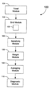
[0094] Figure 7 shows a block diagram of a system 100 for diagnosing
the possibility of disease in a body part using a sensitivity. The system 100
includes a grid module 102 for representing the body part with a grid having a
plurality of finite elements. The grid module 102 includes a finite element
analysis module 103 for performing finite element analysis, as described in
more detail below.
[0095] The system 100 also includes a voxel module 104 for dividing a
volume into a plurality of voxels, the grid being contained by the volume. The
surface of the volume, for example, can correspond to the surface of the grid.
In a different example, the volume could be larger than the grid, such as a
box
enclosing the grid.
[0096] A sensitivity module 105 computes sensitivities, such that each
voxei is assigned a sensitivity. In one embodiment, the sensitivity is
approximately constant throughout the voxel. Typically, a voxel is larger than
a finite element, containing several such elements (e.g., approximately one
hundred). However, this need not be true in general.
[0097] The system 100 further includes a weight module 106 that uses
a model of the body part to compute a set of weights associated with a
particular one of the plurality of voxels. A diagnostic module 108 computes a
diagnostic at the particular voxel to diagnose the possibility of disease in
the
body part, wherein the diagnostic is a function of the set of weights, and a
measured electrical property of the body part obtained with the electrode
array 12. An averaging module 110 calculates an average of a function
f (Z;,Z' ), defined below, at the I~" voxel.
CA 02528303 2005-11-28
-28-
[0098] Figure 8 shows a three-dimensional grid 112 and an enclosing
volume 114 formed by the grid module 102 and voxel module 104,
respectively, of Figure 7. The volume 114 is box shaped and is divided into
smaller box-shaped voxels 116. The voxels 116 span the volume, but in
Figure 8 only a few voxels are shown for clarity. As described in more detail
below, to each voxel is assigned a sensitivity. In one embodiment, the
sensitivity is approximately constant throughout the voxel 116. The grid 112
is divided into a collection of finite elements 118, which in the example
shown
are three-dimensional triangular wedges. Again, for clarity, only a few finite
elements 118 are shown. Typically, a voxel 116 is larger than a finite element
118.
[0099] The finite element analysis module 103 computes DV;,u, the
gradient of the electric potential arising when conditions ace employed
corresponding to injection of current between the ith pair of current
injection
electrodes. The finite element analysis module 103 also computes DV,,,b, the
gradient of the electric potential arising when conditions are employed
corresponding to injection of current between the pair of voltage electrodes
associated with the ith pair of current injection electrodes.
[00100] The sensitivity module 105 uses the gradients DV;,p and DV,..6
within a kt" voxel to obtain a set of sensitivities, {du,k, ouZk,..., Dun~~k],
where
Du;~ is the sensitivity at the kt" voxel obtained from DV;,a and DV;.6. The
set of
weights are calculated by the weight module 106 according to
~u;k
W ik - nc~
~~ujk
j=1
[00101] The sensitivity module 105 obtains the sensitivity Dusk using the
formula
~u;k = - fJJ ~ KR; DVia ' DV,b dv ,
Rt
CA 02528303 2005-11-28
-29-
where R~ is the volume of the kth voxel, and ~KRk is a deviation of a
conductivity at the kth voxel.
[00102] Diagnosing the possibility of disease in a body part using the
sensitivity proceeds in a similar manner as above, but with sensitivities
being
used instead of current densities.
[00103) Thus, the grid module 102 uses the model of the body part to
obtain a set of baseline impedances ~Z,, Z2,..., Z~~, } where Z; is the
impedance associated with the it" electrode pair.
[00104] The averaging module 110 of Figure 7 calculates an average of
a function f(Z;,ZM) at the kt" voxel, the average given by
( fk) _ ~ w;k f (Z;,Z,"' ) . The diagnostic at the kt" voxel is defined to be
( fk~ . For
;.,
example,
Z.
M
.f(Z,~Z, )- M .
Z;
where the Z"' are the impedances measured with the electrode array, as
described above.
[00105] The electrode array 12, the grid module 102, the sensitivity
module 105 and the weight module 106 are used to calculate diagnostics at
the other voxels, which together with the particular one, comprise the
plurality
of voxels. The diagnostic module averages the diagnostics at the voxels 116
to find an averaged diagnostic ~ f ~, and calculates a second averaged
diagnostic, t f~mo), corresponding to a homologous body part.
[00106] The diagnostic module 108 can calculate several quantities
having diagnostic value, such as the difference (f~-(fhomo) or
CA 02528303 2005-11-28
-30-
(.f)-(.fhomo) that are indicative of the possibility of disease in the body
part
2 ((.f ) + (.fhomo))
or the homologous body part.
[00107] Referring to Figure 9, shown is a plot of images of different
layers (i.e. slices) of respective right and left breasts of an actual subject
that
were obtained using a system in accordance with an embodiment of the
invention. The subject has a carcinoma in the left breast, which is generally
indicated by 132. The first layer (i.e. anterior, top layer) is the front-most
layer. The gray patterns in the plot represent a relative difference between
two homologous areas between right and left breasts. That is the darkness or
intensity of a grey pattern increases as the homologous difference of the
diagnostic becomes more profound. The carcinoma 132 is the darkest grey
pattern in the left breast. More specifically, the carcinoma 132 is located at
the middle depth at approximately three o'clock. The three o'clock angle is
clearly visible in all layers while the darkest area deminates the plot of the
middle layer. The right breast, on the other hand, is completely white
because all of the corresponding WVG valves on the left breast are higher
than those on the right breast.
[00108] The quantity hf;~n,~-~,fe~~~ is, by convention, plotted on the side
having the larger WEVaI; i.e., on the right breast for elements where
~yght > ~ \J IeR ~ and on the left breast where ~ fe ft ~ > ~ f~~,, ~ . These
differences
are scaled in the figure to the maximum level of black. Sixteen different
levels
of gray are presented, and some contrasting has been added to emphasize
areas where the differences are highest. However, none of these seating
methods appreciably influenced the results.
[00109] Different computer systems can be used to implement the
method for diagnosing disease in a body part. The computer system can
include a monitor for displaying diagnostic information using one of several
visual methods. In one embodiment, the method can be implemented on a 2
GHz PentiumT"" 4 system with 512 MB RAM.
CA 02528303 2005-11-28
-31 -
[00110] Although emphasis has been placed on describing a system for
diagnosing breast cancer, the principles of the present invention can also be
advantageously applied to other diseases of other body parts. These body
parts need not have a homolog. Also, although the main measured electrical
property described herein is impedance, it should be understood that other
electrical properties, such as functions of the electrical impedance, may also
be used in accordance with the principles of the present invention.
[00111] The expression for the voltage across a lead b for a unit current
injection over lead a is:
J'fVads f fVads
'b J'fl~ds J'fl~ds
i~Z Iii
J' Q Jb ds J'J'Vp Jb ds 1 = i = f 'J'Jb ' ds
f~~ds
- J'~~Jb ~ ds Jb ~ ds = 0 on S which is not in ~J3, U (3z )
- J'~'~ ~ (VaJb )dv Divergence Theorem : f'J'G ~ ds = J'f~ ' Gdv , B is volume
a s a
- -~'J'J'VaO ~ Jn dv - J'J'fJh ~ oVu dv product rule of differentiation
a a
- J'J'J'Jb ~ v va av v ~ Jb = 0 on volume B
a
_ ~'~'~'ko Va ~ V Vb dv Jb = -ko Vh
a
[00112] It should be understood that various modifications and
adaptations could be made to the embodiments described and illustrated
herein, without departing from the present invention, the scope of which is
defined in the appended claims.