Note: Descriptions are shown in the official language in which they were submitted.
CA 02528919 2005-12-09
WO 2005/004735 PCT/US2003/018676
VESSEL SEALER AND DIVIDER FOR USE WITH SMALL
TROCARS AND CANNULAS
BACKGROUND
The present disclosure relates to an electrosurgical forceps and more
particularly, the present disclosure relates to an endoscopic bipolar
electrosurgical forceps for sealing and/or cutting tissue.
Technical Field
Electrosurgical forceps utilize both mechanical clamping action and
electrical energy to effect hemostasis by heating the tissue and blood vessels
to coagulate, cauterize and/or seal tissue. As an alternative to open forceps
for use with open surgical procedures, many modern surgeons use
endoscopes and endoscopic instruments for remotely accessing organs
through smaller, puncture-like incisions. As a direct result thereof, patients
tend to benefit from less scarring and reduced healing time.
Endoscopic instruments are inserted into the patient through a cannula,
or port, which has been made with a trocar. Typical sizes for cannulas range
from three millimeters to twelve millimeters. Smaller cannulas are usually
preferred, which, as can be appreciated, ultimately presents a design
challenge
to instrument manufacturers who must find ways to make endoscopic
instruments that fit through the smaller cannulas.
Many endoscopic surgical procedures require cutting or ligating blood
vessels or vascular tissue. Due to the inherent spatial considerations of the
surgical cavity, surgeons often have difficulty suturing vessels or performing
other traditional methods of controlling bleeding, e.g., clamping and/or tying-
off
transected blood vessels. By utilizing an endoscopic electrosurgical forceps,
a
surgeon can either cauterize, coagulate/desiccate and/or simply reduce or slow
bleeding simply by controlling the intensity, frequency and duration of the
CA 02528919 2005-12-09
WO 2005/004735 PCT/US2003/018676
electrosurgical energy applied through the jaw members to the tissue. Most
small blood vessels, i.e., in the range below two millimeters in diameter, can
often be closed using standard electrosurgical instruments and techniques.
However, if a larger vessel is ligated, it may be necessary for the surgeon to
convert the endoscopic procedure into an open-surgical procedure and thereby
abandon the benefits of endoscopic surgery. Alternatively, the surgeon can
seal the larger vessel or tissue.
It is thought that the process of coagulating vessels is fundamentally
different than electrosurgical vessel sealing. For the purposes herein,
"coagulation" is defined as a process of desiccating tissue wherein the tissue
cells are ruptured and dried. "Vessel sealing" or "tissue sealing" is defined
as
the process of liquefying the collagen in the tissue so that it reforms into a
fused mass. Coagulation of small vessels is sufficient to permanently close
them, while larger vessels need to be sealed to assure permanent closure.
In order to effectively seal larger vessels (or tissue) two predominant
mechanical parameters must be accurately controlled - the pressure applied to
the vessel (tissue) and the gap distance between the electrodes - both of
which
are affected by the thickness of the sealed vessel. More particularly,
accurate
application of pressure is important to oppose the walls of the vessel; to
reduce
the tissue impedance to a low enough value that allows enough electrosurgical
energy through the tissue; to overcome the forces of expansion during tissue
heating; and to contribute to the end tissue thickness which is an indication
of a
good seal. It has been determined that a typical fused vessel wall is optimum
between 0.001 and 0.006 inches. Below this range, the seal may shred or tear
and above this range the lumens may not be properly or effectively sealed.
With respect to smaller vessels, the pressure applied to the tissue tends
to become less relevant whereas the gap distance between the electrically
conductive surfaces becomes more significant for effective sealing. In other
words, the chances of the two electrically conductive surfaces touching during
activation increases as vessels become smaller.
Many known instruments include blade members or shearing members
which simply cut tissue in a mechanical and/or electromechanical manner and
2
CA 02528919 2005-12-09
WO 2005/004735 PCT/US2003/018676
are relatively ineffective for vessel sealing purposes. Other instruments rely
on
clamping pressure alone to procure proper sealing thickness and are not
designed to take into account gap tolerances and/or parallelism and flatness
requirements which are parameters which, if properly controlled, can assure a
consistent and effective tissue seal. For example, it is known that it is
difficult
to adequately control thickness of the resulting sealed tissue by controlling
clamping pressure alone for either of two reasons: 1) if too much force is
applied, there is a possibility that the two poles will touch and energy will
not be
transferred through the tissue resulting in an ineffective seal; or 2) if too
low a
force is applied the tissue may pre-maturely move prior to activation and
sealing and/or a thicker, less reliable seal may be created.
As mentioned above, in order to properly and effectively seal larger
vessels or tissue, a greater closure force between opposing jaw members is
required. It is known that a large closure force between the jaws typically
requires a large moment about the pivot for each jaw. This presents a design
challenge because the jaw members are typically affixed with pins which are
positioned to have small moment arms with respect to the pivot of each jaw
member. A large force, coupled with a small moment arm, is undesirable
because the large forces may shear the pins. As a result, designers must
compensate for these large closure forces by either designing instruments with
metal pins and/or by designing instruments which at least partially offload
these
closure forces to reduce the chances of mechanical failure. As can be
appreciated, if metal pivot pins are employed, the metal pins must be
insulated
to avoid the pin acting as an alternate current path between the jaw members
which may prove detrimental to effective sealing.
Increasing the closure forces between electrodes may have other
undesirable effects, e.g., it may cause the opposing electrodes to come into
close contact with one another which may result in a short circuit and a small
closure force may cause pre-mature movement of the tissue during
compression and prior to activation. As a result thereof, providing an
instrument which consistently provides the appropriate closure force between
opposing electrode within a preferred pressure range will enhance the chances
3
CA 02528919 2011-06-02
of a successful seal. As can be appreciated, relying on a surgeon to manually
provide the appropriate closure force within the appropriate range on a
consistent basis would be difficult and the resultant effectiveness and
quality of
the seal may vary. Moreover, the overall success of creating an effective
tissue
seal is greatly reliant upon the user's expertise, vision, dexterity, and
experience in judging the appropriate closure force to uniformly, consistently
and effectively seal the vessel. In other words, the success of the seal would
greatly depend upon the ultimate skill of the surgeon rather than the
efficiency
of the instrument.
It has been found that the pressure range for assuring a consistent and
effective seal is between about 3 kg/cm2 to about 16 kg/cm2 and, preferably,
within a working range of 7 kg/cm2 to 13 kg/cm2. Manufacturing an instrument
which is capable of providing a closure pressure within this working range has
been shown to be effective for sealing arteries, tissues and other vascular
bundles.
Various force-actuating assemblies have been developed in the past for
providing the appropriate closure forces to effect vessel sealing. For
example,
one such actuating assembly has been developed by Valleylab Inc., a division
of Tyco Healthcare LP, for use with Valleylab's vessel sealing and dividing
instrument commonly sold under the trademark LIGASURE ATLAS . This
assembly includes a four-bar mechanical linkage, a spring and a drive
assembly which cooperate to consistently provide and maintain tissue
pressures within the above working ranges. The LIGASURE ATLAS is
presently designed to fit through a 10mm cannula and includes a bi-lateral jaw
closure mechanism which is activated by a foot switch. A trigger assembly
extends a knife distally to separate the tissue along the tissue seal. A
rotating
mechanism is associated with distal end of the handle to allow a surgeon to
selectively rotate the jaw members to facilitate grasping tissue. U.S. Patent
Publication 2003/0018331, and U.S. Patent Publication 2002/0188294 describe
in detail the operating features of the LIGASURE ATLAS and various methods
relating thereto.
4
CA 02528919 2011-06-02
It would be desirous to develop a smaller, simpler endoscopic vessel
sealing instrument which can be utilized with a 5mm cannula. Preferably, the
instrument would include a simpler and more mechanically advantageous drive
assembly to facilitate grasping and manipulating vessels and tissue. In
addition, it would be desirous to manufacture an instrument which includes a
hand switch and a unilateral jaw closure mechanism. Moreover, it would be
advantageous to provide a vessel sealing instrument which effectively,
reliably
and accurately divides the tissue across the tissue seal.
SUMMARY
The present disclosure relates to a bipolar forceps for sealing and
dividing tissue which is preferably designed to be utilized with a 5mm trocar
or
cannula and includes a housing and a shaft affixed to the distal end of the
housing. The shaft includes first and second jaw members attached to the
distal end thereof which are movable relative to one another from a first
spaced-apart position to a second position for grasping tissue. At least one
of
the jaw members includes a knife channel disposed substantially along the
length thereof. The knife channel has a depth, a width and an aspect ratio
which is defined as the depth of the knife channel divided by the width of the
knife channel.
Preferably the aspect ratio of the knife channel is at least 1.3. The
aspect ratio is dependant upon, inter alia, closure pressure, tissue
thickness,
tissue type, and moisture content of the tissue. For example, in one
embodiment according to the present disclosure, the closure pressure is
advantageously in the range of abut 7 kg/cm2 to about 11 kg/cm2 which
warrants an aspect ratio of about 1.9 to optimize tissue cutting.
The forceps is connected to a source of electrosurgical energy and also
includes an actuator for moving the jaw members relative to one another.
Advantageously, a knife assembly is included which allows a user to
selectively
move a knife to cut tissue disposed between the jaw members. The source of
5
CA 02528919 2005-12-09
WO 2005/004735 PCT/US2003/018676
electrosurgical energy carries electrical potentials to each respective jaw
member such that the jaw members are capable of conducting bipolar energy
through tissue held therebetween to effect a tissue seal.
In one embodiment, the first jaw member and the second jaw member
each include includes an elongated slot which run in opposition substantially
along the respective lengths thereof such that the two opposing elongated
slots
form the knife channel for reciprocating the knife to divide tissue disposed
between the two jaw members.
In yet another embodiment, at least one of the jaw members includes
one or more non-conductive stop members disposed thereon which controls
the distance between the jaw members when tissue is held therebetween.
Advantageously, the stop members maintain a gap distance of about 0.001
inches to about 0.006 inches between the jaw members when tissue is
compressed between the jaw members. In still another embodiment, the
actuator is selectively lockable to maintain a closure pressure in the range
of
about 3 kg/cm2 to about 16 kg/cm2 and, preferably, about 7 kg/cm2 to about
13 kg/cm2 between the jaw members which is advantageous in producing
effective and reliable tissue seals.
Advantageously, the forceps includes a unilateral jaw assembly, i.e., the
first jaw member is movable relative to the second jaw member and the second
jaw member is substantially fixed. In another embodiment, the forceps may
also include a rotating assembly for rotating the jaw members about a
longitudinal axis defined through the shaft.
Still another embodiment of the present disclosure relates to a bipolar
forceps for sealing and dividing tissue which includes a housing having a
shaft
affixed thereto having first and second jaw members attached to a distal end
thereof. At least one of the jaw members includes a knife channel disposed
substantially along the length of the jaw member. The forceps also includes an
actuator for moving jaw members relative to one another from a first position
wherein the jaw members are disposed in spaced relation relative to one
another to a second position wherein the jaw members cooperate to grasp
tissue therebetween. The forceps is connected to a source of electrosurgical
6
CA 02528919 2005-12-09
WO 2005/004735 PCT/US2003/018676
energy connected to each jaw member such that the jaw members are capable
of conducting bipolar energy through tissue held therebetween to effect a
tissue seal.
Advantageously, a knife assembly is included which has an elongated
knife bar for supporting a knife with a leading cutting edge. The elongated
knife bar is selectively moveable within the knife channel to force tissue
disposed within the knife channel into engagement with the cutting edge of the
knife upon distal movement thereof which, in turn, cuts tissue disposed
between the jaw members. Preferably, the elongated knife bar includes a
chamfered edge which directs tissue from the knife channel and towards the
cutting edge of the knife. As can be appreciated, having the leading edge of
the knife bar chamfered insures accurate and effective tissue separation.
A rotating assembly may also be included for rotating the jaw members
about the longitudinal axis defined through the shaft. Preferably, the
rotating
assembly is located near the proximal end of the housing and near the hand
switch to facilitate rotation.
Advantageously, the movable jaw member includes a first electrical
potential and the fixed jaw member includes a second electrical potential. A
lead connects the movable jaw member to the first potential and a conductive
tube (which is disposed through the shaft) conducts a second electrical
potential to the fixed jaw member. Preferably, the conductive tube is
connected to the rotating assembly to permit selective rotation of the jaw
members.
In still yet another embodiment, a spring is included with the drive
assembly to facilitate actuation of the movable handle and to assure the
closure force is maintained within a working range of about 3kg/cm2 to about
16 kg/cm2. At least one of the jaw members may include a series of stop
members disposed thereon for regulating the distance between the jaw
members (i.e., creating a gap between the two opposing jaw members) during
the sealing process.
7
CA 02528919 2005-12-09
WO 2005/004735 PCT/US2003/018676
BRIEF DESCRIPTION OF THE DRAWINGS
Various embodiments of the subject instrument are described herein
with reference to the drawings wherein:
Fig. 1 is a left, perspective view of an endoscopic bipolar forceps
showing a housing, a shaft and an end effector assembly according to the
present disclosure;
Fig. 2 is a top view of the forceps of Fig. 1;
Fig. 3 is a left, side view of the forceps of Fig. 1;
Fig. 4 is a left, perspective view of the forceps of Fig. 1 showing the
rotation of the end effector assembly about a longitudinal axis "A";
Fig. 5 is a front view of the forceps of Fig. 1;
Figs. 6 is an enlarged view of the indicated area of detail of Fig. 5
showing an enhanced view of the end effector assembly detailing a pair of
opposing jaw members;
Fig. 7 is an enlarged, rear perspective view of the housing;
Fig. 8 is an enlarged, left perspective view of the end effector assembly
with the jaw members shown in open configuration;
Fig. 9 is an enlarged, side view of the end effector assembly;
Fig. 10 is an enlarged, perspective view of the underside of the upper
jaw member of the end effector assembly;
Fig. 11 is an enlarged, broken perspective view showing the end effector
assembly and highlighting a cam-like closing mechanism which cooperates
with a reciprocating pull sleeve to move the jaw members relative to one
another;
Fig. 12 is a full perspective view of the end effector assembly of Fig. 11;
Fig. 13 is an enlarged, perspective view of the housing and the internal
working components thereof;
Fig. 14 is top, perspective view of the housing of Fig. 13 with parts
separated;
Fig. 15 is a left, perspective view of a rotating assembly, drive assembly,
knife assembly and lower jaw member according to the present disclosure;
8
CA 02528919 2005-12-09
WO 2005/004735 PCT/US2003/018676
Fig. 16 is a rear, perspective view of the rotating assembly, drive
assembly and knife assembly;
Fig. 17 is an enlarged, top, perspective view of the end effector
assembly with parts separated;
Fig. 18 is an enlarged, perspective view of the knife assembly;
Fig. 19 is an enlarged, perspective view of the rotating assembly;
Fig. 20 is an enlarged, perspective view of the drive assembly;
Fig. 21 is an enlarged, perspective view of the knife assembly with parts
separated;
Fig. 22 is an enlarged view of the indicated area of detail of Fig. 21;
Fig. 23 is a greatly-enlarged, perspective view of a distal end of the knife
assembly;
Fig. 24 is a greatly-enlarged, perspective view of a knife drive of the
knife assembly;
Fig. 25 is an enlarged, perspective view of the rotating assembly and
lower jaw member with parts separated;
Fig. 26 is a cross section of the area indicated in detail in Fig. 25;
Fig. 27 is a greatly-enlarged, perspective view of the lower jaw member;
Fig. 28 is an enlarged, perspective view of the drive assembly;
Fig. 29 is an enlarged perspective view of the drive assembly of Fig. 28
with parts separated;
Fig. 30 is an internal, side view of the housing showing the inner-working
components thereof;
Fig. 31 is a cross-section of the housing with the end effector shown in
open configuration and showing the internal, electrical routing of an
electrosurgical cable and electrical leads;
Fig. 32 is a greatly-enlarged view of the indicated area of detail of Fig.
31;
Fig. 33 is a greatly-enlarged view of the indicated area of detail of Fig.
31;
Fig. 34 is a greatly-enlarged, cross section of the shaft taken along line
34-34;
9
CA 02528919 2005-12-09
WO 2005/004735 PCT/US2003/018676
Fig. 35 is a side, cross section of the shaft and end effector assembly;
Fig. 36 is a perspective view showing the forceps of the present
disclosure being utilized with a 5mm cannula;
Fig. 37 is a side, cross section of the housing showing the moving
components of the drive assembly during actuation;
Fig. 38 is a greatly-enlarged, perspective view of a handle locking
mechanism for use with the drive assembly;
Fig. 39 is a greatly-enlarged view of the indicated area of detail in Fig.
37;
Fig. 40 is a greatly-enlarged view of the indicated area of detail in Fig.
37;
Fig. 41 is an enlarged, rear, perspective view of the end effectors shown
grasping tissue;
Fig. 42 is an enlarged view of a tissue seal;
Fig. 43 is a side, cross section of a tissue seal;
Fig. 44 is a cross section of the housing with the handle in a locked
configuration and showing the moving components of the knife assembly
during activation;
Fig. 45 is an enlarged view of the area indicated in detail in Fig. 44;
Fig. 46 is a side, cross section of a tissue seal after separation by the
knife assembly;
Fig. 47 is a side, cross section of the housing showing the release of the
knife assembly and release of the drive assembly to open the jaw members
and release the tissue;
Fig. 48 is a greatly-enlarged view of the indicated area of detail in Fig.
47;
Fig. 49 is a greatly-enlarged view of the indicated area of detail in Fig.
47;
Fig. 50 is a greatly-enlarged schematic diagram of an upper knife
channel of the movable jaw member showing one preferred configuration to
facilitate tissue separation;
CA 02528919 2005-12-09
WO 2005/004735 PCT/US2003/018676
Fig. 51 is a greatly-enlarged end cross section showing the knife being
supported by a knife bar which rides within a lower knife channel disposed in
the fixed jaw member; and
Fig. 52 is a greatly-enlarged schematic view of a knife which is spring-
biased to expand fully within the knife channel upon reciprocation of the
knife
assembly.
DETAILED DESCRIPTION
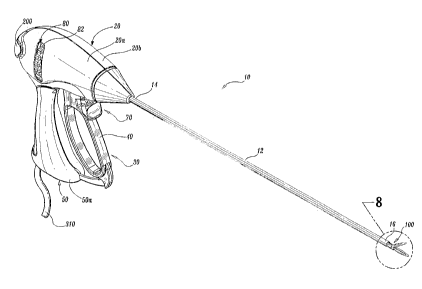
Turning now to Figs. 1-3, one embodiment of an endoscopic bipolar
forceps 10 is shown for use with various surgical procedures and generally
includes a housing 20, a handle assembly 30, a rotating assembly 80, a trigger
assembly 70 and an end effector assembly 100 which mutually cooperate to
grasp, seal and divide tubular vessels and vascular tissue 420 (Fig. 36).
Although the majority of the figure drawings depict a bipolar forceps 10 for
use
in connection with endoscopic surgical procedures, the present disclosure may
be used for more traditional open surgical procedures. For the purposes
herein, the forceps 10 is described in terms of an endoscopic instrument,
however, it is contemplated that an open version of the forceps may also
include the same or similar operating components and features as described
below.
Forceps 10 includes a shaft 12 which has a distal end 16 dimensioned
to mechanically engage the end effector assembly 100 and a proximal end 14
which mechanically engages the housing 20. Details of how the shaft 12
connects to the end effector are described in more detail below with respect
to
Fig. 25. The proximal end 14 of shaft 12 is received within the housing 20 and
the connections relating thereto are described in detail below with respect to
Figs. 13 and 14. In the drawings and in the descriptions which follow, the
term
"proximal", as is traditional, will refer to the end of the forceps 10 which
is
closer to the user, while the term "distal" will refer to the end which is
further
from the user.
As best seen in Fig. 1, forceps 10 also includes an electrosurgical cable
310 which connects the forceps 10 to a source of electrosurgical energy, e.g.,
11
CA 02528919 2011-06-02
a generator (not shown). Preferably, generators such as those sold by
Valleylab - a division of Tyco Healthcare LP, located in Boulder Colorado are
used as a source of electrosurgical energy, e.g., FORCE EZTM Electrosurgical
Generator, FORCE FXTM Electrosurgical Generator, FORCE ICTM, FORCE 2TM
Generator, SurgiStatTM II. One such system is described in commonly-owned
U.S. Patent No. 6,033,399 entitled "ELECTROSURGICAL GENERATOR
WITH ADAPTIVE POWER CONTROL".
Other systems have been described in commonly-owned
U.S. Patent No. 6,187,003 entitled "BIPOLAR ELECTROSURGICAL
INSTRUMENT FOR SEALING VESSELS".
Preferably, the generator includes various safety and performance
features including isolated output, independent activation of accessories.
Preferably, the electrosurgical generator includes Valleylab's Instant
ResponseTM technology features which provides an advanced feedback
system to sense changes in tissue 200 times per second and adjust voltage
and current to maintain appropriate power. The Instant ResponseTM
technology is believed to provide one or more of the following benefits to
surgical procedure:
0 Consistent clinical effect through all tissue types;
Reduced thermal spread and risk of collateral tissue damage;
= Less need to "turn up the generator"; and
= Designed for the minimally invasive environment.
Cable 310 is internally divided into cable lead 310a, 310b and 310c
which each transmit electrosurgical energy through their respective feed paths
through the forceps 10 to the end effector assembly 100 as explained in more
detail below with respect to Figs. 14 and 30.
Handle assembly 30 includes a fixed handle 50 and a movable handle
40. Fixed handle 50 is integrally associated with housing 20 and handle 40 is
movable relative to fixed handle 50 as explained in more detail below with
12
CA 02528919 2005-12-09
WO 2005/004735 PCT/US2003/018676
respect to the operation of the forceps 10. Rotating assembly 80 is preferably
integrally associated with the housing 20 and is rotatable approximately 180
degrees in either direction about a longitudinal axis "A" (See Fig. 4).
Details of
the rotating assembly 80 are described in more detail with respect to Figs.
13,
14, 15 and 16
As best seen in Figs. 2, 13 and 14, housing 20 is formed from two (2)
housing halves 20a and 20b which each include a plurality of interfaces 27a-
27f which are dimensioned to mechanically align and engage one another to
form housing 20 and enclose the internal working components of forceps 10.
As can be appreciated, fixed handle 50 which, as mentioned above, is
integrally associated with housing 20, takes shape upon the assembly of the
housing halves 20a and 20b.
It is envisioned that a plurality of additional interfaces (not shown) may
disposed at various points around the periphery of housing halves 20a and 20b
for ultrasonic welding purposes, e.g., energy direction/deflection points. It
is
also contemplated that housing halves 20a and 20b (as well as the other
components described below) may be assembled together in any fashion'
known in the art. For example, alignment pins, snap-like interfaces, tongue
and groove interfaces, locking tabs, adhesive ports, etc. may all be utilized
either alone or in combination for assembly purposes.
Rotating assembly 80 includes two halves 82a and 82b which, when
assembled, form the rotating assembly 80 which, in turn, houses the drive
assembly 150 and the knife assembly 140 (See Figs. 13, 14 and 25). Half 80a
includes a series of detents/flanges 375a, 375b, 375c and 375d (Fig. 25) which
are dimensioned to engage a pair of corresponding sockets or other
mechanical interfaces (not shown) disposed within rotating half 80a. Movable
handle 40 and trigger assembly 70 are preferably of unitary construction and
are operatively connected to the housing 20 and the fixed handle 50 during the
assembly process.
As mentioned above, end effector assembly 100 is attached at the distal
end 14 of shaft 12 and includes a pair of opposing jaw members 110 and 120.
Movable handle 40 of handle assembly 30 is ultimately connected to a drive
13
CA 02528919 2005-12-09
WO 2005/004735 PCT/US2003/018676
assembly 150 which, together, mechanically cooperate to impart movement of
the jaw members 110 and 120 from an open position wherein the jaw members
110 and 120 are disposed in spaced relation relative to one another, to a
clamping or closed position wherein the jaw members 110 and 120 cooperate
to grasp tissue 420 (Fig. 36) therebetween.
It is envisioned that the forceps 10 may be designed such that it is fully
or partially disposable depending upon a particular purpose or to achieve a
particular result. For example, end effector assembly 100 may be selectively
and releasably engageable with the distal end 16 of the shaft 12 and/or the
proximal end 14 of shaft 12 may be selectively and releasably engageable with
the housing 20 and the handle assembly 30. In either of these two instances,
the forceps 10 would be considered "partially disposable" or "reposable",
i.e., a
new or different end effector assembly 100 (or end effector assembly 100 and
shaft 12) selectively replaces the old end effector assembly 100 as needed.
As can be appreciated, the presently disclosed electrical connections would
have to be altered to modify the instrument to a reposable forceps.
Turning now to the more detailed features of the present disclosure as
described with respect to Figs. I - 14, movable handle 40 includes a finger
loop 41 which has an aperture 42 defined therethrough which enables a user
to grasp and move the handle 40 relative to the fixed handle 50. Handle .40
also includes an ergonomically-enhanced gripping element 43 disposed along
the inner peripheral edge of aperture 42 which is designed to facilitate
gripping
of the movable handle 40 during activation. It is envisioned that gripping
element 43 may include one or more protuberances, scallops and/or ribs to
enhance gripping. As best seen in Fig. 14, movable handle 40 is selectively
moveable about a pair of pivot pins 29a and 29b from a first position relative
to
fixed handle 50 to a second position in closer proximity to the fixed handle
50
which, as explained below, imparts movement of the jaw members 110 and
120 relative to one another. The movable handle include a clevis 45 which
forms a pair of upper flanges 45a and 45b each having an aperture 49a and
49b, respectively, at an upper end thereof for receiving the pivot pins 29a
and
29b therethrough and mounting the upper end of the handle 40 to the housing
14
CA 02528919 2005-12-09
WO 2005/004735 PCT/US2003/018676
20. In turn, each pin 29a and 29b mounts to a respective housing half 20a and
20b.
Each upper flange 45a and 45b also includes a force-actuating flange or
drive flange 47a and 47b, respectively, which are aligned along longitudinal
axis "A" and which abut the drive assembly 150 such that pivotal movement of
the handle 40 forces actuating flange against the drive assembly 150 which, in
turn, closes the jaw members 110 and 120. For the purposes herein, 47a and
47b which act simultaneously on the drive assembly are referred to as "driving
flange 47". A more detailed explanation of the inter-cooperating components
of the handle assembly 30 and the drive assembly 150 is discussed below.
As best seen in Fig. 14, the lower end of the movable handle 40
includes a flange 90 which is preferably mounted to the movable handle 40 by
pins 94a and 94b which engage a corresponding pair of apertures 91 a and 91 b
disposed within the lower portion of handle 40 and apertures 97a and 97b
disposed within flange 90, respectively. Other methods of engagement are
also contemplated, snap-lock, spring tab, etc. Flange 90 also includes a t-
shaped distal end 95 which rides within a predefined channel 51 disposed
within fixed handle 50 to lock the movable handle 40 relative to the fixed
handle 50. Additional features with respect to the t-shaped end 95 are
explained below in the detailed discussion of the operational features of the
forceps 10.
Movable handle 40 is designed to provide a distinct mechanical
advantage over conventional handle assemblies due to the unique position of
the pivot pins 29a and 29b (i.e., pivot point) relative to the longitudinal
axis "A"
of the shaft 12 and the disposition of the driving flange 47 along
longitudinal
axis "A". In other words, it is envisioned that by positioning the pivot pins
29a
and 29b above the driving flange 47, the user gains lever-like mechanical
advantage to actuate the jaw members 110 and 120 enabling the user to close
the jaw members 110 and 120 with lesser force while still generating the
required forces necessary to effect a proper and effective tissue seal. It is
also
envisioned that the unilateral design of the end effector assembly 100 will
also
increase mechanical advantage as explained in more detail below.
CA 02528919 2005-12-09
WO 2005/004735 PCT/US2003/018676
As shown best in Figs. 6-12, the end effector assembly 100 includes
opposing jaw members 110 and 120 which cooperate to effectively grasp
tissue 420 for sealing purposes. The end effector assembly 100 is designed
as a unilateral assembly, i.e., jaw member 120 is fixed relative to the shaft
12
and jaw member 110 pivots about a pivot pin 103 to grasp tissue 420.
More particularly, the unilateral end effector assembly 100 includes one
stationary or fixed jaw member 120 mounted in fixed relation to the shaft 12
and pivoting jaw member 110 mounted about a pivot pin 103 attached to the
stationary jaw member 120. A reciprocating sleeve 60 is slidingly disposed
within the shaft 12 and is remotely operable by the drive assembly 150. The
pivoting jaw member 110 includes a detent or protrusion 117 which extends
from jaw member 110 through an aperture 62 disposed within the reciprocating
sleeve 60 (Fig. 12). The pivoting jaw member 110 is actuated by sliding the
sleeve 60 axially within the shaft 12 such that a distal end 63 of the
aperture 62
abuts against the detent 117 on the pivoting jaw member 110 (See Figs. 11
and 12). Pulling the sleeve 60 proximally closes the jaw members 110 and 120
about tissue 420 grasped therebetween and pushing the sleeve 60 distally
opens the jaw members 110 and 120 for grasping purposes.
As best illustrated in Figs. 8 and 10, a knife channel 115a and 115b runs
through the center of the jaw members 110 and 120, respectively, such that a
blade 185 from the knife assembly 140 can cut the tissue 420 grasped
between the jaw members 110 and 120 when the jaw members 110 and 120
are in a closed position. More particularly, the blade 185 can only be
advanced
through the tissue 420 when the jaw members 110 and 120 are closed thus
preventing accidental or premature activation of the blade 185 through the
tissue 420. Put simply, the knife channel 115 (made up of half channels 115a
and 115b) is blocked when the jaws members 110 and 120 are opened and
aligned for distal activation when the jaw members 110 and 120 are closed
(See Figs. 35 and 39). It is also envisioned that the unilateral end effector
assembly 100 may be structured such that electrical energy can be routed
through the sleeve 60 at the protrusion 117 contact point with the sleeve 60
or
using a "brush" or lever (not shown) to contact the back of the moving jaw
16
CA 02528919 2005-12-09
WO 2005/004735 PCT/US2003/018676
member 110 when the jaw member 110 closes. In this instance, the electrical
energy would be routed through the protrusion 117 to the stationary jaw
member 120. Alternatively, the cable lead 311 may be routed to energize the
stationary jaw member 120 and the other electrical potential may be conducted
through the sleeve 60 and transferred to the pivoting jaw member 110 which
establishes electrical continuity upon retraction of the sleeve 60. It is
envisioned that this particular envisioned embodiment will provide at least
two
important safety features: 1) the blade 185 cannot extend while the jaw
members 110 and 120 are opened; and 2) electrical continuity to the jaw
members 110 and 120 is made only when the jaw members are closed. The
illustrated forceps 10 only includes the novel knife channel 115.
As best shown in Fig. 8, jaw member 110 also includes a jaw housing
116 which has an insulative substrate or insulator 114 and an electrically
conducive surface 112. Insulator 114 is preferably dimensioned to securely
engage the electrically conductive sealing surface 112. This may be
accomplished by stamping, by overmolding, by overmolding a stamped
electrically conductive sealing plate and/or by overmolding a metal injection
molded seal plate. For example and as shown in Fig. 17, the electrically
conductive sealing plate 112 includes a series of upwardly extending flanges
111 a and 111 b which are designed to matingly engage the insulator 114. The
insulator 114 includes a shoe-like interface 107 disposed at a distal end
thereof
which is dimensioned to engage the outer periphery 116a of the housing 116 in
a slip-fit manner. The shoe-like interface 107 may also be overmolded about
the outer periphery of the jaw 110 during a manufacturing step. It is
envisioned
that lead 311 terminates within the shoe-like interface 107 at the point where
lead 311 electrically connects to the seal plate 112 (not shown). The movable
jaw member 110 also includes a wire channel 113 which is designed to guide
cable lead 311 into electrical continuity with sealing plate 112 as described
in
more detail below.
All of these manufacturing techniques produce jaw member 110 having
an electrically conductive surface 112 which is substantially surrounded by an
insulating substrate 114. The insulator 114, electrically conductive sealing
17
CA 02528919 2005-12-09
WO 2005/004735 PCT/US2003/018676
surface 112 and the outer, non-conductive jaw housing 116 are preferably
dimensioned to limit and/or reduce many of the known undesirable effects
related to tissue sealing, e.g., flashover, thermal spread and stray current
dissipation. Alternatively, it is also envisioned that the jaw members 110 and
120 may be manufactured from a ceramic-like material and the electrically
conductive surface(s) 112 are coated onto the ceramic-like jaw members 110
and 120.
Jaw member 110 includes a pivot flange 118 which includes protrusion
117. Protrusion 117 extends from pivot flange 118 and includes an arcuately-
shaped inner surface 111 dimensioned to matingly engage the aperture 62 of
sleeve 60 upon retraction thereof. Pivot flange 118 also includes a pin slot
119
which is dimensioned to engage pivot pin 103 to allow jaw member 110 to
rotate relative to jaw member 120 upon retraction of the reciprocating sleeve
60. As explained in more detail below, pivot pin 103 also mounts to the
stationary jaw member 120 through a pair of apertures 101a and 101b
disposed within a proximal portion of the jaw member 120.
It is envisioned that the electrically conductive sealing surface 112 may
also include an outer peripheral edge which has a pre-defined radius and the
insulator 114 meets the electrically conductive sealing surface 112 along an
20, adjoining edge of the sealing surface 112 in a generally tangential
position.
Preferably, at the interface, the electrically conductive surface 112 is
raised
relative to the insulator 114. These and other envisioned embodiments are
discussed in co-pending, commonly assigned Application Serial No.
PCT/US01/11412 entitled "ELECTROSURGICAL INSTRUMENT WHICH
REDUCES COLLATERAL DAMAGE TO ADJACENT TISSUE" by Johnson et
al. and co-pending, commonly assigned Application Serial No.
PCT/US01/11411 entitled "ELECTROSURGICAL INSTRUMENT WHICH IS
DESIGNED TO REDUCE THE INCIDENCE OF FLASHOVER" by Johnson et
al.
Preferably, the electrically conductive surface 112 and the insulator 114,
when assembled, form a longitudinally-oriented slot 115a defined therethrough
for reciprocation of the knife blade 185. It is envisioned that the knife
channel
18
CA 02528919 2011-06-02
115a cooperates with a corresponding knife channel 115b defined in stationary
jaw member 120 to facilitate longitudinal extension of the knife blade 185
along
a preferred cutting plane to effectively and accurately separate the tissue
420
along the formed tissue seal 450 (See Figs. 42 and 46).
Jaw member 120 includes similar elements to jaw member 110 such as
jaw housing 126 having an insulator 124 and an electrically conductive sealing
surface 122 which is dimensioned to securely engage the insulator 124.
Likewise, the electrically conductive surface 122 and the insulator 124, when
assembled, include a longitudinally-oriented channel 115a defined
therethrough for reciprocation of the knife blade 185. As mentioned above,
when the jaw members 110 and, 120 are closed about tissue 420, knife
channels 115a and 115b form a complete knife channel 115 to allow
longitudinal extension of the knife 185 in a distal fashion to sever tissue
420
along the tissue seal 450. It is also envisioned that the knife channel 115
may
be completely disposed in one of the two jaw members, e.g., jaw member 120,
depending upon a particular purpose. It is envisioned that the fixed jaw
member 120 may be assembled in a similar manner as described above with
respect to jaw member 110.
As best seen in Fig. 8, jaw member 120 includes a series of stop
members 750 preferably disposed on the inner facing surfaces of the
electrically conductive sealing surface 122 to facilitate gripping and
manipulation of tissue and to define a gap "G" (Fig. 24) between opposing jaw
members 110 and 120 during sealing and cutting of tissue. It is envisioned
that the series of stop members 750 may be employed on one or both jaw
members 110 and 120 depending upon a particular purpose or to achieve a
desired result. A detailed discussion of these and other envisioned stop
members 750 as well as various manufacturing and assembling processes for
attaching and/or affixing the stop members 750 to the electrically
conductive sealing surfaces 112, 122 are described in WO 02/080796.
19
CA 02528919 2005-12-09
WO 2005/004735 PCT/US2003/018676
Jaw member 120 is designed to be fixed to the end of a rotating tube
160 which is part of the rotating assembly 80 such that rotation of the tube
160
will impart rotation to the end effector assembly 100 (See Figs. 25 and 27).
Jaw member 120 includes a rear C-shaped cuff 170 having a slot 177 defined
therein which is dimensioned to receive a slide pin 171. More particularly,
slide pin 171 includes a slide rail 176 which extends substantially the length
thereof which is dimensioned to slide into friction-fit engagement within slot
177. A pair of chamfered plates 172a and 172b extend generally radially from
the slide rail 176 and include a radius which is substantially the same radius
as
the outer periphery of the rotating tube 160 such that the shaft 12 can
encompass each of the same upon assembly.
As explained in more detail below, fixed jaw member 120 is connected
to a second electrical potential through tube 160 which is connected at its
proximal end to lead 310c. More particularly, fixed jaw 120 is welded to the
rotating tube 160 and includes a fuse clip, spring clip or other electro-
mechanical connection which provides electrical continuity to the fixed jaw
member 120 from lead 310c (See Fig. 32). As best shown in Figs. 25 and 26,
the rotating tube 160 includes an elongated guide slot 167 disposed in an
upper portion thereof which is dimensioned to carry lead 311 therealong. The
chamfered plates 172a and 172b also form a wire channel 175 which is
dimensioned to guide the cable lead 311 from the tube 160 and into the
movable jaw member 110 (See Fig. 8). Lead 311 carries a first electrical
potential to movable jaw 110. As explained in more detail below with respect
to the internal electrical connections of the forceps, a second electrical
connection from lead 310c is conducted through the tube 160 to the fixed jaw
member 120.
As shown in Fig. 25, the distal end of the tube 160 is generally C-
shaped to include two upwardly extending flanges 162a and 162b which define
a cavity 165 for receiving the proximal end of the fixed jaw member 120
inclusive of C-shaped cuff 170 and slide pin 171 (See Fig. 27). Preferably,
the
tube cavity 165 retains and secures the jaw member 120 in a friction-fit
manner, however, the jaw member 120 may be welded to the tube 160
CA 02528919 2005-12-09
WO 2005/004735 PCT/US2003/018676
depending upon a particular purpose. Tube 160 also includes an inner cavity
169 defined therethrough which reciprocates the knife assembly 140 upon
distal activation thereof and an elongated guide rail 163 which guides the
knife
assembly 140 during distal activation. The details with respect to the knife
assembly are explained in more detail with respect to Figs. 21-24. The
proximal end of tube 160 includes a laterally oriented slot 168 which is
designed to interface with the rotating assembly 80 as described below.
Fig. 25 also shows the rotating assembly 80 which includes C-shaped
rotating halves 82a and 82b which, when assembled about tube 160, form a
generally circular rotating member 82. More particularly, each rotating half,
e.g., 82b, includes a series of mechanical interfaces 375a, 375b, 375c and
375d which matingly engage a corresponding series of mechanical interfaces
in half 82a to form rotating member 82. Half 82b also includes a tab 89b which
together with a corresponding tab 89a disposed on half 82a (phantomly
illustrated) cooperate to matingly engage slot 168 disposed on tube 160. As
can be appreciated, this permits selective rotation of the tube 160 about axis
"A" by manipulating the rotating member 82 in the direction of the arrow "B"
(see Fig. 4).
As best shown in the exploded view of Fig. 17, jaw members 110 and
120 are pivotably mounted with respect to one another such that jaw member
110 pivots in a unilateral fashion from a first open position to a second
closed
position for grasping and manipulating tissue 420. More particularly, fixed
jaw
member 120 includes a pair of proximal, upwardly extending flanges 125a and
125b which define a cavity 121 dimensioned to receive flange 118 of movable
jaw member 110 therein. Each of the flanges 125a and 125b includes an
aperture 101a and 101b, respectively, defined therethrough which secures
pivot pin 103 on opposite sides of pivot mount 119 disposed within jaw member
110. As explained in detail below with respect to the operation of the jaw
members 110 and 120, proximal movement of the tube 60 engages detent 117
to pivot the jaw member 110 to a closed position.
Figs. 13 and 14 show the details of the housing 20 and the component
features thereof, namely, the drive assembly 150, the rotating assembly 80,
the
21
CA 02528919 2005-12-09
WO 2005/004735 PCT/US2003/018676
knife assembly 140, the trigger assembly 70 and the handles 40 and 50. More
particularly, Fig. 13 shows the above-identified assemblies and components in
an assembled form in the housing 20 and Fig. 14 shows an exploded view of
each of the above-identified assemblies and components.
As shown best in Fig. 14, the housing includes halves 20a and 20b
which, when mated, form housing 20. As can be appreciated, housing 20,
once formed, houses the various assemblies identified above which will enable
a user to selectively manipulate, grasp, seal and sever tissue 420 in a
simple,
effective, and efficient manner. Preferably, each half of the housing, e.g.,
half
20b, includes a series of mechanical interfacing component, e.g., 27a - 27f
which align and/or mate with a corresponding series of mechanical interfaces
(not shown) to align the two housing halves 20a and 20b about the inner
components and assemblies. The housing halves 20a and 20b are then
preferably sonic welded to secure the housing halves 20a and 20b once
assembled.
As mentioned above, the movable handle 40 includes clevis 45 which
forms upper flanges 45a and 45b which pivot about pins 29a and 29b to pull
the reciprocating sleeve 60 along longitudinal axis "A" and force during
flange
47 against the drive assembly 150 which, in turn, closes the jaw members 110
and 120. As mentioned above, the lower end of the movable handle 40
includes a flange 90 which has a t-shaped distal end 95 which rides within a
predefined channel 51 disposed within fixed handle 50 to lock the movable
handle 40 in a preset orientation relative to the fixed handle 50. The
arrangement of the upper flanges 45a and 45b and the pivot point of the
movable handle 40 provides a distinct mechanical advantage over
conventional handle assemblies due to the unique position of the pivot pins
29a and 29b (i.e., pivot point) relative to the longitudinal axis "A" of the
driving
flange 47. In other words, by positioning the pivot pins 29a and 29b above the
driving flange 47, the user gains lever-like mechanical advantage to actuate
the
jaw members 110 and 120. This reduces the overall amount of mechanical
force necessary to close the jaw members 110 and 120 to effect a tissue seal.
22
CA 02528919 2005-12-09
WO 2005/004735 PCT/US2003/018676
Handle 40 also includes a finger loop 41 which defines opening 42
which is dimensioned to facilitate grasping the handle 40. Preferably, finger
loop 41 includes rubber insert 43 which enhances the overall ergonomic "feel"
of the handle member 40. A locking flange 44 is disposed on the outer
periphery of the handle member 40 above the finger loop 41. Locking flange
44 prevents the trigger assembly 70 from firing when the handle member 40 is
oriented in a non-actuated position, i.e., the jaw members 110 and 120 are
open. As can be appreciated, this prevents accidental or premature severing
of tissue 420 prior to completion of the tissue seal 450.
Fixed handle 50 includes halves 50a and 50b which, when assembled,
form handle 50. Fixed handle 50 includes a channel 51 defined therein which
is dimensioned to receive flange 90 in a proximal moving manner when
movable handle 40 is actuated. The t-shaped free end 95 of handle 40 is
dimensioned for facile reception within channel 51 of handle 50. It is
envisioned that flange 90 may be dimensioned to allow a user to selectively,
progressively and/or incrementally move jaw members 110 and 120 relative to
one another from the open to closed positions. For example, it is also
contemplated that flange 90 may include a ratchet-like interface which
lockingly
engages the movable handle 40 and, therefore, jaw members 110 and 120 at
selective, incremental positions relative to one another depending upon a
particular purpose. Other mechanisms may also be employed to control
and/or limit the movement of handle 40 relative to handle 50 (and jaw
members 110 and 120) such as, e.g., hydraulic, semi-hydraulic, linear
actuator(s), gas-assisted mechanisms and/or gearing systems.
As best illustrated in Fig. 13, housing halves 20a and 20b when
assembled form an internal cavity 52 which predefines the channel 51 within
fixed handle 50 such that an entrance pathway 54 and an exit pathway 58 are
formed for reciprocation of the t-shaped flange end 95 therein. When
assembled, two generally triangular-shaped members 57 (one disposed in
each handle half 50a and 50b) are positioned in close abutment relative to one
another to define a rail or track 192, therebetween. During movement of the
flange 90 along the entrance and exit pathways 54 and 58, respectively, the t-
23
CA 02528919 2005-12-09
WO 2005/004735 PCT/US2003/018676
shaped end 95 rides along track 192 between the two triangular members 57
according to the particular dimensions of the triangularly-shaped members 57,
which, as can be appreciated, predetermines part of the overall pivoting
motion
of handle 40 relative to fixed handle 50.
Once actuated, handle 40 moves in a generally arcuate fashion towards
fixed handle 50 about pivot pins 29a and 29b which forces driving flange 47
proximally against the drive assembly 150 which, in turn, pulls reciprocating
sleeve 60 in a generally proximal direction to close jaw member 110 relative
to
jaw member 120. Moreover, proximal rotation of the handle 40 causes the
locking flange 44 to release, i.e., "unlock", the trigger assembly 70 for
selective
actuation. This feature is shown in detail with reference to Figs. 33, 37 and
44
and the explanation of the operation of the knife assembly 70 explained below.
The operating features and relative movements of the internal working
components of the forceps 10 are shown by phantom representation in the
various figures. As mentioned above, when the forceps 10 is assembled a
predefined channel 52 is formed within the fixed handle 50. The channel
includes entrance pathway 51 and an exit pathway 58 for reciprocation of the
flange 90 and the t-shaped end 95 therein. Once assembled, the two
generally triangular-shaped members 57 are positioned in close abutment
relative to one another and define track 192 disposed therebetween.
As the handle 40 is squeezed and flange 90 is incorporated into channel
51 of fixed handle 50, the driving flange 47, through the mechanical advantage
of the above-the-center pivot points, biases flange 154 of drive ring 159
which,
in turn, compresses a spring 67 against a rear ring 156 of the drive assembly
150 (Fig. 40). As a result thereof, the rear ring 156 reciprocates sleeve 60
proximally which, in turn, closes jaw member 110 onto jaw member 120. It is
envisioned that the utilization of an over-the-center pivoting mechanism will
enable the user to selectively compress the coil spring 67 a specific distance
which, in turn, imparts a specific pulling load on the reciprocating sleeve 60
which is converted to a rotational torque about the jaw pivot pin 103. As a
result, a specific closure force can be transmitted to the opposing jaw
members
110 and 120.
24
CA 02528919 2005-12-09
WO 2005/004735 PCT/US2003/018676
Figs. 37 and 38 show the initial actuation of handle 40 towards fixed
handle 50 which causes the free end 95 of flange 90 to move generally
proximally and upwardly along entrance pathway 51. During movement of the
flange 90 along the entrance and exit pathways 51 and 58, respectively, the t-
shaped end 95 rides along track 192 between the two triangular members 57.
Once the desired position for the sealing site is determined and the jaw
members 110 and 120 are properly positioned, handle 40 may be compressed
fully such that the t-shaped end 95 of flange 90 clears a predefined rail edge
193 located atop the triangular-shaped members 57. Once end 95 clears edge
193, releasing movement of the handle 40 and flange 90 is redirected into a
catch basin 194 located at the proximal end of the triangular member 57.
More particularly, upon a slight reduction in the closing pressure of handle
40
against handle 50, the handle 40 returns slightly distally towards entrance
pathway 51 but is re-directed towards exit pathway 58. At this point, the
release or return pressure between the handles 40 and 50 which is attributable
and directly proportional to the release pressure associated with the
compression of the drive assembly 150 causes the end 95 of flange 90 to
settle or lock within catch basin 194. Handle 40 is now secured in position
within fixed handle 50 which, in turn, locks the jaw members 110 and 120 in a
closed position against the tissue 420.
As mentioned above, the jaw members 110 and 120 may be opened,
closed and rotated to manipulate tissue 420 until sealing is desired. This
enables the user to position and re-position the forceps 10 prior to
activation
and sealing. As illustrated in Fig. 4, the end effector assembly 100 is
rotatable
about longitudinal axis "A" through rotation of the rotating assembly 80. As
explained in more detail below, it is envisioned that the unique feed path of
the
cable lead 311 through the rotating assembly 80, along shaft 12 and,
ultimately, to the jaw member 110 enables the user to rotate the end effector
assembly 100 about 180 degrees in both the clockwise and counterclockwise
direction without tangling or causing undue strain on cable lead 311. Cable
lead 310c is fused or clipped to the proximal end of tube 160 and is generally
CA 02528919 2005-12-09
WO 2005/004735 PCT/US2003/018676
unaffected by rotation of the jaw members 110 and 120. As can be
appreciated, this facilitates the grasping and manipulation of tissue 420.
Again as best shown in Figs. 13 and 14, trigger assembly 70 mounts
atop movable handle 40 and cooperates with the knife assembly 140 to
selectively translate knife 185 through a tissue seal 450. More particularly,
the
trigger assembly 70 includes a finger actuator 71 and a U-shaped upwardly-
extending flange 74 having legs 74a and 74b. A pivot pin 73 mounts the
trigger assembly 70 between housing halves 20a and 20b for selective rotation
thereof. A pair of safety tabs 76a and 76b are disposed atop finger actuator
71
and are dimensioned to abut the locking flange 44 on handle 40 when the
handle 40 is disposed in a non-actuated position, i.e., the jaw members 110
and 120 are opened.
As best seen in Fig. 14, the legs 74a and 74b of the U-shaped flange
74 each include a respective slot 77a and 77b defined therein which are each
dimensioned to receive a free end of an elongated drive bar 75. Drive bar 75,
in turn, is dimensioned to sit within a drive slot 147 which is part of the
knife
assembly 140 explained in detail below. The trigger assembly 70 is mounted
atop the donut-like drive ring 141 of the knife assembly 140. Proximal
activation of the finger actuator 71 rotates the trigger assembly 70 about
pivot
pin 73 which, in turn, forces the drive bar 75 distally, which, as explained
in
more detail below, ultimately extends the knife 185 through the tissue 420. A
spring 350 biases the knife assembly 70 in a retracted position such that
after
severing tissue 420 the knife 185 and the knife assembly 70 are automatically
returned to a pre-firing position.
As mentioned above, the locking flange 44 abuts tabs 76a and 76b
when the handle 40 is disposed in a non-actuated position. When the handle
40 is actuated and flange 90 is fully reciprocated within channel 51 of the
fixed
handle 50, the locking flange 44 moves proximally allowing activation of the
trigger assembly 70 (See Figs. 37 and 44).
Drive assembly 150 includes reciprocating sleeve 60, drive housing 158,
spring 67, drive ring 159, drive stop 155 and guide sleeve 157 which all
cooperate to form the drive assembly 150. More particularly and as best
26
CA 02528919 2005-12-09
WO 2005/004735 PCT/US2003/018676
shown in Figs. 28 and 29, the reciprocating sleeve 60 includes a distal end 65
which as mentioned above has an aperture 62 formed therein for actuating the
detent 117 of jaw member 110. The distal end 65 preferably includes a scoop
like support member 69 for supporting the proximal end of the fixed jaw
member 120 therein. The proximal end 61 of the reciprocating sleeve 60
includes a slot 68 defined therein which is dimensioned to slidingly support
the
knife assembly 70 for longitudinal reciprocation thereof to sever tissue 420.
The slot 68 also permits retraction of the reciprocating sleeve 60 over the
knife
assembly 140 during the closing of jaw member 110 relative to jaw member
120.
The proximal end 61 of the reciprocating sleeve 60 is positioned within
an aperture 151 in drive housing 158 to permit selective reciprocation thereof
upon actuation of the movable handle 40. The spring 67 is assembled atop the
drive housing 158 between a rear stop 156 of the drive housing 158 and a
forward stop 154 of the drive ring 159 such that movement of the forward stop
154 compresses the spring 67 against the rear stop 156 which, in turn,
reciprocates the drive sleeve 60. As a result thereof, the jaw members 110
and 120 and the movable handle 40 are biased by spring 67 in an open
configuration. The drive stop 155 is fixedly positioned atop the drive housing
158 and biases the upper flanges 45a and 45b of the movable handle 40 when
actuated such that the driving flange 47 forces the stop 154 of the drive ring
159 proximally against the force of the spring 67. The spring 67, in turn,
forces
the rear stop 156 proximally to reciprocate the sleeve 60 (See Fig. 40).
Preferably, the rotating assembly 80 is located proximate the driving flange
47
to facilitate rotation of the end effector assembly 100. The guide sleeve 157
mates with the proximal end 61 of the reciprocating sleeve 60 and affixes to
the drive housing 158. The assembled drive assembly 150 is shown best in
Fig. 20.
As best shown in Figs. 18 and 21-24, the knife assembly 140 includes
an elongated rod 182 having a bifurcated distal end comprising prongs 182a
and 182b which cooperate to receive a knife bar 184 therein. The knife
assembly 180 also includes a proximal end 183 which is keyed to facilitate
27
CA 02528919 2005-12-09
WO 2005/004735 PCT/US2003/018676
insertion into tube 160 of the rotating assembly 80. A knife wheel 148 is
secured to the knife bar 182 by a pin 143. More particularly, the elongated
knife rod 182 includes apertures 181a and 181b which are dimensioned to
receive and secure the knife wheel 148 to the knife rod 182 such that
longitudinal reciprocation of the knife wheel 148, in turn, moves the
elongated
knife rod 182 to sever tissue 420.
The knife wheel 148 is preferably donut-like and includes rings 141 a and
141b which define a drive slot 147 designed to receive the drive bar 75 of the
trigger assembly 70 such that proximal actuation of the trigger assembly 70
forces the drive bar 75 and the knife wheel 148 distally. It is envisioned
that
aperture 181a may be used for a particular trigger assembly 70 configuration
and aperture 181b may be used for a different trigger assembly 70
configuration. As such, pin 143 is designed for attachment through either
aperture 181 a or 181 b to mount the knife wheel 148 (See Fig. 24). Knife
wheel
148 also includes a series of radial flanges 142a and 142b which are
dimensioned to slide along both channel 163 of tube 160 and slot 68 of the
reciprocating sleeve 60 (See Fig. 15).
As mentioned above, the knife rod 182 is dimensioned to mount the
knife bar 184 between prongs 182a and 182b preferably in friction-fit
engagement. The knife bar 184 includes a series of steps 186a, 186b and
186c which reduce the profile of the knife bar 184 towards the distal end
thereof. The distal ends of the knife bar 184 includes a knife support 188
which is dimensioned to retain knife blade 185. It is envisioned that the
knife
blade 185 may be welded to the knife support 188 of secured in any manner
known in the trade.
As best shown in the exploded view of the Figs. 14 and 30-32, the
electrical leads 310a, 310b, 310c and 311 are fed through the housing 20 by
electrosurgical cable 310. More particularly, the electrosurgical cable 310 is
fed into the bottom of the housing 20 through fixed handle 50. Lead 310c
extends directly from cable 310 into the rotating assembly 80 and connects
(via
a fused clip or spring clip or the like) to tube 60 to conduct the second
electrical potential to fixed jaw member 120. Leads 310a and 310b extend
28
CA 02528919 2011-06-02
from cable 310 and connect to the hand switch or joy-stick-like toggle switch
200.
Switch 200 includes an ergonomically dimensioned toggle plate 205
having a pair of wings 207a and 207b which preferably conform to the outer
shape of housing 20 (once assembled). It is envisioned that the switch 200
permits the user to selectively activate the forceps 10 in a variety of
different
orientations, i.e., multi-oriented activation. As can be appreciated, this
simplifies activation. A pair of prongs 204a and 204b extend distally and mate
with a corresponding pair of mechanical interfaces 21a and 21b disposed
within housing 20 (See Fig. 32). Prongs 204a and 204b preferably snap-fit to
the housing 20 during assembly. Toggle plate 205 also includes a switch
interface 203 with mates with a switch button 202 which, in turn, connects to
electrical interface 201. The electrical leads 310a and 310b are electrically
connected to electrical interface 201. When the toggle plate 205 is depressed,
trigger lead 311 carries the first electrical potential to jaw member 110.
More
particularly, lead 311 extends from interface 201 through a plurality of slots
84a, 84b and 84c of the rotating assembly 80 (See Figs. 25 and 30) and along
the upper portion of tube 160 and eventually connects to the movable jaw
member 110 as described above (See Figs. 32, 34 and 35).
When the switch 200 is depressed, electrosurgical energy is transferred
through leads 311 and 310c to jaw members 110 and 120, respectively. It is
envisioned that a safety switch or circuit (not shown) may be employed such
that the switch cannot fire unless the jaw members 110 and 120 are closed
and/or unless the jaw members 110 and 120 have tissue 420 held
therebetween. In the latter instance, a sensor (not shown) may be employed to
determine if tissue 420 is held therebetween. In addition, other sensor
mechanisms may be employed which determine pre-surgical, concurrent
surgical (i.e., during surgery) and/or post surgical conditions. The sensor
mechanisms may also be utilized with a closed-loop feedback system coupled
to the electrosurgical generator to regulate the electrosurgical energy based
upon one or more pre-surgical, concurrent surgical or post surgical
conditions.
Various sensor mechanisms and feedback systems are known.
29
CA 02528919 2011-06-02
Preferably, the jaw members 110 and 120 are electrically isolated from
one another such that electrosurgical energy can be effectively transferred
through the tissue 420 to form seal 450. For example and as best illustrated
in
Figs. 32, 34 and 35, each jaw member, e.g., 110, includes a uniquely-
designed electrosurgical cable path disposed therethrough which transmits
electrosurgical energy to the electrically conductive sealing surface 112. It
is
envisioned that jaw member 110 may include one or more cable guides or
crimp-like electrical connectors to direct cable lead 311 towards electrically
conductive sealing surface 112. Preferably, cable lead 311 is held loosely but
securely along the cable path to permit rotation of the jaw member 110 about
pivot 103. As can be appreciated, this isolates electrically conductive
sealing
surface 112 from the remaining operative components of the end effector
assembly 100, jaw member 120 and shaft 12. As explained in detail above,
the second electrical potential is conducted, to jaw member 120 through tube
160. The two potentials are isolated from one another by virtue of the
insulative sheathing surrounding cable lead 311.
It is contemplated that utilizing a cable feed path for cable lead 311 and
by utilizing a conductive tube 160 to carry the first and second electrical
potentials not only electrically isolates each jaw member 110 and 120 but also
allows the jaw members 110 and 120 to pivot about pivot pin 103 without
unduly straining or possibly tangling cable lead 311. Moreover, it is
envisioned
that the simplicity of the electrical connections greatly facilitates the
manufacturing and assembly process and assures a consistent and tight
electrical connection for the transfer of energy through the tissue 420.
As mentioned above, it is envisioned that cable leads 311 and 310c are
fed through respective halves 82a and 82b of the rotating assembly 80 in such
a manner to allow rotation of the shaft 12 (via rotation of the rotating
assembly
80) in the clockwise or counter-clockwise direction without unduly tangling or
CA 02528919 2005-12-09
WO 2005/004735 PCT/US2003/018676
twisting the cable leads 311 and 310c. More particularly, each cable lead 311
and 310c is fed through a series of conjoining slots 84a, 84b, 84c and 84d
located in the two halves 82a and 82b of the rotating assembly 80. Preferably
each conjoining pair of slots, e.g., 84a, 84b and 84c, 84d, are large enough
to
permit rotation of the rotating assembly 80 without unduly straining or
tangling
the cable leads 311 and 310c. The presently disclosed cable lead feed path is
envisioned to allow rotation of the rotation assembly approximately 180
degrees in either direction.
Turning back to Fig. 14 which shows the exploded view of the housing
20, rotating assembly 80, trigger assembly 70, movable handle 40 and fixed
handle 50, it is envisioned that all of these various component parts along
with
the shaft 12 and the end effector assembly 100 are assembled during the
manufacturing process to form a partially and/or fully disposable forceps 10.
For example and as mentioned above, the shaft 12 and/or end effector
assembly 100 may be disposable and, therefore, selectively/releasably
engagable with the housing 20 and rotating assembly 80 to form a partially
disposable forceps 10 and/or the entire forceps 10 may be disposable after
use.
As best seen in Fig. 13, once assembled, spring 67 is poised for
compression atop drive housing 158 upon actuation of the movable handle 40.
More particularly, movement of the handle 40 about pivot pins 29a and 29b
reciprocates the flange 90 into fixed handle 50 and forces drive flange 47
against flange 154 of drive ring 159 to compress spring 67 against the rear
stop 156 to reciprocate the sleeve 60 (See Fig. 40).
Preferably, the trigger assembly 70 is initially prevented from firing by
the locking flange 44 disposed on movable handle 40 which abuts against the
trigger assembly 70 prior to actuation. It is envisioned that the opposing jaw
members 110 and 120 may be rotated and partially opened and closed without
unlocking the trigger assembly 70 which, as can be appreciated, allows the
user to grip and manipulate the tissue 420 without premature activation of the
knife assembly 140. As mentioned below, only when the t-shaped end 95 of
flange 90 is completely reciprocated within channel 51 of the fixed handle 50
31
CA 02528919 2005-12-09
WO 2005/004735 PCT/US2003/018676
and seated within pre-defined catch basin 194 will the locking flange allow
activation of the trigger assembly 70. The operating features and relative
movements of these internal working components of the forceps 10 are shown
by phantom representation and directional arrows and are best illustrated in
Figs. 36-49.
Fig. 36 shows the forceps approximating tissue. As the handle 40 is
squeezed and flange 90 is incorporated into channel 54 of fixed handle 50, the
drive flange 47, through the mechanical advantage of the over the center pivot
pins 29a and 29b is rotated generally proximally to compress spring 67.
Simultaneously, the reciprocating sleeve 60 is pulled proximally by the
movement of rear ring 156 which, in turn, causes aperture 62 of sleeve 60 to
proximally cam detent 117 and close the jaw member 110 relative to jaw
member 120 (See Figs. 37-40).
It is envisioned that the mechanical advantage of the over-the-center
pivot will enable the user to selectively compress the coil spring 67 a
specific
distance which, in turn, imparts a specific load on the reciprocating sleeve
60.
The reciprocating sleeve's 60 load is converted to a torque about the jaw
pivot
103. As a result, a specific closure force can be transmitted to the opposing
jaw members 110 and 120. As mentioned above, the jaw members 110 and
120 may be opened, closed and rotated to manipulate tissue 420 until sealing
is desired without unlocking the trigger assembly 70. This enables the user to
position and re-position the forceps 10 prior to activation and sealing. More
particularly, as illustrated in Fig. 4, the end effector assembly 100 is
rotatable
about longitudinal axis "A" through rotation of the rotating assembly 80.
Once the desired position for the sealing site is determined and the jaw
members 110 and 120 are properly positioned, handle 40 may be compressed
fully such that the t-shaped end 95 of flange 90 clears a predefined rail edge
193 located atop the triangular-shaped members 57. Once end 95 clears edge
193, the end is directed into catch basin 194 located within the exit pathway
58. More particularly, upon a slight reduction in the closing pressure of
handle
against handle 50, the handle 40 returns slightly distally towards entrance
pathway 54 but is re-directed towards exit pathway 58 into catch basin 194
32
CA 02528919 2005-12-09
WO 2005/004735 PCT/US2003/018676
(See Fig. 38). At this point, the release or return pressure between the
handles
40 and 50 which is attributable and directly proportional to the release
pressure
associated with the compression of the drive assembly 150 causes the end 95
of flange 90 to settle or lock within catch basin 194. Handle 40 is now
secured
in position within fixed handle 50 which, in turn, locks the jaw members 110
and 120 in a closed position against the tissue 420.
At this point the jaws members 110 and 120 are fully compressed about
the tissue 420 (Fig. 26). Moreover, the forceps 10 is now ready for selective
application of electrosurgical energy and subsequent separation of the tissue
420, i.e., as t-shaped end 95 seats within catch basin 194, locking flange 44
moves into a position to permit activation of the trigger assembly 70 (Figs.
44
and 45).
As the t-shaped end 95 of flange 90 becomes seated within catch basin
194, a proportional axial force on the reciprocating sleeve 60 is maintained
which, in turn, maintains a compressive force between opposing jaw members
110 and 120 against the tissue 420. It is envisioned that the end effector
assembly 100 and/or the jaw members 110 and 120 may be dimensioned to
off-load some of the excessive clamping forces to prevent mechanical failure
of
certain internal operating elements of the end effector 100.
As can be appreciated, the combination of the mechanical advantage of
the over-the-center pivot along with the compressive force associated with the
compression spring 67 facilitate and assure consistent, uniform and accurate
closure pressure about the tissue 420 within the desired working pressure
range of about 3 kg/cm2 to about 16 kg/cm2 and, preferably about 7 kg/cm2 to
about 13 kg/cm2. By controlling the intensity, frequency and duration of the
electrosurgical energy applied to the tissue 420, the user can either
cauterize,
coagulate/desiccate, seal and/or simply reduce or slow bleeding. As
mentioned above, two mechanical factors play an important role in determining
the resulting thickness of the sealed tissue and effectiveness of the seal
450,
i.e., the pressure applied between opposing jaw members 110 and 120 and the
gap distance "G" between the opposing sealing surfaces 112, 122 of the jaw
members 110 and 120 during the sealing process. However, thickness of the
33
CA 02528919 2005-12-09
WO 2005/004735 PCT/US2003/018676
resulting tissue seal 450 cannot be adequately controlled by force alone. In
other words, too much force and the two jaw members 110 and 120 would
touch and possibly short resulting in little energy traveling through the
tissue
420 thus resulting in a bad tissue seal 450. Too little force and the seal 450
would be too thick.
Applying the correct force is also important for other reasons: to oppose
the walls of the vessel; to reduce the tissue impedance to a low enough value
that allows enough current through the tissue 420; and to overcome the forces
of expansion during tissue heating in addition to contributing towards
creating
the required end tissue thickness which is an indication of a good seal 450.
Preferably, the electrically conductive sealing surfaces 112, 122 of the
jaw members 110, 120, respectively, are relatively flat to avoid current
concentrations at sharp edges and to avoid arcing between high points. In
addition and due to the reaction force of the tissue 420 when engaged, jaw
members 110 and 120 are preferably manufactured to resist bending. For
example, the jaw members 110 and 120 may be tapered along the width
thereof which is advantageous for two reasons: 1) the taper will apply
constant
pressure for a constant tissue thickness at parallel; 2) the thicker proximal
portion of the jaw members 110 and 120 will resist bending due to the reaction
force of the tissue 420.
As mentioned above, at least one jaw member, e.g., 120, may include a
stop member 750 which limits the movement of the two opposing jaw members
110 and 120 relative to one another. Preferably, the stop member 750
extends from the sealing surface 122 a predetermined distance according to
the specific material properties (e.g., compressive strength, thermal
expansion,
etc.) to yield a consistent and accurate gap distance "G" during sealing (Fig.
41). Preferably, the gap distance between opposing sealing surfaces 112 and
122 during sealing ranges from about 0.001 inches to about 0.006 inches and,
more preferably, between about 0.002 and about 0.003 inches. Preferably, the
non-conductive stop members 750 are molded onto the jaw members 110 and
120 (e.g., overmolding, injection molding, etc.), stamped onto the jaw members
110 and 120 or deposited (e.g., deposition) onto the jaw members 110 and
34
CA 02528919 2005-12-09
WO 2005/004735 PCT/US2003/018676
120. For example, one technique involves thermally spraying a ceramic
material onto the surface of the jaw member 110 and 120 to form the stop
members 750. Several thermal spraying techniques are contemplated which
involve depositing a broad range of heat resistant and insulative materials on
various surfaces to create stop members 750 for controlling the gap distance
between electrically conductive surfaces 112 and 122.
As energy is being selectively transferred to the end effector assembly
100, across the jaw members 110 and 120 and through the tissue 420, a tissue
seal 450 forms isolating two tissue halves 420a and 420b. At this point and
with other known vessel sealing instruments, the user must remove and
replace the forceps 10 with a cutting instrument (not shown) to divide the
tissue
halves 420a and 420b along the tissue seal 450. As can be appreciated, this
is both time consuming and tedious and may result in inaccurate tissue
division
across the tissue seal 450 due to misalignment or misplacement of the cutting
instrument along the ideal tissue cutting plane.
As explained in detail above, the present disclosure incorporates knife
assembly 140 which, when activated via the trigger assembly 70, progressively
and selectively divides the tissue 420 along an ideal tissue plane in precise
manner to effectively and reliably divide the tissue 420 into two sealed
halves
420a and 420b (See Fig. 46) with a tissue gap 475 therebetween. The knife
assembly 140 allows the user to quickly separate the tissue 420 immediately
after sealing without substituting a cutting instrument through a cannula or
trocar port. As can be appreciated, accurate sealing and dividing of tissue
420
is accomplished with the same forceps 10.
It is envisioned that knife blade 185 may also be coupled to the same or
an alternative electrosurgical energy source to facilitate separation of the
tissue
420 along the tissue seal 450 (Not shown). Moreover, it is envisioned that the
angle of the knife blade tip 185 may be dimensioned to provide more or less
aggressive cutting angles depending upon a particular purpose. For example,
the knife blade 185 may be positioned at an angle which reduces "tissue wisps"
associated with cutting. More over, the knife blade 185 may be designed
having different blade geometries such as serrated, notched, perforated,
CA 02528919 2005-12-09
WO 2005/004735 PCT/US2003/018676
hollow, concave, convex etc. depending upon a particular purpose or to
achieve a particular result.
Once the tissue 420 is divided into tissue halves 420a and 420b, the jaw
members 110 and 120 may be opened by re-grasping the handle 40 as
explained below. It is envisioned that the knife assembly 140 generally cuts
in
a progressive, uni-directional fashion (i.e., distally).
As best shown in Figs. 47-49, re-initiation or re-grasping of the handle
40 again moves t-shaped end 95 of flange 90 generally proximally along exit
pathway 58 until end 95 clears a lip 196 disposed atop triangular-shaped
members 57 along exit pathway 58. Once lip 196 is sufficiently cleared, handle
40 and flange 90 are fully and freely releasable from handle 50 along exit
pathway 58 upon the reduction of grasping/gripping pressure which, in turn,
returns the jaw members 110 and 120 to the open, pre-activated position.
In one embodiment according to the present disclosure, the knife
channel 11 5a disposed within the movable jaw member 110 includes a specific
aspect ratio (depth or height "h" divided by width "w" - "h"P'w") to
facilitate and
enhance tissue separation. It has been discovered that several factors affect
the ideal aspect ratio for cutting tissue for the knife channel 115a and
include:
tissue type, tissue thickness, tissue desiccation, closure pressure, jaw size
and
blade configuration. In general, higher jaw pressure, softer tissue, thicker
tissue and tissue with higher water content all tend to contribute to the need
for
a higher aspect ratio.
More particularly and as best shown in Fig. 50, one or both of the jaw
members 110 and 120 may be designed to have a specific aspect ratio which
controls the influx of and shape of tissue within the knife channel 115a when
tissue 420 is clamped between jaw members 110 and 120. As can be
appreciated, since the length of the cutting edge of the knife 185 is
substantially the same depth or height "h" of the knife channel 115a, the
likelihood that the knife 185 will "miss" cutting across the entire tissue
seal 450
is substantially reduced when the tissue does not bulge completely into the
knife channel 115a. Since the tissue 420 is prevented from bulging completely
36
CA 02528919 2005-12-09
WO 2005/004735 PCT/US2003/018676
into the knife channel, all of the tissue remains in the cutting path of the
knife
(See Fig. 50).
Preferably, the aspect ratio of the knife channel 115a (and/or 115b if
applicable) is about 1.3 or higher. In one embodiment, the knife channel 115a
is approximately 0.012 inches wide and 0.023 inches high (or deep) yielding an
aspect ratio of about 1.9. It is envisioned that an aspect ratio of about 1.9
is
ideal for closure forces within the range of about 7 kg/cm2 to about 11 kg/cm2
between the jaw members 110 and 120. As can be appreciated, the ideal
aspect ratio may change for closure pressures outside the above working
ranges or depending upon tissue type, thickness and moisture level.
Fig. 51 shows yet another embodiment of the present disclosure
wherein the knife bar 184 rides within the knife channel 11 5b of fixed jaw
120.
It is envisioned that the knife bar 184 which supports the knife 185 thereon,
forces tissue 420 out of the channel 115b and into engagement with the knife
185 during distal movement of the knife bar 184. Preferably, the knife bar 184
includes a chamfer 188a on the leading edge thereof which is designed to
force the tissue 420 over the knife bar 184 and into the cutting path of the
knife
185 (See Fig. 21). In one embodiment, the knife bar 184 is designed to extend
from the leading edge of the knife 185 (e.g., within about 0.010 inches to
about
0.100 inches) to ensure that the tissue 420 is lifted from the knife channel
11 5b
in advance of the cutting edge of the knife 185. In this instance, less
emphasis
is placed on the overall aspect ratio of the knife channel 115b.
It is envisioned that the opposing knife channels 115a and 115b may
have the same or different configurations or, alternatively, one channel,
e.g.,
115a, may be configured to have a specific aspect ratio while the other
channel, e.g., 115b, may be dimensioned to house the knife bar 184 as
described above.
In another embodiment according to the present disclosure, the knife
285 may be automatically adjustable depending upon the tissue thickness such
that the knife 285 expands fully within the depths of the knife channel 115a,
115b upon reciprocation thereof. More particularly, it is contemplated that
the
knife 285 may include two halves 286a and 286b which are spring-biased in an
37
CA 02528919 2005-12-09
WO 2005/004735 PCT/US2003/018676
open configuration to expand from a minimum height "h1" to a maximum height
"h2" and any position therebetween depending upon the tissue thickness,
tissue type, closure pressure, etc. (See Fig. 52). In other words, the knife
285
is designed to ride fully within the knife channel 11 5a, 11 5b irrespective
of the
tissue parameters. As can be appreciated, upon distal movement thereof, the
configuration or height of the knife 285 changes to expand fully within the
knife
channel 11 5a, 11 5b to reliably cut across the entire tissue seal 450. It is
also
envisioned that this particular configuration will produce reliable and
consistent
tissue division should the jaw members 110 and 120 bulge, skew or become
slightly off -parallel.
As best seen in Fig. 52, the knife 285 include two halves 286a and 286b
which are biased towards the open configuration by a spring 287. It is
envisioned that the halves 286a and 286b may be adjacent one another or
telescopically disposed within one another to expand within the knife channel
115a and 115b upon distal movement of the knife bar 184. The upper and
lower tips 289a and 289b of the halves 286a and 286b may be dimensioned to
slide against the inner periphery of the upper and lower knife channels 115a
and 115b to facilitate reciprocation, e.g., blunt edges, Teflon coated, etc.
Alternatively, the halves 286a and 286b may be biased about a pivot (not
shown) to accomplish a similar purpose, i.e., ride fully along the knife
channel
115a and 115b.
From the foregoing and with reference to the various figure drawings,
those skilled in the art will appreciate that certain modifications can also
be
made to the present disclosure without departing from the scope of the same.
For example, it may be preferable to add other features to the forceps 10,
e.g.,
an articulating assembly to axially displace the end effector assembly 100
relative to the elongated shaft 12.
It is also contemplated that the forceps 10 (and/or the electrosurgical
generator used in connection with the forceps 10) may include a sensor or
feedback mechanism (not shown) which automatically selects the appropriate
amount of electrosurgical energy to effectively seal the particularly-sized
tissue
grasped between the jaw members 110 and 120. The sensor or feedback
38
CA 02528919 2011-06-02
mechanism may also measure the impedance across the tissue during sealing
and provide an indicator (visual and/or audible) that an effective seal has
been
created between the jaw members 110 and 120.
Moreover, it is contemplated that the trigger assembly 70 may include
other types of recoil mechanism which are designed to accomplish the same
purpose, e.g., gas-actuated recoil, electrically-actuated recoil (i.e.,
solenoid),
etc. It is also envisioned that the forceps 10 may be used to cut tissue 420
without sealing. Alternatively, the knife assembly 70 may be coupled to the
same or alternate electrosurgical energy source to facilitate cutting of the
tissue
420.
Although the figures depict the forceps 10 manipulating an isolated
vessel 420, it is contemplated that the forceps 10 may be used with non-
isolated vessels as well. Other cutting mechanisms are also contemplated to
cut tissue 420 along the ideal tissue plane.
It is envisioned that the outer surface of the end effector assembly 100
may include a nickel-based material, coating, stamping, metal injection
molding
which is designed to reduce adhesion between the jaw members 110 and 120
with the surrounding tissue during activation and sealing. Moreover, it is
also
contemplated that the conductive surfaces 112 and 122 of the jaw members
110 and 120 may be manufactured from one (or a combination of one or more)
of the following materials: nickel-chrome, chromium nitride, MedCoat 2000
manufactured by The Electrolizing Corporation of OHIO, inconel 600 and tin-
nickel. The tissue conductive surfaces 112 and 122 may also be coated with
one or more of the above materials to achieve the same result, i.e., a "non-
stick surface". As can be appreciated, reducing the amount that the tissue
"sticks" during sealing improves the overall efficacy of the instrument.
One particular class of materials disclosed herein has demonstrated
superior non-stick properties and, in some instances, superior seal quality.
For
39
CA 02528919 2005-12-09
WO 2005/004735 PCT/US2003/018676
example, nitride coatings which include, but not are not limited to: TiN, ZrN,
TiAIN, and CrN are preferred materials used for non-stick purposes. CrN has
been found to be particularly useful for non-stick purposes due to its overall
surface properties and optimal performance. Other classes of materials have
also been found to reducing overall sticking. For example, high nickel/chrome
alloys with a Ni/Cr ratio of approximately 5:1 have been found to
significantly
reduce sticking in bipolar instrumentation. One particularly useful non-stick
material in this class is Inconel 600. Bipolar instrumentation having sealing
surfaces 112 and 122 made from or coated with Ni200, Ni201 (-100% Ni) also
showed improved non-stick performance over typical bipolar stainless steel
electrodes.
As can be appreciated, locating the switch 200 on the forceps 10 has
many advantages. For example, the switch 200 reduces the amount of
electrical cable in the operating room and eliminates the possibility of
activating
the wrong instrument during a surgical procedure due to "line-of-sight"
activation. Moreover, decommissioning the switch 200 when the trigger is
actuated eliminates unintentionally activating the device during the cutting
process. It is also envisioned that the switch 200 may be disposed on another
part of the forceps 10, e.g., the fixed handle 40, rotating assembly 80,
housing
20, etc.
While several embodiments of the disclosure have been shown in the
drawings, it is not intended that the disclosure be limited thereto, as it is
intended that the disclosure be as broad in scope as the art will allow and
that
the specification be read likewise. Therefore, the above description should
not
be construed as limiting, but merely as exemplifications of preferred
embodiments. Those skilled in the art will envision other modifications within
the scope and spirit of the claims appended hereto.